Review

Polysaccharides and Lectins: A Natural Complementary Approach against the SARS-CoV-2 Pandemic

by
Radu Lefter
1,
Prairna Balyan
2,
Ioana-Miruna Balmus
3,4,
Abdellah Ech-Chahad
5,
Ahmad Ali
2,
Alin Ciobica
6,7,8,
Antoneta Dacia Petroaie
9,
Gabriela Halitchi
10,*,
Bogdan Novac
9,
Catalina Ionescu
6,10 and
Fatima Zahra Kamal
5,11,*
1
Biomedical Research Center, Romanian Academy, Iași Branch, 8th Carol I Avenue, 700506 Iași, Romania
2
Department of Life Sciences, University of Mumbai, Vidyanagari, Santacruz (E), Mumbai 400098, India
3
Department of Exact Sciences and Natural Sciences, Institute of Interdisciplinary Research, Alexandru Ioan Cuza University of Iași, 700057 Iași, Romania
4
CENEMED Platform for Interdisciplinary Research, Grigore T. Popa University of Medicine and Pharmacy, University St. 16th, 700115 Iași, Romania
5
Laboratory of Physical Chemistry of Processes and Materials, Faculty of Sciences and Techniques, Hassan First University, B.P. 539, Settat 26000, Morocco
6
Department of Biology, Faculty of Biology, Alexandru Ioan Cuza University of Iași, Bd. Carol I No. 20A, 700505 Iași, Romania
7
Center of Biomedical Research, Romanian Academy, Iași Branch, Teodor Codrescu 2, 700481 Iași, Romania
8
Academy of Romanian Scientists, Str. Splaiul Independentei No. 54, Sector 5, 050094 Bucharest, Romania
9
Faculty of Medicine, Grigore T. Popa University of Medicine and Pharmacy, 700115 Iași, Romania
10
Preclinical Department, Apollonia University, Păcurari Street 11, 700511 Iași, Romania
11
Higher Institute of Nursing Professions and Health Technical (ISPITS), Marrakech 40000, Morocco
*
Authors to whom correspondence should be addressed.
Microbiol. Res. 2024, 15(2), 525-549; https://doi.org/10.3390/microbiolres15020035
Submission received: 29 February 2024 / Revised: 31 March 2024 / Accepted: 6 April 2024 / Published: 9 April 2024

Abstract

:
Infection with the novel coronavirus SARS-CoV-2, the cause of coronavirus disease (COVID-19), has emerged as a global pandemic, with a high toll on casualties, economic impact, and human lifestyle. Despite the recent approval of various vaccines against the virus, challenges remain, including the limited availability of these vaccines, the prevalent rejection of vaccination by a large proportion of the population, and the recurrent appearance of new variants of the virus due to mutations. This context raises the alarm for scientists and clinicians to seek alternative and complementary therapies. In this context, natural products and their derivatives serve as reservoirs for potential therapeutic compounds that can be exploited in the research and production of antiviral drugs against COVID-19. Among these substances, lectin and polysaccharides isolated from fauna and flora emerge as complementary strategies for treating coronavirus infection. The review objective is to cover and analyze the specific role of polysaccharides and lectins and their synergy in the fight against this deadly SARS-CoV-2 virus. For this purpose, a primary literature search was conducted on Google Scholar, PubMed, and Web of Sciences using relevant keywords like “SARS-CoV-2 Variants”; “Antiviral Strategies”; “Antiviral Polysaccharides”; “Antiviral Lectins”; and “Synergistic effect”. The results demonstrate that lectins and polysaccharides exhibit antiviral activities against SARS-CoV-2 via mechanisms related to binding and steric blocking, the binding of glycan-based decoys, chemical reactions, virus particle disruption strategies, and steric blocking for competitive inhibition to block SARS-CoV-2 and its variants’ entry. In addition, this review analyzes the rationale behind combining polysaccharides and lectins, emphasizing complementary mechanisms of action. By simultaneously targeting multiple stages of the viral life cycle, this dual strategy aims to comprehensively inhibit viral propagation and enhance the durability of antiviral strategies over time.

1. Introduction

Severe acute respiratory syndrome coronavirus 2 (SARS-CoV-2) emergence has plunged the world into an unparalleled global health crisis [1]. In a few months, SARS-CoV-2 has profoundly disrupted our lives, exerting immense pressure on healthcare systems, global economies, and daily routines. Compounding this challenge, a new variant of the virus, referred to as “variant JN1”, has heightened concerns due to its presumed increased transmissibility [2].
The race against SARS-CoV-2 has seen vaccines emerge, but the pressing need for antiviral strategies remains [3]. The deployment of vaccines against SARS-CoV-2 faces difficulties, including limited distribution, vaccine reluctance, concerns about side effects, and widespread rejection of vaccination by a large proportion of the population [4,5]. Vaccination campaigns therefore come up against resistance that prevents collective immunity from being achieved. The constant evolution of new virus variants due to mutations adds an additional layer of complexity, requiring adaptability in our approach to counteracting the virus [6]. It is therefore imperative to explore effective antiviral solutions to improve the response to the pandemic.
This context highlights the critical need to diversify therapeutic avenues, fully justifying the in-depth exploration of alternative and complementary therapies such as natural substances. With their rich diversity and complex chemical compositions, natural products stand out as reservoirs of potentially groundbreaking therapeutic compounds. Among these, lectins and polysaccharides emerge as particularly intriguing candidates for their unique properties and biological activities [7,8]. Lectin and polysaccharide exploration in the context of antiviral strategies against SARS-CoV-2 is not merely a scientific curiosity; it is a response to the limitations and challenges posed by current therapeutic approaches [9]. Lectins, proteins with the ability to bind to specific carbohydrates, have garnered attention for their involvement in various cellular processes, including immune responses [10]. Polysaccharides, on the other hand, are complex carbohydrates with multifaceted roles in biological systems, ranging from structural support to immunomodulation [11].
This review aims to explore the specific roles played by lectins and polysaccharides in combating the SARS-CoV-2 virus. Furthermore, it analyzes the synergistic approach, investigating the combined use of polysaccharides and lectins to enhance antiviral effects. This strategy aims not only to impede the SARS-CoV-2 entry but also to comprehensively inhibit viral propagation by targeting multiple stages of the viral life cycle.

2. SARS-CoV-2: Structure and Replication

Human coronaviruses are the main group of viruses causing respiratory system infections, including coronaviruses responsible for the common cold (229E, NL63, OC43, HKU1) and rarer coronaviruses that determine severe respiratory illness (the Middle East respiratory syndrome virus, and severe acute respiratory syndrome viruses) [12,13]. Besides human coronaviruses, previous research in animal epidemiology showed that coronaviruses are also found to infect other mammals, causing gastrointestinal inflammation (cattle, pigs, and mice) [14,15].
The newer severe acute respiratory syndrome coronavirus 2 (SARS-CoV 2), which was the etiologic agent of the most recent global epidemic, is also part of the coronavirus group. Thus, SARS-CoV 2 morphology and virulence mechanisms have been thoroughly described [16,17,18]. Similar to the other coronaviruses, SARS-CoV-2, the causative agent of coronavirus disease (COVID-19), is a spherical-shaped single-stranded RNA virus condensed in a helically symmetric nucleocapsid [19]. The virus particle has a characteristic crown structure due to the presence of spike (S)-shaped proteins on its surface that play a crucial role in viral entry into host cells. The viral envelope surrounds the nucleocapsid, housing the viral genome in RNA. Besides S-proteins, other structural proteins such as envelope (E) proteins and the membrane (M) contribute to the stability of the viral particle and are essential for the assembly and release of new virions. The virulence mechanism includes the coupling of viral protein S to the angiotensin converting enzyme 2 (ACE2) receptor causing ACE/ACE2 imbalance and renin-angiotensin–aldosterone system (RAAS) activation [20]. SARS-CoV-2 replication involves the synthesis of viral RNA and the translation of viral proteins within host cells [21]. The viral life cycle includes attachment and entry into host cells, the translation and replication of the viral genome, the assembly of virions, and, finally, their release [22]. Potential targets for intervention include inhibition of the interaction between the viral S-protein and the ACE2 receptor, targeting replicase proteins involved in viral RNA synthesis, and the use of protease inhibitors to disrupt viral replication [23]. Understanding the structure and replication mechanisms of SARS-CoV-2 is essential for the development of targeted intervention strategies, including vaccines and antiviral drugs, aimed at combating COVID-19.

3. Polysaccharides in Biological Diversity: Structure, Sources, and Potential Applications against SARS-CoV-2

3.1. Overview of Polysaccharides: Structure, Sources and Classification

Polysaccharides (carbohydrates or complex glycans), are an essential macromolecule from a variety of sources, including marine micro- and macro-algae, bacterial and fungal microorganisms, animals, and plants [24,25,26].
The classification of polysaccharides can be based on their monomer type, structure, function, monosaccharide composition, chemical nature, charge, and sources. Homopolysaccharides, such as starch and cellulose, comprise a single monomer type, while heteropolysaccharides, like heparin and chondroitin sulfate, consist of multiple monomer types. Structurally, they may be linear, like amylose, or branched, like amylopectin. Functionally, they serve as storage (e.g., glycogen) or structural (e.g., cellulose) components, or regulatory agents (e.g., hyaluronic acid). Their composition varies, with glucans dominated by glucose units, fructans by fructose, and galactans by galactose. Chemically, they can be homopolysaccharides (e.g., chitin) or heteropolysaccharides (e.g., hyaluronic acid). Polysaccharides may bear a neutral (e.g., cellulose) or charged (e.g., carrageenan) nature, and they derive from plants (e.g., pectin), algae (e.g., agar), fungi (e.g., chitin), animals (e.g., chondroitin sulfate), or bacteria (e.g., xanthan). These classifications provide insights into their roles, structures, and sources in biological systems [27].
In 1983, Painter emphasized the structural complexity of polysaccharides by mentioning the numerous methods for analyzing their structure, such as chromatography and spectroscopy [28]. He concluded that the complexity of polysaccharides is due to the combination of various types of monosaccharides and other components that form the molecule [28]. In general, the formula for polysaccharides is [Cx (H2O)y]n−(where x is usually a number between 200 and 2500 and y is usually x − 1) [29]. All polysaccharides, including homopolysaccharides, heteropolysaccharides, and glycoproteins are formed by the same basic process in which identical or dissimilar monosaccharides are linked together by glycosidic bonds [30]. These glycosidic bonds result from a dehydration synthesis reaction and consist of an oxygen molecule connecting two carbon rings: carbon-1 of one residue and carbon-4 of the other residue [31]. Monosaccharide composition, configuration, glycosidic linkage, and the sequence of the combined molecules affect the structures and properties of the resulting polysaccharide [32]. Understanding the different structures of polysaccharides is crucial for better understanding their properties and potential applications. In the plant kingdom, polysaccharides are involved in the structural support of cells. For example, cellulose, the main component of plant cell walls, provides rigidity and strength [33]. Additional polysaccharides, such as starch, act as energy stores in seeds and tubers [34], while, in the animal kingdom, polysaccharides play a crucial role, with glycogen, a glucose polymer, serving as an energy reserve stored primarily in the liver and muscles [35]. Another polysaccharide type, glycosaminoglycan, plays a major role in connective tissue, contributing to joint structure and lubrication [36,37]. Bacteria also have the ability to synthesize polysaccharides, including lipopolysaccharide (LPS), an essential component of the cell wall of Gram-negative bacteria [38]. This component is potentially of crucial importance in bacterial pathogenicity, triggering immune responses in hosts [38].
In addition to their structural functions, polysaccharides may possess beneficial biological properties [27]. The increasing enthusiasm for medical and nutritional research is driven by the observed immunomodulatory, anti-inflammatory, and antioxidant properties exhibited by specific extracts from plants and mushrooms rich in polysaccharides [27]. Apart from their biological implications, the physical, chemical, and structural properties of polysaccharides influence their industrial applications. They are used in various sectors, such as agriculture, food, chemistry, petroleum, and pharmaceuticals, for various applications, notably as viscosity-promoting additives, prebiotics, gelling agents, sensory activators, stabilizers, dietary fibers, and substitutes of fat and resistant starch [39,40,41,42].

3.2. Polysaccharides as Guardians: Exploring Their Potential against SARS-CoV-2

Polysaccharides, the most abundant natural biopolymers, comprising about two-thirds of the dry weight of the total biomass [43], have very diverse structures and properties, as well as sources, from animal and plant to fungal and microbial. The animal-sourced polysaccharides with antiviral interest can be broadly grouped into several classes of glycosaminoglycan (GAG) mucopolysaccharides with polyanionic character, made of a linear chain of two repeating sugar units, a N-acetylated or N-sulfated hexosamine (glucosamine or galactosamine) and either a uronic acid (glucuronic acid or iduronic acid) or galactose. The four main GAGs notable for their potential against coronaviruses are the heparins, the chondroitin sulfates, the dermatan sulfates/keratan sulfates, and the hyaluronans [44,45]. However, the sulfated polysaccharides, which retain their antiviral activity due to their anionic charge, a counter to the positive charge of the pathogen surface receptors [46], are predominantly the marine polysaccharides extracted from algae [47].
Heparin, the most widely used clinical anticoagulant worldwide, is a highly sulfated polysaccharide extracted from animal tissues such as bovine lung and porcine intestine [48]. Its glycosaminoglycan-type (or repeating disaccharide units) structure is made of tri-sulfated disaccharide glucosamine and uronic acid repeating units, together with less sulfated and variably sulfated domains [48]. The linear highly sulfated glycosaminoglycans units of heparin are known as heparan sulfates (HSs) [49]. Besides a variety of beneficial physiological roles derived from its binding ability at the surface of cells [50], heparins, however, particularly the heparan sulfate, act as first pathogen receptors enabling the coronavirus to anchor to its proteoglycans before interacting with the ACE2 entry receptor of the host cells (Table 1) [51,52,53,54]. Multiple binding motif sequences have been identified at different sites within the SARS-CoV-2 spike S-protein receptor-binding domain, such as the S1/S2 proteolytic cleavage motif and Y453–S459 and P681–S686, S2′ proteolytic cleavage S810–S816, [55,56] or near various amino acid-rich residues in different Omicron variants [57]. These sites bind tightly to heparin or heparan sulfate through electrostatic interactions driven by the higher degree of sulfation [56]. However, the versatility of heparin as an effective inhibitor of viral attachment was also suggested based on its polyanionic nature from as early as 2020 [58]. The use of soluble HS and other xyloside GAGs, such as heparin, dermatan, and chondroitin sulfates as competitive inhibitors to SARS-CoV-2, preventing the binding at the HS attachment sites, is suggested [53] based on recent evidence [59]. Preclinical evidence was brought by a series of studies that reported that exogenous heparin and derivatives such as enoxaparin can inhibit the viral infection by inducing conformational changes in the spike protein receptor-binding domain S1 in a dose-dependent manner and at concentrations used during anti-coagulation therapy [59,60].
A systematic assessment of the antiviral activity of seven different preparations of unfractionated heparins or low-molecular-weight heparins in vitro against live wild-type SARS-CoV-2 using the plaque inhibition assay with Vero E6 cells confirmed that heparin binds and destabilizes the RBD protein and directly inhibits the binding of RBD (Table 2) [61]. Interestingly, the unfractionated heparin preparations showed a significantly stronger antiviral activity compared to low-molecular-weight heparin (LMWH). Synthetic mimetics such as the heparan sulfate mimetic pixatimod, tested in vitro against four SARS-CoV-2 variants of concern and in vivo in a K18-hACE2 mouse model (Table 2) [62], and Pentosan Polysulfate, a sulfated plant-derived xylan with a similar structure as heparin [63], inhibited the viral invasion by the same S1-RBD binding mechanism; in mice, the pixatimod mimetic reduced viral titers in the upper respiratory tract and virus-induced weight loss [62]. More well-established beneficial effects of heparin and low-molecular-weight heparin in COVID-19 patients are derived from their anticoagulant properties, neutralization of chemokines, cytokines, and other inflammatory factors, and neutralization of the extracellular cytotoxic histones often encountered in patients [64]. In this direction, heparin has been evaluated as a thromboprophylactic drug in a series of randomized clinical trials in hospitalized patients with COVID-19 with risk for venous and arterial thromboembolism [65,66]. Largely, the conclusions of these trials confirm heparin as reducing major thromboembolism and decreasing the odds of death but not being significantly linked with a decrease in the primary outcome [65,66].
Chondroitin sulfates are synthesized from the precursor chondroitin, a linear polysaccharide composed of repeating disaccharide units of (1,3)-β-N-acetyl-galactosamine and (1,4)-β-glucuronic acid that are sulfated in four main sulfation patterns according to the position of the sulfated C in the sugar unit [45]. Chondroitin sulfate (CS) can usually be isolated from bovine, porcine, and chicken cartilaginous tissues, shark cartilage, and numerous other animal sources [45,67]; the fucosylated chondroitin sulfate (CS with sulfated fucosyl branching units linked to the acid residues) is obtained from other marine animals, such as holothurians [68]. CSs play pivotal biological roles in various processes, such as the construction and maintenance of the extracellular matrix, tissue development, cell signaling to the extracellular matrix, angiogenesis, skeletal and brain development, pathogen adhesion, and others [69], and has very wide-ranging clinical applications, among which the principal is the treatment of osteoarthritis symptoms and cartilage repair [70]. The antiviral activity of CS is well established against human immunodeficiency virus (HIV), dengue virus (DENV), and herpes simplex virus (HSV) and seems to be specifically correlated to the CS-E sulfation pattern, which occurs often in the CS from marine-animal-derived sources [71,72]. In a high-sensitivity screening of 20 distinct GAGs’ binding specificity to the spike protein of SARS-CoV-2, the CS-E was reported for its competitive specific binding to the S2 subunit in a concentration-dependent manner [73]. The inhibitory activity of CS has been investigated in in vitro studies and has provided promising results: while the shark CS-C type yielded modest results [74], the holothurian fucosylated chondroitin sulfates from Pentacta pygmaea and Isostichopus badionotus exerted highly competitive inhibition of the S-protein RBD binding with host surface HS at IC50 values up to 25 times more efficient than heparin (Table 1). In this case, the molecular weight and homogenous composition of α-Fuc2,4S units were more relevant than the sulfation degree for the antiviral activity [75]. The bovine low-sulfated chondroitin sulfate sCS3 was also reported to efficiently inhibit in vitro the viral replication of Alpha and Beta SARS-CoV-2 and the related bovine coronavirus BCoV at low IC50 concentrations of 1.756 µM [76]. Interesting, in this study, assessing also the very efficient antiviral activity of another GAG, the modified high-sulfated hyaluronan sHA3, is the observation that the molecular weight and structure particularities of the disaccharide-repeating units determine the efficiency of the binding behaviors of sHA3 and sCS3, which have a similar degree of sulfation [76].
Hyaluronans or hyaluronic acids are anionic, non-sulfated GAGs, composed of repeating D-glucuronic acid and D-N-acetylglucosamine residues, that are not covalently attached to a peptide (Table 1) [77]. Key regulators of endothelial cell functions, hyaluronans may serve as a binding site for the SARS-CoV-2 spike glycoprotein and were found to promote acute respiratory distress syndrome in COVID-19 and contribute to the cytokine storm and to vascular manifestations [78,79]. However, due to its biodegradable nature, hyaluronic acid may represent an alternative for new vaccine delivery systems or for various chemical modifications [80]. The in silico testing of a hyaluronan–hydroxychloroquine conjugate and a hyaluronic acid derivative was carried out to assess the antiviral activity toward different SARS-CoV-2 proteases and helicase targets. Superior binding affinity and interactions with the majority of the screened SARS-CoV-2 molecular target proteins were reported for the hyaluronan–hydroxychloroquine conjugate (Table 2) [81].
Marine polysaccharides composed of monosaccharides with α- or β-glycosidic linkages, which have a high degree of sulfation, are among the most efficient polysaccharides with antiviral activity (Table 1) [82]. Algae function as a primary source for the extraction of polysaccharides. Ulvans are derived from green macroalgae, carrageenans and agar from red macroalgae, and fucoidans and laminarians from brown macroalgae. Salih et al. [83] have compiled a list of approximately 80 studies spanning the last 25 years, focusing on marine sulfated polysaccharides with antiviral effects against a total of 22 different viral strains. Out of their 80 entrances, less than 10 studies report compounds with anti-SARS-CoV-2 activity, the majority of which belong to the carrageenans [83]. The antiviral potential of marine polysaccharides is, arguably, related primarily to the overall sulfate content and the sulfate group position within the chemical structure [84]. Additionally, sugar composition, molecular weight, and the branched-chain length, particularly the polyanionic charge, significantly contribute to inhibiting the positively charged region of viral glycoproteins [47].
Galactans and sulfated galactans are the main polysaccharide structures in red seaweeds (Rhodophyceae), chains of alternating three-linked β-D-Galp and four-linked α-Galp residues, the latter with a D-configuration in the carrageenans group or L-configuration in the agarans group (Table 1) [71]. Carrageenans are sulfated polymers with a high molecular weight, featuring repeating disaccharide units composed of D-galactose residues connected through alternating β-1-4- and -1-3-glycosidic bonds. There are three primary forms of carrageenans—λ-, ι-, and κ-carrageenan—distinguished by variations in sulfation degree, solubility, and gelling properties [85]. Their antiviral properties have been described at multiple levels: inhibition of viral binding and penetration by forming a barrier on the mucosal cellular surface, thereby preventing further interaction between virus and surface; also, the suppression of the allosteric activity or of the replication process of the virus [86]. Carrageenans have been described as effective nasal spray in respiratory viruses [87,88], and Salih et al. [83] describe their activity against a spectrum of 12 different viruses, encompassing SARS-CoV-2, Rift Valley fever virus (RVFV), HSV, influenza virus (InfV), hRV, DENV, hCoV-OC43, HIV, hCV, human papillomavirus (HPV), tobacco mosaic virus (TMV), and Japanese encephalitis virus (JEV). A series of in vitro studies mention the particular efficacy of ι-carrageenan as nasal or oral spray against SARS-CoV-2 pseudovirion variants [89,90] in inhibiting the cell entry or blocking the production of progeny virions, but with cytotoxic effects in some cases [91]. While the other forms of carrageenan, such as λ-, have been reported previously to successfully block the viral S-protein attachment to cellular receptors [92], the majority of the studies consistently indicate that ι-carrageenan is the most potent in inhibiting the in vitro SARS-CoV-2 systems’ viral replication, including the spreading variants of concern (VOCs), Alpha, Beta, Gamma, and Delta [93]. The antiviral activity of ι-carrageenan reaches up to a ten-fold superior efficacy compared to λ- and κ-carrageenans at similar IC50 values [89]. The action mechanism of ι-carrageenan is supposedly the same as that of other polysaccharides in nebulized forms: the binding of its negatively charged molecules to the positively charged virions at the nasal cavity level, steric hindrance, and non-specific enveloping of the viruses, followed by mucociliary clearance [93]. More importantly, the antiviral ι-carrageenan nasal spray was tested in several randomized clinical trials in adults or children with symptoms of the common cold (Table 2) [87,94,95], showing robust antiviral efficacy in comparison to a placebo, along with a favorable safety profile [96], and in patients recovering from coronavirus infections, in which it showed an impressive 139% increase in the recovery rate [97].
Agarans are polysaccharides composed of neutral agarose and highly sulfated agaropectin units [98]. The structure consists of disaccharide repeat units, specifically 1,3-linked β-D-galactopyranose and 1,4-linked 3,6-anhydro-α-L-galactopyranose (the agarose units). The agaropectin units, forming the main chain, feature methoxyl, sulfate, and/or pyruvate substituents at various positions [98]. More than 53% of the global agar production is derived from two primary species of red seaweed, namely Gracilariopsis and Gracilaria, as highlighted by Nasrollahzadeh et al. [98]. The high antiviral activity of agarans, evaluated mainly against herpes simplex virus HSV-1 and HSV-2 from the encapsulated viruses [99,100,101,102], is considered to be correlated with the high-molecular-weight galactan sulfate fractions [103] and is mediated through direct interaction with the virus particle rather than by interaction with the host cells or with erythrocytes [104]. Currently, there is only one study, that of Farfour et al., 2022, that has investigated the dynamics of SARS-CoV-2 directly in agar media. In this study, the viability and persistence of a suspension of SARS-CoV-2 was evaluated following incubation on different agar plate types in order to determine the risk of infections for laboratory workers when handling bacterial culture plates inoculated with samples from infected patients. The incubation varied between 1 and 42 days at room temperature or at 35 °C. The results showed that viral RNA could be detected for up to 42 days; however, no viable virus was detected in the culture media [105].
The fucans of brown algae, also called fucoidans [106], are fucose-rich sulfated polysaccharides that make up to 25–30% of seaweed dry weight and consist of alternating α-(1-3) and α-(1-4)-linked L-fucopyranose residues with a sulfate group mainly substituted on C-2 or C-4, and can also contain various amounts of mannose, galactose, and glucose [107]. Fucoidans are noted for their numerous biological, pharmacological, and pharmaceutical applications [108], and have been investigated for their antiviral activity against multiple viruses, such as human immunodeficiency virus type-1 (HIV-1) [109], herpes simplex virus (HSV) [110], hepatitis virus, [111], influenza virus [112], and, recently, against SARS-CoV-2. The screening of specific sulfated polysaccharides, including glycosaminoglycans such as heparin, heparan sulfates, and fucoidans, in terms of their in vitro binding affinity to the S-protein of SARS-CoV-2, using a docking model of the chimeric RBD–angiotensin-converting enzyme 2 (ACE2) complex (surface plasmon resonance), evidenced the fucoidans RPI-27 and RPI-28 extracted from the seaweed Saccharina japonica [113] as the compounds with the highest competitive binding profile and lowest cytotoxicity [114]. Supposedly, the high-branched basic structure of RPI-27 and RPI-28 would confer multivalent interactions with the SARS-CoV-2 S-protein in three-dimensional space [114]. However, linear polysaccharides, such as heparin and its chemo-enzymatically derivative tri-sulfated TriS-heparin, showed similar competitive binding and were suggested for further testing in human primary epithelial cells under a nebulized form. [114]. Fucoidans extracted from brown algae also inhibited the in vitro SARS-CoV-2 infection at a concentration as low as 15.6 μg/mL more efficiently than red algae ι-carrageenans, which prevented the infection, in the same conditions, but at concentrations of 125 μg/mL [74]. Differences in the antiviral activity can be explained by structural features, such as the sulfate content richness, which in fucoidans was reported at a value of 22.8%, and mainly by a certain structural flexibility, essential for the binding of the viral S-glycoproteins—which would otherwise manifest its specific preference to the heparan sulfate chains on the host cell surface proteoglycans [115].
Sulfated fucans can also be isolated from other marine animals such as echinoderms [116], particularly from holothurians, sea cucumbers, which are a rich source of fucans and specific fucosylated chondroitin sulfates (FucCSs), disaccharide chains composed of alternating residues of N-acetylgalactosamine and glucuronic acid, with fucosyl branches linked to GlcA and/or GalNAc residues [117]. FucCSs extracted from different species of sea cucumbers (Isostichopus badionotus, Holothuria floridana, and Pentacta pygmaea) or sulfated fucans from sea cucumbers and the sea urchin (Lytechinus variegatus) generally exhibit a similar and (slightly) weaker inhibitory activity against SARS-CoV-2 Wuhan and Delta strains compared to heparin, but this antiviral activity could not be correlated with structural features such as the sulfation degree or monosaccharide composition [44]. However, in the case of a sulfated polysaccharide, extracted from another sea cucumber species, Stichopus japonicus, mainly comprising FucCS and fucoidan, a very strong SARS-CoV-2 antiviral activity, superior to brown algae fucoidans and ι-carrageenan, was correlated with its high sulfation degree [74].
Alginates are another class of highly acidic and linear polysaccharides extracted from brown algae, composed of alternating α-L-guluronic acid (G blocks) and β-D-mannuronic acid (M blocks) residues with no sulfate ester (Table 1) [104,118]. The brown algae, including Laminaria sp., Macrocystis pyrifera, and Ascophyllum nodosum, but also two genera of bacteria, Pseudomonas and Azotobacter, are recognized as important sources of alginate [119]. Alginates are commonly used as gels and hydrogels, can be combined with other materials for specific applications, are reportedly nontoxic, biocompatible, and biodegradable, and are invaluable in biomedicine as encapsulation vehicles for biomolecules and biologically active agents [120]. In terms of antiviral activity, the alginate gel has been tested against HIV, hepatitis C virus (HCV) infection, and HSV-1 tobacco mosaic virus infection with promising results [47,104]. The 911 alginate-containing polysaccharide inhibited in vitro the acute and chronic infection of HIV-1 and also interfered in vivo in the virus replication cycle. The mechanisms underlying the anti-HIV actions were the blockade of the binding of HIV-1 to MT_(4) and H_(9) cells and suppression of the activity of reverse transcriptase [121]. The results of the chemical derivatives of alginate, such as sulfated alginate, which was reported for its inhibition activity against HSV-1 [122], and alginate nanocomposite films with therapeutic compounds incorporated into them, such as green tea extract and grape seed extract, which showed significant antiviral activity against murine norovirus and hepatitis A virus [123], seem very promising. In light of these and other reports, Serrano-Aroca et al. [124], who described in a recent review no fewer than 17 types of viruses that alginate-based materials displayed antiviral activity against, suggest the potential for this family of materials in treating the coronavirus. However, only one study to date brings more direct evidence of the antiviral potential on the alginate against SARS-CoV-2. Yang et al. [125] reported that a derivative of alginate, the phosphate of polymannuronate (PMP), exhibits favorable spike/ACE2 inhibitory activity. PMP, a derivative of the polymannuronate, the linear polymer composed of the M blocks of β-d-mannuronic acid, was investigated in this study due to its recently identified antidiabetic effects and the study being based on the close relationship between T2DM and COVID-19 infection [126]. The PMP mechanism of action is dependent on the presence of the monophosphate group and involves binding via hydrogen bonds to the spike RBD site that would otherwise interact with ACE2. In addition, the sulfated derivatives polyguluronate sulfate and propylene glycol alginate sodium sulfate shows excellent 3-chymotrypsin-like protease (3CLpro) inhibitory activities. These compounds achieve this inhibition by disrupting the dimerization interface interactions of 3CLpro, which is the primary viral protease. The structure–activity studies indicate that both the polyguluronate backbone and the sulfate were essential for their inhibitory activity [126].
Several sulfated polysaccharides extracted from marine green algae (Chlorophytae) species, such as ulvans and caulerpins, were tested against SARS-CoV-2, with good results in blocking the replication phase of enveloped viruses or destabilizing the otherwise highly stable SARS-CoV-2 spike protein in combination with other medications [127,128]. Caulerpin, a polysaccharide with two indole rings, isolated from Caulerpa racemosa and the red alga Chondrus armatus, showed an in silico high-binding affinity for the crystal structure replications of the main protease Mpro 6LU7 and the 6VYB spike protein [128]. Ulvans, gelling sulfated polysaccharides, derived particularly from green algae such as Ulva sp., are mainly composed of repeating disaccharides composed of a uronic acid linked to sulfated neutral sugars (xylose and rhamnose) and have multiple applications, including antiviral, antioxidant, anticancer, or immunostimulatory [129]. In a very recent study, a total of twelve phyco-compounds obtained from Ulva fasciata methanolic extract were characterized for their in silico binding affinity with the 6Y84 target protein, the catalytic site of the COVID-19 main protease. Promising results were reported for the acyclic hydrogenated diterpene alcohol 3,7,11,15-tetramethyl-2-hexadecen-1-OL, recommended for future in vivo experimental evaluations [84]. The extraction method is reported to have a significant impact on various key fractions of the extracts, determining their antiviral effectiveness. The ulvan extracts from U. rigida and U. lactuca obtained by an ammonium-oxalate-based extraction protocol exhibited an antiviral activity 11.3-fold higher than the HCl-based extracts in a cytopathic assay on VERO E6 cells (Table 2) [130], the mammalian cell lineage used for expression of the angiotensin-converting enzyme 2 receptor to which SARS-CoV-2 typically binds [131].
The ubiquitous blue–green cyanobacteria are also rich in sulfated polysaccharides, which can present antiviral activity, and the effects of calcium spirulan from Arthrospira platensis (Spirulina), nostoflan, from the terrestrial cyanobacterium Nostoc flagelliforme, or naviculan, derived from the diatom Navicula directa, have been reviewed elsewhere for their inhibitory proved effects against enveloped viruses such as HSV-1, HCMV (human cytomegalovirus), MeV (measles virus), MuV (mumps virus), influenza A virus, human herpesvirus 6 (HHV-6), and others [132,133]. Some earlier studies of Hayashi et al. [134,135] evaluating the effect of Spirulina calcium spirulan against HIV-1 and HSV-2 in mice describe significantly higher antiviral potential compared with the standard dextran sulfate via a selective inhibition of penetration mediated through a chelating mechanism of sulfate groups [136]. Interestingly, the dry powder of Arthrospira maxima dissolved in Dulbecco’s modified Eagle medium (DMEM) or dimethyl sulfoxide (DMSO) solvents was reported to provide good antiviral protection against the SARS-CoV-2 infection of Vero cells at 50 TCID50/mL and partial protection at 100 TCID50/mL; in the same in vitro conditions, the dry fucoidan powder showed minor antiviral activity [137]. This could reflect an important antiviral action of Spirulina polysaccharides given that the polysaccharide yield of the algae dry weight mass reaches around 8.3% [138]; however, the exact role of these nutraceutical-derived polysaccharides in SARS-CoV-2 infections remains insufficiently studied.
Plants, particularly medicinal plants, are a major source of polysaccharides that could have significant antiviral activity against SARS-CoV-2 and post-COVID complications (Table 1). A great diversity of plant polysaccharides, such as fucoidans, glucans, xylans, sulfated xylomannans, glucoarabinans, etc., as well as diverse chemical sulfated derivatives, exhibit antiviral and also immunomodulating and anti-inflammatory activities [139,140]. Ginseng polysaccharides, including arabinogalactan, pectin, and acidic polysaccharides [141], are one of the most important active ingredients of ginseng, which was demonstrated to inhibit inflammasome-activated inflammatory responses in SARS-CoV-2 infection [142]. Actually, polysaccharides are the main active ingredients of traditional Chinese medicine decoctions, which have shown notable therapeutic effects in COVID-19 patients [143]. However, given the complexity of these traditional prescriptions, whether these polysaccharides exert antiviral activity remains an open question [144]. Biobran, an arabinoxylan extracted from rice bran and treated with hydrolyzing enzymes from shiitake mushrooms, has been shown to significantly inhibit in vitro ACE2--SARS-CoV-2 S-protein RBD binding and to significantly suppress SARS-CoV-2 gene expression and protein levels [145]. An interesting perspective on cereals as a source of bioactive compounds with anti-hypertensive activity describes their role as inhibitors of the angiotensin-converting enzyme, suggesting that their consumption could be indirectly associated with reducing the virulence of COVID-19. Among these bioactive compounds are mentioned the fibers polysaccharides, β-glucans, arabinoxylan, cellulose, and fructans [146].
In mushrooms, there are high concentrations of polysaccharides with important biological activities, including immunomodulatory, antioxidant, antiviral, and anti-inflammatory activity (Table 1) [147]. Polysaccharides derived from both edible and medicinal mushrooms have demonstrated antiviral activity against a wide range of pathogenic viruses [148]. For instance, the acidic protein-bound polysaccharide extracted from Ganoderma lucidum hinders the entry of HSV-1 and HSV-2 into Vero cells [148,149], lentinan, obtained from Lentinus edodes (shiitake mushroom), impedes the viral replication of the infectious hematopoietic necrosis virus (IHNV) [148,150,151], and mycelial extracts of Auriporia aurea, Trametes versicolor, Pleurotus ostreatus, and Fomes fomentarius exhibit antiviral activity against influenza virus and HSV-2 [152]. The structure and especially the sulfation degree significantly influence the direct antiviral activity of fungal polysaccharides [153]. The modified sulfated forms of Auricularia auricula polysaccharide by the chlorosulfonic acid–pyridine method showed a higher in vitro inhibitory rate of the Newcastle disease virus [154]. Structurally, polysaccharides are a very heterogeneous group, made up of sugar monomers linked either linearly or as branched chains; alpha- and beta-glucans, galactans, mannans, xylans, and chitin are the most important molecular forms [155]. There is a relatively low number of studies carried out to assess the antiviral activity of fungal polysaccharides in the context of coronavirus disease, the majority of these following the different classes of beta-glucans, polymers of β-D-glucose. Notably, the extracts of lentinan, a class of 1,3-1,6-β-glucans from the edible shiitake mushroom, Lentinus edodes, demonstrated effective immunomodulatory and anti-inflammatory properties in a human epithelial cell model of lung injury [156]. These results are confirmed by another study in which the extract of Lentinus edodes alleviated the in vivo acute lung injury induced in mice, prevented oxidative stress, and reduced the pulmonary levels of inflammatory factors [157]. This suggests a promising perspective for counteracting the SARS-CoV-2 acute respiratory distress syndrome [156]. In another mice model with dihydrotestosterone-induced transmembrane protease serine 2 (AR-TMPRSS2) overexpression, which is a putative receptor for coronavirus entry into the host cells, the oral administration of a β-glucan-rich preparation from Agaricus bisporus (a white button mushroom) interrupted the expression of AR-TMPRSS2 in putative COVID-19-targeted organs and attenuated the serum pro-inflammatory cytokines, reminiscent of a cytokine storm [158]. Another promising compound for SARS-CoV-2 infection repression may be schizophyllan, a β-1-3-glucan extracted from the mushroom Schizophyllum commune that significantly diminishes the ACE2 and TMPRSS2 protein expression in vitro (on HEK 293T and HepG2 cell lines) and in vivo in mice without inducing cell damage [159]. Pullulan, consisting of maltotriose units linked with α-1,6 glycosidic bonds, is a biodegradable polymer secreted by Aureobasidium pullulans remarkable for its capacity to produce biogenic silver nanoparticles and for forming nanocomposites suited for the delivery of macromolecules. A future application of pullulan could lie in the delivery of transmucosal protein, with a specific emphasis on nasal and pulmonary routes [160,161].

4. Lectin and SARS-CoV-2 Virus

4.1. Lectins: Molecular Recognition and Functional Diversity

Lectins are a diverse group of naturally occurring proteins that bind to carbohydrates in a reversible manner with high selectivity for mono- and oligosaccharides. Lectins may bind to glycoprotein sugar complexes, and all lectins contain two or more carbohydrate-binding sites with the critical ability to agglutinate cells without altering carbohydrate properties [162]. The molecular weights of lectins range from 60 to 400 kDa. Lectins are adaptable in their ability to recognize the glycan-specific framework found on cell surfaces, resulting in an interaction that results in cell signaling [163].
Numerous scientific fields, like medicine, cellular and molecular biology, biochemistry, immunology, clinical analysis, and pharmacology, have accepted lectins as a crucial tool because of their variety and capacity to attach to carbohydrates. As a result, in addition to their antiviral action, which is the major topic of this article, they also have the unique ability to stimulate mitogenesis and drive dormant cells to grow and multiply. Another application area for lectins is cancer diagnosis and therapy [164,165,166]. Future research on antivirals might benefit greatly from a thorough examination of the lectin sector.
Even though lectins were initially discovered in plants (abrin from Abrus precatorius and ricin from Ricinus communis), it has now been discovered that they are present in all living things [167]. Lectins’ fundamental job in every living thing is to bind and recognize carbohydrates biologically so that they may be transported or stored. Lectins are crucial to the colonization of microbes because they aid in cell surface attachment [168]. Pathogenic viruses, fungi, bacteria, and symbiotic microbes must engage with the host through these interactions, which are crucial for their survival. The interaction between cells in animals depends on lectins. Although their precise function in plants is not entirely understood, lectins are believed to be a part of the plant defense system [169]. The ability of lectins is to selectively detect various groupings of carbohydrates and their glycoconjugates and form a reversible reaction-based bond with them. The lectins themselves may exist as free proteins or may be linked to various cell surfaces by means of certain sites of binding [170].
Lectins are architecturally variable proteins that have a high level of selectivity for various carbohydrates. The three-dimensional (3D) layout of these proteins reveals the existence of a conserved amino acid (AA) sequence determining their specificity. These proteins contain a non-catalytic carbohydrate binding (CBD) site. The CBD has hydrophobic as well as metal ion binding sites that help to increase binding specificity and affinity by keeping AA residues in their designated locations within the protein and preserving the integrity of subunits [167,170]. Depending on the lectin’s type, oligomerization state, and specific carbohydrate, lectins can contain 1 or up to 12 interaction sites [171]. Lectins can combine with various effectors to produce reversible complexes that result in multifunctional supramolecules. The co-functional characteristics of distinct lectins demonstrate their evolutionary interaction with one another [167].
Lectins are typically classified according to the similarity of their structure and sequence, localization within a cell or organism, origin and source, number of binding sites, organization of their domains and epitopes, specificity of their carbohydrate-binding, etc., as shown in Table 2. Each grouping overlaps with the others. Finally, according to the many functions that they carry out, lectins are divided into toxins, adhesins, hemagglutinins, galectins, collectins, selectins, and pentraxins (Table 3).

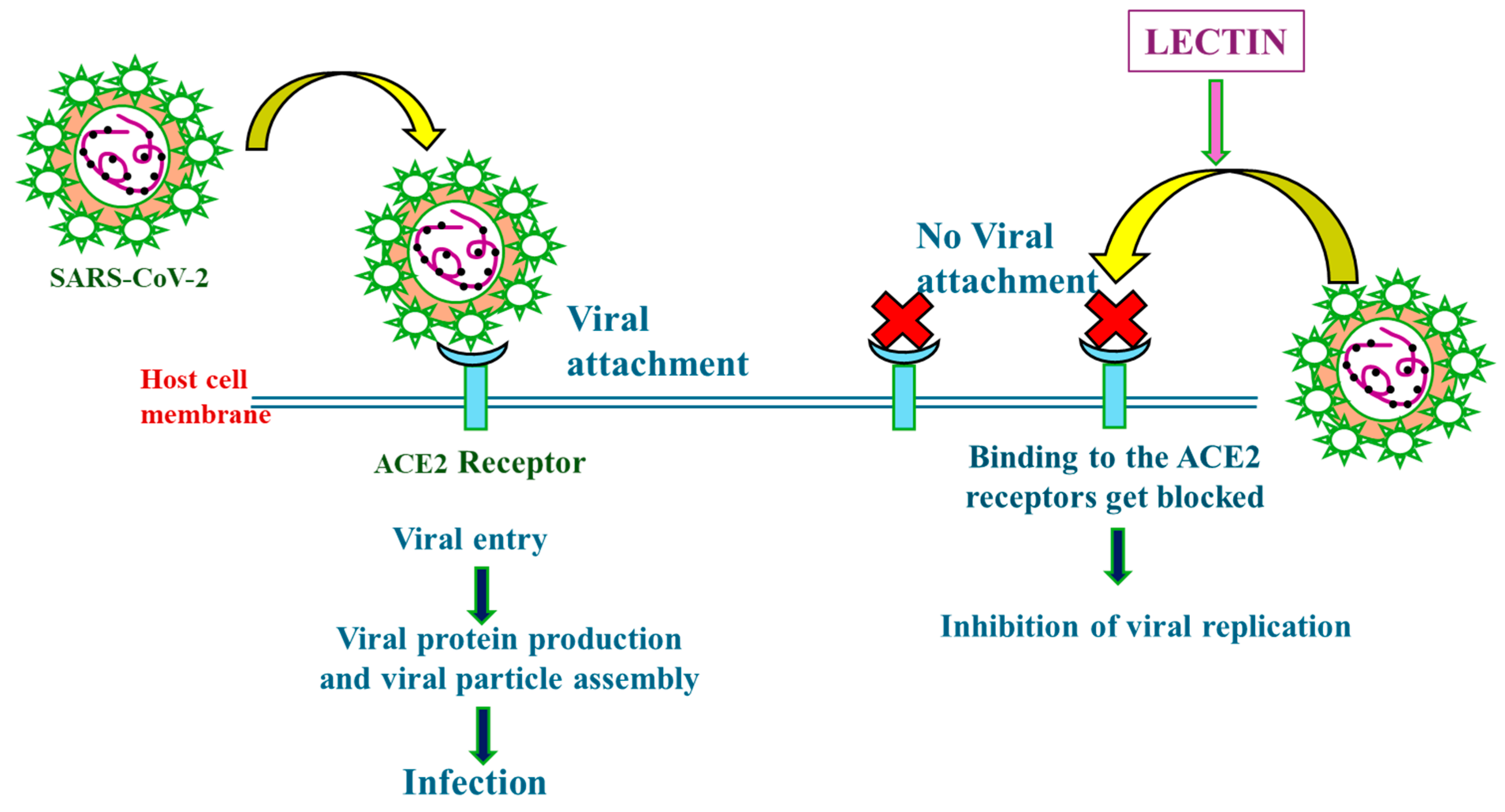
4.2. Mechanisms of Action of Lectins in Combating Severe Acute Respiratory Syndrome Coronavirus 2 (SARS-CoV-2)

Lectins have been the tool of choice for the biochemical and histochemical analysis of cell glycol conjugates because of their high specificity for different glycans. Many biological events, including viral adherence to host cell membranes, depend on the interaction between lectins and glycoproteins. By interacting with glycoproteins, many lectins prevent the reproduction of viruses [177]. It has been reported that numerous lectins have antiviral activities against a variety of viruses, such as influenza, hepatitis C virus (HCV), respiratory syncytial virus (RSV), and herpes simplex virus type 2 (HSV-2) viruses, as well as human immunodeficiency virus (HIV). Lectins have also been shown to suppress Middle East respiratory syndrome coronavirus (MERS-CoV), severe acute respiratory syndrome coronavirus (SARS-CoV), and other avian and mammalian coronaviruses [165,178,179]. While the glycan shield of the SARS-CoV-2 spike protein is similar to that of other coronaviruses, variations in surface glycan patterns and glycosylation sites do exist. The effectiveness of lectins against SARS-CoV-2 and its antigenic variants remains unclear. The two possible sites for antiviral intervention in SARS-CoV-2 are the virus’ replication cycle, and toward the end of the infectious virus cycle. Infection prevention may be aided by targeting the cellular receptors or structural proteins of SARS-CoV-2, such as the human angiotensin-converting enzyme 2 (ACE2) gene and S-protein (Figure 1) [180].
The SARS-CoV spike protein is extensively glycosylated, featuring 23 potential N-glycosylation sites, with 12 confirmed to be efficiently glycosylated [181]. Consequently, lectins capable of recognizing the glycans present in the spike glycoprotein should be effective in suppressing coronavirus infectivity. The non-catalytic carbohydrate recognition domain (CRD), often known as the lectin domain, is one of the numerous molecular domains found in each lectin polypeptide. Its capacity to detect and interact with particular glycoconjugates without changing their structure is due to the lectin domain.
For various RNA viruses, including SARS-CoV-2, lectins exhibit strong anti-infectivity capabilities. Lectins exhibit a strong affinity for the N-glycosylated spike glycoprotein of the SARS-CoV virus [182]. It has been shown that mannose-specific/mannose-binding lectins (MBLs) have strong complement cascade induction, anti-infectivity, DC-SIGN antagonists, immunoadjuvants, and glycomimetic approach efficacies that may be useful against COVID-19, as well as antiviral immunity and glyco-biological aspects of SARS-CoV-2 infections [183]. For instance, in vivo, in vitro, and in silico tests have demonstrated that the mannose-specific lectins Frutalin (FRIL), BanLac, Griffithsin (GRFT) from red algae, and lentil can suppress and neutralize the infection with SARS-CoV-2.
Plants have been the focus of extensive research on lectin domains [184]. The plant lectins’ antiviral activity spectrum varies greatly depending upon the specificity of their sugar binding. MBLs are generally very potent against SARS-CoV-2. Depending on the location of the glycans targeted, plant lectins with different specificities may potentially obstruct distinct targets necessary for viral entry [171]. Significantly reducing viral entry into ACE2-expressing HEK293T cells was achieved by genetically and chemically inhibiting N-glycan production during the oligomannose stage. Partial viral entry was also prevented by inhibiting O-glycan elaboration [185].
Recent studies indicate that FRIL, a plant-derived lectin with the ability to directly bind to virus particles, demonstrates antiviral activity against a SARS-CoV-2 strain originating from Taiwan (Table 4) [186]. According to reports, the wheat germ agglutin/lectin exhibits antiviral efficacy against SARS-CoV-2 and its two main variants of concern (VoCs), Alpha and Beta (Table 4) [187]. The lentil lectin derived from Lens culinaris, exhibiting a specific binding affinity for oligomannose-type glycans and GlcNAc at the non-reducing end terminus, demonstrated highly potent and broad-spectrum antiviral activity (Table 4). This efficacy extended to various mutant strains and variants, encompassing epidemic variants such as B.1.1.7, B.1.351, and P.1, as well as naturally occurring amino acid mutants and artificially induced mutants at the N-/O-linked glycosylation site [188]. Recombinant lectins can serve as new anti-SARSCoV-2 agents by targeting SARS-CoV-2-associated glycans [189]. According to previous coronavirus investigations, mannose-specific plant lectins may be useful in preventing human infection with SARS-CoV-2 [190]. Ahan et al. [191] showed that GRFT may bind to the SARS-CoV-2 spike protein and prevent it from infecting IFNAR−/− mouse model and Vero E6 cell lines (Table 4). Another study found that the H84T-banana lectin (H84T-BanLec), which was rationally engineered, can effectively inhibit SARS-CoV-2, MERS-CoV, and other human-pathogenic coronaviruses at nanomolar concentrations by specifically recognizing high mannose present on viral proteins but not frequently on healthy human cells (Table 4) [192]. NTL-125, a new plant lectin, blocks SARS-CoV-2 interaction with human angiotensin-converting enzyme 2 (hACE2) (Table 3) [193]. Lectins are effective antiviral agents with a bright future in diagnosing and treating viral diseases [194]. Acmella japonica rhizome lectin (AcmJRL), a jacalin-related lectin produced from pineapple, binds the SARS-CoV-2 spike protein in a carbohydrate-dependent manner (Table 4) [195].
These findings indicate that lectins have robust and specific anti-coronavirus action that should spur a future investigation into the identification of additional carbohydrate-binding molecules, such as synthetic low-molecular-weight compounds.

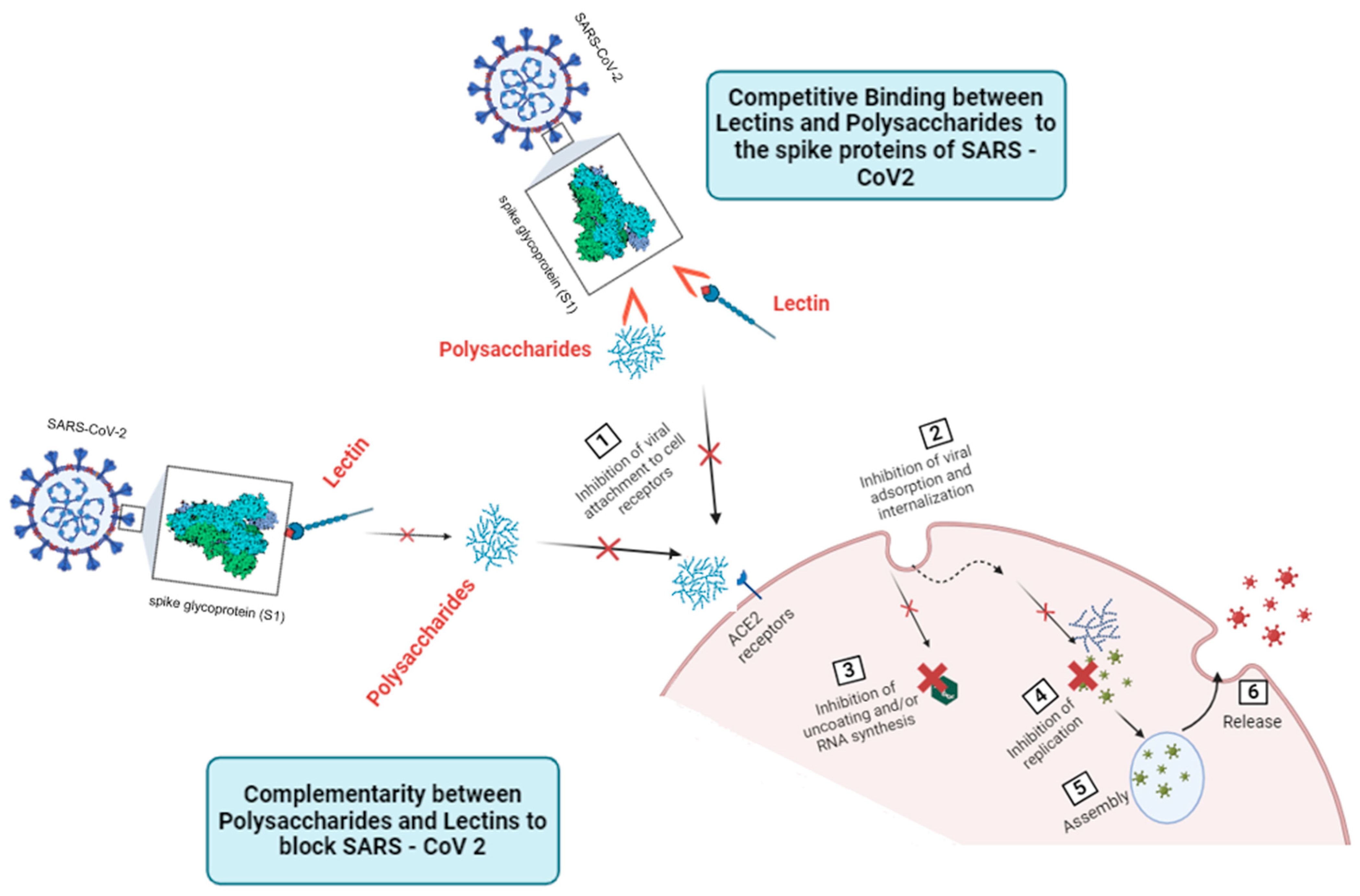
5. Unveiling the Interplay: Complementary and Competitive Actions of Lectins and Polysaccharides in Combating SARS-CoV-2

Polysaccharides, essential macromolecules found in various organisms, and lectins, proteins playing a crucial role in sugar recognition, offer unique characteristics enhancing antiviral effects when combined together. This synergistic approach arises from the understanding of the complementary mechanisms of action of both compounds.
Alsaidi et al. [196] revealed the synergistic antiviral effects of carrageenan (sulfated polysaccharide) and griffithsin (lectin) against SARS-CoV-1 and SARS-CoV-2, showcasing potent activity as well as a promising combination for neutralizing both SARS-CoV-1 and SARS-CoV-2. The synergistic mechanism involves griffithsin targeting viral spikes through binding to high-mannose arrays, while sulfated polysaccharides inhibit viral entry by potentially binding directly to viral particles, preventing adsorption and internalization into host cells. This finding highlights a potential broad-spectrum approach against coronaviruses.
By harnessing the synergistic capabilities of polysaccharides and lectins, a distinctive opportunity arises for targeting multiple crucial points in the viral life cycle simultaneously. The potential rationale behind combining lectins and polysaccharides against SARS-CoV-2 might involve the following considerations:
Complementary Mechanisms of Action: Polysaccharides and lectins often target various phases of the viral life cycle and employ diverse mechanisms of action [47,197]. Polysaccharides may interfere with viral entry, inhibit viral replication, or modulate the host immune response (Figure 2) [198]. On the other hand, lectins may recognize specific glycoproteins on the viral surface, blocking viral attachment or entry into host cells (Figure 2) [199]. By combining these two classes of compounds, there is an opportunity to target the virus at multiple points simultaneously, increasing the likelihood of inhibiting viral propagation comprehensively (Figure 2).
Interference with Viral Binding and Entry: The ability of lectins to bind specifically to glycosylated viral proteins can interfere with the initial steps of viral infection, preventing the virus from attaching to host cells [199]. Polysaccharides may complement this action by modifying the viral envelope or glycoproteins, making them less prone to interactions with cellular receptors [47]. By taking a dual strategy, the possibility of a successful infection could be decreased by erecting a strong barrier against viral entry.
Potential Reduction in Viral Resistance: Combining polysaccharides and lectins may mitigate the risk of viral resistance development. Viruses, including SARS-CoV-2, can adapt and evolve rapidly, potentially acquiring resistance to single-target antiviral agents [200,201]. However, a combination therapy targeting different aspects of the viral life cycle makes it challenging for the virus to develop resistance simultaneously against both polysaccharides and lectins. This may enhance the durability and sustainability of the antiviral strategy over time.
Improved Immune Modulation: Polysaccharides, known for their immunomodulatory effects, stimulate various components of the immune system [147]. Through their interactions with glycoproteins, lectins can further amplify the immune response [202]. Polysaccharides and lectins, when used in combination, may create a synergistic effect, potentiating the activation of immune cells, like dendritic cells, macrophages, and lymphocytes [203,204]. This enhanced immune modulation not only inhibits the direct replication of viruses but also plays a vital role in establishing a strong and enduring antiviral defense.
Enhanced antiviral synergies between lectins and polysaccharides mark significant progress in scientific understanding and potential therapeutic strategies against the pandemic of SARS-CoV-2 and possibly other coronaviruses.
The interaction between polysaccharides and lectins in combating SARS-CoV-2 reveals a fascinating interplay of complementarity action. Polysaccharides, with their intricate structures mimicking those of host cell surface glycans, and lectins, known for their selective binding to viral envelope glycoproteins, operate synergistically and competitively in the battle against the virus. However, this complementary relationship also entails competition between polysaccharides and lectins (Figure 2). Both compounds vie for binding sites on viral envelope glycoproteins and cellular receptors, including those crucial for virus attachment and entry. This competitive dynamic shapes the efficacy of antiviral strategies, as polysaccharides and lectins strive to outcompete each other in inhibiting viral infection.
Polysaccharides and lectins, both integral players in the battle against viruses, exhibit a complex interplay shaped by their shared affinity for sugar molecules. Lectins, characterized by their ability to bind to viral envelope glycoproteins, impede virus attachment and entry into host cells by competitively occupying sugar-binding sites, including those utilized by the spike protein to latch onto host receptors like angiotensin-converting enzyme 2 (ACE2) (Figure 2) [205]. On the other hand, polysaccharides, with their structural complexity, mimic host cell surface glycans, engaging in competitive interactions with viral glycoproteins and cellular receptors like ACE2, thus impeding virus attachment and entry (Figure 2) [206]. In the context of targeting viruses, the shared affinity for sugar units presents a scenario of competition between lectins and polysaccharides. When both lectins and polysaccharides are present, they may compete for binding sites on the surface of viral particles or host cells. This competition can influence the effectiveness of each agent in inhibiting viral infection and replication.
This competition between lectins and polysaccharides for binding sites on viral and cellular targets outlines a dynamic scenario where both compounds vie for supremacy in inhibiting viral infection. It is important to note that while competition between lectins and polysaccharides may occur, there may also be complementary actions between these two agents. Despite competing for binding sites, lectins and polysaccharides may exhibit synergistic effects in inhibiting viral infection when used together. Their combined action could enhance the overall antiviral activity by targeting different stages of the viral life cycle or by modulating immune responses.

Author Contributions

Conceptualization, F.Z.K., A.C., A.A. and R.L.; writing—original draft preparation, R.L., F.Z.K., A.A., P.B. and I.-M.B.; writing—review and editing, F.Z.K., P.B. and C.I.; visualization, A.E.-C., A.D.P., G.H. and B.N.; supervision, F.Z.K.; project administration, A.C., A.D.P., G.H. and B.N.; funding acquisition, A.C., A.D.P., G.H. and B.N. All authors have read and agreed to the published version of the manuscript.

Funding

This research received no external funding.

Institutional Review Board Statement

Not applicable.

Informed Consent Statement

Not applicable.

Data Availability Statement

Data are contained within the article.

Conflicts of Interest

The authors declare no conflicts of interest.

References

  1. Karami, H.; Karimi, Z.; Karami, N. SARS-CoV-2 in Brief: From Virus to Prevention. Osong Public Health Res. Perspect. 2022, 13, 394–406. [Google Scholar] [CrossRef] [PubMed]
  2. Altamimi, I.; Alabdulkarim, I.M.; Alhumimidi, A.S.; Albabtain, M.A.; Temsah, M.-H. Navigating Novel Uncertainties of COVID-19: The Rise of the JN.1 Variant. Cureus 2024, 16, e51497. [Google Scholar] [CrossRef] [PubMed]
  3. Hu, Y.; Liu, L.; Zhang, S.; Su, S.; Liang, X. The Application of Biomaterials for the Vaccine, Treatment, and Detection of SARS-CoV-2. ACS Omega 2024, 9, 5175–5192. [Google Scholar] [CrossRef] [PubMed]
  4. Amit, A.M.L.; Pepito, V.C.F.; Sumpaico-Tanchanco, L.; Dayrit, M.M. COVID-19 vaccine brand hesitancy and other challenges to vaccination in the Philippines. PLoS Glob. Public Health 2022, 2, e0000165. [Google Scholar] [CrossRef] [PubMed]
  5. Alotaibi, W.; Alomary, F.; Mokni, R. COVID-19 Vaccine Rejection Causes Based on Twitter People’s Opinions Analysis Using Deep Learning. Soc. Netw. Anal. Min. 2023, 13, 62. [Google Scholar] [CrossRef] [PubMed]
  6. Islam, M.A. A Review of SARS-CoV-2 Variants and Vaccines: Viral Properties, Mutations, Vaccine Efficacy, and Safety. Infect. Med. 2023, 2, 247–261. [Google Scholar] [CrossRef] [PubMed]
  7. Bah, C.S.F.; Fang, E.F.; Ng, T.B. Medicinal Applications of Plant Lectins. In Antitumor Potential and Other Emerging Medicinal Properties of Natural Compounds; Springer: Berlin/Heidelberg, Germany, 2013; pp. 55–74. [Google Scholar]
  8. Sharma, A.; Shahid, A.; Banerjee, R.; Kumar, K.J. Emerging Insights into the Structure-Activity Relationship of Water-Soluble Polysaccharides in Antiviral Therapy. Eur. J. Med. Chem. Rep. 2024, 10, 100122. [Google Scholar] [CrossRef]
  9. Usman, Τ.; Rasheed, A. Lectins and Polysaccharides against SARS-CoV-2; Elsevier: New York, NY, USA, 2023; pp. 223–252. [Google Scholar]
  10. Vijayan, M.; Chandra, N. Lectins. Curr. Opin. Struct. Biol. 1999, 9, 707–714. [Google Scholar] [CrossRef] [PubMed]
  11. Murphy, E.J.; Fehrenbach, G.W.; Abidin, I.Z.; Buckley, C.; Montgomery, T.; Pogue, R.; Murray, P.; Major, I.; Rezoagli, E. Polysaccharides—Naturally Occurring Immune Modulators. Polymers 2023, 15, 2373. [Google Scholar] [CrossRef] [PubMed]
  12. Honein, M.A.; Christie, A.; Rose, D.A.; Brooks, J.T.; Meaney-Delman, D.; Cohn, A.; Sauber-Schatz, E.K.; Walker, A.; McDonald, L.C.; Liburd, L.C.; et al. Summary of Guidance for Public Health Strategies to Address High Levels of Community Transmission of SARS-CoV-2 and Related Deaths, December 2020. Morb. Mortal. Wkly. Rep. 2020, 69, 1860–1867. [Google Scholar] [CrossRef]
  13. Santacroce, L.; Charitos, I.A.; Carretta, D.M.; De Nitto, E.; Lovero, R. The Human Coronaviruses (HCoVs) and the Molecular Mechanisms of SARS-CoV-2 Infection. J. Mol. Med. 2021, 99, 93–106. [Google Scholar] [CrossRef] [PubMed]
  14. Alluwaimi, A.M.; Alshubaith, I.H.; Al-Ali, A.M.; Abohelaika, S. The Coronaviruses of Animals and Birds: Their Zoonosis, Vaccines, and Models for SARS-CoV and SARS-CoV2. Front. Vet. Sci. 2020, 7, 582287. [Google Scholar] [CrossRef] [PubMed]
  15. Compton, S.R. Overview of Coronaviruses in Veterinary Medicine. Comp. Med. 2021, 71, 333–341. [Google Scholar] [CrossRef] [PubMed]
  16. Kumar, S.; Nyodu, R.; Maurya, V.K.; Saxena, S.K. Morphology, Genome Organization, Replication, and Pathogenesis of Severe Acute Respiratory Syndrome Coronavirus 2 (SARS-CoV-2). In Coronavirus Disease 2019 (COVID-19); Saxena, S.K., Ed.; Medical Virology: From Pathogenesis to Disease Control; Springer: Singapore, 2020; pp. 23–31. ISBN 9789811548130. [Google Scholar]
  17. Bhattacharyya, P.; Das, S.; Aich, S.; Sarkar. COVID-19: Morphology and Mechanism of the SARS-CoV-2, Global Outbreak, Medication, Vaccines and Future of the Virus. Front. Biosci. 2021, 13, 272. [Google Scholar] [CrossRef]
  18. Jackson, C.B.; Farzan, M.; Chen, B.; Choe, H. Mechanisms of SARS-CoV-2 Entry into Cells. Nat. Rev. Mol. Cell Biol. 2022, 23, 3–20. [Google Scholar] [CrossRef] [PubMed]
  19. Sriwilaijaroen, N.; Suzuki, Y. Host Receptors of Influenza Viruses and Coronaviruses—Molecular Mechanisms of Recognition. Vaccines 2020, 8, 587. [Google Scholar] [CrossRef] [PubMed]
  20. Beyerstedt, S.; Casaro, E.B.; Rangel, É.B. COVID-19: Angiotensin-Converting Enzyme 2 (ACE2) Expression and Tissue Susceptibility to SARS-CoV-2 Infection. Eur. J. Clin. Microbiol. Infect. Dis. 2021, 40, 905–919. [Google Scholar] [CrossRef] [PubMed]
  21. V’kovski, P.; Kratzel, A.; Steiner, S.; Stalder, H.; Thiel, V. Coronavirus Biology and Replication: Implications for SARS-CoV-2. Nat. Rev. Microbiol. 2021, 19, 155–170. [Google Scholar] [CrossRef] [PubMed]
  22. Zhang, Q.; Xiang, R.; Huo, S.; Zhou, Y.; Jiang, S.; Wang, Q.; Yu, F. Molecular Mechanism of Interaction between SARS-CoV-2 and Host Cells and Interventional Therapy. Sig Transduct. Target. Ther. 2021, 6, 233. [Google Scholar] [CrossRef] [PubMed]
  23. Aboul-Fotouh, S.; Mahmoud, A.N.; Elnahas, E.M.; Habib, M.Z.; Abdelraouf, S.M. What Are the Current Anti-COVID-19 Drugs? From Traditional to Smart Molecular Mechanisms. Virol. J. 2023, 20, 241. [Google Scholar] [CrossRef] [PubMed]
  24. Ren, Y.; Bai, Y.; Zhang, Z.; Cai, W.; Del Rio Flores, A. The Preparation and Structure Analysis Methods of Natural Polysaccharides of Plants and Fungi: A Review of Recent Development. Molecules 2019, 24, 3122. [Google Scholar] [CrossRef]
  25. Díaz-Montes, E. Polysaccharides: Sources, Characteristics, Properties, and Their Application in Biodegradable Films. Polysaccharides 2022, 3, 480–501. [Google Scholar] [CrossRef]
  26. Torres, F.G.; Troncoso, O.P.; Pisani, A.; Gatto, F.; Bardi, G. Natural Polysaccharide Nanomaterials: An Overview of Their Immunological Properties. Int. J. Mol. Sci. 2019, 20, 5092. [Google Scholar] [CrossRef] [PubMed]
  27. Mohammed, A.S.A.; Naveed, M.; Jost, N. Polysaccharides; Classification, Chemical Properties, and Future Perspective Applications in Fields of Pharmacology and Biological Medicine (A Review of Current Applications and Upcoming Potentialities). J. Polym. Environ. 2021, 29, 2359–2371. [Google Scholar] [CrossRef]
  28. Painter, T.J. Algal polysaccharides. Polysaccharides 1983, 2, 195–285. [Google Scholar]
  29. Venugopal, V. Marine Polysaccharides; CRC Press: Boca Raton, FL, USA, 2016; ISBN 978-1-4398-1527-4. [Google Scholar]
  30. Qiang, M.; Cai, P.; Ao, M.; Li, X.; Chen, Z.; Yu, L. Polysaccharides from Chinese Materia Medica: Perspective towards Cancer Management. Int. J. Biol. Macromol. 2023, 224, 496–509. [Google Scholar] [CrossRef]
  31. Nikonenko, N.A.; Buslov, D.K.; Sushko, N.I.; Zhbankov, R.G. Spectroscopic Manifestation of Stretching Vibrations of Glycosidic Linkage in Polysaccharides. J. Mol. Struct. 2005, 752, 20–24. [Google Scholar] [CrossRef]
  32. BeMiller, J.N. Carbohydrate Chemistry for Food Scientists; Elsevier: New York, NY, USA, 2019. [Google Scholar]
  33. Cosgrove, D.J. Diffuse Growth of Plant Cell Walls. Plant Physiol. 2018, 176, 16–27. [Google Scholar] [CrossRef]
  34. MacNeill, G.J.; Mehrpouyan, S.; Minow, M.A.A.; Patterson, J.A.; Tetlow, I.J.; Emes, M.J. Starch as a Source, Starch as a Sink: The Bifunctional Role of Starch in Carbon Allocation. J. Exp. Bot. 2017, 68, 4433–4453. [Google Scholar] [CrossRef]
  35. Daghlas, S.A.; Mohiuddin, S.S. Biochemistry, Glycogen; StatPearls Publishing: Treasure Island, FL, USA, 2024. [Google Scholar]
  36. Song, E.-h.; Shang, J.; Ratner, D.M. Polysaccharides; Elsevier: New York, NY, USA, 2012; pp. 137–155. [Google Scholar]
  37. Climov, M.; Leavitt, T.; Molnár, J.; Orgill, D.P. Natural Biomaterials for Skin Tissue Engineering; Elsevier: New York, NY, USA, 2016; pp. 145–161. [Google Scholar]
  38. Farhana, A.; Khan, Y.S. Biochemistry, Lipopolysaccharide; StatPearls Publishing: Treasure Island, FL, USA, 2024. [Google Scholar]
  39. Ullah, S.; Khalil, A.A.; Shaukat, F.; Song, Y. Sources, Extraction and Biomedical Properties of Polysaccharides. Foods 2019, 8, 304. [Google Scholar] [CrossRef]
  40. Morin-Crini, N.; Lichtfouse, E.; Torri, G.; Crini, G. Applications of Chitosan in Food, Pharmaceuticals, Medicine, Cosmetics, Agriculture, Textiles, Pulp and Paper, Biotechnology, and Environmental Chemistry. Environ. Chem. Lett. 2019, 17, 1667–1692. [Google Scholar] [CrossRef]
  41. Niyigaba, T.; Liu, D.; de Dieu Habimana, J. The Extraction, Functionalities and Applications of Plant Polysaccharides in Fermented Foods: A Review. Foods 2021, 10, 3004. [Google Scholar] [CrossRef]
  42. Xia, S.; Zhang, L.; Davletshin, A.; Li, Z.; You, J.; Tan, S. Application of Polysaccharide Biopolymer in Petroleum Recovery. Polymers 2020, 12, 1860. [Google Scholar] [CrossRef] [PubMed]
  43. BeMiller, J.N. Polysaccharides. In Encyclopedia of Life Sciences; Wiley: New York, NY, USA, 2014; ISBN 978-0-470-01617-6. [Google Scholar]
  44. Dwivedi, R.; Samanta, P.; Sharma, P.; Zhang, F.; Mishra, S.K.; Kucheryavy, P.; Kim, S.B.; Aderibigbe, A.O.; Linhardt, R.J.; Tandon, R.; et al. Structural and Kinetic Analyses of Holothurian Sulfated Glycans Suggest Potential Treatment for SARS-CoV-2 Infection. J. Biol. Chem. 2021, 297, 101207. [Google Scholar] [CrossRef] [PubMed]
  45. Couto, M.R.; Rodrigues, J.L.; Rodrigues, L.R. Heterologous Production of Chondroitin. Biotechnol. Rep. 2022, 33, e00710. [Google Scholar] [CrossRef] [PubMed]
  46. Chen, X.; Han, W.; Wang, G.; Zhao, X. Application Prospect of Polysaccharides in the Development of Anti-Novel Coronavirus Drugs and Vaccines. Int. J. Biol. Macromol. 2020, 164, 331–343. [Google Scholar] [CrossRef]
  47. Claus-Desbonnet, H.; Nikly, E.; Nalbantova, V.; Karcheva-Bahchevanska, D.; Ivanova, S.; Pierre, G.; Benbassat, N.; Katsarov, P.; Michaud, P.; Lukova, P.; et al. Polysaccharides and Their Derivatives as Potential Antiviral Molecules. Viruses 2022, 14, 426. [Google Scholar] [CrossRef]
  48. Onishi, A.; St Ange, K.; Dordick, J.S.; Linhardt, R.J. Heparin and anticoagulation. Front. Biosci. 2016, 21, 1372–1392. [Google Scholar]
  49. Hippensteel, J.A.; LaRiviere, W.B.; Colbert, J.F.; Langouët-Astrié, C.J.; Schmidt, E.P. Heparin as a Therapy for COVID-19: Current Evidence and Future Possibilities. Am. J. Physiol.-Lung Cell. Mol. Physiol. 2020, 319, L211–L217. [Google Scholar] [CrossRef]
  50. Mikhailov, D.; Young, H.C.; Linhardt, R.J.; Mayo, K.H. Heparin Dodecasaccharide Binding to Platelet Factor-4 and Growth-Related Protein-α. J. Biol. Chem. 1999, 274, 25317–25329. [Google Scholar] [CrossRef]
  51. Peysselon, F.; Ricard-Blum, S. Heparin–Protein Interactions: From Affinity and Kinetics to Biological Roles. Application to an Interaction Network Regulating Angiogenesis. Matrix Biol. 2014, 35, 73–81. [Google Scholar] [CrossRef] [PubMed]
  52. Belouzard, S.; Millet, J.K.; Licitra, B.N.; Whittaker, G.R. Mechanisms of Coronavirus Cell Entry Mediated by the Viral Spike Protein. Viruses 2012, 4, 1011–1033. [Google Scholar] [CrossRef] [PubMed]
  53. Cheudjeu, A. The SARS-CoV-2 Entry Inhibition Mechanisms of Serine Protease Inhibitors, OM-85, Heparin and Soluble HS Might Be Linked to HS Attachment Sites. Molecules 2022, 27, 1947. [Google Scholar] [CrossRef] [PubMed]
  54. Milewska, A.; Nowak, P.; Owczarek, K.; Szczepanski, A.; Zarebski, M.; Hoang, A.; Berniak, K.; Wojarski, J.; Zeglen, S.; Baster, Z.; et al. Entry of Human Coronavirus NL63 into the Cell. J. Virol. 2018, 92, e01933-17. [Google Scholar] [CrossRef] [PubMed]
  55. Coutard, B.; Valle, C.; de Lamballerie, X.; Canard, B.; Seidah, N.G.; Decroly, E. The Spike Glycoprotein of the New Coronavirus 2019-NCoV Contains a Furin-like Cleavage Site Absent in CoV of the Same Clade. Antivir. Res. 2020, 176, 104742. [Google Scholar] [CrossRef] [PubMed]
  56. Kim, S.Y.; Jin, W.; Sood, A.; Montgomery, D.W.; Grant, O.C.; Fuster, M.M.; Fu, L.; Dordick, J.S.; Woods, R.J.; Zhang, F.; et al. Characterization of Heparin and Severe Acute Respiratory Syndrome-Related Coronavirus 2 (SARS-CoV-2) Spike Glycoprotein Binding Interactions. Antivir. Res. 2020, 181, 104873. [Google Scholar] [CrossRef]
  57. Shi, D.; Bu, C.; He, P.; Song, Y.; Dordick, J.S.; Linhardt, R.J.; Chi, L.; Zhang, F. Structural Characteristics of Heparin Binding to SARS-CoV-2 Spike Protein RBD of Omicron Sub-Lineages BA.2.12.1, BA.4 and BA.5. Viruses 2022, 14, 2696. [Google Scholar] [CrossRef]
  58. Thachil, J. The Versatile Heparin in COVID-19. J. Thromb. Haemost. 2020, 18, 1020–1022. [Google Scholar] [CrossRef] [PubMed]
  59. Tandon, R.; Sharp, J.S.; Zhang, F.; Pomin, V.H.; Ashpole, N.M.; Mitra, D.; McCandless, M.G.; Jin, W.; Liu, H.; Sharma, P.; et al. Effective Inhibition of SARS-CoV-2 Entry by Heparin and Enoxaparin Derivatives. J. Virol. 2021, 95, e01987-20. [Google Scholar] [CrossRef] [PubMed]
  60. Mycroft-West, C.J.; Su, D.; Pagani, I.; Rudd, T.R.; Elli, S.; Gandhi, N.S.; Guimond, S.E.; Miller, G.J.; Meneghetti, M.C.Z.; Nader, H.B.; et al. Heparin Inhibits Cellular Invasion by SARS-CoV-2: Structural Dependence of the Interaction of the Spike S1 Receptor-Binding Domain with Heparin. Thromb. Haemost. 2020, 120, 1700–1715. [Google Scholar] [CrossRef] [PubMed]
  61. Tree, J.A.; Turnbull, J.E.; Buttigieg, K.R.; Elmore, M.J.; Coombes, N.; Hogwood, J.; Mycroft-West, C.J.; Lima, M.A.; Skidmore, M.A.; Karlsson, R.; et al. Unfractionated Heparin Inhibits Live Wild Type SARS-CoV-2 Cell Infectivity at Therapeutically Relevant Concentrations. Br. J. Pharmacol. 2021, 178, 626–635. [Google Scholar] [CrossRef] [PubMed]
  62. Guimond, S.E.; Mycroft-West, C.J.; Gandhi, N.S.; Tree, J.A.; Le, T.T.; Spalluto, C.M.; Humbert, M.V.; Buttigieg, K.R.; Coombes, N.; Elmore, M.J.; et al. Synthetic Heparan Sulfate Mimetic Pixatimod (PG545) Potently Inhibits SARS-CoV-2 by Disrupting the Spike–ACE2 Interaction. ACS Cent. Sci. 2022, 8, 527–545. [Google Scholar] [CrossRef] [PubMed]
  63. Bertini, S.; Alekseeva, A.; Elli, S.; Pagani, I.; Zanzoni, S.; Eisele, G.; Krishnan, R.; Maag, K.P.; Reiter, C.; Lenhart, D.; et al. Pentosan polysulfate Inhibits Attachment and Infection by SARS-CoV-2 In Vitro: Insights into Structural Requirements for Binding. Thromb. Haemost. 2022, 122, 984–997. [Google Scholar] [CrossRef] [PubMed]
  64. Buijsers, B.; Yanginlar, C.; Maciej-Hulme, M.L.; de Mast, Q.; van der Vlag, J. Beneficial Non-Anticoagulant Mechanisms Underlying Heparin Treatment of COVID-19 Patients. eBioMedicine 2020, 59, 102969. [Google Scholar] [CrossRef] [PubMed]
  65. Spyropoulos, A.C.; Goldin, M.; Giannis, D.; Diab, W.; Wang, J.; Khanijo, S.; Mignatti, A.; Gianos, E.; Cohen, M.; Sharifova, G.; et al. Efficacy and Safety of Therapeutic-Dose Heparin vs Standard Prophylactic or Intermediate-Dose Heparins for Thromboprophylaxis in High-Risk Hospitalized Patients with COVID-19: The HEP-COVID Randomized Clinical Trial. JAMA Intern. Med. 2021, 181, 1612. [Google Scholar] [CrossRef] [PubMed]
  66. Sholzberg, M.; Tang, G.H.; Rahhal, H.; AlHamzah, M.; Kreuziger, L.B.; Áinle, F.N.; Alomran, F.; Alayed, K.; Alsheef, M.; AlSumait, F.; et al. Effectiveness of Therapeutic Heparin versus Prophylactic Heparin on Death, Mechanical Ventilation, or Intensive Care Unit Admission in Moderately Ill Patients with COVID-19 Admitted to Hospital: RAPID Randomised Clinical Trial. BMJ 2021, 375, n2400. [Google Scholar] [CrossRef] [PubMed]
  67. Zhao, T.; Zhang, J.; Liu, X.; Wang, F. Analysis of Chondroitin Sulfate from Different Sources of Cartilage by Electrophoretically Mediated Microanalysis. RSC Adv. 2015, 5, 52314–52319. [Google Scholar] [CrossRef]
  68. Pomin, V.H. Holothurian Fucosylated Chondroitin Sulfate. Mar. Drugs 2014, 12, 232–254. [Google Scholar] [CrossRef]
  69. Mizumoto, S.; Kwok, J.C.F.; Whitelock, J.M.; Li, F.; Perris, R. Editorial: Roles of Chondroitin Sulfate and Dermatan Sulfate as Regulators for Cell and Tissue Development. Front. Cell Dev. Biol. 2022, 10, 941178. [Google Scholar] [CrossRef]
  70. Henrotin, Y.; Mathy, M.; Sanchez, C.; Lambert, C. Chondroitin Sulfate in the Treatment of Osteoarthritis: From In Vitro Studies to Clinical Recommendations. Ther. Adv. Musculoskelet. 2010, 2, 335–348. [Google Scholar] [CrossRef] [PubMed]
  71. Ray, B.; Ali, I.; Jana, S.; Mukherjee, S.; Pal, S.; Ray, S.; Schütz, M.; Marschall, M. Antiviral Strategies Using Natural Source-Derived Sulfated Polysaccharides in the Light of the COVID-19 Pandemic and Major Human Pathogenic Viruses. Viruses 2021, 14, 35. [Google Scholar] [CrossRef]
  72. Abidine, Y.; Liu, L.; Wallén, O.; Trybala, E.; Olofsson, S.; Bergström, T.; Bally, M. Cellular Chondroitin Sulfate and the Mucin-like Domain of Viral Glycoprotein C Promote Diffusion of Herpes Simplex Virus 1 While Heparan Sulfate Restricts Mobility. Viruses 2022, 14, 1836. [Google Scholar] [CrossRef] [PubMed]
  73. Watanabe, T.; Takeda, K.; Hiemori, K.; Minamisawa, T.; Tateno, H. A Glycosaminoglycan Microarray Identifies the Binding of SARS-CoV-2 Spike Protein to Chondroitin Sulfate E. FEBS Lett. 2021, 595, 2341–2349. [Google Scholar] [CrossRef] [PubMed]
  74. Song, S.; Peng, H.; Wang, Q.; Liu, Z.; Dong, X.; Wen, C.; Ai, C.; Zhang, Y.; Wang, Z.; Zhu, B. Inhibitory Activities of Marine Sulfated Polysaccharides against SARS-CoV-2. Food Funct. 2020, 11, 7415–7420. [Google Scholar] [CrossRef] [PubMed]
  75. Dwivedi, R.; Sharma, P.; Farrag, M.; Kim, S.B.; Fassero, L.A.; Tandon, R.; Pomin, V.H. Inhibition of SARS-CoV-2 Wild-Type (Wuhan-Hu-1) and Delta (B.1.617.2) Strains by Marine Sulfated Glycans. Glycobiology 2022, 32, 849–854. [Google Scholar] [CrossRef] [PubMed]
  76. Möller, S.; Theiß, J.; Deinert, T.I.L.; Golat, K.; Heinze, J.; Niemeyer, D.; Wyrwa, R.; Schnabelrauch, M.; Bogner, E. High-Sulfated Glycosaminoglycans Prevent Coronavirus Replication. Viruses 2022, 14, 413. [Google Scholar] [CrossRef] [PubMed]
  77. Albtoush, N.; Petrey, A.C. The Role of Hyaluronan Synthesis and Degradation in the Critical Respiratory Illness COVID-19. Am. J. Physiol.-Cell Physiol. 2022, 322, C1037–C1046. [Google Scholar] [CrossRef]
  78. Queisser, K.A.; Mellema, R.A.; Middleton, E.A.; Portier, I.; Manne, B.K.; Denorme, F.; Beswick, E.J.; Rondina, M.T.; Campbell, R.A.; Petrey, A.C. COVID-19 Generates Hyaluronan Fragments That Directly Induce Endothelial Barrier Dysfunction. JCI Insight 2021, 6, e147472. [Google Scholar] [CrossRef] [PubMed]
  79. Yang, S.; Tong, Y.; Chen, L.; Yu, W. Human Identical Sequences, Hyaluronan, and Hymecromone—The New Mechanism and Management of COVID-19. Mol. Biomed. 2022, 3, 15. [Google Scholar] [CrossRef] [PubMed]
  80. Mallakpour, S.; Azadi, E.; Hussain, C.M. Chitosan, Alginate, Hyaluronic Acid, Gums, and β-Glucan as Potent Adjuvants and Vaccine Delivery Systems for Viral Threats Including SARS-CoV-2: A Review. Int. J. Biol. Macromol. 2021, 182, 1931–1940. [Google Scholar] [CrossRef] [PubMed]
  81. Thirumalaisamy, R.; Aroulmoji, V.; Iqbal, M.N.; Deepa, M.; Sivasankar, C.; Khan, R.; Selvankumar, T. Molecular Insights of Hyaluronic Acid-Hydroxychloroquine Conjugate as a Promising Drug in Targeting SARS-CoV-2 Viral Proteins. J. Mol. Struct. 2021, 1238, 130457. [Google Scholar] [CrossRef] [PubMed]
  82. Damonte, E.; Matulewicz, M.; Cerezo, A. Sulfated Seaweed Polysaccharides as Antiviral Agents. Curr. Med. Chem. 2004, 11, 2399–2419. [Google Scholar] [CrossRef] [PubMed]
  83. Salih, A.E.M.; Thissera, B.; Yaseen, M.; Hassane, A.S.I.; El-Seedi, H.R.; Sayed, A.M.; Rateb, M.E. Marine Sulfated Polysaccharides as Promising Antiviral Agents: A Comprehensive Report and Modeling Study Focusing on SARS CoV-2. Mar. Drugs 2021, 19, 406. [Google Scholar] [CrossRef] [PubMed]
  84. Kalasariya, H.S.; Patel, N.B.; Gacem, A.; Alsufyani, T.; Reece, L.M.; Yadav, V.K.; Awwad, N.S.; Ibrahium, H.A.; Ahn, Y.; Yadav, K.K.; et al. Marine Alga Ulva fasciata-Derived Molecules for the Potential Treatment of SARS-CoV-2: An In Silico Approach. Mar. Drugs 2022, 20, 586. [Google Scholar] [CrossRef] [PubMed]
  85. Álvarez-Viñas, M.; Souto, S.; Flórez-Fernández, N.; Torres, M.D.; Bandín, I.; Domínguez, H. Antiviral Activity of Carrageenans and Processing Implications. Mar. Drugs 2021, 19, 437. [Google Scholar] [CrossRef] [PubMed]
  86. Ahmadi, A.; Zorofchian Moghadamtousi, S.; Abubakar, S.; Zandi, K. Antiviral Potential of Algae Polysaccharides Isolated from Marine Sources: A Review. BioMed Res. Int. 2015, 2015, 825203. [Google Scholar] [CrossRef] [PubMed]
  87. Koenighofer, M.; Lion, T.; Bodenteich, A.; Prieschl-Grassauer, E.; Grassauer, A.; Unger, H.; Mueller, C.A.; Fazekas, T. Carrageenan Nasal Spray in Virus Confirmed Common Cold: Individual Patient Data Analysis of Two Randomized Controlled Trials. Multidiscip. Respir. Med. 2014, 9, 57. [Google Scholar] [CrossRef] [PubMed]
  88. Graf, C.; Bernkop-Schnürch, A.; Egyed, A.; Koller, C.; Prieschl-Grassauer, E.; Morokutti-Kurz, M. Development of a Nasal Spray Containing Xylometazoline Hydrochloride and Iota-Carrageenan for the Symptomatic Relief of Nasal Congestion Caused by Rhinitis and Sinusitis. Int. J. Gen. Med. 2018, 11, 275–283. [Google Scholar] [CrossRef] [PubMed]
  89. Morokutti-Kurz, M.; Fröba, M.; Graf, P.; Große, M.; Grassauer, A.; Auth, J.; Schubert, U.; Prieschl-Grassauer, E. Iota-Carrageenan Neutralizes SARS-CoV-2 and Inhibits Viral Replication In Vitro. PLoS ONE 2021, 16, e0237480. [Google Scholar] [CrossRef] [PubMed]
  90. Bansal, S.; Jonsson, C.B.; Taylor, S.L.; Figueroa, J.M.; Dugour, A.V.; Palacios, C.; Vega, J.C. Iota-Carrageenan and Xylitol Inhibit SARS-CoV-2 in Vero Cell Culture. PLoS ONE 2021, 16, e0259943. [Google Scholar] [CrossRef] [PubMed]
  91. Schütz, D.; Conzelmann, C.; Fois, G.; Groß, R.; Weil, T.; Wettstein, L.; Stenger, S.; Zelikin, A.; Hoffmann, T.K.; Frick, M.; et al. Carrageenan-Containing over-the-Counter Nasal and Oral Sprays Inhibit SARS-CoV-2 Infection of Airway Epithelial Cultures. Am. J. Physiol.-Lung Cell. Mol. Physiol. 2021, 320, L750–L756. [Google Scholar] [CrossRef]
  92. Jang, Y.; Shin, H.; Lee, M.K.; Kwon, O.S.; Shin, J.S.; Kim, Y.; Kim, C.W.; Lee, H.-R.; Kim, M. Antiviral Activity of Lambda-Carrageenan against Influenza Viruses and Severe Acute Respiratory Syndrome Coronavirus 2. Sci. Rep. 2021, 11, 821. [Google Scholar] [CrossRef] [PubMed]
  93. Fröba, M.; Große, M.; Setz, C.; Rauch, P.; Auth, J.; Spanaus, L.; Münch, J.; Ruetalo, N.; Schindler, M.; Morokutti-Kurz, M.; et al. Iota-Carrageenan Inhibits Replication of SARS-CoV-2 and the Respective Variants of Concern Alpha, Beta, Gamma and Delta. Int. J. Mol. Sci. 2021, 22, 13202. [Google Scholar] [CrossRef] [PubMed]
  94. Eccles, R.; Meier, C.; Jawad, M.; Weinmüllner, R.; Grassauer, A.; Prieschl-Grassauer, E. Efficacy and Safety of an Antiviral Iota-Carrageenan Nasal Spray: A Randomized, Double-Blind, Placebo-Controlled Exploratory Study in Volunteers with Early Symptoms of the Common Cold. Respir. Res. 2010, 11, 108. [Google Scholar] [CrossRef] [PubMed]
  95. Eccles, R.; Winther, B.; Johnston, S.L.; Robinson, P.; Trampisch, M.; Koelsch, S. Efficacy and Safety of Iota-Carrageenan Nasal Spray versus Placebo in Early Treatment of the Common Cold in Adults: The ICICC Trial. Respir. Res. 2015, 16, 121. [Google Scholar] [CrossRef] [PubMed]
  96. Bichiri, D.; Rente, A.R.; Jesus, Â. Safety and Efficacy of a Iota-Carrageenan Nasal Spray in Treatment and Prevention of the Common Cold. Med. Pharm. Rep. 2021, 94, 28–34. [Google Scholar] [CrossRef]
  97. Hemilä, H.; Chalker, E. Carrageenan Nasal Spray May Double the Rate of Recovery from Coronavirus and Influenza Virus Infections: Re-analysis of Randomized Trial Data. Pharmacol. Res. Perspec. 2021, 9, e00810. [Google Scholar] [CrossRef]
  98. Nasrollahzadeh, M.; Sajjadi, M.; Nezafat, Z.; Shafiei, N. Polysaccharide Biopolymer Chemistry. In Biopolymer-Based Metal Nanoparticle Chemistry for Sustainable Applications; Elsevier: New York, NY, USA, 2021; pp. 45–105. [Google Scholar]
  99. Mazumder, S.; Ghosal, P.K.; Pujol, C.A.; Carlucci, M.J.; Damonte, E.B.; Ray, B. Isolation, Chemical Investigation and Antiviral Activity of Polysaccharides from Gracilaria corticata (Gracilariaceae, Rhodophyta). Int. J. Biol. Macromol. 2002, 31, 87–95. [Google Scholar] [CrossRef] [PubMed]
  100. Chattopadhyay, K.; Ghosh, T.; Pujol, C.A.; Carlucci, M.J.; Damonte, E.B.; Ray, B. Polysaccharides from Gracilaria corticata: Sulfation, Chemical Characterization and Anti-HSV Activities. Int. J. Biol. Macromol. 2008, 43, 346–351. [Google Scholar] [CrossRef] [PubMed]
  101. Duarte, M.E.R.; Cauduro, J.P.; Noseda, D.G.; Noseda, M.D.; Gonçalves, A.G.; Pujol, C.A.; Damonte, E.B.; Cerezo, A.S. The Structure of the Agaran Sulfate from Acanthophora spicifera (Rhodomelaceae, Ceramiales) and Its Antiviral Activity. Relation between Structure and Antiviral Activity in Agarans. Carbohydr. Res. 2004, 339, 335–347. [Google Scholar] [CrossRef]
  102. Duarte, M.E.R.; Noseda, D.G.; Noseda, M.D.; Tulio, S.; Pujol, C.A.; Damonte, E.B. Inhibitory Effect of Sulfated Galactans from the Marine Alga Bostrychia Montagnei on Herpes Simplex Virus Replication In Vitro. Phytomedicine 2001, 8, 53–58. [Google Scholar] [CrossRef] [PubMed]
  103. Panggabean, J.A.; Adiguna, S.P.; Rahmawati, S.I.; Ahmadi, P.; Zainuddin, E.N.; Bayu, A.; Putra, M.Y. Antiviral Activities of Algal-Based Sulfated Polysaccharides. Molecules 2022, 27, 1178. [Google Scholar] [CrossRef] [PubMed]
  104. Geetha Bai, R.; Tuvikene, R. Potential Antiviral Properties of Industrially Important Marine Algal Polysaccharides and Their Significance in Fighting a Future Viral Pandemic. Viruses 2021, 13, 1817. [Google Scholar] [CrossRef] [PubMed]
  105. Farfour, E.; Lebourgeois, S.; Chenane, H.R.; Charpentier, C.; Pascreau, T.; Jolly, E.; Descamps, D.; Vasse, M.; Visseaux, B. SARS-CoV-2 Viability and Viral RNA Persistence on Microbiological Agar Plates. J. Hosp. Infect. 2023, 132, 140–142. [Google Scholar] [CrossRef] [PubMed]
  106. Berteau, O. Sulfated Fucans, Fresh Perspectives: Structures, Functions, and Biological Properties of Sulfated Fucans and an Overview of Enzymes Active toward This Class of Polysaccharide. Glycobiology 2003, 13, 29R–40R. [Google Scholar] [CrossRef] [PubMed]
  107. Kadam, S.U.; Álvarez, C.; Tiwari, B.K.; O’Donnell, C.P. Extraction of Biomolecules from Seaweeds; Elsevier: New York, NY, USA, 2015; pp. 243–269. [Google Scholar]
  108. Zayed, A.; Ulber, R. Fucoidans: Downstream Processes and Recent Applications. Mar. Drugs 2020, 18, 170. [Google Scholar] [CrossRef] [PubMed]
  109. Prokofjeva, M.; Imbs, T.; Shevchenko, N.; Spirin, P.; Horn, S.; Fehse, B.; Zvyagintseva, T.; Prassolov, V. Fucoidans as Potential Inhibitors of HIV-1. Mar. Drugs 2013, 11, 3000–3014. [Google Scholar] [CrossRef] [PubMed]
  110. Hayashi, K.; Nakano, T.; Hashimoto, M.; Kanekiyo, K.; Hayashi, T. Defensive Effects of a Fucoidan from Brown Alga Undaria Pinnatifida against Herpes Simplex Virus Infection. Int. Immunopharmacol. 2008, 8, 109–116. [Google Scholar] [CrossRef]
  111. Li, H.; Li, J.; Tang, Y.; Lin, L.; Xie, Z.; Zhou, J.; Zhang, L.; Zhang, X.; Zhao, X.; Chen, Z.; et al. Fucoidan from Fucus Vesiculosus Suppresses Hepatitis B Virus Replication by Enhancing Extracellular Signal-Regulated Kinase Activation. Virol. J. 2017, 14, 178. [Google Scholar] [CrossRef] [PubMed]
  112. Wang, W.; Wu, J.; Zhang, X.; Hao, C.; Zhao, X.; Jiao, G.; Shan, X.; Tai, W.; Yu, G. Inhibition of Influenza A Virus Infection by Fucoidan Targeting Viral Neuraminidase and Cellular EGFR Pathway. Sci. Rep. 2017, 7, 40760. [Google Scholar] [CrossRef] [PubMed]
  113. Jin, W.; Wang, J.; Ren, S.; Song, N.; Zhang, Q. Structural Analysis of a Heteropolysaccharide from Saccharina Japonica by Electrospray Mass Spectrometry in Tandem with Collision-Induced Dissociation Tandem Mass Spectrometry (ESI-CID-MS/MS). Mar. Drugs 2012, 10, 2138–2152. [Google Scholar] [CrossRef]
  114. Kwon, P.S.; Oh, H.; Kwon, S.-J.; Jin, W.; Zhang, F.; Fraser, K.; Hong, J.J.; Linhardt, R.J.; Dordick, J.S. Sulfated Polysaccharides Effectively Inhibit SARS-CoV-2 In Vitro. Cell Discov. 2020, 6, 50. [Google Scholar] [CrossRef]
  115. Chan, J.F.-W.; Kok, K.-H.; Zhu, Z.; Chu, H.; To, K.K.-W.; Yuan, S.; Yuen, K.-Y. Genomic Characterization of the 2019 Novel Human-Pathogenic Coronavirus Isolated from a Patient with Atypical Pneumonia after Visiting Wuhan. Emerg. Microbes Infect. 2020, 9, 221–236. [Google Scholar] [CrossRef]
  116. Pereira, M.S.; Mulloy, B.; Mourão, P.A.S. Structure and Anticoagulant Activity of Sulfated Fucans. J. Biol. Chem. 1999, 274, 7656–7667. [Google Scholar] [CrossRef]
  117. Kiselevskiy, M.V.; Anisimova, N.Y.; Bilan, M.I.; Usov, A.I.; Ustyuzhanina, N.E.; Petkevich, A.A.; Shubina, I.Z.; Morozevich, G.E.; Nifantiev, N.E. Prospects for the Use of Marine Sulfated Fucose-Rich Polysaccharides in Treatment and Prevention of COVID-19 and Post-COVID-19 Syndrome. Russ. J. Bioorg Chem. 2022, 48, 1109–1122. [Google Scholar] [CrossRef]
  118. Rahman, M.M.; Islam, M.R.; Shohag, S.; Hossain, M.E.; Shah, M.; khan shuvo, S.; Khan, H.; Chowdhury, M.A.R.; Bulbul, I.J.; Hossain, M.S.; et al. Multifaceted Role of Natural Sources for COVID-19 Pandemic as Marine Drugs. Environ. Sci. Pollut. Res. 2022, 29, 46527–46550. [Google Scholar] [CrossRef]
  119. Smidsrød, O.; Skjåk-Bræk, G. Alginate as Immobilization Matrix for Cells. Trends Biotechnol. 1990, 8, 71–78. [Google Scholar] [CrossRef]
  120. Spoială, A.; Ilie, C.-I.; Ficai, D.; Ficai, A.; Andronescu, E. From Biomedical Applications of Alginate towards CVD Implications Linked to COVID-19. Pharmaceuticals 2022, 15, 318. [Google Scholar] [CrossRef]
  121. Xin, X.; Geng, M.; Guan, H.; Li, Z. Study on the Mechanism of Inhibitory Action of 911 on Replication of HIV-1 In Vitro. Chin. J. Mar. Drugs 2000, 19, 15–18. [Google Scholar]
  122. Bandyopadhyay, S.S.; Navid, M.H.; Ghosh, T.; Schnitzler, P.; Ray, B. Structural Features and In Vitro Antiviral Activities of Sulfated Polysaccharides from Sphacelaria Indica. Phytochemistry 2011, 72, 276–283. [Google Scholar] [CrossRef]
  123. Fabra, M.J.; Falcó, I.; Randazzo, W.; Sánchez, G.; López-Rubio, A. Antiviral and Antioxidant Properties of Active Alginate Edible Films Containing Phenolic Extracts. Food Hydrocoll. 2018, 81, 96–103. [Google Scholar] [CrossRef]
  124. Serrano-Aroca, Á.; Ferrandis-Montesinos, M.; Wang, R. Antiviral Properties of Alginate-Based Biomaterials: Promising Antiviral Agents against SARS-CoV-2. ACS Appl. Bio Mater. 2021, 4, 5897–5907. [Google Scholar] [CrossRef]
  125. Yang, C.; Li, D.; Wang, S.; Xu, M.; Wang, D.; Li, X.; Xu, X.; Li, C. Inhibitory Activities of Alginate Phosphate and Sulfate Derivatives against SARS-CoV-2 In Vitro. Int. J. Biol. Macromol. 2023, 227, 316–328. [Google Scholar] [CrossRef]
  126. Li, D.; Zhang, S.; Yang, C.; Li, Q.; Wang, S.; Xu, X.; Hao, J.; Li, C. A Novel PTP1B Inhibitor-Phosphate of Polymannuronic Acid Ameliorates Insulin Resistance by Regulating IRS-1/Akt Signaling. Int. J. Mol. Sci. 2021, 22, 12693. [Google Scholar] [CrossRef]
  127. Shah, S.A.A.; Hassan, S.S.u.; Bungau, S.; Si, Y.; Xu, H.; Rahman, M.H.; Behl, T.; Gitea, D.; Pavel, F.-M.; Corb Aron, R.A.; et al. Chemically Diverse and Biologically Active Secondary Metabolites from Marine Phylum chlorophyta. Mar. Drugs 2020, 18, 493. [Google Scholar] [CrossRef]
  128. Ahmed, S.A.; Abdelrheem, D.A.; El-Mageed, H.R.A.; Mohamed, H.S.; Rahman, A.A.; Elsayed, K.N.M.; Ahmed, S.A. Destabilizing the Structural Integrity of COVID-19 by Caulerpin and Its Derivatives along with Some Antiviral Drugs: An In Silico Approaches for a Combination Therapy. Struct. Chem. 2020, 31, 2391–2412. [Google Scholar] [CrossRef]
  129. Kidgell, J.T.; Magnusson, M.; de Nys, R.; Glasson, C.R.K. Ulvan: A Systematic Review of Extraction, Composition and Function. Algal Res. 2019, 39, 101422. [Google Scholar] [CrossRef]
  130. Shefer, S.; Robin, A.; Chemodanov, A.; Lebendiker, M.; Bostwick, R.; Rasmussen, L.; Lishner, M.; Gozin, M.; Golberg, A. Fighting SARS-CoV-2 with Green Seaweed Ulva Sp. Extract: Extraction Protocol Predetermines Crude Ulvan Extract Anti-SARS-CoV-2 Inhibition Properties in In Vitro Vero-E6 Cells Assay. PeerJ 2021, 9, e12398. [Google Scholar] [CrossRef]
  131. Lan, J.; Ge, J.; Yu, J.; Shan, S.; Zhou, H.; Fan, S.; Zhang, Q.; Shi, X.; Wang, Q.; Zhang, L.; et al. Structure of the SARS-CoV-2 Spike Receptor-Binding Domain Bound to the ACE2 Receptor. Nature 2020, 581, 215–220. [Google Scholar] [CrossRef]
  132. Mazur-Marzec, H.; Cegłowska, M.; Konkel, R.; Pyrć, K. Antiviral Cyanometabolites—A Review. Biomolecules 2021, 11, 474. [Google Scholar] [CrossRef]
  133. Wei, Q.; Fu, G.; Wang, K.; Yang, Q.; Zhao, J.; Wang, Y.; Ji, K.; Song, S. Advances in Research on Antiviral Activities of Sulfated Polysaccharides from Seaweeds. Pharmaceuticals 2022, 15, 581. [Google Scholar] [CrossRef] [PubMed]
  134. Hayashi, K.; Hayashi, T.; Kojima, I. A Natural Sulfated Polysaccharide, Calcium Spirulan, Isolated from Spirulina platensis: In Vitro and Ex Vivo Evaluation of Anti-Herpes Simplex Virus and Anti-Human Immunodeficiency Virus Activities. AIDS Res. Hum. Retroviruses 1996, 12, 1463–1471. [Google Scholar] [CrossRef] [PubMed]
  135. Hayashi, T.; Hayashi, K.; Maeda, M.; Kojima, I. Calcium Spirulan, an Inhibitor of Enveloped Virus Replication, from a Blue-Green Alga Spirulina platensis. J. Nat. Prod. 1996, 59, 83–87. [Google Scholar] [CrossRef] [PubMed]
  136. Ratha, S.K.; Renuka, N.; Rawat, I.; Bux, F. Prospective Options of Algae-Derived Nutraceuticals as Supplements to Combat COVID-19 and Human Coronavirus Diseases. Nutrition 2021, 83, 111089. [Google Scholar] [CrossRef] [PubMed]
  137. Garcia-Ruiz, D.; Villalobos-Sánchez, E.; Alam-Escamilla, D.; Elizondo-Quiroga, D. In Vitro Inhibition of SARS-CoV-2 Infection by Dry Algae Powders. Sci. Rep. 2022, 12, 17101. [Google Scholar] [CrossRef] [PubMed]
  138. Chaiklahan, R.; Chirasuwan, N.; Triratana, P.; Loha, V.; Tia, S.; Bunnag, B. Polysaccharide Extraction from Spirulina Sp. and Its Antioxidant Capacity. Int. J. Biol. Macromol. 2013, 58, 73–78. [Google Scholar] [CrossRef] [PubMed]
  139. Sen, I.K.; Chakraborty, I.; Mandal, A.K.; Bhanja, S.K.; Patra, S.; Maity, P. A Review on Antiviral and Immunomodulatory Polysaccharides from Indian Medicinal Plants, Which May Be Beneficial to COVID-19 Infected Patients. Int. J. Biol. Macromol. 2021, 181, 462–470. [Google Scholar] [CrossRef] [PubMed]
  140. Mukherjee, P.K.; Efferth, T.; Das, B.; Kar, A.; Ghosh, S.; Singha, S.; Debnath, P.; Sharma, N.; Bhardwaj, P.K.; Haldar, P.K. Role of Medicinal Plants in Inhibiting SARS-CoV-2 and in the Management of Post-COVID-19 Complications. Phytomedicine 2022, 98, 153930. [Google Scholar] [CrossRef] [PubMed]
  141. Zeng, P.; Li, J.; Chen, Y.; Zhang, L. The Structures and Biological Functions of Polysaccharides from Traditional Chinese Herbs. Prog. Mol. Biol. Transl. Sci. 2019, 143, 423–444. [Google Scholar]
  142. Yi, Y.-S. Potential Benefits of Ginseng against COVID-19 by Targeting Inflammasomes. J. Ginseng Res. 2022, 46, 722–730. [Google Scholar] [CrossRef] [PubMed]
  143. Ren, J.; Zhang, A.-H.; Wang, X.-J. Traditional Chinese Medicine for COVID-19 Treatment. Pharmacol. Res. 2020, 155, 104743. [Google Scholar] [CrossRef] [PubMed]
  144. Cao, P.; Wu, S.; Wu, T.; Deng, Y.; Zhang, Q.; Wang, K.; Zhang, Y. The Important Role of Polysaccharides from a Traditional Chinese Medicine-Lung Cleansing and Detoxifying Decoction against the COVID-19 Pandemic. Carbohydr. Polym. 2020, 240, 116346. [Google Scholar] [CrossRef] [PubMed]
  145. Ghoneum, M.; Abdulmalek, S.; Fadel, H.H. Biobran/MGN-3, an Arabinoxylan Rice Bran, Protects against Severe Acute Respiratory Syndrome Coronavirus 2 (SARS-CoV-2): An In Vitro and In Silico Study. Nutrients 2023, 15, 453. [Google Scholar] [CrossRef] [PubMed]
  146. García-Castro, A.; Román-Gutiérrez, A.D.; Castañeda-Ovando, A.; Cariño-Cortés, R.; Acevedo-Sandoval, O.A.; López-Perea, P.; Guzmán-Ortiz, F.A. Cereals as a Source of Bioactive Compounds with Anti-Hypertensive Activity and Their Intake in Times of COVID-19. Foods 2022, 11, 3231. [Google Scholar] [CrossRef]
  147. Barbosa, J.R.; de Carvalho Junior, R.N. Polysaccharides Obtained from Natural Edible Sources and Their Role in Modulating the Immune System: Biologically Active Potential That Can Be Exploited against COVID-19. Trends Food Sci. Technol. 2021, 108, 223–235. [Google Scholar] [CrossRef] [PubMed]
  148. Arunachalam, K.; Sasidharan, S.P.; Yang, X. A Concise Review of Mushrooms Antiviral and Immunomodulatory Properties That May Combat against COVID-19. Food Chem. Adv. 2022, 1, 100023. [Google Scholar] [CrossRef] [PubMed]
  149. Eo, S.-K.; Kim, Y.-S.; Lee, C.-K.; Han, S.-S. Possible Mode of Antiviral Activity of Acidic Protein Bound Polysaccharide Isolated from Ganoderma lucidum on Herpes Simplex Viruses. J. Ethnopharmacol. 2000, 72, 475–481. [Google Scholar] [CrossRef]
  150. Lee, H.H.; Lee, J.S.; Cho, J.Y.; Kim, Y.E.; Hong, E.K. Study on immunostimulating activity of macrophage treated with purified polysaccharides from liquid culture and fruiting body of Lentinus edodes. J.Microbiol.Biotechnol. 2009, 19, 566–572. [Google Scholar] [CrossRef] [PubMed]
  151. Rincão, V.P.; Yamamoto, K.A.; Silva Ricardo, N.M.P.; Soares, S.A.; Paccola Meirelles, L.D.; Nozawa, C.; Carvalho Linhares, R.E. Polysaccharide and Extracts from Lentinula Edodes: Structural Features and Antiviral Activity. Virol. J. 2012, 9, 37. [Google Scholar] [CrossRef]
  152. Krupodorova, T.; Rybalko, S.; Barshteyn, V. Antiviral Activity of Basidiomycete Mycelia against Influenza Type A (Serotype H1N1) and Herpes Simplex Virus Type 2 in Cell Culture. Virol. Sin. 2014, 29, 284–290. [Google Scholar] [CrossRef]
  153. Guo, Y.; Chen, X.; Gong, P. Classification, Structure and Mechanism of Antiviral Polysaccharides Derived from Edible and Medicinal Fungus. Int. J. Biol. Macromol. 2021, 183, 1753–1773. [Google Scholar] [CrossRef] [PubMed]
  154. Nguyen, T.L.; Chen, J.; Hu, Y.; Wang, D.; Fan, Y.; Wang, J.; Abula, S.; Zhang, J.; Qin, T.; Chen, X.; et al. In Vitro Antiviral Activity of Sulfated Auricularia Auricula Polysaccharides. Carbohydr. Polym. 2012, 90, 1254–1258. [Google Scholar] [CrossRef] [PubMed]
  155. Mirończuk-Chodakowska, I.; Kujawowicz, K.; Witkowska, A.M. Beta-Glucans from Fungi: Biological and Health-Promoting Potential in the COVID-19 Pandemic Era. Nutrients 2021, 13, 3960. [Google Scholar] [CrossRef] [PubMed]
  156. Murphy, E.J.; Masterson, C.; Rezoagli, E.; O’Toole, D.; Major, I.; Stack, G.D.; Lynch, M.; Laffey, J.G.; Rowan, N.J. β-Glucan Extracts from the Same Edible Shiitake Mushroom Lentinus edodes Produce Differential In-Vitro Immunomodulatory and Pulmonary Cytoprotective Effects—Implications for Coronavirus Disease (COVID-19) Immunotherapies. Sci. Total Environ. 2020, 732, 139330. [Google Scholar] [CrossRef] [PubMed]
  157. Zhang, Y.; Cui, Y.; Feng, Y.; Jiao, F.; Jia, L. Lentinus edodes Polysaccharides Alleviate Acute Lung Injury by Inhibiting Oxidative Stress and Inflammation. Molecules 2022, 27, 7328. [Google Scholar] [CrossRef] [PubMed]
  158. Chen, S.; Wang, X.; Ha, D.; Yoshitake, R. White Button Mushroom (Agaricus bisporus) Interrupts Tissue AR-TMPRSS2 Expression and Attenuates Pro-Inflammatory Cytokines in C57BL/6 Mice: Implication for COVID-19 Dietary Intervention; Review. 2021. Available online: https://www.ncbi.nlm.nih.gov/pmc/articles/PMC8010737/ (accessed on 5 April 2024).
  159. Sun, T.-K.; Huang, W.-C.; Sun, Y.-W.; Deng, J.-S.; Chien, L.-H.; Chou, Y.-N.; Jiang, W.-P.; Lin, J.-G.; Huang, G.-J. Schizophyllum commune Reduces Expression of the SARS-CoV-2 Receptors ACE2 and TMPRSS2. Int. J. Mol. Sci. 2022, 23, 14766. [Google Scholar] [CrossRef] [PubMed]
  160. Nakahashi-Ouchida, R.; Yuki, Y.; Kiyono, H. Cationic Pullulan Nanogel as a Safe and Effective Nasal Vaccine Delivery System for Respiratory Infectious Diseases. Hum. Vaccines Immunother. 2018, 14, 2189–2193. [Google Scholar] [CrossRef] [PubMed]
  161. Rai, M.; Wypij, M.; Ingle, A.P.; Trzcińska-Wencel, J.; Golińska, P. Emerging Trends in Pullulan-Based Antimicrobial Systems for Various Applications. Int. J. Mol. Sci. 2021, 22, 13596. [Google Scholar] [CrossRef]
  162. Lam, S.K.; Ng, T.B. Lectins: Production and Practical Applications. Appl. Microbiol. Biotechnol. 2011, 89, 45–55. [Google Scholar] [CrossRef] [PubMed]
  163. Van Damme, E.J.M. Lectins as Tools to Select for Glycosylated Proteins. In Methods in Molecular Biology; Springer: Berlin/Heidelberg, Germany, 2011; pp. 289–297. [Google Scholar]
  164. Kilpatrick, D.C. Mechanisms and Assessment of Lectin-Mediated Mitogenesis. Mol. Biotechnol. 1999, 11, 55–65. [Google Scholar] [CrossRef] [PubMed]
  165. Keyaerts, E.; Vijgen, L.; Pannecouque, C.; Van Damme, E.; Peumans, W.; Egberink, H.; Balzarini, J.; Van Ranst, M. Plant Lectins Are Potent Inhibitors of Coronaviruses by Interfering with Two Targets in the Viral Replication Cycle. Antivir. Res. 2007, 75, 179–187. [Google Scholar] [CrossRef] [PubMed]
  166. Patel, S.; Goyal, A. Recent Developments in Mushrooms as Anti-Cancer Therapeutics: A Review. 3 Biotech 2012, 2, 1–15. [Google Scholar] [CrossRef] [PubMed]
  167. Lakhtin, V.; Lakhtin, M.; Alyoshkin, V. Lectins of Living Organisms. The Overview. Anaerobe 2011, 17, 452–455. [Google Scholar] [CrossRef] [PubMed]
  168. Nizet, V.; Varki, A.; Aebi, M. Microbial Lectins: Hemagglutinins, Adhesins and Toxins. In Essentials of Glycobiology, 3rd ed.; Cold Spring Harbor Laboratory Press: New York, NY, USA, 2017. [Google Scholar]
  169. Goldstein, I.J.; Hayes, C.E. The lectins: Carbohydrate-binding proteins of plants and animals. Adv. Carbohydr. Chem. Biochem. 1978, 35, 127–340. [Google Scholar] [CrossRef] [PubMed]
  170. Santos, A.F.S.; Da Silva, M.D.C.; Napoleão, T.H.; Paiva, P.M.G.; Correia, M.T.S.; Coelho, L.C.B.B. Lectins: Function, structure, biological properties and potential applications. Curr. Top. Pept. Protein Res. 2014, 15, 41–62. [Google Scholar]
  171. Balzarini, J. Inhibition of HIV Entry by Carbohydrate-Binding Proteins. Antivir. Res. 2006, 71, 237–247. [Google Scholar] [CrossRef]
  172. Martínez-Alarcón, D.; Blanco-Labra, A.; García-Gasca, T. Expression of Lectins in Heterologous Systems. Int. J. Mol. Sci. 2018, 19, 616. [Google Scholar] [CrossRef] [PubMed]
  173. Catanzaro, E.; Calcabrini, C.; Bishayee, A.; Fimognari, C. Antitumor Potential of Marine and Freshwater Lectins. Mar. Drugs 2019, 18, 11. [Google Scholar] [CrossRef]
  174. Anderson, K.N.; Evers, D.L.; Rice, K.G. Structure and Function of Mammalian Carbohydrate-Lectin Interactions; Springer: Berlin/Heidelberg, Germany, 2008; pp. 2445–2482. [Google Scholar]
  175. De Bolle, M.F.C.; David, K.M.M.; Rees, S.B.; Vanderleyden, J.; Cammue, B.P.A.; Broekaert, W.F. Cloning and Characterization of a CDNA Encoding an Antimicrobial Chitin-Binding Protein from Amaranth, Amaranthus caudatus. Plant Mol. Biol. 1993, 22, 1187–1190. [Google Scholar] [CrossRef] [PubMed]
  176. Van Damme, E. Plant Lectins: A Composite of Several Distinct Families of Structurally and Evolutionary Related Proteins with Diverse Biological Roles. Crit. Rev. Plant Sci. 1998, 17, 575–692. [Google Scholar] [CrossRef]
  177. Roth, J. Lectins for Histochemical Demonstration of Glycans. Histochem. Cell Biol. 2011, 136, 117–130. [Google Scholar] [CrossRef] [PubMed]
  178. Millet, J.K.; Séron, K.; Labitt, R.N.; Danneels, A.; Palmer, K.E.; Whittaker, G.R.; Dubuisson, J.; Belouzard, S. Middle East Respiratory Syndrome Coronavirus Infection Is Inhibited by Griffithsin. Antivir. Res. 2016, 133, 1–8. [Google Scholar] [CrossRef] [PubMed]
  179. O’Keefe, B.R.; Giomarelli, B.; Barnard, D.L.; Shenoy, S.R.; Chan, P.K.S.; McMahon, J.B.; Palmer, K.E.; Barnett, B.W.; Meyerholz, D.K.; Wohlford-Lenane, C.L.; et al. Broad-Spectrum In Vitro Activity and In Vivo Efficacy of the Antiviral Protein Griffithsin against Emerging Viruses of the Family Coronaviridae. J. Virol. 2010, 84, 2511–2521. [Google Scholar] [CrossRef] [PubMed]
  180. Sohrab, S.S.; Suhail, M.; Kamal, M.A.; Ahmad, F.; Azhar, E.I. The Emergence of Human Pathogenic Coronaviruses: Lectins as Antivirals for SARS-CoV-2. Curr. Pharm. Des. 2020, 26, 5286–5292. [Google Scholar] [CrossRef]
  181. Tian, Y.; Parsons, L.M.; Jankowska, E.; Cipollo, J.F. Site-Specific Glycosylation Patterns of the SARS-CoV-2 Spike Protein Derived From Recombinant Protein and Viral WA1 and D614G Strains. Front. Chem. 2021, 9, 767448. [Google Scholar] [CrossRef] [PubMed]
  182. Mitchell, C.A.; Ramessar, K.; O’Keefe, B.R. Antiviral Lectins: Selective Inhibitors of Viral Entry. Antivir. Res. 2017, 142, 37–54. [Google Scholar] [CrossRef] [PubMed]
  183. Ahmed, M.N.; Jahan, R.; Nissapatorn, V.; Wilairatana, P.; Rahmatullah, M. Plant Lectins as Prospective Antiviral Biomolecules in the Search for COVID-19 Eradication Strategies. Biomed. Pharmacother. 2022, 146, 112507. [Google Scholar] [CrossRef] [PubMed]
  184. Surya, P.H.; Deepti, M.; Elyas, K.K. Plant Lectins: Sugar-Binding Properties and Biotechnological Applications; Springer: Berlin/Heidelberg, Germany, 2020; pp. 401–439. [Google Scholar]
  185. Yang, Q.; Hughes, T.A.; Kelkar, A.; Yu, X.; Cheng, K.; Park, S.; Huang, W.-C.; Lovell, J.F.; Neelamegham, S. Inhibition of SARS-CoV-2 Viral Entry upon Blocking N- and O-Glycan Elaboration. eLife 2020, 9, e61552. [Google Scholar] [CrossRef] [PubMed]
  186. Liu, Y.-M.; Shahed-Al-Mahmud, M.; Chen, X.; Chen, T.-H.; Liao, K.-S.; Lo, J.M.; Wu, Y.-M.; Ho, M.-C.; Wu, C.-Y.; Wong, C.-H.; et al. A Carbohydrate-Binding Protein from the Edible Lablab Beans Effectively Blocks the Infections of Influenza Viruses and SARS-CoV-2. Cell Rep. 2020, 32, 108016. [Google Scholar] [CrossRef]
  187. Auth, J.; Fröba, M.; Große, M.; Rauch, P.; Ruetalo, N.; Schindler, M.; Morokutti-Kurz, M.; Graf, P.; Dolischka, A.; Prieschl-Grassauer, E.; et al. Lectin from Triticum vulgaris (WGA) Inhibits Infection with SARS-CoV-2 and Its Variants of Concern Alpha and Beta. Int. J. Mol. Sci. 2021, 22, 10205. [Google Scholar] [CrossRef] [PubMed]
  188. Wang, W.; Li, Q.; Wu, J.; Hu, Y.; Wu, G.; Yu, C.; Xu, K.; Liu, X.; Wang, Q.; Huang, W.; et al. Lentil Lectin Derived from Lens culinaris Exhibit Broad Antiviral Activities against SARS-CoV-2 Variants. Emerg. Microbes Infect. 2021, 10, 1519–1529. [Google Scholar] [CrossRef] [PubMed]
  189. Abdullah Saad, D.A. Recombinant Lectins as Pioneering Anti-Viral Agents against COVID-19. Hematol. Transfus. Int. J. 2021, 9, 77–79. [Google Scholar] [CrossRef]
  190. Gupta, A.; Gupta, G.S. Status of Mannose-Binding Lectin (MBL) and Complement System in COVID-19 Patients and Therapeutic Applications of Antiviral Plant MBLs. Mol. Cell Biochem. 2021, 476, 2917–2942. [Google Scholar] [CrossRef] [PubMed]
  191. Ahan, R.E.; Hanifehnezhad, A.; Kehribar, E.Ş.; Oguzoglu, T.C.; Földes, K.; Özçelik, C.E.; Filazi, N.; Öztop, S.; Palaz, F.; Önder, S.; et al. A Highly Potent SARS-CoV-2 Blocking Lectin Protein. ACS Infect. Dis. 2022, 8, 1253–1264. [Google Scholar] [CrossRef] [PubMed]
  192. Chan, J.F.-W.; Oh, Y.J.; Yuan, S.; Chu, H.; Yeung, M.-L.; Canena, D.; Chan, C.C.-S.; Poon, V.K.-M.; Chan, C.C.-Y.; Zhang, A.J.; et al. A Molecularly Engineered, Broad-Spectrum Anti-Coronavirus Lectin Inhibits SARS-CoV-2 and MERS-CoV Infection In Vivo. Cell Rep. Med. 2022, 3, 100774. [Google Scholar] [CrossRef] [PubMed]
  193. Sarkar, A.; Paul, S.; Singh, C.; Chowdhury, N.; Nag, P.; Das, S.; Kumar, S.; Sharma, A.; Das, D.K.; Dutta, D.; et al. A Novel Plant Lectin, NTL-125, Interferes with SARS-CoV-2 Interaction with HACE2. Virus Res. 2022, 315, 198768. [Google Scholar] [CrossRef] [PubMed]
  194. Nabi-Afjadi, M.; Heydari, M.; Zalpoor, H.; Arman, I.; Sadoughi, A.; Sahami, P.; Aghazadeh, S. Lectins and Lectibodies: Potential Promising Antiviral Agents. Cell Mol. Biol. Lett. 2022, 27, 37. [Google Scholar] [CrossRef]
  195. Meiers, J.; Dastbaz, J.; Adam, S.; Rasheed, S.; Kirsch, S.H.; Meiser, P.; Gross, P.; Müller, R.; Titz, A. Pineapple Lectin AcmJRL Binds SARS-CoV-2 Spike Protein in a Carbohydrate-Dependent Fashion. ChemBioChem 2023, 24, e202200463. [Google Scholar] [CrossRef] [PubMed]
  196. Alsaidi, S.; Cornejal, N.; Mahoney, O.; Melo, C.; Verma, N.; Bonnaire, T.; Chang, T.; O’Keefe, B.R.; Sailer, J.; Zydowsky, T.M.; et al. Griffithsin and Carrageenan Combination Results in Antiviral Synergy against SARS-CoV-1 and 2 in a Pseudoviral Model. Mar. Drugs 2021, 19, 418. [Google Scholar] [CrossRef]
  197. Mazalovska, M.; Kouokam, J.C. Lectins as Promising Therapeutics for the Prevention and Treatment of HIV and Other Potential Coinfections. BioMed Res. Int. 2018, 2018, 3750646. [Google Scholar] [CrossRef]
  198. Lu, W.; Yang, Z.; Chen, J.; Wang, D.; Zhang, Y. Recent Advances in Antiviral Activities and Potential Mechanisms of Sulfated Polysaccharides. Carbohydr. Polym. 2021, 272, 118526. [Google Scholar] [CrossRef] [PubMed]
  199. Hassan, S.; Donia, A.; Sial, U.; Zhang, X.; Bokhari, H. Glycoprotein- and Lectin-Based Approaches for Detection of Pathogens. Pathogens 2020, 9, 694. [Google Scholar] [CrossRef] [PubMed]
  200. Pozzi, C.; Vanet, A.; Francesconi, V.; Tagliazucchi, L.; Tassone, G.; Venturelli, A.; Spyrakis, F.; Mazzorana, M.; Costi, M.P.; Tonelli, M. Antitarget, Anti-SARS-CoV-2 Leads, Drugs, and the Drug Discovery–Genetics Alliance Perspective. J. Med. Chem. 2023, 66, 3664–3702. [Google Scholar] [CrossRef]
  201. Sendi, P.; Razonable, R.R.; Nelson, S.B.; Soriano, A.; Gandhi, R.T. First-Generation Oral Antivirals against SARS-CoV-2. Clin. Microbiol. Infect. 2022, 28, 1230–1235. [Google Scholar] [CrossRef] [PubMed]
  202. van Kooyk, Y.; Rabinovich, G.A. Protein-Glycan Interactions in the Control of Innate and Adaptive Immune Responses. Nat. Immunol. 2008, 9, 593–601. [Google Scholar] [CrossRef] [PubMed]
  203. Loh, S.H.; Park, J.-Y.; Cho, E.H.; Nah, S.-Y.; Kang, Y.-S. Animal Lectins: Potential Receptors for Ginseng Polysaccharides. J. Ginseng Res. 2017, 41, 1–9. [Google Scholar] [CrossRef] [PubMed]
  204. Breuilh, L.; Vanhoutte, F.; Fontaine, J.; van Stijn, C.M.W.; Tillie-Leblond, I.; Capron, M.; Faveeuw, C.; Jouault, T.; van Die, I.; Gosset, P.; et al. Galectin-3 Modulates Immune and Inflammatory Responses during Helminthic Infection: Impact of Galectin-3 Deficiency on the Functions of Dendritic Cells. Infect. Immun. 2007, 75, 5148–5157. [Google Scholar] [CrossRef] [PubMed]
  205. Chiodo, F.; Bruijns, S.C.; Rodriguez, E.; Li, R.E.; Molinaro, A.; Silipo, A.; Di Lorenzo, F.; Garcia-Rivera, D.; Valdes-Balbin, Y.; Verez-Bencomo, V. Novel ACE2-Independent Carbohydrate-Binding of SARS-CoV-2 Spike Protein to Host Lectins and Lung Microbiota. bioRxiv 2020. [Google Scholar] [CrossRef]
  206. Douma, M.; Boualy, B.; Manaut, N.; Hammal, R.; Byadi, S.; Lahlali, M.; Eddaoudi, F.-E.; Mallouk, S. Sulphated polysaccharides from seaweeds as potential entry inhibitors and vaccine adjuvants against SARS-CoV-2 RBD spike protein: A computational approach. J. Taibah Univ. Sci. 2021, 15, 649–655. [Google Scholar] [CrossRef]
Figure 1. Potential anti-SARS-CoV-2 activity of lectins.
Microbiolres 15 00035 g001
Figure 2. Interplay of lectins and polysaccharides: targeting multiple stages of SARS-CoV-2 viral life cycle.
Microbiolres 15 00035 g002
Table 1. Summary of the antiviral activity of polysaccharides against SARS-CoV-2.
PolysaccharideSourceStructureAntiviral Activity against SARS-CoV-2References
HeparinAnimal tissuesHighly sulfated glycosaminoglycanBinds to RBD protein, inhibits viral attachment, induces conformational changes in spike protein receptor-binding domain, reduces viral titers[48,49,51,52,53,54,55,56,58,59,60,61,62,63,64,65,66]
Chondroitin SulfatesBovine, porcine, chicken cartilage, shark cartilageLinear polysaccharide with varying sulfation patternsCompetitive inhibitor of S-protein RBD binding, inhibit viral replication[45,67,68,69,70,71,72,73,74,75,76]
HyaluronansNon-sulfated GAGRepeating D-glucuronic acid and D-N-acetylglucosamine residuesBind to SARS-CoV-2 spike glycoprotein, promote ARDS, contribute to cytokine storm[77,78,79,80,81]
Marine PolysaccharidesAlgaeVaried structures with high degree of sulfationBlock replication phase, destabilize SARS-CoV-2 spike protein[82,83,84,85,86,87,88,89,90,91,92,93,94,95,96,97,98,99,100,101,102,103,104,105,106,107,108,109,110,111,112,113,114,115,116,117,118,119,120,121,122,123,124,125,126,127,128]
Galactans, Sulfated GalactansRed seaweedsChains of alternating residues with sulfationInhibit viral binding and penetration, suppress viral replication[129,130,131,132,133,134,135,136,137,138]
AlginateBrown algaeAlternating α-L-guluronic acid and β-D-mannuronic acid residuesInhibits ACE2-S-protein RBD binding, suppresses viral gene expression[104,118,119,120,121,122,123,124,125,126]
Plant PolysaccharidesMedicinal plantsDiverse structures and derivativesInhibit S-protein binding, suppress viral replication[139,140,141,142,143,144,145,146]
Mushroom PolysaccharidesEdible, medicinal mushroomsImmunomodulatory, antioxidant, antiviralInhibit viral entry, replication, and protein expression[147,148,149,150,151,152,153,154,155,156,157,158,159,160,161]
Table 2. Methods for evaluating antiviral activity of polysaccharides against SARS-CoV-2.
MethodDescriptionIn Vitro/In VivoAdvantagesDisadvantages
Plaque Inhibition AssayAssesses antiviral activity of heparins against live SARS-CoV-2 by measuring plaque formation in Vero E6 cells.In VitroProvides direct evidence of antiviral activity.Limited to assessing activity in cell culture; may not fully replicate in vivo interactions.
Docking ModelsEvaluate binding affinity of specific polysaccharides to S-protein of SARS-CoV-2 using docking models.In VitroEnable screening of compounds for further testing.Results may not always correlate with experimental data; simplifications in computational model.
Cytopathic AssayDetermines antiviral activity of ulvan extracts from Ulva sp. against SARS-CoV-2 in VERO E6 cells.In VitroProvides quantitative data on antiviral activity.Relies on cell culture systems; may not fully replicate in vivo conditions.
Binding Affinity StudiesEvaluate binding affinity of polysaccharides to COVID-19 main protease using in silico methods.In VitroOffer insights into potential therapeutic targets.Computational results may not fully represent biological reality.
Mouse Model StudiesAssess in vivo antiviral activity of synthetic mimetics and natural polysaccharides in K18-hACE2 mouse models.In VivoProvide insights into in vivo efficacy and safety.Results may not always translate to humans; ethical considerations limit use of animal models.
Randomized Clinical TrialsEvaluate therapeutic effects of heparin in hospitalized COVID-19 patients for thromboprophylaxis.In VivoProvide crucial data on therapeutic efficacy.Time-consuming and expensive; large sample sizes may be needed; ethical considerations; results may not generalize to all patient populations.
Table 3. Classification of lectins.
Basis of ClassificationCategoriesReferences
Based on their sourcePlant lectins, animal lectins, microbial lectins, etc.[167]
Based on sequence and evolutionary similarityGalectins, C-type lectins, P-type lectins, etc.[172,173]
Based on their structural characteristics Helix-rich lectins, β-sheet-rich lectins, etc.[174]
Based on their number of binding sitesMonovalent lectins, bivalent lectins, multivalent lectins, etc.[175]
Based on cellular localizationIntracellular lectins, extracellular lectins, membrane-bound lectins, etc.[176]
Based on lectin specificity for binding carbohydrates Galactose-specific lectins, Mannose-specific lectins, etc.[172,173]
Table 4. Antiviral activity of lectins against SARS-CoV-2.
LectinSourceAntiviral ActivityReferences
Mannose-specific/mannose-binding lectins (MBLs)Various sourcesStrongly complement cascade induction, anti-infectivity, DC-SIGN antagonists, immunoadjuvants, and glycomimetic approach efficacies useful against COVID-19 and SARS-CoV-2 infections[183]
FRILPlant-derivedDirectly binds to virus particles; demonstrates antiviral activity against a SARS-CoV-2 strain originating from Taiwan[186]
Wheat germ agglutin/lectinPlant-derivedExhibits antiviral efficacy against SARS-CoV-2 and its variants of concern (VoCs), Alpha and Beta[187]
Lentil lectinDerived from Lens culinarisDemonstrates highly potent and broad-spectrum antiviral activity against various SARS-CoV-2 mutant strains and variants, including epidemic variants such as B.1.1.7, B.1.351, and P.1[188]
Recombinant lectinsSyntheticServe as new anti-SARS-CoV-2 agents by targeting SARS-CoV-2-associated glycans[189]
Plant lectinsVarious sourcesVaried antiviral activity spectrum against SARS-CoV-2; potent against viral entry targets[171,185,190]
Griffithsin lectin (GRFT)Source unspecifiedBinds to the SARS-CoV-2 spike protein and prevents infection[191]
H84T-banana lectin (H84T-BanLec)EngineeredInhibits SARS-CoV-2, MERS-CoV, and other human-pathogenic coronaviruses at nanomolar concentrations[192]
NTL-125New plant lectinBlocks SARS-CoV-2 interaction with hACE2[193]
AcmJRLPineapple-derivedBinds the SARS-CoV-2 spike protein in a carbohydrate-dependent manner[195]
Disclaimer/Publisher’s Note: The statements, opinions and data contained in all publications are solely those of the individual author(s) and contributor(s) and not of MDPI and/or the editor(s). MDPI and/or the editor(s) disclaim responsibility for any injury to people or property resulting from any ideas, methods, instructions or products referred to in the content.

