Article

Selective Isolation of Bioactive-Pigmented Bacteria from Saline Agricultural Soil and Assessment of Their Antimicrobial Potential against Plant Pathogens

1
Department of Biotechnology, Quaid-i-Azam University, Islamabad 45320, Pakistan
2
Environmental and Health Sciences (Biology), University of North Carolina, Greensboro, NC 27413, USA
3
Department of Plant Sciences, Quaid-i-Azam University, Islamabad 45320, Pakistan
*
Author to whom correspondence should be addressed.
Sustainability 2022, 14(20), 13574; https://doi.org/10.3390/su142013574
Submission received: 3 August 2022 / Revised: 28 September 2022 / Accepted: 1 October 2022 / Published: 20 October 2022
(This article belongs to the Special Issue Biocontrol for Sustainable Crop and Livestock Production)

Abstract

:
The high incidence of disease and pests and their resistance to chemical control agents pose serious threats to both the agriculture sector and the environment. The present study assessed the antagonistic potential of bioactive pigment-producing bacteria isolated from the saline agricultural fields of Gujrat, Pakistan, against plant pathogenic fungi and bacteria. The seeded agar overlay method was used to selectively isolate bioactive pigment-producing colonies. Isolates were identified as Nonomurae salmonae, Streptomyces chromofuscus, and Actinocorallia libanotica using 16S rRNA gene sequence analysis. All the isolates and their crude pigment extracts were screened to assess antifungal activity against five fungal phytopathogens, namely Fusarium oxysporum (F. oxysporum), Fusarium solani (F. solani), Aspergillus flavus (A. flavus), Aspergillus niger (A. niger), and Alternaria alternata (A. alternata), as well as two bacterial phytopathogens, namely Psuedomonas syringae (P. syringae) and Xanthomonas axonopodis (X. axonopodis). Of these, Streptomyces chromofuscus was found to be active against most of the fungal and bacterial phytopathogens tested, followed by Nonomurae salmonae. Actinocorallia libanotica showed little to no activity against the tested microbes. Nonomurae salmonae and Actinocorallia libanotica are rare actinomycetes and the current study is the first to assess their antimicrobial activity against plant pathogens, specifically, plant pathogenic bacteria, i.e., P. syringae and X. axonopodis. The isolation of these species suggests that the chances of the isolation of rare species of microbes, which can serve as promising new sources of bioactive compounds, can be increased by using enhanced techniques for isolation. The results of this preliminary study assessing the antagonistic effect of bioactive pigment-producing bacterial isolates against plant pathogens are encouraging, and suggest a detailed research on the modes of action, optimum working conditions, and active components involved in an antagonism of these bioactive pigment-producing bacteria.

1. Introduction

Agriculture plays a critical role in the economy of many countries [1]. However, the agricultural sector is facing several challenges, one being the high incidence of disease and pests. Chemical control agents are commonly used to solve this problem with much success. However, their consistent usage poses a serious impact on both the environment and human health [2], along with an increasing incidence of chemical resistance in plant pathogens. These concerns about chemical control agents highlight the need to search for new and safe plant protectants [3] with minimal side effects. In this context, natural control agents are increasingly being studied, and are considered safe and sustainable alternatives to chemical control agents.
Among natural control agents, soil microbial communities can be considered as a potential source, since these microbes are observed to have a role in suppressing plant diseases [4]. These communities are very diverse, complex, and important assemblies of organisms in the biosphere [5], and are considered an important source for the search for novel antimicrobial agents and molecules with biotechnological importance. Soil microbial communities, however, are influenced by soil composition, and salinity is one of the most important factors shaping microbial community composition [5]. The reason for this is that saline habitats have high ionic contents (mainly NaCl), and to grow in such conditions, saline-inhabiting microbes have to adapt to cope with the stress conditions generated by high ionic content. To do so, they synthesize unique molecules and physiological pathways that differ from those found in microbes from non-saline habitats. Thus, they are reported to produce several novel compounds, such as exopolysaccharides and enzymes, e.g., alpha-amylases, endoglucanases, or lipases, that exhibit unique properties and promising perspectives for biotechnological exploitation [4].
Among natural control agents, new antimicrobial agents with different modes of action from existing antimicrobial agents are currently needed [3]. In this regard, bacteria producing varying bioactive pigments can be considered a potential tool. These natural pigments do not just consist of color, but are a mixture of diverse chemical components with multifaceted biological activities [6]. Chemically, pigments produced by bacteria are pyrole, carotenoid, phenozyne, quinine, xanthophylls, and quinone derivatives. Hence, these can be considered as promising new antimicrobial sources against various pathogenic and antibiotic-resistant microbes [7].
The cosmetics, food, textile, and pharmaceutical industries are studying microbial pigments more often as safe and sustainable alternatives to various chemical counterparts [8]. These pigments are used as plant growth promoters [9], antioxidative agents, antibacterial agents [10,11], protectors against light damage [12], enhancers of the immune response, antiprotozoal agents, anticancer agents [11], potential antidiabetic drugs [13], and for the treatment of eye-related diseases. Utilizing pigment-producing bacteria as a biocontrol agent against plant pathogens has been relatively less explored. This study aims to isolate bioactive pigment-producing bacteria from saline agricultural fields and to evaluate their potential to exhibit antimicrobial activities against different plant pathogenic microorganisms.

2. Materials and Methods

2.1. Sample Collection

Soil samples were collected from three different saline agricultural fields of the village Kassoki in Gujrat, Pakistan, at a latitude and longitude of 32.658080° N and 74.303055° E, respectively. Soil was clayey and usually used to grow fodder and wheat. At each sampling site, surface and subsurface soil samples were collected to isolate the maximum number of bacteria with the desired characteristics. Surface samples were collected by directly picking the surface soil off the field after carefully removing surface dirt, and subsurface soil was collected after digging in the sampling site up to 1 foot deep. About 300 g of soil was collected with a clean spatula and placed in an appropriately sized clean zip-lock bag. Samples were immediately stored at 4 °C until assayed, as described below.

2.2. Isolation of Bioactive Pigment-Producing Bacteria

A serial dilution method using three different bacterial growth media, namely TSA (tryptic soy agar) medium, GYM (glucose yeast extract malt extract medium) agar medium and R2A (Reasoner 2A) agar medium, supplemented with 2% NaCl, was used for the initial isolation of bacteria. About 10 g of all types of soil samples collected were mixed together in 100 mL sterile normal saline solution. From that, up to 10−6 serial dilutions were prepared and 50 µL of each serial dilution was spread on above mentioned media plates in triplicates. The inoculated plates were incubated at 30 ± 2 °C for up to 10 days and checked for bacterial growth until desire sized colonies appeared.

Selective Isolation of Bioactive Pigment-Producing Bacteria

To selectively isolate bioactive pigment-producing bacteria, a seeded agar overlay method was used. In this method, culture plates, having substantial sized bacterial colonies, that were initially inoculated with serially diluted soil suspension were inoculated again with an isolate of Bacillus subtilis (B. subtilis) that was sensitive to the number of antibiotics. For this, freshly prepared B. subtilis culture in an antibiotic assay agar was overlaid over the isolation plates, followed by incubation for 3 h to allow any antibiotic in the bacterial isolation medium layer (agar medium used for isolation) to diffuse up into the seeded layer. Plates were then incubated again overnight at room temperature. After 24 h 1.5 mL of a 2 mg/mL aqueous solution of 2,3,5 triphenyltetrazolium chloride (MTT) was spread over the agar overlay and incubated again at 30 ± 2 °C for 3 h. Clear inhibition zones appear above colonies where an antibiotic has killed the inoculating B. subtilis colonies. Only three pigmented colonies show bioactivity, and hence, were selected for further analysis. Selected colonies were purified through a rigorous procedure as pure cultures and were checked again to confirm bioactivity against B. subtilis cultures. All the isolates were maintained later in GYM agar medium, as they showed good pigment production in said medium.

2.3. Screening Isolates for Antimicrobial Activity

Bioactive-pigmented bacteria isolated as a result of above mentioned procedure were screened to possess antimicrobial activity against phytopathogenic microbes.

2.3.1. Phytopathogenic Microorganisms Tested

Plant pathogenic fungi and bacteria used in this study were obtained from First Fungal Culture Bank of Pakistan (FCBP) and American Type Culture Collection (ATCC). Fungal and bacterial pathogens used in the study with their accession numbers were Fusarium oxysporum (F. oxysporum) (FCBP-PTF-0082), Fusarium solani (F. solani) (ATCC 12823), Aspergillus flavus (A. flavus) (FCBP-0064), Aspergillus niger (A. niger) (FCBP-0198), Alternaria alternata (A. alternata) (FCBP-PTF-0003), Psuedomonas syringae (P. syringae) (FCBP, 009) and Xanthomonas axonopodis (X. axonopodis) (FCBP, 001).

2.3.2. Screening Bioactive Pigment-Producing Bacteria for Antifungal Activity

The antagonistic activity of purified bioactive pigment-producing bacteria against fungal phytopathogens was determined by the conventional dual culture method [14]. A 5 mm fungal plug was placed at the center of the plate, and tested bacterial cultures were spot inoculated at about 25 mm from the center of the plate. A plate with a fungal disk alone served as negative control and a plate having fungal plug and an amphotericin-impregnated disk served as a positive control. Inoculated plates were incubated at 30 ± 2 °C for 5–7 days. After incubation zones of inhibition were recorded by measuring the distance between growth of the fungal mycelium and the growth of tested bacteria. Experiment was performed in triplicate and was repeated without running triplicate for confirmation. The following formula was used to determine percent inhibition of the radial growth (PIRG):
PIRG = [(R1 − R2)/R1] × 100
In this formula,
R1 = radius of growth of fungal mycelium in negative
R2 = radius of fungal mycelium towards treatment [15].

2.3.3. Screening Bioactive Pigment-Producing Bacteria for Antibacterial Activity

The antibacterial potential of bioactive pigment-producing bacteria was evaluated by a previously reported agar disk diffusion method [16]. Phytpathogenic bacterial cultures were refreshed in nutrient broth, culture densities were adjusted according to Mcfarland 0.5, and bacterial lawns were prepared by evenly spreading above mentioned phytopathogenic bacterial culture broths on TSA plates. Sterile filter paper disks impregnated with freshly prepared cultures of isolated bacteria with pre-set densities were placed on bacterial lawns. The antagonistic activity was recorded by measuring halo zones after incubation at 30 ± 2 °C for 24 h. Negative and positive controls used were filter paper disks impregnated with 1% DMSO and Streptomycin (10 μg/mL), respectively. The average diameters of clear zones were measured and recorded in mm.

2.4. Strain Identification

Isolates were identified using 16S rRNA gene sequence analysis. Bacterial genomic DNA was extracted using PureLink™ Microbiome DNA following the manufacturer’s instruction. Bacterial universal primers i.e., 27F and 1492R were used to amplify the required sequence for identification [17]. PureLink™ Quick PCR Purification Kit (Invitrogen) was used to purify PCR products. Amplified and purified products were sequenced commercially by Macrogen (South Korea) using 27F primer. To determine the closest matching strain EzBioCloud database and Mega BLAST algorithm by National Centre for Biotechnology Information (NCBI) (http://www.ncbi.nlm.nih.gov/BLAST, accessed on 30 July 2020) were used [18].

2.5. Crude Bioactive Pigment Extract Preparation

Bioactive pigment-producing bacteria were separately assessed to check optimum pigment production in different culture media at different incubation temperatures and media. In the present case, all three bioactive pigment-producing bacteria showed good growth and maximum pigment production in a solid GYM medium at 30 ± 2 °C. To get crude extract in the desired quantity for analysis, bacteria were inoculated in bulk on GYM agar plates and incubated at 30 ± 2 °C for 7–10 days. Growth in plates was monitored regularly and checked for the presence of any contamination, and contaminated plates were subsequently eliminated. After achieving maximum growth, crude pigment extract was prepared by cutting culture-containing media in all plates using a clean spatula and dumping it into a single sterile beaker. After that, analytical grade ethyl acetate was added enough to cover all media with bacterial cultures and sonicated for half an hour at 30 ± 2 °C. Crude extract was filtered out by vacuum filtration. The process was repeated until no further pigment was extracted from the culture medium.

2.6. Antifungal Activity of Crude Bioactive Pigment Extract

The antifungal activity of crude bioactive pigment extract of isolated strains was determined using the agar disk diffusion assay [19]. The phytopathogenic fungal spore suspensions (having turbidity set according to McFarland 0.5) were prepared and swabbed on SDA (Sabouraud Dextrose agar) plates. A bioactive pigment-producing bacterial crude extract was prepared at a concentration of 4 mg/mL in 1% DMSO. Sterile 5 mm filter paper disks impregnated with 5 μL of sample extracts were placed on prepared seeded plates. DMSO (1%) was used as negative control and Amphotericin B (4 mg/mL) was used as positive control. Zones of inhibition were recorded in mm after 48 h of incubation at 30 ± 2 °C. Crude bioactive pigment extracts with zones of inhibition greater than 10 mm were further analyzed to determine minimum inhibitory concentrations (MICs).

Determination of Minimum Inhibitory Concentration (MIC) against Fungal Phytopathogens

MIC was determined by a broth dilution method [20]. Spore suspensions were prepared for all phytopathogenic fungal strains with turbidity set according to McFarland 0.5. Prepared suspensions were used to inoculate 5 mL SDB (Sabouraud dextrose broth) incubated at 30 ± 2 °C for 48 h. Bioactive pigment extracts (2000 μg/mL) of isolated strains of bacteria were prepared in 1% DMSO and two-fold serial dilutions of the extracts were prepared for MIC (100–0.781 μg/mL). Fungal growth in SDB was checked after 48 h of incubation at 30 ± 2 °C in the presence of crude bioactive extract (having final concentrations of 100 μg/mL, 50 μg/mL, 25 μg/mL, 12.5 μg/mL, 6.25 μg/mL, 3.125 μg/mL, 1.562 μg/mL, and 0.781 μg/mL). To check MIC, 10 μL of broth culture from each tube was spread on SDA plates and growth was checked after 48 h of incubation at 30 ± 2 °C. MIC was recorded as the lowest concentration of the crude pigment extract that inhibited the visible growth of phytopathogens completely after 48 h.

2.7. Antifungal Activity on Mycelial Radial Growth (MRG)

A previously reported procedure known as the poisoned food technique was used to check the antifungal activity of crude bioactive pigment extract on mycelial growth [20]. For assessment, crude pigment extract at a concentration of 20 mg/mL (in 1% DMSO) was mixed with warm (45–50 °C) SDA medium to get a final concentration of 100 μg/mL. This mixture of crude extract and SDA was then poured into sterilized petri dishes. A similar process was used to prepare negative and positive control plates using 1% DMSO and Amphotericin B. A 5 mm fungal disk was then placed carefully at the center of each petri plate, sealed with parafilm, and incubated at 30 ± 2 °C until the control mycelium fully covered the plates. Assay was performed in triplicate, colony radii were measured, and the below mentioned formula was used to calculate the percent inhibition of the mycelial radial growth:
Percent inhibition of mycelial growth = [(C − T) C] × 100
Here, C represents the colony diameter in negative control, and T is the colony diameter in treatment [21].

2.8. Antibacterial Activity of Crude Bioactive Pigment Extract against Bacterial Phytopathogens

The antibacterial activity of crude bioactive pigment extract was determined using the agar disk diffusion assay [16]. Stock solution (4 mg/mL) of crude bioactive pigment extract was prepared in 1% DMSO. Test bacterial cultures were refreshed in TSB in test tubes and their turbidity was set according to McFarland 0.5. Test cultures were then spread on TSA plates and sterile filter paper disks impregnated in crude extract were placed carefully on inoculated plates. Plates were checked for clear zones after incubation at 30 ± 2 °C for 24 h. DMSO (1%) and ampicillin (4 mg/mL) impregnated disks served as a negative and positive control respectively. Samples having zones of inhibition greater than 10 mm were further screened for MIC.

Determination of MIC of Crude Bioactive Pigment Extract against Phytopathogenic Bacteria

MIC against phytopathogenic bacteria was determined by a previously reported method [22]. Sample stocks (5120 μg/mL) of crude pigmented extracts were prepared in 1% DMSO. Working stocks were prepared by a two-fold serial dilution method in TSB. Filter paper disks impregnated with phytopathogenic bacterial cultures (5 μL), having turbidity set according to McFarland 0.5, were placed on the above-mentioned solidified plates. MIC was determined after incubation at 30 ± 2 °C for 24 h.

2.9. Statistical Analysis

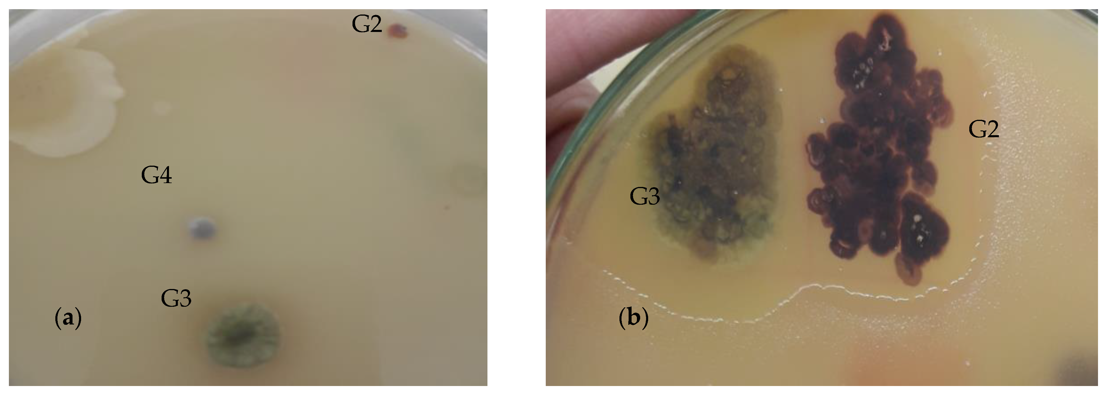
The experiments were performed in triplicate, and the obtained results were analyzed statistically by Kruskel–Wallis test, followed by Dunn’s post hoc test for data that did not follow Normal distribution and by One way Analysis of Variance (ANOVA) followed by Dunnett’s Multiple Comparison test for data that followed Normal distribution. In these analyses, bioactive-pigmented bacterial treatments named as Nonomurae salmonae (G2), Streptomyces chromofuscus (G3), and Actinocorallia libanotica (G4) are Explanatory variables while F. oxysporum, F. solani, A. flavus, A. alternata, and A. niger are Response variables i.e., changing bacterial treatments affect growth inhibition of F. oxysporum, F. solani, A. flavus, A. alternate, and A. niger. All the analyses reported here were performed using GraphPad Prism 5 (5.01). Data have been expressed as mean ± SD.

3. Results

3.1. Isolation and Identification of Bioactive Pigment-Producing Bacteria

As a result of the selective isolation procedure, bioactive bacterial colonies, which were making clear zones against antibiotic-sensitive B. subtilis, exhibiting distant colors (i.e., pigmented colonies), were selected and purified for further studies. Three such colonies were selected, having purple, light green, and dark green colors (Figure 1). The morphological description of the selected colonies is given in Table 1.
All three isolates were found to belong to the single phylum Actinobacteria and three different genera, namely, Actinocorallia, Streptomyces, and Nonomuraea. Bacterial sequences obtained in this study were submitted to GenBank with accession numbers MT071630 to MT071632 (Table 2). Neighbor joining phylogenetic tree was constructed using MEGA 5.2 software to further confirm the identification of isolates (Figure 2).

3.2. Screening Bioactive Pigment-Producing Bacteria for Antimicrobial Activity

All the three bioactive pigment-producing colonies selected were initially screened to possess antimicrobial activity against phytopathogenic microorganisms. Antifungal activity was assessed using the dual culture technique. All three isolates were seen to possess antifungal activity against more than one phytopathogenic fungi tested, and hence, were further assessed to check the antifungal activity of their crude extracts. Percent inhibition of the radial growth based on screening by dual culture technique was calculated and given in Figure 3. Images of a confirmatory experiment of the antifungal activity of isolates performed later are given in Figure 4. For this, a fungal plug was placed in the center of a plate, and as many as three bacterial isolates were inoculated on three sides on a petri plate, while the fourth side of the plate was left uninoculated, serving as negative control (Figure 4).
The antibacterial activity of pigment-producing isolates against bacterial phytopathogens was assessed, as well. The screening was performed using the disk diffusion method. Streptomyces chromofuscus was found active against both of the pathogenic bacterial strains. Nonomurae salmonae was found active against P. syringae, and Actinocorallia libanotica was found inactive against both of the tested strains. Solvent control was used as negative control (1% DMSO), showing no activity against both of the pathogenic bacterial strains. Streptomycin (4 mg/mL) was used as positive control, showing good antibacterial activity against both of the pathogenic strains. The results of screening isolates for antibacterial activity against bacterial phytopathogens are shown in Table 3.
Results obtained were analyzed using the Kruskal–Wallis test followed by Dunn’s post hoc test. Kruskal–Wallis test compares mean ranks and Dunn’s test is used to perform pairwise comparisons between each independent group, which, in this case, are bacterial treatments (i.e., Nonomurae salmonae (G2), Streptomyces chromofuscus (G3), and Actinocorallia libanotica (G4)). Statistically significantly different groups are found in Dunn’s post hoc test at some level of α. The results of Dunn’s post hoc test are given in Table 4.

3.3. Antifungal Activity of Crude Bioactive Pigment Extract and Determination of MIC

The antifungal activity of crude bioactive pigment extract was determined at a concentration of 4 mg/mL and zones of inhibition were measured in mm. Streptomyces chromofuscus showed good antifungal results against all the phytopathogenic fungi compared to the other two isolates. Nonomurae salmonae was found inactive against A. flavus and showed little to no activity against F. oxysporum and F. solani. However, it showed very good activity against A. niger. Actinocorallia libanotica showed good activity against A. alternata and A. niger, however, it showed little to no activity against other phytopathogenic fungi tested. The results of zones of inhibition were given in Table 5.
MIC was determined for bioactive pigment extracts showing zones of inhibition greater than 10 mm. Streptomyces chromofuscus showed the lowest MIC against A. niger. The results are given in Table 5.

3.4. Antifungal Activity on Mycelial Radial Growth (MRG)

The ability of crude bioactive pigment extract to inhibit mycelial radial growth (MRG) of the tested phytopathogenic fungi was assessed. The percent inhibitory MRG values varied greatly among all the samples (Table 6). Streptomyces chromofuscus crude extract showed a wide range of antifungal activity. It inhibited MRG of all the phytopathogens tested and showed maximum percent inhibition of MRG against A. flavus (36.62), followed by A. niger (32.86), and A. alternata (30.99). Nonomurae salmonae showed mediocre results against F. oxysporum (15.49), F. solani (13.14), and A. alternata (14.56), no activity against A. flavus, and a good inhibition percentage of MRG against A. niger (23.01). Actinocorallia libanotica showed no inhibition of MRG against F. oxysporum and A. flavus. It showed maximum percent inhibition of MRG against F. solani (22.01), and a moderate activity against A. alternata (17.37), followed by A. niger (7.98).

3.5. Antibacterial Activity and MIC Determination

The antibacterial activity of crude pigment extract was determined by the agar disk diffusion assay. Zones of inhibition were measured in mm and are presented in Figure 5. Streptomyces chromofuscus crude extract inhibited the growth of both of the phytopathogenic bacteria making 19.67 mm and 18.67 mm zones of inhibition against P. syringae and X. axonopodis, respectively. Nonomurae salmonae inhibited the growth of P. syringae (17 mm) and was found inactive against X. axonopodis. Actinocorallia libanotica crude extract showed no activity against both of the phytopathogens tested. Streptomycin (4 mg/mL) was used as positive control and was seen to show good activity against both of the phytopathogens. 1% DMSO was used as a solvent to make a crude pigment extract solution and as a negative control.
MIC in the case of bacterial phytopathogens was determined only for those crude pigment extracts that made zones of inhibition greater than 10 mm. Results of MIC are given in Table 7.

4. Discussion

The current research was initiated to search for novel, new, and sustainable plant protectants. In this regard, bacteria have been increasingly investigated as a sustainable and safe alternative to chemical plant protectants. Bioactive pigment-producing bacteria can be considered a relatively new and less-investigated source in this context.
Bioactive-pigmented bacteria from saline agricultural soil were purposely targeted in the current research because the soil is a rich source of beneficial bacteria with a high percentage still uncultured and uninvestigated. Furthermore, as a general perception, the probability of isolating plant-beneficial bacteria is greater in agricultural soil as compared to non-agricultural soils. It was deduced from previous reports that bacterial inhabitants of saline environments tend to produce different and diverse useful metabolites/compounds that differ in properties from those found in non-saline habitats [4]. Extreme or stressed ecosystems are also reported to cause the evolution of novel secondary metabolic pathways that enhance the chances of finding new biological functions of bioactive compounds [23].
As a result of above-mentioned selective isolation, three bioactive colored colonies were isolated and were identified as Nonomurae salmonae, Streptomyces chromofuscus, and Actinocorallia libanotica, using 16S rRNA gene sequencing. All the isolates were found to be Actinobacteria, belonging to three different genera, namely, Actinocorallia, Streptomyces, and Nonomuraea. This is in congruence with previous studies that actinobacteria occur most commonly in soil [24]. The reporting on the isolation of bioactive-pigmented actinobacteria only, in the present study, agrees with previous reports on actinobacteria as producers of instinctive pigments on the media, which are red, green, yellow, brown, and black in color [25], and are an important and attractive source of bioactive secondary metabolites [20,26,27]. Because of this ability, these bacteria are one of the most explored microbes among prokaryotes [28].
Of all the three isolates of the current study, Streptomyces chromofuscus showed good antimicrobial activity against plant pathogens as compared to other isolates. It showed broad-spectrum antifungal activity against plant pathogenic fungi. These results are in congruence with previous reports about Streptomyces being an active producer of bioactive natural products as compared to other actinobacteria, and that more than 70% of the natural product having bioactive potential have been isolated from Streptomyces [26,29,30]. One of the previous studies reported that novel formulations of some of the strains of Streptomyces in the form of encapsulation are being developed as biocontrol agents against different plant pathogens [31]. Our results assessing the antimicrobial potential of Streptomyces chromofuscus are very encouraging, showing a broad-spectrum antimicrobial potential of this isolate.
Here, we also report the isolation of two rare actinobacteria, namely, Nonomurae salmonae and Actinocorallia libanotica, from saline agricultural fields. Rare actinobacteria are actinobacteria that are difficult to isolate, are abundant in different habitats but need specialized protocols for isolation, and are believed to be a source of diverse new chemical compounds [32]. Rare actinobacteria are considered as an unexplored source of novel bioactive metabolites and hence are increasingly being investigated [33]. Our study provides useful preliminary information about the antimicrobial potential of isolated rare actinobacteria against plant pathogens.
Our study is the first to assess the antimicrobial potential in general and antibacterial potential in specific of Nonomurae salmonae and Actinocorallia libanotica against P. syringae and X. axonopodis. It was deduced from the results that Nonomurae salmonae was active against P. syringae only and Actinocorallia libanotica was inactive against both of the bacterial pathogens. Furthermore, both isolates exhibited moderate to good antifungal activity. These results encourage us to further assess the active component involved in the activity and study different conditions required for the optimum production of the active component. Some previous studies on the members of the same genera of rare actinobacteria can be related to our results. One of the studies on Nonomuraea sp. IB 2015I9-2 grown on different media reported bioactivity of its crude extract against St. carnosus culture at varying concentrations [34]. In another study, some members of genus Nonomuraea were revealed to be a valuable source of novel metabolites for medical and industrial purposes [35]. Hence, we can say that preliminary information on the bioactivity of Nonomurae salmonae from our research is in congruence with previous findings on the members of the same genus.
A previous study on Actinocorallia libanotica reported its antimicrobial activity as weak to moderate against E. coli, S. aureus, M. luteus, and B. cereus [33]. This can be related to our results, where Actinocorallia libanotica was observed to show no activity against different bacterial pathogens and moderate activity against fungal plant pathogens. Another study on Actinocorallia aurantiaca reported the isolation of three new bioactive compounds, namely, aurantiadioic acids A-B and aurantoic acid A [36]. The apparent inactivity or weak activity of rare actinomycetes in the present study can be attributed to the requirement of special conditions for the growth and production of the active metabolite. Detailed studies to assess the conditions under which these rare actinobacteria produce active metabolites are required.

5. Conclusions

Bioactive-pigmented bacteria are relatively less investigated as biocontrol agents. Our study is one of the few studies on the biocontrol potential of pigmented bacteria against plant pathogenic bacteria and fungi. It was a preliminary study with encouraging results, demanding detailed research to isolate, identify, and assess the active components involved in bioactivity. Furthermore, the present study reports, for the first time, the bioactivity of two rare actinobacterial species, namely, Nonomurae salmonae and Actinocorallia libanotica, against bacterial plant pathogens. Detailed studies are needed and are underway by our group to work out the optimum conditions for the induction of production of active metabolites by rare isolates and the characterization of active components in the crude pigmented extract.

Author Contributions

S.B. design the experiment, carried out research work and wrote manuscript. M.N. carried out data analysis. Z.K.S. supervised the research work and guided experimental. All authors have read and agreed to the published version of the manuscript.

Funding

External funding was not received for this research.

Institutional Review Board Statement

Not applicable.

Informed Consent Statement

Not applicable.

Data Availability Statement

Not applicable.

Conflicts of Interest

The authors have no conflict of interest.

References

  1. Ajmal, A.W.; Saroosh, S.; Mulk, S.; Hassan, M.N.; Yasmin, H.; Jabeen, Z.; Nosheen, A.; Shah, S.M.U.; Naz, R.; Hasnain, Z. Bacteria isolated from wastewater irrigated agricultural soils adapt to heavy metal toxicity while maintaining their plant growth promoting traits. Sustainability 2021, 13, 7792. [Google Scholar] [CrossRef]
  2. Zhou, L.; Song, C.; Li, Z.; Kuipers, O.P. Antimicrobial activity screening of rhizosphere soil bacteria from tomato and genome-based analysis of their antimicrobial biosynthetic potential. BMC Genom. 2021, 22, 29. [Google Scholar] [CrossRef] [PubMed]
  3. Wang, X.; Radwan, M.M.; Taráwneh, A.H.; Gao, J.; Wedge, D.E.; Rosa, L.H.; Cutler, H.G.; Cutler, S.J. Antifungal activity against plant pathogens of metabolites from the endophytic fungus Cladosporium cladosporioides. J. Agric. Food Chem. 2013, 61, 4551–4555. [Google Scholar] [CrossRef] [PubMed] [Green Version]
  4. Torres, M.; Dessaux, Y.; Llamas, I. Saline environments as a source of potential quorum sensing disruptors to control bacterial infections: A review. Mar. Drugs 2019, 17, 191. [Google Scholar] [CrossRef] [Green Version]
  5. Rashid, M.; Fakruddin, M.; Mazumdar, R.M.; Kaniz, F.; Chowdhury, M. Anti-Bacterial Activity of Pigments Isolated From Pigment-Forming Soil Bacteria. J. Pharm. Res. Int. 2014, 4, 880–894. [Google Scholar] [CrossRef]
  6. Ramesh, C.; Vinithkumar, N.V.; Kirubagaran, R.; Venil, C.K.; Dufossé, L. Multifaceted applications of microbial pigments: Current knowledge, challenges and future directions for public health implications. Microorganisms 2019, 7, 186. [Google Scholar] [CrossRef] [PubMed] [Green Version]
  7. Sekurova, O.N.; Schneider, O.; Zotchev, S.B. Novel bioactive natural products from bacteria via bioprospecting, genome mining and metabolic engineering. Microb. Biotechnol. 2019, 12, 828–844. [Google Scholar] [CrossRef] [Green Version]
  8. Venil, C.K.; Dufossé, L.; Renuka Devi, P. Bacterial Pigments: Sustainable Compounds With Market Potential for Pharma and Food Industry. Front. Sustain. Food Syst. 2020, 4, 1–17. [Google Scholar] [CrossRef]
  9. Ongena, M.; Jacques, P. Bacillus lipopeptides: Versatile weapons for plant disease biocontrol. Trends Microbiol. 2008, 16, 115–125. [Google Scholar] [CrossRef] [PubMed]
  10. Mumtaz, R.; Bashir, S.; Numan, M.; Shinwari, Z.K.; Ali, M. Pigments from Soil Bacteria and Their Therapeutic Properties: A Mini Review. Curr. Microbiol. 2018, 76, 1–8. [Google Scholar] [CrossRef] [PubMed]
  11. Banerjee, D.; Chatterjee, S.; Banerjee, U.; Guha, A.K.; Ray, L. Green Pigment from Bacillus cereus M 1 16 (MTCC 5521): Production Parameters and Antibacterial Activity. Appl. Biochem. Biotechnol. 2011, 164, 767–779. [Google Scholar] [CrossRef] [PubMed]
  12. Drewnowska, J.M.; Zambrzycka, M.; Kalska-Szostko, B.; Fiedoruk, K.; Swiecicka, I. Melanin-like pigment synthesis by soil Bacillus weihenstephanensis isolates from Northeastern Poland. PLoS ONE 2015, 10, e0125428. [Google Scholar] [CrossRef] [Green Version]
  13. Kim, H.; Han, S.; Lee, C.; Lee, K.; Park, S.; Kim, Y. Use of Prodigiosin for Treating Diabetes Mellitus. US 6,638,968 B1, 28 October 2003. [Google Scholar]
  14. Sakthivel, N.; Gnanamanickam, S. Toxicity of Pseudomonas fluorescens towards rice sheath-rot pathogen Acrocylindrium oryzae Saw. Curr. Sci. 1986, 55, 106–107. [Google Scholar]
  15. Chen, Y.; Zhou, D.; Qi, D.; Gao, Z.; Xie, J.; Luo, Y. Growth promotion and disease suppression ability of a Streptomyces sp. CB-75 from banana rhizosphere soil. Front. Microbiol. 2018, 8, 2704. [Google Scholar] [CrossRef] [PubMed]
  16. Sharma, A.; Chandraker, S.; Patel, V.; Ramteke, P. Antibacterial activity of medicinal plants against pathogens causing complicated urinary tract infections. Indian J. Pharm. Sci. 2009, 71, 136. [Google Scholar] [CrossRef] [Green Version]
  17. Chen, L.; Luo, S.; Xiao, X.; Guo, H.; Chen, J.; Wan, Y.; Li, B.; Xu, T.; Xi, Q.; Rao, C. Application of plant growth-promoting endophytes (PGPE) isolated from Solanum nigrum L. for phytoextraction of Cd-polluted soils. Appl. Soil Ecol. 2010, 46, 383–389. [Google Scholar] [CrossRef]
  18. Kim, O.-S.; Cho, Y.-J.; Lee, K.; Yoon, S.-H.; Kim, M.; Na, H.; Park, S.-C.; Jeon, Y.S.; Lee, J.-H.; Yi, H. Introducing EzTaxon-e: A prokaryotic 16S rRNA gene sequence database with phylotypes that represent uncultured species. Int. J. Syst. Evol. Microbiol. 2012, 62 Pt 3, 716–721. [Google Scholar] [CrossRef] [Green Version]
  19. Fatima, H.; Khan, K.; Zia, M.; Ur-Rehman, T.; Mirza, B.; Haq, I.-U. Extraction optimization of medicinally important metabolites from Datura innoxia Mill.: An in vitro biological and phytochemical investigation. BMC Complement. Altern. Med. 2015, 15, 376. [Google Scholar] [CrossRef] [Green Version]
  20. Sharma, P.; Kalita, M.C.; Thakur, D. Broad Spectrum Antimicrobial Activity of Forest-Derived Soil Actinomycete, Nocardia sp. PB-52. Front. Microbiol. 2016, 7, 347. [Google Scholar] [CrossRef] [PubMed] [Green Version]
  21. Nimaichand, S.; Devi, A.M.; Tamreihao, K.; Ningthoujam, D.S.; Li, W.-J. Actinobacterial diversity in limestone deposit sites in Hundung, Manipur (India) and their antimicrobial activities. Front. Microbiol. 2015, 6, 413. [Google Scholar] [CrossRef] [PubMed]
  22. Ahameethunisa, A.R.; Hopper, W. Antibacterial activity of Artemisia nilagirica leaf extracts against clinical and phytopathogenic bacteria. BMC Complement. Altern. Med. 2010, 10, 6. [Google Scholar] [CrossRef]
  23. Qi, D.; Zou, L.; Zhou, D.; Chen, Y.; Gao, Z.; Feng, R.; Zhang, M.; Li, K.; Xie, J.; Wang, W. Taxonomy and broad-spectrum antifungal activity of Streptomyces sp. SCA3-4 isolated from rhizosphere soil of Opuntia stricta. Front. Microbiol. 2019, 10, 1390. [Google Scholar] [CrossRef] [PubMed]
  24. Human, Z.R.; Moon, K.; Bae, M.; De Beer, Z.W.; Cha, S.; Wingfield, M.J.; Slippers, B.; Oh, D.-C.; Venter, S.N. Antifungal Streptomyces spp. associated with the infructescences of Protea spp. in South Africa. Front. Microbiol. 2016, 7, 1657. [Google Scholar] [CrossRef] [Green Version]
  25. Sapkota, A.; Thapa, A.; Budhathoki, A.; Sainju, M.; Shrestha, P.; Aryal, S. Isolation, characterization, and screening of antimicrobial-producing actinomycetes from soil samples. Int. J. Microbiol. 2020, 2020, 2716584. [Google Scholar] [CrossRef] [PubMed]
  26. Ryandini, D.; Radjasa, O. Bioactive compounds derived from Streptomyces sp. SA32: Antibacterial activity, chemical profile, and their related genes. IOP Conference Series: Earth and Environmental Science. In Proceedings of the 4th International Conference on Biosciences (ICoBio 2021), Bogor, Indonesia, 11–12 August 2021; p. 012062. [Google Scholar]
  27. Bundale, S.; Singh, J.; Begde, D.; Nashikkar, N.; Upadhyay, A. Rare actinobacteria: A potential source of bioactive polyketides and peptides. World J. Microbiol. Biotechnol. 2019, 35, 92. [Google Scholar] [CrossRef] [PubMed]
  28. Selim, M.S.M.; Abdelhamid, S.A.; Mohamed, S.S. Secondary metabolites and biodiversity of actinomycetes. J. Genet. Eng. Biotechnol. 2021, 19, 1–13. [Google Scholar] [CrossRef] [PubMed]
  29. Ding, T.; Yang, L.J.; Zhang, W.D.; Shen, Y.H. The secondary metabolites of rare actinomycetes: Chemistry and bioactivity. RSC Adv. 2019, 9, 21964–21988. [Google Scholar] [CrossRef] [PubMed] [Green Version]
  30. Dezfully, N.K.; Ramanayaka, J.G. Isolation, identification and evaluation of antimicrobial activity of Streptomyces flavogriseus, strain ACTK2 from soil sample of Kodagu, Karnataka State (India). Jundishapur J. Microbiol. 2015, 8, e15107. [Google Scholar]
  31. Pacios-Michelena, S.; Aguilar, C.N.; Alvarez-Perez, O.B.; Rodriguez-Herrera, R.; Chávez-González, M.L.; Arredondo Valdés, R.; Ascacio Valdes, J.A.; Govea Salas, M.; Ilyina, A. Application of Streptomyces antimicrobial compounds for the control of phytopathogens. Front. Sustain. Food Syst. 2021, 310, 1–8. [Google Scholar] [CrossRef]
  32. Amin, D.H.; Abdallah, N.A.; Abolmaaty, A.; Tolba, S.; Wellington, E.M. Microbiological and molecular insights on rare Actinobacteria harboring bioactive prospective. Bull. Natl. Res. Cent. 2020, 44, 1–12. [Google Scholar] [CrossRef]
  33. Messaoudi, O.; Wink, J.; Bendahou, M. Diversity of actinobacteria isolated from date palms rhizosphere and saline environments: Isolation, identification and biological activity evaluation. Microorganisms 2020, 8, 1853. [Google Scholar] [CrossRef] [PubMed]
  34. Axenov-Gribanov, D.V.; Kostka, D.V.; Vasilieva, U.A.; Shatilina, Z.M.; Krasnova, M.E.; Pereliaeva, E.V.; Zolotovskaya, E.D.; Morgunova, M.M.; Rusanovskaya, O.O.; Timofeyev, M.A. Cultivable actinobacteria first found in baikal endemic algae is a new source of natural products with antibiotic activity. Int. J. Microbiol. 2020, 2020, 5359816. [Google Scholar] [CrossRef] [PubMed]
  35. Sungthong, R.; Nakaew, N. The genus Nonomuraea: A review of a rare actinomycete taxon for novel metabolites. J. Basic Microbiol. 2015, 55, 554–565. [Google Scholar] [CrossRef] [PubMed]
  36. Han, K.-Y.; Wu, X.; Jiang, C.; Huang, R.; Li, Z.-H.; Feng, T.; Chen, H.-P.; Liu, J.-K. Three new compounds from the actinomycete Actinocorallia aurantiaca. Nat. Prod. Bioprospect. 2019, 9, 351–354. [Google Scholar] [CrossRef] [PubMed]
Figure 1. (a) Initial isolation plate with three distant colored colonies labelled as G2, G3, and G4 (b) colored colonies in overlaid plates showing antagonistic activity against B. subtilis overlay.
Sustainability 14 13574 g001
Figure 2. Neighbor joining tree of isolates using Mega 5.2 software.
Sustainability 14 13574 g002
Figure 3. Percent inhibition of fungal radial growth by bioactive pigment-producing bacterial isolates.
Sustainability 14 13574 g003
Figure 4. Antifungal activity of G2, G3 and G4 against F. solani. NC represents uninoculated side of petri plate serving as negative control, while SNA represents sample not included in this study.
Sustainability 14 13574 g004
Figure 5. Antibacterial activity of crude bioactive bacterial pigment based on zones of inhibition (mm).
Sustainability 14 13574 g005
Table 1. Morphological description of selected colonies.
Bacterial IsolatesColorShapeMarginElevationTextureOpacitySizeSpore Color
G2Dark purple/magentaIrregularIrregularRaisedHard, dry, breaks apartOpaque3 mmWhite with very slight purplish tinch
G3Light greenIrregularUndulateRaisedHardOpaque5–7 mmBlackish
G4Dark greenRound EntireConvexVery hard, cannot be easily pickedOpaque2 mmWhite
Table 2. Isolates with accession numbers and closet matching strain in NCBI.
Bacterial IDsAccession No.Closet Match in NCBI with Accession No.Link to NCBI Closet MatchSimilarity %
G2MT071630Nonomurae salmonae strain JCM 3324 (MT760449)https://www.ncbi.nlm.nih.gov/nucleotide/MT760449.1?report=genbank&log$=nuclalign&blast_rank=7&RID=G8YD90N001R (accessed on 30 July 2020)100
G3MT071631Streptomyces chromofuscus strain AS-2 (JX442508)https://www.ncbi.nlm.nih.gov/nucleotide/JX442508.1?report=genbank&log$=nucltop&blast_rank=2&RID=G8YNXKZ0013 (accessed on 30 July 2020)100
G4MT071632Actinocorallia libanotica strain 11-2 (KJ571069)https://www.ncbi.nlm.nih.gov/nucleotide/KJ571069.1?report=genbank&log$=nucltop&blast_rank=5&RID=G8YWRK2S016 (accessed on 30 July 2020)99.64
Table 3. Screening isolates against phytopathogenic bacteria.
Bacterial PathogensNonomurae salmonae (G2)Streptomyces chromofuscus (G3)Actinocorallia libanotica (G4)Kruskal Wallis Test Summary
P. syringae14.33 ± 0.5817.66 ± 0.580 ± 0P = 0.0006
Kruskal–Wallis statistics = 10.73
X. axonopodis0 ± 09 ± 10 ± 0P = 0.0182, Kruskal–Wallis statistics = 10.73
Data in the table are means ± SD.
Table 4. Dunn’s multiple comparisons to compare mean ranks of control group to bioactive pigment-producing bacterial samples.
Dunn’s Multiple ComparisonsP. syringaeX. axonopodis
Mean Rank DifferenceSignificantSummaryAdjusted p ValueMean Rank DifferenceSignificantSummaryAdjusted p Value
NC to N. salmonae (G2)0Nons>0.9999−4.5Nons0.3041
NC to S. chromofuscus (G3)−6Yes*0.0224−7.5Yes*0.0190
NC to A. libanotica (G4)0Nons>0.99990Nons>0.9999
Data in the table are means ± SD. NC represents Negative Control, ns represents non-significant difference, while * represents significant difference among mean ranks.
Table 5. Antifungal activity of crude bioactive pigment extract and MIC values (in μg/mL) of bioactive crude pigment extract.
Nonomurae salmonae (G2)Streptomyces chromofuscus (G3)Actinocorallia libanotica (G4)ANOVA Summary
Inhibition Zone (mm)MIC (μg/mL)Inhibition Zone (mm)MIC (μg/mL)Inhibition Zone (mm)MIC (μg/mL)
F. oxysporum7 ± 1 a<5011 ± 1 a<500.67 ± 0.57 cNot in rangeF = 142, P < 0.0001, DF = 11
F. solani6.67 ± 1.5 b<2516.7 ± 0.4 a<2513.3 ± 1.5 a<100F = 128.5, P < 0.0001, DF = 11
A. flavus0 ± 0 cNo activity28 ± 1 a<12.50 ± 0 cNo activityF = 2352, P < 0.0001, DF = 11
A. alternata11.3 ± 1.15 a<5022.67 ± 0.5 a<12.511 ± 1 a<100F = 385.5, P < 0.0001, DF = 11
A. niger19.33 ± 1.15 a<5026.33 ± 1.5 a<6.254 ± 0.7 b<100F = 436.5, P < 0.0001, DF = 11
Data in the table are means ± SD. Lower case letter in superscript in the same row represent significant difference at p < 0.05 by Dunnett’s test. Lower case letters “a–c” represent; “a” is highly significant difference, “b” is slightly significant difference, and “c “is nonsignificant difference from negative control at p < 0.05.
Table 6. Percent inhibition of mycelial radial growth.
Pathogenic FungiNonomurae salmonae (G2)Streptomyces chromofuscus (G3)Actinocorallia libanotica(G4)ANOVA Summary
F. oxysporum15.49 ± 0.81 a24.88 ± 0.81 a0.94 ± 0.81 cF = 610.6, P < 0.0001, df = 11
F. solani13.14 ± 0.7 a30.98 ± 0.8 a22.07 ± 0.8 aF = 812, P < 0.0001, df = 11
A. flavus2.82 ± 1.4 c36.62 ± 0.8 a0.94 ± 0.8 cF = 1078, P < 0.0001, df = 11
A. alternata14.56 ± 0.7 a30.99 ± 1.07 a17.37 ± 0.8 aF = 591, P < 0.0001, df = 11
A. niger23.01 ± 0.8 a32.86 ± 1.2 a7.98 ± 0.8 aF = 718, P < 0.0001, df = 11
Data in the table are means ± SD. Lower case letter in superscript in the same row represent significant difference at p < 0.05 by Dunnett’s test. Lower case letters “a–c” represent; “a“ is highly significant difference,” b” is slightly significant difference, and “c” is nonsignificant difference from negative control at p < 0.05.
Table 7. MIC values (in μg/mL) of bioactive crude pigment extract.
Pathogenic BacteriaNonomurae salmonaeStreptomyces chromofuscusStreptomycin
P. syringae15.4924.883.906
X. axonopodisNA30.981.953
Publisher’s Note: MDPI stays neutral with regard to jurisdictional claims in published maps and institutional affiliations.

