Article

The Antagonistic Activity of Beneficial Fungi and Mechanisms Underlying Their Protective Effects on Plants Against Phytopathogens

by
Yelena Brazhnikova
1,2,
Andrey Belimov
3,
Lyudmila Ignatova
1,2,*,
Togzhan Mukasheva
1,
Tatyana Karpenyuk
1 and
Alla Goncharova
1
1
Department of Biology and Biotechnology, Al-Farabi Kazakh National University, Al-Farabi Ave. 71, Almaty 050038, Kazakhstan
2
Scientific Research Institute of Biology and Biotechnology Problems, Al-Farabi Kazakh National University, Al-Farabi Ave. 71, Almaty 050038, Kazakhstan
3
All-Russia Research Institute for Agricultural Microbiology, Sh. Podbelskogo 3, Pushkin, 196608 Saint-Petersburg, Russia
*
Author to whom correspondence should be addressed.
Sustainability 2025, 17(2), 450; https://doi.org/10.3390/su17020450
Submission received: 16 December 2024 / Revised: 4 January 2025 / Accepted: 6 January 2025 / Published: 9 January 2025

Abstract

:
Among the different strategies of plant protection from phytopathogens, the use of beneficial fungi has been described as a sustainable, eco-friendly approach. The aim of the present work was to evaluate the antagonistic activity of beneficial fungal strains in vitro and in vivo. The studied strains (Beauveria bassiana T7, Beauveria bassiana T15, Metarhizium robertsii An1, Talaromyces pinophilus T14) had pronounced antagonistic activity against three phytopathogens (the growth inhibition was 18.2–51%). In pot experiments, the studied strains significantly reduced the level of stress in barley plants caused by phytopathogenic load. The beneficial effect of the strains consisted of an increase in the morphometric parameters of plants and a positive effect on photosynthetic pigments and proline levels. The 1-aminocyclopropane-1-carboxylate deaminase level of the strains varied from 0.95 to 2.73 µM α-KB mg protein−1 h−1. The most significant mechanisms of antagonistic action of the M. robertsii An1 strain were the following: the production of hydrolytic enzymes (chitinase and glucanase activity amounted to 0.23 U mL−1 and 3.42 U mL−1, respectively) and the synthesis of soluble volatile and non-volatile compounds with antifungal properties, including destruxin E, destruxin A, and hydroxyanthraquinones. The results obtained revealed the potential of the studied strains for their integration into a sustainable agricultural system.

1. Introduction

Due to rapid population growth, innovative approaches to modern agriculture and environmental sustainability are needed to ensure food security [1], which is in line with the UN’s Sustainable Development Goal 2, “End hunger, achieve food security and improved nutrition, and promote sustainable agriculture” [2].
Barley (Hordeum vulgare L.) is one of the most important cereal crops in the world. It is mainly used as grain for livestock feed, as malt for beer production, and as human food. The increased interest in barley as a human food ingredient is linked to its special chemical composition and health benefits. Barley grains are an excellent source of dietary fiber (particularly β-glucan), vitamins (particularly vitamin E), and tocols, which are beneficial to human health [3].
The most important pests of barley are fungal pathogens, which have been estimated to cause an average annual yield loss of 15% worldwide [4]. The use of fungicides to protect plants is not always effective due to the spread of resistance in phytopathogenic fungi. Promising plant protection tools include the application of beneficial microorganisms that can be used as biocontrol agents to regulate the growth of phytopathogens, enhance plant growth, and promote plant resistance against adverse factors [5].
Among the various groups of microorganisms, fungi are of particular interest, since they are one of the main integral structural and functional components of biocenoses. They also have a number of advantages over other groups of microorganisms. Thus, fungi are distinguished by a high linear growth rate (1–2 orders higher than that of bacteria), which enables them to spread over greater distances and colonize the substrate more effectively [6]. In addition, fungi are characterized by extreme metabolic plasticity and high adaptive potential to the action of unfavorable environmental factors [7]. It is known that fungi in the soil form more stable interspecies networks [8,9].
It is known that beneficial fungi provide a number of positive effects on plants by involving the following main mechanisms: the synthesis of phytohormones and signaling molecules, increased availability of nutrients, protection of plants from abiotic stress factors, biocontrol of phytopathogens, influence on soil fertility, and induction of systemic resistance [5,10]. Plant protection from phytopathogens is realized by fungi through the following physiological and biochemical mechanisms: synthesis of extracellular hydrolytic enzymes; production of soluble non-volatile substances with antibiotic activity; release of volatile compounds; competition with phytopathogenic microflora for nutrients and a niche for colonization; elicitor activity; and the induction of systemic plant resistance [11,12].
The ability of fungal strains to produce extracellular hydrolases (chitinases, chitosanases, glucanases, cellulases, proteases, lipases, xylanases, mannases, etc.) is the most important mechanism of their antagonistic action against phytopathogenic microorganisms. These enzymes are capable of destroying the structural components of the cell wall, disrupting spore germination, and lysing germ tubes and hyphae of phytopathogenic fungi [11,12]. Another key mechanism of biocontrol of phytopathogens is the synthesis of various non-volatile metabolites with antibiotic properties. These metabolites are a chemically heterogeneous group of organic low-molecular-weight compounds produced by fungi, which even in low concentrations negatively affect the growth and/or metabolic activity of other microorganisms [11,12]. Microorganisms, including fungi, are capable of producing a large number of volatile substances with antimicrobial activity, the vast majority of which are volatile organic compounds (VOCs) [13,14,15].
Fungi are able to suppress the development of diseases in plants not only due to the synthesis of various metabolites with antifungal activity, but also due to an indirect mechanism—by activating the systemic resistance of plants regulated by signaling compounds (salicylic, abscisic, jasmonic acids, ethylene). An important indirect mechanism of the anti-stress effect of fungi on plants is their ability to produce 1-aminocyclopropane-1-carboxylate (ACC) deaminase. This enzyme hydrolyzes the amino acid ACC, which is a precursor of ethylene, the biosynthesis of which increases significantly under stressful conditions, including the effect of phytopathogens [16,17]. In addition, the protective activity of fungi is realized in improving the mineral nutrition of plants (potassium and phosphate mobilization).
In recent years, numerous reports presented considerable information on the antifungal activity of filamentous fungi against phytopathogens. However, many studies investigated the antifungal activity of the strains in vitro [18,19], and information on their effects on plants is insufficient. Moreover, most of the studies are focused primarily on the entomopathogenic role of Beauveria and Metarhizium [20,21,22,23], and little attention has been paid to their antifungal activity against phytopathogens. Furthermore, the antagonistic activity of these fungi against phytopathogens such as Fusarium oxysporum, F. graminearum, and Botrytis cinerea has been studied the most [19,24,25], while information on the antifungal activity against Alternaria is limited. However, this phytopathogen is one of the most widespread fungi with wide host diversity in agricultural crops [26]. In addition, studying the local beneficial fungi may be the most suitable way to control local pathogens and develop an effective and environmentally friendly biofungicide formula.
Thus, a more detailed study on the characteristics and mechanisms of antagonistic activity of beneficial fungal strains, as well as their application for protecting plants against phytopathogens, is needed to expand and deepen existing knowledge.
The present study aimed to evaluate the antagonistic activity of beneficial fungal strains and reveal the mechanisms of their antagonism against the most significant phytopathogens of agriculture. The main objectives of this research are as follows: (1) an evaluation of the antagonistic activity of four beneficial fungal strains in vitro, (2) an investigation of the antagonistic and plant-growth-promoting properties of beneficial fungi on barley plants grown under biotic stress conditions caused by the phytopathogen F. graminearum, and (3) a study of the mechanisms underlying the antagonism of the strain having the highest level of antagonistic activity in vitro and in vivo.
This study is significant in providing a better understanding of the mechanisms underlying the antagonistic activity of beneficial fungi. In this work, the ability of filamentous fungi of the species B. bassiana, M. robertsii, and T. pinophilus to synthesize 1-aminocyclopropane-1-carboxylate deaminase was demonstrated for the first time. The results obtained indicate the potential of the studied strains for their integration into a sustainable agricultural system.

2. Materials and Methods

2.1. Materials

Strains of beneficial fungi that have phosphate-mobilizing activity, the ability to synthesize phytohormones and signaling molecules [27], resistance to heavy metals [28], and plant-growth-promoting activity in relation to various agricultural crops [29] were used in the present study. Two of the studied strains were endophytes isolated from soybean (Glycine max L.): Beauveria bassiana T7 (accession No. MG966197) and Beauveria bassiana T15 (accession No. MG970260). Two strains were isolated from Kastanozem soil: Metarhizium robertsii An1 (accession No. PQ526686) and Talaromyces pinophilus T14 (accession No. MT364484). Detailed information on the sampling and identification of these strains is presented in previous studies [27,28].
Strains of phytopathogenic fungi (Fusarium oxysporum P8, Fusarium graminearum P12, and Alternaria alternata P15), obtained from the local collection of the Department of Biology and Biotechnology of Al-Farabi Kazakh National University, were used as test cultures.

2.2. Methods

2.2.1. Study of Antagonistic Activity

Strains of beneficial fungi and phytopathogenic fungi were grown on the surface of Sabouraud Agar for 5 days; then, plugs of 5 mm in diameter were cut out using sterile cork borer. In a Petri dish on the surface of the medium, a plug with a phytopathogen culture was placed on one edge, and a plug of the studied antagonistic strain was placed on the other edge. The distance between the plugs was 30 mm. Dishes with only the phytopathogen culture served as a control [30]. The experiment was conducted with five replicates. The growth inhibition zone was determined by measuring the size of the zone where the phytopathogen did not grow. The growth inhibition (GI) of phytopathogenic fungi was calculated using Equation (1):
G I = ( R 1 R 2 ) R 1 × 100 %
where GI—growth inhibition, %; R1—size of the phytopathogen colony in the control variant, mm; R2—size of the phytopathogen colony in the variant with the antagonist, mm.

2.2.2. Pot Experiments

To conduct pot experiments, we used seeds of spring barley (Hordeum vulgare L. var. Baysheshek), which were pre-sterilized by soaking them in 75% ethyl alcohol for 5 min and in 1% sodium hypochlorite for 10 min, followed by thorough rinsing in sterile distilled water.
To prepare the fungal inoculum, the fungal strains (each strain separately) were grown in liquid Sabouraud medium for 7 days at 180 rpm and 25 °C. After incubation, Tween-80 was added to the medium, and the spores were collected in a test tube with sterile water. The spore suspension was centrifuged at 9000× g for 1 min, after which the resulting sediment was resuspended in sterile distilled water. The spore concentration was brought to 107 spores mL−1.
The soil for pot experiments was classified as Kastanozem and had alkaline conditions with a pH (H2O) value of 8.4. The soil properties: bulk density of 1.25 g cm−3, total C of 2.75%, total N of 1.9 g kg−1, total phosphorus of 1.8 g kg−1, and CaCO3 of 7.8%. Dry soil (300 g) was placed in plastic pots (65 × 65 × 100 mm) and moistened to 60% with distilled water. Seeds were placed in pots (15 seeds per pot, 5 pots for each treatment), and each seed was inoculated with 1 mL of fungal inoculum. Control seeds were treated with sterile water.
The conditions of phytopathogenic load for plants were simulated by introducing a suspension of the phytopathogenic fungus F. graminearum P12 with a titer of 109 spores mL−1 into the soil.
The experimental design included 6 treatments: (1) sterile soil without inoculation, (2) soil with F. graminearum P12 without inoculation with antagonistic strains, (3) soil with F. graminearum P12 + inoculation with M. robertsii An1, (4) soil with F. graminearum P12 + inoculation with B. bassiana T15, (5) soil with F. graminearum P12 + inoculation with B. bassiana T7, and (6) soil with F. graminearum P12 + inoculation with T. pinophilus T14. The pots were arranged according to a completely randomized design for one variable (treatment type) with five replicates for each variant. All pots were uniformly moistened every three days. After incubation for 14 days, the plants were collected and the following parameters were examined: growth characteristics (length and mass of roots and stems), biochemical defense reaction (proline content), and activity of the photosynthetic system (content and ratio of chlorophyll pigments).

2.2.3. Determination of the Content of Photosynthetic Pigments

A sample of leaves (2 g) was thoroughly homogenized, extracted in 10 mL of 96% ethanol, kept in the dark for 1 h, then centrifuged at 6000× g for 15 min. The resulting extracts were used for spectrophotometric measurement of absorption at 649 and 665 nm. The content of chlorophyll was expressed as mg per g of leaf weight [31].

2.2.4. Determination of Proline Content

A sample of leaves (0.5 g) was thoroughly homogenized, extracted in 10 mL of 3% sulfosalicylic acid, and centrifuged at 11,000× g for 15 min. Glacial acetic acid and acidic ninhydrin reagent were added to the resulting extract in equal volumes. The mixture was kept in a water bath at 100 °C for 1 h, after which it was used for spectrophotometric measurement at 520 nm [32].

2.2.5. Determination of ACC Deaminase Activity

To identify ACC-utilizing strains among the studied beneficial fungi, three variants of the medium were used: with ACC as the only nitrogen source, with NH4NO3 (positive control), and without adding a nitrogen source (negative control). Petri dishes with inoculated strains were incubated at 25 °C for 7 days, after which a visual assessment of the growth intensity of the studied strains was performed. Strains capable of growing on a medium with ACC and showing more intensive growth on this medium compared to the positive and negative controls were selected for further determination of ACC deaminase activity.
ACC-utilizing strains were grown on a medium supplemented with ACC at 25 °C and 180 rpm for 5 days. After incubation, the cultures were centrifuged for 15 min at 14,000× g and 4 °C and washed with 0.1M Tris-HCl buffer (pH 7.5), and the pellet was resuspended in 0.1M Tris-HCl buffer (pH 8.5). Homogenization was performed in a UD-20 ultrasonic disintegrator (3 alternating periods of sonication for 60 s at 150 W) at 4 °C. The resulting suspension was centrifuged for 15 min at 14,000× g and 4 °C, and the supernatant was used to determine the enzyme activity by the colorimetric method for the formation of α-ketobutyrate (α-KB) [33]; 0.5M ACC was used as a substrate and the mixture was incubated for 30 min at 28 °C. The reaction was stopped by adding 1 mL of 0.56M HCl, and the mixture was centrifuged for 5 min at 14,000× g. Then, 100 μL of 0.56M HCl and 150 μL of 0.2% 2,4-dinitrophenylhydrazine solution in 2M HCl were added to 0.6 mL of the supernatant, and the mixture was incubated for 30 min at 28 °C. The reaction mixture was neutralized with 2M NaOH, and the optical density was measured at 540 nm. The protein content was determined by the Bradford colorimetric method [34]. Enzyme activity was expressed as the amount (in μM) of α-KB formed per 1 mg of cellular protein over an hour.

2.2.6. Determination of the Activity of Hydrolytic Enzymes

The M. robertsii An1 strain having the highest level of antagonistic activity was grown in liquid Sabouraud medium (50 mL) with the addition of chitin (for determining chitinase) or sodium carboxymethyl cellulose (Na-CMC) (for determining glucanase) for 7 days at 180 rpm and 25 °C, after which the cultures were centrifuged at 11,000× g for 15 min. The supernatant was used to determine chitinase and β-1,3-glucanase activity. Chitinase activity was determined spectrophotometrically with dinitrosalicylic acid based on the amount of N-acetylglucosamine released from colloidal chitin used as a substrate. The unit of chitinase activity (U) was taken as the amount of enzyme catalyzing the formation of 1 μg of N-acetylglucosamine per minute. The activity of β-1,3-glucanase was determined spectrophotometrically with dinitrosalicylic acid based on the amount of reducing sugars formed as a result of the hydrolysis of Na-CMC used as a substrate. The amount of enzyme catalyzing the formation of 1 μg of reducing sugars per minute was taken as a unit of activity (U) of β-1,3-glucanase [30].

2.2.7. Determination of VOC-Producing Capacity

In one Petri dish, the antagonistic strain was streaked onto the surface of the Sabouraud medium and incubated at 25 °C for 2 days. In another dish, a 5-day-old 5 mm block of the phytopathogenic fungus culture was placed on the surface of the medium. Both dishes were combined in such a way as to avoid direct contact between the cultures, and the edges were sealed with two layers of parafilm. A pair of dishes served as a control, one of which was inoculated with the phytopathogen, and the other contained only the Sabouraud medium. The dishes were incubated at 25 °C for 3 days [30]. Then, the GI was determined using the formula described above.

2.2.8. Extraction of Metabolites from M. robertsii An1 Culture and Evaluation of Antagonistic Activity of Extracts

The M. robertsii An1 strain having the highest level of antagonistic activity was grown on liquid Sabouraud and Czapek Dox media for 7 days at 180 rpm and 25 °C. At the end of the incubation, the biomass was separated by filtering the culture through a paper filter (Whatman No. 2). The metabolites were extracted in an ultrasonic bath for 10 min: from the filtrate with ethyl acetate and from the dry crushed mycelium with hexane and then with ethyl acetate. The solvent was distilled off at 40 °C on a Hei-VAP Precision rotary vacuum evaporator (Heidolph Instruments GmbH & Co KG, Schwabach, Germany), and the mass of the dry residue was determined. The resulting dry residue was suspended in sterile water to obtain solutions with a concentration of 5 mg mL−1 [35]. The antagonistic activity of the obtained extracts was assessed using the paper disk method on cultures of the phytopathogenic fungi F. oxysporum P8, F. graminearum P12, and A. alternata P15. Pieces of sterile filter paper with diameters of 7 mm were placed on each plate with the phytopathogen; then, 15 μL of extract with a concentration of 5 mg mL−1 was added to the filter paper sheets [36]. After incubation at 25 °C for 5 days, the size of inhibition zone was observed and the GI was calculated.

2.2.9. Chromatographic Analysis of Extracts

Extracts were separated using an ACQUITY UPLC H-Class (Waters, Milford, CT, USA) and diode-matrix UV detector on an ACQUITY UPLC BEH C8 reversed-phase column (50 × 2.1 mm, sorbent particle diameter 1.7 μm) (Waters, Milford, CT, USA). Elution was performed in an acetonitrile–0.1% formic acid system in the following gradient: 10–30% acetonitrile for 2 min, 30% acetonitrile for 2 min, 30–100% acetonitrile for 3 min, 100% acetonitrile for 2 min. The flow rate was 300 μL per min; the column temperature was 40 °C; the injected sample volume was 1 μL of extract. Extract samples were prepared by dissolving the dry residue in acetonitrile to a concentration of 5 mg mL−1. The detection of substances was carried out by scanning in the wavelength range from 200 to 800 nm (using the MaxPlot function of the Empower 3, Waters program 2475). The substances were compared by retention time and their UV spectra.

2.3. Statistical Analysis

Statistical data processing was performed using the Statistica version 10.0 software package (TIBCO Software Inc., Palo Alto, CA, USA). All data are presented as mean ± standard deviation. Prior to analysis, equality of variances and normality of the data were tested using Levene’s test and a Shapiro–Wilk test, respectively. Data analysis was performed using one-way analysis of variance (ANOVA) followed by Tukey’s HSD test for multiple comparisons.

3. Results and Discussion

3.1. Antagonistic Activity of Beneficial Fungi In Vitro

The studied strains had pronounced antagonistic properties against three phytopathogenic fungi. The zones of growth inhibition varied in the range from 10 to 28 mm; the GI was in the range from 18.2 to 51% (Table 1).
According to the nature of interaction with phytopathogenic fungi, two types of manifestation of antagonistic activity of the studied fungal strains were noted (Figure 1).
Fungi B. bassiana T7 (Figure 1b) and B. bassiana T15, without contact with phytopathogens, completely suppressed their development with the formation of “clean” inhibition zones. This may be due to the production of extracellular metabolites, which, diffusing into the thickness of the agar, inhibited the growth of phytopathogens [11,12].
Two other strains, M. robertsii An1 and T. pinophilus T14, characterized by rapid growth, actively grew, lysing the culture of the phytopathogen in the areas of direct contact (Figure 1d). This type of interaction may be associated with competition for the substrate, which is possible due to the rapid growth of fungal strains, which gives them an advantage in obtaining nutrients. In addition, these strains probably produce hydrolytic enzymes that degrade the mycelium of phytopathogens, as well as compounds with antifungal activity.
When examined microscopically, changes in the morphological structure of the mycelium were observed in phytopathogenic fungi under the influence of the studied antagonistic strains: the suppression of sporulation occurred; the structure of the strands became sparser and thinner; hyphal cells were noticeably rounded and shortened. It should be noted that as the antagonist strain colony approached, changes in the structure of the phytopathogen mycelium were more intense, while outside the zone of action of the metabolites secreted by the fungi, the pathogens formed a normal mycelium (Figure 1). This indicates that the degree of antagonistic action of antagonistic fungi on the phytopathogen is due to the radial gradient of the concentration of extracellular metabolites.

3.2. Antagonistic Activity of Fungal Strains In Vivo

Fusarium graminearum is a major crop contaminant that infects cereals, especially barley. This ascomycete fungal pathogen is the causal agent of Fusarium head blight in barley. It leads not only to significant yield losses but also contaminates grains through mycotoxins that are dangerous to humans and animals [37].
In the present study, when barley grew under conditions of phytopathogenic load caused by F. graminearum P12, the values of all the studied growth parameters decreased by 20–32%. Inoculation with antagonistic strains ensured more intensive development of plants under biotic stress. In the inoculated variants, barley developed a more powerful and long root system. In addition, the treated plants were taller than the control ones under phytopathogenic load. In particular, the length of the stem and root increased by 14–25% and 13–19%; the biomass of the aboveground and underground parts exceeded the non-inoculated variant by 11–12% and 12–14%, respectively. Inoculated plants grown on contaminated soil, in terms of growth parameters, were close to plants grown under normal conditions without exposure to the stress factor (Table 2).
The amount of photosynthetic pigments in leaves is an indirect indicator of the stress resistance of a crop, since this indicator changes during plant adaptation to environmental conditions, including the influence of various stress factors; i.e., it reflects the plant’s response to growing conditions. The ratio of chlorophylls a/b, which ensures the stability of the photosynthetic system, is of great importance; a decrease in this indicator and a decrease in the total chlorophyll content indicate a disruption in the photosynthetic activity of plants [38].
Under biotic stress conditions caused by the phytopathogen F. graminearum P12, the total chlorophyll content in barley leaves decreased by 22% compared to this value in plants grown under normal conditions and amounted to 1.88 ± 0.06 mg g−1 (Table 3). In addition, under phytopathogenic load, a decrease in the chlorophyll a/b ratio was observed, indicating changes in the pigment–protein complexes of light-harvesting antennae and reaction centers of both photosystems. The use of beneficial fungal strains had a stimulating effect on the photosynthetic activity of barley when grown in soil with an increased infectious background. The positive effect of studied strains was expressed in an increase in the Chl a content in leaves by 11–21%, Chl b content by 9–13%, and total Chl (a + b) content by 19–18%. The maximum effect was achieved in the variant with the M. robertsii An1 strain (Table 3).
In response to stress factors, the plant’s defense system is activated. An increase in proline content is one of the characteristic plant reactions to various types of stress, including biotic stress, which ensures the first stage of plant adaptation [39,40]. A higher level of proline in barley leaves was noted when plants were grown in soil with an increased infectious background compared to untreated plants in sterile soil. In the non-inoculated variant, under stressful conditions, the proline concentration exceeded this indicator in plants grown under favorable conditions by 1.9 times and amounted to 2.44 μmol g−1 of fresh weight. In plants treated with beneficial fungi, the content of proline varied in the range from 1.65 to 1.96 μmol g−1 depending on the strain (Table 3). The results obtained suggest that the studied strains significantly reduce the level of stress caused by phytopathogenic load.
Under stress conditions, the biosynthesis of ethylene is activated in plant tissues. This is a signal substance that triggers a cascade of non-specific stress reactions leading to the inhibition of physiological and growth processes in the plant. One of the mechanisms of positive effects on plants under normal and, in particular, stress conditions is the synthesis by some microorganisms of the enzyme ACC deaminase, which hydrolyzes the amino acid ACC, which is a precursor of ethylene, to α-ketobutyrate (α-KB) and ammonium. Due to the action of ACC deaminase, the level of ethylene in plant tissues is noticeably reduced, which leads to an increase in plant resistance to biotic and abiotic stress [16,41].
The activity of ACC deaminase in fungi is one of the key factors that facilitates growth and promotes the stress resistance of plants under unfavorable conditions. In this regard, an important stage of this work was the revealing ability of the studied strains to synthesize ACC deaminase.
All four strains studied demonstrated the ability to grow on a medium containing the amino acid ACC as the only nitrogen source, indicating the synthesis of the enzyme ACC deaminase by these cultures. When quantitatively assessing the activity of ACC deaminase in liquid culture, it was found that this indicator varied from 0.95 to 2.73 µM α-KB mg−1 protein h−1. The maximum activity of this enzyme was characteristic of the strains M. robertsii An1 and T. pinophilus T14 (Figure 2).
The positive role of ACC-hydrolyzing bacteria in the processes of plant growth, development, and resistance to biotic and abiotic stresses has been confirmed by many researchers [41,42,43,44,45]. However, information on plant-growth-promoting fungi containing ACC deaminase is limited. In this work, the ability of filamentous fungi of the species B. bassiana, M. robertsii, and T. pinophilus to synthesize this enzyme was demonstrated for the first time.

3.3. Study of Mechanisms of Antagonism Against Phytopathogenic Fungi

The strain M. robertsii An1, which exhibited maximum antagonistic activity in vitro and in vivo, was selected to decipher the mechanisms underlying the antagonism.
One of the important mechanisms involved in the interaction of antagonist strains with phytopathogens is the production of a complex of specific hydrolytic enzymes that destroy the structural polymers of the cell wall, which leads to the suppression of development and/or complete inhibition of the growth of phytopathogens. Chitinases and glucanases are of primary importance among these enzymes [12].
The M. robertsii An1 strain demonstrated the ability to grow on media containing colloidal chitin and Na CMC as the sole carbon source. The utilization of these substrates indicates the ability of the strain to produce chitinase and β-1,3-glucanase, respectively. Quantitative assessment revealed a high level of activity of these enzymes: 0.23 ± 0.01 U for chitinase and 3.42 ± 0.1 U for β-1,3-glucanase. The data obtained indicate that the synthesis of extracellular hydrolases is one of the important criteria that determine the antagonistic properties of the M. robertsii An1 strain. The mechanism of the antagonistic action of these enzymes is that chitinases cause the degradation of the main structural polysaccharide of the cell wall of pathogenic fungi—chitin. And glucanases hydrolyze glycosidic bonds in glucans, which are the second main component of the cell wall of fungi after chitin, and also play an important role during cell division and vegetative growth. It is also known that as a result of the action of chitinases, not only the growth of the pathogen is limited, but also chitooligosaccharides are formed, which are effective elicitors of systemic plant resistance [11,12].
Another key mechanism for suppressing the growth of pathogenic microorganisms is the ability of fungi to synthesize substances with antibiotic activity.
VOCs are an important class of substances with antifungal properties. They can suppress and stimulate the growth of microorganisms, induce systemic resistance in plants, and act as attractants or repellents for insects, nematodes, and other organisms [13,14,15]. At present, VOCs identified in fungi of the genus Metarhizium are mainly characterized by a modulating effect on insects [15], while information on their effect on phytopathogenic fungi is extremely limited. The antagonistic activity of VOCs of the strain M. robertsii An1 was studied using the double-plate method against three phytopathogenic fungi. It was shown that the VOCs of the studied strain exhibited weak antifungal activity only against two tested phytopathogens, F. oxysporum P8 and A. alternata P15, where the GI was 12.7% and 6.7%, respectively (Table 4). The results obtained indicate that VOC production is not the main mechanism of the antagonistic effect of the studied strain.
In addition to VOCs, antagonistic strains synthesize a wide range of non-volatile organic compounds with antagonistic properties [11,12]. At the next stage of this work, non-volatile exo- and endometabolites were studied, obtained by extraction from the filtrate and mycelium of the M. robertsii An1 culture, respectively.
The yield of extractive substances varied in the range from 0.22 to 0.48 g L−1 depending on the localization (filtrate and biomass) and nutrient medium (Czapek Dox and Sabouraud) (Table 5). Sabouraud medium was more favorable for the formation of endo- and exometabolites: the yield of extractive substances of the strain on this medium reached maximum values and amounted to 0.34 ± 0.01 and 0.48 ± 0.02 g L−1, respectively, while on Czapek Dox medium, this indicator did not exceed 0.29 ± 0.01 g L−1 (Table 5). The yield of extractive substances from the filtrate was significantly higher (p < 0.05) compared to mycelium, which indicates predominantly extracellular accumulation of metabolites.
Analysis of the antagonistic activity of extracts from the M. robertsii An1 strain revealed a number of features. The most sensitive to the studied extracts were the phytopathogenic fungi F. oxysporum P8 and A. alternata P15. The GI of F. graminearum P12 was significantly lower in all variants compared to other test cultures of phytopathogens (Table 6), indicating the greatest resistance of this pathogen to the metabolites of M. robertsii An1.
Extracts from the filtrates showed higher inhibitory activity against all phytopathogenic fungi than similar extracts from the mycelium (Table 6). This result suggests that compounds with antagonistic activity are released to a greater extent by the M. robertsii An1 strain into the environment, and only some of them accumulate intracellularly.
The composition of the nutrient medium had a significant effect on the degree of antagonistic activity of the fungal extracts. The antagonistic activity of both endo- and exometabolites obtained when growing the strain on Sabouraud medium was significantly higher than when growing the strain on Czapek Dox medium (Table 6). Thus, the most favorable nutrient medium for the formation of endo- and exometabolites with a high level of antagonistic activity was Sabouraud medium.
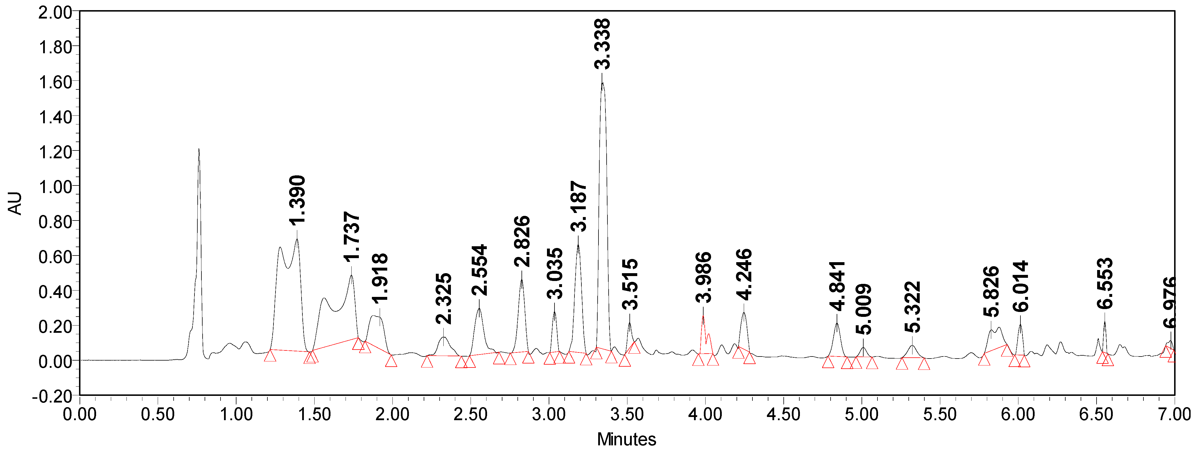
Analysis of the obtained chromatograms of extracts from the M. robertsii An1 culture showed the multicomponent nature of these fractions (Figure 3 and Figure 4). The metabolite complexes of extracts from the mycelium and culture liquid of the strain differed significantly in chemical composition.
As can be seen from the data presented in Figure 3, in the extract from the mycelium of the strain, the main compounds had absorption spectra in the far region of the UV spectrum (λ max = 196.7 nm, 193.2 nm, 190.9 nm, 194.4 nm, 192.0 nm), which is typical for aliphatic compounds, including those with multiple isolated bonds.
In the extracts from the culture filtrate of the M. robertsii An1 strain, absorptions were recorded in the range from 230 to 290 nm (Figure 4), which indicates the presence of substances with conjugated diene and polyene bonds. At the same time, the detected absorptions in the UV spectrum at λ from 230 to 250 nm indicate the presence of heteroannular cyclic systems, and those from 265 to 285 nm indicate homoannular cyclic systems. In addition, absorption in this region of the UV spectrum is characteristic of substances that have single C–C bonds in the structure and polar chromophore groups conjugated with each other.
In the extract from the culture filtrate, the compound with the retention time tR = 5.33 min is probably destruxin E, since it has a similar retention time and the characteristic absorption maximum in the UV spectrum at λ 271.2 nm for this destruxin. Also, another major substance in the extract from the culture liquid with tR = 7.48 min and a UV spectrum with an absorption maximum at λ max = 290.2 nm is destruxin A (Figure 4). These destruxins, which belong to cyclic depsipeptides, are found in various strains of Metarhizium and have pronounced insecticidal, antibiotic, and cytotoxic properties [46,47,48].
Analysis of the metabolite profiles of the extracts showed that in addition to destruxins, the strain produces yellow pigments that are not related to aurovertins. These pigments, judging by the UV spectra and retention time, most likely belong to the group of hydroxyanthraquinones [49,50].
The obtained data are consistent with previous studies. Thus, a number of authors report a high level of antagonistic activity of extracts obtained from cultures of various Metarhizium species. The extracted metabolites were characterized by antifungal properties against the phytopathogenic fungi Verticillium dahliae, Phytophthora megasperma, Fusarium oxysporum, Cladosporium herbarum, and Curvularia clavata [35,51,52]. Unlike previous studies, this work not only examined the antimicrobial activity of volatile and non-volatile metabolites of M. robertsii An1, but for the first time demonstrated another mechanism of the antagonistic action of the strain—the synthesis of hydrolytic enzymes.

4. Conclusions

The ability of beneficial fungal strains to suppress the growth and development of phytopathogenic fungi has been demonstrated. The complex nature of the antagonism of the M. robertsii An1 strain has been revealed. The production of extracellular hydrolytic enzymes (chitinases, glucanases) and the synthesis of non-volatile soluble compounds with antimicrobial activity (mainly destruxins) are the most significant mechanisms of the antagonistic action of this strain. Also, a certain role in the manifestation of antagonism is played by the emitted VOCs with antifungal properties. The results of the studies suggest that one of the mechanisms involved in protecting plants from phytopathogens is the ability of beneficial fungi to synthesize ACC deaminase.
The results obtained indicate the prospects for the use of beneficial fungi to enhance plant adaptation and improve their growth under the influence of biotic stress factors.

Author Contributions

Conceptualization, Y.B.; methodology, Y.B., A.B. and L.I.; software, T.M., T.K. and A.G.; validation, Y.B., T.M. and L.I.; formal analysis, Y.B.; investigation, Y.B., A.B. and L.I.; resources, A.B. and L.I.; data curation, A.B. and T.M.; writing—original draft preparation, Y.B. and L.I.; writing—review and editing, Y.B. and L.I.; visualization, Y.B.; supervision, Y.B., T.M. and L.I.; project administration, A.B. and L.I.; funding acquisition, A.B., L.I., T.K. and A.G. All authors have read and agreed to the published version of the manuscript.

Funding

This research was funded by the Science Committee of the Ministry of Science and Higher Education of the Republic of Kazakhstan (Grant No. AP19679444), whereas the determination of ACC deaminase activity on the fungi was funded by the Russian Science Foundation (Grant No. 24-16-00166).

Institutional Review Board Statement

Not applicable.

Informed Consent Statement

Not applicable.

Data Availability Statement

The data that support the findings of this study are available upon request from the corresponding author.

Conflicts of Interest

The authors declare no conflicts of interest.

References

  1. Giller, K.E.; Delaune, T.; Silva, J.V.; Descheemaeker, K.; van de Ven, G.; Schut, A.G.; van Wijk, M.; Hammond, J.; Hochman, Z.; Taulya, G.; et al. The future of farming: Who will produce our food? Food Secur. 2021, 13, 1073–1099. [Google Scholar] [CrossRef]
  2. United Nations Sustainable Development Goals. Goal 2: Zero Hunger. Available online: https://www.un.org/sustainabledevelopment/hunger/#:~:text=To%20achieve%20zero%20hunger%20by,global%20nutrition%20and%20food%20security (accessed on 12 December 2024).
  3. Geng, L.; Li, M.; Zhang, G.; Ye, L. Barley: A potential cereal for producing healthy and functional foods. Food Qual. Saf. 2022, 6, fyac012. [Google Scholar] [CrossRef]
  4. Stetkiewicz, S.; Burnett, F.J.; Ennos, R.A.; Topp, F.E.C. The impact of fungicide treatment and Integrated Pest Management on barley yields: Analysis of a long term field trials database. Eur. J. Agron. 2019, 105, 111–118. [Google Scholar] [CrossRef]
  5. Hossain, M.M.; Sultana, F. Application and mechanisms of plant growth promoting fungi (PGPF) for Phytostimulation. In Organic Agriculture; Das, S.K., Ed.; IntechOpen: London, UK, 2020; pp. 1–31. [Google Scholar] [CrossRef]
  6. Moore, D.; Robson, G.D.; Trinci, A.P.J. Hyphal cell biology and growth on solid substrates. In 21st Century Guidebook to Fungi; Moore, D., Robson, G.D., Trinci, A.P.J., Eds.; Cambridge University Press: Cambridge, UK, 2020; pp. 79–96. [Google Scholar] [CrossRef]
  7. Frąc, M.; Hannula, S.E.; Bełka, M.; Jędryczka, M. Fungal Biodiversity and Their Role in Soil Health. Front. Microbiol. 2018, 9, 707. [Google Scholar] [CrossRef] [PubMed]
  8. Chen, J.; Nan, J.; Xu, D.; Mo, L.; Zheng, Y.; Chao, L.; Bao, Y. Response differences between soil fungal and bacterial communities under opencast coal mining disturbance conditions. Catena 2020, 194, e104779. [Google Scholar] [CrossRef]
  9. de Vries, F.T.; Griffiths, R.I.; Bailey, M.; Craig, H.; Girlanda, M.; Gweon, H.S.; Hallin, S.; Kaisermann, A.; Keith, A.M.; Kretzschmar, M.; et al. Soil bacterial networks are less stable under drought than fungal networks. Nat. Commun. 2018, 9, 3033. [Google Scholar] [CrossRef] [PubMed]
  10. Baron, N.C.; Rigobelo, E.C. Endophytic fungi: A tool for plant growth promotion and sustainable agriculture. Mycology 2021, 13, 39–55. [Google Scholar] [CrossRef] [PubMed]
  11. Ghorbanpour, M.; Omidvari, M.; Abbaszadeh-Dahaji, P.; Omidvar, R.; Kariman, K. Mechanisms underlying the protective effects of beneficial fungi against plant diseases. Biol. Control 2018, 117, 47–157. [Google Scholar] [CrossRef]
  12. Roca-Couso, R.; Flores-Félix, J.D.; Rivas, R. Mechanisms of Action of Microbial Biocontrol Agents against Botrytis cinerea. J Fungi 2021, 7, 1045. [Google Scholar] [CrossRef]
  13. Karsli, A.; Şahin, Y.S. The role of fungal volatile organic compounds (FVOCs) in biological control. Türk. Biyolojik Mücadele Derg. 2021, 12, 79–92. [Google Scholar] [CrossRef]
  14. Morath, S.U.; Hung, R.; Bennett, J.W. Fungal volatile organic compounds: A review with emphasis on their biotechnological potential Fungal Biol. Rev. 2012, 26, 73–83. [Google Scholar] [CrossRef]
  15. Hummadi, E.H.; Cetin, Y.; Demirbek, M.; Kardar, N.M.; Khan, S.; Coates, C.J.; Eastwood, D.C.; Dudley, E.; Maffeis, T.; Loveridge, J.; et al. Antimicrobial Volatiles of the Insect Pathogen Metarhizium brunneum. J Fungi 2022, 8, 326. [Google Scholar] [CrossRef] [PubMed]
  16. Chandwani, S.; Amaresan, N. Role of ACC deaminase producing bacteria for abiotic stress management and sustainable agriculture production. Environ. Sci. Pollut. Res. Int. 2022, 29, 22843–22859. [Google Scholar] [CrossRef] [PubMed]
  17. Belimov, A.A.; Zinovkina, N.Y.; Safronova, V.I.; Litvinsky, V.A.; Nosikov, V.V.; Zavalin, A.A.; Tikhonovich, I.A. Rhizobial ACC deaminase contributes to efficient symbiosis with pea (Pisum sativum L.) under single and combined cadmium and water deficit stress. Environ. Exp. Bot. 2019, 167, 103859. [Google Scholar] [CrossRef]
  18. Erdoğan, O.; Sağlan, Z. Antifungal activity of local isolates of Beauveria bassiana (Balsamo) Vuillemin against Verticillium dahlia Kleb. causing wilt disease of cotton. Egypt. J. Biol. Pest. Control 2023, 33, 52. [Google Scholar] [CrossRef]
  19. Russo, M.L.; Vianna, M.F.; Scorsetti, A.C.; Ferreri, N.; de Abajo, J.M.; Troncozo, M.I.; Pelizza, S.A. Entomopathogenic Fungi as Dual Control Agents against Two Phytopathogens and the Lepidopteran Pest Rachiplusia nu in Soybean (Glycine max (L.) Merr). J. Fungi 2024, 10, 93. [Google Scholar] [CrossRef]
  20. Kisaakye, J.; Fourie, H.; Coyne, D.; Cortada, L.; Masinde, S.; Subramanian, S.; Haukeland, S. Evaluation of the Entomopathogenic Potential of Beauveria bassiana, Metarhizium anisopliae and Isaria fumosorosea for Management of Cosmopolites sordidus Germar (Coleoptera: Curculionidae). Agriculture 2021, 11, 1290. [Google Scholar] [CrossRef]
  21. Carolino, A.T.; Paula, A.R.; Silva, C.P.; Butt, T.M.; I Samuels, R. Monitoring persistence of the entomopathogenic fungus Metarhizium anisopliae under simulated field conditions with the aim of controlling adult Aedes aegypti (Diptera: Culicidae). Parasites Vectors 2014, 7, 198. [Google Scholar] [CrossRef]
  22. Mnyone, L.L.; Ng’habi, K.R.; Mazigo, H.D.; Katakweba, A.A.; Lyimo, I.N. Entomopathogenic fungi, Metarhizium anisopliae and Beauveria bassiana reduce the survival of Xenopsylla brasiliensis larvae (Siphonaptera: Pulicidae). Parasites Vectors 2012, 5, 204. [Google Scholar] [CrossRef] [PubMed]
  23. Kirkland, B.H.; Westwood, G.S.; Keyhani, N.O. Pathogenicity of Entomopathogenic Fungi Beauveria bassiana and Metarhizium anisopliae to Ixodidae Tick Species Dermacentor variabilis, Rhipicephalus sanguineus, and Ixodes scapularis. J. Med. Entomol. 2004, 41, 705–711. [Google Scholar] [CrossRef] [PubMed]
  24. Sui, L.; Lu, Y.; Zhou, L.; Li, N.; Li, Q.; Zhang, Z. Endophytic Beauveria bassiana promotes plant biomass growth and suppresses pathogen damage by directional recruitment. Front. Microbiol. 2023, 14, 1227269. [Google Scholar] [CrossRef]
  25. Muhorakeye, M.C.; Namikoye, E.S.; Khamis, F.M.; Wanjohi, W.; Akutse, K.S. Biostimulant and antagonistic potential of endophytic fungi against fusarium wilt pathogen of tomato Fusarium oxysporum f. sp. lycopersici. Sci. Rep. 2024, 14, 15365. [Google Scholar] [CrossRef] [PubMed]
  26. Carrascal-Hernández, D.C.; Flórez-López, E.; Peralta-Ruiz, Y.; Chaves-López, C.; Grande-Tovar, C.D. Eco-Friendly Biocontrol Strategies of Alternaria Phytopathogen Fungus: A Focus on Gene-Editing Techniques. Agriculture 2022, 12, 1722. [Google Scholar] [CrossRef]
  27. Brazhnikova, Y.V.; Shaposhnikov, A.I.; Sazanova, A.L.; Belimov, A.A.; Mukasheva, T.D.; Ignatova, L.V. Phosphate Mobilization by Culturable Fungi and Their Capacity to Increase Soil P Availability and Promote Barley Growth. Curr. Microbiol. 2022, 79, 240. [Google Scholar] [CrossRef] [PubMed]
  28. Ignatova, L.; Kistaubayeva, A.; Brazhnikova, Y.; Omirbekova, A.; Mukasheva, T.; Savitskaya, I.; Karpenyuk, T.; Goncharova, A.; Egamberdieva, D.; Sokolov, A. Characterization of cadmium-tolerant endophytic fungi isolated from soybean (Glycine max) and barley (Hordeum vulgare). Heliyon 2021, 7, e08240. [Google Scholar] [CrossRef]
  29. Brazhnikova, Y.; Ignatova, L.; Omirbekova, A.; Mukasheva, T.; Kistaubayeva, A.; Savitskaya, I.; Egamberdieva, D.; Usmanova, A.; Batlutskaya, I. Effect of plant growth promotion fungi on agricultural crops. Proc. BIO Web Conf. 2021, 40, 01004. [Google Scholar] [CrossRef]
  30. Khunnamwong, P.; Lertwattanasakul, N.; Jindamorakot, S.; Suwannarach, N.; Matsui, K.; Limtong, S. Evaluation of antagonistic activity and mechanisms of endophytic yeasts against pathogenic fungi causing economic crop diseases. Folia Microbiol. 2020, 65, 573–590. [Google Scholar] [CrossRef] [PubMed]
  31. Lichtestaller, H.K. Determination of total carotenoids and chlorophylls a and b of leaves extracts in different solvents. Biochem. Soc. Trans. 1983, 11, 591–592. [Google Scholar] [CrossRef]
  32. Bates, L.S.; Waldren, R.P.; Teare, I.D. Rapid determination of free proline for water-stress studies. Plant Soil 1973, 39, 205–207. [Google Scholar] [CrossRef]
  33. Honma, M.; Shimomura, T. Metabolism of 1-aminocyclopropane-1-carboxylic acid. Agric. Biol. Chem. 1978, 42, 1825–1831. [Google Scholar] [CrossRef]
  34. Bradford, M.M. A rapid and sensitive method for the quantitation of microgram quantities of protein utilizing the principle of protein-dye binding. Anal. Biochem. 1976, 72, 248–254. [Google Scholar] [CrossRef]
  35. Lozano-Tovar, M.D.; Garrido-Jurado, I.; Quesada-Moraga, E.; Raya-Ortega, M.C.; Trapero-Casas, A. Metarhizium brunneum and Beauveria bassiana release secondary metabolites with antagonistic activity against Verticillium dahliae and Phytophthora megasperma olive pathogens. Crop Prot. 2017, 100, 186–195. [Google Scholar] [CrossRef]
  36. Huang, X.; Han, Y.; Du, J.; Guo, P.; Wang, Y.; Ma, K.; Li, N.; Zhang, Z.; Li, Y.; Pan, J. Inhibitory Effect of Cinnamaldehyde on Main Destructive Microorganisms of Nanhai No. 1 Shipwreck. Appl. Sci. 2021, 11, 5262. [Google Scholar] [CrossRef]
  37. Yang, F.; Jacobsen, S.; Jørgensen, H.J.L.; Collinge, D.B.; Svensson, B.; Finnie, C. Fusarium graminearum and its interactions with cereal heads: Studies in the proteomics era. Front. Plant Sci. 2013, 4, 37. [Google Scholar] [CrossRef] [PubMed]
  38. Sharkey, T.D. Emerging research in plant photosynthesis. Emerg. Top Life Sci. 2020, 4, 137–150. [Google Scholar] [CrossRef] [PubMed]
  39. Liang, X.; Zhang, L.; Natarajan, S.K.; Becker, D.F. Proline mechanisms of stress survival. Antioxid. Redox Signal. 2013, 19, 998–1011. [Google Scholar] [CrossRef] [PubMed]
  40. Fabro, G.; Kovács, I.; Pavet, V.; Szabados, L.; Alvarez, M.E. Proline accumulation and AtP5CS2 gene activation are induced by plant-pathogen incompatible interactions in Arabidopsis. Mol. Plant-Microbe Interact. 2004, 17, 343–350. [Google Scholar] [CrossRef] [PubMed]
  41. Belimov, A.A.; Safronova, V.I.; Sergeyeva, T.A.; Egorova, T.N.; Matveyeva, V.A.; Tsyganov, V.E.; Borisov, A.Y.; Tikhonovich, I.A.; Kluge, C.; Preisfeld, A.; et al. Characterization of plant growth-promoting rhizobacteria isolated from polluted soils and containing 1-aminocyclopropane-1-carboxylate deaminase. Can. J. Microbiol. 2001, 47, 642–652. [Google Scholar] [CrossRef] [PubMed]
  42. Begum, N.; Hu, Z.; Cai, Q.; Lou, L. Influence of PGPB inoculation on HSP70 and HMA3 gene expression in switchgrass under cadmium stress. Plants 2019, 8, 504. [Google Scholar] [CrossRef] [PubMed]
  43. Mitra, S.; Pramanik, K.; Sarkar, A.; Ghosh, P.K.; Soren, T.; Maiti, T.K. Bioaccumulation of cadmium by Enterobacter sp. and enhancement of rice seedling growth under cadmium stress. Ecotoxicol. Environ. Saf. 2018, 156, 183–196. [Google Scholar] [CrossRef]
  44. Gupta, S.; Pandey, S. Enhanced salinity tolerance in the common bean (Phaseolus vulgaris) plants using twin ACC deaminase producing rhizobacterial inoculation. Rhizosphere 2020, 16, 100241. [Google Scholar] [CrossRef]
  45. Robison, M.M.; Shah, S.; Tamot, B.; Pauls, K.P.; Moffatt, B.A.; Glick, B.R. Reduced symptoms of Verticillium wilt in transgenic tomato expressing a bacterial ACC deaminase. Mol. Plant Pathol. 2001, 2, 135–145. [Google Scholar] [CrossRef]
  46. Golo, P.S.; Gardner, D.R.; Grilley, M.M.; Takemoto, J.Y.; Krasnoff, S.B.; Pires, M.S.; Fernandes, É.K.K.; Bittencourt, V.R.E.P.; Roberts, D.W. Production of destruxins from Metarhizium spp. fungi in artificial medium and in endophytically colonized cowpea plants. PLoS ONE 2014, 9, e104946. [Google Scholar] [CrossRef] [PubMed]
  47. Sree, K.S.; Padmaja, V. Destruxin from Metarhizium anisopliae induces oxidative stress effecting larval mortality of the polyphagous pest Spodoptera litura. J. Appl. Entomol. 2008, 132, 68–78. [Google Scholar] [CrossRef]
  48. Yadav, R.N.; Mahtab, R.M.; Zaidi, N.W.; Kumar, R.; Singh, H.B. Secondary Metabolites of Metarhizium spp. and Verticillium spp. and Their Agricultural Applications. In Secondary Metabolites of Plant Growth Promoting Rhizomicroorganisms; Singh, H.B., Keswani, C., Reddy, M.S., Sansinenea, E., García-Estrada, C., Eds.; Springer: Singapore, 2019; pp. 27–58. [Google Scholar] [CrossRef]
  49. Liu, J.; Leng, L.; Liu, Y.; Gao, H.; Yang, W.; Chen, S.; Liu, A. Identification and quantification of target metabolites combined with transcriptome of two rheum species focused on anthraquinone and flavonoids biosynthesis. Sci. Rep. 2020, 10, 20241. [Google Scholar] [CrossRef] [PubMed]
  50. Zhao, X.; Wei, J.; Yang, M. Simultaneous Analysis of Iridoid Glycosides and Anthraquinones in Morinda officinalis Using UPLC-QqQ-MS/MS and UPLC-Q/TOF-MSE. Molecules 2018, 23, 1070. [Google Scholar] [CrossRef] [PubMed]
  51. Ravindran, K.; Chitra, S.; Wilson, A.; Sivaramakrishnan, S. Evaluation of Antifungal Activity of Metarhizium anisopliae Against Plant Phytopathogenic Fungi. In Microbial Diversity and Biotechnology in Food Security; Kharwar, R.N., Upadhyay, R.S., Dubey, N.K., Raghuwanshi, R., Eds.; Springer: New Delhi, India, 2014; pp. 251–255. [Google Scholar] [CrossRef]
  52. Guigón-López, C.; Holguín-Ibarra, P.D.; Torres-Zapien, J.H.; García- Cruz, I.; Villapando, I.; Salas-Salazar, N.A. Metarhizium anisopliae reduces conidial germination and mycelium growth of the apple gray mold Botrytis cinerea. Biol. Control 2021, 160, 104660. [Google Scholar] [CrossRef]
Figure 1. Antagonism between antagonistic strains and phytopathogenic fungi: (a) phytopathogen Alternaria alternata (control); (b) Beauveria bassiana T7 against Alternaria alternata; (c) phytopathogen Fusarium oxysporum (control); (d) Talaromyces pinophilus T14 against Fusarium oxysporum. The left column represents fungal colonies on the Sabouraud plates. The right column represents microscopic morphology (×400).
Sustainability 17 00450 g001aSustainability 17 00450 g001b
Figure 2. Activity of ACC deaminase in fungal strains. Different letters indicate statistically significant differences between values according to Tukey’s test at p < 0.05.
Sustainability 17 00450 g002
Figure 3. HPLC-UV chromatogram of the mycelial extract of M. robertsii An1.
Sustainability 17 00450 g003
Figure 4. HPLC-UV chromatogram of the extract from the culture filtrate of M. robertsii An1.
Sustainability 17 00450 g004
Table 1. Antagonistic activity of fungal strains against phytopathogens.
Antagonistic StrainGrowth Inhibition, %Size of Growth Inhibition Zone, mm
Fusarium oxysporum P8Fusarium graminearum P12Alternaria alternata P15Fusarium oxysporum P8Fusarium graminearum P12Alternaria alternata P12
Beauveria bassiana T723.6 ± 1.1 b32.3 ± 1.2 b37.8 ± 1.6 b13 ± 0.4 b21 ± 0.8 c17 ± 0.7 b
Beauveria bassiana T1530.9 ± 1.2 c27.7 ± 0.8 c22.2 ± 1 a17 ± 0.7 c18 ± 0.7 b10 ± 0.4 a
Metarhizium robertsii An151 ± 1.5 d41.5 ± 1.1 d48.9 ± 1.8 c28 ± 0.9 d27 ± 1.1 d22 ± 0.8 c
Talaromyces pinophilus T1418.2 ± 0.7 a20 ± 0.3 a35.6 ± 1.5 b10± 0.3 a13 ± 0.5 a16 ± 0.8 b
Note: Different letters in the same column indicate statistically significant differences between values according to Tukey’s test at p < 0.05.
Table 2. Effect of inoculation with antagonistic fungal strains on growth parameters of barley under phytopathogenic load.
TreatmentShoot Length, cmRoot Length, cmDry Weight of Shoot, gDry Weight of Root, g
Sterile soil17.1 ± 0.5 d7.9 ± 0.2 c0.045 ± 0.0021 c0.019 ± 0.0014 c
Soil with Fusarium graminearum P12
Without inoculation13.1 ± 0.52 a6.3 ± 0.05 a0.035 ± 0.0022 a0.013 ± 0.0014 a
Beauveria bassiana T715.9 ± 0.4 c7.3 ± 0.22 b0.041 ± 0.0012 b0.018 ± 0.0013 bc
Beauveria bassiana T1514.9 ± 0.3 b7.1 ± 0.2 b0.039 ± 0.001 b0.016 ± 0.0012 b
Metarhizium robertsii An116.5 ± 0.5 cd7.5 ± 0.3 bc0.042 ± 0.002 bc0.018 ± 0.0009 bc
Talaromyces pinophilus T1415.2 ± 0.2 b7.1 ± 0.14 b0.039 ± 0.0013 b0.016 ± 0.0011 b
Note: Different letters in the same column indicate statistically significant differences between values according to Tukey’s test at p < 0.05.
Table 3. Effect of inoculation with antagonistic fungal strains on physiological parameters of barley under phytopathogenic load.
TreatmentChl a, mg g−1Chl b, mg g−1Chl (a + b), mg g−1Chl a/bProline, µmol g−1
Sterile soil1.76 ± 0.05 d0.64 ± 0.02 b2.4 ± 0.08 d2.75 ± 0.1 c1.28 ± 0.045 a
Soil with Fusarium graminearum P12
Without inoculation1.33 ± 0.05 a0.55 ± 0.01 a1.88 ± 0.06 a2.42 ± 0.05 a2.44 ± 0.08 d
Beauveria bassiana T71.58 ± 0.06 c0.61 ± 0.021 b2.19 ± 0.05 bc2.59 ± 0.1 b1.96 ± 0.08 c
Beauveria bassiana T151.55 ± 0.05 bc0.62 ± 0.024 b2.17 ± 0.1 bc2.5 ± 0.05 a1.88 ± 0.06 c
Metarhizium robertsii An11.61 ± 0.07 c0.6 ± 0.023 b2.21 ± 0.08 c2.68 ± 0.1 b1.65 ± 0.07 b
Talaromyces pinophilus T141.48 ± 0.03 b0.56 ± 0.012 a2.04 ± 0.04 b2.64 ± 0.08 b1.72 ± 0.052 b
Note: Different letters in the same column indicate statistically significant differences between values according to Tukey’s test at p < 0.05.
Table 4. Antagonistic activity of volatile organic compounds of M. robertsii An1 strain against phytopathogens.
PhytopathogenGrowth Inhibition, %Size of Growth Inhibition Zone, mm
Fusarium oxysporum P812.7 ± 0.57 ± 1
Fusarium graminearum P1200
Alternaria alternata P156.7 ± 0.13 ± 0.5
Table 5. Yield of extractive substances of the M. robertsii An1 strain.
Extract TypeExtractive Substances Yield, g L−1
Czapek Dox medium
Filtrate0.29 ± 0.01 b
Biomass0.22 ± 0.016 a
Sabouraud medium
Filtrate0.48 ± 0.02 b
Biomass0.34 ± 0.015 a
Note: Different letters in the same column indicate statistically significant differences between values according to Tukey’s test at p < 0.05.
Table 6. Antagonistic activity of different types of extracts from the M. robertsii An1 strain.
Extract TypeGrowth Inhibition, %
Fusarium oxysporum P8Fusarium graminearum P12Alternaria alternata P15
Czapek Dox medium
Filtrate34.5 ± 1.1 b27.7 ± 0.8 b33.3 ± 1.4 b
Biomass20 ± 0.5 a15.4 ± 0.4 a17.8 ± 0.5 a
Sabouraud Medium
Filtrate55.5 ± 1.5 b45.4 ± 1.5 b54.4 ± 1.6 b
Biomass35.5 ± 0.7 a33 ± 1 a36.7 ± 0.8 a
Note: Different letters in the same column indicate statistically significant differences between values according to Tukey’s test at p < 0.05.
Disclaimer/Publisher’s Note: The statements, opinions and data contained in all publications are solely those of the individual author(s) and contributor(s) and not of MDPI and/or the editor(s). MDPI and/or the editor(s) disclaim responsibility for any injury to people or property resulting from any ideas, methods, instructions or products referred to in the content.