Article

RASAL1 and ROS1 Gene Variants in Hereditary Breast Cancer

1
Genetic Laboratory, Department of Medical and Surgical Sciences, University of Bologna, 40138 Bologna, Italy
2
Unit of Medical Genetics, Azienda Ospedaliero-Universitaria Policlinico S. Orsola-Malpighi, 40138 Bologna, Italy
*
Authors to whom correspondence should be addressed.
Cancers 2020, 12(9), 2539; https://doi.org/10.3390/cancers12092539
Submission received: 16 July 2020 / Revised: 27 August 2020 / Accepted: 3 September 2020 / Published: 7 September 2020

Abstract

:

Simple Summary

Breast cancer is the second leading cause of death in women. Identifying novel genetic factors conferring BC predisposition is crucial to predict who is at increased risk of developing the disease, allowing for early detection and therapy, and optimized patient management. We identified germline pathogenic variants in familial breast cancer patients in ROS1 and RASAL1 genes. Further analysis in independent patient group will help understanding the role of these novel genes in breast cancer predisposition.

Abstract

Breast cancer (BC) is the second leading cause of death in women. BC patients with family history or clinical features suggestive of inherited predisposition are candidate to genetic testing to determine whether a hereditary cancer syndrome is present. We aimed to identify new predisposing variants in familial BC patients using next-generation sequencing approaches. We performed whole exome sequencing (WES) in first-degree cousin pairs affected by hereditary BC negative at the BRCA1/2 (BReast CAncer gene 1/2) testing. Targeted analysis, for the genes resulting mutated via WES, was performed in additional 131 independent patients with a suspected hereditary predisposition (negative at the BRCA1/2 testing). We retrieved sequencing data for the mutated genes from WES of 197 Italian unrelated controls to perform a case-controls collapsing analysis. We found damaging variants in NPL (N-Acetylneuraminate Pyruvate Lyase), POLN (DNA Polymerase Nu), RASAL1 (RAS Protein Activator Like 1) and ROS1 (ROS Proto-Oncogene 1, Receptor Tyrosine Kinase), shared by the corresponding cousin pairs. We demonstrated that the splice site alterations identified in NPL and ROS1 (in two different pairs, respectively) impaired the formation of the correct transcripts. Target analysis in additional patients identified novel and rare damaging variants in RASAL1 and ROS1, with a significant allele frequency increase in cases. Moreover, ROS1 achieved a significantly higher proportion of variants among cases in comparison to our internal control database of Italian subjects (p = 0.0401). Our findings indicate that germline variants in ROS1 and RASAL1 might confer susceptibility to BC.

1. Introduction

Breast cancer (BC) is the most common cancer in women worldwide and the second leading cause of cancer death, exceeded only by lung cancer [1].
BC patients with a family history or clinical features (such as bilateral, or early-onset BC) suggestive of inherited predisposition are candidate for genetic testing to determine whether a hereditary cancer syndrome is present. The two major genes BRCA1 (BReast CAncer gene 1) and BRCA2 (BReast CAncer gene 2), together with rare high penetrance genes (TP53 (Tumor protein 53), PTEN (Phosphatase and tensin homolog), CDH1 (Cadherin-1) and STK11 (Serine/threonine kinase 11)) and moderate penetrance genes collectively explain ~30% of familial BC risk [2].
Increasing the knowledge on genetic predisposition is crucial to extend the benefits of targeted surveillance/prevention to still unidentified high-risk women. As an example, among BRCA1 mutation carriers, the estimated lifetime risk of BC is 67%, and lifetime risk of ovarian cancer is 45%. BRCA2 mutation carriers face a similar risk for BC, with an ovarian cancer risk estimated to be 12% [3,4]. Therefore, lifetime risk is dramatically increased if compared to the general population, in which BC risk is approximately 13% and the risk of ovarian cancer is 1.5%, with significant implications for prevention and early detection [3,4]. In recent years, large genome-wide association studies (GWAS) have discovered many additional low-penetrance common variants associated with BC risk, with more than 100 SNPs (single nucleotide polymorphisms) independently associated with BC risk [5,6,7]. A recent association study in women of European origin focused on ER-negative disease, or BRCA1 mutation carriers, who are more likely to develop ER-negative disease (70–80% of cases) [5,8]. The Authors identified independent associations at genome-wide significance level with 10 variants at 9 novel loci. Replicated association was also detected for 10 of 11 variants previously reported in ER-negative disease or BRCA1 mutation carrier GWAS and consistent association with ER-negative disease was also detected for 105 susceptibility variants identified by other studies [5]. Together, these 125 variants explained ~14% of an assumed twofold increased risk of developing ER-negative disease for the first-degree female relatives of women affected with this subtype and ~40% of the estimated familial risk. An additional study identified 65 novel loci for breast cancer susceptibility [6]. Collectively, the >180 identified low-risk loci can explain 18% of the familial risk. Further studies of the altered molecular pathways are demanded in order to translate the association data into actionable targets for prevention/treatment of the disease.
In parallel, the advent of next-generation sequencing (NGS) has led to a timely testing of multiple genes, many of which with differing estimated cancer risks. This expansion introduced significant issues in clinical settings, most notably who to test and which genes should be included, since for multiple genes there is little clinical information available to guide patients if a pathogenic variant is identified, and larger panels have a higher incidence of finding variants of unknown significance [9]. Affordable wholeexome sequencing (WES) has become a common approach to identify rare variants, by performing a staged study starting with sequencing in a small cohort of cases with familial aggregation of BC, assuming that genetic contribution is higher. This approach led to the recent identification of rare risk variants in REQL (RecQ-like) and BLM (BLM RecQ Like Helicase) genes [10,11].
We undertook a WES-based approach in affected first-degree cousins, to identify candidate gene variants for hereditary BC (HBC), and identified damaging variants in NPL (N-Acetylneuraminate Pyruvate Lyase), POLN (DNA Polymerase Nu), RASAL1 (RAS Protein Activator Like 1) and ROS1 (ROS Proto-Oncogene 1, Receptor Tyrosine Kinase)genes. We then performed a targeted NGS analysis of these genes identifying additional pathogenic variants in ROS1 and RASAL1 in an independent cohort of 131 familial BC patients.

2. Results

2.1. Whole Exome Sequencing (WES) Analysis

To identify novel predisposing BC loci, we carried out a WES analysis in affected first-degree cousin-pairs with familial BC, who tested negative for BRCA1/2. Since first-degree cousins, based on their relationship, share 1/8 of the entire genome, this strategy helped filtering and selecting shared candidate variants, according to an autosomal dominant model of inheritance.
Three cousin-pairs were analyzed through WES and candidate variants were identified in different genes (Figure 1a–c; Table 1).
Variant prioritization for shared heterozygous variants was performed considering (i) a minor allele frequency (MAF) <0.01 in the Genome Aggregation database, gnomAD [12], and an in-house database of 650 Italian exomes; (ii) the pathogenicity of the variants (predicted by the Mendelian Clinically Applicable Pathogenicity (M-CAP) Score [13] and PolyPhen-2 [14] programs for missense variants, and Human Splice Finder v3.1 (HSF v3.1) [15], for the analysis of potential splicing alterations). This led to the identification of the following variants shared by the corresponding couples of cousins (Table 1): a single nucleotide variant abolishing the canonical donor splice site in intron 37 of the gene ROS1 (c.6005 + 2T > G) in family ITA_BC01; two rare variants in family ITA_BC02; a missense change in RASAL1 (p.Phe602Leu), present in ClinVar with conflicting data on pathogenicity and a predicted damaging missense change in POLN (p.Asp229Ala); a splice site deletion in NPL (c.68 + 2delT) in family ITA_BC03. All variants were confirmed by direct sequencing.
As variants in these genes are not known to be associated to HBC, to explore their role in BC, we investigated whether they are de-regulated in BC, compared to normal tissues. To this aim, we first evaluated the expression profiles of NPL, POLN, RASAL1 and ROS1 in human normal tissues, as reported in GTEx [16] and Human Protein Atlas [17] databases. NPL, POLN and RASAL1 showed a widespread expression in many human tissues, whereas ROS1 showed a more restricted expression in normal conditions, mainly in lung and brain (Figure S1A). BRCA1 and BRCA2 also showed a variable degree of expression in human normal tissues (Figure S1B).
As reported in the Catalogue of Somatic Mutation in Cancer (COSMIC) [18] database, NPL was overexpressed in 1.18% of BC cases (13/1104), POLN in 3.99% (44/1104), RASAL1 in 6.97% (77/1104) and ROS1 in 1.36% (15/1104). In comparison, overexpression of BRCA1 was observed in 11.02% of cases (122/1104) and overexpression of BRCA2 in 9.42% of cases (104/1104).
In the Human Protein Atlas [17] NPL, POLN and RASAL1 showed generally an increased expression in most cancer tissues (Figure S1C). In particular, RASAL1 overexpression resulted significantly correlated with a worse outcome (increased death) in endometrial cancer (Figure S1D), whereas NPL overexpression was correlated to a worse outcome in liver and renal cancers (Figure S1E). However, in other studies RASAL1 was found significantly downregulated in cancer [19,20], whereas its overexpression blocked cancer progression [21].

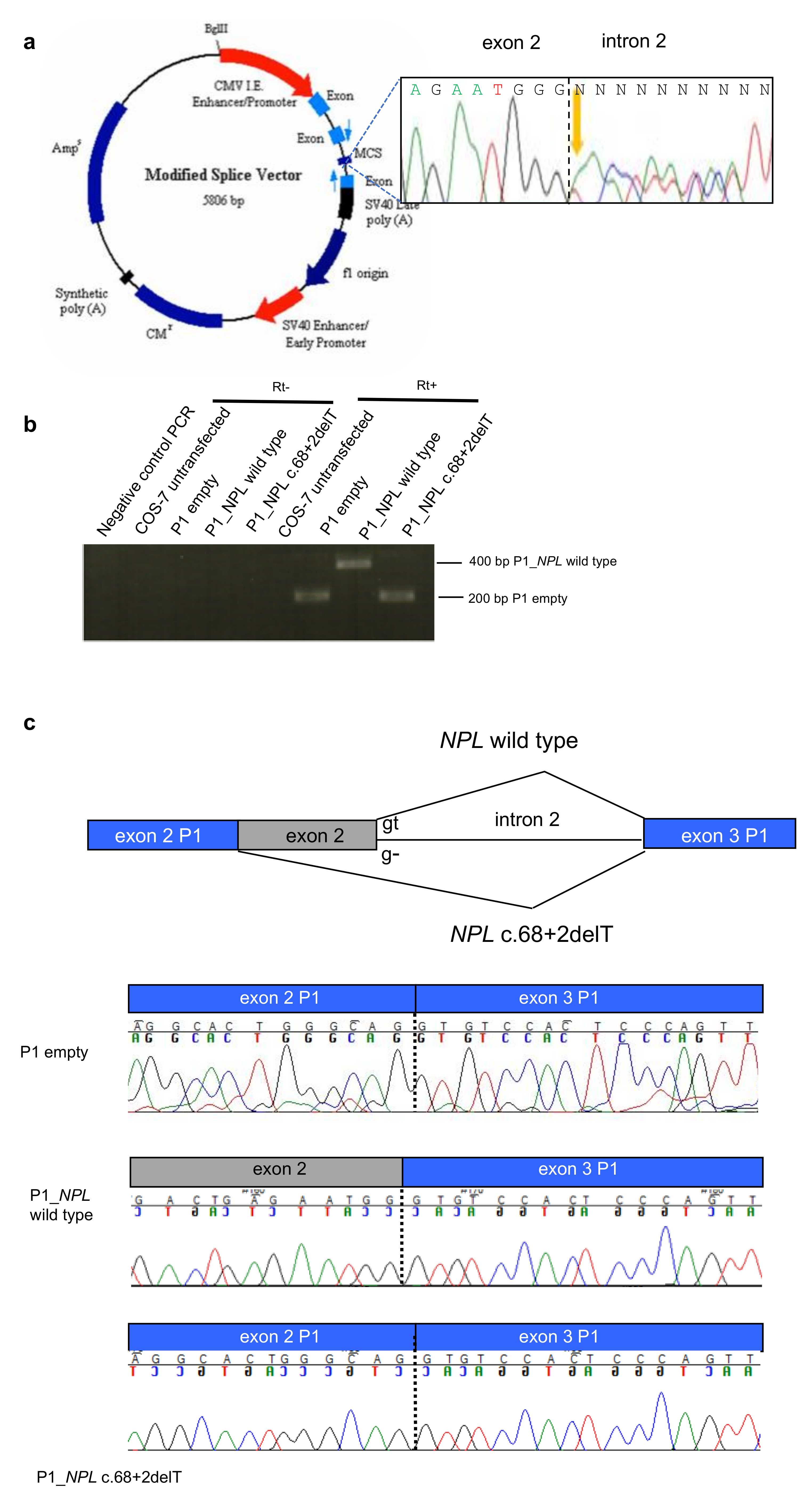
2.2. Splice Site Alterations in NPL and ROS1

As two of the variants identified via WES affected canonical splice sites, in NPL and ROS1, we evaluated their effect on the final transcripts. The NPL variant altered the canonic splice site in intron 2, through a deletion of the T in position + 2 (c.68 + 2delT). Since no fresh RNA was available from patients’ tissues, we used an in vitro system to study the splicing event in the presence of the variant. We inserted the wild-type or mutant genomic region surrounding exon 2 into the minigene plasmid P1-Altermax, that carries synthetic exons separated by an intronic region where the multiple cloning site maps (Figure 2a). The vectors were transiently transfected into simian COS7 cells, because they have a splicing apparatus comparable to the human one [22]. After RNA extraction and Reverse Transcriptase-PCR (RT-PCR) with primers specific for the synthetic exons, PCR products were sequenced (Figure 2b,c). In presence of the mutant allele, NPL exon 2, which contains the ATG start codon, was not included in the final transcript, suggesting that the protein would not be correctly translated (Figure 2c).
The ROS1 variants identified via WES affected a canonic donor splice site through the substitution in position + 2T > G in the intron 37 of ROS1. Since no fresh RNA was available from the patients’ tissues, we used the in vitro system to study the splicing events in the presence of the mutation. We inserted the wild-type or mutant genomic region surrounding exon 37 into the minigene plasmid P1-Altermax (Figure 3a) and the vectors were transiently transfected into simian COS7 cells. After RNA extraction and RT-PCR with primers specific for the synthetic exons, PCR products were sequenced (Figure 3b,c) in the presence of ROS1 mutant plasmids the splicing machinery used a cryptic intronic splice site 31 bp downstream of the canonical one, leading to the insertion of a premature stop-codon in the protein product of the mutant ROS1 transcript (Figure 3c).

2.3. Variant Screening in BC Patients Negative at BRCA1/BRCA2 Testing

To evaluate the frequency of damaging/possibly pathogenic variants in NPL, POLN, RASAL1 and ROS1 in familial BC, we used a target panel covering all exons and exon-intron boundaries for these genes (custom-made TruSeq amplicon kit, designed with the DesignStudio software [23]). The variant screening was performed in 131 independent BC patients who had previously tested negative at BRCA1 and BRCA2 analysis (Table S1). The filtering criteria were the same used for the WES analysis. In POLN we identified two rare variants and 1 in NPL in non-coding regions (Table S2), therefore ruling out a major role of these genes in predisposing to BC in this group of patients.
In the gene ROS1 we identified a total of 15 different heterozygous variants, eight of which showed a significant increased allele frequency in our sample vs. gnomAD [12] database (European non-Finnish population used as controls) and two were novel variants, p.Ile1305Phe and p.Gly1991Arg (Table 2). Several variants recurred among patients, such as (i) the c.-32C > T variant in the 5′UTR region, present in two unrelated HBC individuals and absent in the control European group; (ii) the missense variant p.Glu1902Lys, present in three unrelated patients, and showing an increased frequency compared to the controls (p = 0.0024; Fisher’s exact test, Table 2); (iii) the same splice site variant c.6005 + 2T > G, identified via WES was detected in another unrelated patient (BC14177); (iv) the missense variant p.Arg2126Gln, present in three unrelated patients, showing a significant difference in allele distribution compared to gnomAD [12] (p = 0.0008; Table 2).
Rare/novel heterozygous damaging variants were also identified in RASAL1 including: (i) the premature stop codon c.158G > A p.Trp53Ter (found in cis with the novel missense change p.Ile83Asn); (ii) a novel frameshift p.Leu292CysfsTer5 in another patient; (iii) the novel missense changes, predicted damaging, p.Ala43Thr and p.Ser466Asn in two unrelated patients (Table 3), all pointing to a loss-of-function mechanism for RASAL1 in BC, in concordance with previous data indicating that it might act as a tumor suppressor gene in thyroid cancer [24]. Moreover, germline RASAL1 variants may be relatively frequent in patients with apparently sporadic thyroid carcinoma with follicular features [25].
The 22 patients carrying ROS1 variants had a mean age at BC diagnosis of 41.36 years, while the 5 patients with RASAL1 variants had been diagnosed with BC at 36.2 years of age on average, compared to 38.29 years of patients with no variant detected in ROS1/RASAL1. More than half patients with ROS1 variants had a relevant family history, while RASAL1 carriers were mainly clinically sporadic early-onset cases. Features of patients are reported in Table S3.
Sequencing data for ROS1 and RASAL1 genes were retrieved in silico from WES analysis of 197 Italian subjects with no reported cancer history, who were selected as controls. For collapsing analysis comparing gene frequencies between cases (131) and controls (197), we used stringent criteria as previously described by Povysil et al. [26], selecting only non-synonymous coding changes, such as missense, nonsense, frameshift, splice acceptor and donor variants, having a minor allele frequency (MAF) <0.001 in gnomAD database, both in BC cases and in control group. According to these guidelines, we removed three ROS1 variants (c.-32C > T, p.Ser79=, and p.Ser370Pro) and a synonymous RASAL1 variant (p.Ile84=) in the number of variants identified in the BC cases.
A significantly higher proportion of ROS1 non-synonymous variant carriers was observed among BC cases in comparison to controls (p = 0.0401; Fisher’s exact test, Table 4).
We also evaluated the somatic mutations in ROS1 and RASAL1 present in BC in COSMIC database (v.91). 4.53% mutated cases in ROS1 (217/4792) and 1.28% cases in RASAL1 (33/2583) were reported. Somatic mutations in BRCA1 were reported in 2.83% BC cases (229/8090), and in 4.06% in BRCA2 (306/7532). Comparing these frequencies, in RASAL1 a lower number of mutated cases was reported, compared to BRCA1 and BRCA2 genes (p < 0.00001 for each test; Chi-square test; Table S4). Conversely, an increased number of ROS1-mutated BC cases, compared to BRCA1-mutated BC cases, was detected (p < 0.00001; Table S4), further supporting a role for ROS1 in BC.

3. Discussion

Next-generation sequencing has made available the analysis of multiple genomic regions simultaneously, shortening the time and cost of gene tests, and high-throughput association studies have unveiled the presence of many independent predisposing BC loci, each contributing to a relatively small portion of familial BC risk [5,6,7,8]. However, integrating these data in the clinical management of patients present significant controversies, in particular for genes contributing moderate risks. Epidemiological studies have identified a number of modifiable risk factors (e.g., weight, use of hormones, alcohol consumption, physical activity, and breastfeeding, diet), each explaining a modest proportion of the variation in disease risk individually, but with a substantial effect on BC predisposition, when combined together and with unmodifiable factors (e.g., genes, menstrual and/or reproductive history, family history of BC, and prior benign breast disease) [27,28]. It has been shown that changing the risk factor profile (low weight gain, no alcohol consumption, high physical activity level, breastfeeding, and no hormone use) could reduce the rate of postmenopausal BC by more than 34% [29], and that the reduction in absolute risk achievable by changing modifiable risk factors is larger for those who are at higher risk from nonmodifiable factors [30]. In addition, women found to be at elevated risk because of unmodifiable factors (e.g., pathogenic gene variant carriers) could be more motivated to adopt healthier lifestyles to lower their risk of BC if they have a better understanding of the potential gains. Identifying the fraction of the preventable cases can also better tailor the risk factor modification actions only to high-risk cases.
Moreover, several studies have identified specific molecular signatures with actionable targets in BC, generating a growing catalog of genomic mutations that facilitate tailored approaches to advanced BC, based on molecular analyses [31,32,33]. Therefore, an integrated molecular testing has to become an integral part of BC management. The use of whole exome sequencing in unexplained familial BC clusters may help identify novel of causal/predisposing variants in BC patients. In our study, we selected for the analysis affected first-degree cousins, who share 1/8 of their genome, therefore reducing the number of variants shared due to identity-by-descent. Under this assumption, we were able to identify novel or rare predicted pathogenic variants in ROS1, RASAL1 and POLN, and NPL in the three cousin pairs included in the exome analysis. NPL encodes for N-acetylneuraminate pyruvate lyase, an enzyme regulating the cellular concentrations of N-acetyl-neuraminic acid (sialic acid) [34,35]. As reported in Human Protein Atlas, NPL overexpression seemed to correlate with poor survival in liver and renal cancers, but little is known regarding the corresponding altered molecular pathways. POLN encodes for the DNA Polymerase nu involved in cross-link repair, homologous recombination and binding to BRCA1 [36]. ROS1 and RASAL1 are two genes with established roles in tumor development and progression in different types of cancer [21,24,25,37,38].
Targeted gene screening allowed us to identify additional pathogenic variants in RASAL1 and ROS1. In particular, we found a significant increase in the number of ROS1-mutated individuals in BC cases (131), compared to population controls (197 Italian individuals). Instead, the contribution of NPL and POLN to BC predisposition was not supported by the additional screening of cancer cases. In the 131 independent BC patients, we detected mainly RASAL1 loss-of-function variants (Table 2), in concordance with a tumor suppressor role of RASAL1 in cancer [19,24,25]. RASAL1 stimulates the GTPase activity of normal RAS p21, but not its oncogenic counterpart. Acting as a suppressor of RAS function, the protein enhances the intrinsic GTPase activity of RAS proteins resulting in the inactive GDP-bound form of RAS, thereby controlling cellular proliferation and differentiation [19,20]. Recent data have shown that exogenous expression of RASAL1 in MCF-7 and MDA-MB-231 sensitized the response to hypoxia treatments, associated with its ability to directly reduce HIF-1α (Hypoxia-inducible factor 1-alpha) expression, inhibiting migration and decreasing the accumulation of reactive oxygen species [21]. Conversely, RASAL1 knockdown reversed the cellular response to hypoxia [21]. Somatic driver gene point mutations in RASAL1 in follicular thyroid cancer have also been reported [24]. Germline mutations in RASAL1 have been identified in differentiated thyroid cancer (DTC) and in patients with Cowden Syndrome characterized by both BC and DTC features [25]. Intriguingly, in the family ITA_BC02 carrying the RASAL1 variant there were also DTC cases. Therefore, we suggest that, similarly to other cancers characterized by RASAL1 mutations, the loss of this tumor suppressor might promote BC development.
ROS1 encodes for an orphan tyrosine kinase receptor and its somatic rearrangements leading to the fusion of the kinase domain to different genes have been described in sarcomas, glioblastoma multiforme and lung tumors (non-small cell lung cancer): for a review, see [39,40,41]. The natural ligand of ROS1 is still unknown, whereas the proteins derived by the genomic rearrangements present a plethora of different N-terminal domains from different genes fused in frame with the cytoplasmic portion of ROS1, including the tyrosine kinase [42]). These rearranged proteins are abnormally activated and stimulated unrestricted cell growth and proliferation, with the activation of several pathways, including SHP2/PI3K/Akt/mTOR pathways [43]. The ROS1 rearrangements act as oncogenic drivers, and many tyrosine kinase inhibitors (TKIs) have been developed to target these driver oncogene products [44,45]. In lobular BC, the loss of E-cadherin expression that occurs early in the tumorigenic process [46], could be rescue by ROS1 synthetic lethality, an effect clinically actionable using ROS1 inhibitors [47]. However, many patients presenting tumors with somatic ROS1 rearrangements develop a resistance to TKIs, due to the acquisition of secondary mutations in the ROS1 tyrosine kinase domain that are refractory to TKI inhibition and maintain the receptor abnormally activated [48,49]. The progress in developing new TKIs to overcome ROS1 acquired secondary mutations is currently producing novel drugs for a targeted therapy [50,51].
Recently, ROS1 pathogenic somatic variants were identified in inflammatory breast cancer [52] and ROS1 somatic mutations are present in the COSMIC database. However, germline pathogenic variants have not been reported in hereditary BC. We characterized the splicing effect of the specific germline ROS1 variant c.6005 + 2T > G identified in one of the cousin pairs analyzed by WES and in one unrelated BC patient undergone NGS target sequencing. We could establish that the variant induced the use of a cryptic splice site, generating a premature stop codon in the cytoplasmic tail of ROS1 receptor in the in-vitro cell model COS7 cells, widely used to study human splicing alterations, because they present a splicing apparatus comparable to the one in H. sapiens [22,53]. Although patient RNA is usually preferred for splicing analysis, several issues, such as availability, or degradation of aberrant transcripts through nonsense-mediated mRNA decay (NMD), may make it difficult to assess the effect of a variant allele on splicing. Minigene assays is an alternative method that show high sensitivity and specificity in the assessment of aberrant splicing caused by genetic sequence variants [54,55]. The unavailability of the corresponding breast cancer cells or pathological breast cancer tissues from the affected individuals carrying the constitutive germline variant c.6005 + 2T > G in ROS1 prevented us from investigating whether the truncated protein was produced and could act with a dominant negative effect, shown for example by the truncated TrkB receptor [56] or promote an aberrant signal activation as shown by the truncated and constitutively active form of the EGF receptor, a key determinant of tumor growth and progression in cancer [57,58]. Interestingly, the majority of the other germline predicted pathogenic variants, found with a significant increase in the BC patient group, clustered to the cytoplasmic tail of ROS1, similarly to the variants conferring resistance to classic TKIs in ROS1-rearranged cancers and for which novel drugs are currently developed [50,51].
Our study provided the identification of novel and rare pathogenic constitutive variants in ROS1 in hereditary BC, but the case study is relatively small; therefore, further studies in additional independent cohorts of familial BC cases is warranted to investigate the contribution of germline ROS1 variants to breast cancer predisposition.
The discovery of additional genes predisposing to familial BC is crucial to identify the altered pathways in BC and is paramount in the development of novel agents that can target non-endocrine pathways, cellular proliferation and tumor progression. Moreover, identifying novel genetic factors conferring BC predisposition is crucial to predict who is at increased risk of developing the disease, allowing for early detection and therapy, and optimized patient management.

4. Materials and Methods

4.1. Patients

At the Cancer Genetics Clinic of the Hospital S.Orsola-Malpighi, BRCA testing is offered to patients with BC suspected to carry hereditary predisposition according to regional criteria [59]; about 18% of patients tested are found to carry a pathogenic variant and 6% carry variants of unknown significance; the remaining 76% test negative for BRCA variants [60]. Among kindreds highly suspicious for HBC with no BRCA variants found, we selected for WES analysis three families in which two 1st degree cousins had been tested. The pedigrees of the three families included in the Whole exome sequencing (WES) analysis are presented in Figure 1.
A consecutive series of 131 patients with familial and/or early-onset breast cancer who had undergone complete BRCA1/2 analysis (sequencing and MLPA) with negative results were then enrolled in the study as confirmation set, upon informed consent. The study was approved by the Ethical Board of Hospital S.Orsola-Malpighi, Bologna, Italy (113/2013/O/Tess) and was in compliance with the Helsinki declaration. Features of the patients are reported in Table S1.

4.2. WES Analysis

WES was performed on genomic DNA extracted from peripheral blood (QIAGEN mini kit) from three affected first-degree cousin-pairs, who tested negative for BRCA1/2. Dual-index paired-end libraries followed by exome enrichment were prepared starting from 100 ng genomic DNA, according to the Nextera Rapid Capture Enrichment protocol (Illumina, San Diego, CA USA). The captured regions were sequenced on the Illumina HiScanSQ platform for 200 cycles (100 cycles paired-ends; Illumina). The read files were aligned to hg19 version of the human genome sequencing, annotation and variant prioritization was performed according to our internal pipeline for exome annotation as previously reported [61]. The identified variants were confirmed by PCR and direct sequencing. Sequencing data for RASAL1, ROS1 genes were retrieved in silico from 197 WES data of Italian subjects without reporting cancer history, who were selected as controls. Variants were called using GATK “HaplotypeCaller” and variant calls were recalibrated before being annotated with the Ensembl tool Variant Effect Predictor (VEP). The number of rare (MAF < 0.001) single nucleotide variants and small indels predicted to functionally impact the RASAL1 and ROS1 proteins, was obtained by custom scripts. Fisher’s exact test was used to test the null hypothesis of equality of proportions of subjects with at least one variant in these genes.

4.3. Splice Site Analysis

The genomic regions surrounding ROS1 exon 37 and NPL exon 2 from the corresponding heterozygous carriers were amplified using primers 5′-GAATTCGTCTTGCTGGGAA-3′(ROS1) and 5′-GGGGCATTCGGACTTTGCCTGGGAGTTTT-3′ (NPL), inserting an EcoRI restriction site, and primers 5′-TCAGCAGACAAACTCCAAGCTCTAGA-3′ (ROS1), and 5′-CCTCTAGATTAGAAAGGCCAAGGCTGTG-3′ (NPL) inserting a XbaI restriction site, respectively. We generated the mini-gene reporter as previously described [22]. The PCR products were cloned into the digested P1-pAltermax and plasmids sequenced in order to identify the plasmids with the wild-type (wt) or the variant alleles. The splicing alteration analysis was carried out in COS7 cells (derived from monkey kidney tissue) grown in DMEM, 10% fetal bovine serum, 2 mM L-glutamine, 100 U/ mL penicillin and 100 μg/mL streptomycin (Sigma-Aldrich; St Louis, MO, USA), in a humidified incubator at 37 °C with 5% CO2. 3 × 10⁵ COS7 cells were plated in six-well plate for transfection using Lipofectamine reagent, according to the manufacturer’s instructions (Lipofectamine 2000, Thermo Fisher Scientific, Waltham, MA, USA). Total RNA was extracted after 48 h from transfection using the RNeasy kit (QIAGEN, Hilden, Germany) and subjected to DNase I digestion (Fermentas, Thermo Fisher Scientific) for 30 min at 37 °C. 1 μg of DNase I–treated RNA was used for reverse transcription with random hexamers and the Multiscribe RT system (Thermo Fisher Scientific) at 48 °C for 40 min in a final volume of 50 μL. RT-PCR was performed with primers specific for the P1-pAltermax vector, forward T7EE 5′-AAGGCTAGAGTACTTAATACGA-3′ and reverse PMaxR 5′-TATCATGTCTGCTCGAAGCATTA-3′. RT-PCR was carried out in the following conditions: 2 μL cDNA, 2.5 mM MgCl2, 0.5 mM dNTPs, 0.5 μM primers, 5% dimethyl sulfoxide in a final volume of 20 μL using the 2× KAPA Fast Taq Polymerase Master mix (KAPA Biosystems, Sigma-Aldrich). The RT-PCR products were run on 2.5% w/v agarose gel in TBE 1× buffer and visualized under UV light using ethidium bromide (Sigma-Aldrich).

4.4. Target Gene Screening

The Illumina TruSeq Custom Amplicon Kit was used to capture all exons and the flanking sequences of NPL, POLN, RASAL1 and ROS1 genes. For all genes, custom oligos specific to our targeted regions of interest were designed using DesignStudio [23] and amplicon length averaged 250 base pairs (2 × 150 base pairs reads length in paired-end mode). The number of amplicons per gene in the panel varies from 15 to 70, with a total of 143 amplicons and a mean coverage of 200×. Genomic DNA from peripheral blood was extracted using QIAGEN DNA mini kit (QIAGEN). The TruSeq Custom Amplicon sequencing assay was performed according to the manufacturer’s protocol (Illumina). All DNA samples were diluted to 10 ng/L and then were hybridized to the custom oligo pool (CAT). Upstream and downstream oligos were extended and ligated. Then, the result products containing the targeted regions of interest were amplified. Sample normalization of each library was performed according to Illumina bead-based normalization manufacturer’s protocol for balanced representation in pooled libraries. Pooled Amplicon Library (PAL) preparation was performed by combining 5 μL of each normalized library in one tube. PAL was quantified by Qubit dsDNA HS Assay kit (ThermoFisher Scientific) and diluted to a final concentration of 10 pM to be loaded on the flow cell. Runs were performed on Illumina MiSeq sequencer with a V2 flow cell (300 cycles). Bioinformatics analysis (including demultiplexing, reads alignment and variant calling) was performed using the MiSeq provided software (Illumina). Variant annotation and prioritization were performed using the dedicated cloud-based software BaseSpace, using the Variant Studio program (Illumina).

5. Conclusions

In our study, we focused on the identification of germline variants in familial BC, through whole exome and target NGS sequencing. Considering the relatively small number of familial BC individuals analyzed, the evaluation of ROS1 and RASAL1 as potential predisposing genes is warranted through further screening in independent patient groups. Identifying novel genetic factors conferring BC predisposition is crucial to predict who is at increased risk of developing the disease, allowing for early detection and therapy, and optimized patient management.

Supplementary Materials

The following are available online at https://www.mdpi.com/2072-6694/12/9/2539/s1, Figure S1: Gene expression in human tissues, Table S1: Clinical and familial features of the BC patients included in the target NGS screening, Table S2: Novel/rare variants in POLN, NPL genes detected through target NGS analysis, Table S3: Features of patients according to ROS1 and RASAL1 test results, Table S4: Mutation frequencies of cases in BRCA1, BRCA2, ROS1, RASAL1 in breast cancer cases, as reported in COSMIC.

Author Contributions

Conceptualization, E.B. and D.T.; Methodology, F.I. and I.B.; Software, F.I.; Investigation, F.I., I.B., S.F., G.I., L.G.; Resources, D.T, L.G., G.I.; Data Curation, D.T., L.G.; Writing–Original Draft Preparation, E.B., D.T., F.I., I.B.; Review and Editing, S.F.; Supervision, E.B., D.T.; Funding Acquisition, E.B. All authors have read and agreed to the published version of the manuscript.

Funding

This research was funded by the ER-Programma Giovani Ricercatori to E.B.

Acknowledgments

The authors thank all the patients and family members who participated to the study. The authors thank F. Buscherini and R. Zuntini for technical help in sequencing.

Conflicts of Interest

The authors declare no conflict of interest.

References

  1. Siegel, R.L.; Miller, K.D.; Jemal, A. Cancer Statistics, 2020. CA Cancer J. Clin. 2020, 70, 7–30. [Google Scholar] [CrossRef] [PubMed]
  2. Antoniou, A.; Pharoah, P.D.; Narod, S.; Risch, H.A.; Eyfjord, J.E.; Hopper, J.L.; Loman, N.; Olsson, H.; Johannsson, O.; Borg, A.; et al. Average risks of breast and ovarian cancer associated with BRCA1 or BRCA2 mutations detected in case Series unselected for family history: A combined analysis of 22 studies. Am. J. Hum. Genet. 2003, 72, 1117–1130. [Google Scholar] [CrossRef] [PubMed] [Green Version]
  3. Antoniou, A.C.; Cunningham, A.P.; Peto, J.; Evans, D.G.; Lalloo, F.; Narod, S.A.; Risch, H.A.; Eyfjord, J.E.; Hopper, J.L.; Southey, M.C.; et al. The BOADICEA model of genetic susceptibility to breast and ovarian cancers: Updates and extensions. Br. J. Cancer 2008, 98, 1457–1466. [Google Scholar] [CrossRef] [PubMed]
  4. Hartmann, L.C.; Lindor, N.M. The Role of Risk-Reducing Surgery in Familial Breast and Ovarian Cancer. N. Engl. J. Med. 2016, 374, 454–468. [Google Scholar] [CrossRef]
  5. Michailidou, K.; Lindström, S.; Dennis, J.; Beesley, J.; Hui, S.; Kar, S.; Lemaçon, A.; Soucy, P.; Glubb, D.; Rostamianfar, A.; et al. Association analysis identifies 65 new breast cancer risk loci. Nature 2017, 551, 92–94. [Google Scholar] [CrossRef] [Green Version]
  6. Michailidou, K.; Beesley, J.; Lindstrom, S.; Canisius, S.; Dennis, J.; Lush, M.J.; Maranian, M.J.; Bolla, M.K.; Wang, Q.; Shah, M.; et al. Genome-wide Association Analysis of More Than 120,000 Individuals Identifies 15 New Susceptibility Loci for Breast Cancer. Nat. Genet. 2015, 47, 373–380. [Google Scholar] [CrossRef] [Green Version]
  7. Wendt, C.; Margolin, S. Identifying Breast Cancer Susceptibility Genes—A Review of the Genetic Background in Familial Breast Cancer. Acta Oncol. 2019, 58, 135–146. [Google Scholar] [CrossRef] [Green Version]
  8. Mavaddat, N.; Barrowdale, D.; Andrulis, I.L.; Domchek, S.M.; Eccles, D.; Nevanlinna, H.; Ramus, S.J.; Spurdle, A.; Robson, M.; Sherman, M.; et al. Pathology of breast and ovarian cancers among BRCA1 and BRCA2 mutation carriers: Results from the Consortium of Investigators of Modifiers of BRCA1/2 (CIMBA). Cancer Epidemiol. Biomarkers Prev. 2012, 21, 134–147. [Google Scholar] [CrossRef] [Green Version]
  9. Beitsch, P.D.; Whitworth, P.W.; Hughes, K.; Patel, R.; Rosen, B.; Compagnoni, G.; Baron, P.; Simmons, R.; Smith, L.A.; Grady, I.; et al. Underdiagnosis of hereditary breast cancer: Are genetic testing guidelines a tool or an obstacle? J. Clin. Oncol. 2019, 37, 453–460. [Google Scholar] [CrossRef]
  10. Cybulski, C.; Carrot-Zhang, J.; Kluźniak, W.; Rivera, B.; Kashyap, A.; Wokołorczyk, D.; Giroux, S.; Nadaf, J.; Hamel, N.; Zhang, S.; et al. Germline RECQL mutations are associated with breast cancer susceptibility. Nat. Genet. 2015, 47, 643–646. [Google Scholar] [CrossRef]
  11. Thompson, E.R.; Doyle, M.A.; Ryland, G.L.; Rowley, S.M.; Choong, D.Y.H.; Tothill, R.W.; Thorne, H.; kConFab; Barnes, D.R.; Li, J.; et al. Exome sequencing identifies rare deleterious mutations in DNA repair genes FANCC and BLM as potential breast cancer susceptibility alleles. PLoS Genet. 2012, 8, e1002894. [Google Scholar] [CrossRef] [PubMed] [Green Version]
  12. Genome Aggregation Database (gnomAD). Available online: http://gnomad.broadinstitute.org/ (accessed on 16 July 2020).
  13. Mendelian Clinically Applicable Pathogenicity Score (M-CAP). Available online: http://bejerano.stanford.edu/mcap/ (accessed on 16 July 2020).
  14. Prediction of Functional Effects of Human nsSNPs (PolyPhen-2). Available online: http://genetics.bwh.harvard.edu/pph2/ (accessed on 16 July 2020).
  15. Human Splicing Finder (HSF), v.3.1. Available online: http://www.umd.be/HSF3/index.html (accessed on 16 July 2020).
  16. Genotype-Tissue Expression (GTEx). Available online: https://commonfund.nih.gov/gtex (accessed on 16 July 2020).
  17. The Human Protein Atlas. Available online: https://www.proteinatlas.org/ (accessed on 16 July 2020).
  18. Catalogue of Somatic Mutations in Cancer (COSMIC). Available online: https://cancer.sanger.ac.uk/cosmic (accessed on 16 July 2020).
  19. Ohta, M.; Seto, M.; Ijichi, H.; Miyabayashi, K.; Kudo, Y.; Mohri, D.; Asaoka, Y.; Tada, M.; Tanaka, Y.; Ikenoue, T.; et al. Decreased expression of the RAS-GTPase activating protein RASAL1 is associated with colorectal tumor progression. Gastroenterology 2009, 136, 206–216. [Google Scholar] [CrossRef] [PubMed]
  20. Seto, M.; Ohta, M.; Ikenoue, T.; Sugimoto, T.; Asaoka, Y.; Tada, M.; Mohri, D.; Kudo, Y.; Ijichi, H.; Tateishi, K.; et al. Reduced expression of RAS protein activator like-1 in gastric cancer. Int. J. Cancer. 2011, 128, 1293–1302. [Google Scholar] [CrossRef]
  21. Huang, Z.; Wang, R.; Wei, G.; Jiang, R.; Zheng, Y.; Tian, Y.; Chen, B.; Ye, C.; Xue, M.; Yu, C. RAS protein activator-like 1 is functionally involved in hypoxia resistance in breast cancer cells by targeting hypoxia inducible factor-1α. Oncol. Lett. 2017, 14, 3839–3845. [Google Scholar] [CrossRef] [Green Version]
  22. Bonora, E.; Evangelisti, C.; Bonichon, F.; Tallini, G.; Romeo, G. Novel Germline Variants Identified in the Inner Mitochondrial Membrane Transporter TIMM44 and Their Role in Predisposition to Oncocytic Thyroid Carcinomas. Br. J. Cancer 2006, 95, 1529–1536. [Google Scholar] [CrossRef] [PubMed]
  23. DesignStudio. Available online: http://designstudio.illumina.com/ (accessed on 16 July 2020).
  24. Liu, D.; Yang, C.; Bojdani, E.; Murugan, A.K.; Xing, M. Identification of RASAL1 as a major tumor suppressor gene in thyroid cancer. J. Natl. Cancer Inst. 2013, 105, 1617–1627. [Google Scholar] [CrossRef] [PubMed] [Green Version]
  25. Ngeow, J.; Ni, Y.; Tohme, R.; Chen, F.S.; Bebek, G.; Eng, C. Germline alterations in RASAL1 in Cowden syndrome patients presenting with follicular thyroid cancer and in individuals with apparently sporadic epithelial thyroid cancer. J. Clin. Endocrinol. Metab. 2014, 99, E1316–E1321. [Google Scholar] [CrossRef] [PubMed] [Green Version]
  26. Povysil, G.; Petrovski, S.; Hostyk, J.; Aggarwal, V.; Allen, A.S.; Goldstein, D.B. Rare-variant collapsing analyses for complex traits: Guidelines and applications. Nat. Rev. Genet. 2019, 20, 747–759. [Google Scholar] [CrossRef]
  27. Parkin, D.M.; Boyd, L.; Walker, L.C. The fraction of cancer attributable to lifestyle and environmental factors in the UK in 2010. Br. J. Cancer 2011, 105, S77–S81. [Google Scholar] [CrossRef]
  28. Madigan, M.P.; Ziegler, R.G.; Benichou, J.; Byrne, C.; Hoover, R.N. Proportion of breast cancer cases in the United States explained by well-established risk factors. J. Natl. Cancer Inst. 1995, 87, 1681–1685. [Google Scholar] [CrossRef] [Green Version]
  29. Tamimi, R.M.; Spiegelman, D.; Smith-Warner, S.A.; Wang, M.; Pazaris, M.; Willett, W.C.; Eliassen, A.H.; Hunter, D.J. Population Attributable Risk of Modifiable and Nonmodifiable Breast Cancer Risk Factors in Postmenopausal Breast Cancer. Am. J. Epidemiol. 2016, 184, 884–893. [Google Scholar] [CrossRef] [PubMed] [Green Version]
  30. Maas, P.; Barrdahl, M.; Joshi, A.D.; Auer, P.L.; Gaudet, M.M.; Milne, R.L.; Schumacher, F.R.; Anderson, W.F.; Check, D.; Chattopadhyay, S.; et al. Breast Cancer Risk From Modifiable and Nonmodifiable Risk Factors Among White Women in the United States. JAMA Oncol. 2016, 2, 1295–1302. [Google Scholar] [CrossRef] [PubMed]
  31. Litton, J.K.; Rugo, H.S.; Ettl, J.; Hurvitz, S.A.; Gonçalves, A.; Lee, K.H.; Fehrenbacher, L.; Yerushalmi, R.; Mina, L.A.; Martin, M.; et al. Talazoparib in patients with advanced breast cancer and a germline BRCA mutation. N. Engl. J. Med. 2018, 379, 753–763. [Google Scholar] [CrossRef] [PubMed]
  32. National Comprehensive Cancer Network. Breast Cancer. Version 4. Available online: https://www.nccn.org/professionals/physician_gls/pdf/breast_blocks.pdf (accessed on 16 July 2020).
  33. Litton, J.K.; Burstein, H.J.; Turner, N.C. Molecular Testing in Breast Cancer. Am. Soc. Clin. Oncol. Educ. Book 2019, 39, e1–e7. [Google Scholar] [CrossRef]
  34. Sood, R.; Bonner, T.I.; Makalowska, I.; Stephan, D.A.; Robbins, C.M.; Connors, T.D.; Morgenbesser, S.D.; Su, K.; Faruque, M.U.; Pinkett, H.; et al. Cloning and characterization of 13 novel transcripts and the human RGS8 gene from the 1q25 region encompassing the hereditary prostate cancer (HPC1) locus. Genomics 2001, 73, 211–222. [Google Scholar] [CrossRef] [Green Version]
  35. Chu, H.Y.; Zheng, Q.C.; Zhao, Y.S.; Zhang, H.X. Homology modeling and molecular dynamics study on N-acetylneuraminate lyase. J. Mol. Model. 2009, 15, 323–328. [Google Scholar] [CrossRef]
  36. Takata, K.; Tomida, J.; Reh, S.; Swanhart, L.M.; Takata, M.; Hukriede, N.A.; Woodl, R.A. Conserved overlapping gene arrangement, restricted expression, and biochemical activities of DNA polymerase ν (POLN). J. Biol Chem. 2015, 290, 24278–24293. [Google Scholar] [CrossRef] [Green Version]
  37. Ye, M.; Zhang, X.; Li, N.; Zhang, Y.; Jing, P.; Chang, N. ALK and ROS1 as targeted therapy paradigms and clinical implications to overcome crizotinib resistance. Oncotarget 2016, 7, 12289–12304. [Google Scholar] [CrossRef] [Green Version]
  38. Eom, M.; Lkhagvadorj, S.; Oh, S.S.; Han, A.; Park, K.H. ROS1 expression in invasive ductal carcinoma of the breast related to proliferation activity. Yonsei Med. J. 2013, 54, 650–657. [Google Scholar] [CrossRef]
  39. Acquaviva, J.; Wong, R.; Charest, A. The Multifaceted Roles of the Receptor Tyrosine Kinase ROS in Development and Cancer. Biochim. Biophys. Acta. 2009, 1795, 37–52. [Google Scholar] [CrossRef]
  40. Duma, N.; Santana-Davila, R.; Molina, J.R. Non-Small Cell Lung Cancer: Epidemiology, Screening, Diagnosis, and Treatment. Mayo Clin. Proc. 2019, 94, 1623–1640. [Google Scholar] [CrossRef] [PubMed]
  41. Morris, T.A.; Khoo, C.; Solomon, B.J. Targeting ROS1 Rearrangements in Non-small Cell Lung Cancer: Crizotinib and Newer Generation Tyrosine Kinase Inhibitors. Drugs 2019, 79, 1277–1286. [Google Scholar] [CrossRef] [PubMed]
  42. Davies, K.D.; Doebele, R.C. Molecular Pathways: ROS1 Fusion Proteins in Cancer. Clin. Cancer Res. 2013, 19, 4040–4045. [Google Scholar] [CrossRef] [PubMed] [Green Version]
  43. Charest, A.; Wilker, E.W.; McLaughlin, M.E.; Lane, K.; Gowda, R.; Coven, S.; McMahon, K.; Kovach, S.; Feng, Y.; Yaffe, M.B.; et al. ROS Fusion Tyrosine Kinase Activates a SH2 Domain-Containing phosphatase-2/phosphatidylinositol 3-kinase/mammalian Target of Rapamycin Signaling Axis to Form Glioblastoma in Mice. Cancer Res. 2006, 66, 7473–7481. [Google Scholar] [CrossRef] [Green Version]
  44. Shaw, A.T.; Riely, G.J.; Bang, Y.J.; Kim, D.W.; Camidge, D.R.; Solomon, B.J.; Varella-Garcia, M.; Iafrate, A.J.; Shapiro, G.I.; Usari, T.; et al. Crizotinib in ROS1-rearranged non-small-cell lung cancer. N. Engl. J. Med. 2014, 371, 1963–1971. [Google Scholar] [CrossRef] [PubMed] [Green Version]
  45. Roys, A.; Chang, X.; Liu, Y.; Xu, X.; Wu, Y.; Zuo, D. Resistance mechanisms and potent-targeted therapies of ROS1-positive lung cancer. Cancer Chemother. Pharmacol. 2019, 84, 679–688. [Google Scholar] [CrossRef]
  46. Ciriello, G.; Gatza, M.L.; Beck, A.H.; Wilkerson, M.D.; Rhie, S.K.; Pastore, A.; Zhang, H.; McLellan, M.; Yau, C.; Kandoth, C.; et al. Comprehensive Molecular Portraits of Invasive Lobular Breast Cancer. Cell 2015, 163, 506–519. [Google Scholar] [CrossRef] [Green Version]
  47. Bajrami, I.; Marlow, R.; Van de Ven, M.; Brough, R.; Pemberton, H.N.; Frankum, J.; Song, F.; Rafiq, R.; Konde, A.; Krastev, D.B.; et al. E-Cadherin/ROS1 Inhibitor Synthetic Lethality in Breast Cancer. Cancer Discov. 2018, 8, 498–515. [Google Scholar] [CrossRef] [Green Version]
  48. McCoach, C.E.; Le, A.T.; Gowan, K.; Jones, K.; Schubert, L.; Doak, A.; Estrada-Bernal, A.; Davies, K.D.; Merrick, D.T.; Bunn, P.A.J.; et al. Resistance Mechanisms to Targeted Therapies in ROS1(+) and ALK(+) Non-small Cell Lung Cancer. Clin. Cancer Res 2018, 2, 3334–3347. [Google Scholar] [CrossRef] [Green Version]
  49. Facchinetti, F.; Loriot, Y.; Kuo, M.S.; Mahjoubi, L.; Lacroix, L.; Planchard, D.; Besse, B.; Farace, F.; Auger, N.; Remon, J.; et al. Crizotinib-Resistant ROS1 Mutations Reveal a Predictive Kinase Inhibitor Sensitivity Model for ROS1- and ALK-Rearranged Lung Cancers. Clin. Cancer Res. 2016, 22, 5983–5991. [Google Scholar] [CrossRef] [Green Version]
  50. Drilon, A.; Ou, S.I.; Cho, B.C.; Kim, D.W.; Lee, J.; Lin, J.J.; Zhu, V.W.; Ahn, M.J.; Camidge, D.R.; Nguyen, J.; et al. Repotrectinib (TPX-0005) Is a Next-Generation ROS1/TRK/ALK Inhibitor That Potently Inhibits ROS1/TRK/ALK Solvent- Front Mutations. Cancer Discov. 2018, 8, 1227–1236. [Google Scholar] [CrossRef] [PubMed] [Green Version]
  51. Drilon, A.; Siena, S.; Ou, S.I.; Patel, M.; Ahn, M.J.; Lee, J.; Bauer, T.M.; Farago, A.F.; Wheler, J.J.; Liu, S.V.; et al. Safety and Antitumor Activity of the Multitargeted Pan-TRK, ROS1, and ALK Inhibitor Entrectinib: Combined Results from Two Phase I Trials (ALKA-372-001 and STARTRK-1). Cancer Discov. 2017, 7, 400–409. [Google Scholar] [CrossRef] [PubMed] [Green Version]
  52. Liang, X.; Vacher, S.; Boulai, A.; Bernard, V.; Baulande, S.; Bohec, M.; Bièche, I.; Lerebours, F.; Callens, C. Targeted next-generation sequencing identifies clinically relevant somatic mutations in a large cohort of inflammatory breast cancer. Breast Cancer Res. 2018, 20, 88. [Google Scholar] [CrossRef] [PubMed]
  53. Diquigiovanni, C.; Bergamini, C.; Evangelisti, C.; Isidori, F.; Vettori, A.; Tiso, N.; Argenton, F.; Costanzini, A.; Iommarini, L.; Anbunathan, H.; et al. Mutant MYO1F alters the mitochondrial network and induces tumor proliferation in thyroid cancer. Int. J. Cancer 2018, 143, 1706–1719. [Google Scholar] [CrossRef] [Green Version]
  54. Tournier, I.; Vezain, M.; Martins, A.; Charbonnier, F.; Baert-Desurmont, S.; Olschwang, S.; Wang, Q.; Buisine, M.P.; Soret, J.; Tazi, J.; et al. A large fraction of unclassified variants of the mismatch repair genes MLH1 and MSH2 is associated with splicing defects. Hum. Mutat. 2008, 29, 1412–1424. [Google Scholar] [CrossRef]
  55. Van der Klift, H.M.; Jansen, A.M.; Van der Steenstraten, N.; Bik, E.C.; Tops, C.M.J.; Devilee, P.; Wijnen, J.T. Splicing analysis for exonic and intronic mismatch repair gene variants associated with Lynch syndrome confirms high concordance between minigene assays and patient RNA analyses. Mol. Genet. Genomic Med. 2015, 3, 327–345. [Google Scholar] [CrossRef]
  56. Fenne, B.M. Truncated TrkB: Beyond a Dominant Negative Receptor. Cytokine Growth Factor Rev. 2012, 23, 15–24. [Google Scholar] [CrossRef]
  57. Hu, J.; Jo, M.; Cavenee, W.K.; Furnari, F.; VandenBerg, S.R.; Gonias, S.L. Crosstalk Between the Urokinase-Type Plasminogen Activator Receptor and EGF Receptor Variant III Supports Survival and Growth of Glioblastoma Cells. Proc. Natl Acad. Sci. USA 2011, 108, 15984–15989. [Google Scholar] [CrossRef] [Green Version]
  58. Wee, P.; Wang, Z. Epidermal Growth Factor Receptor Cell Proliferation Signaling Pathways. Cancers (Basel) 2017, 9, 52. [Google Scholar] [CrossRef] [Green Version]
  59. Servizio Sanità Pubblica; Regione Emilia-Romagna. Contributo n. 91/2016: “Protocollo Assistenziale Nelle Donne a Rischio Ereditario di Tumore Della Mammella e/o Ovaio”; Regione Emilia-Romagna: Bologna, Italy, 2016; ISSN 2464-9252. [Google Scholar]
  60. Zuntini, R.; Ferrari, S.; Bonora, E.; Buscherini, F.; Bertonazzi, B.; Grippa, M.; Godino, L.; Miccoli, S.; Turchetti, D. Dealing with BRCA1/2 unclassified variants in a cancer genetics clinic: Does cosegregation analysis help? Front. Genet. 2018, 9, 378. [Google Scholar] [CrossRef]
  61. Diquigiovanni, C.; Bergamini, C.; Diaz, R.; Liparulo, I.; Bianco, F.; Masin, L.; Baldassarro, V.A.; Rizzardi, N.; Tranchina, A.; Buscherini, F.; et al. A novel mutation in SPART gene causes a severe neurodevelopmental delay due to mitochondrial dysfunction with complex I impairments and altered pyruvate metabolism. FASEB J. 2019, 33, 11284–11302. [Google Scholar] [CrossRef] [PubMed] [Green Version]
Figure 1. Family trees of the families included in the whole exome sequencing (WES) analysis. We reported in the legend the different types of tumors identified in the corresponding individuals in the different generations. Red circles indicate the subjects analyzed via WES. (a) Family tree of the first-degree cousins carrying the ROS1 (ROS Proto-Oncogene 1, Receptor Tyrosine Kinase) splice variant. (b) Family tree of the first-degree cousins carrying the RASAL1 (RAS Protein Activator Like 1) and POLN (DNA Polymerase Nu) missense variants. (c) Family tree of the first-degree cousin carrying the NPL (N-Acetylneuraminate Pyruvate Lyase) splice variant.
Cancers 12 02539 g001
Figure 2. The NPL c.68 + 2delT mutation alters the splicing. (a) Map of the minigene system. In the MCS (multiple cloning site) the genomic region surrounding the mutation was inserted starting from the PCR product of one heterozygous patient (electropherogram in black box). (b) Reverse Transcriptase-PCR (RT-PCR) from RNA extracted from COS7 cells transfected with the different plasmids: empty, carrying NPL wild type, carrying the NPL variant. (c) Electropherograms of the RT-PCR products showing that wild type NPL produced a correct splicing, whereas the mutant NPL induced exon 2 skipping.
Cancers 12 02539 g002
Figure 3. The ROS1 c.6005 + 2T > G variant alters the splicing. (a) Map of the minigene system. In the MCS the genomic region surrounding the mutation was inserted starting from the PCR product of one heterozygous patient (electropherogram in black box). (b) RT-PCR of cDNA from COS7 cells transfected with the different plasmids: empty, carrying ROS1 wild type; carrying the ROS1 variant. (c) Electropherograms of the RT-PCR products, showing that wild type ROS1 allele produces a correct splicing, whereas in the variant one a cryptic splice site in the intron 37 is used, generating a longer transcript with the insertion of a premature stop codon in the open reading frame.
Cancers 12 02539 g003
Table 1. Single nucleotide variants/indels identified by WES in the three families with hereditary breast cancer.
Family IDGenomic Coordinates (hg19)GeneFunctional EffectdbSNPPolyPhen-2 ScoresM-CAP
Score
MAF gnomAD (European) a
ITA_BC016: g.117639349A > CROS1 (NM_002944)c.6005 + 2T > Grs200482357----0.000008850
ITA_BC0212: g.113543542A > GRASAL1 (NM_001193521)p.Phe602Leurs142556970 bHumDiv = 0.136 (benign) HumVar = 0.217 (benign)0.207 (Possibly pathogenic)0.002692452
ITA_BC024: g.2209742T > GPOLN (NM_181808)p.Asp229Alars747058815HumDiv = 1.000 (Probably damaging) HumVar = 0.993 (Probably damaging)0.032 (Possibly pathogenic)3.53638 × 10⁻⁵
ITA_BC031:g.182763575GT > GNPL (NM_030769)c.68 + 2delTrs755026414----0.000425763
(a) Allele frequency in European non-Finnish population, as accessed in gnomAD v2.1. [12].
Table 2. Rare/novel single nucleotide variants in the ROS1 gene, identified in breast cancer (BC) patients via next-generation sequencing (NGS) target analysis.
Patient IDchr6 (hg19)cDNA NM_002944Functional EffectdbSNPPolyPhen-2 ScoresM-CAP
Score
MAF gnomAD (European) a
BC14623 BC12674g.117746850C > Tc.-32C > T5′UTRrs376700858----0/127478 (p < 0.00001) b
BC12132 d BC11298 BC15651g.117730797C > Tc.237G > Ap.Ser79= (removing ESE)rs55736087----0.004862 628/129166 (p = 0.1371) b
BC10557 BC10536g.117715381A > Gc.1108T > Cp.Ser370Prors56274823HumDiv = 0.917 Possibly damaging HumVar = 0.446 Benign0.086 Possibly pathogenic0.003072 390/126944 (p = 0.1937) b
BC9762g.117710875C > Tc.1397G > Ap.Arg466Glnrs140178288HumDiv = 0.957 Probably damaging HumVar = 0.246 Benign0.048 Possibly pathogenic0.000007752 1/129000 (p = 0.004) b
BC9631g.117710828G > Tc.1444C > Ap.Leu482Metrs757273336HumDiv = 1.000 Probably damaging HumVar = 0.998 Probably damaging0.075 Possibly pathogenic0/113636 (p = 0.0023) b
BC14139g.117710578G > Ac.1694C > Tp.Ser565Leurs142303126HumDiv = 0.830 Possibly damaging HumVar = 0.124 Benign0.042 Possibly pathogenic0.0005425 70/129034 (p = 0.1342) b
BC11084g.117709072T > Cc.1885A > Gp.Ile629Val (inserting splice donor site)rs142877218HumDiv = 0.000 Benign HumVar = 0.001 Benign0.027 Possibly pathogenic0.00009297 12/129076 (p = 0.026) b
BC13285g.117704600C > Gc.2376G > Cp.Met792Ile (removing ESE)rs140511382HumDiv = 0.279 Benign HumVar = 0.081 Benign--0.00002323 3/129128 (p = 0.0081) b
BC11925g.117704565G > Tc.2411C > Ap.Thr804Asnrs200615700HumDiv = 0.999 Probably damaging HumVar = 0.922 Probably damaging0.085 Possibly pathogenic0.0003331 43/129096 (p = 0.0854) b
BC12188g.117678020T > Ac.3913A > Tp.Ile1305Phe--HumDiv = 0.553 Possibly damaging HumVar = 0.110 Benign0.058 Possibly pathogenicNovel
BC14203 BC17333 BC18206g.117642495C > Tc.5704G > Ap.Glu1902Lysrs9489124HumDiv = 0.923 Probably damaging HumVar = 0.122 Benign--0.0009854 127/128884 (p = 0.0024) b
BC14840g.117642468C > Gc.5731G > Cp.Gly1911Arg--HumDiv = 0.997 Probably damaging HumVar = 0.925 Benign0.033 Possibly pathogenicNovel
BC14177 cg.117639349A > Cc.6005 + 2T > GAltered donor splice siters200482357----0.000008850 1/112990 (p = 0.0046) b
BC12132 dg.117622137C > Tc.6733G > Ap.Gly2245Serrs142264513HumDiv = 0.196 Benign HumVar = 0.063 Benign0.032 Possibly pathogenic0.001065 124/116398 (p = 0.2451) b
BC13141 BC13389 BC16373g.117631301C > Tc.6377G > Ap.Arg2126Glnrs199882276HumDiv = 1.000 Probably damaging HumVar = 0.998 Probably damaging0.096 Possibly pathogenic0.0006670 86/128930 (p = 0.0008) b
(a) Allele frequency in European non-Finnish population, as accessed in gnomAD v2.1. [12]. In the second row, we reported the number of variant alleles vs the total alleles for the specific position in the database. (b) p values reported for Fisher’s exact test, calculated considering the number of variant alleles for the BC cases (n = 131 cases analyzed; 262 total alleles), and the frequency of the variant alleles as reported in gnomAD v2.1. [12]. Significant p values are indicated in italics. (c) The splice variant identified in individual BC14177 is the same identified in the unrelated family ITA_BC01 via WES analysis. (d) Patient BC12132 carried two different variants in ROS1. Abbreviations as reported for Table 1.
Table 3. Rare/novel single nucleotide variants in the RASAL1 gene, identified in BC patients via target NGS analysis.
Patient IDchr.12 (hg19)cDNA NM_001193520Functional EffectdbSNPPolyPhen-2 ScoresM-CAP ScoreMAF gnomAD (European) a
BC14290g.113128174G > Ac.127C > Tp.Ala43Thr--HumDiv = 0.986 Probably damaging HumVar = 0.969 Probably damaging--Novel
BC14401(1) g.113565948C > T (2) g.113127862T > A(1) c.158G > A (2) c.248A > T(1) p.Trp53Ter (2) p.Ile83Asnrs139329607(2) HumDiv = 0.880 Possibly damaging HumVar = 0.718 Possibly damaging--(1) 0.000008797 1/113678 (p = 0.0046) b (2) Novel
BC17895g.113565663G > Ac.252C > Tp.Ile84=rs61759864----0.001356 175/129046 (p = 0.3004) b
BC18101g.113546005C > Tc.1400G > Ap.Ser466Asn--HumDiv = 0.494 Possibly damaging HumVar = 0.474 Possibly damaging0.099 Possibly pathogenicNovel
BC16851g.113553568 delAc.875 delTp.Leu292CysfsTer5------Novel
(a) Allele frequency in European non-Finnish population, as accessed in gnomAD v2.1. [12]. In the second row, we reported the number of variant alleles vs the total alleles for the specific position in the database. (b) p values reported for Fisher’s exact test, calculated considering the number of variant alleles for the BC cases (n = 131 cases analyzed; 262 total alleles), and the frequency of the variant alleles as reported in gnomAD v2.1. [12]. Significant p values are indicated in italics.
Table 4. Contingency table of mutated subjects in ROS1 and RASAL1 genes, identified among cases and controls.
IndividualsROS1RASAL1
CasesControlsTotalCasesControlsTotal
Mutated161127448
Not mutated115186301127193320
Total131197328131197328
p = 0.0401 ap = 0.7179 a
(a) Fisher’s exact test was used to test the null hypothesis of equality of proportions of subjects with at least one variant in these genes.

