Article

Evidence of Cooperation between Hippo Pathway and RAS Mutation in Thyroid Carcinomas

by
Thaise Nayane Ribeiro Carneiro
1,
Larissa Valdemarin Bim
1,
Vanessa Candiotti Buzatto
2,
Vanessa Galdeno
2,
Paula Fontes Asprino
2,
Eunjung Alice Lee
3,
Pedro Alexandre Favoretto Galante
2 and
Janete Maria Cerutti
1,*
1
Genetic Bases of Thyroid Tumors Laboratory, Division of Genetics, Department of Morphology and Genetics, Escola Paulista de Medicina, Universidade Federal de São Paulo, Pedro de Toledo 669, 11 Andar, São Paulo, SP 04039-032, Brazil
2
Centro de Oncologia Molecular, Hospital Sírio-libanês, Rua Professor Daher Cutait 69, Bela Vista, São Paulo, SP 01308-060, Brazil
3
Division of Genetics and Genomics, Boston Children’s Hospital and Harvard Medical School, 3 Blackfan Circle, CLS (Center for Life Science) Building 15th Floor, Office 15020 | Lab 15072, Boston, MA 02115, USA
*
Author to whom correspondence should be addressed.
Cancers 2021, 13(10), 2306; https://doi.org/10.3390/cancers13102306
Submission received: 22 February 2021 / Revised: 30 March 2021 / Accepted: 6 April 2021 / Published: 12 May 2021
(This article belongs to the Special Issue Advances in Thyroid Carcinoma)

Abstract

:

Simple Summary

RAS mutations have been reported in a wide range of thyroid cancer histological types, from benign to aggressive phenotypes. The presence of RAS mutations in benign lesions suggests that the mutation alone is unlikely to lead to a malignant transformation per se, and thus, additional aberrations are necessary for this transformation. In this study, we initially screened a cohort of 120 thyroid carcinomas with a panel of known driver mutations and identified 11 RAS-mutated samples. An RNA-Seq analysis of those 11 RAS-positive samples identified that the Hippo pathway was both mutated and differentially expressed in the RAS-positive tumors. The gene expression analysis of 60 RAS-positive The Cancer Genome Atlas (TCGA) papillary thyroid carcinomas (PTC) samples supported our findings.

Abstract

Thyroid cancer incidences have been steadily increasing worldwide and are projected to become the fourth leading cancer diagnosis by 2030. Improved diagnosis and prognosis predictions for this type of cancer depend on understanding its genetic bases and disease biology. RAS mutations have been found in a wide range of thyroid tumors, from benign to aggressive thyroid carcinomas. Based on that and in vivo studies, it has been suggested that RAS cooperates with other driver mutations to induce tumorigenesis. This study aims to identify genetic alterations or pathways that cooperate with the RAS mutation in the pathogenesis of thyroid cancer. From a cohort of 120 thyroid carcinomas, 11 RAS-mutated samples were identified. The samples were subjected to RNA-Sequencing analyses. The mutation analysis in our eleven RAS-positive cases uncovered that four genes that belong to the Hippo pathway were mutated. The gene expression analysis revealed that this pathway was dysregulated in the RAS-positive samples. We additionally explored the mutational status and expression profiling of 60 RAS-positive papillary thyroid carcinomas (PTC) from The Cancer Genome Atlas (TCGA) cohort. Altogether, the mutational landscape and pathway enrichment analysis (gene set enrichment analysis (GSEA) and Kyoto Encyclopedia of Genes and Genome (KEGG)) detected the Hippo pathway as dysregulated in RAS-positive thyroid carcinomas. Finally, we suggest a crosstalk between the Hippo and other signaling pathways, such as Wnt and BMP.

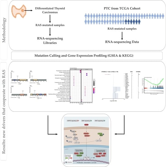
Graphical Abstract

1. Introduction

The incidence of thyroid cancer has been steadily increasing worldwide in both men (5.4%) and women (6.5%), and it is projected to become the fourth leading cancer diagnosis by 2030 [1]. Hence, it is essential to prepare for the increasing rate of diagnosis and to better discriminate the low-risk disease that may represent the early stage of the high-risk disease from those low-risk diseases that are indolent, and active surveillance has been proposed as the treatment modality. It is also important to predict the high-risk disease that will almost surely do poorly and improve the treatment of patients with advanced and aggressive disease. Ongoing efforts to improve the diagnosis and better predict the prognosis for thyroid cancer depend on a deep and more precise understanding of the genetic bases and disease biology of this tumor.
The Cancer Genome Atlas (TCGA), a landmark cancer genomics program, sequenced the genomes and transcriptomes of nearly 500 papillary thyroid carcinomas (PTC), the most common type of thyroid cancer, to identify novel drivers (e.g., genes or mutations) that could improve the molecular diagnosis of thyroid cancer [2]. They also developed a 71-gene expression signature, named the BRAF-RAS Score (BRS), designed to quantify the gene expression profile of a given tumor that resembles either the BRAF V600E or RAS-mutant profiles, the most common mutations found in PTC. Using this approach, PTC samples were, therefore, divided into BRAF-like or RAS-like groups [2].
Although it has been created a comprehensive atlas of the somatic genetic changes involved in the pathogenesis of classical variant of PTC cases, that is not completely true for other thyroid carcinomas, such as the follicular variant of papillary thyroid carcinomas (FVPTC), follicular thyroid carcinomas (FTC) and Hurthle cell carcinoma (HCC) [3,4,5,6,7,8,9]. Additionally, while the BRAF-like group has been widely investigated, studies that have analyzed molecular differences in the RAS-like group are limited.
Importantly, RAS mutations have been found in a wide range of thyroid tumors, from benign follicular thyroid adenoma (FTA) and noninvasive follicular thyroid neoplasm with papillary-like nuclear features (NIFTP), to poorly differentiated thyroid carcinomas (PDTC) and undifferentiated thyroid carcinomas (UTC) [10,11,12,13,14,15,16,17].
Although mutations in the RAS gene predispose cells to large-scale genomic abnormalities [18], it has been suggested that RAS induces growth arrest and premature senescence in primary cells, unless it cooperates with other driver mutations [19]. Some in vivo studies confirmed that the RAS mutation alone is not sufficient to trigger a thyroid transformation and is unable to completely activate the MAPK pathway [20,21,22,23,24,25]. However, double-mutant mice, KrasG12D and Pten−/−, developed invasive and metastatic FTC [24].
Genomic studies on PDTCs and UTCs have unveiled that mutations in RAS genes co-occur with mutations in other genes, such as TERT, TP53, EIF1AX or PTEN, as well as with the 22q loss of heterozygosity [10,26,27,28]. It has also been shown that the acquisition of an activating KRAS G12V mutation acts as a potential resistance mechanism in patients with BRAF V600E-mutated PTC and treated with BRAF and MEK inhibitors [29].
Altogether, these findings suggest that RAS mutations do not lead to a malignant transformation per se; thus, additional mutations are necessary for this transformation. In this study, we aimed to identify the genetic alterations or pathways that cooperate with RAS mutations in the pathogenesis of thyroid cancer. We screened a cohort of 120 thyroid carcinomas and identified 11 RAS-mutated samples that were subjected to RNA-Sequencing (RNA-Seq) analyses. We further evaluated 60 RAS-positive PTCs from the TCGA cohort. We identified mutations in novel genes and a pathway that likely cooperates with RAS in thyroid tumor pathogenesis.

2. Materials and Methods

2.1. Patients

This study was approved by the institutional Research Ethics Committee of the Universidade Federal de São Paulo (UNIFESP) and Universidade de São Paulo (USP). The study included patients who underwent thyroidectomy at Hospital São Paulo (Universidade Federal de São Paulo) and Hospital das Clínicas (Faculdade de Medicina, Universidade de São Paulo) as the initial treatment for thyroid cancer and received no prior treatment for their disease. A portion of the thyroid specimen was taken from the patient at the time of surgery, snap-frozen and stored at −80 °C. The original cohort was comprised of 120 fresh-frozen samples obtained from patients diagnosed with thyroid cancer. The validation cohort included an independent set of formalin-fixed, paraffin-embedded (FFPE) surgical specimens that were obtained from the files of the Department of Pathology from UNIFESP. This set of samples included five RAS-positive and five BRAF V600E-positive samples, as well as five normal adjacent thyroid tissues. H&E-stained (Hematoxylin and Eosin) tissue sections from each tumor were reviewed by a pathologist to confirm the diagnosis.

2.2. Study Design

A total of 120 specimens from patients with PTC, FVPTC and FTC were screened for a panel of point mutations (K-N-HRAS and BRAF V600E) and fusions (RET-PTC1, RET/PTC2, RET/PTC3, AGK-BRAF, ETV6-NTRK3, STRN-ALK and PAX8-PPARG). The discovery cohort was comprised of eleven samples (5 FTC and 6 FVPTC) confirmed as positive for RAS mutations. The control group included sixteen samples negative for the panel of mutations and fusions. Twenty-seven samples were submitted to RNA-sequencing in an Illumina NextSeq platform (Figure 1).
The demographic; clinical and pathological features, such as age at diagnosis, gender, histological subtype, tumor size, tumor focality, extrathyroidal extension, vascular invasion, presence of metastasis and RAS mutational profile, are detailed in Table 1.

2.3. RNA and DNA Isolation

For the RNA-Seq experiment, total RNA was isolated from fresh-frozen samples using TRIzol reagent (Invitrogen Corp., Carlsbad, CA, USA) according to the manufacturer’s instructions. RNA Integrity Number (RIN) was measured using an Agilent 2100 Bio-analyzer (Agilent Technologies Inc., Santa Clara, CA, USA). To validate our findings, FFPE thyroid tissue sections were used. Representative tumor blocks containing a percentage of tumor cells greater than 70% were preferentially selected. For samples containing less than 70% of tumor cells, the tumor-containing area was marked by the pathologist and manually macro-dissected to enrich the proportion of neoplastic cells. DNA and RNA were isolated from FFPE sections using the NucleoSpin Tissue Kit (Macherey-Nagel GmbH & Co. KG, Düren, Germany) and RNeasy FFPE Kit (Qiagen, Hilden, Germany), respectively. Nucleic acid was quantified using a NanoDrop spectrophotometer (Thermo Fisher Scientific Inc., Waltham, MA, USA).

2.4. RNA Library Preparation

Briefly, 0.5–1 micrograms of RNA from each sample were used to prepare the sequencing libraries. RNA libraries were prepared using a TruSeq Total RNA Sample preparation kit v.2 (Illumina Inc., San Diego, CA, USA), according to the manufacturer’s protocols. Library concentrations and quality were measured using a Qubit fluorometer and Quant-iT RNA kit (Life Technologies, Carlsbad, CA, USA). Samples were sequenced for paired-end reads on an Illumina NextSeq 500 platform (Illumina Inc.), targeting 100 million raw reads per sample, at the Centro de Oncologia Molecular, Hospital Sírio-libanês, São Paulo, Brazil.

2.5. RNA-Sequencing and Data Analysis

RNA-Seq reads were assessed for quality control using the FastQC format (Babraham Institute, Babraham, UK). The total set of paired-end reads was mapped against the human genome (GRCh38) for gene expression analysis and mutation calling (GRCh37) using STAR (version 2.7; default parameters with transcript-aware) [30]. Transcript abundance was estimated using HTSeq count (version 0.11.4; parameters -m union -s reverse -a 20) [31] and GENCODE (version 24) as reference for the human transcriptome. Nucleotide variants (Single-Nucleotide Variations (SNVs) and Insertions or Deletions (InDels)) were called using Genomic Analysis Toolkit (GATK) HaplotyperCaller [32]. We excluded the reads with low mapping quality (Q < 20) and further selected only variants with minor allele frequencies (MAF) reported by a genome aggregation database (gnomAD) lower than 0.1%. We used healthy thyroid samples from the ENCODE database (https://www.encodeproject.org, accessed on 20 February 2019) as the normal control.
The mutation-calling pipeline was the same described for tumor samples, and the variants found in normal thyroid tissue were filtered out from tumor mutation results. We subsequently recalled only variants with predicted damaging impact scores on the protein function produced by SIFT and PolyPhen-2, which are inserted in the dbNSFP database [33]. Finally, we selected the variants likely pathogenic (by SIFT and PolyPhen-2) and that were not represented at the Brazilian genomic variants (ABraOM) database [34]. Maftools [35] was used to detect genes exclusively mutated in RAS samples (default parameters: min mut = 2, p-value ≤ 0.1, odds ratio analysis) compared to 16-gene mutation panel negative samples. Genes found exclusively mutated were enriched using the Kyoto Encyclopedia of Genes and Genome (KEGG) database through Enrichr (https://maayanlab.cloud/Enrichr, accessed on 28 July 2020). The workflow with the pipeline to the RNA call variants and expression is detailed in Figure 1.

2.6. Experimental Validation by Sanger Sequencing

We carried out Sanger sequencing on a subset of tumors from the discovery cohort to confirm the mutations found in RAS-positive samples. The PCR reaction included 1 μL of cDNA or 50 ng of DNA, 0.2 μM of each specific primer, 1.5-mM MgCl2, 0.8-mM dNTP mix and 2 units of Platinum Taq DNA polymerase (Invitrogen) in a 25-μL final volume. The amplified products were analyzed by gel electrophoresis on a 2.0% agarose gel, visualized in the Gel Doc EZ system (Bio-Rad, Hercules, CA, USA) and purified using illustra ExoProStar S (GE Healthcare, Waukesha, WI, USA). Sequencing was performed with the Big Dye TerminatorCycle v3.1 Sequencing Ready Reaction kit in the ABI 3130 platform, according to the manufacturer’s instructions (Applied Biosystems, Foster City, CA, USA). All samples were sequenced at least twice.

2.7. In Silico Analysis

The HOPE server (https://www3.cmbi.umcn.nl/hope/, accessed on 28 September 2020) was used to predict the structural and functional effects of a point mutation on protein 3D structure and function. For this, server relies on sequence annotations from the UniProt database, predictions from the Reprof software for a mutational analysis and calculations on the 3D protein structure [36].

2.8. Differentially Expressed Genes Analysis in a RAS-Positive Cohort

R package DESeq2 (version 1.30.0) [37] was used to analyze differentially expressed genes (DEGs) amongst RAS-positive samples and 16-gene mutation panel negative samples. The adjusted p-value ≤ 0.05 was taken as the cut-off value (Figure 1). A total of 1765 genes were found with significantly different expressions in the discovery cohort, 1280 downregulated and 485 upregulated. For biological function and pathway enrichment, the top 10% differentially expressed genes were filtered, resulting in a list of 177 genes with the lowest adjusted p-values.

2.9. TCGA Cohort Mutation Analysis

The RNA-seq data of 492 PTC and 58 normal thyroid samples from the TCGA cohort were downloaded from the TCGA portal (https://portal.gdc.cancer.gov, accessed on 24 July 2020). To detect somatic variants, the maf open file created with Mutect was used. In this file with annotation, RAS-positive PTC samples (n = 60) were compared to the remaining PTC samples, called group “others” (n = 432), including those with BRAF mutations. Maftools was used to detect recurrent mutations exclusively found in the RAS-positive group. The list of exclusive genes was enriched to signaling pathways using KEGG data through Enrichr (https://maayanlab.cloud/Enrichr/, accessed on 12 July 2020).

2.10. TCGA Expression Analysis

R package DESeq2 was used to identify DEGs between 60 PTC harboring RAS mutations versus 550 RAS-negative samples, hereafter named the others group, including normal adjacent samples. The adjusted p-value < 0.05 was taken as the cut-off value. Hence, the top-ranked differentially expressed genes (top 10%) were submitted to a gene set enrichment analysis (GSEA) and Kyoto Encyclopedia of Genes and Genome (KEGG) enrichment analysis, as described for the discovery cohort in Figure 1.

2.11. Pathway Enrichment Analysis Using GSEA and KEGG

To mine significant biological processes and pathways we used as input the list of recurrent and exclusively mutated genes and 10% DEG in both the discovery cohort and TCGA cohort analysis. Gene set enrichment analysis (GSEA), which focused on the annotation of gene sets to the biological process, molecular functions and cellular components, was performed through the gseGO function (default parameters: nPerm = 10,000, minGSSize = 3, maxGSSize = 800, p = 0.05) [38,39].
ClusterProfiler, an R/Bioconductor package (version 3.16.1), and its R function gseKEGG (nPerm = 10,000, minGSSize = 3, maxGSSize = 800, p = 0.05; if any pathway was enriched under this p-value, more ample values were used, since the gene-enriched list holds the ones with the lowest adjusted p-values) were used for Kyoto Encyclopedia of Genes and Genome (KEGG) pathway enrichment [40]. Further, gageData and pathview packages were used to evaluate specific signaling pathways, once this package draws KEGG pathway maps shading the molecules according to their degree of up- and downregulation [41].

2.12. Expression Analysis by Quantitative RT-PCR (qRT-PCR)

To validate the signaling effect triggered by the presence of mutations, we measured the mRNA expression of downstream target genes by qRT-PCR. Total RNA (1 μg) was treated with DNAse and reverse-transcribed into cDNA with 50-μM oligo(dT)20 using a Superscript III transcriptase kit (Thermo Fisher Scientific). About 2 μL of cDNA was used in a 12-μL PCR reaction containing 1× SYBR Green PCR Master Mix (Applied Biosystems) and 3 pmol of each specific primer for the target gene. As a reference gene, we used ribosomal protein S8 (RPS8). The reaction was performed in triplicate on a QuantStudioTM 12K Flex (Applied Biosystems). The relative expression levels were calculated based on Delta-Delta-CT (ddCT), as previously reported [42,43]. The Kruskal-Wallis test was applied to analyze our set of candidates.

3. Results

3.1. Outline of RNA-Seq Data

We generated RNA-Seq data for 11 RAS-mutated thyroid cases with 89 million total reads per sample on average (35–163 million reads) (Table 1). About 99% of the reads were mapped to the human reference genome. The percentage of reads with Quality Q20, a sequencing score quality that indicates a call accuracy of 99%, ranged from 89.6% to 93.2%.

3.2. Somatic Single-Nucleotide Variants in the RAS-Positive Cohort

To detect Somatic Single-Nucleotide Variants (SNVs) that are likely clinically relevant and cooperate with RAS in the pathogenesis or progression of thyroid cancer, we extensively explored the RNA-Seq data by using two major approaches: (i) by calling SNVs and Indels and (ii) by exploring the gene expression profile (see pipeline detailed in Figure 1). The SNVs were filtered to keep only rare variants (population allele frequency <0.1%) using the gnomAD and the largest online archive of Brazilian mutations (ABraOM), with a total of 2,382,573 variants [34]. Data from Encode [44] was also used to remove variants present in normal thyroid tissue.
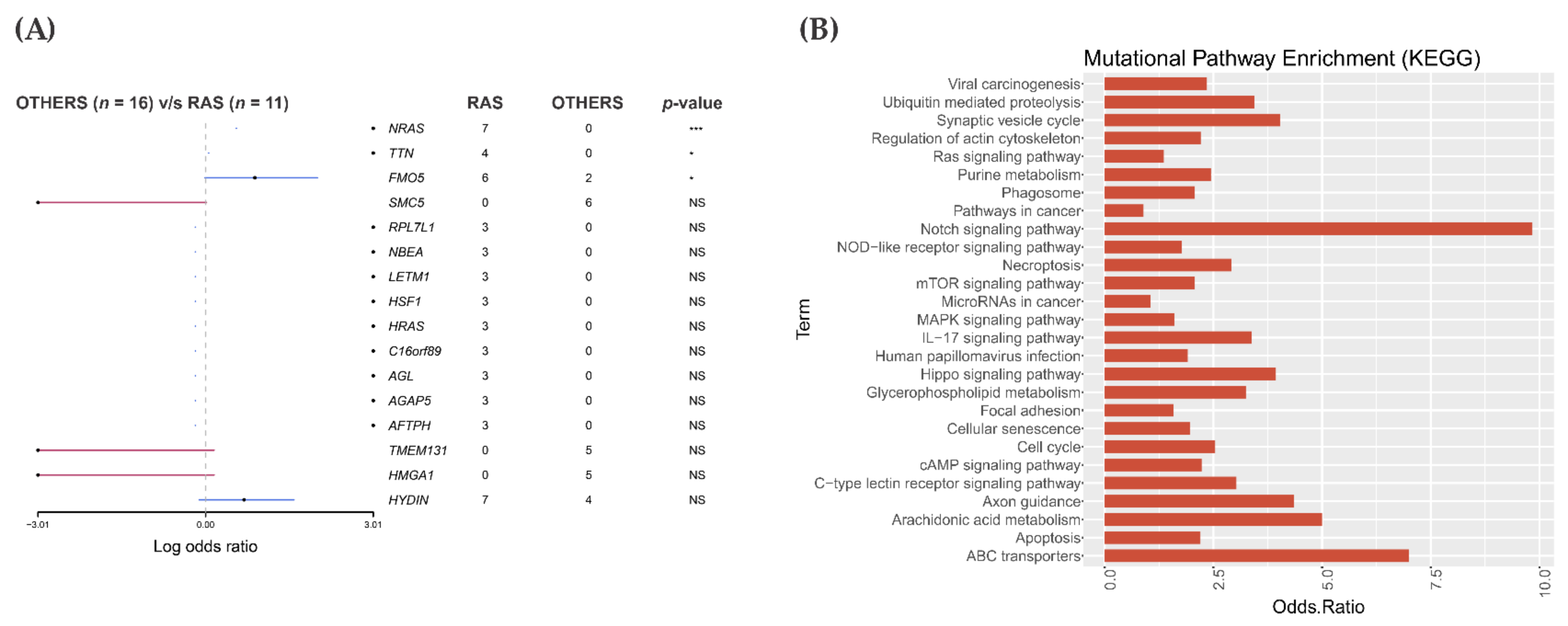
After a comparison of RAS-positive samples with 16 panel negative samples, we obtained a list of 126 genes that were exclusively and recurrently altered in the RAS-positive cohort (Figure 2A and Figure S1). Those genes were enriched for pathways using the KEGG database through Enrich.

3.3. Pathway Enrichment Analysis in the RAS-Positive Cohort

Numerous pathway databases have been made available to infer biologically relevant pathway activity. To gain insights into pathways affected by RAS-associated mutations, the list of all 126 genes exclusively and recurrently mutated in this cohort was analyzed with the Enrichr web-based application (http://amp.pharm.mssm.edu/Enrichr, accessed on 28 July 2020), using the KEGG pathway information [45,46]. The data was plotted with odds ratio values calculated based on the total number of genes in each pathway and how many of them were found mutated (Figure 2B).
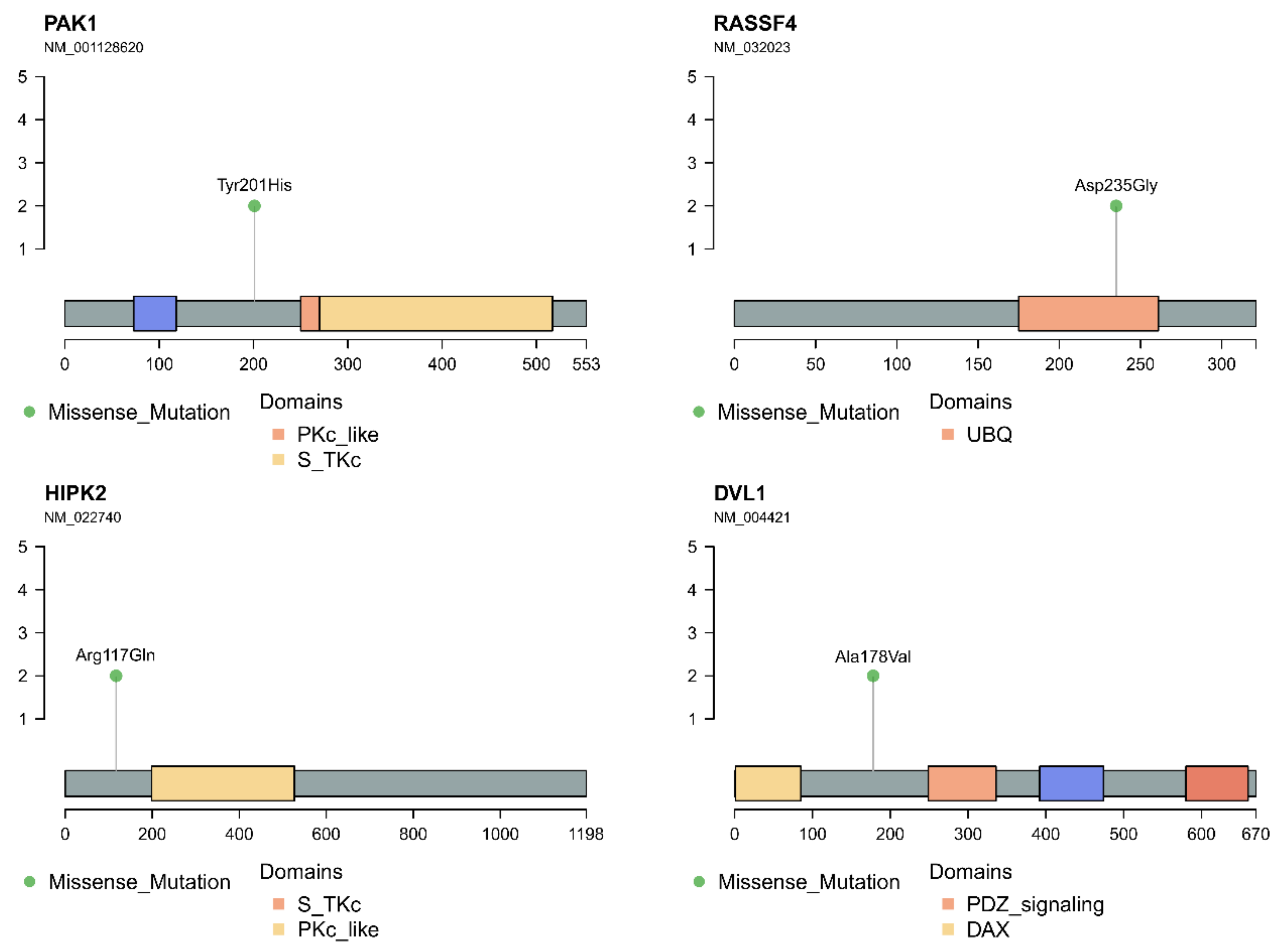
A pathway was considered affected if it contained at least two or more mutated genes. We found between two and five genes mutated in the same pathways. Some genes were found to be affecting multiple pathways. A plot with the number of genes affected (odds ratio) in each pathway group is shown (Figure 2B). The pathways that present higher odds ratios and p-values < 0.05 are: Axon guidance (5/181 genes: ABLIM1, PAK1, UNC5B, SEMA3D and RHOD); Hippo (4/160: PAK1, RASSF4, HIPK2 and DVL1); Notch (3/48 genes; HDAC2, DVL1 and PSEN2) and ABC transporters (2/45 genes; ABCC4 and ABCA5). Among them, the Hippo pathway has been recurrently associated with tumor progression and selected for further validation.
Sanger sequencing confirmed the presence of the mutations of PAK1, RASSF4, HIPK2 and DVL1 in the discovery cohort (Figure S2A,B).

3.4. In Silico Analysis of Hippo Pathway Mutated Genes in RAS-Positive Samples and Its Mutational Impact

The PAK1 missense mutation (rs775172015) promoted a tyrosine to a histidine substitution at codon 201 (p.Y201H) (Figure 3). Tyrosines 153, 201 and 285 were reported as required for a PAK1 kinase activity and identified as a JAK2 tyrosyl phosphorylation site by mass spectrometry and two-dimensional peptide mapping [47]. In fact, this alteration was classified as deleterious (sift score 0.04) and possibly damaging (PolyPhen2 score 0.789). The HOPE analysis indicated that the p.Y201H substitution was located within a stretch of residues annotated in UniProt as a special region of interaction with CRIPaK, a cysteine-rich inhibitor of Pak1 (CRIPak). Therefore, the difference in amino acid properties between the wild type and the mutant likely affects its function. Due to the smaller size and less hydrophobicity of the mutant residue, hydrophobic and other interactions either in the core of the protein or on the surface may be lost.
The RASSF4 missense mutation resulted in the p.D235G change and was classified as tolerated (sift score 0.25) and benign (PolyPhen2 score 0.075) (Figure 3). Interestingly, the mutation is located within the Ras associating site, which interacts with the Ras GTPase protein family, controlling cellular processes such as membrane trafficking, apoptosis and proliferation. The changed amino acid found in RASSF4 is smaller, neutral and more hydrophobic than the wild type (negative charge), which can lead to a loss of interaction and, also, a loss of hydrogen bonds and/or disturb the correct folding. The mutation introduced a glycine, a very flexible amino acid, likely disturbs the required rigidity of the protein at this position.
The HIPK2 mutation promoted the p.R117Q substitution, previously described in the dbSNP database (rs764542823) and classified as tolerated (sift score 0.2) and probably damaging (PolyPhen2 score 0.978) (Figure 3). The HOPE analysis revealed that the mutant and the wild-type amino acid differ in size and charge. While the wild type is positive, the mutant is neutral and smaller. The p.R117Q mutation is located within a stretch of residues annotated in UniProt as a transcriptional co-repression site. As the wild-type residue is highly conserved, the effect of replacing the R117 of HIPK2 with other residue that has differences in amino acid properties likely disrupt the three-dimensional structure of the protein and its function. Although neither the mutant residue described here nor other residue with similar properties was described at this site, based on conservation scores the mutation is probably damaging.
The DVL1 mutation promoted the p.A178V substitution previously described in the dbSNP (rs139645212) as deleterious (sift score 0.02) and benign (PolyPhen2 score 0.036) (Figure 3). The interaction between the DLV protein and YAP/TAZ rely on specific conserved domains of the protein, like N-terminal DIX, central PDZ and c-terminal DEP domains. Studies indicate that the PDZ domain and WW domain, present in both the YAP and TAZ resulting proteins, are responsible for their interaction. The change described in this study is located next to a PDZ domain, which may interfere with this interaction.
Additionally, the integrative Onco Genomics analysis (intOgen https://www.intogen.org/search, accessed on 2 December 2020), which evaluated the mutational status in 28,076 samples of 66 cancer types, acknowledged different mutations in PAK1, RASSF4, DVL1 and HIPK2 genes. Recurrent mutations in these genes in many types of cancer suggest their action in the tumorigenesis process (Figure S3A) [48].

3.5. Mutational Landscape and Pathway Enrichment in the RAS-Positive Samples from TCGA Cohort

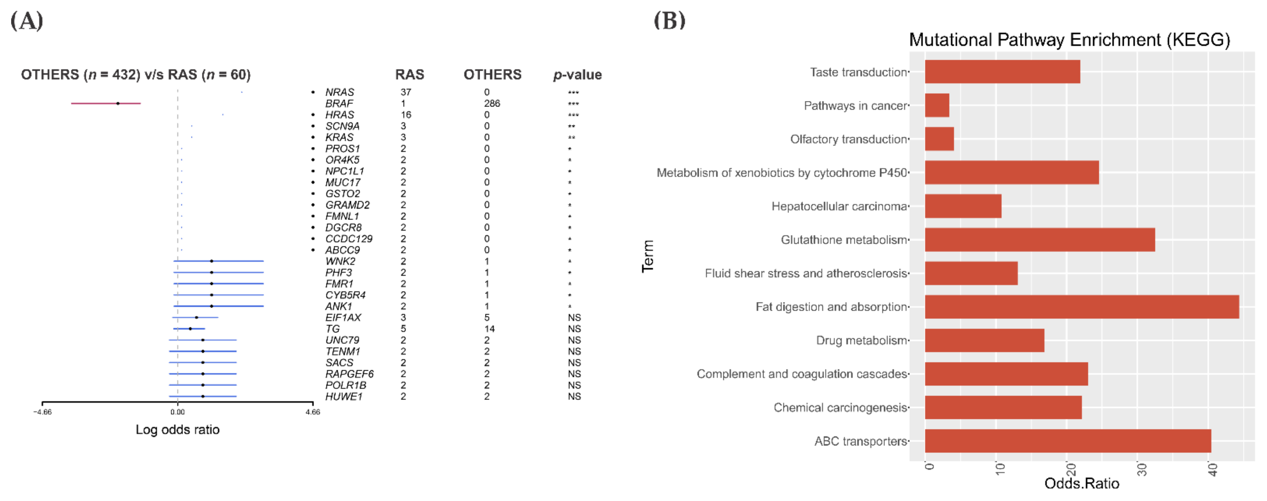
The analysis of 60 RAS-positive PTCs from the TCGA cohort was performed against the 432 PTCs designated as “others”. As expected, NRAS, HRAS and KRAS showed the highest mutation rank in the RAS-mutated group. Other 11 genes were mutated recurrently and exclusively in the RAS-positive PTC samples (Figure 4A). BRAF V600E was the most frequently mutated gene in the PTC group classified as others. None of the genes that were mutated in our RAS-positive samples were detected in the RAS-positive samples from the TCGA cohort, including PAK1, RASSF4, HIPK2 and DVL1, (Figure S3B). However, most of our RAS-positive samples are FTC and FVPTC while the RAS-positive PTC samples from TCGA cohort are the classic variant of PTC.
The pathway enrichment analysis showed that most of the 11 genes belonged to one pathway. Some of the genes were enriched for more than one signaling pathway. The odds ratio varied according to the number of genes mutated in each pathway (Figure 4B).
Remarkably, only two pathways that were enriched in the RAS-positive PTC from the TCGA cohort were also found in our RAS-positive cohort: ABC transporters (p-values ≤ 0.05) and pathways in cancer. However, none of the genes (ABCC9 and GSTO2) mutated in the TCGA were found mutated in our set of samples.

3.6. Gene Expression Pattern in the RAS-Positive Samples

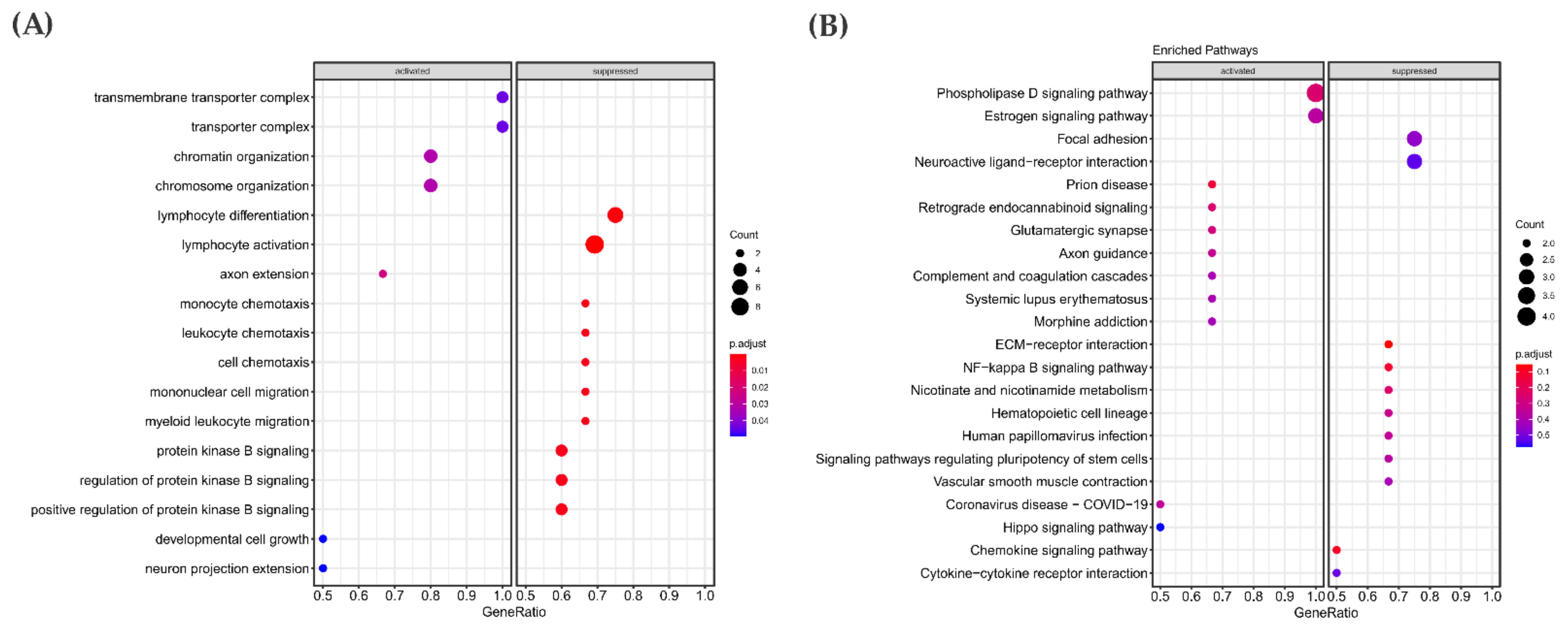
By comparing RAS-positive samples from the discovery cohort against the “others”, we identified 1765 genes that were found differentially expressed (adjusted p-value ≤ 0.05). About 485 genes were upregulated in the RAS-positive samples, while 1280 genes were downregulated. To gain insights into altered pathways, we performed the Gene Set Enrichment Analysis (GSEA and KEGG), using top 10% ranked genes DEGs (177 genes up- and down-regulated with the lowest adjusted p-value) (Figure 5).
GSEA functional analyses revealed that genes involved in cell growth development, chromatin organization and chromosome organization were differentially expressed (Figure 5A). KEGG pathway enrichment showed that pathways such as NF-Kappa beta and ECM receptor interaction contained genes that were downregulated in the discovery cohort. Among the pathways whose genes were enriched as upregulated, the most interesting was the Hippo signaling pathway (Figure 5B). Remarkably, this pathway showed significant enrichment in the discovery cohort in both mutational and gene expression analysis.
Next, to verify if the 11 RAS-positive samples in the discovery cohort and the TCGA RAS-positive samples show similar expression profile, the BAM files from 60 RAS-positive samples and 25 BRAF V600E-positive PTC samples from TCGA were processed according to our pipeline (Figure 1), removing the batch effect due to the difference in the library construction. A principal component analysis (PCA) performed on DEGs showed that the RAS-positive samples from a discovery cohort showed an expression profile similar to that observed in RAS-positive samples from the TCGA cohort, as most samples grouped into the same cluster. Instead, the BRAF V600E-positive samples from the TCGA cohort grouped into a distinct cluster (Figure S4).
Accordingly, an enrichment analysis was also performed in the 10% ranked genes obtained after performing differential gene expression (adjusted p-value ≤ 0.05) in the 60 RAS-positive PTC from the TCGA cohort (1700 genes with the lowest adjusted p-value).
The GSEA functional analyses showed that genes involved with nucleoside and ATP metabolism were upregulated, and the genes associated with cellular processes such as the regulation of DNA replication were downregulated (Figure 6A). Regarding KEGG pathway enrichment, the pathway with the larger number of genes differentially expressed refers to the immune response (Figure 6B).
Among the pathways already known to contribute to cancer progression, three of them were enriched in both a RAS-positive discovery cohort (11 samples) and RAS-positive PTC from a TCGA cohort: ECM-receptor interaction, NF-kappa B and Hippo signaling (Figure 6C).
We used the gageDate package to analyze which genes were altered in the Hippo pathway in RAS-positive samples from both discovery and the TCGA cohorts. The data are summarized in Figure 7. Briefly, the genes RASSF6, DLG1, AREG, ITGB2, BIRC2, YWHAQ (14-3-3), BMPR1B and WNT11 were downregulated, while the genes GDF6, ID2, CCND1 and FZD1 were upregulated. Remarkable, GDF6 was one of the most upregulated, with an expression 6.97-fold higher than its expression in the others group. The fact that GDF6 and FZD1 are upregulated may indicate a crosstalk between BMP, Wnt and Hippo signaling, which is reinforced by the fact that the target genes of both pathways are highly expressed (ID2 and CCND1, respectively).
Another interesting result assembling our mutational and differential expression analysis is the fact that the PAK1 gene found exclusively mutated in the RAS-positive discovery cohort was also found significantly upregulated (0.5-fold) (p-value= 0.002, adjusted p-value = 0.043).
Regarding the RAS-positive samples from the TCGA cohort, all the genes above mentioned as downregulated in the RAS-positive discovery group were also downregulated in the RAS-positive samples from the TCGA cohort. We additionally found other genes that play a significant role-regulating the Hippo pathway downregulated, such as KIBRA, MOB1A and LATS1 (Hippo kinase activator) (Figure 7), whereas the GDF6, ID2 and SMAD5 from the BMP pathway were upregulated.
The higher PAK1 expression, which was concomitant with a decreased NF2 and LATS1 expression, strongly suggests that the Hippo pathway kinase core is off, and YAP/TAZ proteins are active in the nucleus. In fact, target genes that are normally expressed when YAP is localized in the nucleus, such as AFP and FGF1, were upregulated. Finally, another evidence comes from the increased expression of the PP2R2C gene, which encodes the protein phosphatase PPA2A that dephosphorylate MST1/MST2, causing the activation of YAP/TAZ.
DVL genes (DVL1, 2 and 3) and DSNK1E, which encodes the CK1-epsilon kinase, were also found downregulated. As DVL suppresses the YAP/TAZ nuclear abundance and TEAD transcriptional activity, these findings indicate that a DVL protein interaction with YAP/TAZ in the cytoplasm is reduced, resulting in the YAP/TAZ nuclear localization and increased TEAD activity. A possible activation of the Wnt pathway is reinforced by the fact that AXIN2 and CCND1, target genes of this pathway that are normally expressed when YAP is in the nucleus, showed increased expressions.
Remarkable, the genes encoding proteins that regulate the canonical TGFβ signaling (TGFBI, TGFBR1, TGFB2, TGFB3, TGFBR3, SMAD2 and SMAD3), showed lower expression, indicating that the TGFβ pathway is likely inactive in the RAS-positive samples from the TCGA cohort.
Altogether, the results of the differential gene expression in the RAS data from discovery and the TCGA cohorts suggest that the Hippo pathway is inactive in RAS-positive thyroid carcinomas, and, therefore, YAP/TAZ might be localized in the nucleus regulating genes that act in cell proliferation, migration and survival. Moreover, the activation of key genes belonging to the BMP and Wnt pathways raises the possibility of a hypothetical crosstalk among these pathways.

3.7. Gene Expression Analysis: Proof of Concept

In order to test the hypothesis that the Hippo pathway might be dysregulated and, therefore, YAP/TAZ were able to locate in the nucleus and regulate the genes associated with Wnt and BMP signaling, we performed real-time qPCR of key genes associated with these pathways: YAP1, PAK1, NF2, LATS1, ID1, SMAD1, SMAD5 and DVL1. The expression analysis was performed in five PTC-positive for RAS mutations. As controls, five PTC-positive for BRAF V600E and five normal adjacent thyroid samples were used.
YAP1 showed a higher expression in RAS-positive samples when compared to BRAF V600E-positive PTC (p < 0.05). Although not considered significant, BRAF V600E-positive PTCs showed lower levels of YAP1 expression than the normal thyroid tissue. PAK1 also showed a higher expression in the RAS group compared to BRAF V600E group (Figure S5).
On the other hand, the NF2 expression was significantly diminished in the RAS cohort compared to normal adjacent tissue. Remarkable, NF2 and LATS1 were also significantly downregulated in the RAS-positive PTC from the TCGA cohort (Figure 7).
Considering the genes belonging to the BMP and Wnt pathways, the SMAD1 expression was significantly higher in the RAS group compared to the BRAF group. SMAD5 did not show a significant expression difference when compared to both the BRAF and normal adjacent groups. The ID1 expression was significantly lower in the BRAF group compared to normal tissue. These results, especially SMAD1 upregulation, are in agreement with TCGA-RAS differential expression analysis and reinforce the fact that BMP is active when the Hippo pathway is inactive (Figure S5).
The difference of LATS1 and DVL1 expression was not considered significant when compared RAS group with BRAF V600E and normal thyroid adjacent groups.

4. Discussion

In recent years, several studies identified genes that cooperate with RAS mutations in the pathogenesis or progression of thyroid and other types of cancers, such as NF1, APC, PTEN, KEAP1, NF2 and others [49,50,51,52,53].
Mathematical prediction predicted synergy between specific combinations of mutations. The subsequent analysis of Pan-Cancer TCGA dataset and Cancer Cell line Encyclopedia (CCLE) uncovered that NF1 mutations promote cancer not only by increasing Ras signaling but, also, by increasing the number of mutations that would further increase the Ras signaling. In other words, the number of potential noncanonical and canonical RAS point mutations capable of promoting cancer was greater in the NF1-deficient context [50].
Others demonstrated that the loss of Nf2 or Ras activation was insufficient to independently induce thyroid cancers, while the cooperation of Ras and Nf2 led to poorly differentiated thyroid carcinomas with increased MAPK signaling [53]. In human thyroid cancers, a high frequency of 22q loss was preferentially associated with RAS-mutated PTC and poorly differentiated thyroid carcinomas as compared to BRAF-mutated tumors. Remarkably, the authors demonstrated that Nf2 loss promoted Ras signaling, in part through Hippo pathway inactivation and YAP-induced transcriptional activation of oncogenic and wild-type RAS [53].
The Hippo pathway regulates tissue growth and cell fate. It was first postulated to play a role in human cancer on the basis of the notorious overgrowth of Drosophila melanogaster tissues that harbor mutations in genes that encode proteins associated with this pathway [54,55,56].
The core of the Hippo pathway consists of the serine/threonine kinases MST1/2 and LATS1/2. MST kinase activates LATS, via a membrane-associated complex containing the tumor suppressor NF2, which, in turn, phosphorylates the transcriptional coactivators YAP and TAZ on multiple sites. Once phosphorylated, the YAP/TAZ complex is inactivated through cytoplasm sequestration via binding to 14-3-3 or by its increased ubiquitination and degradation. Conversely, inhibition of the Hippo pathway leads to dephosphorylation of the YAP/TAZ complex, its increased nuclear abundance and transcriptional enhancer activation domain (TEAD) transcriptional activity, which promotes cell proliferation [56,57]. Beside the TEAD family of transcription factors, YAP/TAZ also interacts with other transcription factors, including Smad, p63 and PAX [56,58,59].
Although mutations and altered expressions of a group of Hippo pathway genes that lead to the increased activity of coactivators YAP and TAZ and an overgrowth phenotype have been observed in human cancer [56,60,61], the core Hippo pathway genes are infrequently mutated. As far as we know, NF2 is the only commonly mutated driver gene in the Hippo pathway in thyroid cancer [56].
Importantly, beyond the main components of the Hippo pathway, several upstream regulatory branches have been reported to modulate the Hippo pathway, such as Wnt and BMP/Transforming Growth Factor β [59].
To identify genes that cooperate with RAS in thyroid tumorigenesis, in this study, we performed an RNA-Seq analysis of FVPTC and FTC with the canonical RAS mutation. Here, we describe missense mutations in four components of the Hippo pathway.
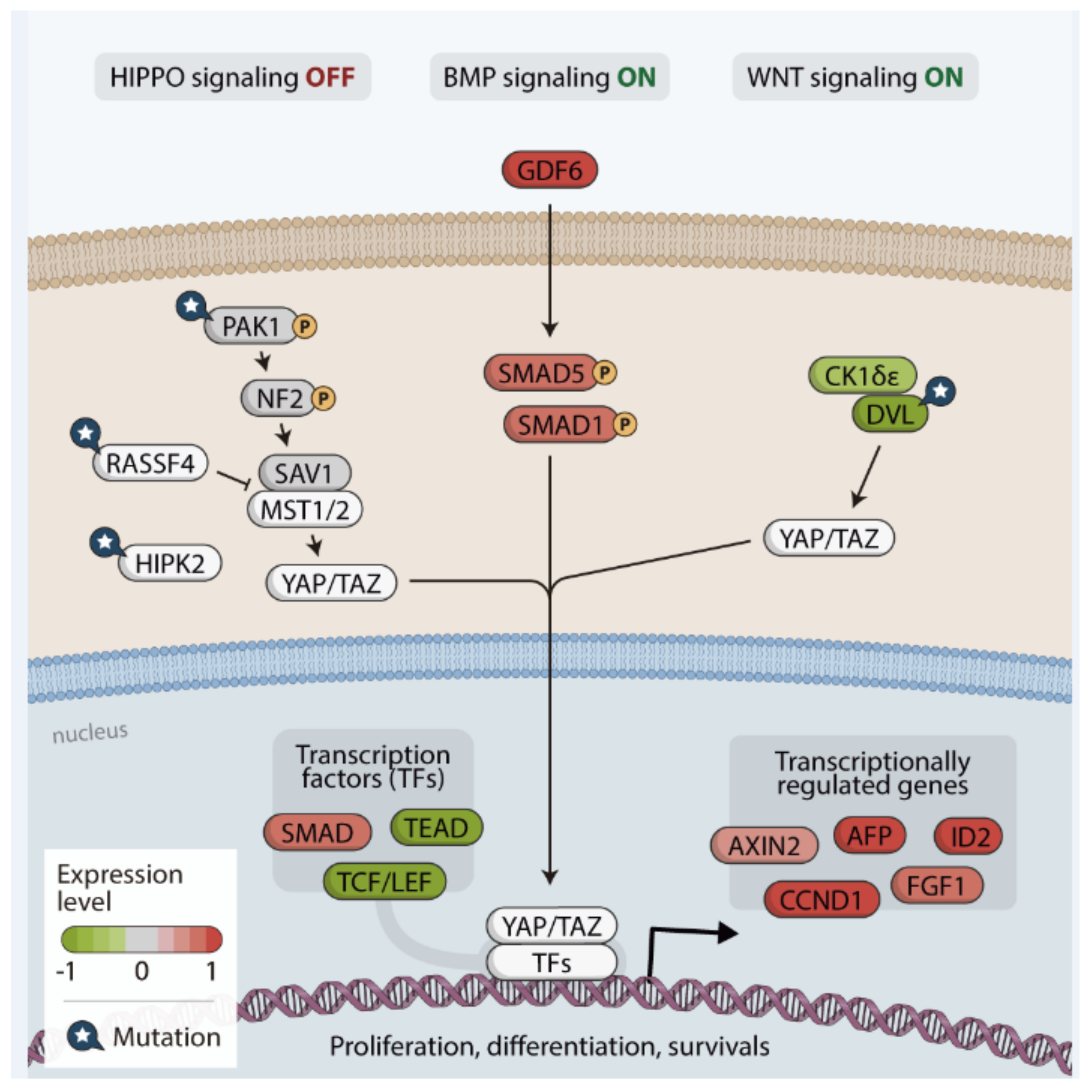
The genes PAK1 (p21-activated kinase 1), RASSF4 (RAS association domain-containing family protein), HIPK2 (homeodomain-interacting protein kinase 2) and DVL1 (the human homolog of the Drosophila dishevelled gene) were mutated in the FVPTC and FTC samples (Figure 8).
The mutations found in the PAK1 and HIPK2 genes may affect the Hippo pathway functions through NF2-reduced expression and YAP/TAZ phosphorylation, respectively. Although controversial, it has been suggested that these proteins promote Hippo pathway repression, YAP/TAZ increased nuclear abundance and the transcriptional activity of several transcription factors, including TEAD [62,63,64,65,66,67].
Though further studies are needed to clarify whether HIPK2 regulates YAP phosphorylation and its activity either in the cytoplasm or at a nuclear level, the mutation described here likely interferes with the HIPK2 function and transcription regulation of Hippo target genes [67,68].
Interestingly, HIPK family members are reported to have distinct and contradictory effects on cell proliferation and tissue growth [69,70]. The HIPK2 gene is located on chromosome 7q, near the BRAF locus. The increased expression of HIPK2 was reported in sporadic pilocytic astrocytoma, likely due to the copy number gain [71]. HIPK2 amplification was also reported in melanoma [72]. Moreover, it has been reported that HIPK2 promotes the activation of conserved pathways implicated in cancer, such as Wnt and Hippo [73].
It is known that the Hippo and Wnt pathways equally regulate the nuclear transcriptional activity and are closely connected to each other on multiple levels [74,75,76]. It has been reported that CK1ɛ isoform phosphorylates YAP/TAZ after priming phosphorylation from LATS1/2 and following CK1ɛ binding to MST1/2. Under these conditions phosphorylation of DVL is reduced, leading to inhibition of Wnt Signaling. Also, YAP/TAZ complex bind to the cytoplasmic DVL1, preventing phosphorylation and abrogating the complex translocation to the nucleus [74,75]. The A178V DVL1 substitution here described may promote Wnt activation and the expression of target genes by interfering with YAP/TAZ binding sites or preventing the CK1-dependend phosphorylation of DVL1.
RASSF4, a member of tumor suppressor genes family, is broadly expressed in normal tissues. However, it was found down regulated in several tumors subtypes and cancer cell lines by promoter methylation. RASSF proteins can associate, via its SARAH domain, with downstream kinases of Hippo pathway such as MST1/2 and SAV1 in order to promote apoptosis. Therefore, mutations describe here may affect its ability to associate with these kinases and Hippo pathway signalling [62].
While Hippo genes have been rarely reported as mutated in human cancers, the recurrent mutations reported in PAK1, RASSF4, HIPK2 and DVL1 in many types of cancer by the Integrative Onco Genomics, a web site that explores driver genes in cancer, suggest they likely play a role in tumorigenesis process.
Beyond the main components of the Hippo pathway described above as mutated, many other additional proteins that have been reported to modulate this pathway were dysregulated at expression levels. For example, RASSF6, KIBRA, NF2, LATS1 and MOB1A showed lower expression in RAS-positive samples from both discovery and TCGA cohorts, suggesting YAP/TAZ reduced phosphorylation in RAS positive samples.
We additionally found increased expression of PAK1 in RAS positive samples. Hence, PAK1 increased expression, associated with NF2 and LATS1 reduced expression, indicates a new potential regulation mechanism of Hippo pathway inactivation in RAS-positive samples.
Reduced expression of tumor suppressor NF2, due to 22q loss, was previously associated with RAS mutant poorly differentiated thyroid carcinoma, undifferentiated thyroid carcinoma and in PTC from TCGA cohort [53]. The authors demonstrated that loss of Nf2 or Ras activation is insufficient to independently induce thyroid cancer in mice, but their combination was highly tumorigenic. They suggested that NF2 loss cooperates with mutant RAS to increase signaling via MAPK, acting in part through YAP-induced transcriptional activation of oncogenic and wild-type RAS, providing a novel mechanism of promotion of RAS-induced [53].
As Hippo pathway suppresses cell growth through phosphorylation of YAP, which disrupts its ability to promote TEAD-dependent transcription of genes involved in proliferation and survival [77], we next investigated the expression of YAP/TAZ target genes.
We here observed an increased expression of YAP/TAZ target genes AFP, FGF1, suggesting that YAP/TAZ complex was able to promote their transcriptional activation. Another relevant finding was that ID2, CCND1, and AXIN2 genes, associated with BMP and Wnt pathways, showed increased expression. These findings raised the hypothesis of a crosstalk between Hippo, BMP and Wnt signalling pathways. In other words, nuclear YAP might be able to bind to other transcriptions factors, other than TEAD, to activate transcriptional program.
The TCGA cohort analysis corroborates with this hypothesis since all three DVL genes were down regulated and the AXIN2, CCND1, CTNNB1, AXIN1 were detected with higher expression in this cohort, suggesting that Wnt signalling pathway was active. In addition to the Wnt target gene CCND1, we found increased expression of FZD1 (in both cohorts), which triggers Wnt activation.
Another finding that corroborates with our hypothesis is the fact that we found increased expression of GDF6, also named BMP13, in the RAS-positive discovery cohort reported in this study and in PTC from TCGA cohort. GDF6 is a BMP ligand that was previously described as an oncogene in melanoma. Its increased expression was detected in melanoma, compared to the normal tissue and benign skin lesions [78].
Furthermore, increased expression of other components of BMP signalling pathway, SMAD5 and SMAD1, were identified in RAS-positive samples from the TCGA cohort. These findings, in association with further validation analysis of SMAD1 in our RAS cohort, reinforce the possibility that activation of the BMP pathway leads to YAP/TAZ shift to the nucleus to induce target gene expression.
Altogether, our findings suggest that the Hippo pathway is dysregulated in thyroid cancer RAS-positive samples, resulting in the translocation of the YAP/TAZ complex to the nucleus. Hence, the pathway inactivation can occur not only through NF2 loss but also through mutations or changes in the expression of genes that act or regulate proteins that act in the kinase core. Once in the nucleus, YAP/TAZ complex can interact with sequence specific transcription factors other than TEADs. The complex can cooperate with SMADs and T-cell specific transcription factor (LEF/TCF), among others, to regulate expression of Hippo target genes such as AFP, FGF1, ID2, AXIN2 and CCND1 that are involved in cell proliferation and cell survival. Besides, BMP and Wnt pathways are active due to a cross talk with effectors of Hippo signalling (Figure 8). Our data suggest a possible mechanism whereby dysregulation of the Hippo pathway and activation of pathways such as BMP and Wnt may contribute for tumorigenesis in RAS-positive tumors. If the mechanism of Hippo dysregulation described here hyperactivate MAPK signalling needs further investigation.
As it has been shown that this network contributes to thyroid cancer progression, we believe that these findings will not only help diagnosis and prognosis of thyroid cancer but also the identification of new therapeutic approaches for targeting the whole Hippo pathway, therefore, helping to improve the treatment of thyroid cancer.

5. Conclusions

In summary, our study suggests that mutations and/or genes expression alteration that lead to Hippo pathway inactivation can contribute to tumorigenesis in RAS positive thyroid tumors. The mutations found in PAK1, HIPK2 and RASSF4 genes may affect Hippo pathway function through NF2 reduced expression, YAP/TAZ phosphorylation, and reduce association with MST1/2 and SAV1 respectively. Also, DVL1 substitution may promote Wnt activation and the expression of target genes by interfering with YAP/TAZ binding sites or preventing the CK1-dependend phosphorylation of DVL1.
Increased expression of YAP/TAZ target genes AFP, FGF1 were observed, suggesting that YAP/TAZ complex is likely in the nucleus promoting their transcriptional activity, an event only possible if Hippo pathway is inactive. RASSF6, KIBRA, NF2, LATS1, MOB1A, down regulation corroborates with these findings. On the other hand AXIN2, CCND1, CTNNB1, AXIN1, FZD1 higher expression in this cohort, suggest Wnt activity. GDF6, ID2, SMAD5, and SMAD1 up-regulation reinforce the hypothesis of activation of the BMP pathway.
In summary, our findings suggest that the Hippo pathway is dysregulated in thyroid cancer, mainly in RAS-positive tumours. Our data also suggests a cross talk of Hippo with Wnt and BMP pathways, providing potential mechanistic bases for the synergism between RAS, Hippo, Wnt and BMP pathways and, thus, novel opportunities for effective targeted therapies.

Supplementary Materials

The following are available online at https://www.mdpi.com/article/10.3390/cancers13102306/s1, Figure S1: List of 126 genes found exclusively and recurrently mutated in RAS discovery cohort (11 samples). The 11 RAS-mutated samples were classified according tumor histology (FTC and FVPTC), stage and risk for recurrence. Figure S2: (A) Distribution of PAK1, RASSF4, HIPK2, and DVL1 in RAS-positive discovery cohort, the four genes are mutated exclusively in the RAS group. (B) Sanger sequencing confirming the nucleotide change in the four genes mutated in the Hippo Path: PAK1 (Chr11:77066884, A/G, c.T601C:p.Y201H), RASSF4(Chr10:45486414, A/G, c.A704G:p.D235G), HIPK2 (Chr7:139416484, C/T, c.G350A:p.R117Q) and DVL1(Chr1:1277119, G/A, C533T:p.A178V), Figure S3: (A) PAK1, RASSF4, HIPK2 and DVL1, mutational distribution in Integrative Onco Genomics cancer cohorts. (B) Mutations found in PAK1, RASSF4, HIPK2 and DVL1 are absent in TCGA-THCA cohort, showing no co-occurrence of mutations in these genes with mutations in the BRAF and RAS genes, Figure S4: Principal component analysis (PCA) showing a similar expression profile of the RAS discovery cohort (11 samples) when compared to RAS-positive TCGA-THCA, Figure S5: Real time polymerase chain reaction (qRT-PCR) performed on additional RAS, BRAF V600E and normal adjacent thyroid tissue (referred as normal adj in graph). Key genes from Hippo, BMP and Wnt signaling pathways altered in RAS-positive TCGA-THCA cohort were evaluated. YAP1, NF2 and PAK1 from Hippo pathway and SMAD1 and ID1 from BMP signaling had significant results.

Author Contributions

Conceptualization and design, T.N.R.C., L.V.B. and J.M.C.; RNA library preparation; P.F.A.; and Bioinformatics analysis; T.N.R.C., L.V.B., V.C.B., V.G. and P.A.F.G.; Data curation, T.N.R.C.; Analysis and interpretation of the data, T.N.R.C. and J.M.C., Validation analysis, T.N.R.C.; TCGA data processing and data analysis, T.N.R.C. and E.A.L. Writing—original draft preparation, T.N.R.C. and J.M.C. Writing-review and editing, T.N.R.C., L.V.B., V.C.B., P.A.F.G., E.A.L. and J.M.C.; Supervision, J.M.C.; Funding acquisition, J.M.C. All authors have read and agreed to the published version of the manuscript.

Funding

The study was supported by research grants from The São Paulo State Research Foundation (FAPESP, 2014/06570-6). JMC is a recipient of a scholarship of Research Productivity from CNPq (304534/2018-8). We also thank Coordenação de Aperfeiçoamento de Pessoal de Nível Superior (CAPES) for their financial support.

Institutional Review Board Statement

The study was conducted under the approval of the Ethical Committee of the Universidade Federal de São Paulo—UNIFESP/EPM (number 03233218.4.0000.5505).

Informed Consent Statement

Informed consent was obtained from all subjects involved in the study.

Data Availability Statement

TCGA data used in this project can be access at https://portal.gdc.cancer.gov, accessed on 24 July 2020, ENCODE normal thyroid can be found at https://www.encodeproject.org, accessed on 20 February 2019.

Conflicts of Interest

The authors declare that the research was conducted in in the absence of any commercial or financial relationships that could be interpreted as a potential conflict of interest.

References

  1. Rahib, L.; Smith, B.D.; Aizenberg, R.; Rosenzweig, A.B.; Fleshman, J.M.; Matrisian, L.M. Projecting cancer incidence and deaths to 2030: The unexpected burden of thyroid, liver, and pancreas cancers in the United States. Cancer Res. 2014, 74, 2913–2921. [Google Scholar] [CrossRef] [Green Version]
  2. Agrawal, N.; Akbani, R.; Aksoy, B.A.; Ally, A.; Arachchi, H.; Asa, S.L.; Auman, J.T.; Balasundaram, M.; Balu, S.; Baylin, S.B.; et al. Integrated genomic characterization of papillary thyroid carcinoma. Cell 2014, 159, 676–690. [Google Scholar] [CrossRef] [PubMed] [Green Version]
  3. Bastos, A.U.; De Jesus, A.C.; Cerutti, J.M. ETV6-NTRK3 and STRN-ALK kinase fusions are recurrent events in papillary thyroid cancer of adult population. Eur. J. Endocrinol. 2018, 178, 83–91. [Google Scholar] [CrossRef]
  4. Hemerly, J.P.; Bastos, A.U.; Cerutti, J.M. Identification of several novel non-p.R132 IDH1 variants in thyroid carcinomas. Eur. J. Endocrinol. 2010, 163, 747–755. [Google Scholar] [CrossRef] [PubMed] [Green Version]
  5. Oler, G.; Cerutti, J.M. High prevalence of BRAF mutation in a Brazilian cohort of patients with sporadic papillary thyroid carcinomas: Correlation with more aggressive phenotype and decreased expression of iodide-metabolizing genes. Cancer 2009, 115, 972–980. [Google Scholar] [CrossRef]
  6. Chen, H.; Luthra, R.; Routbort, M.J.; Patel, K.P.; Cabanillas, M.E.; Broaddus, R.R.; Williams, M.D. Molecular profile of advanced thyroid carcinomas by next-generation sequencing: Characterizing tumors beyond diagnosis for targeted therapy. Mol. Cancer Ther. 2018, 17, 1575–1584. [Google Scholar] [CrossRef] [Green Version]
  7. Duan, H.; Liu, X.; Ren, X.; Zhang, H.; Wu, H.; Liang, Z. Mutation profiles of follicular thyroid tumors by targeted sequencing. Diagn. Pathol. 2019, 14, 1–10. [Google Scholar] [CrossRef] [Green Version]
  8. Nicolson, N.G.; Murtha, T.D.; Dong, W.; O Paulsson, J.; Choi, J.; Barbieri, A.L.; Brown, T.C.; Kunstman, J.W.; Larsson, C.; Prasad, M.L.; et al. Comprehensive genetic analysis of follicular thyroid carcinoma predicts prognosis independent of histology. J. Clin. Endocrinol. Metab. 2018, 103, 2640–2650. [Google Scholar] [CrossRef] [Green Version]
  9. Ohba, K.; Mitsutake, N.; Matsuse, M.; Rogounovitch, T.; Nishino, N.; Oki, Y.; Goto, Y.; Kakudo, K. Encapsulated papillary thyroid tumor with delicate nuclear changes and a KRAS mutation as a possible novel subtype of borderline tumor. J. Pathol. Transl. Med. 2019, 53, 136–141. [Google Scholar] [CrossRef]
  10. Landa, I.; Ibrahimpasic, T.; Boucai, L.; Sinha, R.; Knauf, J.A.; Shah, R.H.; Dogan, S.; Ricarte-Filho, J.C.; Krishnamoorthy, G.P.; Xu, B.; et al. Genomic and transcriptomic hallmarks of poorly differentiated and anaplastic thyroid cancers. J. Clin. Investig. 2016, 126, 1052–1066. [Google Scholar] [CrossRef] [PubMed] [Green Version]
  11. Fagin, J.A. Minireview: Branded from the start—Distinct oncogenic initiating events may determine tumor fate in the thyroid. Mol. Endocrinol. 2002, 16, 903–911. [Google Scholar] [CrossRef] [Green Version]
  12. Nikiforova, M.N. Molecular testing of thyroid FNA samples. In Diagnostic Pathology and Molecular Genetics of the Thyroid; Nikiforov, Y.E., Biddinger, P.W., Thompson, L.D.R., Eds.; Lippincott Williams & Wilkins: Baltimore, MD, USA, 2009; pp. 94–102. [Google Scholar]
  13. Motoi, N.; Sakamoto, A.; Yamochi, T.; Horiuchi, H.; Motoi, T.; Machinami, R. Role of Ras mutation in the progression of thyroid carcinoma of follicular epithelial origin. Pathol. Res. Pract. 2000, 196, 1–7. [Google Scholar] [CrossRef]
  14. Nikiforov, Y.E.; Nikiforova, M.N. Molecular genetics and diagnosis of thyroid cancer. Nat. Rev. Endocrinol. 2011, 7, 569–580. [Google Scholar] [CrossRef]
  15. Patel, S.G.; Carty, S.E.; McCoy, K.L.; Ohori, N.P.; Lebeau, S.O.; Seethala, R.R.; Nikiforova, M.N.; Nikiforov, Y.E.; Yip, L. Preoperative detection of RAS mutation may guide extent of thyroidectomy. Surgery 2017, 161, 168–175. [Google Scholar] [CrossRef] [Green Version]
  16. Garcia-Rostan, G.; Zhao, H.; Camp, R.L.; Pollan, M.; Herrero, A.; Pardo, J.; Wu, R.; Carcangiu, M.L.; Costa, J.; Tallini, G. Ras mutations are associated with aggressive tumor phenotypes and poor prognosis in thyroid cancer. J. Clin. Oncol. 2003, 21, 3226–3235. [Google Scholar] [CrossRef] [PubMed]
  17. Paniza, A.C.D.J.; Mendes, T.B.; Vianna, M.D.B.; Thomaz, D.M.D.; O Chiappini, P.B.; Colazza-Gama, G.A.; Lindsey, S.C.; De Carvalho, M.B.; Alves, V.A.F.; Curione, O.; et al. Revised criteria for diagnosis of NIFTP reveals a better correlation with tumor biological behavior. Endocr. Connect. 2019, 8, 1529–1538. [Google Scholar] [CrossRef] [PubMed] [Green Version]
  18. Saavedra, H.I.; Knauf, J.A.; Shirokawa, J.M.; Wang, J.; Ouyang, B.; Elisei, R.; Stambrook, P.J.; Fagin, J.A. The RAS oncogene induces genomic instability in thyroid PCCL3 cells via the MAPK pathway. Oncogene 2000, 19, 3948–3954. [Google Scholar] [CrossRef] [Green Version]
  19. Serrano, M.; Lin, A.W.; McCurrach, M.E.; Beach, D.; Lowe, S.W. Oncogenic Ras provokes premature cell senescence associated with accumulation of p53 and p16INK4a. Cell 1997, 88, 593–602. [Google Scholar] [CrossRef] [Green Version]
  20. Tuveson, D.A.; Shaw, A.T.; Willis, N.A.; Silver, D.P.; Jackson, E.L.; Chang, S.; Mercer, K.L.; Grochow, R.; Hock, H.; Crowley, D.; et al. Endogenous oncogenic K-rasG12D stimulates proliferation and widespread neoplastic and developmental defects. Cancer Cell 2004, 5, 375–387. [Google Scholar] [CrossRef] [Green Version]
  21. Haigis, K.M.; Kendall, K.R.; Wang, Y.; Cheung, A.; Haigis, M.C.; Glickman, J.N.; Niwa-Kawakita, M.; Sweet-Cordero, A.; Sebolt-Leopold, J.; Shannon, K.M.; et al. Differential effects of oncogenic K-Ras and N-Ras on proliferation, differentiation and tumor progression in the colon. Nat. Genet. 2008, 40, 600–608. [Google Scholar] [CrossRef] [Green Version]
  22. Schuhmacher, A.J.; Guerra, C.; Sauzeau, V.; Cañamero, M.; Bustelo, X.R.; Barbacid, M. A mouse model for Costello syndrome reveals an Ang II-mediated hypertensive condition. J. Clin. Investig. 2008, 118, 2169–2179. [Google Scholar] [CrossRef] [Green Version]
  23. Franco, A.T.; Malaguarnera, R.; Refetoff, S.; Liao, X.-H.; Lundsmith, E.; Kimura, S.; Pritchard, C.; Marais, R.; Davies, T.F.; Weinstein, L.S.; et al. Thyrotrophin receptor signaling dependence of braf-induced thyroid tumor initiation in mice. Proc. Natl. Acad. Sci. USA 2011, 108, 1615–1620. [Google Scholar] [CrossRef] [Green Version]
  24. Miller, K.A.; Yeager, N.; Baker, K.; Liao, X.-H.; Refetoff, S.; Di Cristofano, A. Oncogenic Kras requires simultaneous PI3K signaling to induce ERK activation and transform thyroid epithelial cells in vivo. Cancer Res. 2009, 69, 3689–3694. [Google Scholar] [CrossRef] [Green Version]
  25. Chen, X.; Mitsutake, N.; Laperle, K.; Akeno, N.; Zanzonico, P.; Longo, V.A.; Mitsutake, S.; Kimura, E.T.; Geiger, H.; Santos, E.; et al. Endogenous expression of HrasG12V induces developmental defects and neoplasms with copy number imbalances of the oncogene. Proc. Natl. Acad. Sci. USA 2009, 106, 7979–7984. [Google Scholar] [CrossRef] [PubMed] [Green Version]
  26. Krishnamoorthy, G.P.; Davidson, N.R.; Leach, S.D.; Zhao, Z.; Lowe, S.W.; Lee, G.; Landa, I.; Nagarajah, J.; Saqcena, M.; Singh, K.; et al. EIF1AX and RAS mutations cooperate to drive thyroid tumorigenesis through ATF4 and c-MYC. Cancer Discov. 2019, 9, 264–281. [Google Scholar] [CrossRef] [PubMed] [Green Version]
  27. Ibrahimpasic, T.; Ghossein, R.; Shah, J.P.; Ganly, I. Poorly differentiated carcinoma of the thyroid gland: Current status and future prospects. Thyroid 2019, 29, 311–321. [Google Scholar] [CrossRef] [PubMed]
  28. Yoo, S.-K.; Song, Y.S.; Lee, E.K.; Hwang, J.; Kim, H.H.; Jung, G.; A Kim, Y.; Kim, S.-J.; Cho, S.W.; Won, J.-K.; et al. Integrative analysis of genomic and transcriptomic characteristics associated with progression of aggressive thyroid cancer. Nat. Commun. 2019, 10, 1–12. [Google Scholar] [CrossRef] [Green Version]
  29. Owen, D.H.; Konda, B.; Sipos, J.; Liu, T.; Webb, A.; Ringel, M.D.; Timmers, C.D.; Shah, M.H. KRAS G12V mutation in acquired resistance to combined BRAF and MEK inhibition in papillary thyroid cancer. J. Natl. Compr. Cancer Netw. 2019, 17, 409–413. [Google Scholar] [CrossRef] [Green Version]
  30. Dobin, A.; Davis, C.A.; Schlesinger, F.; Drenkow, J.; Zaleski, C.; Jha, S.; Batut, P.; Chaisson, M.; Gingeras, T.R. STAR: Ultrafast universal RNA-seq aligner. Bioinformatics 2013, 29, 15–21. [Google Scholar] [CrossRef]
  31. Anders, S.; Pyl, P.T.; Huber, W. HTSeq—A Python framework to work with high-throughput sequencing data. Bioinformatics 2015, 31, 166–169. [Google Scholar] [CrossRef] [PubMed]
  32. Depristo, M.A.; Banks, E.; Poplin, R.; Garimella, K.V.; Maguire, J.R.; Hartl, C.; Philippakis, A.A.; Del Angel, G.; Rivas, M.A.; Hanna, M.; et al. A framework for variation discovery and genotyping using next-generation DNA sequencing data. Nat. Genet. 2011, 43, 491–498. [Google Scholar] [CrossRef] [PubMed]
  33. Adzhubei, I.; Jordan, D.M.; Sunyaev, S.R. Predicting functional effect of human missense mutations using PolyPhen-2. Curr. Protoc. Hum. Genet. 2013, 76. [Google Scholar] [CrossRef] [PubMed] [Green Version]
  34. Naslavsky, M.S.; Yamamoto, G.L.; De Almeida, T.F.; Ezquina, S.A.M.; Sunaga, D.Y.; Pho, N.; Bozoklian, D.; Sandberg, T.O.M.; Brito, L.A.; Lazar, M.; et al. Exomic variants of an elderly cohort of Brazilians in the ABraOM database. Hum. Mutat. 2017, 38, 751–763. [Google Scholar] [CrossRef]
  35. Mayakonda, A.; Lin, D.-C.; Assenov, Y.; Plass, C.; Koeffler, H.P. Maftools: Efficient and comprehensive analysis of somatic variants in cancer. Genome Res. 2018, 28, 1747–1756. [Google Scholar] [CrossRef] [Green Version]
  36. Venselaar, H.; Beek, T.A.H.T.; Kuipers, R.K.P.; Hekkelman, M.L.; Vriend, G. Protein structure analysis of mutations causing inheritable diseases. An e-Science approach with life scientist friendly interfaces. BMC Bioinform. 2010, 11, 548. [Google Scholar] [CrossRef] [Green Version]
  37. Love, M.I.; Huber, W.; Anders, S. Moderated estimation of fold change and dispersion for RNA-seq data with DESeq2. Genome Biol. 2014, 15, 550. [Google Scholar] [CrossRef] [PubMed] [Green Version]
  38. Yu, G.; He, Q.-Y. ReactomePA: An R/Bioconductor package for reactome pathway analysis and visualization. Mol. BioSyst. 2016, 12, 477–479. [Google Scholar] [CrossRef] [PubMed]
  39. Subramanian, A.; Tamayo, P.; Mootha, V.K.; Mukherjee, S.; Ebert, B.L.; Gillette, M.A.; Paulovich, A.; Pomeroy, S.L.; Golub, T.R.; Lander, E.S.; et al. Gene set enrichment analysis: A knowledge-based approach for interpreting genome-wide expression profiles. Proc. Natl. Acad. Sci. USA 2005, 102, 15545–15550. [Google Scholar] [CrossRef] [Green Version]
  40. Yu, G.; Wang, L.-G.; Han, Y.; He, Q.-Y. ClusterProfiler: An R Package for comparing biological themes among gene clusters. OMICS J. Integr. Biol. 2012, 16, 284–287. [Google Scholar] [CrossRef]
  41. Luo, W.; Friedman, M.S.; Shedden, K.; Hankenson, K.D.; Woolf, P.J. GAGE: Generally applicable gene set enrichment for pathway analysis. BMC Bioinform. 2009, 10, 161. [Google Scholar] [CrossRef] [Green Version]
  42. Cerutti, J.M.; Delcelo, R.; Amadei, M.J.; Nakabashi, C.; Maciel, R.M.; Peterson, B.; Shoemaker, J.; Riggins, G.J. A preoperative diagnostic test that distinguishes benign from malignant thyroid carcinoma based on gene expression. J. Clin. Investig. 2004, 113, 1234–1242. [Google Scholar] [CrossRef] [PubMed] [Green Version]
  43. Cordioli, M.I.C.V.; Moraes, L.; Alves, M.T.D.S.; Delcelo, R.; Monte, O.; Longui, C.A.; Cury, A.N.; Cerutti, J.M. Thyroid-specific genes expression uncovered age-related differences in pediatric thyroid carcinomas. Int. J. Endocrinol. 2016, 2016, 1–9. [Google Scholar] [CrossRef] [Green Version]
  44. The ENCODE Project Consortium. An integrated encyclopedia of DNA elements in the human genome. Nature 2012, 489, 57–74. [Google Scholar] [CrossRef]
  45. Chen, E.Y.; Tan, C.M.; Kou, Y.; Duan, Q.; Wang, Z.; Meirelles, G.V.; Clark, N.R.; Ma’Ayan, A. Enrichr: Interactive and collaborative HTML5 gene list enrichment analysis tool. BMC Bioinform. 2013, 14, 128. [Google Scholar] [CrossRef] [PubMed] [Green Version]
  46. Kuleshov, M.V.; Jones, M.R.; Rouillard, A.D.; Fernandez, N.F.; Duan, Q.; Wang, Z.; Koplev, S.; Jenkins, S.L.; Jagodnik, K.M.; Lachmann, A.; et al. Enrichr: A comprehensive gene set enrichment analysis web server 2016 update. Nucleic Acids Res. 2016, 44, W90–W97. [Google Scholar] [CrossRef] [Green Version]
  47. Rider, L.; Shatrova, A.; Feener, E.P.; Webb, L.; Diakonova, M. JAK2 Tyrosine kinase phosphorylates PAK1 and regulates PAK1 activity and functions. J. Biol. Chem. 2007, 282, 30985–30996. [Google Scholar] [CrossRef] [Green Version]
  48. Martínez-Jiménez, F.; Muiños, F.; Sentís, I.; Deu-Pons, J.; Reyes-Salazar, I.; Arnedo-Pac, C.; Mularoni, L.; Pich, O.; Bonet, J.; Kranas, H.; et al. A compendium of mutational cancer driver genes. Nat. Rev. Cancer 2020, 20, 1–18. [Google Scholar] [CrossRef] [PubMed]
  49. Umetani, N.; Sasaki, S.; Masaki, T.; Watanabe, T.; Matsuda, K.; Muto, T. Involvement of APC and K-Ras mutation in non-polypoid colorectal tumorigenesis. Br. J. Cancer 2000, 82, 9–15. [Google Scholar] [CrossRef] [Green Version]
  50. Stites, E.C.; Trampont, P.C.; Haney, L.B.; Walk, S.F.; Ravichandran, K.S. Cooperation between noncanonical Ras network mutations. Cell Rep. 2015, 10, 307–316. [Google Scholar] [CrossRef] [PubMed] [Green Version]
  51. Jefferies, M.T.; Cox, A.C.; Shorning, B.Y.; Meniel, V.; Griffiths, D.; Kynaston, H.G.; Smalley, M.J.; Clarke, A.R. PTEN loss and activation of K-RAS and β-catenin cooperate to accelerate prostate tumourigenesis: PTEN, K-RAS and β-catenin in prostate cancer. J. Pathol. 2017, 243, 442–456. [Google Scholar] [CrossRef]
  52. Romero, R.; Sayin, V.I.; Davidson, S.M.; Bauer, M.R.; Singh, S.X.; Leboeuf, S.E.; Karakousi, T.R.; Ellis, D.C.; Bhutkar, A.; Sánchez-Rivera, F.J.; et al. Keap1 loss promotes Kras-driven lung cancer and results in dependence on glutaminolysis. Nat. Med. 2017, 23, 1362–1368. [Google Scholar] [CrossRef] [Green Version]
  53. Garcia-Rendueles, M.E.; Ricarte-Filho, J.C.; Untch, B.R.; Landa, I.; Knauf, J.A.; Voza, F.; Smith, V.E.; Ganly, I.; Taylor, B.S.; Persaud, Y.; et al. NF2 loss promotes oncogenic RAS-induced thyroid cancers via YAP-dependent transactivation of RAS proteins and sensitizes them to MEK inhibition. Cancer Discov. 2015, 5, 1178–1193. [Google Scholar] [CrossRef] [PubMed] [Green Version]
  54. Justice, R.W.; Zilian, O.; Woods, D.F.; Noll, M.; Bryant, P.J. The Drosophila tumor suppressor gene warts encodes a homolog of human myotonic dystrophy kinase and is required for the control of cell shape and proliferation. Genes Dev. 1995, 9, 534–546. [Google Scholar] [CrossRef] [PubMed] [Green Version]
  55. Xu, T.; Wang, W.; Zhang, S.; Stewart, R.A.; Yu, W. Identifying tumor suppressors in genetic mosaics: The Drosophila lats gene encodes a putative protein kinase. Development 1995, 121, 1053–1063. [Google Scholar] [PubMed]
  56. Harvey, K.F.; Zhang, X.; Thomas, D.M. The Hippo pathway and human cancer. Nat. Rev. Cancer 2013, 13, 246–257. [Google Scholar] [CrossRef] [PubMed]
  57. Azad, T.; Rezaei, R.; Surendran, A.; Singaravelu, R.; Boulton, S.; Dave, J.; Bell, J.C.; Ilkow, C.S. Hippo signaling pathway as a central mediator of receptors tyrosine kinases (RTKs) in tumorigenesis. Cancers 2020, 12, 2042. [Google Scholar] [CrossRef]
  58. Grusche, F.A.; Richardson, H.E.; Harvey, K.F. Upstream regulation of the hippo size control pathway. Curr. Biol. 2010, 20, R574–R582. [Google Scholar] [CrossRef] [Green Version]
  59. Hansen, C.G.; Moroishi, T.; Guan, K.-L. YAP and TAZ: A nexus for Hippo signaling and beyond. Trends Cell Biol. 2015, 25, 499–513. [Google Scholar] [CrossRef] [Green Version]
  60. Piccolo, S.; Cordenonsi, M.; Dupont, S. Molecular pathways: YAP and TAZ take center stage in organ growth and tumorigenesis. Clin. Cancer Res. 2013, 19, 4925–4930. [Google Scholar] [CrossRef] [Green Version]
  61. Hong, X.; Nguyen, H.T.; Chen, Q.; Zhang, R.; Hagman, Z.; Voorhoeve, P.M.; Cohen, S.M. Opposing activities of the Ras and Hippo pathways converge on regulation of YAP protein turnover. EMBO J. 2014, 33, 2447–2457. [Google Scholar] [CrossRef]
  62. Volodko, N.; Gordon, M.; Salla, M.; Abu Ghazaleh, H.; Baksh, S. RASSF tumor suppressor gene family: Biological functions and regulation. FEBS Lett. 2014, 588, 2671–2684. [Google Scholar] [CrossRef] [Green Version]
  63. Sabra, H.; Brunner, M.; Mandati, V.; Wehrle-Haller, B.; Lallemand, D.; Ribba, A.-S.; Chevalier, G.; Guardiola, P.; Block, M.R.; Bouvard, D. β1 integrin-dependent Rac/group I PAK signaling mediates YAP activation of Yes-associated protein 1 (YAP1) via NF2/merlin. J. Biol. Chem. 2017, 292, 19179–19197. [Google Scholar] [CrossRef] [Green Version]
  64. Park, H.W.; Kim, Y.C.; Yu, B.; Moroishi, T.; Mo, J.-S.; Plouffe, S.W.; Meng, Z.; Lin, K.C.; Yu, F.-X.; Alexander, C.M.; et al. Alternative Wnt signaling activates YAP/TAZ. Cell 2015, 162, 780–794. [Google Scholar] [CrossRef] [Green Version]
  65. Kim, N.H.; Lee, Y.; Yook, J.I. Dishevelling Wnt and Hippo. BMB Rep. 2018, 51, 425–426. [Google Scholar] [CrossRef] [Green Version]
  66. Varelas, X.; Miller, B.W.; Sopko, R.; Song, S.; Gregorieff, A.; Fellouse, F.A.; Sakuma, R.; Pawson, T.; Hunziker, W.; McNeill, H.; et al. The Hippo pathway regulates Wnt/β-catenin signaling. Dev. Cell 2010, 18, 579–591. [Google Scholar] [CrossRef] [Green Version]
  67. Poon, C.L.; Zhang, X.; Lin, J.I.; Manning, S.A.; Harvey, K.F. Homeodomain-interacting protein kinase regulates hippo pathway-dependent tissue growth. Curr. Biol. 2012, 22, 1587–1594. [Google Scholar] [CrossRef] [PubMed] [Green Version]
  68. Chen, J.; Verheyen, E.M. Homeodomain-interacting protein kinase regulates yorkie activity to promote tissue growth. Curr. Biol. 2012, 22, 1582–1586. [Google Scholar] [CrossRef] [PubMed] [Green Version]
  69. Mo, J.; Park, H.W.; Guan, K. The Hippo signaling pathway in stem cell biology and cancer. EMBO Rep. 2014, 15, 642–656. [Google Scholar] [CrossRef] [PubMed] [Green Version]
  70. Meng, Z.; Moroishi, T.; Guan, K.-L. Mechanisms of Hippo pathway regulation. Genes Dev. 2016, 30, 1–17. [Google Scholar] [CrossRef] [PubMed] [Green Version]
  71. Yu, J.; Deshmukh, H.; Gutmann, R.J.; Emnett, R.J.; Rodriguez, F.J.; Watson, M.A.; Nagarajan, R.; Gutmann, D.H. Alterations of BRAF and HIPK2 loci predominate in sporadic pilocytic astrocytoma. Neurology 2009, 73, 1526–1531. [Google Scholar] [CrossRef] [Green Version]
  72. Zhang, X.; Tang, J.Z.; Vergara, I.A.; Zhang, Y.; Szeto, P.; Yang, L.; Mintoff, C.; Colebatch, A.; McIntosh, L.; Mitchell, K.A.; et al. Somatic hypermutation of the YAP oncogene in a human cutaneous melanoma. Mol. Cancer Res. 2019, 17, 1435–1449. [Google Scholar] [CrossRef] [PubMed] [Green Version]
  73. Blaquiere, J.A.; Wong, K.K.L.; Kinsey, S.D.; Wu, J.; Verheyen, E.M. Homeodomain-interacting protein kinase promotes tumorigenesis and metastatic cell behavior. Dis. Model. Mech. 2018, 11, 031146. [Google Scholar] [CrossRef] [PubMed] [Green Version]
  74. Knippschild, U.; Krüger, M.; Richter, J.; Xu, P.; Garcia-Reyes, B.; Peifer, C.; Halekotte, J.; Bakulev, V.; Bischof, J. The CK1 family: Contribution to cellular stress response and its role in carcinogenesis. Front. Oncol. 2014, 4, 96. [Google Scholar] [CrossRef] [PubMed] [Green Version]
  75. Fulcher, L.J.; Sapkota, G.P. Functions and regulation of the serine/threonine protein kinase CK1 family: Moving beyond promiscuity. Biochem. J. 2020, 477, 4603–4621. [Google Scholar] [CrossRef]
  76. Zeng, R.; Dong, J. The Hippo signaling pathway in drug resistance in cancer. Cancers 2021, 13, 318. [Google Scholar] [CrossRef]
  77. Zhang, H.; Liu, C.-Y.; Zha, Z.-Y.; Zhao, B.; Yao, J.; Zhao, S.; Xiong, Y.; Lei, Q.-Y.; Guan, K.-L. TEAD transcription factors mediate the function of TAZ in cell growth and epithelial-mesenchymal transition. J. Biol. Chem. 2009, 284, 13355–13362. [Google Scholar] [CrossRef] [PubMed] [Green Version]
  78. Venkatesan, A.M.; Vyas, R.; Gramann, A.K.; Dresser, K.; Gujja, S.; Bhatnagar, S.; Chhangawala, S.; Gomes, C.B.F.; Xi, H.S.; Lian, C.G.; et al. Ligand-activated BMP signaling inhibits cell differentiation and death to promote melanoma. J. Clin. Investig. 2017, 128, 294–308. [Google Scholar] [CrossRef] [Green Version]
Figure 1. Workflow of the sample selection, screening and RNA-Sequencing pipeline analysis for the discovery cohort.
Cancers 13 02306 g001
Figure 2. Mutational analysis and pathway enrichment in the RAS-positive cohort. (A) Forest plot shows representative genes that were recurrently mutated in the 11 RAS-positive groups or in the “others” group (samples negative for the driver mutation). The black points indicate genes exclusively mutated in the RAS cohort. The blue line shows genes mutated in both groups and the red line the ones mutated exclusively in the “others” group. Recurrent mutations were considered those mutations that occurred in at least two samples. *** p-value = 0.001, * p-value ≤ 0.05, NS = non-significant p-value (B) A KEGG pathway enrichment of the 126 exclusively mutated in the RAS cohort.
Cancers 13 02306 g002
Figure 3. Amino acid change and localization relative to the respective protein domains in the four proteins (PAK1, RASSF4, HIPK2 and DVL1) whose corresponding genes belong to the Hippo pathway and are mutated in a RAS-positive cohort. Brackets from 1 to 5 indicate the number of samples positive for each mutation. In our RAS-positive samples, the alteration of each gene was present in two samples.
Cancers 13 02306 g003
Figure 4. Mutational analysis and pathway enrichment (KEGG) in RAS-positive papillary thyroid carcinomas (PTC) from The Cancer Genome Atlas (TCGA) cohort. (A) A forest plot showing the eleven genes exclusively and recurrently mutated (present in at least two samples) in 60 RAS-positive samples. *** p-value ≤ 0.001, ** p-value ≤ 0.01, * p-value < 0.05, NS = non-significant p-value (B) KEGG pathway enrichment of the 11 exclusively mutated genes in rhe RAS-positive TCGA cohort.
Cancers 13 02306 g004
Figure 5. Differential gene expression analysis in the RAS-positive group from the discovery cohort (11 samples) detected 1765 genes significantly up- and downregulated. The top 10% ranked genes were differentially expressed, which shows that the ones with the lowest adjusted p-values were submitted to GSEA and KEGG enrichment to gain insight into altered biological processes and pathways. (A) GSEA enrichment analysis of biological processes that contain upregulated genes are marked on the left panel and downregulated on the right panel. (B) Pathway enrichment analysis (KEGG) results showed pathways in which most of their genes are upregulated (left) or down-regulated (right) in the RAS-positive cohort.
Cancers 13 02306 g005
Figure 6. Differential gene expression analysis in RAS-positive PTC samples from the TCGA cohort (60 samples). Enrichment analysis (GSEA and KEGG) was performed on the top 10% genes differentially expressed composing the ones with lowest adjusted p-values to gain insights into altered biological process and pathways. (A) GSEA enrichment analysis of the biological processes that contain upregulated genes are marked on the left panel and downregulated on the right panel. (B) Pathway enrichment analysis result showed pathways in which most of their genes are upregulated (left) or downregulated (right) in RAS-positive TCGA cohorts. (C) Common pathways associated with cancer progression differentially expressed in both RAS-positive discovery cohort and RAS-positive PTC from the TCGA cohort.
Cancers 13 02306 g006
Figure 7. Hippo signaling pathway KEGG-based map, showing genes up- (red) and downregulated (green) detected by differential expression (adjusted p-value ≤ 0.05) performed in both the RAS-positive discovery cohort and RAS-positive TCGA cohort.
Cancers 13 02306 g007
Figure 8. Hippo pathway simplified map, summarizing the findings of the mutation and differential gene expression analysis in our RAS-positive cohort supported by the RAS-positive TCGA data analysis. Inactivation of this pathway promotes YAP/TAZ translocation into the nucleus and targets gene expression, which leads to cellular survival and growth. Once Hippo is inactive and YAP/TAZ acting, a crosstalk may occur with the BMP and Wnt pathways.
Cancers 13 02306 g008
Table 1. Clinical pathological features of the RAS-positive cohort and sequencing coverage.
SampleRAS MutationNo. of Reads (Millions)Reads Mapping Mutant Allele (%) Tumor Type *Tumor Size
(cm)
MultifocalExtrathyroidal ExtensionVascular InvasionMetastasesRecurrenceAge at DiagnosisGender
1N Q61R13666FVPTC5.0NYNNN32M
2N Q61K11362FVPTC4.0NNNNN40M
3N Q61R7578FVPTC5.0YNNNN28F
4N Q61R10633FVPTC1.7YNNNN55F
5N Q61R11856FVPTC3.0NNNNN36F
6H Q61R24271FVPTC1.1NNNNN45F
7N Q61R9370FTC3.5YYNYN45F
8K Q61R16822FTC3.2NNYNN48F
9N Q61R13150FTC1.6NYYNN48F
10H Q61K9159FTC7.5NYYYN76F
11H A11G11713FTC10.0NYYYY70M
* No anaplastic or dedifferentiated components were described in these samples. FVPTC, follicular variant of papillary thyroid carcinoma, FTC, follicular thyroid carcinoma, Y = yes and N = no; F: female; and M: male.
Publisher’s Note: MDPI stays neutral with regard to jurisdictional claims in published maps and institutional affiliations.