Review

The Landscape of lncRNAs in Hepatocellular Carcinoma: A Translational Perspective

by
Juan Pablo Unfried
1,*,†,
Paloma Sangro
2,*,†,
Laura Prats-Mari
1,
Bruno Sangro
2,3,4,‡ and
Puri Fortes
1,3,4,‡
1
Center for Applied Medical Research (CIMA), Department of Gene Therapy and Regulation of Gene Expression, Universidad de Navarra (UNAV), 31008 Pamplona, Spain
2
Liver Unit, Clínica Universidad de Navarra (CUN), 31008 Pamplona, Spain
3
Navarra Institute for Health Research (IdiSNA), 31008 Pamplona, Spain
4
Liver and Digestive Diseases Networking Biomedical Research Centre (CIBERehd), 31008 Pamplona, Spain
*
Authors to whom correspondence should be addressed.
These authors contributed equally to this work.
Cancers 2021, 13(11), 2651; https://doi.org/10.3390/cancers13112651
Submission received: 30 April 2021 / Revised: 24 May 2021 / Accepted: 25 May 2021 / Published: 28 May 2021
(This article belongs to the Special Issue New Advances in Hepatocellular Carcinoma: From the Past to the Future)

Abstract

:

Simple Summary

Hepatocellular carcinoma is the major form of liver cancer; it has a high incidence in the global population, and it is a leading cause of the world’s cancer burden. Despite great efforts to understand the disease at the molecular level and develop effective treatments for hepatocellular carcinoma patients, current therapies for advanced cases are only effective in a small percentage of patients. Therefore, it is paramount to discover new tumor targets that can be used to develop alternative therapeutic strategies. Promising novel targets could be long non-coding RNAs. These RNA molecules are frequently found to be involved in the development and progression of cancer. More importantly, they can be safely targeted with several strategies. However, our knowledge about long non-coding RNA biology and their clinical relevance remains underdeveloped. In this review, we summarize current efforts to validate the importance of long non-coding RNAs in hepatocellular carcinoma and to evaluate their potential to impact the clinical setting. To do that we highlight lncRNA implication in key events leading to cancer and we describe how affecting lncRNAs could broaden the repertoire of potentially useful targets to help meet the challenge of hepatocellular carcinoma treatment.

Abstract

LncRNAs are emerging as relevant regulators of multiple cellular processes involved in cell physiology as well as in the development and progression of human diseases, most notably, cancer. Hepatocellular carcinoma (HCC) is a prominent cause of cancer-related death worldwide due to the high prevalence of causative factors, usual cirrhotic status of the tumor-harboring livers and the suboptimal benefit of locoregional and systemic therapies. Despite huge progress in the molecular characterization of HCC, no oncogenic loop addiction has been identified and most genetic alterations remain non-druggable, underscoring the importance of advancing research in novel approaches for HCC treatment. In this context, long non-coding RNAs (lncRNAs) appear as potentially useful targets as they often exhibit high tumor- and tissue-specific expression and many studies have reported an outstanding dysregulation of lncRNAs in HCC. However, there is a limited perspective of the potential role that deregulated lncRNAs may play in HCC progression and aggressiveness or the mechanisms and therapeutic implications behind such effects. In this review, we offer a clarifying landscape of current efforts to evaluate lncRNA potential as therapeutic targets in HCC using evidence from preclinical models as well as from recent studies on novel oncogenic pathways that show lncRNA-dependency.

1. Introduction

HCC is the most common primary liver cancer, with a global incidence of ~750,000 cases per year that nearly matches the mortality rate (700,000 deaths per year) [1,2]. Exposure to risk factors determines the incidence and mortality of HCC and it varies markedly between countries and regions. Advanced hepatic fibrosis or cirrhosis due to hepatitis B virus (HBV), hepatitis C virus (HCV), alcohol intake or the metabolic syndrome are the main well-established predisposing factors [3]. Aflatoxin B1 dietary intake is also a well-known environmental risk factor for HCC development in parts of Asia and Africa [4]. It is estimated that one-third of cirrhotic patients will develop liver cancer during their lifetime [5]. Strategies such as HBV vaccination, eradication of HCV with antiviral agents and community programs to improve lifestyle and eating habits have helped to reduce HCC predisposing factors. However, HCC remains the fourth leading cause of cancer-related deaths. Further, it has been projected that by 2030 the barrier of one million deaths per year will have been surpassed, consolidating HCC as the fastest-rising cause of cancer-related death worldwide [1,5,6,7,8]. Risk factors and factor-induced inflammation and fibrosis may induce DNA damage, epigenetic alterations or mutations leading to the activation of oncogenes or silencing of tumor suppressors, which might influence HCC development. These changes occur mainly in mature hepatocytes, but identification of up to 20% of HCCs with progenitor cell markers suggests that the cell of origin could also be a stem or progenitor cell in those cases [9,10].
Up to 50% of patients with HCC are diagnosed at early stages in developed countries, when radical treatments are feasible [11,12]. However, even though cirrhotic patients are monitored for early detection of HCC with ultrasound and/or alfa-fetoprotein (AFP) levels, many of them show advanced disease at diagnosis [11] or reach this stage after progression to local therapies. HCC is highly resistant to conventional cytotoxic chemotherapy. Sorafenib has been the only systemic treatment available for more than a decade [13]. Other tyrosine kinase inhibitors with an extended target spectrum but a similar antiangiogenic effect, including lenvatinib, regorafenib and cabozantinib, have joined sorafenib as alternative options for the first- or second-line therapy [3,11]. Immune checkpoint inhibitors that target cytotoxic T lymphocyte protein 4 (CTLA-4), programmed death receptor 1 (PD-1) or its ligand (PD-L1) have appeared as a new class of active agents against HCC. Nivolumab and pembrolizumab (anti-PD-1) are active in the second-line scenario [14,15]. Recently, the combination of atezolizumab (anti-PD-L1) with the vascular endothelial growth factor (VEGF)-inhibitor bevacizumab has proven superior to sorafenib in the first-line setting and is now the standard of care for patients naïve to systemic therapy [16]. During this time, several trials have shown no relevant antitumor activity using other agents with diverse molecular targets, including c-MET (tyrosine-protein kinase Met), mTOR (mechanistic target of rapamycin), EGFR (epidermal growth factor receptor) and others [17]. By sequential administration of available therapeutic agents, patients with a stably preserved liver function may survive for prolonged periods of time. However, most patients still do not respond to any of these therapies. The combination of agents targeting the immune response or cellular proliferation could further improve the outcome of HCC patients. However, this will require an exquisite understanding of the biology behind immunomodulation and HCC cell growth, including the identification of tumor-addictive targets within the coding and the non-coding genome.
Ordinarily, HCC studies that search for novel therapeutic strategies have been focused on protein targets. However, a global evaluation of the human genome indicates that only less than 2% accounts for coding sequences (exons that are translated into proteins) [18]. Hence, the non-protein-coding portion of the genome that is transcribed into non-coding RNAs (ncRNAs) has become increasingly valuable in recent years [19]. Out of all ncRNAs, long non-coding RNAs (lncRNAs) are the most numerous and the least studied. The relative novelty and general lack of knowledge of lncRNA functions so far, has prevented the development of classification systems that inform on lncRNA functional relevance or putative mechanisms of action. Therefore, major classifications describe lncRNA genes according to their location in the genome compared to their nearest protein-coding genes. In this context, lncRNAs are either intergenic (interspersed between coding regions) or intragenic (located within or very near to coding genes in either sense or antisense orientation). From a functional perspective, lncRNAs can be classified as trans-acting (which function similar to proteins, in cellular regions located away from the lncRNA transcription site) or cis-acting (which control the expression of neighboring genes co-transcriptionally, usually through transcriptional interference or chromatin modifications) [20]. Such localized function is exclusive to lncRNAs compared to proteins and it is an elegant and fast way of controlling a specific region of the genome without investing in DNA-binding domains. Other than that, lncRNAs, such as proteins, are folded into secondary and tertiary structures and may show domains capable of binding to DNA, other RNAs and/or proteins to form functional complexes and exert their functions. Most of the functions described to date are related to the regulation of coding genes at transcriptional and post-transcriptional levels [21]. Examples exist of lncRNAs that modulate DNA folding and enhancer activity, affect chromatin remodeling and epigenetic modifications and control transcription initiation or elongation. At the post-transcriptional level, lncRNAs have been described that can modulate splicing, translation and RNA stability by different means. In addition, some lncRNAs regulate protein function by influencing post-translational modifications or by acting as cofactors. Finally, although it seems contradictory, some lncRNAs encode small peptides with key functions in different cellular processes [22] (Figure 1). Interestingly, lncRNA function may be sequence dependent or independent. An excellent example of the latter is when the sole act of lncRNA transcription is required for function by transcriptional interference.
Transcription, processing, and regulation of most lncRNAs is very similar to that of their coding mRNA counterparts. Indeed, most lncRNAs are transcribed by polymerase II and are co-transcriptionally modified to undergo 5’capping, and oftentimes, polyadenylation, and splicing. However, splicing is less efficient and lncRNAs are more prone to undergo premature termination [23]. On average, lncRNAs have fewer exons, show less alternative splicing and are shorter than mRNAs. Other major differences are that, compared to mRNAs, lncRNAs are more nuclear, less abundant, more cell-type specific and, those deregulated in cancer, are far more tumor-type specific [24,25]. In fact, lncRNAs have been described as deregulated in various different cancers, including HCC, with implications as biomarkers for diagnosis, prediction of survival and risk of metastasis [26,27,28].

2. LncRNAs in the Hallmarks of HCC

Given the importance of lncRNAs, their ability to regulate gene expression at various levels and the differential profiles of lncRNA expression between human HCCs and non-tumoral tissue [29], lncRNAs are being actively studied for their potential as alternative therapeutic targets. In fact, the literature describes multiple lncRNAs deregulated in HCC with potential roles in therapeutically actionable pathways that strongly affect patient survival. Cell dedifferentiation, epigenetic alterations, chromatin instability and the activation of oncogenic pathways concomitant to hepatocarcinogenesis and tumor progression impact cell expression, leading to lncRNA deregulation. The altered lncRNA transcriptome may also result from increased inflammation and liver fibrosis caused by HCC risk factors. In the case of HCV and HBV patients, several lncRNAs can be deregulated in response to infection that may affect viral oncoproteins and increase hepatocarcinogenesis (reviewed in [30,31]). An interesting example is EGOT, a lncRNA induced after HCV infection that contributes to viral replication and HCC growth [32,33].
In the last decade, HCC–lncRNA research has grown exponentially, which has allowed the study of the increasing repertoire of mechanisms enabled by lncRNAs to impact the hallmarks of liver cancer. However, only a minority of studies have validated these findings in appropriate preclinical models and independent cohorts of patient samples to offer a clearer picture of their potential relevance for clinical translation. In this review, we have selected a collection of lncRNAs that have been published in high-impact journals, with thorough preclinical validation and/or association with relevant clinical parameters as a proxy for their clinical relevance. We are confident that this collection shows a clarifying landscape of the reality of lncRNA activity in HCC. Therefore, we have also grouped these lncRNAs in terms of their general mechanisms of action linked to the hallmarks of cancer they regulate.
In 2000, Hanahan and Weinberg proposed six hallmarks of cancer that all together constitute the key to unlock malignant transformation [34]. As it is well known, tumor formation is a multistage process where cells gain specific features and capacities that allow them to sustain proliferative signaling, evade growth suppressors, resist cell death, enable replicative immortality, induce angiogenesis, and activate invasion and metastasis. Ten years later, as knowledge of tumor behavior progressed, Hanahan and Weinberg proposed additional emerging hallmarks involved in deregulation of cell metabolism, escape from immune destruction, induction of tumor inflammation, genome instability and mutations. Currently, new evidence supports rapidly evolving fields that can be considered as additional emerging hallmarks of cancer, such as the contribution of stem cell programs that can promote cancer initiation, therapy resistance and metastasis [35,36].
Hereafter, we describe a curated list of exceptionally well-characterized lncRNAs, published in journals with impact factors around ten and higher. We analyzed and annotated their described mechanisms of action and the main hallmarks of cancer they enable to promote HCC progression. We believe this helps put lncRNA research in context with current efforts to pharmacologically target the acquired malignant capabilities of liver cancer cells and we suggest that the development of lncRNA-based therapies as stand-alone or combinatorial agents could potentially improve the current suboptimal outcomes of HCC patients.

2.1. Activating Invasion and Metastasis

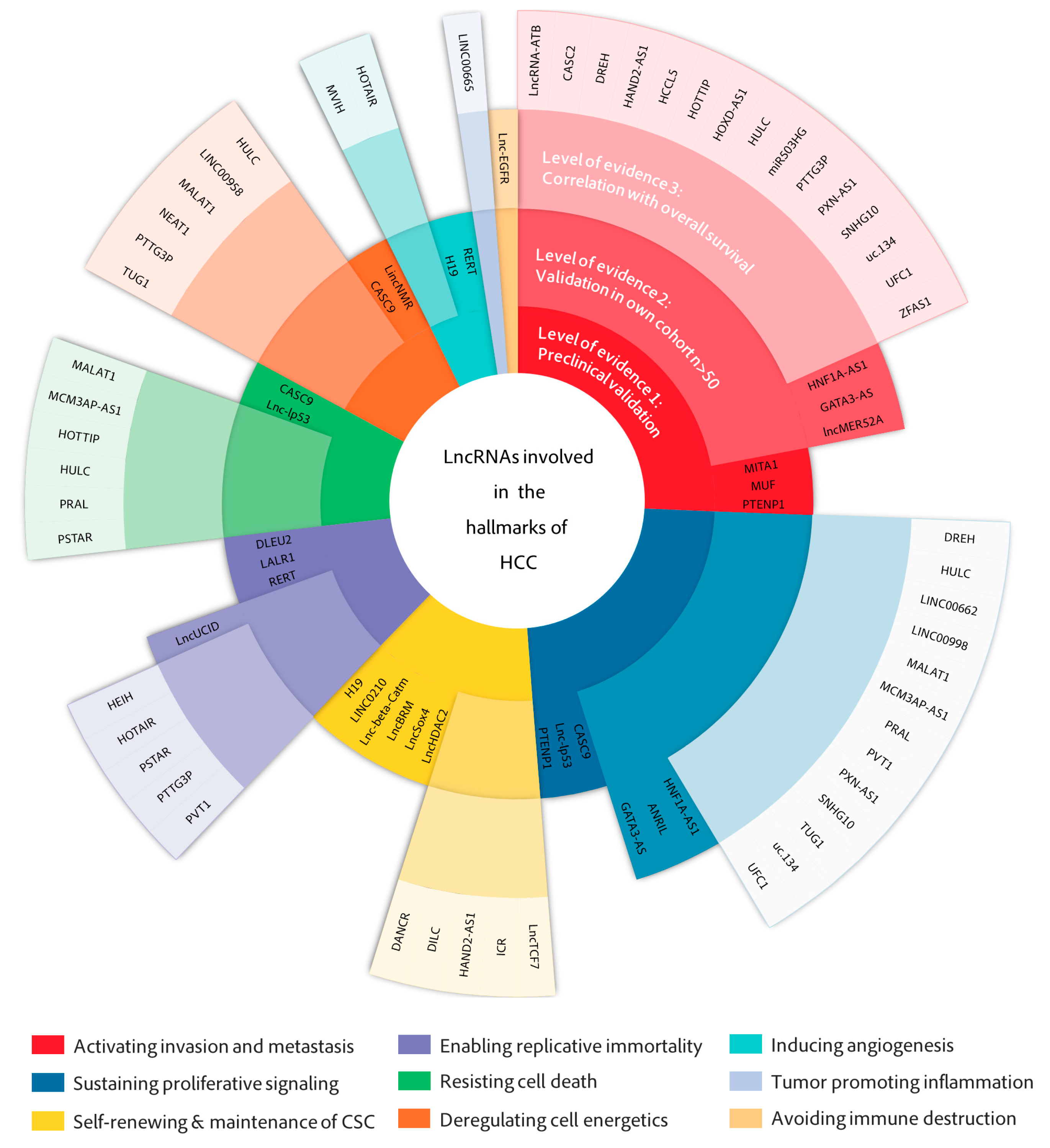
LncRNA deregulation leading to the activation of invasion and metastasis is the most represented hallmark of cancer in our main lncRNA collection (Figure 2). This is not surprising, as HCC shows a high tendency to metastasize and infiltrate adjacent and distant tissues [37]. In fact, the overall poor prognosis of HCC is largely the result of its metastatic nature and the high rate of tumor recurrence after surgery, which remain the main causes of lethal outcomes in HCC [38].
Epithelial-to-mesenchymal transition (EMT) is an essential event in the early stages of invasion required for cell escape and colonization, and therefore is highly regulated in normal cells by several pathways, including the transforming growth factor-β (TGF-β) pathway [106]. TGF-β exerts antiproliferative effects on normal and premalignant cells. However, advanced cancers often become insensitive to the tumor-suppressive actions of TGF-β and instead, benefit from TGF-β’s profound metastasis-promoting effects, such as EMT induction [107]. The EMT prometastatic response to TGF-β is mediated by several transcription factors (ZEB1/2 (zinc finger E-box binding homeobox 1/2), SNAIL1/2 (Snail family transcriptional repressor 1/2), TWIST, SLUG and members of the FOX (forkhead box) family) and other downstream effectors, including cytokines (interleukins (IL)-6, IL-11), growth factors (VEGF), matrix proteins (fibronectin, collagen), cytoskeletal (vimentin) and cell adhesion proteins (E- and N-cadherin), metalloproteases (MMPs) and more recently described factors such as microRNAs (miRNAs) [108] and lncRNAs [109]. LncRNAs are able to promote and sustain molecular events leading to metastasis by enabling several possible mechanisms, predominantly by binding miRNAs (Figure 2) and working as competing endogenous RNAs (ceRNAs) or by regulating more intricate lncRNA–miRNA protein axes.
LncRNA-ATB (activated by TGF-β) is a paradigmatic ceRNA, which increases invasion and metastasis by sequestering members of the miR-200 family (miR-200a, b and c), known EMT regulators that target ZEB1 and ZEB2 [79,110]. Accordingly, in vivo models of LncRNA-ATB upregulation showed increased intrahepatic dissemination, higher levels of circulating tumors cells (CTCs) and multiple-site metastasis. In addition, other mechanisms besides miR binding work to promote metastasis in lncRNA-ATB-expressing cells. LncRNA-ATB binds to and increases the stability of IL-11 mRNA, improving IL-11 secretion and activating STAT3 signaling in an autocrine manner (Figure 1). This confers a survival advantage to metastatic cells [111].
Similarly, many other lncRNAs regulate downstream transcription factors related to canonical TGF-β-mediated EMT. In some cases, the mechanism also involves miRNA binding, such as ZFAS1 (zinc finger antisense 1) [105] which upregulates ZEB1 by sequestering miR-150. LncRNA MUF (mesenchymal stem cells upregulated factor) [89] also acts as a ceRNA for miR-34a, resulting in increased SNAIL transcription and EMT activation. This is in accordance with previous results in colon, breast and lung carcinomas where decreased miR-34 expression was found to de-repress SNAIL transcription [112]. Additional lncRNAs upregulate the expression of EMT transcription factors through unknown mechanisms. LncRNA MITA1 (metabolism-induced tumor activator 1) [88], induced by energy stress, increases SLUG transcription. LncRNA HCCL5 (hepatocellular carcinoma L5) [51] which is transcriptionally regulated by ZEB1 via a super-enhancer, is a general positive regulator of EMT genes SNAIL, SLUG, TWIST and ZEB1 in a positive feedback loop. Of these, SNAIL and TWIST have been directly reported to induce EMT, invasion and metastasis in HCC [113,114].
Cells that undergo EMT reorganize their cytoskeleton to enable cell elongation and directional motility [107]. LncRNA DREH (downregulated expression by hepatitis B virus X protein) [45], represses EMT, likely by binding directly to vimentin to change normal cytoskeleton structure. Other lncRNAs affect Rho GTPases, which control the process of actin remodeling in EMT [115]. By competitively binding to miR19a, lncRNA HOXD-AS1 (homeobox D cluster antisense 1) [58] de-represses Rho GTPase activating protein 11a (ARHGAP11A), while lncMER52A (lncRNA derived from MER52A retrotransposons) [78] stabilizes the cell–cell adhesion regulator p120-catenin (Figure 1). Both proteins are involved in the activation of the Rho GTPases, RAC1 (Ras-related C3 botulinum toxin substrate 1) and CDC42 (cell division cycle 42), which promote the formation of membrane protrusions and cell motility, and the initiation of front-rear polarity. All of these are essential events in EMT that enable directional migration. Finally, an interesting lncRNA is PXN-AS1 (paxillin (PXN) antisense RNA 1). PXN is a cytoskeletal protein located in sites of cell adhesion to the extracellular matrix and has been involved in cancer cell proliferation and invasion. PXN mRNA stability and translation are regulated by PXN-AS1 in a dual manner. When the oncofetal splicing factor MBNL3 is expressed, it induces PXN-AS1 exon 4 inclusion, resulting in increased PXN and tumor promotion. This occurs as PXN-AS1 containing exon 4 binds to the 3´UTR region of PXN mRNA and impedes the negative action of miR-24. Instead, PXN-AS1 lacking exon 4 binds to coding sequences of PXN mRNA and decreases translation. Therefore, MBNL3 knockdown almost completely abolishes HCC growth [80].
As cells acquire motility and invasive capacities, additional factors such as metalloproteinases (MMPs) help the cell move through the surrounding stroma towards a blood or lymphatic vessel potentiating invasiveness [116]. This process can also be enabled by lncRNA–miRNA protein axes. For example, HOXD-AS1, this time by binding to miR-130a, prevents miRNA-dependent degradation of the SOX4 mRNA transcription factor, activating the expression of target genes EZH2 and MMP2 [59]. Similarly, ZFAS1, besides ZEB1, is able to induce MMP14 and MMP16, two membrane MMPs with relevant roles in HCC metastasis and invasion [117,118,119].
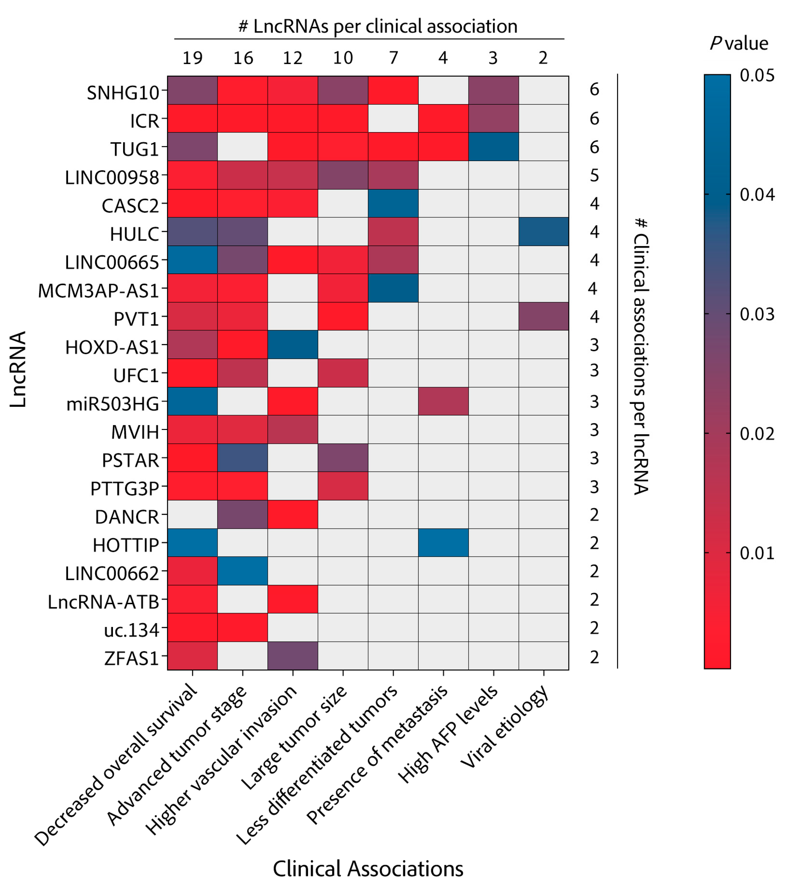
Several other studies have found lncRNAs to promote or protect against metastasis in HCC cells by less frequent mechanisms involving regulatory signaling pathways such as Wnt-β-catenin (lncRNA MUF) [89], NF-κB (lncRNA miR503HG (MIR503 host gene)) [87] or novel metastasis-related factors such as FBXW7 (lncRNA CASC2 (circulating cancer susceptibility 2)) [40] with varying degrees of evidence of clinical relevance. While MUF, MITA1 or PTENP1 (phosphatase and tensin homolog pseudogene 1) [96] lack validation in HCC tissues (Figure 3), other lncRNAs such as miR503HG, CASC2, ZFAS or lncRNA-ATB have validated correlations with overall survival (OS) and importantly, with micro and macrovascular invasion or portal vein tumor thrombus (PVTT), one of the first events in intrahepatic dissemination (Figure 4). These clinical correlations help validate a potential role for these lncRNAs in the invasion and metastasis of HCC and support their targetability with therapeutic purposes.

2.2. Sustaining Proliferative Signaling

The proliferation and survival of normal hepatocytes is under the control of several layers of regulation owing to its remarkable ability to regenerate. However, alterations in signaling pathways arising from mutations or epigenetic or epitranscriptomic changes can render cells independent or refractory to the control of these pathways. In most HCC patients, such alterations are the result of sustained damage exerted by HCC risk factors. Exposure to these factors leads to persistent liver inflammation and progressive liver fibrosis that often leads to cirrhosis. Liver cirrhosis generates a pro-tumorigenic microenvironment that promotes the transformation of pre-malignant lesions into highly proliferating neoplastic foci, a mechanism that accounts for up to 90% of HCC cases [120]. This process is initiated by activating mutations in the telomerase (TERT) gene, considered a gatekeeper of early hepatocarcinogenesis. TERT expression allows cirrhotic hepatocytes to override their senescent state and reactivate proliferation [121]. Furthermore, transcriptomic analyses of premalignant and early HCC samples show that MYC activation also plays a central role in early malignant transformation [122]. This process is strongly enhanced by mutations occurring in one or more oncogenes or tumor suppressors, predominantly β-catenin activation and p53 inactivation. Additional, less frequent mutations add up to a cumulative mutation burden of up to 70 mutations within each tumor [123,124]. This outstanding mutational burden impacts several pathways including PI3K/Akt, Ras/Raf/MEK/ERK, TGF-β, Wnt or NOTCH. All play important roles in supporting HCC growth, frequently promoting invasion and metastasis concomitantly as co-occurring malignant traits.
LncRNAs seem to follow this trend as there is a significant overlap between lncRNAs involved in invasion/metastasis and proliferation. The combination of these two hallmarks accounts for the majority of lncRNAs in our main collection (Figure 2) and even more in our larger collection (Figure S1B). Despite this overlap, the mechanisms of action induced by lncRNAs that affect the proliferative potential of HCC cells seem to differ from those of lncRNAs involved in promoting invasion/metastasis. While miRNA binding is also prominent, lncRNAs involved in the malignant proliferation of HCC cells act by varied mechanisms, including binding to effector proteins and epigenetic and novel epitranscriptomic regulators.
LncRNA SNHG10 (small nucleolar RNA host gene 10) [100] is a good example of an lncRNA working at the intersection between proliferation and metastasis induction. By sponging miRNA-150 and stabilizing the mRNA of RPL4 (receptor-like protein 4), SNHG10 acts to increase the expression and activity of the MYB (myeloblastosis) proto-oncogene. Reciprocally, MYB enhances SNHG10 expression by promoter activation in a positive feedback loop. Further, SNHG10 modulates the expression of its homolog SCARNA13 (small Cajal body-specific RNA 13) through the miR-150/RPL4/MYB pathway. Then, SCARNA13 upregulates the levels of the transcription factor SOX9 (SRY-box transcription factor 9) with repercussions in HCC cell growth and metastasis. This regulatory reinforcement may be behind the drastic phenotype of SNHG10 depletion in subcutaneous and orthotopic mouse models as well as the outstanding correlation of SNHG10 expression with patient prognosis. Indeed, SNHG10 is the lncRNA that correlates with more clinical parameters in our lncRNA collection (Figure 4).
LINC00662 (long intergenic non-protein coding RNA 662) [68] activity is more specific to the induction of HCC proliferation by mediating genome-wide hypomethylation through depletion of SAM (S-adenosylmethionine) and increase in SAH (S-adenosylhomocysteine) levels. The ratio of the methionine cycle intermediates SAM and SAH is commonly regarded as an indicator of the methylation potential of the cell, and changes in its balance have been implicated in liver carcinogenesis [125,126]. LINC00662 binds to the 3′-UTR of MAT1A (S-adenosylmethionine synthase isoform type-1) mRNA to promote its decay and reduce the levels of MAT1A, a liver-specific SAM synthesizing enzyme. At the same time, LINC00662 interacts with and promotes the ubiquitination and degradation of the enzyme AHCY (adenosyl homocysteinase), which hydrolyzes SAH and prevents its accumulation. Therefore, by RNA–RNA and RNA–protein interactions, LINC00662 is able to decrease the SAM/SAH balance and de-represses methylated oncogene promoters, activating known HCC oncogenes such as MYC, CTNNB1 and HRAS.
By enabling similar effector-binding mechanisms, lncRNAs UFC1 (ubiquitin-fold modifier conjugating enzyme 1) [104] and uc.134 (long non-coding RNA uc.134) [103], up and downregulated in HCC, respectively, are also involved in HCC proliferation through oncogene activation. In a multistep regulatory network, lncRNA uc.134 binds to CUL4 (cullin-4A), inhibiting its nuclear export and preventing CUL4-mediated ubiquitinylation and degradation of LATS1 (large tumor suppressor kinase 1), a tumor suppressor. This increases the stability and overall levels of LATS1 which in turn phosphorylates and inactivates the YAP1 (yes1-associated transcriptional regulator) oncoprotein, effectively silencing its target genes, among them c-MYC. Conversely, in a more straightforward mechanism, lncRNA UFC1 is able to bind with the mRNA stabilizing protein HuR (Hu antigen R) to induce the levels of β-catenin mRNA, favoring proliferation of HCC cells and the growth of subcutaneous mouse tumors. An unexpected mechanism of action for an lncRNA is that of LINC00998 (long intergenic non-protein coding RNA 998) [71], that contains an open reading frame and is translated to a small peptide, SMIM30, which promotes HCC growth and metastasis. SMIM30 is required for membrane anchoring and functionality of tyrosine kinases SRC/YES1 leading to MAPK pathway activation (Figure 1). In turn, HNF1A-AS1 (hepatocyte nuclear factor 1 homeobox A antisense RNA 1) is a target of HNF1A, considered a strong tumor suppressor for HCC [53]. HNF1A-AS1 mediates the protective role of HNF1A by binding and increasing the activity of the phosphatase SHP1 (protein tyrosine phosphatase non-receptor type 6), a strong inhibitor of JAK/STAT, NF-κB and AKT pathways [53,127].
Other mechanisms requiring lncRNAs for HCC cell proliferation include epigenetic and epitranscriptomic changes. Epigenetic regulation in HCC has been gaining momentum over the past few years as a promising target that may be actionable using epigenetic drugs [128]. Instead, epitranscriptomic changes have only been recently described to be relevant for HCC progression [129]. ANRIL (cyclin-dependent kinase inhibitor 2B (CDKN2B) antisense RNA (1) [39], a known regulator of the polycomb repressive complex 2 (PRC2), was found to bind and recruit PRC2 to the promoter of the differentiation factor KLF2 (Krüppel-like factor (2) in HCC cells, effectively repressing KLF2 transcription to promote HCC cell growth in vitro and in vivo. GATA3-AS (GATA-binding protein 3) [46] has been shown to regulate m6A deposition on the pre-mRNA of the tumor suppressor GATA3. To do this, GATA3-AS base pairs with GATA3 pre-mRNA and guides KIA1429 (a scaffolding component of the m6A methyltransferase complex) binding. This increases m6A marks in the 3′UTR of GATA3 pre-mRNA that prevent binding of HuR (Figure 1), promote RNA degradation, reduce GATA3 levels and help HCC proliferation and metastasis.
While there is a clear role of lncRNAs in regulating epigenetic or epitranscriptomic events and there is preclinical evidence of their potential from mouse xenografts, many of these lncRNAs, including ANRIL or GATA3-AS, fail to validate their relevance in patient survival or as prognostic factors (Figure 3 and Figure 4). Further efforts need to be dedicated to evaluating their therapeutic potential for HCC patients.

2.3. Deregulating Cell Metabolism

In order to enable malignant cell proliferation, adjustments in cell metabolism have to be made that increase the biomass and provide the energy required for cell division and growth. Interestingly, instead of using oxidative phosphorylation as the main energy source for dividing, tumor cells utilize glycolysis even when enough oxygen is available [130]. This phenomenon is known as the Warburg effect and it usually involves genetic mutations that increase glucose intake. Some lncRNAs have been involved in this process (Figure 2). Similar to ANRIL, TUG1 (taurine upregulated gene 1) epigenetically represses KLF2 transcription by binding PRC2 and recruiting it to the KLF2 promoter [101]. In addition, TUG1 sequesters miR-455-3p, which targets AMPKβ2 mRNA (AMP-activated protein kinase containing the β2 regulatory subunit), leading to an increase in HK2 (hexokinase 2), a rate-limiting factor required for the first step in most glucose metabolism pathways [102]
Interestingly, many lncRNAs have been described to regulate the PI3K/AKT (phosphatidylinositol-3-kinase (PI3K)/protein kinase B(AKT)) pathway in HCC. AKT induces mTOR activity, which promotes glucose metabolism, lipid and nucleotide biosynthesis and protein translation. CASC9 (circulating cancer susceptibility 9) [131] forms a functional cytoplasmic complex with HNRNPL (heterogeneous nuclear ribonucleoprotein L) that regulates AKT function [131]. Likewise, PTTG3P (pituitary tumor-transforming 3, pseudogene) [97] is upregulated in HCC and activates PI3K/AKT signaling [97]. MALAT-1 (metastasis-associated lung adenocarcinoma transcript 1) also leads to activation of the mTOR pathway by modulating the splicing targets of the oncogenic splicing factor SRSF1 (serine- and arginine-rich splicing factor 1), which also promotes the production of anti-apoptotic splicing isoforms [84,85]. HULC (HCC upregulated lncRNA) activates the AKT–PI3K–mTOR pathway after PTEN (phosphatase and tensin homolog) inhibition through miR-15a/P62. In addition, HULC also promotes methylation of the miR-9 promoter and suppresses the targeting of PPARA (peroxisome proliferator-activated receptor alpha), which activates the ACSL1 (Acyl-CoA synthetase 1) promoter and induces lipogenesis. HULC also contributes to HCC growth by phosphorylation of YB-1 (Y-box-binding protein 1), which leads to the release of YB-1 from its bound mRNA and translation of silenced oncogenic mRNAs [60,61,62,63]. Finally, by sequestering miR-327, HULC allows increased levels of PRKACB (protein kinase cAMP-activated catalytic subunit beta), which phosphorylates CREB (cAMP response element-binding) transcription factor, leading to higher levels of CREB target genes such as cFOS or HULC itself [64]. An additional inducer of nucleotide metabolism is LincNMR (long intergenic non-coding RNA-nucleotide metabolism regulator) [72], that binds to YBX1 and locates to the promoter of RRM2 (ribonucleotide reductase subunit M2), TYMS (thymidylate synthetase) and TK1 (thymidine kinase 1) genes, increasing the expression of these essential enzymes for nucleotide synthesis [72].
Other modulators of lipid metabolism are Linc00958 (long intergenic non-coding RNA 00958) and NEAT1 (nuclear paraspeckle assembly transcript 1). Linc00958 functions by sponging miR-3619-5p, increasing HDGF (hepatoma-derived growth factor) expression and resulting in lipogenesis and progression of HCC [70]. Instead, NEAT1 binds to miR-124-3p and allows expression of the adipose triglyceride lipase (ATGL), highly expressed in human HCC. High ATGL, increases the levels of diacylglycerol and free fatty acids and PPARA (peroxisome proliferator-activated receptor alpha) signaling, which works as a predictor of poor prognosis [91].

2.4. Self-Renewal and Maintenance of Cancer Stem Cells

Liver cancer stem cells (LCSCs) can derive from genetic or epigenetic alterations of liver progenitor cells or hepatocytes involved in regeneration or dedifferentiation processes [132]. Thus, stem or progenitor cells can also be the cells of origin of certain HCCs [35]. In fact, many studies suggest that embryonic liver and HCC development share similar alterations in their genetic programs. More importantly, HCC patients with gene expression profiles similar to stem cells show a worse prognosis [133,134]. LCSCs impact cell division and reprogramming but they also affect tumor heterogeneity, relapse, metastasis, and drug resistance. Induction, self-renewal, proliferation, and differentiation of LCSCs are complex processes regulated by a myriad of signaling pathways including JAK/STAT, NF-κB, Wnt/β-catenin, Notch, Hedgehog, Hippo/TAZ/YAP or JNK.
LncRNAs have been shown to regulate HCC by influencing LCSCs. For example, although with certain controversy, lncRNA H19 has been involved in the deregulation of LCSCs in different tumors, including HCC where it contributes to doxorubicin resistance [135,136]. Many LCSC lncRNA regulators affect the activity of chromatin remodeling complexes SWI–SNF (SWItch/sucrose non-fermentable), INO80 (INO80 complex ATPase subunit) or NuRD (nucleosome remodeling deacetylase) complex. LncTCF7 (transcription factor 7)/WSPAR (Wnt signaling pathway activating non-coding RNA) binds SWI–SNF to promote expression of TCF7, a major transcription factor in the Wnt signaling pathway in LCSCs [82]. Of interest, TCF7 is barely detected in normal liver, which makes it a promising biomarker in HCC [82]. In the case of LncBRM (long non-coding RNA for association with Brahma), the interaction is with the SWI–SNF subunit BRM (Brahma), resulting in the activation of YAP1 (yes1-associated transcriptional regulator) signaling [75]. Similarly, lncRNA HAND2-AS1 (heart and neural crest derivatives expressed 2- antisense RNA 1) recruits the INO80 chromatin-remodeling complex to the promoter of BMPR1A (BMP (bone morphogenetic protein) receptor type 1A), thereby inducing its expression and leading to the activation of BMP signaling which activates the initiation of HCC [49]. LncHDAC2 (lncRNA histone deacetylase 2), highly expressed in LCSCs, recruits the NuRD complex to the promoter of PTCH1 (protein-patched homolog 1), impairing PTCH1 expression. This induces the hedgehog signaling pathway and promotes the self-renewal of LCSCs [77].
Instead, other lncRNAs affect the function of major regulators including NF-κB (nuclear factor kappa-beta), STAT3 (signal transducer and activator of transcription 3) or β-catenin. DILC (downregulated in liver cancer stem cells) functions as a tumor suppressor to inhibit LCSCs by preventing NF-κB binding, inhibiting IL-6 transcription and abolishing JAK2/STAT3 pathway activation. Downregulation of DILC in HCC allows stimulation of LCSCs [43,137,138]. Lnc-Sox4 (SRY-box transcription factor 4), upregulated in HCC cells, interacts with the transcription factor STAT3 and recruits it to the promoter of Sox4 to stimulate its transcription. Sox4 overexpression then leads to initiation of LCSCs [81]. Interestingly, Lnc-β-Catm (lncRNA for β-catenin methylation) recruits the PRC2 subunit EZH2 (enhancer of zeste homolog 2) and contributes to EZH2-mediated methylation of β-catenin. This impedes β-catenin ubiquitination and degradation and supports the self-renewal of LCSCs through the Wnt pathway [74]. Similarly, LINC00210 (long intergenic non-protein coding RNA 210), highly expressed in LCSCs, binds to CTNNBIP1 and blocks its inhibitory activity over β-catenin, resulting in activation of the Wnt pathway and promoting self-renewal [67]. β-catenin expression is also regulated at the mRNA level by DANCR (differentiation antagonizing non-protein coding ribonucleic acid). DANCR binding to the 3´UTR of CTNNB1 mRNA impedes the repressing effect of miR-214, 320a and 199a (Figure 1), increasing β-catenin expression and the stemness features of HCC cells [42]. Additionally, acting at the mRNA level, ICR (intercellular adhesion molecule 1 (ICAM-1) related) increases the stability of ICAM-1 mRNA through RNA duplex formation [65]. This is relevant as ICAM-1 is an adhesion molecule highly expressed in LCSCs that plays pivotal roles in carcinogenesis and metastasis.

2.5. Resisting Cell Death

Programmed cell death by apoptosis is considered as an inherent boundary to halt cancer development. Tumoral cells try to attenuate the signaling circuits operating in the apoptotic program to successfully override death cues [35]. A major master regulator of apoptosis is the tumor suppressor p53, whose gene shows inactivating mutations in 12–48% of HCCs [123]. In line with the high relevance of this factor, there are several lncRNAs that function to increase or decrease p53 activity in several tumors including HCC. Lnc-lp53 (lncRNA induced by p53) sits among the negative regulators [73]. Lnc-lp53 attenuates p300-acetylating activity on p53 and binds to HDAC1 (histone deacetylase 1), preventing its degradation and maintaining deacetylated/inactive p53, altogether abrogating p53 activity. Instead, PRAL (P53 regulation-association long non-coding ribonucleic acid), downregulated in HCC, facilitates the interaction of HSP90 and p53, inhibiting MDM2-dependent p53 ubiquitination and resulting in enhanced p53 stability [93]. Similarly, PSTAR (P53-stabilizing and activating RNA) binds to hnRNPK (heterogeneous nuclear ribonucleoprotein K) and enhances its SUMOylation, thereby strengthening the interaction between hnRNPK and p53, which ultimately leads to the accumulation and transactivation of p53 [94].
Other well-known apoptosis regulators are factors required for development, as they need to coordinate several processes, including differentiation, motility, and apoptosis. This includes HOXA genes, homeodomain transcription factors whose expression is induced by HOTTIP (HOXA transcript at the distal tip). HOTTIP is a pro-tumoral cis-acting lncRNA upregulated in HCC that activates transcription of the HOXA locus by local recruitment of the positive regulator WDR5–MLL (WD repeat-containing protein 5–mixed linage leukemia) complex [29,57]. MCM3AP-AS1 (minichromosome maintenance deficient 3-associated protein antisense RNA 1) is involved in HCC cell proliferation by targeting miR-194-5p and subsequently promoting FOXA1 (forkhead box A1) expression in HCC cells [86]. FOXA1 is a pioneer factor and a well-known positive regulator of liver specific gene expression with a fundamental role in impairing liver cell apoptosis.
Other factors may affect apoptosis, including NF-κB. While the role of NF-κB in cancer by regulating immunity and inflammation is well documented, there are some reports that link NF-κB with tumor progression by regulating apoptosis [139], a function of NF-κB that may be mediated in part by lncRNA PDIA3P1 (protein disulfide isomerase family A member 3 pseudogene 1). PDIA3P1 is involved in a regulatory axis with NF-κB, by preventing miR-124 and miR-125 targeting of TRAF6. This leads to activation of the pathway and downstream anti-apoptosis genes, promoting doxycycline-induced apoptosis in vitro and sensitizing mouse xenografts to doxycycline treatment [92].

2.6. Enabling Replicative Immortality

The unlimited proliferative potential of tumor cells as a way to achieve immortality constitutes one of the main keys of cancer. The ability to maintain telomeres protecting the ends of chromosomal DNAs to avert apoptosis is achieved by regulation of telomere expression by different mechanisms [35]. This couples to a remodeling of the cell cycle machinery to promote cell division and restrain checkpoint controls. Several lncRNAs function as regulators of cell-cycle factors. HEIH (high expression in HCC) and DLEU2 (deleted in lymphocytic leukemia 2) modulate EZH2 activity. LncRNA-HEIH enhances EZH2 for post-transcriptional regulation of cyclin-dependent kinase inhibitor genes (p15, p16, p21 and p57), playing a key role in G0/G1 phase of the cell cycle [52]. DLEU2 is upregulated in HCC and linked to HBV. HBx and DLEU2 co-recruitment on the cDNA displaces EZH2 from the viral chromatin to boost transcription and viral replication. In addition, DLEU2-HBx association with target host promoters relieves EZH2 repression and leads to transcriptional activation of the cyclin B2 gene, among others [44].
Other lncRNAs that affect the cell cycle at different levels are LALR1 (liver regeneration 1), which recruits CTCF (CCCTC-binding factor) to repress AXIN1 (protein phosphatase 1) promoter, facilitating cyclin D1 expression through Wnt/β-catenin activation [66]. LncUCID (lncRNA upregulating CDK6 by interacting with DHX9), enhances CDK6 (cyclin-dependent kinase 6) expression by competitively binding to DHX9 (ATP-dependent RNA helicase A) and sequestering DHX9 from CDK6-3’UTR [83]. Finally, PVT1 (plasmacytoma variant translocation 1) upregulates cell cycle genes by increasing the stability of nucleolar protein NOP2 [98].

2.7. Inducing Angiogenesis

The tumor-associated neovasculature, due to the permanent activation of angiogenesis, constitutes another main key for HCC progression [35]. However, few lncRNAs from our main collection have been associated with angiogenesis. One example is lncRNA- MVIH (microvascular invasion in HCC), which is upregulated in HCC, and favors the formation of new vessels and, therefore, HCC metastasis. MVIH represses the secretion of PGK1 (phosphoglycerate kinase 1), an inhibitory regulator of angiogenesis, although the detailed mechanism is still unknown [90]. Another lncRNA related to angiogenesis is H19 (imprinted maternally expressed untranslated transcript) which was the first lncRNA described back in 1990 and it is downregulated in HCC [140]. The reason for this is that H19 is activated by Sox2 (transcription factor SOX-2), and Sox2 is inhibited by TGF-β, whose levels are high in the tumor microenvironment. Therefore, there is a significant negative correlation between the TGF-β gene signature and H19 function [47]. In spite of this, LCSC-derived exosomes contain high levels of H19 which is thought to induce angiogenesis in endothelial cells [48]. In fact, an indirect manner to promote angiogenesis is through exosome secretion. HOTAIR (HOX transcript antisense RNA) accelerates MVB (multivesicular bodies) transport by inducing Ras-related protein Rab-35 (RAB35) expression and localization to MVBs. Further, HOTAIR promotes MVB fusion by regulating the colocalization of vesicle-associated membrane protein 3 (VAMP3) and synaptosome-associated protein 23 (SNAP23) [54,55]. This results in increased exosome secretion, which may contribute to tumor growth by several means and provides a useful source of biomarkers for tumor diagnosis and prognosis.
Finally, lncRNA regulation can also impact on the levels of hypoxia-inducible factors (HIF), whose function is sensing and reacting against hypoxia by triggering angiogenesis. At normal oxygen levels, the alpha subunit of the HIF transcriptional complex (HIF1A) is targeted to degradation by prolyl hydroxylation carried out by EGLN2 (prolyl hydroxylase 1) [141]. Interestingly, a 4-bp insertion/deletion polymorphism (rs10680577), located in a distal promoter of EGLN2, affects RERT-lncRNA (RAB4B-EGLN2 read-through long non-coding RNA) structure and expression, which correlates positively with EGLN2 levels [99]. The deletion allele was significantly associated with increased risk of HCC and this association was more significant in current smokers.

2.8. Tumor-Promoting Inflammation and Avoiding Immune Destruction

Despite their strong relevance for HCC, very few lncRNAs have been described with a role in tumor-promoting inflammation or in avoiding immune destruction. This is the case of LINC00665 and Lnc-EGFR (long non-coding epidermal growth factor receptor). LINC00665 binds to the double-stranded RNA (dsRNA)-activated protein kinase (PKR), blocks its degradation by ubiquitination and increases PKR activity [69]. This activates NF-κB signaling, which contributes to LINC00665 expression, resulting in a positive feedback loop. Interestingly, LINC00665 contributes to enhanced cell proliferation in vivo and to poor prognosis of HCC patients. Instead, Lnc-EGFR is expressed in T regulatory cells (Tregs), correlating positively with tumor size and EGFR expression and negatively with IFN-γ levels. Lnc-EGFR binds to EGFR and blocks its degradation by ubiquitination by the ubiquitin ligase c-CBL (Casitas B-lineage lymphoma). Increased EGFR levels activate AP1/NF-AT1 (activator protein 1/ nuclear factor of activated T cell 1) transcription, resulting in a positive feedback loop that increases EGFR levels and in the promotion of Treg differentiation that contributes to the immunosuppressive state of HCC [76].

3. Concluding Remarks

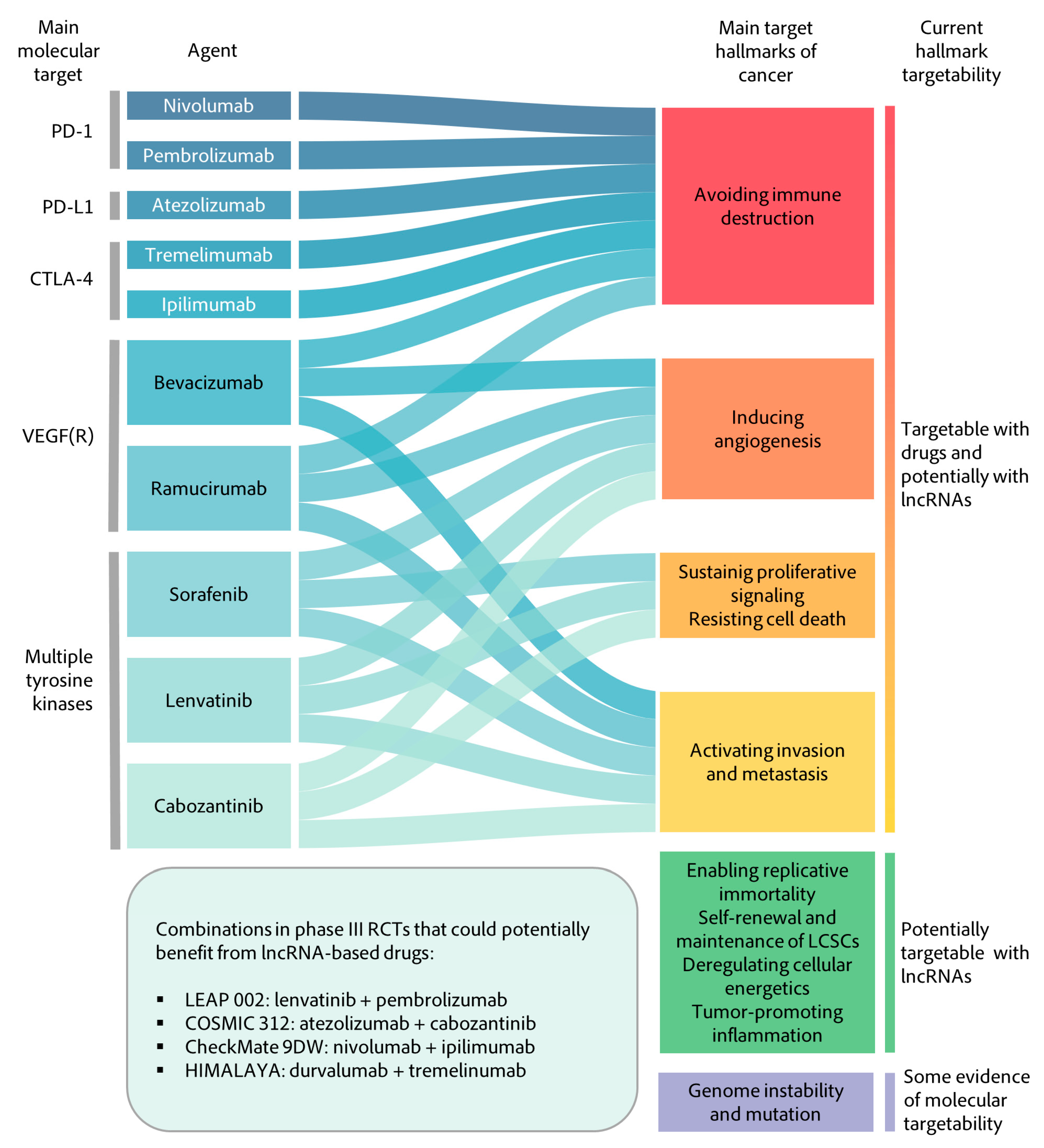
This review is the result of a strong effort that has involved extensive examination of hundreds of lncRNAs related to HCC (Supplementary Materials, Figure S1, Table S1 and Table S2) and the selection of few representative studies (Table 1) with the aim of classifying them according to their mechanism of action (Figure 2), associated cancer hallmarks (Figure 2 and Figure 3) and correlation with relevant clinical parameters (Figure 3 and Figure 4) to find potentially useful associations with therapies that are currently in the clinic or that are expected to arrive in the near future (Figure 5). The classification into cancer hallmarks should be taken with flexibility, as several biological processes involved in tumor progression play relevant roles in overlapping hallmarks and lncRNAs are not an exception. In fact, similar to protein multifunctionality, there are several lncRNAs that follow different mechanisms to impact on single or multiple hallmarks. Examples of this are HOXD-AS1 and ZFAS1 function in invasion and metastasis, HULC in cell metabolism and proliferative signaling or H19 in both the maintenance of cancer stem cells and angiogenesis. Clinical parameters chosen were OS and medical or histological features linked to worse prognosis, such as tumor stage, vascular invasion, tumor size, degree of histological differentiation, presence of metastasis or AFP levels (Figure 4). Significant associations between these clinical parameters and the expression of HCC-related lncRNAs give them a non-negligible degree of clinical relevance. Moreover, some lncRNAs have been studied in both massive public databases and in independent cohorts of HCC patients, which confers these candidates a higher level of evidence for clinical translation (Figure 3).

3.1. Molecular Mechanisms of lncRNAs Relevant for HCC

Despite the identification and publication of large numbers of lncRNAs in the literature, our understanding of their functions is still limited [142]. However, we believe that the collection discussed in this review is a good representation of a larger collection of HCC-relevant lncRNAs and helps clarify the major mechanisms enabled by lncRNAs in HCC cells (Figure S1). LncRNAs can interact with proteins, DNA or other RNAs to modulate gene expression or protein activity. By far, most lncRNAs are described to function primarily as ceRNAs or miRNAs sponges (Figure 2 and Figure S1). This over-representation of such a mechanism of action has generated controversy in the field for several reasons. On one hand, miRNAs are generally expressed to higher levels than lncRNAs. However, ceRNA function should require equimolar or higher levels of lncRNA versus the target miRNA, as the great majority of lncRNAs described to function as ceRNAs (including those described in this review) harbor but one binding site for their suggested miRNA target. The experiments performed for the identification of ceRNAs do not normally take this issue into consideration, as they generally involve ectopic overexpression of the lncRNA and miRNA components. The functional analysis of miRNA-binding site deletion mutants, as has been done in the case of LncRNA-ATB, together with a careful quantification of the molecules present in the cell, should be performed to ensure ceRNA functionality. On the other hand, each mRNA can be targeted by multiple miRNAs and each miRNA is predicted to regulate hundreds of targets. Therefore, the sequestration of a single miRNA by a lncRNA should impact gene expression at several levels and this may vary in different cells depending on their miRNAome. Similar concerns can be applied to lncRNAs described to bind and control the activity of proteins regulators. Examples include factors that regulate mRNA splicing or stability (SRSF1, HuR, hnRNPs) or protein phosphorylation, methylation, acetylation, or ubiquitination (SHP1, EZH2, p300, HDAC1, CUL4). Most studies fail to address whether lncRNA activity over these general regulators can reach certain specificity and how this is achieved. Otherwise, lncRNA expression should affect all targets of these global regulators, and lead to the deregulation of several cellular pathways.
A clearer picture is possible for lncRNAs that bind directly to a specific factor, rather than to a global regulator. LncRNA binding to a specific mRNA may impede miRNA blockade or HuR regulation or may modulate mRNA translation and stability by other molecular mechanisms. Similarly, lncRNA binding to a specific genomic locus may control chromatin remodeling or epigenetic modifications, or the activity of transcription factors impacting on the expression of a specific gene. The ability of lncRNAs to bind mRNAs involves interactions by base pairing and therefore it may be easily acquired evolutionarily just by gaining the expression of genes located antisense to the target gene. Likewise, naïve induction of the expression of lncRNAs that may function in cis co-transcriptionally is an excellent way to control local gene expression at the genomic level. All these are exclusive lncRNA features that should contribute to the relevance of lncRNAs in processes where evolutionary pressure plays a fundamental role. This is the case of carcinogenesis, cancer cell establishment and proliferation in the tumor microenvironment, tumor evasion from the immune system and tumor escape from anticancer therapies. In accordance, therapies targeting such lncRNAs may show unprecedented beneficial effects.

3.2. Therapeutic Implications

The most promising attempt at lncRNA inhibition therapy at the RNA level is grounded on antisense oligonucleotides (ASOs), which can efficiently reduce lncRNA levels [143]. Transcriptional deregulation or depletion by genome editing can also be achieved with CRIPSR-Cas technologies. Currently, the use of ASOs seems the more feasible strategy to drug oncogenic lncRNAs, as their effects are reversible in nature and their development is simplified due to their uniform chemical process. Although the pharmacodynamic and pharmacokinetic properties of ASOs are unconventional and remain to be fully described, ASO-based therapies, such as Nusinersen (SPINRAZA®) for treatment of spinal muscular atrophy, have already been safely used in patients [144]. Alternatively, restoration of tumor suppressor lncRNAs that are downregulated in HCC has several additional barriers to overcome before becoming a potentially useful strategy for HCC treatment. However, the current COVID-19 pandemic has shown, at world scale, the efficacy and safety of lipid nanoparticles and viral vectors for the delivery of RNA or DNA. Lipid nanoparticles, in particular, appear to be a natural and safe way to deliver lncRNAs or ASOs into cells with potential high organ-specific targetability [145]. Once the last frontiers involving delivery and safety issues are broadly surpassed, it is expected that lncRNA targeting will be widely used as a therapy to treat several diseases, including cancer. This could be done as single or combination therapies.
Regarding combination therapies, nowadays, the main HCC molecular targets with commercialized drugs are immune targets (PD-1, PD-L1, CTLA-4), VEGF or multi-TK (tyrosine kinases) inhibitors (Figure 5). Anti-VEGF therapies or anti-TK exert their therapeutic effect by targeting multiple hallmarks of cancer such as angiogenesis, proliferation, evasion of growth suppressors and cell death or activating invasion and metastasis. As described before, some lncRNAs, such as TUG1, MVIH, PVT1 or HOTTIP, have been related to these hallmarks with a significant impact on patient survival. Therefore, combined therapies with both anti-lncRNA and a direct protein target could boost the tumor targeting, increase response rates and impact patient management. Interestingly, other possible coding targets from the enabling replicative immortality, the self-renewal and maintenance of CSCs or the deregulating cell energetics hallmarks do not have any available drugs. These include highly relevant un-druggable factors for HCC such as the tumor suppressor p53 or the oncogenic β-catenin. Since many lncRNAs have been described that modulate the activity of these factors or with a related hallmark function and with effect over OS, many potential targets arise.
Remarkably, only lnc-EGFR from our selection and an additional study on lncRNA FENDRR [146] have been found to regulate immune escape in HCC. However, evidence from studies in other tumors have found clinically relevant lncRNAs associated with antigen release [147], antigen presentation [148,149,150,151], immune cell activation [152] and importantly, with induction of PD-L1 expression and immune escape [153,154]. Therefore, efforts to study lncRNAs involved in immune checkpoint regulation in HCC could potentially help to find new therapeutic targets and potentiate the outstanding improvement in response rates obtained with drugs affecting such signaling pathways. Similarly, other missing lncRNAs in fields of therapeutic relevance are those involved in regulating genome stability or TERT function. Interestingly, it has been recently found that lncRNA NIHCOLE (non-coding RNA involved in HCC with an oncogenic role in ligation efficiency), an lncRNA highly expressed in HCC that associates with OS and several clinical parameters, functions to increase the ligation efficiency of DNA double-strand breaks through the NHEJ pathway of DNA repair (Unfried et al., under review). This provides a new avenue for therapeutic intervention. On the same line, inhibition of TERT with ASOs has been recently shown to impact on HCC growth in model animals. However, clinical translation of this result needs to ensure local delivery of the therapy to guarantee that TERT is not inhibited in tissues other than HCC [155].
Future research in HCC-related lncRNAs is also expected to identify novel candidates that function through the expression of micropeptides. Although contradictory, it is now widely accepted that some RNAs annotated as non-coding harbor small open reading frames (ORFs). The explanation is simple. Short ORFs may occur randomly in the genome and may represent regions that are not truly translated, and therefore they are not considered as actual ORFs in genomic annotations. Nevertheless, microprotein translation is noticeable in the liver and may also extensively occur in liver cancer [156]. Interestingly, some microproteins are secreted or are expressed as transmembrane proteins, as is the case of SMIM30 [71]. Therefore, their activity could be neutralized with specific antibodies. In the case of micropeptides that function as tumor suppressors, their functionality could be mimicked with synthetic peptides that may be introduced in the cell fused to cell-penetrating sequences. In addition, the small domain that constitutes the micropeptide is amenable for the selection of small molecules or specific aptamers that impair functionality, which may have therapeutic implications for oncogenic micropeptides.

3.3. Biomarker Potential

Current efforts to analyze molecular and genetic information from tumor samples are essential for identifying new drug targets, but also new biomarkers to assess patient prognosis, disease progression or treatment selection and follow-up. Nonetheless, this requires a tissue sample which provides only static information of the tumor features at the time of extraction. Further, procedures to biopsy tumors are not always feasible in HCC patients, and conventional clinical HCC biomarkers (AFP, AFP lectin fraction or des- γ- carboxy prothrombin) lack sensitivity and specificity. This underscores the need to investigate more effective markers of disease stage and progression. Given the high HCC specificity of some lncRNAs, they appear as promising biomarker candidates. This has been extensively reviewed recently [157,158]. In fact, the studies that have analyzed lncRNA biomarkers in HCC show that they can be detected in extracellular vesicles (EVs), circulating immune cells or as cell-free lncRNAs in the plasma or serum of HCC patients. This would allow the use of liquid biopsies to enable periodic monitoring of disease status with minimally invasive specimens such as serum or plasma [159]. However, despite cumulating evidence of the biomarker potential of circulating HCC-lncRNAs, further research efforts are required. The clinical implementation of circulating lncRNA HCC biomarkers, either by quantifying multiple-lncRNA panels alone or in combination with other biomarkers such as miRNAs and/or AFP, may drastically impact the assessment of HCC disease in the near future.
In summary, lncRNAs play essential roles in different hallmarks of HCC, becoming excellent putative therapeutic targets for precision medicine. It should be clarified that several studies support that only a fraction of all lncRNAs are functionally relevant [160,161,162,163]. However, in the case of HCC, some of those lncRNAs seem to confer a fitness advantage to cancer cells, offering a unique opportunity to expand the repertoire of potential targets. This is essential for HCC that activates several undruggable pathways [121] and where novel therapies have shown potent responses but only in a fraction of patients [164]. HCC patients could benefit from the additive or synergistic effects of co-targeting with novel first-line treatments and potentially useful lncRNA-based drugs. Finally, in addition to targets for precision medicine, some lncRNAs could be excellent biomarkers for diagnosis and patient stratification. Screening tissue or liquid biopsies for the presence and abundance of HCC-lncRNAs or prognostic lncRNA signatures, together with the therapeutic targeting of lncRNAs, could soon become standard clinical practices, providing a much-needed tool to meet the challenges of HCC treatment.

Supplementary Materials

The following are available online at https://www.mdpi.com/article/10.3390/cancers13112651/s1. Figure S1: Comparison between the lncRNA collection used for the review and a larger collection of HCC-related lncRNAs. (A) LncRNAs divided according to their mechanism of action. (B) LncRNAs stratified according to the hallmarks of cancer they modulate. Note that a single lncRNA may be ascribed to different mechanisms and/or to several cancer hallmarks. The larger collection comprises all lncRNAs published in journals from the first quartile (Q1) in any possible category up to March 2021. The number of lncRNAs and the percentage is indicated for each case, Table S1: Extended annotation of the main collection of lncRNAs described in this review. This list includes the data summarized in Table 1 plus additional information recovered from their respective publications. “Biochemistry” shows the molecular mechanism of the lncRNA. “Preclinic” describes the cell lines and the animal model used. “Clinic” shows the number of patients studied in an independent cohort and the significance of the association between the expression of the lncRNA and overall survival and other clinical parameters. All candidates are in alphabetical order and the last two columns show the journal of publication and associated PMID, Table S2: List of lncRNAs included in the larger collection. This list includes the lncRNAs that were used to generate Figure S1, and their annotated mechanisms of action and hallmarks of cancer.

Author Contributions

J.P.U., P.S., B.S and P.F. wrote, reviewed and edited the article. J.P.U. made the figures. L.P.-M. made the tables. All authors have read and agreed to the published version of the manuscript.

Funding

This research was supported by RTI2018-101759-B-I00_FEDER, EU/Ministerio de Ciencia e Innovación—Agencia Estatal de Investigación to P.F., Scientific Foundation of the Spanish Association Against Cancer (AECC IDEAS20169FORT to P.F.) and by the Instituto de Salud Carlos III, which finances the Fondo de Investigación Sanitaria PI19/00742 to B.S. together with FEDER, EU and the Centro de Investigación Biomédica en Red de Enfermedades Hepáticas y Digestivas (CIBEREhd). L.P-M. is the recipient of a PFIS fellowship (FI20/00074). Investing in your future (FSE).

Data Availability Statement

All data needed to evaluate the conclusions in the paper are present in the paper and/or the Supplementary Materials.

Acknowledgments

The authors would like to thank Ken Flerlage for his assistance in the design of Figure 2 and Figure 5. Figure 1 was created using BioRender.com (Accessed on 20 May 2021). Figure 2 and Figure 5 were created using Tableau, version 2019.2 (Tableau Software. Seattle, WA, USA). Figure 4 was created using GraphPad Prism, version 9.0.0 (GraphPad Software. San Diego, CA, USA). Figure 3 was created using Microsoft Excel, version 2104 (Microsoft. Albuquerque, NM, USA).

Conflicts of Interest

The authors declare no conflict of interest.

Abbreviations

CSCCancer stem cells
CTNNBIP1 Catenin beta interacting protein 1
CUL4Cullin 4A
DNADesoxiribonucleic acid
EZH2Enhancer of zeste 2 polycomb repressive complex 2
FBXW7 F-box and WD repeat domain-containing 7
GTPasesGuanosine triphosphate (GTP) hydrolase
HDAC1Histone deacetylase 1
JAK2Janus kinase 2
m6AN6-methyladenosine
MBNL3Muscleblind-like splicing regulator 3
mRNAMessenger RNA
NF-kBNuclear factor kappa B
NOP2 NOP2 nucleolar protein
p300Histone acetyltransferase p 300
RNARibonucleic acid
SHP1Tyrosine phosphatase SHP1
SLUGSnail family transcription repressor 2
STAT3Signal transducer and activator of transcription 3
TERTTelomerase reverse transcriptase
TWISTTwist family BHLH transcription factor 1
UTRUntranslated region

References

  1. Bray, F.; Ferlay, J.; Soerjomataram, I.; Siegel, R.L.; Torre, L.A.; Jemal, A. Global cancer statistics 2018: Globocan estimates of incidence and mortality worldwide for 36 cancers in 185 countries. CA Cancer J. Clin. 2018, 68, 394–424. [Google Scholar] [CrossRef] [Green Version]
  2. Ferlay, J.; Soerjomataram, I.; Dikshit, R.; Eser, S.; Mathers, C.; Rebelo, M.; Parkin, D.M.; Forman, D.; Bray, F. Cancer incidence and mortality worldwide: Sources, methods and major patterns in GLOBOCAN 2012. Int. J. Cancer 2014, 136, E359–E386. [Google Scholar] [CrossRef] [PubMed]
  3. Llovet, J.M.; Zucman-Rossi, J.; Pikarsky, E.; Sangro, B.; Schwartz, M.; Sherman, M.; Gores, G. Hepatocellular carcinoma. Nat. Rev. Dis. Prim. 2016, 2, 16018. [Google Scholar] [CrossRef] [PubMed]
  4. Kulik, L.; El-Serag, H.B. Epidemiology and Management of Hepatocellular Carcinoma. Gastroenterology 2019, 156, 477–491.e1. [Google Scholar] [CrossRef] [PubMed]
  5. Singal, A.G.; Lampertico, P.; Nahon, P. Epidemiology and surveillance for hepatocellular carcinoma: New trends. J. Hepatol. 2020, 72, 250–261. [Google Scholar] [CrossRef] [PubMed] [Green Version]
  6. Akinyemiju, T.; Abera, S.; Ahmed, M.; Alam, N.; Alemayohu, M.A.; Allen, C.; Al-Raddadi, R.; Alvis-Guzman, N.; Amoako, Y.; Artaman, A.; et al. The Burden of Primary Liver Cancer and Underlying Etiologies From 1990 to 2015 at the Global, Regional, and National Level: Results from the Global Burden of Disease Study 2015. JAMA Oncol. 2017, 3, 1683–1691. [Google Scholar] [CrossRef] [PubMed]
  7. Siegel, R.L.; Miller, K.D.; Jemal, A. Cancer statistics, 2019. CA Cancer J. Clin. 2019, 69, 7–34. [Google Scholar] [CrossRef] [PubMed] [Green Version]
  8. Rahib, L.; Smith, B.D.; Aizenberg, R.; Rosenzweig, A.B.; Fleshman, J.M.; Matrisian, L.M. Projecting cancer incidence and deaths to 2030: The unexpected burden of thyroid, liver, and pancreas cancers in the United States. Cancer Res. 2014, 74, 2913–2921. [Google Scholar] [CrossRef] [Green Version]
  9. Sia, D.; Villanueva, A.; Friedman, S.L.; Llovet, J.M. Liver Cancer Cell of Origin, Molecular Class, and Effects on Patient Prognosis. Gastroenterology 2017, 152, 745–761. [Google Scholar] [CrossRef] [Green Version]
  10. Marquardt, J.U.; Andersen, J.B.; Thorgeirsson, S.S. Functional and genetic deconstruction of the cellular origin in liver cancer. Nat. Rev. Cancer 2015, 15, 653–667. [Google Scholar] [CrossRef]
  11. Galle, P.R.; Forner, A.; Llovet, J.M.; Mazzaferro, V.; Piscaglia, F.; Raoul, J.L.; Schirmacher, P.; Vilgrain, V. European Association for the Study of the Liver. Electronic address eee, European Association for the Study of the L. EASL Clinical Practice Guidelines: Management of hepatocellular carcinoma. J. Hepatol. 2018, 69, 182–236. [Google Scholar] [CrossRef] [Green Version]
  12. Majumdar, A.; Roccarina, D.; Thorburn, D.; Davidson, B.R.; Tsochatzis, E.; Gurusamy, K.S. Management of people with early- or very early-stage hepatocellular carcinoma. Cochrane Database Syst. Rev. 2017. [Google Scholar] [CrossRef]
  13. Estfan, B.; Byrne, M.; Kim, R. Sorafenib in Advanced Hepatocellular Carcinoma. Am. J. Clin. Oncol. 2013, 36, 319–324. [Google Scholar] [CrossRef]
  14. El-Khoueiry, A.B.; Sangro, B.; Yau, T.; Crocenzi, T.S.; Kudo, M.; Hsu, C.; Kim, T.-Y.; Choo, S.-P.; Trojan, J.; Welling, T.H.; et al. Nivolumab in patients with advanced hepatocellular carcinoma (CheckMate 040): An open-label, non-comparative, phase 1/2 dose escalation and expansion trial. Lancet 2017, 389, 2492–2502. [Google Scholar] [CrossRef]
  15. Zhu, A.X.; Finn, R.S.; Edeline, J.; Cattan, S.; Ogasawara, S.; Palmer, D.; Verslype, C.; Zagonel, V.; Fartoux, L.; Vogel, A.; et al. Pembrolizumab in patients with advanced hepatocellular carcinoma previously treated with sorafenib (KEYNOTE-224): A non-randomised, open-label phase 2 trial. Lancet Oncol. 2018, 19, 940–952. [Google Scholar] [CrossRef]
  16. Finn, R.S.; Qin, S.; Ikeda, M.; Galle, P.R.; Ducreux, M.; Kim, T.-Y.; Kudo, M.; Breder, V.; Merle, P.; Kaseb, A.O. Atezolizumab plus Bevacizumab in Unresectable Hepatocellular Carcinoma. N. Engl. J. Med. 2020, 382, 1894–1905. [Google Scholar] [CrossRef] [PubMed]
  17. Bruix, J. Endpoints in clinical trials for liver cancer and their value in evidence-based clinical decision making: An unresolved Gordian knot. J. Hepatol. 2021, 74, 1483–1488. [Google Scholar] [CrossRef]
  18. Fatica, A.; Bozzoni, I. Long non-coding RNAs: New players in cell differentiation and development. Nat. Rev. Genet. 2014, 15, 7–21. [Google Scholar] [CrossRef] [PubMed]
  19. Taft, R.J.; Pheasant, M.; Mattick, J.S. The relationship between non-protein-coding DNA and eukaryotic complexity. BioEssays 2007, 29, 288–299. [Google Scholar] [CrossRef] [PubMed]
  20. Ma, L.; Bajic, V.B.; Zhang, Z. On the classification of long non-coding RNAs. RNA Biol. 2013, 10, 924–933. [Google Scholar] [CrossRef] [PubMed]
  21. Suarez, B.; Prats-Mari, L.; Unfried, J.P.; Fortes, P. LncRNAs in the Type I Interferon Antiviral Response. Int. J. Mol. Sci. 2020, 21, 6447. [Google Scholar] [CrossRef]
  22. Unfried, J.P.; Fortes, P. SMIM30, a tiny protein with a big role in liver cancer. J. Hepatol. 2020, 73, 1010–1012. [Google Scholar] [CrossRef] [PubMed]
  23. Ransohoff, J.D.; Wei, Y.; Khavari, P.A. The functions and unique features of long intergenic non-coding RNA. Nat. Rev. Mol. Cell Biol. 2018, 19, 143–157. [Google Scholar] [CrossRef] [PubMed]
  24. Derrien, T.; Johnson, R.; Bussotti, G.; Tanzer, A.; Djebali, S.; Tilgner, H.; Guernec, G.; Martin, D.; Merkel, A.; Knowles, D.G.; et al. The GENCODE v7 catalog of human long noncoding RNAs: Analysis of their gene structure, evolution, and expression. Genome Res. 2012, 22, 1775–1789. [Google Scholar] [CrossRef] [Green Version]
  25. Unfried, J.P.; Serrano, G.; Suárez, B.; Sangro, P.; Ferretti, V.; Prior, C.; Boix, L.; Bruix, J.; Sangro, B.; Segura, V.; et al. Identification of Coding and Long Noncoding RNAs Differentially Expressed in Tumors and Preferentially Expressed in Healthy Tissues. Cancer Res. 2019, 79, 5167–5180. [Google Scholar] [CrossRef] [PubMed] [Green Version]
  26. Kamel, M.M.; Matboli, M.; Sallam, M.; Montasser, I.F.; Saad, A.S.; El-Tawdi, A.H. Investigation of long noncoding RNAs expression profile as potential serum biomarkers in patients with hepatocellular carcinoma. Transl. Res. 2016, 168, 134–145. [Google Scholar] [CrossRef]
  27. Liao, X.; Yang, C.; Huang, R.; Han, C.; Yu, T.; Huang, K.; Liu, X.; Yu, L.; Zhu, G.; Su, H.; et al. Identification of Potential Prognostic Long Non-Coding RNA Biomarkers for Predicting Survival in Patients with Hepatocellular Carcinoma. Cell. Physiol. Biochem. 2018, 48, 1854–1869. [Google Scholar] [CrossRef] [PubMed]
  28. Gu, J.-X.; Zhang, X.; Miao, R.-C.; Xiang, X.-H.; Fu, Y.-N.; Zhang, J.-Y.; Liu, C.; Qu, K. Six-long non-coding RNA signature predicts recurrence-free survival in hepatocellular carcinoma. World J. Gastroenterol. 2019, 25, 220–232. [Google Scholar] [CrossRef]
  29. Tsang, F.H.; Au, S.L.; Wei, L.; Fan, D.N.; Lee, J.M.; Wong, C.C.; Ng, I.O.; Wong, C.-M. Long non-coding RNA HOTTIP is frequently up-regulated in hepatocellular carcinoma and is targeted by tumour suppressive miR-125b. Liver Int. 2014, 35, 1597–1606. [Google Scholar] [CrossRef]
  30. Qiu, L.; Wang, T.; Xu, X.; Wu, Y.; Tang, Q.; Chen, K. Long Non-Coding RNAs in Hepatitis B Virus-Related Hepatocellular Carcinoma: Regulation, Functions, and Underlying Mechanisms. Int. J. Mol. Sci. 2017, 18, 2505. [Google Scholar] [CrossRef] [Green Version]
  31. Unfried, J.P.; Fortes, P. LncRNAs in HCV Infection and HCV-Related Liver Disease. Int. J. Mol. Sci. 2020, 21, 2255. [Google Scholar] [CrossRef] [Green Version]
  32. Carnero, E.; Barriocanal, M.; Prior, C.; Unfried, J.P.; Segura, V.; Guruceaga, E.; Enguita, M.; Smerdou, C.; Gastaminza, P.; Fortes, P. Long noncoding RNA EGOT negatively affects the antiviral response and favors HCV replication. EMBO Rep. 2016, 17, 1013–1028. [Google Scholar] [CrossRef]
  33. Barriocanal, M.; Prior, C.; Suarez, B.; Unfried, J.P.; Razquin, N.; Hervás-Stubbs, S.; Sangro, B.; Segura, V.; Fortes, P. Long Noncoding RNA EGOT Responds to Stress Signals to Regulate Cell Inflammation and Growth. J. Immunol. 2021, 206. [Google Scholar] [CrossRef] [PubMed]
  34. Hanahan, D.; Weinberg, R.A. The Hallmarks of Cancer. Cell 2000, 100, 57–70. [Google Scholar] [CrossRef] [Green Version]
  35. Hanahan, D.; Weinberg, R.A. Hallmarks of Cancer: The Next Generation. Cell 2011, 144, 646–674. [Google Scholar] [CrossRef] [PubMed] [Green Version]
  36. Lytle, N.K.; Barber, A.G.; Reya, T. Stem cell fate in cancer growth, progression and therapy resistance. Nat. Rev. Cancer 2018, 18, 669–680. [Google Scholar] [CrossRef]
  37. Chen, L.; Chan, T.H.M.; Yuan, Y.; Hu, L.; Huang, J.; Ma, S.; Wang, J.; Dong, S.; Tang, K.H.; Xie, D.; et al. CHD1L promotes hepatocellular carcinoma progression and metastasis in mice and is associated with these processes in human patients. J. Clin. Investig. 2010, 120, 1178–1191. [Google Scholar] [CrossRef] [PubMed] [Green Version]
  38. Xia, L.; Huang, W.; Tian, D.; Zhu, H.; Qi, X.; Chen, Z.; Zhang, Y.; Hu, H.; Fan, D.; Nie, Y.; et al. Overexpression of forkhead box C1 promotes tumor metastasis and indicates poor prognosis in hepatocellular carcinoma. Hepatology 2012, 57, 610–624. [Google Scholar] [CrossRef]
  39. Huang, M.-D.; Chen, W.-M.; Qi, F.-Z.; Xia, R.; Sun, M.; Xu, T.-P.; Yin, L.; Zhang, E.-B.; De, W.; Shu, Y.-Q. Long non-coding RNA ANRIL is upregulated in hepatocellular carcinoma and regulates cell apoptosis by epigenetic silencing of KLF2. J. Hematol. Oncol. 2015, 8, 1–14. [Google Scholar] [CrossRef] [PubMed] [Green Version]
  40. Wang, Y.; Liu, Z.; Yao, B.; Liang, W.; Wang, L.; Wang, C.; Dou, C.; Xu, M.; Liu, Q.; Tu, K. Long non-coding RNA CASC2 suppresses epithelial-mesenchymal transition of hepatocellular carcinoma cells through CASC2/miR-367/FBXW7 axis. Mol. Cancer 2017, 16, 1–15. [Google Scholar] [CrossRef] [Green Version]
  41. Klingenberg, M.; Groß, M.; Goyal, A.; Polycarpou Schwarz, M.; Miersch, T.; Ernst, A.; Leupold, J.; Patil, N.; Warnken, U.; Allgayer, H.; et al. The Long Noncoding RNA Cancer Susceptibility 9 and RNA Binding Protein Heterogeneous Nuclear Ribonucleoprotein L Form a Complex and Coregulate Genes Linked to AKT Signaling. Hepatology 2018, 68, 1817–1832. [Google Scholar] [CrossRef]
  42. Yuan, S.-X.; Wang, J.; Yang, F.; Tao, Q.-F.; Zhang, J.; Wang, L.-L.; Yang, Y.; Liu, H.; Wang, Z.-G.; Xu, Q.-G.; et al. Long noncoding RNADANCRincreases stemness features of hepatocellular carcinoma by derepression ofCTNNB1. Hepatology 2016, 63, 499–511. [Google Scholar] [CrossRef] [PubMed]
  43. Wang, X.; Sun, W.; Shen, W.; Xia, M.; Chen, C.; Xiang, D.; Ning, B.; Cui, X.; Li, H.; Li, X.; et al. Long non-coding RNA DILC regulates liver cancer stem cells via IL-6/STAT3 axis. J. Hepatol. 2016, 64, 1283–1294. [Google Scholar] [CrossRef] [PubMed]
  44. Salerno, D.; Chiodo, L.; Alfano, V.; Floriot, O.; Cottone, G.; Paturel, A.; Pallocca, M.; Plissonnier, M.-L.; Jeddari, S.; Belloni, L.; et al. Hepatitis B protein HBx binds the DLEU2 lncRNA to sustain cccDNA and host cancer-related gene transcription. Gut 2020, 69, 2016–2024. [Google Scholar] [CrossRef] [PubMed] [Green Version]
  45. Huang, J.-F.; Guo, Y.-J.; Zhao, C.-X.; Yuan, S.-X.; Wang, Y.; Tang, G.-N.; Zhou, W.-P.; Sun, S.-H. Hepatitis B virus X protein (HBx)-related long noncoding RNA (lncRNA) down-regulated expression by HBx (Dreh) inhibits hepatocellular carcinoma metastasis by targeting the intermediate filament protein vimentin. Hepatology 2013, 57, 1882–1892. [Google Scholar] [CrossRef]
  46. Lan, T.; Li, H.; Zhang, D.; Xu, L.; Liu, H.; Hao, X.-Y.; Yan, X.; Liao, H.; Chen, X.; Xie, K.; et al. KIAA1429 contributes to liver cancer progression through N6-methyladenosine-dependent post-transcriptional modification of GATA3. Mol. Cancer 2019, 18, 1–19. [Google Scholar] [CrossRef]
  47. Zhang, J.; Han, C.; Ungerleider, N.; Chen, W.; Song, K.; Wang, Y.; Kwon, H.; Ma, W.; Wu, T. A Transforming Growth Facto β and H19 Signaling Axis in Tumor Initiating Hepatocytes That Regulates Hepatic Carcinogenesis. Hepatology 2019, 69, 1549–1563. [Google Scholar] [CrossRef]
  48. Conigliaro, A.; Costa, V.; Dico, A.L.; Saieva, L.; Buccheri, S.; Dieli, F.; Manno, M.; Raccosta, S.; Mancone, C.; Tripodi, M.; et al. CD90+ liver cancer cells modulate endothelial cell phenotype through the release of exosomes containing H19 lncRNA. Mol. Cancer 2015, 14, 155. [Google Scholar] [CrossRef]
  49. Wang, Y.; Zhu, P.; Luo, J.; Wang, J.; Liu, Z.; Wu, W.; Du, Y.; Ye, B.; Wang, D.; He, L.; et al. LncRNA HAND2 AS1 promotes liver cancer stem cell self renewal via BMP signaling. EMBO J. 2019, 38, e101110. [Google Scholar] [CrossRef] [PubMed]
  50. Yang, Y.; Chen, L.; Gu, J.; Zhang, H.; Yuan, J.; Lian, Q.; Lv, G.; Wang, S.; Wu, Y.; Yang, Y.-C.T.; et al. Recurrently deregulated lncRNAs in hepatocellular carcinoma. Nat. Commun. 2017, 8, 14421. [Google Scholar] [CrossRef] [PubMed] [Green Version]
  51. Peng, L.; Jiang, B.; Yuan, X.; Qiu, Y.; Peng, J.; Huang, Y.; Zhang, C.; Zhang, Y.; Lin, Z.; Li, J.; et al. Super-Enhancer–Associated Long Noncoding RNA HCCL5 Is Activated by ZEB1 and Promotes the Malignancy of Hepatocellular Carcinoma. Cancer Res. 2019, 79, 572–584. [Google Scholar] [CrossRef] [PubMed] [Green Version]
  52. Yang, F.; Zhang, L.; Huo, X.-S.; Yuan, J.-H.; Xu, D.; Yuan, S.-X.; Zhu, N.; Zhou, W.-P.; Yang, G.-S.; Wang, Y.-Z.; et al. Long noncoding RNA high expression in hepatocellular carcinoma facilitates tumor growth through enhancer of zeste homolog 2 in humans. Hepatology 2011, 54, 1679–1689. [Google Scholar] [CrossRef]
  53. Ding, C.-H.; Yin, C.; Chen, S.-J.; Wen, L.-Z.; Ding, K.; Lei, S.-J.; Liu, J.-P.; Wang, J.; Chen, K.-X.; Jiang, H.-L.; et al. The HNF1α-regulated lncRNA HNF1A-AS1 reverses the malignancy of hepatocellular carcinoma by enhancing the phosphatase activity of SHP-1. Mol. Cancer 2018, 17, 1–14. [Google Scholar] [CrossRef]
  54. Yang, L.; Peng, X.; Li, Y.; Zhang, X.; Ma, Y.; Wu, C.; Fan, Q.; Wei, S.; Li, H.; Liu, J. Long non-coding RNA HOTAIR promotes exosome secretion by regulating RAB35 and SNAP23 in hepatocellular carcinoma. Mol. Cancer 2019, 18, 1–12. [Google Scholar] [CrossRef] [PubMed] [Green Version]
  55. Fu, W.-M.; Zhu, X.; Wang, W.-M.; Lu, Y.-F.; Hu, B.-G.; Wang, H.; Liang, W.-C.; Wang, S.-S.; Ko, C.-H.; Waye, M.M.-Y.; et al. Hotair mediates hepatocarcinogenesis through suppressing miRNA-218 expression and activating P14 and P16 signaling. J. Hepatol. 2015, 63, 886–895. [Google Scholar] [CrossRef]
  56. Zhang, H.; Xing, Z.; Mani, S.K.K.; Bancel, B.; Durantel, D.; Zoulim, F.; Tran, E.; Merle, P.; Andrisani, O. RNA helicase DEAD box protein 5 regulates Polycomb repressive complex 2/Hox transcript antisense intergenic RNA function in hepatitis B virus infection and hepatocarcinogenesis. Hepatology 2016, 64, 1033–1048. [Google Scholar] [CrossRef] [PubMed]
  57. Quagliata, L.; Matter, M.; Piscuoglio, S.; Arabi, L.; Ruiz, C.; Procino, A.; Kovac, M.; Moretti, F.; Makowska, Z.; Boldanova, T.; et al. Long noncoding RNA HOTTIP/HOXA13 expression is associated with disease progression and predicts outcome in hepatocellular carcinoma patients. Hepatology 2014, 59, 911–923. [Google Scholar] [CrossRef] [Green Version]
  58. Lu, S.; Zhou, J.; Sun, Y.; Li, N.; Miao, M.; Jiao, B.; Chen, H. The noncoding RNA HOXD-AS1 is a critical regulator of the metastasis and apoptosis phenotype in human hepatocellular carcinoma. Mol. Cancer 2017, 16, 1–14. [Google Scholar] [CrossRef] [PubMed]
  59. Wang, H.; Huo, X.; Yang, X.-R.; He, J.; Cheng, L.; Wang, N.; Deng, X.; Jingyuan, F.; Wang, N.; Wang, C.; et al. STAT3-mediated upregulation of lncRNA HOXD-AS1 as a ceRNA facilitates liver cancer metastasis by regulating SOX4. Mol. Cancer 2017, 16, 1–15. [Google Scholar] [CrossRef] [Green Version]
  60. Hämmerle, M.; Gutschner, T.; Uckelmann, H.; Ozgur, S.; Fiskin, E.; Gross, M.; Skawran, B.; Geffers, R.; Longerich, T.; Breuhahn, K.; et al. Posttranscriptional destabilization of the liver-specific long noncoding RNA HULC by the IGF2 mRNA-binding protein 1 (IGF2BP1). Hepatology 2013, 58, 1703–1712. [Google Scholar] [CrossRef]
  61. Cui, M.; Xiao, Z.; Wang, Y.; Zheng, M.; Song, T.; Cai, X.; Sun, B.; Ye, L.; Zhang, X. Long Noncoding RNA HULC Modulates Abnormal Lipid Metabolism in Hepatoma Cells through an miR-9–Mediated RXRA Signaling Pathway. Cancer Res. 2015, 75, 846–857. [Google Scholar] [CrossRef] [PubMed] [Green Version]
  62. Xin, X.; Wu, M.; Meng, Q.; Wang, C.; Lu, Y.; Yang, Y.; Li, X.; Zheng, Q.; Pu, H.; Gui, X.; et al. Long noncoding RNA HULC accelerates liver cancer by inhibiting PTEN via autophagy cooperation to miR15a. Mol. Cancer 2018, 17, 1–16. [Google Scholar] [CrossRef] [Green Version]
  63. Li, D.; Liu, X.; Zhou, J.; Hu, J.; Zhang, D.; Liu, J.; Qiao, Y.; Zhan, Q. Long noncoding RNA HULC modulates the phosphorylation of YB-1 through serving as a scaffold of extracellular signal-regulated kinase and YB-1 to enhance hepatocarcinogenesis. Hepatology 2017, 65, 1612–1627. [Google Scholar] [CrossRef] [PubMed] [Green Version]
  64. Wang, J.; Liu, X.; Wu, H.; Ni, P.; Gu, Z.; Qiao, Y.; Chen, N.; Sun, F.; Fan, Q. CREB up-regulates long non-coding RNA, HULC expression through interaction with microRNA-372 in liver cancer. Nucleic Acids Res. 2010, 38, 5366–5383. [Google Scholar] [CrossRef] [PubMed] [Green Version]
  65. Guo, W.; Liu, S.; Cheng, Y.; Lu, L.; Shi, J.; Xu, G.; Li, N.; Cheng, K.; Wu, M.; Cheng, S.; et al. ICAM-1–Related Noncoding RNA in Cancer Stem Cells Maintains ICAM-1 Expression in Hepatocellular Carcinoma. Clin. Cancer Res. 2016, 22, 2041–2050. [Google Scholar] [CrossRef] [Green Version]
  66. Xu, D.; Yang, F.; Yuan, J.-H.; Zhang, L.; Bi, H.-S.; Zhou, C.-C.; Liu, F.; Wang, F.; Sun, S.-H. Long noncoding RNAs associated with liver regeneration 1 accelerates hepatocyte proliferation during liver regeneration by activating Wnt/β-Catenin signaling. Hepatology 2013, 58, 739–751. [Google Scholar] [CrossRef]
  67. Fu, X.; Zhu, X.; Qin, F.; Zhang, Y.; Lin, J.; Ding, Y.; Yang, Z.; Shang, Y.; Wang, L.; Zhang, Q.; et al. Linc00210 drives Wnt/β-catenin signaling activation and liver tumor progression through CTNNBIP1-dependent manner. Mol. Cancer 2018, 17, 1–14. [Google Scholar] [CrossRef] [PubMed] [Green Version]
  68. Guo, T.; Gong, C.; Wu, P.; Battaglia-Hsu, S.-F.; Feng, J.; Liu, P.; Wang, H.; Guo, D.; Yao, Y.; Chen, B.; et al. LINC00662 promotes hepatocellular carcinoma progression via altering genomic methylation profiles. Cell Death Differ. 2020, 27, 2191–2205. [Google Scholar] [CrossRef]
  69. Ding, J.; Zhao, J.; Huan, L.; Liu, Y.; Qiao, Y.; Wang, Z.; Chen, Z.; Huang, S.; Zhao, Y.; He, X. Inflammation Induced Long Intergenic Noncoding RNA (LINC00665) Increases Malignancy Through Activating the Double Stranded RNA–Activated Protein Kinase/Nuclear Factor Kappa B Pathway in Hepatocellular Carcinoma. Hepatology 2020, 72, 1666–1681. [Google Scholar] [CrossRef]
  70. Zuo, X.; Chen, Z.; Gao, W.; Zhang, Y.; Wang, J.; Wang, J.; Cao, M.; Cai, J.; Wu, J.; Wang, X. M6A-mediated upregulation of LINC00958 increases lipogenesis and acts as a nanotherapeutic target in hepatocellular carcinoma. J. Hematol. Oncol. 2020, 13, 1–20. [Google Scholar] [CrossRef] [PubMed] [Green Version]
  71. Pang, Y.; Liu, Z.; Han, H.; Wang, B.; Li, W.; Mao, C.; Liu, S. Peptide SMIM30 promotes HCC development by inducing SRC/YES1 membrane anchoring and MAPK pathway activation. J. Hepatol. 2020, 73, 1155–1169. [Google Scholar] [CrossRef] [PubMed]
  72. Gandhi, M.; Groß, M.; Holler, J.M.; Coggins, S.A.; Patil, N.; Leupold, J.H.; Munschauer, M.; Schenone, M.; Hartigan, C.R.; Allgayer, H.; et al. The lncRNA lincNMR regulates nucleotide metabolism via a YBX1-RRM2 axis in cancer. Nat. Commun. 2020, 11, 1–15. [Google Scholar] [CrossRef] [PubMed]
  73. Zhang, L.; Yang, J.; Luo, Y.; Liu, F.; Yuan, Y.; Zhuang, S. A p53/lnc Ip53 Negative Feedback Loop Regulates Tumor Growth and Chemoresistance. Adv. Sci. 2020, 7, 2001364. [Google Scholar] [CrossRef] [PubMed]
  74. Zhu, P.; Wang, Y.; Huang, G.; Ye, B.; Liu, B.; Wu, J.; Du, Y.; He, L. lnc-β-Catm elicits EZH2-dependent β-catenin stabilization and sustains liver CSC self-renewal. Nat. Struct. Mol. Biol. 2016, 23, 631–639. [Google Scholar] [CrossRef]
  75. Zhu, P.; Wang, Y.; Wu, J.; Huang, G.; Liu, B.; Ye, B.; Du, Y.; Gao, G.; Tian, Y.; He, L.; et al. LncBRM initiates YAP1 signalling activation to drive self-renewal of liver cancer stem cells. Nat. Commun. 2016, 7, 13608. [Google Scholar] [CrossRef] [Green Version]
  76. Jiang, R.; Tang, J.; Chen, Y.; Deng, L.; Ji, J.; Xie, Y.; Wang, K.; Jia, W.; Chu, W.-M.; Sun, B. The long noncoding RNA lnc-EGFR stimulates T-regulatory cells differentiation thus promoting hepatocellular carcinoma immune evasion. Nat. Commun. 2017, 8, 15129. [Google Scholar] [CrossRef] [Green Version]
  77. Wu, J.; Zhu, P.; Lu, T.; Du, Y.; Wang, Y.; He, L.; Ye, B.; Liu, B.; Yang, L.; Wang, J.; et al. The long non-coding RNA LncHDAC2 drives the self-renewal of liver cancer stem cells via activation of Hedgehog signaling. J. Hepatol. 2019, 70, 918–929. [Google Scholar] [CrossRef]
  78. Wu, Y.; Zhao, Y.-M.; Huan, L.; Zhao, J.; Zhou, Y.; Xu, L.; Hu, Z.; Liu, Y.; Chen, Z.; Wang, L.; et al. An LTR Retrotransposon-Derived Long Noncoding RNA lncMER52A Promotes Hepatocellular Carcinoma Progression by Binding p120-Catenin. Cancer Res. 2019, 80, 976–987. [Google Scholar] [CrossRef]
  79. Yuan, J.-H.; Yang, F.; Wang, F.; Ma, J.-Z.; Guo, Y.-J.; Tao, Q.-F.; Liu, F.; Pan, W.; Wang, T.-T.; Zhou, C.-C.; et al. A Long Noncoding RNA Activated by TGF-β Promotes the Invasion-Metastasis Cascade in Hepatocellular Carcinoma. Cancer Cell 2014, 25, 666–681. [Google Scholar] [CrossRef] [Green Version]
  80. Yuan, J.-H.; Liu, X.-N.; Wang, T.-T.; Pan, W.; Tao, Q.-F.; Zhou, W.-P.; Wang, F.; Sun, S.-H. The MBNL3 splicing factor promotes hepatocellular carcinoma by increasing PXN expression through the alternative splicing of lncRNA-PXN-AS1. Nat. Cell Biol. 2017, 19, 820–832. [Google Scholar] [CrossRef]
  81. Chen, Z.-Z.; Huang, L.; Wu, Y.-H.; Zhai, W.-J.; Zhu, P.-P.; Gao, Y. LncSox4 promotes the self-renewal of liver tumour-initiating cells through Stat3-mediated Sox4 expression. Nat. Commun. 2016, 7, 12598. [Google Scholar] [CrossRef]
  82. Wang, Y.; He, L.; Du, Y.; Zhu, P.; Huang, G.; Luo, J.; Yan, X.; Ye, B.; Li, C.; Xia, P.; et al. The Long Noncoding RNA lncTCF7 Promotes Self-Renewal of Human Liver Cancer Stem Cells through Activation of Wnt Signaling. Cell Stem Cell 2015, 16, 413–425. [Google Scholar] [CrossRef] [PubMed] [Green Version]
  83. Wang, Y.; Liu, J.; Yang, J.; Yu, X.; Chen, Z.; Chen, Y.; Kuang, M.; Zhu, Y.; Zhuang, S. Lnc UCID Promotes G1/S Transition and Hepatoma Growth by Preventing DHX9 Mediated CDK6 Down regulation. Hepatology 2019, 70, 259–275. [Google Scholar] [CrossRef] [PubMed] [Green Version]
  84. Malakar, P.; Shilo, A.; Mogilevsky, A.; Stein, I.; Pikarsky, E.; Nevo, Y.; Benyamini, H.; Elgavish, S.; Zong, X.; Prasanth, K.V.; et al. Long Noncoding RNA MALAT1 Promotes Hepatocellular Carcinoma Development by SRSF1 Upregulation and mTOR Activation. Cancer Res. 2017, 77, 1155–1167. [Google Scholar] [CrossRef] [PubMed] [Green Version]
  85. Malakar, P.; Stein, I.; Saragovi, A.; Winkler, R.; Stern-Ginossar, N.; Berger, M.; Pikarsky, E.; Karni, R. Long Noncoding RNA MALAT1 Regulates Cancer Glucose Metabolism by Enhancing mTOR-Mediated Translation of TCF7L2. Cancer Res. 2019, 79, 2480–2493. [Google Scholar] [CrossRef] [Green Version]
  86. Wang, Y.; Yang, L.; Chen, T.; Liu, X.; Guo, Y.; Zhu, Q.; Tong, X.; Yang, W.; Xu, Q.; Huang, D.; et al. A novel lncRNA MCM3AP-AS1 promotes the growth of hepatocellular carcinoma by targeting miR-194-5p/FOXA1 axis. Mol. Cancer 2019, 18, 1–16. [Google Scholar] [CrossRef] [PubMed]
  87. Wang, H.; Liang, L.; Dong, Q.; Huan, L.; He, J.; Li, B.; Yang, C.; Jin, H.; Wei, L.; Yu, C.; et al. Long noncoding RNA miR503HG, a prognostic indicator, inhibits tumor metastasis by regulating the HNRNPA2B1/NF-κB pathway in hepatocellular carcinoma. Theranostics 2018, 8, 2814–2829. [Google Scholar] [CrossRef] [PubMed]
  88. Ma, M.; Xu, H.; Liu, G.; Wu, J.; Li, C.; Wang, X.; Zhang, S.; Xu, H.; Ju, S.; Cheng, W.; et al. Metabolism induced tumor activator 1 ( MITA1 ), an Energy Stress–Inducible Long Noncoding RNA, Promotes Hepatocellular Carcinoma Metastasis. Hepatology 2019, 70, 215–230. [Google Scholar] [CrossRef]
  89. Yan, X.; Zhang, D.; Wu, W.; Wu, S.; Qian, J.; Hao, Y.; Yan, F.; Zhu, P.; Wu, J.; Huang, G.; et al. Mesenchymal Stem Cells Promote Hepatocarcinogenesis via lncRNA–MUF Interaction with ANXA2 and miR-34a. Cancer Res. 2017, 77, 6704–6716. [Google Scholar] [CrossRef] [Green Version]
  90. Yuan, S.-X.; Yang, F.; Yang, Y.; Tao, Q.-F.; Zhang, J.; Huang, G.; Wang, R.-Y.; Yang, S.; Huo, X.-S.; Zhang, L.; et al. Long noncoding RNA associated with microvascular invasion in hepatocellular carcinoma promotes angiogenesis and serves as a predictor for hepatocellular carcinoma patients’ poor recurrence-free survival after hepatectomy. Hepatology 2012, 56, 2231–2241. [Google Scholar] [CrossRef]
  91. Liu, X.; Liang, Y.; Song, R.; Yang, G.; Han, J.; Lan, Y.; Pan, S.; Zhu, M.; Liu, Y.; Wang, Y.; et al. Long non-coding RNA NEAT1-modulated abnormal lipolysis via ATGL drives hepatocellular carcinoma proliferation. Mol. Cancer 2018, 17, 1–18. [Google Scholar] [CrossRef]
  92. Xie, C.; Zhang, L.; Chen, Z.; Zhong, W.; Fang, J.; Zhu, Y.; Xiao, M.; Guo, Z.; Zhao, N.; He, X.; et al. A hMTR4 PDIA3P1 miR 125/124 TRAF6 Regulatory Axis and Its Function in NF kappa B Signaling and Chemoresistance. Hepatology 2020, 71, 1660–1677. [Google Scholar] [CrossRef]
  93. Zhou, C.-C.; Yang, F.; Yuan, S.-X.; Ma, J.-Z.; Liu, F.; Yuan, J.-H.; Bi, F.-R.; Lin, K.-Y.; Yin, J.-H.; Cao, G.-W.; et al. Systemic genome screening identifies the outcome associated focal loss of long noncoding RNA PRAL in hepatocellular carcinoma. Hepatology 2016, 63, 850–863. [Google Scholar] [CrossRef]
  94. Qin, G.; Tu, X.; Li, H.; Cao, P.; Chen, X.; Song, J.; Han, H.; Li, Y.; Guo, B.; Yang, L.; et al. Long Noncoding RNA p53 Stabilizing and Activating RNA Promotes p53 Signaling by Inhibiting Heterogeneous Nuclear Ribonucleoprotein K deSUMOylation and Suppresses Hepatocellular Carcinoma. Hepatology 2020, 71, 112–129. [Google Scholar] [CrossRef]
  95. Chen, C.-L.; Tseng, Y.-W.; Wu, J.-C.; Chen, G.-Y.; Lin, K.-C.; Hwang, S.-M.; Hu, Y.-C. Suppression of hepatocellular carcinoma by baculovirus-mediated expression of long non-coding RNA PTENP1 and MicroRNA regulation. Biomaterials 2015, 44, 71–81. [Google Scholar] [CrossRef] [PubMed]
  96. Cao, L.-Q.; Yang, X.-W.; Chen, Y.-B.; Zhang, D.-W.; Jiang, X.-F.; Xue, P. Exosomal miR-21 regulates the TETs/PTENp1/PTEN pathway to promote hepatocellular carcinoma growth. Mol. Cancer 2019, 18, 1–14. [Google Scholar] [CrossRef] [PubMed] [Green Version]
  97. Huang, J.-L.; Cao, S.-W.; Ou, Q.-S.; Yang, B.; Zheng, S.-H.; Tang, J.; Chen, J.; Hu, Y.-W.; Zheng, L.; Wang, Q. The long non-coding RNA PTTG3P promotes cell growth and metastasis via up-regulating PTTG1 and activating PI3K/AKT signaling in hepatocellular carcinoma. Mol. Cancer 2018, 17, 1–16. [Google Scholar] [CrossRef] [PubMed]
  98. Wang, F.; Yuan, J.-H.; Wang, S.-B.; Yang, F.; Yuan, S.-X.; Ye, C.; Yang, N.; Zhou, W.-P.; Li, W.-L.; Sun, S.-H. Oncofetal long noncoding RNA PVT1 promotes proliferation and stem cell-like property of hepatocellular carcinoma cells by stabilizing NOP2. Hepatology 2014, 60, 1278–1290. [Google Scholar] [CrossRef] [PubMed]
  99. Zhu, Z.; Gao, X.; He, Y.; Zhao, H.; Yu, Q.; Jiang, D.; Zhang, P.; Ma, X.; Huang, H.; Dong, D.; et al. An Insertion/Deletion Polymorphism within RERT-lncRNA Modulates Hepatocellular Carcinoma Risk. Cancer Res. 2012, 72, 6163–6172. [Google Scholar] [CrossRef] [Green Version]
  100. Lan, T.; Yuan, K.; Yan, X.; Xu, L.; Liao, H.; Hao, X.; Wang, J.; Liu, H.; Chen, X.; Xie, K.; et al. LncRNA SNHG10 Facilitates Hepatocarcinogenesis and Metastasis by Modulating Its Homolog SCARNA13 via a Positive Feedback Loop. Cancer Res. 2019, 79, 3220–3234. [Google Scholar] [CrossRef] [Green Version]
  101. Huang, M.-D.; Chen, W.-M.; Qi, F.-Z.; Sun, M.; Xu, T.-P.; Ma, P.; Shu, Y.-Q. Long non-coding RNA TUG1 is up-regulated in hepatocellular carcinoma and promotes cell growth and apoptosis by epigenetically silencing of KLF2. Mol. Cancer 2015, 14, 1–12. [Google Scholar] [CrossRef] [PubMed] [Green Version]
  102. Lin, Y.-H.; Wu, M.-H.; Huang, Y.-H.; Yeh, C.-T.; Cheng, M.-L.; Chi, H.-C.; Tsai, C.-Y.; Chung, I.-H.; Chen, C.-Y.; Lin, K.-H. Taurine up-regulated gene 1 functions as a master regulator to coordinate glycolysis and metastasis in hepatocellular carcinoma. Hepatology 2018, 67, 188–203. [Google Scholar] [CrossRef] [PubMed]
  103. Ni, W.; Zhang, Y.; Zhan, Z.; Ye, F.; Liang, Y.; Huang, J.; Chen, K.; Chen, L.; Ding, Y. A novel lncRNA uc.134 represses hepatocellular carcinoma progression by inhibiting CUL4A-mediated ubiquitination of LATS1. J. Hematol. Oncol. 2017, 10, 1–17. [Google Scholar] [CrossRef] [PubMed] [Green Version]
  104. Cao, C.; Sun, J.; Zhang, D.; Guo, X.; Xie, L.; Li, X.; Wu, D.; Liu, L. The Long Intergenic Noncoding RNA UFC1, a Target of MicroRNA 34a, Interacts With the mRNA Stabilizing Protein HuR to Increase Levels of β-Catenin in HCC Cells. Gastroenterology 2015, 148, 415–426.e18. [Google Scholar] [CrossRef]
  105. Li, T.; Xie, J.; Shen, C.; Cheng, D.; Shi, Y.; Wu, Z.; Deng, X.; Chen, H.; Shen, B.; Peng, C.; et al. Amplification of Long Noncoding RNA ZFAS1 Promotes Metastasis in Hepatocellular Carcinoma. Cancer Res. 2015, 75, 3181–3191. [Google Scholar] [CrossRef] [Green Version]
  106. Lamouille, S.; Xu, J.; Derynck, R. Molecular mechanisms of epithelial–mesenchymal transition. Nat. Rev. Mol. Cell Biol. 2014, 15, 178–196. [Google Scholar] [CrossRef] [Green Version]
  107. Dongre, A.; Weinberg, R.A. New insights into the mechanisms of epithelial–mesenchymal transition and implications for cancer. Nat. Rev. Mol. Cell Biol. 2019, 20, 69–84. [Google Scholar] [CrossRef]
  108. Lamouille, S.; Subramanyam, D.; Blelloch, R.; Derynck, R. Regulation of epithelial–mesenchymal and mesenchymal–epithelial transitions by microRNAs. Curr. Opin. Cell Biol. 2013, 25, 200–207. [Google Scholar] [CrossRef] [Green Version]
  109. Hao, Y.; Baker, D.; Dijke, P.T. TGF-β-Mediated Epithelial-Mesenchymal Transition and Cancer Metastasis. Int. J. Mol. Sci. 2019, 20, 2767. [Google Scholar] [CrossRef] [Green Version]
  110. Gregory, P.A.; Bert, A.G.; Paterson, E.L.; Barry, S.C.; Tsykin, A.; Farshid, G.; Vadas, M.A.; Khew-Goodall, Y.; Goodall, G.J. The miR-200 family and miR-205 regulate epithelial to mesenchymal transition by targeting ZEB1 and SIP1. Nat. Cell Biol. 2008, 10, 593–601. [Google Scholar] [CrossRef]
  111. Lo, H.-W.; Hsu, S.-C.; Xia, W.; Cao, X.; Shih, J.-Y.; Wei, Y.; Abbruzzese, J.L.; Hortobagyi, G.N.; Hung, M.-C. Epidermal Growth Factor Receptor Cooperates with Signal Transducer and Activator of Transcription 3 to Induce Epithelial-Mesenchymal Transition in Cancer Cells via Up-regulation of TWIST Gene Expression. Cancer Res. 2007, 67, 9066–9076. [Google Scholar] [CrossRef] [PubMed] [Green Version]
  112. Kim, N.H.; Kim, H.S.; Li, X.-Y.; Lee, I.; Choi, H.-S.; Kang, S.E.; Cha, S.Y.; Ryu, J.K.; Yoon, D.; Fearon, E.R.; et al. A p53/miRNA-34 axis regulates Snail1-dependent cancer cell epithelial mesenchymal transition. J. Cell Biol. 2011, 195, 417–433. [Google Scholar] [CrossRef] [PubMed] [Green Version]
  113. Lee, K.W.; Poon, R.T.; Yuen, A.P.; Ling, M.T.; Kwok, W.K.; Wang, X.H.; Wong, Y.C.; Guan, X.Y.; Man, K.; Chau, K.L.; et al. Twist Overexpression Correlates with Hepatocellular Carcinoma Metastasis through Induction of Epithelial-Mesenchymal Transition. Clin. Cancer Res. 2006, 12, 5369–5376. [Google Scholar] [CrossRef] [Green Version]
  114. Werfel, T.A.; Elion, D.L.; Rahman, B.; Hicks, D.J.; Sanchez, V.; Gonzales-Ericsson, P.I.; Nixon, M.J.; James, J.L.; Balko, J.M.; Scherle, P.A.; et al. Treatment-Induced Tumor Cell Apoptosis and Secondary Necrosis Drive Tumor Progression in the Residual Tumor Microenvironment through MerTK and IDO1. Cancer Res. 2019, 79, 171–182. [Google Scholar] [CrossRef] [Green Version]
  115. Jansen, S.; Gosens, R.; Wieland, T.; Schmidt, M. Paving the Rho in cancer metastasis: Rho GTPases and beyond. Pharmacol. Ther. 2018, 183, 1–21. [Google Scholar] [CrossRef]
  116. Scheau, C.; Badarau, I.A.; Costache, R.; Caruntu, C.; Mihai, G.L.; Didilescu, A.C.; Constantin, C.; Neagu, M. The Role of Matrix Metalloproteinases in the Epithelial-Mesenchymal Transition of Hepatocellular Carcinoma. Anal. Cell. Pathol. 2019, 2019, 1–10. [Google Scholar] [CrossRef] [PubMed] [Green Version]
  117. Li, T.; Xie, J.; Shen, C.; Cheng, D.; Shi, Y.; Wu, Z.; Zhan, Q.; Deng, X.; Chen, H.; Shen, B.; et al. miR-150-5p Inhibits Hepatoma Cell Migration and Invasion by Targeting MMP14. PLoS ONE 2014, 9, e115577. [Google Scholar] [CrossRef] [Green Version]
  118. Taras, D.; Blanc, J.-F.; Rullier, A.; Dugot-Senant, N.; Laurendeau, I.; Vidaud, M.; Rosenbaum, J. Pravastatin reduces lung metastasis of rat hepatocellular carcinoma via a coordinated decrease of MMP expression and activity. J. Hepatol. 2007, 46, 69–76. [Google Scholar] [CrossRef] [PubMed]
  119. Shen, Z.; Wang, X.; Yu, X.; Zhang, Y.; Qin, L. MMP16 promotes tumor metastasis and indicates poor prognosis in hepatocellular carcinoma. Oncotarget 2017, 8, 72197–72204. [Google Scholar] [CrossRef] [Green Version]
  120. Llovet, J.M.; Kelley, R.K.; Villanueva, A.; Singal, A.G.; Pikarsky, E.; Roayaie, S.; Lencioni, R.; Koike, K.; Zucman-Rossi, J.; Finn, R.S. Hepatocellular carcinoma. Nat. Rev. Dis. Prim. 2021, 7, 1–28. [Google Scholar] [CrossRef]
  121. Rebouissou, S.; Nault, J.-C. Advances in molecular classification and precision oncology in hepatocellular carcinoma. J. Hepatol. 2020, 72, 215–229. [Google Scholar] [CrossRef] [Green Version]
  122. Kaposi-Novak, P.; Libbrecht, L.; Woo, H.G.; Lee, Y.-H.; Sears, N.C.; Conner, E.A.; Factor, V.M.; Roskams, T.; Thorgeirsson, S.S. Central Role of c-Myc during Malignant Conversion in Human Hepatocarcinogenesis. Cancer Res. 2009, 69, 2775–2782. [Google Scholar] [CrossRef] [Green Version]
  123. Schulze, K.; Imbeaud, S.; Letouzé, E.; Alexandrov, L.B.; Calderaro, J.; Rebouissou, S.; Couchy, G.; Meiller, C.; Shinde, J.; Soysouvanh, F.; et al. Exome sequencing of hepatocellular carcinomas identifies new mutational signatures and potential therapeutic targets. Nat. Genet. 2015, 47, 505–511. [Google Scholar] [CrossRef]
  124. Cancer Genome Atlas Research Network. Electronic address wbe, Cancer Genome Atlas Research N. Comprehensive and Integrative Genomic Characterization of Hepatocellular Carcinoma. Cell 2017, 169, 1327–1341. [Google Scholar] [CrossRef] [PubMed] [Green Version]
  125. Li, T.W.H.; Peng, H.; Yang, H.; Kurniawidjaja, S.; Panthaki, P.; Zheng, Y.; Mato, J.M.; Lu, S.C. S-Adenosylmethionine and methylthioadenosine inhibit β-catenin signaling by multiple mechanisms in liver and colon cancer. Mol. Pharmacol. 2014, 87, 77–86. [Google Scholar] [CrossRef] [Green Version]
  126. Calvisi, D.F.; Simile, M.M.; Ladu, S.; Pellegrino, R.; De Murtas, V.; Pinna, F.; Tomasi, M.L.; Frau, M.; Virdis, P.; De Miglio, M.R.; et al. Altered methionine metabolism and global DNA methylation in liver cancer: Relationship with genomic instability and prognosis. Int. J. Cancer 2007, 121, 2410–2420. [Google Scholar] [CrossRef] [PubMed]
  127. Wen, L.-Z.; Ding, K.; Wang, Z.-R.; Ding, C.-H.; Lei, S.-J.; Liu, J.-P.; Yin, C.; Hu, P.-F.; Ding, J.; Chen, W.-S.; et al. SHP-1 Acts as a Tumor Suppressor in Hepatocarcinogenesis and HCC Progression. Cancer Res. 2018, 78, 4680–4691. [Google Scholar] [CrossRef] [PubMed] [Green Version]
  128. Fernández-Barrena, M.G.; Arechederra, M.; Colyn, L.; Berasain, C.; Avila, M.A. Epigenetics in hepatocellular carcinoma development and therapy: The tip of the iceberg. JHEP Rep. 2020, 2, 100167. [Google Scholar] [CrossRef]
  129. Zhao, Z.; Meng, J.; Su, R.; Zhang, J.; Chen, J.; Ma, X.; Xia, Q. Epitranscriptomics in liver disease: Basic concepts and therapeutic potential. J. Hepatol. 2020, 73, 664–679. [Google Scholar] [CrossRef]
  130. Warburg, O.; Wind, F.; Negelein, E. The Metabolism of Tumors in the Body. J. Gen. Physiol. 1927, 8, 519–530. [Google Scholar] [CrossRef] [Green Version]
  131. Klingenberg, M.; Groß, M.; Goyal, A.; Polycarpou-Schwarz, M.; Miersch, T.; Ernst, A.-S. The lncRNA CASC9 and RNA binding protein HNRNPL form a complex and co-regulate genes linked to AKT signaling. Hepatology 2017, 777, 1–36. [Google Scholar]
  132. Nio, K.; Yamashita, T.; Kaneko, S. The evolving concept of liver cancer stem cells. Mol. Cancer 2017, 16, 1–12. [Google Scholar] [CrossRef] [Green Version]
  133. Lee, J.-S.; Heo, J.; Libbrecht, L.; Chu, I.-S.; Kaposi-Novak, P.; Calvisi, D.F.; Mikaelyan, A.; Roberts, L.R.; Demetris, A.J.; Sun, Z.; et al. A novel prognostic subtype of human hepatocellular carcinoma derived from hepatic progenitor cells. Nat. Med. 2006, 12, 410–416. [Google Scholar] [CrossRef]
  134. Becker, D.; Sfakianakis, I.; Krupp, M.; Staib, F.; Gerhold-Ay, A.; Victor, A.; Binder, H.; Blettner, M.; Maass, T.; Thorgeirsson, S.; et al. Genetic signatures shared in embryonic liver development and liver cancer define prognostically relevant subgroups in HCC. Mol. Cancer 2012, 11, 55. [Google Scholar] [CrossRef] [Green Version]
  135. Lecerf, C.; Peperstraete, E.; Le Bourhis, X.; Adriaenssens, E. Propagation and Maintenance of Cancer Stem Cells: A Major Influence of the Long Non-Coding RNA H19. Cells 2020, 9, 2613. [Google Scholar] [CrossRef]
  136. Schultheiss, C.S.; Laggai, S.; Czepukojc, B.; Hussein, U.K.; List, M.; Barghash, A.; Tierling, S.; Hosseini, K.; Golob-Schwarzl, N.; Pokorny, J.; et al. The long non-coding RNA H19 suppresses carcinogenesis and chemoresistance in hepatocellular carcinoma. Cell Stress 2017, 1, 37–54. [Google Scholar] [CrossRef] [PubMed] [Green Version]
  137. Huang, Z.; Zhou, J.; Peng, Y.; He, W.; Huang, C. The role of long noncoding RNAs in hepatocellular carcinoma. Mol. Cancer 2020, 19, 1–18. [Google Scholar] [CrossRef] [PubMed] [Green Version]
  138. Huo, X.; Han, S.; Wu, G.; Latchoumanin, O.; Zhou, G.; Hebbard, L.; George, J.; Qiao, L. Dysregulated long noncoding RNAs (lncRNAs) in hepatocellular carcinoma: Implications for tumorigenesis, disease progression, and liver cancer stem cells. Mol. Cancer 2017, 16, 1–10. [Google Scholar] [CrossRef] [PubMed]
  139. Roos, W.P.; Thomas, A.; Kaina, B. DNA damage and the balance between survival and death in cancer biology. Nat. Rev. Cancer 2016, 16, 20–33. [Google Scholar] [CrossRef]
  140. Liu, C.; Yang, Z.; Wu, J.; Zhang, L.; Lee, S.; Shin, D.-J.; Tran, M.; Wang, L. Long noncoding RNA H19 interacts with polypyrimidine tract-binding protein 1 to reprogram hepatic lipid homeostasis. Hepatology 2018, 67, 1768–1783. [Google Scholar] [CrossRef] [PubMed] [Green Version]
  141. Choudhry, H.; Harris, A.L. Advances in Hypoxia-Inducible Factor Biology. Cell Metab. 2018, 27, 281–298. [Google Scholar] [CrossRef]
  142. Wong, L.-S.; Wong, C.-M. Decoding the Roles of Long Noncoding RNAs in Hepatocellular Carcinoma. Int. J. Mol. Sci. 2021, 22, 3137. [Google Scholar] [CrossRef]
  143. Stojic, L.; Lun, A.T.L.; Mangei, J.; Mascalchi, P.; Quarantotti, V.; Barr, A.; Bakal, C.; Marioni, J.C.; Gergely, F.; Odom, D.T. Specificity of RNAi, LNA and CRISPRi as loss-of-function methods in transcriptional analysis. Nucleic Acids Res. 2018, 46, 5950–5966. [Google Scholar] [CrossRef] [PubMed] [Green Version]
  144. Li, Q. Nusinersen as a Therapeutic Agent for Spinal Muscular Atrophy. Yonsei Med. J. 2020, 61, 273–283. [Google Scholar] [CrossRef] [PubMed]
  145. Cheng, Q.; Wei, T.; Farbiak, L.; Johnson, L.T.; Dilliard, S.A.; Siegwart, D.J. Selective organ targeting (SORT) nanoparticles for tissue-specific mRNA delivery and CRISPR–Cas gene editing. Nat. Nanotechnol. 2020, 15, 313–320. [Google Scholar] [CrossRef] [PubMed]
  146. Yu, Z.; Zhao, H.; Feng, X.; Li, H.; Qiu, C.; Yi, X.; Tang, H.; Zhang, J. Long Non-coding RNA FENDRR Acts as a miR-423-5p Sponge to Suppress the Treg-Mediated Immune Escape of Hepatocellular Carcinoma Cells. Mol. Ther. Nucleic Acids 2019, 17, 516–529. [Google Scholar] [CrossRef] [Green Version]
  147. Takahashi, K.; Yan, I.K.; Wood, J.; Haga, H.; Patel, T. Involvement of Extracellular Vesicle Long Noncoding RNA (linc-VLDLR) in Tumor Cell Responses to Chemotherapy. Mol. Cancer Res. 2014, 12, 1377–1387. [Google Scholar] [CrossRef] [PubMed] [Green Version]
  148. Hu, Q.; Ye, Y.; Chan, L.-C.; Li, Y.; Liang, K.; Lin, A.; Egranov, S.D.; Zhang, Y.; Xia, W.; Gong, J.; et al. Oncogenic lncRNA downregulates cancer cell antigen presentation and intrinsic tumor suppression. Nat. Immunol. 2019, 20, 835–851. [Google Scholar] [CrossRef]
  149. Wang, P.; Xue, Y.; Han, Y.; Lin, L.; Wu, C.; Xu, S.; Jiang, Z.; Xu, J.; Liu, Q.; Cao, X. The STAT3-Binding Long Noncoding RNA lnc-DC Controls Human Dendritic Cell Differentiation. Science 2014, 344, 310–313. [Google Scholar] [CrossRef] [PubMed]
  150. Iii, C.F.S.; Tossberg, J.T.; Guo, Y.; Collier, S.P.; Crooke, P.S.; Aune, T.M. Expression and functions of long noncoding RNAs during human T helper cell differentiation. Nat. Commun. 2015, 6, 1–12. [Google Scholar] [CrossRef]
  151. Carpenter, S.; Aiello, D.; Atianand, M.K.; Ricci, E.P.; Gandhi, P.; Hall, L.L.; Byron, M.; Monks, B.; Henry-Bezy, M.; Lawrence, J.B.; et al. A Long Noncoding RNA Mediates Both Activation and Repression of Immune Response Genes. Science 2013, 341, 789–792. [Google Scholar] [CrossRef] [PubMed] [Green Version]
  152. Gomez, J.A.; Wapinski, O.L.; Yang, Y.W.; Bureau, J.-F.; Gopinath, S.; Monack, D.M.; Chang, H.Y.; Brahic, M.; Kirkegaard, K. The NeST Long ncRNA Controls Microbial Susceptibility and Epigenetic Activation of the Interferon-γ Locus. Cell 2013, 152, 743–754. [Google Scholar] [CrossRef] [PubMed] [Green Version]
  153. Ma, H.; Chang, H.; Yang, W.; Lu, Y.; Hu, J.; Jin, S. A novel IFNα-induced long noncoding RNA negatively regulates immunosuppression by interrupting H3K27 acetylation in head and neck squamous cell carcinoma. Mol. Cancer 2020, 19, 1–16. [Google Scholar] [CrossRef] [Green Version]
  154. Wang, C.-J.; Zhu, C.-C.; Xu, J.; Wang, M.; Zhao, W.-Y.; Liu, Q.; Zhao, G.; Zhang, Z.-Z. The lncRNA UCA1 promotes proliferation, migration, immune escape and inhibits apoptosis in gastric cancer by sponging anti-tumor miRNAs. Mol. Cancer 2019, 18, 1–12. [Google Scholar] [CrossRef] [Green Version]
  155. Ningarhari, M.; Caruso, S.; Hirsch, T.Z.; Bayard, Q.; Franconi, A.; Védie, A.-L.; Noblet, B.; Blanc, J.-F.; Amaddeo, G.; Ganne, N.; et al. Telomere length is key to hepatocellular carcinoma diversity and telomerase addiction is an actionable therapeutic target. J. Hepatol. 2021, 74, 1155–1166. [Google Scholar] [CrossRef]
  156. van Heesch, S.; Witte, F.; Schneider-Lunitz, V.; Schulz, J.F.; Adami, E.; Faber, A.B.; Kirchner, M.; Maatz, H.; Blachut, S.; Sandmann, C.-L.; et al. The Translational Landscape of the Human Heart. Cell 2019, 178, 242–260.e29. [Google Scholar] [CrossRef] [PubMed] [Green Version]
  157. Klingenberg, M.; Matsuda, A.; Diederichs, S.; Patel, T. Non-coding RNA in hepatocellular carcinoma: Mechanisms, biomarkers and therapeutic targets. J. Hepatol. 2017, 67, 603–618. [Google Scholar] [CrossRef] [PubMed] [Green Version]
  158. Sukowati, C.; Cabral, L.; Tiribelli, C.; Pascut, D. Circulating Long and Circular Noncoding RNA as Non-Invasive Diagnostic Tools of Hepatocellular Carcinoma. Biomedicine 2021, 9, 90. [Google Scholar] [CrossRef]
  159. Tateishi, R.; Yoshida, H.; Matsuyama, Y.; Mine, N.; Kondo, Y.; Omata, M. Diagnostic accuracy of tumor markers for hepatocellular carcinoma: A systematic review. Hepatol. Int. 2007, 2, 17–30. [Google Scholar] [CrossRef] [Green Version]
  160. Rinn, J.L.; Chang, H.Y. Genome Regulation by Long Noncoding RNAs. Annu. Rev. Biochem. 2012, 81, 145–166. [Google Scholar] [CrossRef] [Green Version]
  161. Ulitsky, I. Evolution to the rescue: Using comparative genomics to understand long non-coding RNAs. Nat. Rev. Genet. 2016, 17, 601–614. [Google Scholar] [CrossRef]
  162. Liu, S.J.; Horlbeck, M.A.; Cho, S.W.; Birk, H.S.; Malatesta, M.; He, D.; Attenello, F.J.; Villalta, J.E.; Cho, M.Y.; Chen, Y.; et al. CRISPRi-based genome-scale identification of functional long noncoding RNA loci in human cells. Science 2017, 355, eaah7111. [Google Scholar] [CrossRef] [PubMed] [Green Version]
  163. Bester, A.C.; Lee, J.; Chavez, A.; Lee, Y.-R.; Nachmani, D.; Vora, S.; Victor, J.; Sauvageau, M.; Monteleone, E.; Rinn, J.L.; et al. An Integrated Genome-wide CRISPRa Approach to Functionalize lncRNAs in Drug Resistance. Cell 2018, 173, 649–664.e20. [Google Scholar] [CrossRef] [PubMed]
  164. Sangro, B.; Sarobe, P.; Hervás-Stubbs, S.; Melero, I. Advances in immunotherapy for hepatocellular carcinoma. Nat. Rev. Gastroenterol. Hepatol. 2021. [Google Scholar] [CrossRef] [PubMed]
Figure 1. Mechanisms of action enabled by lncRNAs in HCC. LncRNAs can work at several levels to impact HCC development and progression. See text for details. LncRNA-linked blue arrows depict activation while red arrows indicate inhibition.
Cancers 13 02651 g001
Figure 2. Sankey diagram linking mechanisms of action and hallmarks of cancer of HCC-relevant lncRNAs. LncRNAs selected according to scientific significance, preclinical validation and/or association with relevant clinical parameters have been classified according to their mechanism of action (left) and the cancer hallmark they modulate (right). Note that some lncRNAs have been assigned to several mechanisms of action and/or different cancer hallmarks. LncRNAs downregulated in HCC are shown in blue while those upregulated are in red. The full list of candidates in shown in Table 1.
Cancers 13 02651 g002
Figure 3. Degree of in vivo validation of HCC lncRNAs stratified according to hallmarks of cancer. LncRNAs described in Figure 1 and classified according to the hallmarks of cancer they modulate have been stratified in three steps called “levels of evidence” of their clinical relevance. Level 1 requires preclinical validation in model animals and has been done for all cases. Level 2 indicates that lncRNA expression and/or clinical associations have been studied in at least two independent cohorts of patients (n > 50). Level 3 is granted when the expression of the lncRNA has a significant association with overall survival.
Cancers 13 02651 g003
Figure 4. Heatmap of clinical associations. LncRNAs (left) with at least two clinical associations have been ordered according to the number of significant associations from top (highest) to bottom (lowest). Clinical associations have been also ordered from left to right. The numbers of lncRNAs per clinical association (top) and the clinical associations per lncRNA (right) are also indicated. Significances (p-values) were extracted from the publications describing each lncRNA and are indicated with a color gradient described to the right of the heatmap.
Cancers 13 02651 g004
Figure 5. Sankey diagram of suggested combination therapies. Approved drugs for HCC and their main molecular targets are shown to the left and linked to their main cancer hallmark and the hallmark druggability, including those based on potential lncRNA targeting. The box at the bottom shows ongoing Phase III clinical trials that could also benefit from lncRNA therapy.
Cancers 13 02651 g005
Table 1. List of lncRNAs described in this review. “Biochemistry” shows the molecular mechanism of the lncRNA (target and detail). “Pre-Clinical” describes the cell lines and the animal model used. “Clinical” shows the number of patients studied in an independent cohort and the significance of the association between the expression of the lncRNA and the overall survival. LncRNA names in red denote upregulated lncRNAs, whereas blue denotes downregulated lncRNAs. All candidates are in alphabetical order.
LncRNABiochemistryPre-ClinicalClinical
TargetDetailCell TypeModelOwn Cohort (n > 50)OS (p Value)
ANRIL [39]↑PRC2Epigenetically represses KLF2 by binding PRC2 and recruiting it to KLF2 promoterHepG2SC77
CASC2 [40]↓miR-367Sponges miR-367 and prevents targeting of FBXW7MHCC-97H, Hep-3BM75p < 0.001
CASC9 [41]↑HNRNPLRegulates AKT function and DNA damage sensingHLECAMNo HCC tissues
DANCR [42]↑CTNNB1Associates with CTNNB1 and blocks the repressing effect of miR-214, 0320a and 199a on CTNNB1SMMC7721SC, OT135p = 0.0003
DILC [43]↓IL-6Binds to IL-6 promoter, preventing NF-κB binding and inhibits IL-6 transcription, IL-6/STAT3 signaling and LCSC expansionLM3SC195p = 0.019
DLEU2 [44]↑DLEU2-HBxDLEU2-HBx association with target host promoters relieves EZH2 and leads to transcriptional activationNo animal modelNo HCC tissues
DREH [45]↓VimentinBinds and represses vimentin to change normal cytoskeleton structure and inhibit tumor metastasisHepa1-6SC, OT, M100p = 0.039
GATA3-AS [46]↓GATA3Interacts with KIAA1429 for binding to GATA3 pre-mRNA, competitively preventing binding of HuR and promoting the degradation of GATA3 pre-mRNASK-Hep1, HCCLM3SC, M70Not with lncRNA but
KIAA1429
H19 [47,48]↑VEGF, ICAMCSC-like CD90+-derived exosomes contain high levels of H19 which induces angiogenesis in endothelial cellsNo animal modelNo exosomes
↑TGF-βTGF-β inhibits expression of Sox2 TF and Sox2-mediated activation of H19 in TICsTICsOTNo HCC tissues
HAND2-AS1 [49,50]↑INO80Recruits the INO80 chromatin-remodeling complex to the promoter of BMPR1A, inducing its expression and activation of BMP signalingHUH7, patient-derived samplesSC, DEN60p < 0.05 from TCGA
Screening of recurrently deregulated lncRNAs enriched in co-expressed clusters of genes related to cell adhesionNo animal modelNo HCC tissues
HCCL5 [51] Transcriptionally driven by ZEB1 via a super-enhancer. Activated by TGF-β. Upregulates EMT genesSMMC-7721SC196p < 0.05 from TCGA
HEIH [52]↓EZH2Binds to EZH2, inhibiting EZH2 targets such as p15, p16, p21 and p57HUH7, SMMC-7721SC50/107/85p = 0.014
HNF1A-AS1 [53]↑SHP-1Interacts with SHP-1, increasing phosphatase activityHUH7, MHCC-LM3SC, M277Not with lncRNA but HNF1
HOTAIR [54,55,56]↑RAB35, VAMP3-SNAP23 and p-SNAP23Promotes MVB fusion by regulating the location and phosphorylation of SNAP23 to form the SNARE complexNo animal modelNo exosomes
↑SUZ12Together with DDX5, regulates SUZ12 stability and PRC2-mediated gene repression of EpCAM, pluripotency genes and HBV cccDNA-encoded genes.No tumor: hepatocyte-specific Hnf4a-null mice52
↑miR218Activates P16Ink4a and P14ARF signaling, increasing miR-218 expression and suppressing Bmi-1, resulting in inhibition of P14 and P16HepG2SCNo HCC tissues
HOTTIP [57]↑WDR5Upregulates oncogenic transcription factors such as HOXA13HepG2SC52
HOXD-AS1 [58,59]↓miR-130a-3pBinds miR-130a-3p and prevents SOX4 miRNA-mediated degradation, thus activating the expression of EZH2 and MMP2HUH7M120p = 0.0179
↓miR19aUpregulates Rho GTPase ARHGAP11A by competitively binding to microRNA-19aHCCLM3SC, M60Not with lncRNA but ARHGAP11A
HULC
[60,61,62,63,64]
↑p-YB1Promotes the phosphorylation of YB-1 and release from silenced oncogene mRNAsNo animal model41p = 0.032
↓IGF2BP1Acts as an adaptor protein of the CCR4-NOT that destabilizes HULCNo animal model60
↓miR-15aInhibits PTEN through miR-15a/P62, activating AKT-PI3K-mTORHep3BSC30
↓miR-9Promotes methylation of miR-9 promoter and suppresses targeting of PPARA, which activates the ACSL1 promoter and lipogenesisHepG2, HUH7SC60
↓miR-372Inhibits miR-372, derepresses PRKACB, which induces CREB phosphorylation and HULC transcription in an auto regulatory loopNo animal model14
ICR [65]↑ICAM-1Increases the stability of ICAM-1 mRNA through RNA duplex formationHUH7SC245/372p < 0.001
LALR1 [66]↑CTCFRecruits CTCF to repress AXIN1 promoter, increasing cyclin D1 expression through Wnt/β-catenin activationLiver transfection No HCC tissues
LINC00210 [67]CTNNBIP1Interacts with CTNNBIP1 and blocks its inhibitory role in Wnt/β-catenin activation.Liver TICsSC5
LINC00662 [68] ↑AHCY ↓MAT1ARegulates MAT1A and AHCY that influence SAM and SAH levels to maintain genomic hypomethylationHCCLM9SC, M70p = 0.0071
LINC00665 [69]↑PKRInteracts with PKR, enhances its activation and blocks degradation, resulting in a positive regulation of NF-κBHUH7SC50/122p = 0.0472
LINC00958 [70]↓miR-3619-5pSponges miR-3619-5p to upregulate HDGF expression, facilitating lipogenesis and progressionPrimary HCC cellsSC, OT80p = 0.0039
LINC00998 [71]↑SRC/YES1Codes for the SMIM30 micropeptide that binds SRC/YES1, to drive anchoring and phosphorylation, activating MAPK pathwayHUH7SC369/160p = 0.00014
LINCNMR [72]↑YBX1Binds YBX1 which regulates RRM2, TYMS and TK1 expression binding to their promoter regionsHLECAMNo HCC tissues
Lnc lp53 [73]↑HDAC1, p300Interacts with HDAC1 and p300 to prevent HDAC1 degradation and attenuate p300 activity, resulting in increased CDKN1A and PUMASK-N-SHSCNo HCC tissues
Lnc-beta-Catm [74]↑EZH2Associates with β-catenin and EZH2, promoting β-catenin, methylation and reduced ubiquitination, thus promoting its stability, leading to activation of Wnt–β-cateninPrimary HCC cellsSCNo HCC tissues
LncBRM [75]↓miR-150Binds miR-150 and abrogates its tumor-suppressive function by inhibiting ZEB1, MMP14 and MMP16Primary HCC cellsSC6Not with lncRNA but YAP1
Lnc-EGFR [76]↑EGFRBinds to EGFR inhibiting its interaction with c-CBL and blocking its ubiquitination and sustaining its activityDCs, CD4+, CD8+ T cells and 97HSC125
LNCHDAC2 [77]NuRD or ↓PTCH1In liver CSCs, recruits the NuRD onto PTCH1 promoter to inhibit expression, and activation of Hedgehog signaling SC3/6Not with lncRNA but HDAC
lncMER52A [78]↑ p120-cateninStabilizes p120-catenin and triggers the activation of Rho GTPase and p120-ctn/Rac1/Cdc42 axisHUH7M120p > 0.05
LncRNA-ATB [79]↓miR-200Sponges miR-200s family derepressing ZEB1 and ZEB2SMMC-7721, HCCLM6OT86
p = 0.004
↑IL-11Binds and stabilizes IL-11 mRNA, causing autocrine IL-11 induction and STAT3 signaling-mediated colonization
lncRNA-PXN-AS1 [80]↑PXN mRNAMBNL3 induces lncRNA-PXN-AS1 exon 4 inclusion, which allows PXN-AS1 to bind to PXN mRNA preventing its degradationSMMC-7721, QSG-7701, HUH7SC2790.0402
LncSox4 [81]↑STAT3Interacts with and recruits Stat3 to the Sox4 promoter to initiate the expression of Sox4Primary HCC cellsSCNo HCC tissues
lncTCF7 [82]↑SWI/SNFRecruits SWI/SNF complex and together they activate TCF7 expression. TCF7 activates Wnt signaling, priming LCSC self-renewalHEP3B, HUH7SC37p < 0.05
LncUCID [83]↓DHX9Enhances CDK6 expression by competitively binding to DHX9 and sequestering DHX9 from CDK6-3’UTRQGY-7703SC139
MALAT1 [84,85]↑WntThrough SRSF1, regulates alternative splicing of effector genes (↑RPS6KB1, TEAD1, mTORC1, ↓ BIM, BIN1 ↑Cyclin D1)PHM-1SC35
Prevents gluconeogenesis and promotes glycolysisNo animal modelNo HCC tissues
MCM3AP-AS1 [86]↓miR-194-5pTargets miR-194-5p and subsequently promotes FOXA1 expressionHep3BSC80p = 0.0054
miR503HG [87]↓ HNRNPA2B1Interacts with HNRNPA2B1 and promotes its degradation, decreasing the stability of p52 and p65 mRNA and suppressing the NF-κB signalingHUH7, SMMC-7721OT93p = 0.045
MITA1 [88] Promotes EMT, partially by increasing Slug transcriptionSK-Hep1MNo HCC tissues
MUF [89]↓miR-34aWorks as a ceRNA for miR-34a, leading to Snail1 upregulation and EMTSMMC-7721SC,MNo HCC tissues
↑ANXA2Binds Annexin A2 (ANXA2) and activates Wnt/β-catenin signaling
MVIH [90]↓PGK1Prevents PGK1 secretionHCCLM3SC, OT, M40/215/65p = 0.007
NEAT1 [91]↑miR-124-3pBinds miR-124-3p to regulate ATGL expressionHUH7, MHCC-LM3SC, OT29/40p = 0.0236
PDIA3P1 [92]↓miR-125a/b/miR-124Binds to miR-125a/b/miR-124 and relieves their repression over TRAF6, leading to activation of NF-κB pathwayQGY-7703SC347
PRAL [93]↑HSP90, p53Stem-loop motifs at the 5’end of lncRNA-PRAL facilitate union of HSP90 and p53, inhibiting MDM2-dependent p53 ubiquitination, resulting in enhanced p53 stabilitySMMC-7721SC56/189/102/80p = 0.035 (c.2) p = 0.027 (c.3)
PSTAR [94]↑hnRNPKBinds to hnRNPK and enhances its SUMOylation and interaction between hnRNPK and p53, resulting in accumulation and transactivation of p53HepG2SC, OT38/127/136p = 0.0003
PTENP1 [95,96]↓miR-17, 19b and 20a”Decoys oncomiRs miR-17, miR-19b and miR-20a, preventing targeting of PTEN, PHLPP and autophagy genes ULK1, ATG7 and p62MahlavuSCNo HCC tissues
↑miR-21Exosomes increase miR-21 and downregulate PTEN, PTENP1 and TETs, promoting tumor growthSNU-449SCNo HCC tissues
PTTG3P [97]↑PTTG1Upregulates PTTG1, activates PI3K/AKT signaling and its downstream signalsHepG2SC90p = 0.002
PVT1 [98]↑NOP2Increases stability of nucleolar protein NOP2SMMC-7721SC89p = 0.0104
RERT [99]↑EGLN2rs10680577 affects RERT-lncRNA structure and subsequently EGLN2 expressionNo animal model
SNHG10 [100]↓miR-150-5pSponges miR-150-5p and interacts with RPL4 mRNA to increase the expression and activity of c-Myb. Enhanced SNHG10, SCARNA13 and SOX9 expression.SNU-387 and HCCLM3SC, OT64p = 0.0255
TUG1 [101,102]↑PRC2Epigenetically represses KLF2 transcription by binding PRC2 and recruiting it to KLF2 promoter regionHepG2SC77
↑miR-455-3pInduces miR-455-3p, which targets the 3’UTR of AMPKβ2, which downregulates HK2 promoting glycolysis and metastasisSK-Hep1M239/242p = 0.026 (c.1); p = 0.039 (c.2)
uc.134 [103]↑CUL4Binds to CUL4A, inhibits its nuclear export and ubiquitination of LATS1. Increases p-YAP to silence its target genesMHCC97-H and HCCLM3SC, M170p < 0.001
UFC1 [104]↑HuRInteracts with HuR to increase levels of β-catenin mRNA and proteinSK-Hep1, BEL-7402SC131p < 0.001
ZFAS1 [105]↓miR-150Binds miR-150 and abrogates its tumor-suppressive function over ZEB1, MMP14 and MMP16HUH7SC, M113p < 0.01
(SC) Subcutaneous, (M) Metastasis, (OT) Orthotopic, (CAM) Chorioallantoic Membrane, (DEN) diethylnitrosamine, (c.) cohort, (miR) microRNA, (TCGA) The Cancer Genome Atlas.
Publisher’s Note: MDPI stays neutral with regard to jurisdictional claims in published maps and institutional affiliations.