Article

The Association between a Decrease in On-Treatment Neutrophil-to-Eosinophil Ratio (NER) at Week 6 after Ipilimumab Plus Nivolumab Initiation and Improved Clinical Outcomes in Metastatic Renal Cell Carcinoma

1
Division of Hematology Oncology, Vanderbilt University Medical Center, 1211 Medical Center Drive, Nashville, TN 37232, USA
2
Grandview Cancer Center, Alabama Oncology, 3670 Grandview Pkwy, Birmingham, AL 35243, USA
3
Levine Cancer Institute, Atrium Health, 1021 Morehead Medical Drive, Charlotte, NC 28204, USA
4
Duke Cancer Institute, 2 Seeley Mudd, 10 Bryan Searle Drive, Durham, NC 27710, USA
5
Division of Hematology Oncology, UT Southwestern Medical Center, 5323 Harry Hines Blvd., Dallas, TX 75390, USA
*
Author to whom correspondence should be addressed.
These authors contributed equally to this work.
Cancers 2022, 14(15), 3830; https://doi.org/10.3390/cancers14153830
Submission received: 21 June 2022 / Revised: 23 July 2022 / Accepted: 27 July 2022 / Published: 7 August 2022
(This article belongs to the Special Issue Advances in Immunotherapy for Genitourinary Malignancies)

Abstract

:

Simple Summary

Immune checkpoint inhibitors (ICIs) have significantly changed the treatment paradigm in metastatic renal cell carcinoma (mRCC) and brought an unprecedented durable response. However, there is still a significant proportion of patients who do not response to ICIs, and there are no biomarkers to select responders. In this study, we investigated the change in neutrophil-to-eosinophil ratio (NER) during ipilimumab/nivolumab treatment and clinical response in mRCC. We found that mRCC patients who responded to immunotherapy had lower on-treatment NER during ipilimumab/nivolumab induction. In addition, after accounting for baseline tumor biological characteristics and patient sociodemographic factors, we found that the decrease in NER at week 6 was independently associated with improved outcomes in ipilimumab/nivolumab-treated mRCC. Given that the NER can be easily obtained through routine laboratory work-ups, our results provide initial evidence that the decrease in on-treatment NER during immunotherapy, as a biomarker to predict ICI treatment response, warrants further investigation in prospective studies.

Abstract

A lower baseline neutrophil-to-eosinophil ratio (NER) has been associated with improved responses to immune checkpoint inhibitors (ICI)-treated metastatic renal cell carcinoma (mRCC). This study investigated the decrease in NER at week 6 after ipilimumab/nivolumab (ipi/nivo) initiation and treatment responses in mRCC. A retrospective study of ipi/nivo-treated mRCC at two US academic cancer centers was conducted. A landmark analysis at week 6 was performed to assess the association between the change in NER and clinical responses (progression-free survival (PFS)/overall survival (OS)). Week 6 NER was modeled as a continuous variable, after log transformation (Ln NER), and a categorical variable by percent change. There were 150 mRCC patients included: 78% had clear cell histology, and 78% were IMDC intermediate/poor risk. In multivariable regression analysis, every decrease of 1 unit of Ln NER at week 6 was associated with improved PFS (adjusted hazard ratio (AHR): 0.78, p-value:0.005) and OS (AHR: 0.67, p-value: 0.002). When NER was modeled by percent change, decreased NER > 50% was associated with improved PFS (AHR: 0.55, p-value: 0.03) and OS (AHR: 0.37, p-value: 0.02). The decrease in week 6 NER was associated with improved PFS/OS in ipi/nivo-treated mRCC. Prospective studies are warranted to validate NER change as a biomarker to predict ICI responses.

1. Introduction

Immune checkpoint inhibitors (ICIs) targeting program-cell death 1 or its ligand (anti-PD1/anti-PDL1) and cytotoxic T-lymphocyte associated protein 4 (anti-CTLA-4) have revolutionized the treatment paradigm for multiple cancers and have brought unprecedented deep and durable treatment efficacy to patients with metastatic disease [1,2,3,4,5]. Kidney cancer is one of the leading examples demonstrating how immune checkpoint inhibitors enable sustained long-term overall survival in metastatic disease: after a minimal 5-year follow-up of the CheckMate 214 trial [6], almost 50% of the ipilimumab/nivolumab treated metastatic clear cell renal cell carcinoma (mccRCC) patients were still alive, with a median overall survival of 56 months, compared to that of 38 months in the sunitinib arm among the intention-to-treatment population. In addition, the median duration of response has not yet been reached.
Despite the great success and ongoing efforts in developing immuno-oncology (IO) agents [7], there is a significant fraction of patients who will not respond to IO, and there is a need to develop predictive biomarkers to improve patient selection. The search for biomarkers has led to enhanced understanding of the tumor microenvironment (TME) in IO-treated patients [8,9]: studies have elucidated the complex interactions between infiltrating immune cells, tumor cells, and other surrounding cell, such as fibroblasts and endothelial cells [9,10]. While infiltrating T lymphocytes caught the early attention of researchers in the studies of TME [11], there has been rapidly accumulating evidence supporting the roles of myeloid cells [12,13] in the response of ICIs, such as the myeloid-derived suppressor cells and tumor-associated macrophages [14]. Furthermore, there is growing evidence among pre-clinical studies suggesting that eosinophils are essential cells in the TME for the anti-tumor activity observed with IO [15,16]. Recent clinical studies also reported that a higher baseline eosinophil count was predictive of improved ICI responses in melanoma [17,18], urothelial cancer [19], and head and neck cancer [20]. Besides the predictive value of the baseline eosinophil count, the interval increase in eosinophil count after initiation of ICIs was also observed among responders in lung cancer [21,22], melanoma [23], and kidney cancer [24].
Our group previously investigated the baseline neutrophil-to-eosinophil ratio (NER) in ipilimumab/nivolumab treated mccRCC and demonstrated that a low baseline NER was associated with favorable progression-free survival (PFS), overall survival (OS), and objective response rate (ORR) [25]. Similar improved outcomes were also observed in nivolumab monotherapy-treated mRCC at second or later lines of therapy [26]. Using NER as a predictive biomarker is clinically pertinent to RCC, since neutrophilia is a well-validated International Metastatic RCC Database Consortium (IMDC) prognostic factor [27,28]. Another advantage of adopting a ratio is to reduce the interlaboratory variability of different assays compared to using absolute eosinophil count alone. Building on our prior findings of the favorable prognostic/predictive value of low baseline NER [25], we investigated the dynamic changes of NER during the induction phase of ipilimumab/nivolumab from baseline to week 12 and the association between the on-treatment change of NER with clinical outcomes in ipilimumab/nivolumab-treated mRCC.

2. Methods

2.1. Patient Population

Patients diagnosed with mRCC and treated with the combination of ipilimumab/nivolumab at Vanderbilt-Ingram Cancer Center and Duke Cancer Institute were identified. Patients who had received prior immune checkpoint inhibitors before ipilimumab/nivolumab were not eligible for inclusion. The study population included patients treated between January 2016 to 21 August 2021, January 2016 to 26 March 2021, for patients treated at Vanderbilt and Duke, respectively. All investigators had access to the study population. This study received Institutional Review Board approval from each institute.

2.2. Variables and Endpoints of Interests

The main variable of interest was the neutrophil-to-eosinophil ratio (NER), which was calculated by the absolute neutrophil count (ANC, cell number × 103/µL) divided by the absolute eosinophil count (AEC, cell number × 103/µL). The NER measurements included baseline and throughout the first 12 weeks of the ipilimumab induction (week 3, 6, 9, and 12).
Patient sociodemographic factors included age, race (White, versus non-White), and sex. Tumor characteristics included histology type (clear cell RCC (ccRCC), versus non-clear cell RCC (nccRCC)), and the IMDC risk group [27] (favorable, intermediate, and poor risk). Prior treatment information included nephrectomy (Yes or No) or systemic therapy (Yes or No).
The endpoints of interest were the length of overall survival (OS), progression-free survival (PFS), and objective response rate (ORR) [29].

2.3. Statistical Analysis

Patient sociodemographic factors, tumor characteristics, and prior treatment information were presented with descriptive statistics. Categorical variables were compared using the chi-square test (or Fisher’s exact test, if cell count ≤ 5). The Mantel–Haenszel chi-square test was used to report p-value for trend. The Wilcoxon rank sum test was used to compare AEC, ANC, and NER between responders and non-responders.
Landmark analysis was conducted to investigate the association between on-treatment NER at week 6 and PFS/OS. PFS was calculated from week 6 after treatment initiation until clinical/radiographic progression or death. OS was calculated from week 6 after treatment initiation until death. Given the non-normal distribution of NER, natural log transformation of NER (LnNER) was used in continuous variable analysis. For categorical variable analysis, relative NER change at week 6 from baseline was categorized into three groups (increase, ≤50% decrease, >50% decrease). Multivariable Cox regression analysis was used to assess the association between week 6 NER with PFS or OS after baseline risk adjustments. The Kaplan–Meier method was used to present PFS and OS. A two-sided p-value < 0.05 was considered statistically significant. All statistical analyses were performed using SAS version 9.4 (SAS Institute Inc., Cary, NC, USA), and survival curves were plotted with GraphPad Prism version 9.0 (GraphPad Software Inc., San Diego, CA, USA).

3. Results

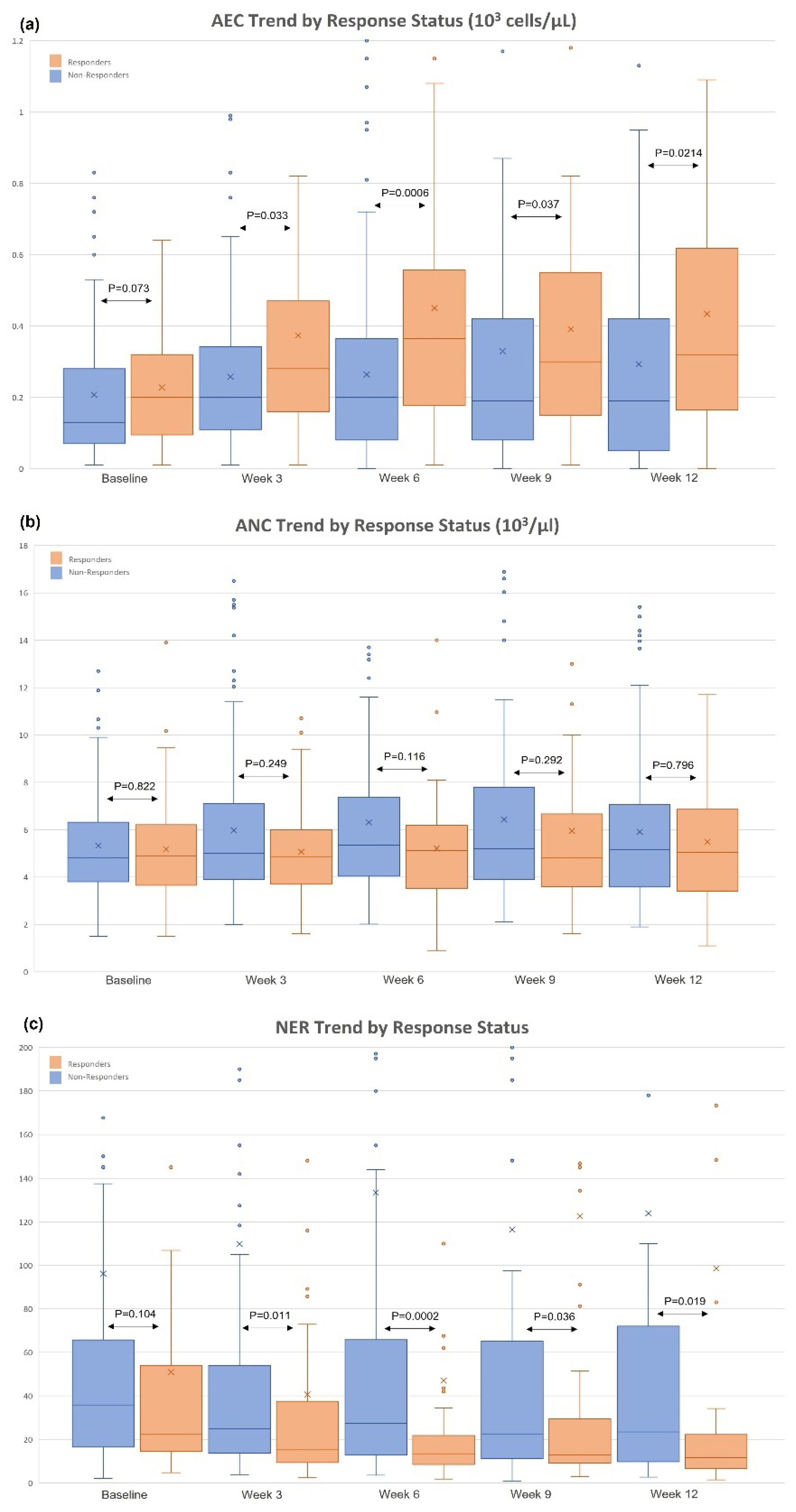
3.1. Changes in AEC/ANC/NER and Patient Baseline Characteristics

The initial study population consisted of 166 patients with mRCC treated with ipilimumab/nivolumab. The trend of AEC, ANC, and NER from baseline to week 12 were stratified by response status (Figure 1). Among responders, the median AEC was 200 cells/µL at baseline, 365 cells/µL at week 6, and 320 cells/µL at week 12; among non-responders, the median AEC was 130 cells/µL at baseline, 200 cells/µL at week 6, and 190 cells/µL at week 12. The most significant AEC difference was observed at week 6 (Figure 1a; p-value: 0.0006). For ANC, there was no statistical difference at either time point (Figure 1b; p-values > 0.05). The median NER was numerically lower in responders from baseline to week 12 (Figure 1c). The most significant difference of NER was observed at week 6 (Figure 1c; p-value: 0.002). Given the above observations, the week 6 NER was further investigated in landmark analyses. The final cohort included 150 patients with mRCC treated with ipilimumab/nivolumab (10 patients with missing week 6 NER, and 6 patients progressed before week 6 were excluded).
There were 150 ipilimumab/nivolumab-treated patients with mRCC included in the final study population (Table 1). The median age was 62 (interquartile range: 53–70), 74% were male, and 80% were White. The majority of patients had clear cell histology (78%); 22% were IMDC favorable risk, 63% were intermediate risk, and 15% were poor risk. There were 104 (69%) patients who had prior nephrectomy, and 49 (33%) patients who had prior systemic therapy. The median follow-up time was 11.6 months.

3.2. Association between Decreased NER at Week 6 and Clinical Outcomes

In the multivariable Cox regression analysis, NER was first modeled as a continuous variable after natural log transformation. After adjusting for age, sex, race, IMDC risk group, baseline LnNER, histology, prior systemic therapy, and prior nephrectomy, every decrease of 1 unit of week 6 LnNER was associated with improved PFS (adjusted hazard ratio (AHR):0.78, 95% CI: 0.66–0.93, p-value: 0.005) and OS (AHR:0.67, 95% CI: 0.52–0.86, p-value: 0.002) (Table 2).
For illustration purposes, baseline NER was subsequently dichotomized at the median (23.8) into high vs. low baseline NER groups, and the NER change from baseline at week 6 were grouped into three groups: (1) NER increase, (2) NER decrease ≤ 50%, and (3) NER decrease > 50%. When stratified by baseline NER (Figure 2a), patients with a low baseline NER had numerically higher ORR than those with a high baseline NER (39% vs. 31%, p-value: 0.30). When considering week 6 NER change alone (Figure 2b), NER decrease >50% had higher ORR compared to NER decrease ≤ 50% and NER increase (43% vs. 36% vs. 25%, p-value for trend: 0.07). When stratified by baseline NER and NER change at week 6 (Figure 2c), patients with NER decrease >50% at week 6 had numerically higher ORR compared to NER decrease ≤ 50% and NER increase in the low baseline NER subgroup (50% vs. 43% vs. 29%, p-value for trend: 0.16), and the high baseline NER subgroup (41% vs. 26% vs. 20%, p-value for trend: 0.11).When stratified by IMDC risk group (Figure 2d), patients with intermediate and poor risk disease had a higher percentage of NER decrease > 50% at week 6 compared to patients with favorable risk disease (33% and 35%, respectively vs. 15%, p-value: 0.048).
When NER was modeled by percent change, NER decrease > 50% at week 6 was associated with improved PFS (AHR:0.55, 95% CI: 0.31–0.95, p-value: 0.03) and OS (AHR: 0.37, 95% CI: 0.16–0.84, p-value: 0.02) when compared to increased NER (Table 2). NER decrease ≤50% at week 6 showed a trend toward improved PFS (AHR:0.63, 95% CI:0.38–1.05, p-value: 0.07) and OS (AHR: 0.49, 95% CI: 0.23–1.06, p-value: 0.07). Stratified analysis was conducted by baseline NER. In the subgroup with a high baseline NER, NER decrease > 50% was associated with improved PFS (AHR: 0.46, 95% CI: 0.22–1.00, p-value: 0.048) and OS (AHR: 0.28, 95% CI: 0.11–0.74, p-value: 0.01). In the subgroup with a low baseline NER, NER decrease > 50% at week 6 was not associated with PFS (AHR: 0.58, 95% CI: 0.22–1.48, p-value: 0.25) or OS (AHR: 0.60, 95% CI: 0.08–4.30, p-value: 0.61). The full models are provided in the Supplementary Material (Tables S1–S4). The Kaplan–Meier curves for PFS and OS by baseline NER and week 6 NER change are presented in Figure 3.

4. Discussion

The current study characterizes the dynamic changes of AEC and NER during the ipilimumab/nivolumab induction from baseline to week 12 in mRCC patients. Patients with an objective response had a higher interval increase in AEC, and there was a prominent decreasing trend of NER from baseline to week 12. After adjusting for patient sociodemographic and tumor characteristics, including the baseline NER [25] and IMDC prognostic risk [27], the decrease in NER at week 6 from the baseline was independently associated with improved PFS and OS in both the continuous and categorial variable analyses. In our subgroup analysis, this association was mostly driven by the subgroup with high baseline NER.
Building on our previous finding that baseline NER is predictive and prognostic in ipilimumab/nivolumab-treated mRCC [25], the current study demonstrates that the on-treatment decrease in NER at week 6 can be an early predictive biomarker of future response of ipilimumab/nivolumab, especially in patients with unfavorable high baseline NER. The current study observed a higher percentage of patients with NER decrease > 50% at week 6 in the IMDC intermediate risk (33%) and poor risk (35%) categories, compared to that in patients with favorable risk disease (15%). This finding is consistent with the CheckMate 214 [6], showing that the superior clinical efficacy of ipilimumab/nivolumab over sunitinib was mainly observed in the IMDC intermediate and poor risk, but not in the favorable risk categories. However, durable long-term responses have been observed among responders in the favorable risk group, but there is no available biomarker to select such patients. If the current biomarker is successfully validated in prospective studies, the decrease in on-treatment NER can be used as an early predictive biomarker in ipilimumab/nivolumab-treated mRCC.
These current findings are supported by previous studies that have observed the elevation of eosinophils after the initiation of ICIs, especially among patients with durable responses in lung cancer [21,22] and melanoma [23]. Prior to the ICI era, this positive association had been observed in IL-2-treated RCC in an early case series [30]. In addition to IO agents, the elevation of eosinophils was also observed in a post hoc analysis of three randomized-controlled phase III trials in sipuleucel-T-treated metastatic castration resistant prostate cancer (mCRPC) patients [31]: there was an association of an elevation of eosinophil at week 6 with improved prostate cancer-specific survival (HR: 0.71, 95% CI: 0.53–0.97, p-value: 0.03) and a trend for improved overall survival (HR: 0.75, 95% CI: 0.56–1.01, p-value: 0.06). The elevation of eosinophils after treatment initiation among responders suggests a certain biological effect of eosinophils in the TME. The anti-tumor activity of eosinophils has been postulated [32], and the roles of eosinophils in the TME have yet to be better elucidated [33]. Eosinophil can leave the bloodstream and migrate into the TME through several integrins-mediated mechanisms [34]. Pre-clinical studies [15,35,36] designed to mechanistically explicate the functions of eosinophils in the TME revealed enhanced antitumor responses through tumor-homing activated eosinophils: the activated eosinophils attracted tumor-specific CD8+ T cells by producing chemokines, such as CCL5, CXCL9, and CXCL10 [15]; the activated eosinophils also normalized the tumor vasculature and polarized the tumor-associated macrophages [15]; anti-tumor activities were also observed in eosinophil-mediated IL-33 [36] and the GM-CSF-IRF5 singling axis in melanoma [36] and colorectal cancer models [35], respectively.
Although our results showed an association between decreased NER and favorable outcomes among patients with ipilimumab/nivolumab-treated mRCC, there were still patients with increased NER at week 6 who had an objective response. Therefore, there are likely additional immune-mediated mechanisms at work in these patients. The current results reflect the complexity of the roles of innate immune cells in the TME, and further research is needed to untangle the involved biological pathways. Eosinophils have the plasticity to shape the TME in opposing directions, between pro-tumorigenic vs. anti-tumorigenic [33], depending on their reciprocal interactions with other cells [32]. Likewise, although tumor-associated neutrophils are traditionally considered to be associated with resistance to ICIs [37], studies revealed that the phenotypes of tumor-associated neutrophils were diverse, and their anti-tumor activities were observed in certain cancer types and at different disease stages [38,39,40]. Our study suggests the possibility of increasing the response of IO agents through enhancing the recruitment of eosinophils to the TME and the augmentation of the anti-tumor activities of the eosinophils. One of the potential targets may be the inhibition of the dipeptidyl peptidase 4 (DPP4) [41], which has been shown by Hollande et al. [42] to increase the chemokine CCL11-mediated eosinophil migration into breast cancer and hepatocellular carcinoma mouse models. In their study, DPP4 inhibition with sitagliptin was shown to increase eosinophil migration into the tumors, and the anti-tumor activity of eosinophils was shown to be independent of T-cells. Tumors treated with sitagliptin, in addition to anti-PD1/anti-CTLA4 antibodies, also demonstrated significantly lower tumor volume compared to controls. Our group previously conducted a small retrospective study of 26 patients with solid tumor diagnosis who were concomitantly taking DPP4 inhibitors for diabetes while on ICI treatment [43]: the objective response rate was 69% (18/26), although the results should be interpreted with caution due to the small sample size and lack of a comparison group. Of note, a phase 1b/2 trial investigating BXCL701, which is an inhibitor of DDP4/DPP8/DPP9, with/without pembrolizumab in mCRPC [44] (NCT03910660) reported a 26% response rate (6/23) and a disease control rate of 63% in this heavily treated patient population [45]. Future correlative studies of this trial may provide further translational data for the anti-tumor activity of eosinophils through DPP inhibitions and the possibility to enhance antitumor activity of ICIs.
In addition to improved clinical outcomes, the increase in eosinophil counts after initiation of ICIs was also reported to be an early biomarker for the development of immune-related adverse events (irAEs). Osawa et al. found that peripheral eosinophilia (defined as AEC ≥ 330/µL) at week 6 was associated with a 2.8-fold higher risk of irAEs [46]. Studies have observed such associations in ICI-induced adrenal insufficiency [47], hypopituitarism [48], cutaneous irAEs [49], and pneumonitis [50]. Given that irAEs have been associated with improved outcomes in ICI-treated cancer patients [51], the association between eosinophil elevation and irAEs is expected. The current dataset did not include granular information on irAEs; therefore, we did not explore such associations in our analysis.
There are several limitations in the current study. First, several peripheral blood count-based biomarkers have been previously investigated in various cancers, before and after the advent of ICIs, such as the neutrophil-to-lymphocyte ratio [52,53,54,55,56], lymphocyte-to-monocyte ratio [52,55,57], and platelet-to-lymphocyte ratio [52,54,58,59,60]. The current study chose to investigate NER (instead of other indices) based on our clinical observation of eosinophil count elevation and the established prognostic role of neutrophilia in mRCC in the IMDC model [27]. However, our results alone are insufficient to compare NER with other indices as a prognostic/predictive biomarker, as this study did not perform direct comparisons with each of these various indices. Prospective validation of NER is warranted. Second, the study design was a retrospective analysis, and inherited biases could not be avoided. The unmeasured confounding and residual confounding may still exist, despite the study-adopted regression method to adjust for baseline characteristics. Third, the median follow-up time was relatively short (11.6 months). Fourth, several other conditions, such as medications, infections, and the inflammatory/autoimmune process, can all affect neutrophil and eosinophil counts, and the current study was not able to account for those factors. As such, the results of the current study should be interpreted as hypothesis generating. Future clinical trials will provide a more ideal setting to prospectively validate the on-treatment NER biomarker. In addition, correlative studies in clinical trials can mechanistically explore the role of eosinophils in cancer immunotherapy by measuring baseline and on-treatment chemokines, such as CCL5/CXCL9/CXCL10/CCL11.

5. Conclusions

In patients with mRCC treated with ipilimumab/nivolumab, our results reveal that the decrease in on-treatment NER at week 6 is associated with improved PFS and overall survival, and the dynamic changes of on-treatment NER implied responses of ICIs. Prospective studies are warrantied to validate this predictive biomarker in IO combinations of mRCC and to further explore the role of eosinophils in TME, along with the therapeutic implications of eosinophil-mediated anti-tumor activities.

Supplementary Materials

The following supporting information can be downloaded at: https://www.mdpi.com/article/10.3390/cancers14153830/s1, Table S1: continuous variable analysis for baseline LnNER and week 6 LnNER. Table S2: categorical variable analysis for week 6 NER change. Table S3: categorical variable analysis for week 6 NER change in subgroup with high baseline NER. Table S4: categorical variable analysis for week 6 NER change in subgroup with low baseline NER.

Author Contributions

Study concept and design: Y.-W.C., M.D.T., B.I.R., and T.Z.; acquisition of data: M.D.T. and L.C.B.; statistical analysis: Y.-W.C. and M.D.T.; analysis and interpretation of data: all authors; drafting of the original manuscript: Y.-W.C. and M.D.T.; critical revision of the manuscript for important intellectual content: all authors; supervision: B.I.R. and T.Z. All authors have read and agreed to the published version of the manuscript.

Funding

This research was supported by the NIH T32 Training Program in Molecular and Genetic Epidemiology (T32 CA160056).

Institutional Review Board Statement

This study was approved by the Vanderbilt University Medical Center IRB (VUMC 160979) and the Duke Cancer Institute IRB (DCI Pro00101984).

Informed Consent Statement

Not required.

Data Availability Statement

The data presented in this study are available upon request. The data are not publicly available, as they contain information that could potentially compromise the privacy of the study subjects.

Conflicts of Interest

YWC: has provided consulting for Deloitte and holds stock in Biogen (an immediate family member). LCB: has provided consulting for Seattle Genetics and Astellas. AJA has received research funding to his institution from Dendreon, Bayer, Pfizer, Novartis, Janssen Oncology, Astellas Pharma, Gilead Sciences, Roche/Genentech, Bristol-Myers Squibb, Constellation Pharmaceuticals, Merck, AstraZeneca, BeiGene, Amgen, and Forma Therapeutics and consulting fees from Bayer, Dendreon, Pfizer, Astellas, AstraZeneca, Merck, Bristol-Myers Squibb, Janssen, FORMA Therapeutics, Novartis, Exelixis, Myovant Sciences, and GoodRx, as well as travel support from Astella Scientific and Medical Affairs, Inc. He has received patents to his institution for circulating tumor cell novel capture technology. KEB has received research funding to her institution from Bristol-Myers Squibb and Merck Sharp & Dohme, as well as consulting fees from Aravive, Exelixis, Bristol-Myers Squibb and Seattle Genetics. NBD has received research funding to her institution from AstraZeneca, Roche, Pfizer, Merck, Incyte, Mirati Therapeutics, Seattle Genetics/Astellas, Calithera Biosciences, Immunomedics, Bristol-Myers Squibb, Exelixis, and Gilead Sciences and consulting fees from Janssen Biotech. MRH has received research funding to his institution from Bristol-Myers Squibb, Genentech, Pfizer, Merck, Astellas Pharma, Bayer, Exelixis, Seattle Genetics, and Propella Therapeutics, as well as consulting fees from Exelixis, Fujifilm, Pfizer, Bristol-Myers Squibb, Seattle Genetics, Gilead Sciences, and Myovant Sciences. He is also on the speaker’s bureau for Exelixis, Eisai, Gilead Sciences, and Myovant Sciences. RKM: has received consulting fees from Immunocore, Sanofi/Regeneron, and Myovant Sciences. KRS has received researching funding to her institution from Tempus and consulting fees from Janssen Biotech. DJG has received research funding to his institution from Exelixis, Janssen Oncology, Novartis, Pfizer, Astellas Pharma, Bristol-Myers Squibb, Acerta Pharma, Bayer, Dendreon, Innocrin Pharma, Calithera Biosciences, and Sanofi/Aventis; consulting fees from Bayer, Exelixis, Pfizer, Sanofi, Astellas Pharma, Innocrin Pharma, Bristol-Myers Squibb, Genentech, Janssen, Merck Sharp & Dohme, Myovant Sciences, AstraZeneca, Michael J. Hennessy Associates, Constellation Pharmaceuticals, Physician’s Education Resources, Propella Therapeutics, RevHealth, and xCures; honoraria from Sanofi, Bayer, Exelixis, EMD Serono, OncLive, Pfizer, UroToday, Acceleron Pharma, the American Association for Cancer Research, Axess Oncology, Janssen Oncology, and Millennium Medical Publishing; travel support from Bayer, Exelixis, Merck, Pfizer, Sanofi, Janssen Oncology, UroToday; and is connected to the leadership of Capio Biosciences. WKR has received research funding to her institution from Peloton Therapeutics, Incyte (an immediate family member) and Sitryx (an immediate family member); consulting fees from Sitryx (an immediate family member), Nirojy (an immediate family member), and Caribou Biosciences (an immediate family member); honoraria from Pfizer (an immediate family member); patents for ClearCode34 Risk prediction biomarker for kidney cancer, hERV 3-2 expression as a biomarker of response to immunotherapy; she holds stock in Sitryx (an immediate family member), Caribou Biosciences (an immediate family member), and Nirogy Therapeutics (an immediate family member). BIR has received research funding to his institution from Pfizer, Roche/Genentech, Bristol-Myers Squibb, Merck, AstraZeneca/MedImmune, Incyte, Arrowhead Pharmaceuticals, Taris, Seattle Genetics, Immunomedics, Surface Oncology, Dragonfly Therapeutics, Aravive, and Exelixis; consulting fees from Pfizer, Merck, Synthrox, Bristol-Myers Squibb, AVEO, Surface Oncology, 3D Medicines, Corvus Pharmaceuticals, Aravive, Arrowhead Pharmaceuticals, Shionogi, Eisai, and GlaxoSmithKline; travel support from Pfizer, Bristol-Myers Squibb, and Merck; is connect to the leadership of MJH Life Sciences, and holds stock in PTC Therapeutics. TZ has received research funding to her institution from Janssen, Pfizer, Merrimack, Stem CentRx, Novartis, OmniSeq, Personal Genome Diagnostics, Regeneron, Merck, Mirati Therapeutics, Loxo/Lilly, and Astellas Pharma; consulting fees from Janssen, Exelixis, Pfizer, Bristol-Myers Squibb, Merck, Seattle Genetics, Dendreon, Calithera Biosciences, QED Therapeutics, Eisai, Aravive, Lilly, AVEO, Merck and Bayer; honoraria from MJH Life Sciences, Pacific Genuity, Aptitude Health, Peerview, Clinical Care Options, and Curio Science; serves on the speaker’s bureau to Genomic Health and Sanofi/Aventis; her institution holds patents for circulating tumor cell novel capture by c-MET technology, and prochelators as targeted prodrugs for prostate cancer; and is connected to the leadership of Capio Biosciences (an immediate family member) and Archimmune Therapeutics (an immediate family member). The remaining authors declare that they have no competing interests.

References

  1. Robert, C. A decade of immune-checkpoint inhibitors in cancer therapy. Nat. Commun. 2020, 11, 3801. [Google Scholar] [CrossRef] [PubMed]
  2. Aroldi, F.; Middleton, M.R. Long-Term Outcomes of Immune Checkpoint Inhibition in Metastatic Melanoma. Am. J. Clin. Dermatol. 2022, 23, 331–338. [Google Scholar] [CrossRef] [PubMed]
  3. Sham, N.O.; Zhao, L.; Zhu, Z.; Roy, T.M.; Xiao, H.; Bai, Q.; Wakefield, M.R.; Fang, Y. Immunotherapy for Non-small Cell Lung Cancer: Current Agents and Potential Molecular Targets. Anticancer Res. 2022, 42, 3275–3284. [Google Scholar] [CrossRef]
  4. Houssiau, H.; Seront, E. The Evolution of Immune Checkpoint Inhibitors in Advanced Urothelial Carcinoma. Cancers 2022, 14, 1640. [Google Scholar] [CrossRef]
  5. Abd El Aziz, M.A.; Facciorusso, A.; Nayfeh, T.; Saadi, S.; Elnaggar, M.; Cotsoglou, C.; Sacco, R. Immune Checkpoint Inhibitors for Unresectable Hepatocellular Carcinoma. Vaccines 2020, 8, 616. [Google Scholar] [CrossRef]
  6. Motzer, R.J.; Tannir, N.M.; McDermott, D.F.; Burotto, M.; Choueiri, T.K.; Hammers, H.J.; Plimack, E.R.; Porta, C.G.; George, S.; Powles, T.B.; et al. Conditional survival and 5-year follow-up in CheckMate 214: First-line nivolumab + ipilimumab (N+I) versus sunitinib (S) in advanced renal cell carcinoma (aRCC). Ann. Oncol. 2021, 32 (Suppl. 5), S678–S724. [Google Scholar] [CrossRef]
  7. Xin, J.X.; Hubbard-Lucey, V.M.; Tang, J. Immuno-oncology drug development goes global. Nat. Rev. Drug Discov. 2019, 18, 899–900. [Google Scholar] [CrossRef] [Green Version]
  8. Darvin, P.; Toor, S.M.; Sasidharan Nair, V.; Elkord, E. Immune checkpoint inhibitors: Recent progress and potential biomarkers. Exp. Mol. Med. 2018, 50, 1–11. [Google Scholar] [CrossRef] [Green Version]
  9. Gerard, C.L.; Delyon, J.; Wicky, A.; Homicsko, K.; Cuendet, M.A.; Michielin, O. Turning tumors from cold to inflamed to improve immunotherapy response. Cancer Treat. Rev. 2021, 101, 102227. [Google Scholar] [CrossRef]
  10. Joyce, J.A.; Fearon, D.T. T cell exclusion, immune privilege, and the tumor microenvironment. Science 2015, 348, 74–80. [Google Scholar] [CrossRef] [Green Version]
  11. Linette, G.P.; Carreno, B.M. Tumor-Infiltrating Lymphocytes in the Checkpoint Inhibitor Era. Curr. Hematol. Malig. Rep. 2019, 14, 286–291. [Google Scholar] [CrossRef]
  12. Liu, X.; Hogg, G.D.; DeNardo, D.G. Rethinking immune checkpoint blockade: ‘Beyond the T cell’. J. Immunother. Cancer 2021, 9, e001460. [Google Scholar] [CrossRef]
  13. Hussain, K.; Cragg, M.S.; Beers, S.A. Remodeling the Tumor Myeloid Landscape to Enhance Antitumor Antibody Immunotherapies. Cancers 2021, 13, 4904. [Google Scholar] [CrossRef]
  14. Nakamura, K.; Smyth, M.J. Myeloid immunosuppression and immune checkpoints in the tumor microenvironment. Cell. Mol. Immunol. 2020, 17, 1–12. [Google Scholar] [CrossRef]
  15. Carretero, R.; Sektioglu, I.M.; Garbi, N.; Salgado, O.C.; Beckhove, P.; Hämmerling, G.J. Eosinophils orchestrate cancer rejection by normalizing tumor vessels and enhancing infiltration of CD8(+) T cells. Nat. Immunol. 2015, 16, 609–617. [Google Scholar] [CrossRef]
  16. Grisaru-Tal, S.; Dulberg, S.; Beck, L.; Zhang, C.; Itan, M.; Hediyeh-Zadeh, S.; Caldwell, J.; Rozenberg, P.; Dolitzky, A.; Avlas, S.; et al. Metastasis-Entrained Eosinophils Enhance Lymphocyte-Mediated Antitumor Immunity. Cancer Res. 2021, 81, 5555–5571. [Google Scholar] [CrossRef]
  17. Martens, A.; Wistuba-Hamprecht, K.; Geukes Foppen, M.; Yuan, J.; Postow, M.A.; Wong, P.; Romano, E.; Khammari, A.; Dreno, B.; Capone, M.; et al. Baseline Peripheral Blood Biomarkers Associated with Clinical Outcome of Advanced Melanoma Patients Treated with Ipilimumab. Clin. Cancer Res. Off. J. Am. Assoc. Cancer Res. 2016, 22, 2908–2918. [Google Scholar] [CrossRef] [Green Version]
  18. Ferrucci, P.F.; Gandini, S.; Cocorocchio, E.; Pala, L.; Baldini, F.; Mosconi, M.; Antonini Cappellini, G.C.; Albertazzi, E.; Martinoli, C. Baseline relative eosinophil count as a predictive biomarker for ipilimumab treatment in advanced melanoma. Oncotarget 2017, 8, 79809–79815. [Google Scholar] [CrossRef] [Green Version]
  19. Mota, J.M.; Teo, M.Y.; Whiting, K.; Li, H.A.; Regazzi, A.M.; Lee, C.H.; Funt, S.A.; Bajorin, D.; Ostrovnaya, I.; Iyer, G.; et al. Pretreatment Eosinophil Counts in Patients With Advanced or Metastatic Urothelial Carcinoma Treated With Anti-PD-1/PD-L1 Checkpoint Inhibitors. J. Immunother. 2021, 44, 248–253. [Google Scholar] [CrossRef]
  20. Minohara, K.; Matoba, T.; Kawakita, D.; Takano, G.; Oguri, K.; Murashima, A.; Nakai, K.; Iwaki, S.; Hojo, W.; Matsumura, A.; et al. Novel Prognostic Score for recurrent or metastatic head and neck cancer patients treated with Nivolumab. Sci. Rep. 2021, 11, 16992. [Google Scholar] [CrossRef]
  21. Sibille, A.; Henket, M.; Corhay, J.L.; Alfieri, R.; Louis, R.; Duysinx, B. White Blood Cells in Patients Treated with Programmed Cell Death-1 Inhibitors for Non-small Cell Lung Cancer. Lung 2021, 199, 549–557. [Google Scholar] [CrossRef] [PubMed]
  22. Alves, A.; Dias, M.; Campainha, S.; Barroso, A. Peripheral blood eosinophilia may be a prognostic biomarker in non-small cell lung cancer patients treated with immunotherapy. J. Thorac. Dis. 2021, 13, 2716–2727. [Google Scholar] [CrossRef]
  23. Simon, S.C.S.; Hu, X.; Panten, J.; Grees, M.; Renders, S.; Thomas, D.; Weber, R.; Schulze, T.J.; Utikal, J.; Umansky, V. Eosinophil accumulation predicts response to melanoma treatment with immune checkpoint inhibitors. Oncoimmunology 2020, 9, 1727116. [Google Scholar] [CrossRef] [PubMed] [Green Version]
  24. Herrmann, T.; Ginzac, A.; Molnar, I.; Bailly, S.; Durando, X.; Mahammedi, H. Eosinophil counts as a relevant prognostic marker for response to nivolumab in the management of renal cell carcinoma: A retrospective study. Cancer Med. 2021, 10, 6705–6713. [Google Scholar] [CrossRef]
  25. Tucker, M.D.; Brown, L.C.; Chen, Y.W.; Kao, C.; Hirshman, N.; Kinsey, E.N.; Ancell, K.K.; Beckermann, K.E.; Davis, N.B.; McAlister, R.; et al. Association of baseline neutrophil-to-eosinophil ratio with response to nivolumab plus ipilimumab in patients with metastatic renal cell carcinoma. Biomark. Res. 2021, 9, 80. [Google Scholar] [CrossRef]
  26. Gil, L.; Alves, F.R.; Silva, D.; Fernandes, I.; Fontes-Sousa, M.; Alves, M.; Papoila, A.; Da Luz, R. Prognostic Impact of Baseline Neutrophil-to-Eosinophil Ratio in Patients With Metastatic Renal Cell Carcinoma Treated With Nivolumab Therapy in Second or Later Lines. Cureus 2022, 14, e22224. [Google Scholar] [CrossRef]
  27. Heng, D.Y.; Xie, W.; Regan, M.M.; Warren, M.A.; Golshayan, A.R.; Sahi, C.; Eigl, B.J.; Ruether, J.D.; Cheng, T.; North, S.; et al. Prognostic factors for overall survival in patients with metastatic renal cell carcinoma treated with vascular endothelial growth factor-targeted agents: Results from a large, multicenter study. J. Clin. Oncol. Off. J. Am. Soc. Clin. Oncol. 2009, 27, 5794–5799. [Google Scholar] [CrossRef]
  28. Ko, J.J.; Xie, W.; Kroeger, N.; Lee, J.L.; Rini, B.I.; Knox, J.J.; Bjarnason, G.A.; Srinivas, S.; Pal, S.K.; Yuasa, T.; et al. The International Metastatic Renal Cell Carcinoma Database Consortium model as a prognostic tool in patients with metastatic renal cell carcinoma previously treated with first-line targeted therapy: A population-based study. Lancet Oncol. 2015, 16, 293–300. [Google Scholar] [CrossRef]
  29. Eisenhauer, E.A.; Therasse, P.; Bogaerts, J.; Schwartz, L.H.; Sargent, D.; Ford, R.; Dancey, J.; Arbuck, S.; Gwyther, S.; Mooney, M.; et al. New response evaluation criteria in solid tumours: Revised RECIST guideline (version 1.1). Eur. J. Cancer 2009, 45, 228–247. [Google Scholar] [CrossRef]
  30. Rodgers, S.; Rees, R.C.; Hancock, B.W. Changes in the phenotypic characteristics of eosinophils from patients receiving recombinant human interleukin-2 (rhIL-2) therapy. Br. J. Haematol. 1994, 86, 746–753. [Google Scholar] [CrossRef]
  31. McNeel, D.G.; Gardner, T.A.; Higano, C.S.; Kantoff, P.W.; Small, E.J.; Wener, M.H.; Sims, R.B.; DeVries, T.; Sheikh, N.A.; Dreicer, R. A transient increase in eosinophils is associated with prolonged survival in men with metastatic castration-resistant prostate cancer who receive sipuleucel-T. Cancer Immunol. Res. 2014, 2, 988–999. [Google Scholar] [CrossRef] [Green Version]
  32. Varricchi, G.; Galdiero, M.R.; Loffredo, S.; Lucarini, V.; Marone, G.; Mattei, F.; Marone, G.; Schiavoni, G. Eosinophils: The unsung heroes in cancer? Oncoimmunology 2018, 7, e1393134. [Google Scholar] [CrossRef] [Green Version]
  33. Reichman, H.; Karo-Atar, D.; Munitz, A. Emerging Roles for Eosinophils in the Tumor Microenvironment. Trends Cancer 2016, 2, 664–675. [Google Scholar] [CrossRef] [PubMed]
  34. Bochner, B.S. Novel Therapies for Eosinophilic Disorders. Immunol. Allergy Clin. North Am. 2015, 35, 577–598. [Google Scholar] [CrossRef] [Green Version]
  35. Arnold, I.C.; Artola-Boran, M.; Gurtner, A.; Bertram, K.; Bauer, M.; Frangez, Z.; Becher, B.; Kopf, M.; Yousefi, S.; Simon, H.U.; et al. The GM-CSF-IRF5 signaling axis in eosinophils promotes antitumor immunity through activation of type 1 T cell responses. J. Exp. Med. 2020, 217, e20190706. [Google Scholar] [CrossRef]
  36. Lucarini, V.; Ziccheddu, G.; Macchia, I.; La Sorsa, V.; Peschiaroli, F.; Buccione, C.; Sistigu, A.; Sanchez, M.; Andreone, S.; D’Urso, M.T.; et al. IL-33 restricts tumor growth and inhibits pulmonary metastasis in melanoma-bearing mice through eosinophils. Oncoimmunology 2017, 6, e1317420. [Google Scholar] [CrossRef] [PubMed] [Green Version]
  37. Faget, J.; Peters, S.; Quantin, X.; Meylan, E.; Bonnefoy, N. Neutrophils in the era of immune checkpoint blockade. J. Immunother. Cancer 2021, 9, e002242. [Google Scholar] [CrossRef] [PubMed]
  38. Fridlender, Z.G.; Sun, J.; Kim, S.; Kapoor, V.; Cheng, G.; Ling, L.; Worthen, G.S.; Albelda, S.M. Polarization of tumor-associated neutrophil phenotype by TGF-beta: “N1” versus “N2” TAN. Cancer Cell 2009, 16, 183–194. [Google Scholar] [CrossRef] [Green Version]
  39. Granot, Z.; Henke, E.; Comen, E.A.; King, T.A.; Norton, L.; Benezra, R. Tumor entrained neutrophils inhibit seeding in the premetastatic lung. Cancer Cell 2011, 20, 300–314. [Google Scholar] [CrossRef] [Green Version]
  40. Eruslanov, E.B.; Bhojnagarwala, P.S.; Quatromoni, J.G.; Stephen, T.L.; Ranganathan, A.; Deshpande, C.; Akimova, T.; Vachani, A.; Litzky, L.; Hancock, W.W.; et al. Tumor-associated neutrophils stimulate T cell responses in early-stage human lung cancer. J. Clin. Investig. 2014, 124, 5466–5480. [Google Scholar] [CrossRef] [Green Version]
  41. Barreira da Silva, R.; Laird, M.E.; Yatim, N.; Fiette, L.; Ingersoll, M.A.; Albert, M.L. Dipeptidylpeptidase 4 inhibition enhances lymphocyte trafficking, improving both naturally occurring tumor immunity and immunotherapy. Nat. Immunol. 2015, 16, 850–858. [Google Scholar] [CrossRef]
  42. Hollande, C.; Boussier, J.; Ziai, J.; Nozawa, T.; Bondet, V.; Phung, W.; Lu, B.; Duffy, D.; Paradis, V.; Mallet, V.; et al. Inhibition of the dipeptidyl peptidase DPP4 (CD26) reveals IL-33-dependent eosinophil-mediated control of tumor growth. Nat. Immunol. 2019, 20, 257–264. [Google Scholar] [CrossRef]
  43. Miranda, K.; Tucker, M.D.; Chen, Y.W.; Beckermann, K.E.; Rini, B.I. Concurrent Immunotherapy And Dipeptidyl Peptidase-4 Inhibition Among Patients With Solid Tumors. J. Immunother. Cancer 2021, 9. [Google Scholar] [CrossRef]
  44. Aggarwal, R.R.; Costin, D.; O’Neill, V.J.; Corsi-Travali, S.; Adurthi, S.; Adedoyin, A.; Healey, D.I.; De Bono, J.S.; Monk, P.; Zhang, J.; et al. Phase 1b study of BXCL701, a novel small molecule inhibitor of dipeptidyl peptidases (DPP), combined with pembrolizumab (pembro), in men with metastatic castration-resistant prostate cancer (mCRPC). J. Clin. Oncol. 2020, 38, e17581. [Google Scholar] [CrossRef]
  45. Monk, P.; Zhang, J.; Costin, D.; Petrylak, D.P.; Tagawa, S.T.; Karsh, L.I.; Zhu, X.; Karsh, L.I.; Zhu, X.; Linch, M.; et al. BXCL701-1st-in-class oral activator of systemic innate immunity-combined with pembrolizumab, in men with metastatic castration-resistant prostate cancer (mCRPC): Phase II results. Ann. Oncol. 2021, 5, S651. [Google Scholar] [CrossRef]
  46. Osawa, H.; Shiozawa, T.; Okauchi, S.; Miyazaki, K.; Kodama, T.; Kagohashi, K.; Nakamura, R.; Satoh, H.; Hizawa, N. Association between time to treatment failure and peripheral eosinophils in patients with non-small cell lung cancer treated with immune checkpoint inhibitors. Pol. Arch. Intern. Med. 2021, 131, 16049. [Google Scholar] [CrossRef]
  47. Takayasu, S.; Mizushiri, S.; Watanuki, Y.; Yamagata, S.; Usutani, M.; Nakada, Y.; Asari, Y.; Murasawa, S.; Kageyama, K.; Daimon, M. Eosinophil counts can be a predictive marker of immune checkpoint inhibitor-induced secondary adrenal insufficiency: A retrospective cohort study. Sci. Rep. 2022, 12, 1294. [Google Scholar] [CrossRef]
  48. Yamada, H.; Washino, S.; Suzuki, D.; Saikawa, R.; Tonezawa, S.; Hagiwara, R.; Funazaki, S.; Yoshida, M.; Konishi, T.; Saito, K.; et al. Hypereosinophilia is a predictive biomarker of immune checkpoint inhibitor-induced hypopituitarism in patients with renal cell carcinoma. BMC Endocr. Disord. 2022, 22, 110. [Google Scholar] [CrossRef]
  49. Bottlaender, L.; Amini-Adle, M.; Maucort-Boulch, D.; Robinson, P.; Thomas, L.; Dalle, S. Cutaneous adverse events: A predictor of tumour response under anti-PD-1 therapy for metastatic melanoma, a cohort analysis of 189 patients. J. Eur. Acad. Dermatol. Venereol. JEADV 2020, 34, 2096–2105. [Google Scholar] [CrossRef]
  50. Chu, X.; Zhao, J.; Zhou, J.; Zhou, F.; Jiang, T.; Jiang, S.; Sun, X.; You, X.; Wu, F.; Ren, S.; et al. Association of baseline peripheral-blood eosinophil count with immune checkpoint inhibitor-related pneumonitis and clinical outcomes in patients with non-small cell lung cancer receiving immune checkpoint inhibitors. Lung Cancer 2020, 150, 76–82. [Google Scholar] [CrossRef]
  51. Petrelli, F.; Grizzi, G.; Ghidini, M.; Ghidini, A.; Ratti, M.; Panni, S.; Cabiddu, M.; Ghilardi, M.; Borgonovo, K.; Parati, M.C.; et al. Immune-related Adverse Events and Survival in Solid Tumors Treated With Immune Checkpoint Inhibitors: A Systematic Review and Meta-Analysis. J. Immunother. 2020, 43, 1–7. [Google Scholar] [CrossRef] [PubMed]
  52. Kumarasamy, C.; Tiwary, V.; Sunil, K.; Suresh, D.; Shetty, S.; Muthukaliannan, G.K.; Baxi, S.; Jayaraj, R. Prognostic Utility of Platelet-Lymphocyte Ratio, Neutrophil-Lymphocyte Ratio and Monocyte-Lymphocyte Ratio in Head and Neck Cancers: A Detailed PRISMA Compliant Systematic Review and Meta-Analysis. Cancers 2021, 13, 4166. [Google Scholar] [CrossRef] [PubMed]
  53. Booka, E.; Kikuchi, H.; Haneda, R.; Soneda, W.; Kawata, S.; Murakami, T.; Matsumoto, T.; Hiramatsu, Y.; Takeuchi, H. Neutrophil-to-Lymphocyte Ratio to Predict the Efficacy of Immune Checkpoint Inhibitor in Upper Gastrointestinal Cancer. Anticancer Res. 2022, 42, 2977–2987. [Google Scholar] [CrossRef] [PubMed]
  54. Yanagisawa, T.; Mori, K.; Katayama, S.; Mostafaei, H.; Quhal, F.; Laukhtina, E.; Rajwa, P.; Motlagh, R.S.; Aydh, A.; König, F.; et al. Hematological prognosticators in metastatic renal cell cancer treated with immune checkpoint inhibitors: A meta-analysis. Immunotherapy 2022, 14, 709–725. [Google Scholar] [CrossRef] [PubMed]
  55. Li, M.; Deng, Q.; Zhang, L.; He, S.; Rong, J.; Zheng, F. The pretreatment lymphocyte to monocyte ratio predicts clinical outcome for patients with urological cancers: A meta-analysis. Pathol. Res. Pract. 2019, 215, 5–11. [Google Scholar] [CrossRef] [PubMed]
  56. Lalani, A.A.; Xie, W.; Martini, D.J.; Steinharter, J.A.; Norton, C.K.; Krajewski, K.M.; Duquette, A.; Bossé, D.; Bellmunt, J.; Van Allen, E.M.; et al. Change in Neutrophil-to-lymphocyte ratio (NLR) in response to immune checkpoint blockade for metastatic renal cell carcinoma. J. Immunother. Cancer 2018, 6, 5. [Google Scholar] [CrossRef]
  57. Facciorusso, A.; Del Prete, V.; Crucinio, N.; Serviddio, G.; Vendemiale, G.; Muscatiello, N. Lymphocyte-to-monocyte ratio predicts survival after radiofrequency ablation for colorectal liver metastases. World J. Gastroenterol. 2016, 22, 4211–4218. [Google Scholar] [CrossRef]
  58. Yamamoto, T.; Kawada, K.; Obama, K. Inflammation-Related Biomarkers for the Prediction of Prognosis in Colorectal Cancer Patients. Int. J. Mol. Sci. 2021, 22, 8002. [Google Scholar] [CrossRef]
  59. Jin, X.; Wang, K.; Shao, X.; Huang, J. Prognostic implications of the peripheral platelet-to-lymphocyte ratio and neutrophil-to-lymphocyte ratio in predicting pathologic complete response after neoadjuvant chemotherapy in breast cancer patients. Gland Surg. 2022, 11, 1057–1066. [Google Scholar] [CrossRef]
  60. Leng, J.; Wu, F.; Zhang, L. Prognostic Significance of Pretreatment Neutrophil-to-Lymphocyte Ratio, Platelet-to-Lymphocyte Ratio, or Monocyte-to-Lymphocyte Ratio in Endometrial Neoplasms: A Systematic Review and Meta-analysis. Front. Oncol. 2022, 12, 734948. [Google Scholar] [CrossRef]
Figure 1. Trend of AEC/ANC/NER by response status.
Cancers 14 03830 g001
Figure 2. ORR by baseline NER and NER change at week 6.
Cancers 14 03830 g002
Figure 3. PFS/OS by baseline NER and NER change at week 6.
Cancers 14 03830 g003
Table 1. Patient baseline characteristics (N = 150).
Median (IQR)
Baseline NER23.8 (15–57)
Week 6 NER 19.8 (10.6–40.8)
N (%)
Week 6 NER change
Decrease > 50%44 (29)
Decrease ≤ 50%58 (39)
Increase48 (32)
Baseline NER
High (≥median)75 (50)
Low (<median)75 (50)
Age(Median: 62 (IQR: 53–70))
<6063 (42)
≥6087 (58)
Sex
Male111 (74)
Female39 (26)
Race
White120 (80)
Non-White24 (14)
Unknown9 (6)
Histology
Clear cell117 (78)
Non-clear cell31 (21)
Unknown2 (1)
IMDC risk
Favorable33 (22)
Intermediate94 (63)
Poor23 (15)
Nephrectomy
Yes104 (69)
No46 (31)
Prior systemic therapy
Yes49 (33)
No100 (67)
Unknown1 (1)
NER: neutrophil-to-eosinophil ratio; IQR: interquartile range; IMDC: International Metastatic RCC Database Consortium.
Table 2. Multivariable regression analysis of NER and clinical outcomes.
PFS OS
AHR (95%)p-ValueAHR (95%)p-Value
Continuous variable
Baseline LnNER0.98 (0.78–1.23)0.840.82 (0.57–1.19)0.30
Week 6 LnNER0.78 (0.66–0.93)0.0050.67 (0.52–0.86)0.002
Week 6 NER change
All patients (N = 150)
Decrease > 50%0.55 (0.31–0.95)0.030.37 (0.16–0.84)0.02
Decrease ≤ 50%0.63 (0.38–1.05)0.070.49 (0.23–1.06)0.07
IncreaseRef Ref
Subgroup with high baseline NER (N = 75)
Decrease > 50%0.46 (0.22–1.00)0.0480.28 (0.11–0.74)0.01
Decrease ≤ 50%0.59 (0.26–1.31)0.190.44 (0.16–1.23)0.12
IncreaseRef Ref
Subgroup with low baseline NER (N = 75)
Decrease > 50%0.58 (0.22–1.48)0.250.60 (0.08–4.30)0.61
Decrease ≤ 50%0.72 (0.34–1.53)0.391.08 (0.25–4.70)0.92
IncreaseRef Ref
PFS: progression-free survival; OS: overall survival; AHR: adjusted hazard ratio. Models were adjusted for age, sex, race, IMDC risk group, baseline NER, histology, prior systemic therapy, and prior nephrectomy. Landmark analysis for PFS and OS were calculated from week 6.
Publisher’s Note: MDPI stays neutral with regard to jurisdictional claims in published maps and institutional affiliations.

