Article

Testing for Lynch Syndrome in Endometrial Carcinoma: From Universal to Age-Selective MLH1 Methylation Analysis

1
Department of Pathology, Helsinki University Hospital, University of Helsinki, 00290 Helsinki, Finland
2
Department of Obstetrics and Gynecology, Helsinki University Hospital, University of Helsinki, 00290 Helsinki, Finland
3
Laboratory of Genetics, HUS Diagnostic Center, Helsinki University Hospital, University of Helsinki, 00209 Helsinki, Finland
4
Department of Medical and Clinical Genetics, University of Helsinki, 00014 Helsinki, Finland
5
Department of Education and Science, Central Finland Health Care District, 40620 Jyväskylä, Finland
6
Department of Surgery, Central Finland Health Care District; 40620 Jyväskylä, Finland
7
Department of Sport and Health Sciences, Jyväskylä University, 40014 Jyväskylä, Finland
8
Applied Tumor Genomics Research Program, University of Helsinki, 00290 Helsinki, Finland
*
Author to whom correspondence should be addressed.
Cancers 2022, 14(5), 1348; https://doi.org/10.3390/cancers14051348
Submission received: 28 December 2021 / Revised: 21 February 2022 / Accepted: 4 March 2022 / Published: 6 March 2022
(This article belongs to the Special Issue Immunohistochemical Markers in Endometrial Cancer)

Abstract

:

Simple Summary

International guidelines recommend universal screening of endometrial carcinoma patients for Lynch syndrome, a hereditary cancer predisposition syndrome. Screening is based on mismatch repair protein immunohistochemistry and reflex MLH1 methylation analysis to exclude the likely sporadic cases of MMR deficiency. As sporadic MLH1 protein loss is common in endometrial carcinoma, the ability to target methylation testing would save efforts and costs. We discovered that limiting methylation testing to patients under 65 years would have significantly reduced the testing effort while maintaining a low false negative rate for MLH1-LS detection (0% and 3% in our clinic and registry-based cohorts, respectively).

Abstract

International guidelines recommend universal screening of endometrial carcinoma (EC) patients for Lynch syndrome (LS). This screening is generally based on mismatch repair (MMR) protein immunohistochemistry followed by MLH1 methylation analysis of MLH1-negative cases to exclude the likely sporadic cases from germline testing. As LS-associated EC is uncommon in the elderly, age-selective methylation testing could improve cost-efficiency. We performed MMR immunohistochemistry on 821 unselected ECs (clinic-based cohort) followed by a MLH1 promoter methylation test of all MLH1/PMS2-negative tumors. Non-methylated MLH1-deficient cases underwent NGS and MLPA-based germline analyses to identify MLH1 mutation carriers. A reduction in the test burden and corresponding false negative rates for LS screening were investigated for various age cut-offs. In addition, the age distribution of 132 MLH1 mutation carriers diagnosed with EC (registry-based cohort) was examined. A germline MLH1 mutation was found in 2/14 patients with non-methylated MLH1-deficient EC. When compared to a universal methylation analysis, selective testing with a cut-off age of 65 years, would have reduced the testing effort by 70.7% with a false negative rate for LS detection of 0% and 3% in the clinic and registry-based cohorts, respectively. The use of age-selective methylation analysis is a feasible way of reducing the costs and laboratory burden in LS screening for EC patients.

1. Introduction

Approximately 3% of endometrial carcinoma (EC) cases are associated with Lynch syndrome (LS), a cancer predisposition syndrome previously referred to as hereditary non-polyposis colorectal cancer (HNPCC) [1]. Individuals with LS have inherited a dysfunctional germline allele of a DNA mismatch repair (MMR) gene (MLH1, PMS2, MSH2, or MSH6). A secondary somatic inactivation of the remaining wild type allele leads to disruption of the MMR system and microsatellite instability (MSI). Microsatellites are DNA regions containing repeated units of 1–6 nucleotides, which are particularly prone to replication errors and an accumulation of mutations in the absence of a functional MMR system. MSI promotes carcinogenesis in various organs including the colorectum, uterus, ovary, small intestine, stomach and upper uroepithelial tract [2]. Cumulative incidence of EC in female carriers of LS is comparable to that of colorectal carcinoma, i.e., up to 50% varying by specific mutations [3]. EC represents the sentinel cancer in 50% of female LS patients with multiple malignancies [4]. In addition to possible hereditary aspects, the hypermutated MSI phenotype has prognostic implications in EC as established by The Cancer Genome Atlas (TCGA) in 2013 [5]. Accordingly, current international guidelines recommend universal tumor-based MMR analysis for all new EC cases [6,7].
MSI/LS screening is generally based on immunohistochemistry, which detects the loss of MMR protein/proteins in the tumor tissue. Molecular MSI analysis is also used, but as a disadvantage, this technique does not provide information on the specific MMR gene to be tested. Further, its sensitivity may be lower given the relatively high frequency of MSH6 loss in EC and the typically lower level of MSI caused by the inactivation of this particular gene [8,9]. As only 10% of all MMR deficient ECs are associated with germline mutation, immunohistochemistry alone is not enough to diagnose LS [1]. The majority of MMRd ECs are associated with loss of MLH1, which is generally a sporadic alteration [10,11]. MLH1 promoter hypermethylation, on the other hand, is uncommon in LS-associated tumors. It has been reported in approximately 5% of LS colorectal carcinomas but only in isolated cases of EC, all of which were associated with constitutional epimutation (germline MLH1 hypermethylation), a rare etiology of LS with non-Mendelian inheritance [12,13,14,15]. Therefore, MLH1 immunohistochemistry and reflex methylation analysis can be used as surrogate markers to confirm the likely sporadic origin of the tumoral MLH1 deficiency. Recently updated ESGO-ESTRO-ESP and NCCN guidelines recommend genetic counseling and subsequent germline mutation analysis for patients with MLH1/PMS2 deficient, non-MLH1 promoter hypermethylated tumors, and to those with a loss of MMR proteins other than MLH1, in order to identify LS [7,16].
As MLH1 deficiency is detected in over 20% of unselected EC samples, reflex methylation testing generates substantial costs and burdens on laboratories [17,18]. Because LS cancer patients are typically younger, the question of whether LS screening and specifically MLH1 methylation analysis could be limited to patients under a certain age limit arises. The objective of our study was to examine the performance of a MLH1-LS screening method based on immunohistochemistry and age selective MLH1 methylation analysis. The age of cancer occurrence in Finnish MLH1 mutation carriers was explored in an unselected clinic-based EC cohort and in a LS registry-based cohort.

2. Materials and Methods

A tissue microarray (TMA) was constructed on 842 primary tumor samples from patients who underwent primary surgical treatment for stage I-IV endometrial cancer at the Department of Obstetrics and Gynecology, Helsinki University Hospital between 2007 and 2012 [19]. Patients with MLH1-negative EC and successful MLH1 methylation analysis formed our clinic-based cohort. The registry-based cohort consisted of 132 MLH1 mutation carriers diagnosed with EC who were identified through the Lynch Syndrome Registry of Finland (LSRFi, accessed on 1 September 2021). The LSRFi is a nation-wide research registry (est. 1982) operating in Jyväskylä and Helsinki, Finland, that organizes surveillance and cancer prevention for LS families.
The following monoclonal antibodies were used for chromogenic immunohistochemistry on multicore TMA slides: MLH1 (ES05, Dako), MSH2 (G219–1129, BD Biosciences), MSH6 (EPR3945, Abcam), PMS2 (EPR3947, Epitomics). The slides were scored by a pathologist blinded to the clinical data. A second investigator (R.B.) examined equivocal cases and a consensus was reached. A mismatch repair protein status was considered deficient (MMR-D) when we observed a complete loss of nuclear expression in the carcinoma cells of 1 or more MMR proteins (MLH1, MSH2, MSH6, PMS2) detected by immunohistochemistry. MMR proteins form heterodimer complexes (MLH1/PMS2 and MSH2/MSH6) and only MLH1 and MSH2 are stable without their dimer partners. Hence, tumors showing the loss of both MLH1 and PMS2 loss were considered MLH1 deficient. Tumors showing an isolated loss of PMS2 or MSH6 in IHC were considered to present inactivation of the homonymous gene. Confirmative whole section immunohistochemistry (MLH1 and PMS2) was performed on all the cases showing a loss of MLH1 in the TMA and negative methylation analysis.
Methylation-specific multiplex ligation-dependent probe amplification (MS-MLPA) was performed to evaluate the MLH1 methylation status of the Deng promotor regions C and D. Hypermethylation of either of these regions correlates with expressional silencing of MLH1 [20,21]. A SALSA MMR MS-MLPA Kit ME011 (MRC-Holland) was used and all MS-MLPA reactions, analyses, and calculations of the methylation dosage ratios were completed according to the manufacturer’s instructions as described before [17]. Tumors with a methylation ratio > 0.15 either in region C or D or both regions were considered hypermethylated [22].
To perform the germline MLH1 mutational analysis, non-neoplastic tissue blocks (fallopian tube, ovary, gallbladder) were retrieved from the 14 patients with non-methylated MLH1 deficient ECs. DNA was extracted from formalin-fixed paraffin-embedded (FFPE) samples using a GeneRead DNA FFPE Kit (Qiagen, Hilden, Germany) with an optimized protocol for FFPE sample material. Library preparation, sequencing and data analysis were performed as described earlier [23]. The only exceptions for the protocol were that the current in-house panel was designed for MLH1 gene analysis and the sequencing was performed with IonProton (ThermoFisher Scientific, Waltham, MA, USA). The in-house panel covers all MLH1 exons and 10 base pairs around the exons. In addition, the panel is designed to detect a Finnish founder mutation MLH1 del ex16 [24,25]. The average base coverage depth for the analyzed samples was 2332. Subsequently, a SALSA MLPA kit P003 from MRC-Holland (Amsterdam, The Netherlands) was used to screen the DNA samples for other large genomic rearrangements in MLH1 as described before [26,27]. A dosage quotient of approximately 0.5 was interpreted as a heterozygous deletion of MLH1 exons. The equivocal cases (dosage quotients of 0.6–0.8) underwent confirmatory testing with the SALSA MLPA kit P248. Variant pathogenicity was assessed using the MMR variant classification criteria by The International Society of Gastrointestinal Hereditary Tumors (InSiGHT) group (www.insight-group.org; accessed on October 25, 2021).
POLE exonuclease domain mutation screening of hot spots in exon 9 (c.857C > G, p.P286R; c.890C > T, p.S297F), exon 13 (c.1231G > C, c1231G > T, p.V411L) and exon 14 (c.1366G > C, p.A456P) was performed by direct sequencing as described before [28].
The false negative rate of selective methylation testing was calculated as the proportion of LS patients lying outside the selected age cut-off (≥60 years, ≥65 years or ≥70 years), i.e., patients that would be missed with restrictive screening. Sensitivity was calculated as 1—false negative rate and was based on the assumption that all LS-associated tumors of the MLH1 mutation carriers presented an MLH1 loss in immunohistochemistry and were nonmethylated. Specificity was calculated as the proportion of non-LS patients (MLH1-negative methylated or nonmethylated tumors in the absence of germline mutation) that based on the patient’s age were correctly classified as probably sporadic. Specificity was only calculated for the clinic-based cohort as the registry-based cohort consisted solely of mutation carriers. Data were analyzed using SPSS version 25 software (IBM Corp., Armonk, NY, USA).

3. Results

MLH1 immunohistochemistry provided conclusive results for 821 cases. A loss of MLH1 (and PMS2) was observed in 225 (27.4%) cases. Seven of these negative cases presented with additional MMR deficiencies (loss of MSH2/MSH6 or MSH6). A MLH1 promoter methylation test by MS-MLPA was successfully carried out on 174 MLH1-negative tumors. Cases with failed methylation analysis due to a lack of tissue blocks, unsuccessful DNA extraction or low-quality DNA (n = 51, 22.7%), were excluded from further analysis (Figure 1).
All cases with multiple MMR anomalies were MLH1 methylation-linked. Pertinent clinicopathological characteristics of the 174 MLH1-negative cases forming our clinic-based study cohort are shown in Table 1.
Hypermethylation (MLH1-Met, methylation ratio > 0.15) was observed in 92% of the MLH1 deficient tumors leaving 14 non-methylated LS suspect cases which were subjected to germline MLH1 testing. Detailed clinicopathological information on the individual cases included in the germline mutational analysis is shown in Table 2.
NGS-based MLH1 mutational analysis on non-neoplastic tissue found germline mutation of MLH1 (LS) in 2/14 (14.3%) of the Lynch-suspect cases. In one case we found exon 16 deletion while the other presented the c.320T > G p.(Ile107Arg) variant (Table 2). These variants were classified as pathogenic. MLPA analysis found no additional cases with germline alterations. The 12/14 patients lacking germline MLH1 mutations were classified as having Lynch-like syndrome (LLS).
The two confirmed LS patients were diagnosed with EC at the age of 43 and 61 years. The median age at diagnosis of patients with hypermethylation-linked EC and LLS were 72 years and 59.5 years, respectively. The median follow-up time for the clinic-based cohort was 74.0 months (range 2–132). Disease-specific mortality occurred in 37/174 (21.3%) patients in the whole MLH1-deficient tumor cohort including 0/2 (0.0%) of LS cases, 1/12 (8.3%) of LLS cases and 36/160 (22.5%) of the patients with MLH1 hypermethylation-linked EC. The distribution of LS, LLS and methylation-based MLH1 deficient EC within the various age groups of patients is shown in Figure 2.
Age distribution of the patients forming the LS registry-based cohort of MLH1 mutation carriers is depicted in Figure 3.
In the registry-based cohort 15/132 (11.4%) of the patients were diagnosed with EC at the age of 60 years or older, 4/132 (3.0%) at 65 years or older and 2/132 (1.5%) at 70 years or older. In the age group of >65 years, two of the four ECs were index cases. In the remaining two cases the patients had a previous or concomitant colon carcinoma suggesting a hereditary background of EC.
As regards the laboratory burden (Table 3), methylation analysis restricted to patients <60 years of age would have excluded 83.3% of these patients from testing compared to universal methylation testing of MLH1-deficient cases. The respective false negative rate for MLH1-LS detection was 50% for the clinic-based cohort and 11.4% for the registry-based cohort (sensitivity 50–88.6%). With the cut-off set at 65 years, 70.7% of the patients would be excluded from testing. This methodology would have resulted in a false negative rate for LS detection of 0% and 3% in the clinic-based and registry-based cohorts, respectively (sensitivity 97–100%). With the cut-off set at 70 years, 56.3% of the patients would be excluded from testing, yielding a false negative rate of 0% and 1.5% in the respective cohorts (sensitivity 98.5–100%). The specificity would have been 83.7%, 71.5% and 43.7% with the respective age cut-offs.
The protocol for methylation testing influences the number of referrals to genetic counseling. With universal methylation testing as a triage method, 14/174 (8.0%) of the patients with MLH1 deficient EC (1.8% of all the 770 EC patients with successful immunohistochemistry and methylation analysis) would have been referred to genetic counselling and successive mutational analysis due to suspected MLH1 mutation carrier status (Table 3). Restricting methylation analysis to patients aged <60, the proportion of patients receiving genetic counselling would have been 7/174 (4%), i.e., a reduction of 50% compared to the triage by universal methylation testing. The corresponding proportion of referrals would be 10/174 (5.7%) for the cut-off <65 years (reduction of 28.6%) and 13/174 (7.4%) for <70 years (reduction of 7.1%) (Table 3).

4. Discussion

The goal of LS screening in EC patients is to provide future cancer surveillance for the newly diagnosed patient as well as germline variant testing for her at-risk relatives. LS screening protocols based on clinical criteria (Amsterdam I and II, Bethesda) suffer from limited sensitivity as they fail to identify over 40% of LS carriers [29,30,31]. The most accurate and cost-effective method of screening appears to be tumor-based MMR immunohistochemistry and MLH1 methylation analysis, which identify patients that presumably benefit from germline mutational testing [8,32,33,34]. Given that MMR deficiency is common in EC and, LS is likely to be rare in elderly patients, it is tempting to propose a selective methylation analysis based on patient age as a tool to improve the cost-effectiveness of LS screening.
In our clinic-based cohort of 174 patients with MLH1-negative EC, 2 of the 14 non-methylation linked cases (14.3%) were associated with pathogenic germline mutations corresponding to LS. In a previous meta-analysis, the corresponding finding was 22.4% [30]. Our lower relative frequency of LS-related EC may partly be explained by active gynecological surveillance and prophylactic surgery that has been offered to LS carriers in Finland after 1995 [35]. The remaining cases lacking MLH1 methylation and germline mutations are generally classified as having Lynch-like syndrome, which may be caused by double somatic mutations [36,37]. Family members of patients with LLS appear to have an increased overall cancer risk, although the risk is lower than in families with LS [38]. As regards the clinical characteristics, such as age and BMI, EC patients with LLS appear to be more similar to patients with MMR proficient EC than to those with LS [39]. Given the variable risk of inheritance and uncertain clinical significance of LLS, it is not clear whether these patients should be dealt with similarly to LS patients or sporadic MMRd cases [38,39].
To determine the optimal age cut-offs for targeted testing, data on the age-specific incidence of EC in LS patients is needed. Given the MMR gene-specific (MLH1, PMS2, MSH2, MSH6) differences regarding the age of cancer occurrence [3], it is essential to determine optimal screening protocols separately for each MMR protein abnormality detected by immunohistochemistry. In a previous registry-based LS study by Møller et al., all of the 110 EC cases occurring in female carriers of the MLH1 mutation were diagnosed before the age of 60 years [3]. By contrast, in MSH2 and MSH6 mutation carriers, EC cases were found up until the end of the observation period (70 years of age) [3].
It should be noted that registry-based cancer syndrome studies may theoretically create bias due to selective inclusion, as most of the patients have been referred to genetic testing based on clinical criteria. This may result in an overrepresentation of families carrying a stronger cancer risk with possibly an earlier age of occurrence; however, this appears not to be the case as regards MLH1 mutation carriers as in the population-based Australian National Endometrial Cancer Study, no MLH1-LS cases were found in EC patients above 60 years of age [14]. Similarly, in our clinic-based cohort of 174 patients, only one MLH1-LS patient was diagnosed with EC at the age of 60 or older (61 years). In the Finnish Lynch registry-based cohort, 11.4% of ECs diagnosed in MLH1 mutation carriers occurred in patients aged 60 years or older, 3.0% in 65 years or older and 1.5% in patients 70 years or older. The prevalence of various LS-related founder mutations varies according to geographical areas, which may explain the small observed differences in the age-specific prevalence of EC arising in MLH1 mutations carriers in different countries [40]. Two of the Finnish LS patients with an age >65 years would have been referred to genetic counselling based on the revised Bethesda criteria regarding the presence of other metachronous or synchronous LS tumors. Importantly, the current guidelines recommend genetic counseling for all patients with familial history highly suspicious of Lynch syndrome (independent of the MMR status), which further improves the sensitivity of selective tumor-based screening strategies [7,16].
The main goal of our study was to explore the trade-off between a reduced testing effort and the sensitivity of targeted LS screening methods. We compared the universal testing of EC patients with selective testing using various age cut-offs. The findings were confirmed in a large registry-based cohort. Based on our results, restricting methylation analysis to patients under 65 years of age would considerably reduce the number of tests performed, excluding 70.7% of the patients with MLH1-negative EC from testing. The corresponding false negative rate, i.e., the proportion of missed MLH1-LS patients, would have been 0–3%. Methylation analysis restricted to patients <60 years would have excluded 83.3% of these patients from testing. A respective false negative rate for MLH1-LS detection was 11.4% for the registry-based cohort. Combining molecular analysis and clinical criteria would further increase the sensitivity of targeted LS screening in EC patients.
A major limitation of our study was the low number of LS patients in the clinic-based cohort, which was due to its unselected nature and the expectedly low prevalence of MLH1-LS; however, our results were confirmed in a sizable registry-based cohort. A low prevalence of germline mutations further supports the usefulness of selective methylation testing. The goal of our study was to investigate the possibility of age-selective methylation testing. Thus, we excluded methylation-unrelated cases presenting an isolated loss of PMS2 or MSH2/MSH6 deficiency. As a consequence, our estimates regarding immunohistochemistry and methylation-based Lynch syndrome screening only apply to patients with MLH1-negative EC. As regards the MMR protein deficiencies other than of MLH1, the prevalence of MSH2/MSH6/PMS2 losses in EC are significantly lower than that of MLH1 loss (1–3% vs. 20%, respectively) [1]. Considering also the older age of the MSH2/MSH6 mutation carriers diagnosed with EC, age-selective screening is not necessary or feasible in the case of MMR deficiency due to other than MLH1 protein loss [3].
To overcome the limitations related to the TMA-based MMR immunohistochemistry, an experienced pathologist carefully selected the tumor areas for punching in order to exclude precursor lesions and poorly fixed areas, which may have produced false negative and positive results, respectively. Further, we included four punch biopsies from each tumor to increase the representability of our TMA results. In addition, all the non-methylated MLH1 negative cases were confirmed by whole section immunohistochemistry. Mutational screening may pose the dilemma of how to address variants of unknown/uncertain significance (VUS). We adopted the MMR variant interpretation criteria established by the InSiGHT group. Germline data were available for the non-methylated cases only, but our estimates on the LS prevalence can be considered accurate given that false negative immunohistochemical results (due to functional mutations not compromising antigen expression) are rare in MMR tumors and MLH1 methylation is extremely rare in LS associated ECs [41]. The age-specific prevalence of EC arising in MLH1 mutation carriers may present geographical variation according to the prevalence of specific MLH1 mutations.

5. Conclusions

Based on our findings, age-guided targeted MLH1 methylation analysis could improve the cost-effectiveness of LS screening procedures. In our Finnish cohort, the age cut-off set at 65 years produced a significant reduction in testing effort and an acceptable false negative rate. Validation of our findings is needed in other populations of EC patients.

Author Contributions

Conceptualization, R.B., M.L. and J.-P.M.; methodology, P.P.; formal analysis, A.P. and K.P.; investigation, A.P., M.L., E.K., P.A., P.P. and A.O.; resources, J.-P.M. and P.P.; data curation, M.L., A.P. and K.P.; writing—original draft preparation, A.P.; writing—review and editing, all authors; project administration, R.B.; funding acquisition, R.B. All authors have read and agreed to the published version of the manuscript.

Funding

This study was supported by Helsinki University Hospital research funds, Jane and Aatos Erkko Foundation, Academy of Finland (grant no. 330606), Cancer Foundation Finland sr, Biomedicum Helsinki Foundation, Sigrid Juselius Foundation and the HiLIFE fellows 2017–2020.

Institutional Review Board Statement

The study was conducted in accordance with the Declaration of Helsinki, and approved by the Institutional Review Board (or Ethics Committee) of the Helsinki University Hospital (journal number 135/13/03/03/2013, date of approval 29 May 2013).

Informed Consent Statement

Participant consent was waived because this was a retrospective study. Instead, the Institutional Review Board called for an approval by the National Supervisory Authority for Welfare and Health, which was granted (journal number 753/06.01.03.01/2016).

Data Availability Statement

All relevant data are within the paper.

Acknowledgments

We thank the technicians Saila Saarinen and Annikki Löfhjelm for performing MLPA and MS-MLPA. Open access funding provided by University of Helsinki.

Conflicts of Interest

The authors declare no conflict of interest.

References

  1. Ryan, N.A.J.; Glaire, M.A.; Blake, D.; Cabrera-Dandy, M.; Evans, D.G.; Crosbie, E.J. The proportion of endometrial cancers associated with Lynch syndrome: A systematic review of the literature and meta-analysis. Genet. Med. 2019, 21, 2167–2180. [Google Scholar] [CrossRef] [PubMed] [Green Version]
  2. Watson, P.; Lynch, H.T. The tumor spectrum in HNPCC. Anticancer Res. 1994, 14, 1635–1639. [Google Scholar]
  3. Moller, P.; Seppala, T.; Bernstein, I.; Holinski-Feder, E.; Sala, P.; Evans, D.G.; Lindblom, A.; Macrae, F.; Blanco, I.; Sijmons, R.; et al. Mallorca Group: Cancer incidence and survival in Lynch syndrome patients receiving colonoscopic and gynaecological surveillance: First report from the prospective Lynch syndrome database. Gut 2017, 66, 464–472. [Google Scholar] [CrossRef]
  4. Lu, K.H.; Dinh, M.; Kohlmann, W.; Watson, P.; Green, J.; Syngal, S.; Bandipalliam, P.; Chen, L.M.; Allen, B.; Conrad, P.; et al. Gynecologic cancer as a "sentinel cancer" for women with hereditary nonpolyposis colorectal cancer syndrome. Obs. Gynecol. 2005, 105, 569–574. [Google Scholar] [CrossRef] [PubMed]
  5. Cancer Genome Atlas Research Network; Kandoth, C.; Schultz, N.; Cherniack, A.D.; Akbani, R.; Liu, Y.; Shen, H.; Robertson, A.G.; Pashtan, I.; Shen, R. Integrated genomic characterization of endometrial carcinoma. Nature 2013, 497, 67–73. [Google Scholar] [PubMed] [Green Version]
  6. Hamilton, C.A.; Pothuri, B.; Arend, R.C.; Backes, F.J.; Gehrig, P.A.; Soliman, P.T.; Thompson, J.S.; Urban, R.R.; Burke, W.M. Endometrial cancer: A society of gynecologic oncology evidence-based review and recommendations. Gynecol. Oncol. 2021, 160, 817–826. [Google Scholar] [CrossRef] [PubMed]
  7. Concin, N.; Matias-Guiu, X.; Vergote, I.; Cibula, D.; Mirza, M.R.; Marnitz, S.; Ledermann, J.; Bosse, T.; Chargari, C.; Fagotti, A.; et al. ESGO/ESTRO/ESP guidelines for the management of patients with endometrial carcinoma. Int. J. Gynecol. Cancer 2021, 31, 12–39. [Google Scholar] [CrossRef] [PubMed]
  8. Palomaki, G.E.; McClain, M.R.; Melillo, S.; Hampel, H.L.; Thibodeau, S.N. EGAPP supplementary evidence review: DNA testing strategies aimed at reducing morbidity and mortality from Lynch syndrome. Genet. Med. 2009, 11, 42–65. [Google Scholar] [CrossRef] [Green Version]
  9. Ukkola, I.; Nummela, P.; Pasanen, A.; Kero, M.; Lepisto, A.; Kytola, S.; Butzow, R.; Ristimaki, A. Detection of microsatellite instability with Idylla MSI assay in colorectal and endometrial cancer. Virchows Arch. 2021, 479, 471–479. [Google Scholar] [CrossRef] [PubMed]
  10. Simpkins, S.B.; Bocker, T.; Swisher, E.M.; Mutch, D.G.; Gersell, D.J.; Kovatich, A.J.; Palazzo, J.P.; Fishel, R.; Goodfellow, P.J. MLH1 promoter methylation and gene silencing is the primary cause of microsatellite instability in sporadic endometrial cancers. Hum. Mol. Genet. 1999, 8, 661–666. [Google Scholar] [CrossRef] [PubMed] [Green Version]
  11. Esteller, M.; Levine, R.; Baylin, S.B.; Ellenson, L.H.; Herman, J.G. MLH1 promoter hypermethylation is associated with the microsatellite instability phenotype in sporadic endometrial carcinomas. Oncogene 1998, 17, 2413–2417. [Google Scholar] [CrossRef] [Green Version]
  12. Joensuu, E.I.; Abdel-Rahman, W.M.; Ollikainen, M.; Ruosaari, S.; Knuutila, S.; Peltomaki, P. Epigenetic signatures of familial cancer are characteristic of tumor type and family category. Cancer Res. 2008, 68, 4597–4605. [Google Scholar] [CrossRef] [PubMed] [Green Version]
  13. Newton, K.; Jorgensen, N.M.; Wallace, A.J.; Buchanan, D.D.; Lalloo, F.; McMahon, R.F.; Hill, J.; Evans, D.G. Tumour MLH1 promoter region methylation testing is an effective prescreen for Lynch Syndrome (HNPCC). J. Med. Genet. 2014, 51, 789–796. [Google Scholar] [CrossRef] [PubMed] [Green Version]
  14. Buchanan, D.D.; Tan, Y.Y.; Walsh, M.D.; Clendenning, M.; Metcalf, A.M.; Ferguson, K.; Arnold, S.T.; Thompson, B.A.; Lose, F.A.; Parsons, M.T.; et al. Tumor mismatch repair immunohistochemistry and DNA MLH1 methylation testing of patients with endometrial cancer diagnosed at age younger than 60 years optimizes triage for population-level germline mismatch repair gene mutation testing. J. Clin. Oncol. 2014, 32, 90–100. [Google Scholar] [CrossRef] [Green Version]
  15. Hitchins, M.P. The role of epigenetics in Lynch syndrome. Fam. Cancer 2013, 12, 189–205. [Google Scholar] [CrossRef] [PubMed]
  16. National Comprehensive Cancer Network. Available online: https://www.nccn.org/professionals/physician_gls/pdf/uterine.pdf (accessed on 1 August 2021).
  17. Pasanen, A.; Loukovaara, M.; Butzow, R. Clinicopathological significance of deficient DNA mismatch repair and MLH1 promoter methylation in endometrioid endometrial carcinoma. Mod. Pathol. 2020, 33, 1443–1452. [Google Scholar] [CrossRef] [PubMed]
  18. Najdawi, F.; Crook, A.; Maidens, J.; McEvoy, C.; Fellowes, A.; Pickett, J.; Ho, M.; Nevell, D.; McIlroy, K.; Sheen, A.; et al. Lessons learnt from implementation of a Lynch syndrome screening program for patients with gynaecological malignancy. Pathology 2017, 49, 457–464. [Google Scholar] [CrossRef] [PubMed]
  19. Pasanen, A.; Tuomi, T.; Isola, J.; Staff, S.; Butzow, R.; Loukovaara, M. L1 cell adhesioN molecule as a predictor of disease-specific survival and patterns of relapse in endometrial cancer. Int. J. Gynecol. Cancer 2016, 26, 1465–1471. [Google Scholar] [CrossRef] [PubMed]
  20. Deng, G.; Chen, A.; Hong, J.; Chae, H.S.; Kim, Y.S. Methylation of CpG in a small region of the hMLH1 promoter invariably correlates with the absence of gene expression. Cancer Res. 1999, 59, 2029–2033. [Google Scholar] [PubMed]
  21. Deng, G.; Chen, A.; Pong, E.; Kim, Y.S. Methylation in hMLH1 promoter interferes with its binding to transcription factor CBF and inhibits gene expression. Oncogene 2001, 20, 7120–7127. [Google Scholar] [CrossRef] [PubMed] [Green Version]
  22. Porkka, N.; Lahtinen, L.; Ahtiainen, M.; Böhm, J.P.; Kuopio, T.; Eldfors, S.; Mecklin, J.; Seppälä, T.T.; Peltomäki, P. Epidemiological, clinical and molecular characterization of Lynch-like syndrome: A population-based study. Int. J. Cancer 2019, 145, 87–98. [Google Scholar] [CrossRef] [PubMed] [Green Version]
  23. Algars, A.; Sundstrom, J.; Lintunen, M.; Jokilehto, T.; Kytola, S.; Kaare, M.; Vainionpaa, R.; Orpana, A.; Osterlund, P.; Ristimaki, A.; et al. EGFR gene copy number predicts response to anti-EGFR treatment in RAS wild type and RAS/BRAF/PIK3CA wild type metastatic colorectal cancer. Int. J. Cancer 2017, 140, 922–929. [Google Scholar] [CrossRef] [Green Version]
  24. Nystrom-Lahti, M.; Kristo, P.; Nicolaides, N.C.; Chang, S.Y.; Aaltonen, L.A.; Moisio, A.L.; Jarvinen, H.J.; Mecklin, J.P.; Kinzler, K.W.; Vogelstein, B. Founding mutations and Alu-mediated recombination in hereditary colon cancer. Nat. Med. 1995, 1, 1203–1206. [Google Scholar] [CrossRef]
  25. Abdel-Rahman, W.M.; Ollikainen, M.; Kariola, R.; Jarvinen, H.J.; Mecklin, J.P.; Nystrom-Lahti, M.; Knuutila, S.; Peltomaki, P. Comprehensive characterization of HNPCC-related colorectal cancers reveals striking molecular features in families with no germline mismatch repair gene mutations. Oncogene 2005, 24, 1542–1551. [Google Scholar] [CrossRef] [Green Version]
  26. Gylling, A.; Ridanpaa, M.; Vierimaa, O.; Aittomaki, K.; Avela, K.; Kaariainen, H.; Laivuori, H.; Poyhonen, M.; Sallinen, S.L.; Wallgren-Pettersson, C.; et al. Large genomic rearrangements and germline epimutations in Lynch syndrome. Int. J. Cancer 2009, 124, 2333–2340. [Google Scholar] [CrossRef]
  27. Olkinuora, A.; Gylling, A.; Almusa, H.; Eldfors, S.; Lepisto, A.; Mecklin, J.P.; Nieminen, T.T.; Peltomaki, P. Molecular basis of mismatch repair protein deficiency in tumors from Lynch suspected cases with negative germline test results. Cancers 2020, 12, 1853. [Google Scholar] [CrossRef] [PubMed]
  28. Pasanen, A.; Ahvenainen, T.; Pellinen, T.; Vahteristo, P.; Loukovaara, M.; Butzow, R. PD-L1 Expression in Endometrial Carcinoma Cells and Intratumoral Immune Cells: Differences Across Histologic and TCGA-based Molecular Subgroups. Am. J. Surg. Pathol. 2020, 44, 174–181. [Google Scholar] [CrossRef] [PubMed]
  29. Mills, A.M.; Liou, S.; Ford, J.M.; Berek, J.S.; Pai, R.K.; Longacre, T.A. Lynch syndrome screening should be considered for all patients with newly diagnosed endometrial cancer. Am. J. Surg. Pathol. 2014, 38, 1501–1509. [Google Scholar] [CrossRef] [PubMed]
  30. Kahn, R.M.; Gordhandas, S.; Maddy, B.P.; Baltich Nelson, B.; Askin, G.; Christos, P.J.; Caputo, T.A.; Chapman-Davis, E.; Holcomb, K.; Frey, M.K. Universal endometrial cancer tumor typing: How much has immunohistochemistry, microsatellite instability, and MLH1 methylation improved the diagnosis of Lynch syndrome across the population? Cancer 2019, 125, 3172–3183. [Google Scholar] [CrossRef] [PubMed]
  31. Ryan, P.; Mulligan, A.M.; Aronson, M.; Ferguson, S.E.; Bapat, B.; Semotiuk, K.; Holter, S.; Kwon, J.; Kalloger, S.E.; Gilks, C.B.; et al. Comparison of clinical schemas and morphologic features in predicting Lynch syndrome in mutation-positive patients with endometrial cancer encountered in the context of familial gastrointestinal cancer registries. Cancer 2012, 118, 681–688. [Google Scholar] [CrossRef]
  32. Ryan, N.A.J.; McMahon, R.; Tobi, S.; Snowsill, T.; Esquibel, S.; Wallace, A.J.; Bunstone, S.; Bowers, N.; Mosneag, I.E.; Kitson, S.J.; et al. The proportion of endometrial tumours associated with Lynch syndrome (PETALS): A prospective cross-sectional study. PLoS Med. 2020, 17, e1003263. [Google Scholar] [CrossRef] [PubMed]
  33. Snowsill, T.M.; Ryan, N.A.J.; Crosbie, E.J.; Frayling, I.M.; Evans, D.G.; Hyde, C.J. Cost-effectiveness analysis of reflex testing for Lynch syndrome in women with endometrial cancer in the UK setting. PLoS ONE 2019, 14, e0221419. [Google Scholar] [CrossRef] [PubMed] [Green Version]
  34. Goverde, A.; Spaander, M.C.; van Doorn, H.C.; Dubbink, H.J.; van den Ouweland, A.M.; Tops, C.M.; Kooi, S.G.; de Waard, J.; Hoedemaeker, R.F.; Bruno, M.J.; et al. Cost-effectiveness of routine screening for Lynch syndrome in endometrial cancer patients up to 70years of age. Gynecol. Oncol. 2016, 143, 453–459. [Google Scholar] [CrossRef]
  35. Kalamo, M.H.; Mäenpää, J.U.; Seppälä, T.T.; Mecklin, J.P.; Huhtala, H.; Sorvettula, K.; Pylvänäinen, K.; Staff, S. Factors associated with decision-making on prophylactic hysterectomy and attitudes towards gynecological surveillance among women with Lynch syndrome (LS): A descriptive study. Fam. Cancer 2020, 19, 177–182. [Google Scholar] [CrossRef] [Green Version]
  36. Xicola, R.M.; Clark, J.R.; Carroll, T.; Alvikas, J.; Marwaha, P.; Regan, M.R.; Lopez-Giraldez, F.; Choi, J.; Emmadi, R.; Alagiozian-Angelova, V.; et al. Implication of DNA repair genes in Lynch-like syndrome. Fam. Cancer 2019, 18, 331–342. [Google Scholar] [CrossRef] [PubMed]
  37. Mensenkamp, A.R.; Vogelaar, I.P.; van Zelst–Stams, W.A.G.; Goossens, M.; Ouchene, H.; Hendriks–Cornelissen, S.J.B.; Kwint, M.P.; Hoogerbrugge, N.; Nagtegaal, I.D.; Ligtenberg, M.J.L. Somatic Mutations in MLH1 and MSH2 Are a Frequent Cause of Mismatch-Repair Deficiency in Lynch Syndrome-Like Tumors. Gastroenterology 2014, 146, 643–646. [Google Scholar] [CrossRef] [PubMed]
  38. Pico, M.D.; Sanchez-Heras, A.B.; Castillejo, A.; Giner-Calabuig, M.; Alustiza, M.; Sanchez, A.; Moreira, L.; Pellise, M.; Castells, A.; Llort, G.; et al. Risk of Cancer in Family Members of Patients with Lynch-Like Syndrome. Cancers 2020, 12, 2225. [Google Scholar] [CrossRef] [PubMed]
  39. Dondi, G.; Coluccelli, S.; De Leo, A.; Ferrari, S.; Gruppioni, E.; Bovicelli, A.; Godino, L.; Coada, C.A.; Morganti, A.G.; Giordano, A.; et al. An Analysis of Clinical, Surgical, Pathological and Molecular Characteristics of Endometrial Cancer According to Mismatch Repair Status. A Multidisciplinary Approach. Int. J. Mol. Sci. 2020, 21, 7188. [Google Scholar] [CrossRef]
  40. Ponti, G.; Castellsagué, E.; Ruini, C.; Percesepe, A.; Tomasi, A. Mismatch repair genes founder mutations and cancer susceptibility in Lynch syndrome. Clin. Genet. 2015, 87, 507–516. [Google Scholar] [CrossRef] [PubMed] [Green Version]
  41. Peltomaki, P. Epigenetic mechanisms in the pathogenesis of Lynch syndrome. Clin. Genet. 2014, 85, 403–412. [Google Scholar] [CrossRef]
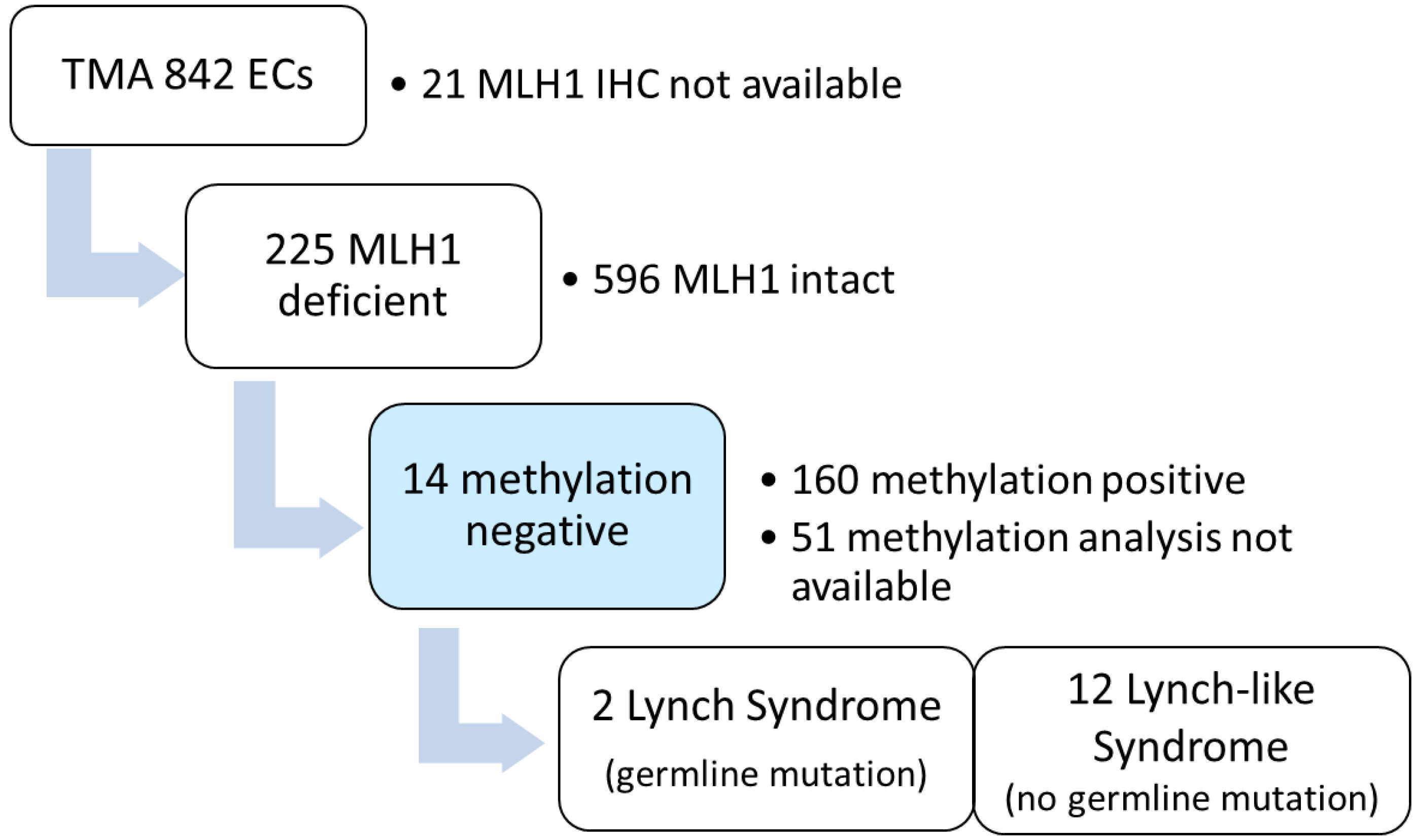
Figure 1. MLH1 immunohistochemistry, methylation analysis and germline mutational analysis performed o 821 unselected EC cases (clinic-based cohort).
Cancers 14 01348 g001
Figure 2. Prevalence of LS, LLS and methylation-linked MLH1-deficient endometrial carcinoma according to age groups (total n = 174 patients).
Cancers 14 01348 g002
Figure 3. Age distribution of MLH1 mutation carriers diagnosed with EC in the LS registry-based cohort (n = 132).
Cancers 14 01348 g003
Table 1. Clinicopathologic data (n = 174 *).
Age (Years) (Median, Range)71.5 (43–94)
Histology (number of cases, percent)
Endometrioid carcinoma160 (92.0)
Clear cell carcinoma4 (2.3)
Serous carcinoma1 (0.6)
Undifferentiated carcinoma6 (3.4)
Carcinosarcoma3 (1.7)
Grade (number of cases, percent) (For endometrioid only, n = 160)
Grade 1 or 2122 (76.3)
Grade 338 (23.8)
FIGO 2009 stage (number of cases, percent)
IA82 (47.1)
IB39 (22.4)
II15 (8.6)
IIIA13 (7.5)
IIIB1 (0.6)
IIIC117 (9.8)
IIIC26 (3.4)
IVA0 (0.0)
IVB1 (0.6)
Adjuvant therapy (number of cases, percent)
No adjuvant therapy19 (10.9)
Vaginal brachytherapy76 (43.7)
Whole pelvic radiotherapy31 (17.8)
Chemotherapy17 (9.7)
Chemotherapy and whole pelvic radiotherapy31 (17.8)
* Cases with MLH1-deficient tumor and successful methylation analysis.
Table 2. Clinicopathological characteristics of patients with non-methylated MLH1-deficient endometrial carcinoma.
MLH1 Germline Mutation (NM_000249.4)Age (Years)HistologyFIGO 2009 StagePOLE
Mut
c.320T > G, (p.Ile107Arg)43Endometrioid G3IA
exon 16 deletion61Clear cell carcinomaIAwt
no48Clear cell carcinomaIAwt
no51Endometrioid G1–2IAwt
no56Endometrioid G1–2IAwt
no57Endometrioid G3IA
no59Endometrioid G3IIIC1wt
no59Endometrioid G3IAwt
no60Endometrioid G3IAwt
no61Endometrioid G1–2IAwt
no66Clear cell carcinomaIIIamut
no67Endometrioid G1–2IAwt
no69Endometrioid G1–2IB
no77Endometrioid G1–2IAwt
Table 3. Comparison of universal and selective MLH1 methylation testing methods according to different age cut-offs.
OutcomeUniversal Met Testing
(n, %)
Cut-Off
<70 Years
(n, %)
Cut-Off
<65 Years
(n, %)
Cut-Off
<60 Years
(n, %)
Patients excluded from methylation testing *0/174 (0.0)98/174 (56.3)123/174 (70.7)145/174 (83.3)
Patients excluded from genetic testing *0/14 (0.0)1/14 (7.1)4/14 (28.6)7/14 (50)
LS/Met-tested cases
(n, %) *
2/174 (1.1)2/76 (2.6)2/51(3.9)1/29 (3.4)
False negative rate (LS) *0/2 (0.0)0/2 (0.0)0/2 (0.0)1/2 (50)
False negative rate (LS) **0/132 (0.0)2/132 (1.5)4/132 (3.0)15/132 (11.4)
LS = Lynch syndrome, Met-tested = methylation tested, * clinic-based cohort, ** registry-based cohort.
Publisher’s Note: MDPI stays neutral with regard to jurisdictional claims in published maps and institutional affiliations.