Review

Radiotherapy for HER 2 Positive Brain Metastases: Urgent Need for a Paradigm Shift

1
Radiation Oncology, Campus Bio-Medico University, Via Alvaro del Portillo 21, 00128 Rome, Italy
2
Radiation Oncology, Tor Vergata University, 00133 Rome, Italy
*
Author to whom correspondence should be addressed.
Cancers 2022, 14(6), 1514; https://doi.org/10.3390/cancers14061514
Submission received: 10 February 2022 / Revised: 7 March 2022 / Accepted: 8 March 2022 / Published: 15 March 2022
(This article belongs to the Special Issue Advanced Breast Cancer: From Biology to Cure)

Abstract

:

Simple Summary

Brain metastases (BMs) are common among patients with advanced HER2 breast cancer. The recent introduction of systemic therapy with central nervous system activity as well as the wider use of brain stereotactic radiotherapy (SRT) are contributing to improving the outcomes for these patients. In this review, we discuss a modified approach to the treatment of HER2-positive BMs from a radiation oncologist point of view, taking into consideration new advances in multimodal therapy and combinations of the most commonly used systemic treatments and brain radiation therapy (RT).

Abstract

Brain metastases (BMs) are common among patients affected by HER2+ metastatic breast cancer (>30%). The management of BMs is usually multimodal, including surgery, radiotherapy, systemic therapy and palliative care. Standard brain radiotherapy (RT) includes the use of stereotactic radiotherapy (SRT) for limited disease and whole brain radiotherapy (WBRT) for extensive disease. The latter is an effective palliative treatment but has a reduced effect on brain local control and BM overall survival, as it is also associated with severe neurocognitive sequelae. Recent advances both in radiation therapy and systemic treatment may change the paradigm in this subset of patients who can experience long survival notwithstanding BMs. In fact, in recent studies, SRT for multiple BM sites (>4) has shown similar efficacy when compared to irradiation of a limited number of lesions (one to three) without increasing toxicity. These findings, in addition to the introduction of new drugs with recognized intracranial activity, may further limit the use of WBRT in favor of SRT, which should be employed for treatment of both multiple-site BMs and for oligo-progressive brain disease. This review summarizes the supporting literature and highlights the need for optimizing combinations of the available treatments in this setting, with a particular focus on radiation therapy.

1. Introduction

Breast cancer is the second most common malignancy associated with increased risk of developing brain metastases (BMs). The incidence of BMs in breast cancer (BC) patients has increased over the past decade. Breast cancer brain metastases (BCBMs) occur in approximately 10–16% of breast cancer patients, especially among patients with HER-2-positive breast cancer [1,2,3].
Before the introduction of trastuzumab, a monoclonal antibody that binds to human epidermal growth factor receptor 2 (HER2), the overall survival (OS) in HER2-positive patients was shorter due to the progression of systemic disease. After the introduction of trastuzumab, OS has been significantly improved due to extracranial control of the disease. More recently, due to the introduction of novel HER2-targeted therapies, the survival of HER2-positive BC patients has further increased, ranging from 12 to 24 months [4,5].
Local treatment of BCBM is multimodal and can include surgery, whole brain radiation (WBRT) and stereotactic radiosurgery (SRS) or fractionated stereotactic radiotherapy (fSRT). Surgery is performed in selected cases and should be preferred for larger and symptomatic lesions. Surgery alone is not sufficient for local control, and postoperative radiotherapy (RT) is recommended [6,7,8].
Historically, the standard treatment for patients with multiple BMs has been WBRT, while SRS was applied only to patients with one or a limited number of BMs (two to four). More recently, Yamamoto et al. reported that there was no difference in median OS between patients with 2–4 BMs and 5–10 BMs treated with SRS. In this study, 10% of patients had breast cancer [9,10]. Moreover, the incidence rate of one or more treatment-related adverse events did not differ between patients with 2–4 or 5–10 BMs.
Furthermore, novel anti-HER2 drugs, such as neratinib, tucatinib and trastuzumab deruxtecan, showed relevant central nervous system (CNS) activity in these patients, which adds to the known action of lapatinib-containing regimens [11].
For all the above reasons, and also due to the longer survival experienced by patients with HER2 BCBMs, the management of these patients represents a unique scenario requiring a multidisciplinary and tailored approach. In this review, we will summarize the supporting literature and discuss the option of a modified approach to the treatment of HER2-positive BMs from a radiation oncologist perspective, taking into consideration new advances in multimodal therapy and combinations of the most commonly used systemic treatments and RT.

2. Brain Radiotherapy: Lessons from the Past and Novel Treatments

Over the years, brain radiotherapy has continued to evolve thanks to advances in radiation techniques including the use of intensity-modulated radiotherapy (IMRT), volumetric-modulated arc therapy (VMAT) and SRT (Figure 1).

2.1. WBRT

WBRT has been used for many years as the mainstay radiation treatment for BMs. The typical dose and fractionation schedule for WBRT is 30 Gray (Gy) in 10 fractions or, alternatively, 20 Gy in 5 fractions. WBRT after either surgical resection or radiosurgery does not improve OS [12,13], while as a palliative treatment it is very effective: 7 out of 10 patients receive benefits in terms of symptom improvement. However, the local control rate using WBRT is limited. Chougule et al. [14] reported a local control of 87%, 91% and 62% for patients treated with SRS (Gamma Knife—GK), SRS + WBRT, and WBRT alone, respectively, suggesting comparatively lower local tumor control for patients undergoing WBRT only.
In order to increase the rates of local control, novel techniques have been developed using a conformational fractionated external beam boost, thereby reaching local control rates above 75%. Dose escalation can be achieved through the use of WBRT + SIB, as well as through the application of WBRT followed by SRS (WBRT + SRS) [15].
The control of dose escalation can be managed with IMRT or VMAT through the use of the simultaneous integrated boost (SIB) technique [16]. Through the use of WBRT + SIB, a boost on BMs can be achieved in a single session with an optimized dose distribution, and the single simulation protocol allows for reduced waiting and dose delivery time. From a radiobiological point of view, SIB techniques reduce the possibility of reoxygenation and re-assortment between fractions. The chance of tumor cell repopulation or a sublet repair process decreases with a reduced time break between WBRT and SRS [17].
Progression outside or in the boost area is strictly related to the applied technique. The incidence of progression outside the dose boost area was found to be significantly lower in patients treated with WBRT + SIB compared with those treated with WBRT + SRS (39.4% vs. 75%). This may be due to the faster reduction in dose outside the BMs in the WBRT + SRS group than in the WBRT + SIB group. The incidence of progression in the dose-boost area was significantly higher in the WBRT + SIB group (60.6%) than in the WBRT + SRS group (25%). This could be due to the higher biologically effective dose (BED) to the P-GTV in the WBRT + SRS group than in the group treated with WBRT + SIB [18].
WBRT is associated with several side effects, such as temporary increase of brain edema and/or hair loss, but mainly neurocognitive function (NCF) impairment [19], that can negatively impact quality of life (QoL) [20]. The most common neurocognitive disfunction is short-term memory loss [21]. NCF decrease can occur 3–6 months after WBRT and can be irreversible and progressive [22].
Since the pathological process behind NCF decline has been mainly attributed to the exposure of neural stem cells of the hippocampus dentate gyrus to radiation, the hippocampal-avoidance technique (HA-WBRT) has been developed in order to reduce NFC decline [23]. This can be feasible, as there is low risk of metastases and recurrence in the hippocampal region [24].
A phase II multi-institutional trial demonstrated that HA-WBRT helps preserve neurocognitive function and quality of life compared with historical controls (p < 0.001) [25]. The phase III NRG Oncology CC001 study demonstrated a lower risk of neurocognitive failure after HA-WBRT plus memantine compared with WBRT plus memantine (adjusted hazard ratio, 0.74; 95% CI, 0.58–0.95; p = 0.02). Furthermore, HA-WBRT showed no differences in intracranial progression-free survival (PFS), OS and toxicity [26]. Moreover, HA-WBRT without memantine showed better memory preservation at 6-month follow-up compared with WBRT alone [27].
Other strategies to improve NCF include the use of neuroprotective drugs, such as memantine, an antagonist of the N-methyl-D-aspartate (NMDA) receptor used in the setting of vascular dementia. RTOG 0614 aimed to investigate the protective effects of memantine on cognitive function in patients receiving WBRT. In this randomized study, patients enrolled in the memantine arm experienced significantly later onsets of cognitive decline (hazard ratio 0.78, 95% confidence interval 0.62–0.99, p = 0.01) [28].
With available technology, it is also possible to specifically and simultaneously deliver fractionated radiation doses to tumors and at-risk brain areas while sparing hippocampal structures [29].
In a study by Westover, a single-arm phase II trial, WBRT (20 Gy in 10 fractions over 2–2.5 weeks) were used together with a simultaneous integrated boost (40 Gy in 10 fractions to identified metastatic lesions) and a hippocampal sparing approach. Patients were then evaluated for NCF decline after treatment using the HVLT-R DR (Hopkins Verbal Learning Test-Revised Delayed Recall), and the results showed significantly better outcomes in patients treated with HSIB-WBRT (hippocampal sparing integrated boost WBRT) compared with historical outcomes and with historical outcomes in patients treated with non-Hippocampal-sparing WBRT and comparable outcomes with those of patients treated with SRS alone; the intracranial control rates were also similar to patients treated with WBRT + SRS [30].

2.2. SRS and Fractionated Stereotactic Radiotherapy (fSRT)

SRS is a technique based on the use of intersected beams that can be delivered in both single RT or fSRT. Compared with WBRT, it allows for a more precise target irradiation to a specific area in order to obtain, through the delivery of a highly conformal and high dose of radiation, an ablative effect on target tissue. Its precise targeting allows for a reduction of radiation exposure to the area surrounding the lesion, thereby reducing the damage to adjacent healthy tissue [31].
SRS treatment can, however, be associated with late toxicity characterized mainly by the occurrence of radionecrosis (RN) [32]. RN results from direct brain damage or from necrosis due to an inflammatory cascade mediated by vascular endothelial injury. It can be asymptomatic or it can cause several neurological symptoms, such as seizures, cognitive deficits, headaches and vomiting. The etiology of RN is multifactorial and can depend on RT dose, fractionation, the volume of BMs, the site of irradiation and the volume of healthy brain irradiated. In the literature, the reported incidence rate ranges from 3% to 24% [32].
According to the RTOG 90–05 protocol, the rates of RN observed in patients with recurrent BMs undergoing a new course of brain RT with single-fraction radiosurgery were 8% at 12 months and 11% at 24 months [33].
Increased rates of RN can be observed if SRS is delivered concurrently with systemic therapies. However, the effective incidence cannot be properly quantified as its definition as study endpoint has been inconsistent. Moreover, no imaging modality is the gold standard test to discriminate between RN and tumor progression, even if the ongoing research is testing the role of PET-based or MRI-based imaging [32].
SRS has recently also been employed to treat multiple BMs. VMAT is a new technique used for linac-based SRS or fSRT, which allows, through the use of a single plan with one isocenter, the treatment of multiple targets simultaneously [34,35]. The use of non-coplanar arcs and the simultaneous variation of MLC leaf positions, dose rate, and gantry rotation speed during the treatment dose delivery allows VMAT to be effective in distributing a highly conformational dose [34]. Treating multiple targets at the same time leads to shortened treatment time, possibly reducing the adverse dosimetric effects linked to intrafraction motion [36].
The difference between WBRT and SRS in patients with five or more BMs still remains unclear. A phase III randomized trial compared SRS with WBRT in 5–15 BMs [9]. The trial has demonstrated that there is no difference in the median OS of 10.4 months in the SRS group and 8.4 months in the WBRT group (p = 0.45). Thus, the authors concluded that avoiding WBRT may also be possible for patients with more than five metastases [9]. Furthermore, neurocognitive function did not differ between the two groups [10]. Two currently ongoing studies are investigating quality of life differences between HA-WBRT and SRS in patients with 5–20 BMs (NCT03075072 study) and the control rate for 1–10 BMs using fSRT (NCT04061408 study).

3. Combination of Brain RT with Systemic Therapy in the Treatment of BCBMs

3.1. Brain RT + Trastuzumab

Trastuzumab is the humanized form of the murine antibody directed to the external domain of HER2. Trastuzumab inhibits the growth of a variety of cancer cells overexpressing HER2 and accrues significant clinical benefit in the metastatic and adjuvant settings.
Preclinical studies demonstrated the critical role played by trastuzumab as a radiosensitizer. In fact, the exposure of breast cancer cells with high levels of HER2 to trastuzumab markedly increased the apoptosis rate caused by RT. Moreover, in cells with experimentally elevated levels of HER2, downregulation of HER2 by trastuzumab restored cellular radiosensitivity [37].
Despite the limited blood–brain permeability, several studies demonstrated that trastuzumab alone for the treatment of HER2-positive breast cancer results in a prolonged time to BMs and longer survival time after the diagnosis of BMs [38]. Moreover, even though trastuzumab is a large molecule, studies on 64Cu-DOTA-trastuzumab or 89Zr-trastuzumab PET imaging showed some BBB penetration and metastatic brain lesions in HER2-positive breast cancer patients [39].
Chargary et al. investigated the outcome of 31 patients treated with WBRT and trastuzumab and reported a radiological response in 74.2% and clinical response in 87.1%. No Grade 2 or higher acute toxicity was observed [40]. Miller et al., in a large retrospective series, including 187 HER2+ patients treated with SRS to 374 BCBMs, reported a longer median survival with the concurrent use of HER2 antibodies (17.9 months vs. 15.1 months; p = 0.04). Nearly all HER2 antibodies were treated with trastuzumab alone (94%). The reported toxicity was acceptable, with a rate of RN of 3.8% [41]. See Table 1 for details.

3.2. Brain RT + Trastuzumab–Pertuzumab

Pertuzumab is a humanized monoclonal antibody which is directed against the extracellular domain of the HER2 receptor and inhibits the interaction of HER2 with HER3. Similar to trastuzumab, pertuzumab stimulates both antibody-dependent and cell-mediated cytotoxicity. In preclinical HER2-positive models, the dual blockade showed increased antitumor effect compared with either agent alone. Dual blockade is also likely to be a powerful radiosensitizer in anti-HER2 therapy. Even if pertuzumab is a large molecule, studies using positron emission tomography–computed tomography (PET-CT) with 89Zr-pertuzumab showed some blood–brain barrier permeability [42]. In the phase II single arm PATRICIA trial, the combination of trastuzumab and pertuzumab achieved an ORR of 11% with a 6-month clinical benefit of 51% in 40 patients progressing after radiotherapy [43].
Not many data are available on the association between dual blockade with pertuzumab and trastuzumab and brain RT. In the phase III CLEOPATRA trial, 23 patients received RT to the brain and only 5 SRS in the pertuzumab–trastuzumab arm [44]. Bergen et al. reported the outcome of 252 HER2-positive BC patients. Among these patients, only 26 were treated with dual blockade and 14 were eligible for response. Of these, 12 patients underwent local therapy (either surgery or WBRT or SRS). The ORR and iCBR were 92.9% and 100.0%, respectively. The only patient who did not receive local therapy (surgery, WBRT or SRT) reported a partial response (PR) [45].
Recently, our institution reported a case series of 10 patients treated with fSRT to 32 BMs. The ORR was 68.7%, while only 1 RN was reported with a median follow-up of 18.3 months. Overall BM median survival was 33.9 months (95% CI 24.1–43.6). The mean duration of dual blockade in these patients was 27.9 months (range: 10.1–53.7 months) [46]. See Table 1 for details.
Table 1. Combination of systemic therapy and brain RT: trastuzumab, trastuzumab–pertuzumab.
AuthorsStudyYearPopulationTreatmentOutcomeAdverse Effects
Chargari et al.
[42]
Retrospective2011- 31 patients
-
25 patients: WBRT 30 Gy/10 fr
-
6 patients: WBRT other fractionation
-
17 patients: trastuzumab 2 mg/kg
-
Weekly
-
14 patients: 6 mg/kg repeated every 21 days
OR: 74.2%
Clinical Response: 87.1%
RN
/
OTHER
Related to WBRT:
Grade 1 -Headache 8 (26%)
-Nausea/vomiting 4 (13%)
Related to trastuzumab
Allergic-like reaction 2 (6.5%)
Decrease in LVEF 3 (9.5%)
Miller
et al.
[43]
Retrospective2017- 187 patients (374 lesions)
-
36% upfront SRS
-
78% upfront WBRT
-
15% underwent upfront surgery
-
83% received hormone therapy
-
80% received HER2 antibodies
-
38% received HER2/EGFR TKIs
-
63% received cytotoxic chemotherapy
OS
Concurrent HER2 antibodies 17.9 months vs. local therapy alone 15.1 months (p = 0.04)
RN
-
SRS alone = 5.6%
-
SRS + trastuzumab = 3.8%
-
SRS + lapatinib = 1.3%
Ippolito E et al.
[47]
Retrospective2021- 10 patients (32 lesions)
-
fSRT Median dose 27 Gy (range 12–27 Gy)
OR
68.7%
iCB
100%
DIF
4/10 patients (40.0%)
Median OBMS
33.9 months (95%CI 24.1–43.6)
Mean duration of PT treatment
27.9 months (range: 10.1–53.7 months)
RN
-
- 1/32 = 3.1%
-
All concurrent pertuzumab and trastuzumab
ABBREVIATIONS: WBRT = whole brain radiotherapy; SRS = stereotactic radiosurgery; fSRT = fractionated stereotactic radiotherapy; OR = overall response rate; iCB = intracranial clinical benefit; DIF = distant intracranial failure DIF; OBMS = overall brain metastases survival.

3.3. Brain RT + Lapatinib

Lapatinib ditosylate is a dual receptor TKI targeting simultaneously two members of the HER family of receptors, HER1 (EGFR1/ErbB1) and HER2/c-neu (ErbB2), that acts by reversibly binding to the cytoplasmic ATP-binding site of the tyrosine kinase domain, producing the inhibition of various downstream signaling cascades involved in anti-proliferative and pro-apoptotic effects [48].
A small molecule can cross the blood–brain barrier and can therefore reach BMs. In a study using radiolabelled lapatinib, the positron emission PET scan showed selective radioactivity uptake in HER2+ brain metastasis compared with normal brain tissue [47]. Morikawa provided evidence for drug uptake of lapatinib in a surgical series of resected, mostly non-irradiated BCBMs [49]. In a clinical setting, in patients not pretreated with RT, the upfront use of lapatinb resulted in an objective response rate of 66% and progression-free survival of 5.5 months [50].
Furthermore, lapatinib has demonstrated in an in vitro experiment that it can increase radiosensibility in HER2+ breast cancer cells. In particular, lapatinib improved the radiosensitivity of breast SKBR3 and BT474 cells, thereby hindering the repair of DNA damage, as suggested by the prolongation of radiation-induced γH2AX foci and the downregulation of phosphorylated DNA-dependent protein kinase and the catalytic subunit (pDNAPKcs) shown using a clonogenic assay [51]. Lapatinib can also potentiate the radiation-induced irreversible arrest of cell proliferation by increasing expression of caspase 3 and the rate of apoptosis and senescence [52].
In a phase 1 trial aimed at evaluating the maximum tolerated dose (MTD) and the feasibility of lapatinib being given concurrently with WBRT, Lin et al. reported, out of 28 eligible patients, an encouraging CNS objective response rate (ORR) of 79%, with 46% of patients remaining progression-free (CNS or non-CNS) at 6 months [53].
In a large retrospective cohort of 132 HER2+ BCBMs, Kim et al. showed how lapatinib given concurrently with SRS resulted in an improved complete response (35% vs. 11%) and the best objective response (median 100% vs. 70% reduction) compared with SRS alone [54].
In a subsequent cohort of 126 patients with a total of 157 HER2+ BCBMs treated at the same institution with SRS and lapatinib, Parsai et al. reported that the efficacy of combining lapatinib and SRS was greater for smaller lesions, with a greater reduction of 12 months local failure compared with SRS alone (5.7% vs. 15.1%). Moreover, it was unexpectedly noted that the rate of radiation necrosis was also reduced when SRS was given concurrently with lapatinib (1.3% vs. 6.3%) [55].
In a recent meta-analysis, including six studies with 843 HER2 + BCBMs, SRS in association with concurrent lapatinib was also investigated. In two studies, increased survival was reported with the association of SRS and any use of lapatinib (Shireen et al.: 27.3 vs. 19.5 months, p = 0.03; Kim et al.: 33.3 vs. 23.6 months, p = 0.009). Local control was also significantly increased with SRS plus lapatinib based on the meta-analysis of three studies (HR 0.47 [0.33, 0.66], p = 0.0001), in particular when lapatinib was given concurrently. In addition, two studies reported a lower radiation necrosis rate with concurrent lapatinib and SRS [56]. See Table 2 for details.

3.4. Brain RT + TDM1

Trastuzumab emtansine (T-DM1) is an immunoconjugated drug consisting of the monoclonal antibody trastuzumab bound to emtansine, a potent maitansin-derived microtubule inhibitor.
Even if it is well demonstrated that HER2 overexpression induces radioresistance, in preclinical studies, T-DM1 failed to induce radiosensitivity in human breast cancer cells expressing HER2. In fact, T-DM1 induced cellular death due to the intracellular action of emtansine. However, the radiation dose needed to achieve 10% cell survival did not decrease when radiotherapy was combined with T-DM1 [57]. In the exploratory analysis of the KAMILLA trial evaluating the safety and efficacy of T-DM1 in 398 patients with stable or occult brain metastases, the intracranial response rate evaluable in 126 patients was 21.4%, with an intracranial clinical benefit of 42.9%. Among 67 patients with measurable BMs not treated with brain RT, a volume reduction >30% was achieved in 49% of patients [59].
Some studies have tried to evaluate the efficacy and safety of a combined treatment with T-DM1 and SRT (see Table 3) [59]. In the majority of these studies, T-DM1 administration in association with brain RT appears to be linked with an increased risk of RN.
One of the first published studies described the outcome of a total of 13 breast cancer patients receiving SRS, of which 7 were also treated with T-DM1: 57% of patients treated with SRS + T-DM1 developed RN [65].
In another small series of 12 patients (PS = 0–1) with HER2+ BCBMs treated from 2014 to 2015 with T-DM1 (every 3 weeks) and concurrent or sequential radiosurgery with or without WBRT, a radiological response was obtained in three out of four cases (75%), with PD in 25% of patients (5.5 months for local progression and 6.5 months for brain progression). In the arm where the sequential SRS + T-DM1 approach was combined, the RN rate was 33.3%, with an edema rate of 28.6%, and 50% in the concurrent group, with an edema rate of 25% [66].
Stumpf et al. analyzed the safety profile of T-DM1 and SRS in a total of 45 patients with BCBMs treated between 2014 and 2017. Of these, 30/45 patietns were HER2-positive. Out of 45 patients, 23 received T-DM1, of which 16 received T-DM1 with SRS/FSRT (7 T-DM1 + sequential SRS–6 T-DM1 + concurrent SRS), while 22/45 received T-DM1 alone. Approximately 22% of patients were found to develop clinically significant radionecrosis, especially in the combined approach, with concomitant SRS of 23 patients who received T-DM1 + RT (vs. 1 out of 22 patients who did not receive T-DM1). The authors additionally provided pre-clinical evidence of the increased rate of RN, explaining how unintended T-DM1 targeting of reactive astrocytes in healthy nearby brain tissue is a mechanism underlying T-DM1/SRS-induced toxicity [60].
More recently, Mills et al. reported on the outcome of 16 patients with HER2 + BCBMs who received SRS or fSRT with concomitant or sequential T-DM1 from December 2013 to December 2019. Overall, 40 BCBMs were treated, of which 24 were treated with SRS (median dose 21 Gy) and 16 with fSRT (median dose 25 Gy delivered in 3 to 5 fractions). Moreover, 19 metastases were treated with RT + concurrent T-DM1, 11 with RT delivered before T-DM1, 10 with RT delivered after T-DM1. After SRS or FSRT, local control (LC) and distant intracranial control (DIC) at 20 months was 75% and 50%, respectively. Moreover, OS at 20 months was 67% after SRS or FSRT and 79% following diagnosis of BCBMs. In this series, only 1 case of radionecrosis was reported (179 days after combined therapy with T-DM1 and SRS on 5 lesions, with the first dose 8 days before SRT). Furthermore, in 8/18 treatment sessions, mild radiation-related side effects were reported (such as headaches and fatigue) [61].
One study, published in abstract form only, showed that T-DM1 exposure post-SRS was independently associated with a higher risk of RN in HER2+ BMBC patients. There were 67 patients with HER2 + BMBC (223 lesions) treated with SRS identified; among these, 21 (31.3%) were treated with post-SRS T-DM1, with a median follow-up of 15.6 months. Considering independent risk factors of RN, either T-DM1 treatment post-SRS (HR 2.5, 95% CI 1.2–5.3, p = 0.02) or an RT dose with a BED >50.4 Gy (HR 2.4, 95% CI 1.1–5.1, p = 0.02) was given; patients treated with T-DM1 and SRS had a 25.2% (95% CI 12.8–37.6%) risk of RN at both 1- and 2-years post-T-DM1, with 80% of all RN cases occurring within 12 months of T-DM1 treatment [62]. See Table 3 for details.

3.5. Limitations

Evaluating the safety and efficacy of combined treatment is difficult and limited by the retrospective nature of the available studies. Moreover, as more new drugs with intracranial activity are going to be used in the near future, combination with RT will be more complex. In fact, in order to extract information that could add to current clinical practice in this area, it would be useful to include data on brain radiotherapy treatments (i.e., type, dose, interval time from delivery of RT to the beginning of systemic therapy). However, most clinical trials available do not provide such data.
On the other hand, only a few prospective studies of combined treatments are ongoing (see Table 4).
Finally, regarding the occurrence of RN, the main limitation is related to the absence of a consistent defined endpoint across combination studies. In fact, different grades of RN are often reported together, not even distinguishing symptomatic from asymptomatic events. This can be crucial, as symptomatic RN is less common but clinically meaningful.

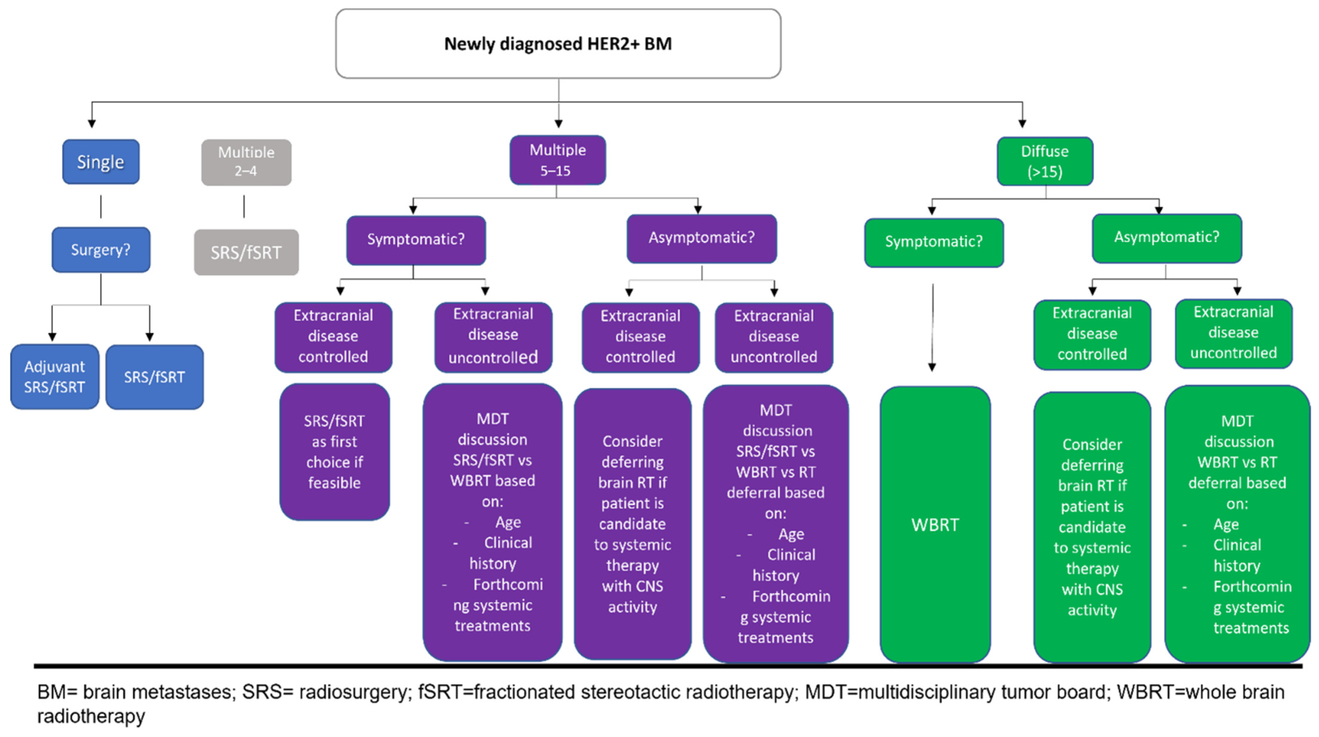
4. Future Developments: How the Therapeutic Algorithm Should Be Reshaped

With this knowledge of all the advances in HER2+ BMs treatment, the optimal treatment algorithm for these patients cannot be the same as that used to treat BMs from other malignancies. However, a strategy based on patient performance status and symptoms, the intracranial volume of disease, the burden of the extracranial disease and the planned systemic therapy could define the best approach for both first BM occurrence or subsequent relapse (see Figure 2 and Figure 3).

4.1. When Should WBRT Be Delivered?

WBRT for newly diagnosed BMs should be delivered in very selected cases as a palliative treatment or to increase intracranial control in patients with symptomatic disease or disease rapidly progressing after brain SRT. See Table 5 for details.
When WBRT is the indicated treatment, clinical and technical approaches to reduce NCF impairment should always be used for patients with a life expectancy >6 months.

4.2. When Should SRS/fSRT Be Delivered?

SRS/fSRT should always be the first RT option for patients with HER2+ BCBMs. Recent guidelines recommend the use of this treatment option for patients with 1–4 metastases [63]. However, in this setting, patients with multiple BMs could experience long survival [64,67,68]. For this reason, once a patient has been accurately selected (good PS, extracranial disease controlled, first occurrence), SRT/fSRT should only be used after a multidisciplinary discussion.
SRS/fSRT should also be used for late relapse (>6 months) after a first RT treatment course (either WBRT or SRS/fSRT). It can also be used as a boost dose to larger BMs after WBRT, taking into account the increased risk of RN.

4.3. When Can RT Deferral Be an Option?

RT deferral can be a valid option for patients with diffuse asymptomatic BMs who are eligible for a systemic therapy with intracranial activity, providing close MRI follow-up evaluations are undertaken to properly treat progression. Recent guidelines recommend this approach for patients to be treated with tucatinib combination therapy [68], even though lapatinib combinations may play a role in this setting.

4.4. How to Manage the Association of Systemic Therapy with Brain RT?

Figure 4 summarizes the rate of RN reported in the studies investigating the association between SRT and the most commonly used systemic therapies in HER2 patients.
Trastuzumab, as well as trastuzumab together with pertuzumab, could be administered concurrently with RT, even though there are not many data available on the association with brain RT. With regard to T-DM1, due to the high frequency of brain radionecrosis reported in retrospective studies, concurrent administration of T-DM1 and brain SBRT should be avoided, and, if necessary, strategies to reduce the rate of RN should be employed (for instance, preferring fSRT or hypofractionated SRT, especially for larger lesions, to limit the volume of healthy brain receiving high RT doses). As retrospective studies suggest, the association of a low frequency of brain RN with the concurrent administration of lapatinib and brain RT should be considered as the safest option to treat a large number of metastases and larger lesions. However, given the lack of clinical comparative data, it is difficult to evaluate whether the combination of lapatinib and brain RT would be more effective than novel TKIs, such as neratinib or tucatinib. Moreover, to the best of our knowledge, no data are available on the combination with brain RT for novel drugs, such as new generation TKIs, as well as trastuzumab deruxetan in combination with brain RT. Therefore, patients undergoing these therapies should not be treated concurrently with brain RT and side effects should be carefully monitored.

Author Contributions

Conceptualization, E.I. and S.R.; data curation, S.S., P.M. and C.G.; formal analysis, M.F.; methodology, R.M.D.; supervision, M.F. and S.R.; writing—original draft, S.C., C.T. (Claudia Talocco), C.T. (Claudia Tacconi) and V.P.; writing—review and editing, E.I., M.F. and S.R. All authors have read and agreed to the published version of the manuscript.

Funding

This research did not receive any specific grant from funding agencies in the public, commercial, or not-for-profit sectors.

Conflicts of Interest

The authors declare no conflict of interest.

References

  1. Sammons, S.; Van Swearingen, A.E.D.; Chung, C.; Anders, C.K. Advances in the management of breast cancer brain metastases. Neurooncol. Adv. 2021, 3 (Suppl. 5), v63–v74. [Google Scholar] [CrossRef] [PubMed]
  2. Yuan, P.; Gao, S.L. Management of breast cancer brain metastases: Focus on human epidermal growth factor receptor 2-positive breast cancer. Chronic Dis. Transl. Med. 2017, 3, 21–32. [Google Scholar] [CrossRef]
  3. Clayton, A.J.; Danson, S.; Jolly, S.; Ryder, W.D.; Burt, P.A.; Stewart, A.L.; Wilkinson, P.M.; Welch, R.S.; Magee, B.; Wilson, G.; et al. Incidence of cerebral metastases in patients treated with trastuzumab for metastatic breast cancer. Br. J. Cancer 2004, 91, 639–643. [Google Scholar] [CrossRef]
  4. Bendell, J.C.; Domchek, S.M.; Burstein, H.J.; Harris, L.; Younger, J.; Kuter, I.; Bunnell, C.; Rue, M.; Gelman, R.; Winer, E. Central nervous system metastases in women who receive trastuzumab-based therapy for metastatic breast carcinoma. Cancer 2003, 97, 2972–2977. [Google Scholar] [CrossRef] [PubMed]
  5. Stemmler, H.J.; Kahlert, S.; Siekiera, W.; Untch, M.; Heinrich, B.; Heinemann, V. Characteristics of patients with brain metastases receiving trastuzumab for HER2 overexpressing metastatic breast cancer. Breast 2006, 15, 219–225. [Google Scholar] [CrossRef] [PubMed]
  6. Patchell, R.A.; Tibbs, P.A.; Walsh, J.W.; Dempsey, R.J.; Maruyama, Y.; Kryscio, R.J.; Markesbery, W.R.; Macdonald, J.S.; Young, B. A randomized trial of surgery in the treatment of single metastases to the brain. N. Engl. J. Med. 1990, 322, 494–500. [Google Scholar] [CrossRef] [PubMed]
  7. Vecht, C.J.; Haaxma-Reiche, H.; Noordijk, E.M.; Padberg, G.W.; Voormolen, J.H.; Hoekstra, F.H.; Tans, J.T.; Lambooij, N.; Metsaars, J.A.; Wattendorff, A.R.; et al. Treatment of single brain metastasis: Radiotherapy alone or combined with neurosurgery? Ann. Neurol. 1993, 33, 583–590. [Google Scholar] [CrossRef] [PubMed]
  8. Mintz, A.H.; Kestle, J.; Rathbone, M.P.; Gaspar, L.; Hugenholtz, H.; Fisher, B.; Duncan, G.; Skingley, P.; Foster, G.; Levine, M. A randomized trial to assess the efficacy of surgery in addition to radiotherapy in patients with a single cerebral metastasis. Cancer 1996, 78, 1470–1476. [Google Scholar] [CrossRef] [PubMed]
  9. Yamamoto, M.; Serizawa, T.; Shuto, T.; Akabane, A.; Higuchi, Y.; Kawagishi, J.; Yamanaka, K.; Sato, Y.; Jokura, H.; Yomo, S.; et al. Stereotactic radiosurgery for patients with multiple brain metastases (JLGK0901): A multi-institutional prospective observational study. Lancet Oncol. 2014, 15, 387–395. [Google Scholar] [CrossRef] [PubMed]
  10. Yamamoto, M.; Serizawa, T.; Higuchi, Y.; Sato, Y.; Kawagishi, J.; Yamanaka, K.; Shuto, T.; Akabane, A.; Jokura, H.; Yomo, S.; et al. A Multi-institutional Prospective Observational Study of Stereotactic Radiosurgery for Patients With Multiple Brain Metastases (JLGK0901 Study Update): Irradiation-related Complications and Long-term Maintenance of Mini-Mental State Examination Scores. Int. J. Radiat. Oncol. Biol. Phys. 2017, 99, 31–40. [Google Scholar] [CrossRef] [PubMed] [Green Version]
  11. Stavrou, E.; Winer, E.P.; Lin, N.U. How we treat HER2-positive brain metastases. ESMO Open 2021, 6, 100256. [Google Scholar] [CrossRef] [PubMed]
  12. Aoyama, H.; Shirato, H.; Tago, M.; Nakagawa, K.; Toyoda, T.; Hatano, K.; Kenjyo, M.; Oya, N.; Hirota, S.; Shioura, H.; et al. Stereotactic radiosurgery plus whole-brain radiation therapy vs stereotactic radiosurgery alone for treatment of brain metastases: A randomized controlled trial. JAMA 2006, 295, 2483–2491. [Google Scholar] [CrossRef] [PubMed]
  13. Kocher, M.; Soffietti, R.; Abacioglu, U.; Villà, S.; Fauchon, F.; Baumert, B.G.; Fariselli, L.; Tzuk-Shina, T.; Kortmann, R.D.; Carrie, C.; et al. Adjuvant whole-brain radiotherapy versus observation after radiosurgery or surgical resection of one to three cerebral metastases: Results of the EORTC 22952-26001 study. J. Clin. Oncol. 2011, 29, 134–141. [Google Scholar] [CrossRef] [PubMed] [Green Version]
  14. Chougule, P.; Burton-Williams, M.; Saris, S.; Zheng, Z.; Ponte, B.; Noren, G.; Alderson, L.; Friehs, G.; Wazer, D.; Epstein, M. Randomized treatment of brain metastasis with gamma knife radiosurgery, whole brain radiotherapy or both. Int. J. Radiat. Oncol. Biol. Phys. 2000, 48, 114. [Google Scholar] [CrossRef]
  15. Casanova, N.; Mazouni, Z.; Bieri, S.; Combescure, C.; Pica, A.; Weber, D.C. Whole brain radiotherapy with a conformational external beam radiation boost for lung cancer patients with 1-3 brain metastasis: A multi institutional study. Radiat. Oncol. 2010, 5, 13. [Google Scholar] [CrossRef] [PubMed] [Green Version]
  16. Weber, D.C.; Caparrotti, F.; Laouiti, M.; Malek, K. Simultaneous in-field boost for patients with 1 to 4 brain metastasis/es treated with volumetric modulated arc therapy: A prospective study on quality-of-life. Radiat. Oncol. 2011, 6, 79. [Google Scholar] [CrossRef] [PubMed] [Green Version]
  17. Bruzzaniti, V.; Abate, A.; Pedrini, M.; Benassi, M.; Strigari, L. IsoBED: A tool for automatic calculation of biologically equivalent fractionation schedules in radiotherapy using IMRT with a simultaneous integrated boost (SIB) technique. J. Exp. Clin. Cancer Res. 2011, 30, 52. [Google Scholar] [CrossRef] [Green Version]
  18. Lin, B.; Huang, D.; Du, H.; Fan, J.; Zhang, Y.; Feng, G.; Gao, F.; Du, X.B. Whole-Brain Radiation Therapy With Simultaneous Integrated Boost Versus Whole-Brain Radiation Therapy Plus Stereotactic Radiosurgery for the Treatment of Brain Metastasis From Lung Cancer. Front. Oncol. 2021, 11, 631422. [Google Scholar] [CrossRef]
  19. Roman, D.D.; Sperduto, P.W. Neuropsychological effects of cranial radiation: Current knowledge and future directions. Int. J. Radiat. Oncol. Biol. Phys. 1995, 31, 983–998. [Google Scholar] [CrossRef] [PubMed]
  20. Soffietti, R.; Kocher, M.; Abacioglu, U.M.; Villa, S.; Fauchon, F.; Baumert, B.G.; Fariselli, L.; Tzuk-Shina, T.; Kortmann, R.D.; Carrie, C.; et al. A European Organisation for Research and Treatment of Cancer phase III trial of adjuvant whole-brain radiotherapy versus observation in patients with one to three brain metastases from solid tumors after surgical resection or radiosurgery: Quality-of-life results. J. Clin. Oncol. 2013, 31, 65–72. [Google Scholar] [CrossRef] [PubMed]
  21. Chang, E.L.; Wefel, J.S.; Hess, K.R.; Allen, P.K.; Lang, F.F.; Kornguth, D.G.; Arbuckle, R.B.; Swint, J.M.; Shiu, A.S.; Maor, M.H.; et al. Neurocognition in patients with brain metastases treated with radiosurgery or radiosurgery plus whole-brain irradiation: A randomised controlled trial. Lancet Oncol. 2009, 10, 1037–1044. [Google Scholar] [CrossRef] [PubMed]
  22. Greene-Schloesser, D.; Moore, E.; Robbins, M.E. Molecular pathways: Radiation-induced cognitive impairment. Clin. Cancer Res. 2013, 19, 2294–2300. [Google Scholar] [CrossRef] [PubMed] [Green Version]
  23. Gondi, V.; Tomé, W.A.; Mehta, M.P. Why avoid the hippocampus? A comprehensive review. Radiother. Oncol. 2010, 97, 370–376. [Google Scholar] [CrossRef] [PubMed] [Green Version]
  24. Sun, B.; Huang, Z.; Wu, S.; Shen, G.; Cha, L.; Meng, X.; Ding, L.; Wang, J.; Song, S. Incidence and relapse risk of intracranial metastases within the perihippocampal region in 314 patients with breast cancer. Radiother. Oncol. 2016, 118, 181–186. [Google Scholar] [CrossRef] [PubMed]
  25. Gondi, V.; Pugh, S.L.; Tome, W.A.; Caine, C.; Corn, B.; Kanner, A.; Rowley, H.; Kundapur, V.; DeNittis, A.; Greenspoon, J.N.; et al. Preservation of memory with conformal avoidance of the hippocampal neural stem-cell compartment during whole-brain radiotherapy for brain metastases (RTOG 0933): A phase II multi-institutional trial. J. Clin. Oncol. 2014, 32, 3810–3816. [Google Scholar] [CrossRef] [PubMed]
  26. Brown, P.D.; Gondi, V.; Pugh, S.; Tome, W.A.; Wefel, J.S.; Armstrong, T.S.; Bovi, J.A.; Robinson, C.; Konski, A.; Khuntia, D.; et al. Hippocampal Avoidance During Whole-Brain Radiotherapy Plus Memantine for Patients With Brain Metastases: Phase III Trial NRG Oncology CC001. J. Clin. Oncol. 2020, 38, 1019–1029. [Google Scholar] [CrossRef]
  27. Yang, W.C.; Chen, Y.F.; Yang, C.C.; Wu, P.F.; Chan, H.M.; Chen, J.L.; Chen, G.Y.; Cheng, J.C.; Kuo, S.H.; Hsu, F.M. Hippocampal avoidance whole-brain radiotherapy without memantine in preserving neurocognitive function for brain metastases: A phase II blinded randomized trial. Neuro Oncol. 2021, 23, 478–486, Erratum in Neuro Oncol. 2021, 23, 2125. [Google Scholar] [CrossRef] [PubMed]
  28. Brown, P.D.; Pugh, S.; Laack, N.N.; Wefel, J.S.; Khuntia, D.; Meyers, C.; Choucair, A.; Fox, S.; Suh, J.H.; Roberge, D.; et al. Memantine for the prevention of cognitive dysfunction in patients receiving whole-brain radiotherapy: A randomized, double-blind, placebo-controlled trial. Neuro Oncol. 2013, 15, 1429–1437. [Google Scholar] [CrossRef]
  29. Gutiérrez, A.N.; Westerly, D.C.; Tomé, W.A.; Jaradat, H.A.; Mackie, T.R.; Bentzen, S.M.; Khuntia, D.; Mehta, M.P. Whole brain radiotherapy with hippocampal avoidance and simultaneously integrated brain metastases boost: A planning study. Int. J. Radiat. Oncol. Biol. Phys. 2007, 69, 589–597. [Google Scholar] [CrossRef] [PubMed] [Green Version]
  30. Westover, K.D.; Mendel, J.T.; Dan, T.; Kumar, K.; Gao, A.; Pulipparacharuv, S.; Iyengar, P.; Nedzi, L.; Hannan, R.; Anderson, J.; et al. Phase II trial of hippocampal-sparing whole brain irradiation with simultaneous integrated boost for metastatic cancer. Neuro Oncol. 2020, 22, 1831–1839. [Google Scholar] [CrossRef] [PubMed]
  31. Hall, E.J.; Brenner, D.J. The radiobiology of radiosurgery: Rationale for different treatment regimes for AVMs and malignancies. Int. J. Radiat. Oncol. Biol. Phys. 1993, 25, 381–385. [Google Scholar] [CrossRef] [PubMed]
  32. Lupattelli, M.; Alì, E.; Ingrosso, G.; Saldi, S.; Fulcheri, C.; Borghesi, S.; Tarducci, R.; Aristei, C. Stereotactic Radiotherapy for Brain Metastases: Imaging Tools and Dosimetric Predictive Factors for Radionecrosis. J. Pers. Med. 2020, 10, 59. [Google Scholar] [CrossRef]
  33. Shaw, E.; Scott, C.; Souhami, L.; Dinapoli, R.; Kline, R.; Loeffler, J.; Farnan, N. Single dose radiosurgical treatment of recurrent previously irradiated primary brain tumors and brain metastases: Final report of RTOG protocol 90-05. Int. J. Radiat. Oncol. Biol. Phys. 2000, 47, 291–298. [Google Scholar] [CrossRef] [PubMed]
  34. Clark, G.M.; Popple, R.A.; Young, P.E.; Fiveash, J.B. Feasibility of single-isocenter volumetric modulated arc radiosurgery for treatment of multiple brain metastases. Int. J. Radiat. Oncol. Biol. Phys. 2010, 76, 296–302. [Google Scholar] [CrossRef] [PubMed]
  35. Kang, J.; Ford, E.C.; Smith, K.; Wong, J.; McNutt, T.R. A method for optimizing LINAC treatment geometry for volumetric modulated arc therapy of multiple brain metastases. Med. Phys. 2010, 37, 4146–4154. [Google Scholar] [CrossRef] [PubMed]
  36. Guckenberger, M.; Roesch, J.; Baier, K.; Sweeney, R.A.; Flentje, M. Dosimetric consequences of translational and rotational errors in frame-less image-guided radiosurgery. Radiat. Oncol. 2012, 7, 63. [Google Scholar] [CrossRef] [Green Version]
  37. Liang, K.; Lu, Y.; Jin, W.; Ang, K.K.; Milas, L.; Fan, Z. Sensitization of breast cancer cells to radiation by trastuzumab. Mol. Cancer Ther. 2003, 2, 1113–1120. [Google Scholar] [PubMed]
  38. Park, Y.H.; Park, M.J.; Ji, S.H.; Yi, S.Y.; Lim, D.H.; Nam, D.H.; Lee, J.I.; Park, W.; Choi, D.H.; Huh, S.J.; et al. Trastuzumab treatment improves brain metastasis outcomes through control and durable prolongation of systemic extracranial disease in HER2-overexpressing breast cancer patients. Br. J. Cancer 2009, 24, 100, 894–900. [Google Scholar] [CrossRef] [PubMed]
  39. Kurihara, H.; Hamada, A.; Yoshida, M.; Shimma, S.; Hashimoto, J.; Yonemori, K.; Tani, H.; Miyakita, Y.; Kanayama, Y.; Wada, Y.; et al. (64) Cu-DOTA-trastuzumab PET imaging and HER2 specificity of brain metastases in HER2-positive breast cancer patients. EJNMMI Res. 2015, 5, 8. [Google Scholar] [CrossRef] [Green Version]
  40. Chargari, C.; Idrissi, H.R.; Pierga, J.Y.; Bollet, M.A.; Diéras, V.; Campana, F.; Cottu, P.; Fourquet, A.; Kirova, Y.M. Preliminary results of whole brain radiotherapy with concurrent trastuzumab for treatment of brain metastases in breast cancer patients. Int. J. Radiat. Oncol. Biol. Phys. 2011, 81, 631–636. [Google Scholar] [CrossRef] [PubMed]
  41. Miller, J.A.; Kotecha, R.; Ahluwalia, M.S.; Mohammadi, A.M.; Chao, S.T.; Barnett, G.H.; Murphy, E.S.; Vogelbaum, M.A.; Angelov, L.; Peereboom, D.M.; et al. Overall survival and the response to radiotherapy among molecular subtypes of breast cancer brain metastases treated with targeted therapies. Cancer 2017, 123, 2283–2293. [Google Scholar] [CrossRef] [PubMed]
  42. Ulaner, G.A.; Lyashchenko, S.K.; Riedl, C.; Ruan, S.; Zanzonico, P.B.; Lake, D.; Jhaveri, K.; Zeglis, B.; Lewis, J.S.; O’Donoghue, J.A. First-in-Human Human Epidermal Growth Factor Receptor 2-Targeted Imaging Using 89Zr-Pertuzumab PET/CT: Dosimetry and Clinical Application in Patients with Breast Cancer. J. Nucl. Med. 2018, 59, 900–906. [Google Scholar] [CrossRef] [PubMed] [Green Version]
  43. Lin, N.U.; Kumthekar, P.; Sahebjam, S.; Ibrahim, N.; Fung, A.; Cheng, A.; Nicholas, A.; Wang, B.; Pegram, M. Pertuzumab (P) plus high-dose trastuzumab (H) for the treatment of central nervous system (CNS) progression after radiotherapy (RT) in patients (pts) with HER2-positive metastatic breast cancer (MBC): Primary efficacy analysis results from the phase II PATRICIA study. Cancer Res. 2020, 80, P1-18-03. [Google Scholar]
  44. Swain, S.M.; Baselga, J.; Miles, D.; Im, Y.H.; Quah, C.; Lee, L.F.; Cortés, J. Incidence of central nervous system metastases in patients with HER2-positive metastatic breast cancer treated with pertuzumab, trastuzumab, and docetaxel: Results from the randomized phase III study Cleopatra. Ann. Oncol. 2014, 25, 1116–1121. [Google Scholar] [CrossRef]
  45. Bergen, E.S.; Binter, A.; Starzer, A.M.; Heller, G.; Kiesel, B.; Tendl-Schulz, K.; Bago-Horvath, Z.; Furtner, J.; Leitner, J.; Exner, R.; et al. Favourable outcome of patients with breast cancer brain metastases treated with dual HER2 blockade of trastuzumab and pertuzumab. Ther. Adv. Med. Oncol. 2021, 13, 17588359211009002. [Google Scholar] [CrossRef]
  46. Ippolito, E.; Silipigni, S.; Matteucci, P.; Greco, C.; Pantano, F.; D’Auria, G.; Quattrocchi, C.C.; Floreno, B.; Fiore, M.; Gamucci, T.; et al. Stereotactic Radiation and Dual Human Epidermal Growth Factor Receptor 2 Blockade with Trastuzumab and Pertuzumab in the Treatment of Breast Cancer Brain Metastases: A Single Institution Series. Cancers 2022, 14, 303. [Google Scholar] [CrossRef]
  47. Saleem, A.; Searle, G.E.; Kenny, L.M.; Huiban, M.; Kozlowski, K.; Waldman, A.D.; Woodley, L.; Palmieri, C.; Lowdell, C.; Kaneko, T.; et al. Lapatinib access into normal brain and brain metastases in patients with Her-2 overexpressing breast cancer. EJNMMI Res. 2017, 5, 30, Erratum in EJNMMI Res. 2017, 7, 74. [Google Scholar] [CrossRef] [Green Version]
  48. Kennecke, H.; Yerushalmi, R.; Woods, R.; Cheang, M.C.; Voduc, D.; Speers, C.H.; Nielsen, T.O.; Gelmon, K. Metastatic behavior of breast cancer subtypes. J. Clin. Oncol. 2010, 28, 3271–3277. [Google Scholar] [CrossRef] [PubMed]
  49. Morikawa, A.; Peereboom, D.M.; Thorsheim, H.R.; Samala, R.; Balyan, R.; Murphy, C.G.; Lockman, P.R.; Simmons, A.; Weil, R.J.; Tabar, V.; et al. Capecitabine and lapatinib uptake in surgically resected brain metastases from metastatic breast cancer patients: A prospective study. Neuro Oncol. 2015, 17, 289–295, Erratum in Neuro Oncol. 2015, 17, 1423. [Google Scholar] [CrossRef] [Green Version]
  50. Bachelot, T.; Romieu, G.; Campone, M.; Diéras, V.; Cropet, C.; Dalenc, F.; Jimenez, M.; Le Rhun, E.; Pierga, J.Y.; Gonçalves, A.; et al. Lapatinib plus capecitabine in patients with previously untreated brain metastases from HER2-positive metastatic breast cancer (LANDSCAPE): A single-group phase 2 study. Lancet Oncol. 2013, 14, 64–71. [Google Scholar] [CrossRef] [PubMed]
  51. Yu, T.; Cho, B.J.; Choi, E.J.; Park, J.M.; Kim, D.H.; Kim, I.A. Radiosensitizing effect of lapatinib in human epidermal growth factor receptor 2-positive breast cancer cells. Oncotarget 2016, 7, 79089–79100. [Google Scholar] [CrossRef] [PubMed] [Green Version]
  52. Angelini, P.D.; Zacarias Fluck, M.F.; Pedersen, K.; Parra-Palau, J.L.; Guiu, M.; Bernadó Morales, C.; Vicario, R.; Luque-García, A.; Navalpotro, N.P.; Giralt, J.; et al. Constitutive HER2 signaling promotes breast cancer metastasis through cellular senescence. Cancer Res. 2013, 73, 450–458, Erratum in Cancer Res. 2013, 73, 6095. [Google Scholar] [CrossRef] [PubMed] [Green Version]
  53. Lin, N.U.; Freedman, R.A.; Ramakrishna, N.; Younger, J.; Storniolo, A.M.; Bellon, J.R.; Come, S.E.; Gelman, R.S.; Harris, G.J.; Henderson, M.A.; et al. A phase I study of lapatinib with whole brain radiotherapy in patients with Human Epidermal Growth Factor Receptor 2 (HER2)-positive breast cancer brain metastases. Breast Cancer Res. Treat. 2013, 142, 405–414. [Google Scholar] [CrossRef] [PubMed]
  54. Kim, J.M.; Miller, J.A.; Kotecha, R.; Chao, S.T.; Ahluwalia, M.S.; Peereboom, D.M.; Mohammadi, A.M.; Barnett, G.H.; Murphy, E.S.; Vogelbaum, M.A.; et al. Stereotactic radiosurgery with concurrent HER2-directed therapy is associated with improved objective response for breast cancer brain metastasis. Neuro Oncol. 2019, 21, 659–668. [Google Scholar] [CrossRef] [PubMed]
  55. Parsai, S.; Miller, J.A.; Juloori, A.; Chao, S.T.; Kotecha, R.; Mohammadi, A.M.; Ahluwalia, M.S.; Murphy, E.S.; Barnett, G.H.; Vogelbaum, M.A.; et al. Stereotactic radiosurgery with concurrent lapatinib is associated with improved local control for HER2-positive breast cancer brain metastases. J. Neurosurg. 2019, 132, 503–511. [Google Scholar] [CrossRef] [PubMed]
  56. Khan, M.; Zhao, Z.; Arooj, S.; Zheng, T.; Liao, G. Lapatinib Plus Local Radiation Therapy for Brain Metastases From HER-2 Positive Breast Cancer Patients and Role of Trastuzumab: A Systematic Review and Meta-Analysis. Front. Oncol. 2020, 10, 576926. [Google Scholar] [CrossRef] [PubMed]
  57. Mignot, F.; Kirova, Y.; Verrelle, P.; Teulade-Fichou, M.P.; Megnin-Chanet, F. In vitro effects of Trastuzumab Emtansine (T-DM1) and concurrent irradiation on HER2-positive breast cancer cells. Cancer Radiother. 2021, 25, 126–134. [Google Scholar] [CrossRef] [PubMed]
  58. Montemurro, F.; Delaloge, S.; Barrios, C.H.; Wuerstlein, R.; Anton, A.; Brain, E.; Hatschek, T.; Kelly, C.M.; Peña-Murillo, C.; Yilmaz, M.; et al. Trastuzumab emtansine (T-DM1) in patients with HER2-positivemetastatic breast cancer and brain metastases: Exploratory final analysis of cohort 1 from KAMILLA, a single-arm phase IIIb clinical trials. Ann. Oncol. 2020, 10, 1350–1358. [Google Scholar] [CrossRef]
  59. Kowalczyk, L.; Bartsch, R.; Singer, C.F.; Farr, A. Adverse Events of Trastuzumab Emtansine (T-DM1) in the Treatment of HER2-Positive Breast Cancer Patients. Breast Care 2017, 12, 401–408. [Google Scholar] [CrossRef]
  60. Stumpf, P.K.; Cittelly, D.M.; Robin, T.P.; Carlson, J.A.; Stuhr, K.A.; Contreras-Zarate, M.J.; Lai, S.; Ormond, D.R.; Rusthoven, C.G.; Gaspar, L.E.; et al. Combination of Trastuzumab Emtansine and Stereotactic Radiosurgery Results in High Rates of Clinically Significant Radionecrosis and Dysregulation of Aquaporin-4. Clin. Cancer Res. 2019, 25, 3946–3953. [Google Scholar] [CrossRef] [PubMed] [Green Version]
  61. Mills, M.N.; Walker, C.; Thawani, C.; Naz, A.; Figura, N.B.; Kushchayev, S.; Etame, A.; Yu, H.M.; Robinson, T.J.; Liu, J.; et al. Trastuzumab Emtansine (T-DM1) and stereotactic radiation in the management of HER2+ breast cancer brain metastases. BMC Cancer 2021, 21, 223. [Google Scholar] [CrossRef] [PubMed]
  62. Id Said, B.; Chen, H.; Jerzak, K.; Myrehaug, S.D.; Tseng CLDetsky, J.; Husain, Z.A.; Sahgal, A.; Soliman, H. T-DM1 Increases the Risk of SRS-Induced Radiation Necrosis in Her2+ Breast Cancer. Int. J. Radiat. Oncol. Biol. Phys. 2021, 111, e568. [Google Scholar] [CrossRef]
  63. Vogelbaum, M.A.; Brown, P.D.; Messersmith, H.; Brastianos, P.K.; Burri, S.; Cahill, D.; Dunn, I.F.; Gaspar, L.E.; Gatson, N.T.N.; Gondi, V.; et al. Treatment for Brain Metastases: ASCO-SNO-ASTRO Guideline. J. Clin. Oncol. 2021, 40, 452–516. [Google Scholar] [CrossRef] [PubMed]
  64. Gori, S.; Puglisi, F.; Moroso, S.; Fabi, A.; La Verde, N.; Frassoldati, A.; Tarenzi, E.; Garrone, O.; Vici, P.; Laudadio, L.; et al. The HERBA Study: A Retrospective Multi-Institutional Italian Study on Patients With Brain Metastases From HER2-Positive Breast Cancer. Clin. Breast Cancer 2019, 19, e501–e510. [Google Scholar] [CrossRef] [PubMed]
  65. Carlson, J.A.; Nooruddin, Z.; Rusthoven, C.; Elias, A.; Borges, V.F.; Diamond, J.R.; Kavanagh, B.; Kabos, P. Trastuzumab emtansine and stereotactic radiosurgery: An unexpected increase in clinically significant brain edema. Neuro Oncol. 2014, 16, 1006–1009. [Google Scholar] [CrossRef] [PubMed] [Green Version]
  66. Geraud, A.; Xu, H.P.; Beuzeboc, P.; Kirova, Y.M. Preliminary experience of the concurrent use of radiosurgery and T-DM1 for brain metastases in HER2-positive metastatic breast cancer. J. Neurooncol. 2017, 131, 69–72. [Google Scholar] [CrossRef] [PubMed]
  67. Russillo, M.; Ferretti, G.; Vidiri, A.; Gasparro, S.; Cognetti, F.; Pellegrini, D.; Fabi, A. Impressive Long-term Response with Pertuzumab and Trastuzumab in HER2-positive Breast Cancer with Brain Metastasis. Vivo 2018, 32, 839–842. [Google Scholar] [CrossRef]
  68. Silipigni, S.; Ippolito, E.; Matteucci, P.; Santo, B.; Gangemi, E.; La Cesa, A.; Santini, D.; Greco, C.; Ramella, S. Repeated courses of radiation treatment in an HER2-positive breast cancer patient with diffuse brain metastases: A case report. Breast J. 2020, 26, 1370–1371. [Google Scholar] [CrossRef] [PubMed]
Figure 1. Evolution of brain radiotherapy (from left to right): (1) WBRT; (2) HA-WBRT; (3) WBRT + SIB; (4) multiple-site SRT.
Cancers 14 01514 g001
Figure 2. Proposed algorithm for newly diagnosed HER2 BMs.
Cancers 14 01514 g002
Figure 3. Proposed algorithm for recurrent HER2 BMs after brain RT.
Cancers 14 01514 g003
Figure 4. RN incidence reported in studies investigating the association between SRT and systemic therapies.
Cancers 14 01514 g004
Table 2. Combination of systemic therapy and brain RT: lapatinib.
AuthorsStudyYearPopulationTreatmentOutcomeAdverse Effects
Lin et al.
[55]
Phase I2013- 28 patients
-
WBRT (37.5 Gy/15 fr)
-
Lapatinib Day 1, a loading dose of 750 mg for two doses. Day 2, three DLs once daily: 1000 (DL1), 1250 (DL2), and 1500 (DL3) mg
ORR
79%
6 mPFS
46% at 6 months
RN
/
Parsai et al.
[57]
Retrospective 2019- 126 patients (479 lesions)
-
24 patients concurrent lapatinib + SRS
-
SRS +/− WBRT
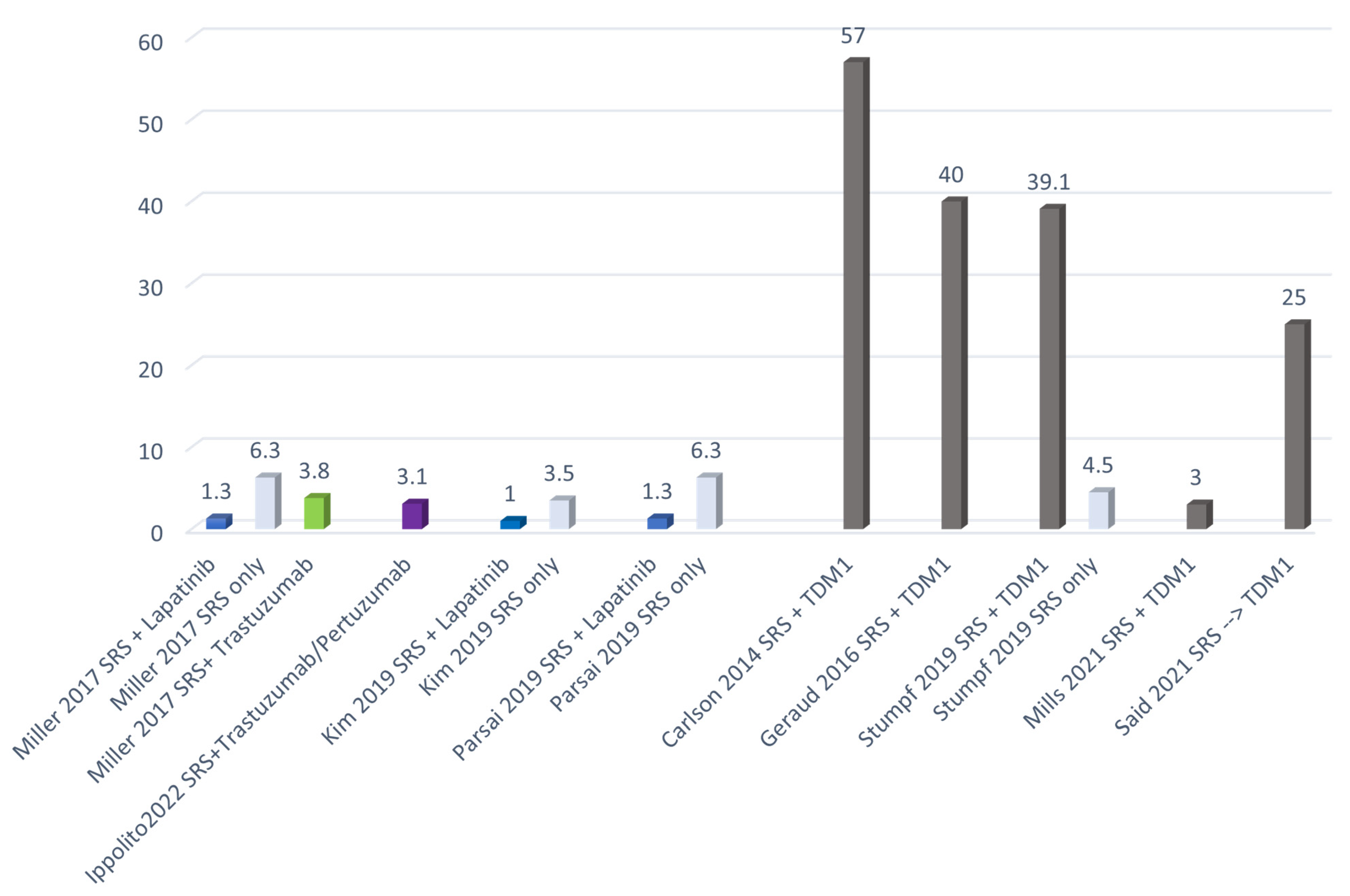
LF
- Concurrent lapatinib + SRS
12-month (5.7% vs. 15.1%, p < 0.01), 24-month (6.5% vs. 18.0%, p < 0.01)
RN
- 1.3% cohort treated with lapatinib
- SRS alone 6.3%
(p< 0.01)
Kim et al.
[56]
Retrospective 2019- 84 patients
(487 lesions)
SRS (18–24 Gy)+/−WBRT
- 132 lesions (27%) SRS +
lapatinib
- 355 lesions (73%) only SRS
CR
- Lapatinib + SRS 35%
- Only SRS
11%
(p = 0.008)
OR
- Lapatinib + SRS -100%
- Only SRS
- 70%
(p < 0.001)
RN
- SRS + lapatinib 1.0%
- Only SRS 3.5% (p = 0.27)
M Khan et al.
[58]
Review–meta-analysis2020
-
6 studies
-
843 HER2+ patients
-
SRS +/− WBRT
-
WBRT
279 patients: lapatinib +/− anti-HER2 therapy
- 610 patients: anti-HER2 therapy (mainly trastuzumab)
- 227 patients: no anti-HER2 therapy
OS
- Lapatinib-based therapy was associated with increase in OS (HR 0.63 p < 0.00001)
LC
significantly increased with concurrent lapatinib + SRS (HR 0.47 (0.33, 0.66), p = 0.0001)
RN
Lower rate for concurrent lapatinib + SRS:
- Miller: 1.3% vs. 6.3% (p 0.001)
- Kim: 1% vs. 3.5% (p = 0.134)
- Parsai: 1.3% vs. 6.3% (p = 0.001)
ABBREVIATIONS: SRS = stereotactic radiosurgery; BCBMs = breast cancer brain metastases; ORR = objective response rate; CR = complete response; OR = objective response; WBRT = whole brain radotherapy; LF = local failure; OS = overall survival; LC = local control; RN = Radionecrosis.
Table 3. Combination of systemic therapy and brain RT: TDM1.
AuthorsStudyYearPopulationTreatmentOutcomeAdverse Effects
Carlson J
et al.
[60]
Retrospective2014- 13 patients7/13 patients T-DM1 + SRSNRRN
57% (4/7) SRS + T-DM1
Geraud A
et al.
[61]
Retrospective2017- 12 patients
-
4/12 SRS + Concurrent T-DM1
-
8/12 SRS + Sequencial T-DM1
SRS + Concurrent T-DM1
- OR 75%
- PD 25%
SRS + Sequential T-DM1
- OR 75%
- PD 25%
RN
- 33.3% (4/12) in the sequential group
- 50% (2/4) in the concurrent group
OTHER
- Edema rate of 28.6% in the sequential group
- Edema rate of 25% in the concurrent group
Stumpf PK et al.
[62]
Retrospective 2019-45 patients TDM1 + RT:
-23/45 (51.1%):
-16 SRS/FSRT + Concurrent T-DM1:
-7 SRS and sequential T-DM1
- Only SRS 22/45
-15 patients also received WBRT
NRRN
10/45 (22.2%) clinically significant:
- 9 of 23 (39.1%) patients received T-DM1 (3 sequential SRS, 6 concurrent SRS)
- 1 of 22 (4.5%) patients did not receive T-DM1
Mills MN
et al.
[63]
Retrospective 2021- 16 patients (40 lesions)RT:
- 24 SRS (median dose 21 Gy)
- 16 FSRT (median dose 25 Gy/3–5 fractions)
T-DM1:
- 19 RT + Concurrent T-DM1
- 11 RT and sequential T-DM1
- 10 T-DM1 and sequential RT
- 20 months LC 75% (SRS or FSRT)
- 20 months DIC 50% (SRS or FSRT)
- 20 months OS 67% (SRS or FSRT) and 79% (following BCBM diagnosis)
RN
1 patient (3%)
OTHER:
45% G1–G2 Headaches and fatigue
Id Said B
et al.
[64]
Retrospective 2021- 67 patients (223 lesions)21/67 patients (31.3%)
SRS and sequential T-DM1
/RN
- Post-SRS
1-year 6.7% (95% CI 2.7–10.7%) 2-year 15.2% (95% CI 9.2–21.3%)
- SRS and sequential T-DM1
1- and 2-years 25.2% (95% CI 12.8–37.6%)
ABBREVIATIONS: TDM1 = trastuzumab emtansine; BCBMs = breast cancer brain metastases; fSRT = fractionated stereotactic radiotherapy; SRS = Stereotactic radiosurgery; RT = radiotherapy; LC = local control; OS = overall survival; WBRT = whole brain radiotherapy; OR = overall response rate; PD = progression disease; RN = radionecrosis.
Table 4. Ongoing clinical trials of combinations of brain RT with systemic therapy in the treatment of BCBMs.
Clinicaltrials
Identifier
TitlePhaseN° PatientsTreatment ArmsEndpoint
NCT05042791Pyrotinib Combined With Brain Radiotherapy in Breast Cancer Patients With BMs2 Randomized362Experimental: fSRT combined with pyrotinib and capecitabine
Active Comparator: WBRT combined with pyrotinib and capecitabine
1-year objective response rate of central nervous system
NCT04582968A Study of Pyrotinib Plus Capecitabine Combined With SRT in HER2+ MBC With BMsI/II47fSRT or WBRT
drug: pyrotinib combined with capecitabine pyrotinib, 400 mg once daily; capecitabine 1000 mg/m2 per day on day 1 through 14, every 21 days
1. Safety and tolerability of pyrotinib plus capecitabine combined with brain radiotherapy
2. Intracranial local tumor control rate
ABBREVIATIONS: BMs = brain metastases; fSRT = fractionated stereotactic radiotherapy; WBRT = whole brain radiotherapy.
Table 5. Patient candidates for WBRT.
WBRT IntentPatient Category
PalliativePatients with a life expectancy of less than 6 months
Increase intracranial controlPatients diagnosed with symptomatic diffuse BMs or multiple BMs (5–15) who are not suitable for SRS/fSRT. Multidisciplinary evaluation is recommended in this latter case for an accurate risk/benefit assessment in terms of disease control and neurocognitive side effects, with particular reference to age, patient preferences and therapeutic options to control extracranial disease
Increase intracranial controlPatients who rapidly progress (<6 months) after a first course of brain RT treatment or with associated systemic progression, with an evaluation of the therapeutic options available on the basis of the patient’s clinical history
Publisher’s Note: MDPI stays neutral with regard to jurisdictional claims in published maps and institutional affiliations.

