
Impact of Positive Radial Margin on Recurrence and Survival in Perihilar Cholangiocarcinoma

 , , , , , , ,
  , ,
, ,  and
and 
Abstract
:Simple Summary
Abstract
1. Introduction
2. Materials and Methods
2.1. Study Population
2.2. Preparation for Surgery and Surgical Procedure
2.3. Pathologic Evaluation
2.4. Study Design and Statistical Analysis
- R0 group: patients with negative DM and RM
- RM group: patients with positive RM and negative DM
- DM ± RM group: patients with positive DM regardless of RM status
3. Results
3.1. Characteristics of the Patients and Surgical Procedures
3.2. Pathologic Features
3.3. Survival Analysis
3.4. Recurrence Analysis
4. Discussion
5. Conclusions
Author Contributions
Funding
Institutional Review Board Statement
Informed Consent Statement
Data Availability Statement
Conflicts of Interest
References
- Lauterio, A.; De Carlis, R.; Centonze, L.; Buscemi, V.; Incarbone, N.; Vella, I.; De Carlis, L. Current Surgical Management of Peri-Hilar and Intra-Hepatic Cholangiocarcinoma. Cancers 2021, 13, 3657. [Google Scholar] [CrossRef] [PubMed]
- Cillo, U.; Fondevila, C.; Donadon, M.; Gringeri, E.; Mocchegiani, F.; Schlitt, H.J.; Ijzermans, J.N.; Vivarelli, M.; Zieniewicz, K.; Olde Damink, S.W.; et al. Surgery for cholangiocarcinoma. Liver Int. 2019, 39, 143–155. [Google Scholar] [CrossRef] [PubMed] [Green Version]
- Lamarca, A.; Edeline, J.; McNamara, M.G.; Hubner, R.A.; Nagino, M.; Bridgewater, J.; Primrose, J.; Valle, J.W. Current standards and future perspectives in adjuvant treatment for biliary tract cancers. Cancer Treat Rev. 2020, 84, 101936. [Google Scholar] [CrossRef] [PubMed]
- Ben-Josef, E.; Guthrie, K.A.; El-Khoueiry, A.B.; Corless, C.L.; Zalupski, M.M.; Lowy, A.M.; Thomas, C.R., Jr.; Alberts, S.R.; Dawson, L.A.; Micetich, K.C.; et al. SWOG S0809: A Phase II Intergroup Trial of Adjuvant Capecitabine and Gemcitabine Followed by Radiotherapy and Concurrent Capecitabine in Extrahepatic Cholangiocarcinoma and Gallbladder Carcinoma. J. Clin. Oncol. 2015, 33, 2617–2622. [Google Scholar] [CrossRef] [Green Version]
- Rizzo, A.; Brandi, G. Pitfalls, challenges, and updates in adjuvant systemic treatment for resected biliary tract cancer. Expert Rev. Gastroenterol. Hepatol. 2021, 15, 547–554. [Google Scholar] [CrossRef]
- Shinohara, K.; Ebata, T.; Shimoyama, Y.; Mizuno, T.; Yokoyama, Y.; Yamaguchi, J.; Onoe, S.; Watanabe, N.; Nagino, M. A Study on Radial Margin Status in Resected Perihilar Cholangiocarcinoma. Ann. Surg. 2021, 273, 572–578. [Google Scholar] [CrossRef]
- Bismuth, H.; Corlette, M.B. Intrahepatic cholangioenteric anastomosis in carcinoma of the hilus of the liver. Surg. Gynecol. Obstet. 1975, 140, 170–178. [Google Scholar]
- Rosa, F.; Costamagna, G.; Doglietto, G.B.; Alfieri, S. Classification of nodal stations in gastric cancer. Transl. Gastroenterol. Hepatol. 2017, 2, 2. [Google Scholar] [CrossRef] [Green Version]
- Pang, Y.Y.; Strasberg, S.M. The Brisbane 2000 Terminology of Liver Anatomy and Resections. HPB 2002, 4, 99–100. [Google Scholar] [CrossRef] [Green Version]
- Dindo, D.; Demartines, N.; Clavien, P.A. Classification of Surgical Complications: A New Proposal With Evaluation in a Cohort of 6336 Patients and Results of a Survey. Ann. Surg. 2004, 240, 205–213. [Google Scholar] [CrossRef]
- Benson, A.B.; D’Angelica, M.I.; Abbott, D.E. Guidelines Insights: Hepatobiliary Cancers, Version 2.2019. J. Natl. Compr. Canc. Netw. 2019, 17, 302–310. [Google Scholar] [CrossRef] [PubMed] [Green Version]
- Amin, M.B.; Edge, S.B.; Greene, F.L.; Byrd, D.R.; Brookland, R.K.; Washington, M.K.; Gershenwald, J.E.; Compton, C.C.; Hess, K.R.; Sullivan, D.C.; et al. AJCC Cancer Staging Manual; Springer International Publishing: Chicago, IL, USA, 2017. [Google Scholar]
- Bird, N.T.E.; McKenna, A.; Dodd, J.; Poston, G.; Jones, R.; Malik, H. Meta-analysis of prognostic factors for overall survival in patients with resected hilar cholangiocarcinoma. Br. J. Surg. 2018, 105, 1408–1416. [Google Scholar] [CrossRef] [PubMed]
- Tang, Z.; Yang, Y.; Zhao, Z.; Wei, K.; Meng, W.; Li, X. The clinicopathological factors associated with prognosis of patients with resectable perihilar cholangiocarcinoma: A systematic review and meta-analysis. Medicine 2018, 97, e11999. [Google Scholar] [CrossRef]
- Liang, L.; Li, C.; Jia, H.D.; Diao, Y.K.; Xing, H.; Pawlik, T.M.; Lau, W.Y.; Shen, F.; Huang, D.S.; Zhang, C.W.; et al. Prognostic factors of resectable perihilar cholangiocarcinoma: A systematic review and meta-analysis of high-quality studies. Ther. Adv. Gastrointest. Endosc. 2021, 14, 2631774521993065. [Google Scholar] [CrossRef]
- Mueller, M.; Breuer, E.; Mizuno, T.; Bartsch, F.; Ratti, F.; Benzing, C.; Ammar-Khodja, N.; Sugiura, T.; Takayashiki, T.; Hessheimer, A.; et al. Perihilar Cholangiocarcinoma–Novel Benchmark Values for Surgical and Oncological Outcomes from 24 Expert Centers. Ann. Surg. 2021; publish ahead of print. [Google Scholar] [CrossRef]
- Koerkamp, B.G.; Wiggers, J.K.; Allen, P.J.; Besselink, M.G.; Blumgart, L.H.; Busch, O.R.; Coelen, R.J.; D’Angelica, M.I.; DeMatteo, R.P.; Gouma, D.J.; et al. Recurrence Rate and Pattern of Perihilar Cholangiocarcinoma after Curative Intent Resection. J. Am. Coll. Surg. 2015, 221, 1041–1049. [Google Scholar] [CrossRef] [PubMed] [Green Version]
- Ke, Q.; Chen, Y.; Huang, Q.; Lin, N.; Wang, L.; Liu, J. Does additional resection of a positive microscopic ductal margin benefit patients with perihilar cholangiocarcinoma: A systematic review and meta-analysis. PLoS ONE 2020, 15, e0232590. [Google Scholar] [CrossRef]
- Endo, I.; House, M.G.; Klimstra, D.S.; Gönen, M.; D’Angelica, M.; DeMatteo, R.P.; Fong, Y.; Blumgart, L.H.; Jarnagin, W.R. Clinical Significance of Intraoperative Bile Duct Margin Assessment for Hilar Cholangiocarcinoma. Ann. Surg. Oncol. 2008, 15, 2104–2112. [Google Scholar] [CrossRef]
- Roos, E.; Franken, L.C.; Soer, E.C.; van Hooft, J.E.; Takkenberg, R.B.; Klümpen, H.J.; Wilmink, J.W.; van de Vijver, M.J.; van Gulik, T.M.; Verheij, J. Lost in translation: Confusion on resection and dissection planes hampers the interpretation of pathology reports for perihilar cholangiocarcinoma. Virchows Arch. 2019, 475, 435–443. [Google Scholar] [CrossRef] [Green Version]
- Chatelain, D.; Farges, O.; Fuks, D.; Trouillet, N.; Pruvot, F.R.; Regimbeau, J.M. Assessment of pathology reports on hilar cholangiocarcinoma: The results of a nationwide, multicenter survey performed by the AFC-HC-2009 study group. J. Hepatol. 2012, 56, 1121–1128. [Google Scholar] [CrossRef]
- Sakamoto, Y.; Kosuge, T.; Shimada, K.; Sano, T.; Ojima, H.; Yamamoto, J.; Yamasaki, S.; Takayama, T.; Makuuchi, M. Prognostic factors of surgical resection in middle and distal bile duct cancer: An analysis of 55 patients concerning the significance of ductal and radial margins. Surgery 2005, 137, 396–402. [Google Scholar] [CrossRef]
- Stremitzer, S.; Stift, J.; Laengle, J.; Schwarz, C.; Kaczirek, K.; Jones, R.P.; Quinn, L.M.; Fenwick, S.W.; Diaz-Nieto, R.; Poston, G.J.; et al. Prognosis and Circumferential Margin in Patients with Resected Hilar Cholangiocarcinoma. Ann. Surg. Oncol. 2021, 28, 1493–1498. [Google Scholar] [CrossRef] [PubMed]
- Zhang, X.F.; Squires, M.H.; Bagante, F.; Ethun, C.G.; Salem, A.; Weber, S.M.; Tran, T.; Poultsides, G.; Son, A.Y.; Hatzaras, I.; et al. The Impact of Intraoperative Re-Resection of a Positive Bile Duct Margin on Clinical Outcomes for Hilar Cholangiocarcinoma. Ann. Surg. Oncol. 2018, 25, 1140–1149. [Google Scholar] [CrossRef] [PubMed]
- Wiggers, J.K.; Koerkamp, B.G.; Cieslak, K.P.; Doussot, A.; van Klaveren, D.; Allen, P.J.; Besselink, M.G.; Busch, O.R.; D’Angelica, M.I.; DeMatteo, R.P.; et al. Postoperative Mortality after Liver Resection for Perihilar Cholangiocarcinoma: Development of a Risk Score and Importance of Biliary Drainage of the Future Liver Remnant. J. Am. Coll. Surg. 2016, 223, 321–331.e1. [Google Scholar] [CrossRef] [PubMed] [Green Version]


| R0 (n = 41) | RM (n = 17) | DM ± RM (n = 17) | Combined (n = 75) | p Value | |
|---|---|---|---|---|---|
| Age, median (IQR) | 68.6 (62.7–75.6) | 63.5 (51.5–68.1) | 64.1 (60.3–66.7) | 65.7 (60.1–73.5) | 0.07 |
| Gender, male | 30 (73%) | 12 (71%) | 10 (59%) | 52 (69%) | 0.55 |
| Bilirubin (mg/dL), median (IQR) | 2.9 (1.0–6.2) | 2.1 (0.7–4.2) | 1.7 (1.1–5.5) | 2.5 (1–5.5) | 0.41 |
| CA19-9 (kU/L), median (IQR) | 108.0 (38.8–444.6) | 312.3 (42.1–1650.0) | 612.2 (250.8–2172.0) | 243.5 (44.7–1171.5) | 0.08 |
| CEA (ng/mL), median (IQR) | 2.9 (1.9–3.6) | 1.6 (1.2–2.5) | 2.2 (1.6–2.9) | 2.20 (1.5–3.5) | 0.24 |
| Pre-operative drain (PTBD or ERCP) | 23 (56%) | 12 (71%) | 13 (76%) | 48 (64%) | 0.28 |
| Bismuth type | 0.79 | ||||
| I | 1 (2.4%) | 1 (6.2%) | 0 (0%) | 2 (2.7%) | |
| II | 7 (17%) | 1 (6.2%) | 4 (24%) | 12 (16%) | |
| IIIa | 13 (32%) | 5 (31%) | 3 (18%) | 21 (28%) | |
| IIIb | 14 (34%) | 5 (31%) | 7 (41%) | 26 (35%) | |
| IV | 6 (15%) | 4 (25%) | 3 (18%) | 13 (18%) | |
| Surgical procedure | 0.68 | ||||
| Left hepatectomy ± S1 | 24 (59%) | 7 (41%) | 11 (65%) | 42 (56%) | |
| Right hepatectomy ± S1 | 12 (29%) | 7 (41%) | 3 (18%) | 22 (29%) | |
| Central hepatectomy ± S1 | 1 (2.4%) | 1 (5.9%) | 0 (0%) | 2 (2.7%) | |
| Isolated bile duct resection | 3 (7.3%) | 1 (5.9%) | 2 (12%) | 6 (8%) | |
| Others | 1 (2.4%) | 1 (5.9%) | 1 (5.9 %) | 3 (4%) | |
| Number of resected segments, median (IQR) | 4 (3–4) | 4 (4–4) | 4 (4–4) | 4 (3–4) | 0.66 |
| Vascular reconstruction | 8 (20%) | 6 (35%) | 1 (5.9%) | 15 (20%) | 0.11 |
| Adjuvant chemotherapy | 26 (63%) | 15 (88%) | 8 (47%) | 49 (65%) | 0.039 |
| Adjuvant radiotherapy | 7 (17%) | 8 (47%) | 1 (5.9%) | 16 (21.3%) | 0.010 |
| R0 (n = 41) | RM (n = 17) | DM ± RM (n = 17) | Combined (n = 75) | p Value | |
|---|---|---|---|---|---|
| Dimension (mm), Median (IQR) | 30 (20–42.5) | 35 (30–40) | 40 (30–40) | 30 (22.5–40) | 0.37 |
| Grade, median (IQR) | 2 (2–2) | 2 (2–3) | 2 (2–2) | 2 (2–2) | 0.37 |
| Grade | 0.73 | ||||
| 1 | 6 (15%) | 1 (5.9%) | 1 (7.1%) | 8 (11%) | |
| 2 | 25 (64%) | 10 (59%) | 10 (71%) | 45 (64%) | |
| 3 | 8 (21%) | 6 (35%) | 3 (21%) | 17 (24%) | |
| T | 0.14 | ||||
| 1 | 6 (15%) | 0 (0%) | 1 (5.9%) | 7 (9.3%) | |
| 2a | 14 (34%) | 4 (24%) | 2 (12%) | 20 (27%) | |
| 2b | 16 (39%) | 13 (76%) | 11 (64.7%) | 40 (53.3%) | |
| 3 | 2 (4.9%) | 0 (0%) | 1 (5.9%) | 3 (4%) | |
| 4 | 2 (4.9%) | 0 (0%) | 2 (12%) | 4 (5.3%) | |
| x | 1 (2.4%) | 0 (0%) | 0 (0%) | 1 (1.3%) | |
| N | 0.52 | ||||
| 0 | 23 (56%) | 10 (59%) | 8 (47%) | 41 (55%) | |
| + | 15 (37%) | 4 (24%) | 8 (47%) | 27 (36%) | |
| x | 3 (7.3%) | 3 (18%) | 1 (5.9%) | 7 (9.3%) | |
| Stage | 0.16 | ||||
| I | 7 (17%) | 0 (0%) | 0 (0%) | 7 (9.3%) | |
| II | 16 (39%) | 13 (76%) | 7 (41%) | 36 (48%) | |
| IIIa | 1 (2%) | 0 (0%) | 0 (0%) | 1 (1%) | |
| IIIb | 3 (7%) | 0 (0%) | 2 (12%) | 5 (7%) | |
| IIIc | 10 (24%) | 4 (24%) | 6 (35%) | 20 (27%) | |
| IV | 4 (9.8%) | 0 (0%) | 2 (12%) | 6 (8%) | |
| Vascular invasion | 27 (71%) | 14 (82%) | 11 (69%) | 52 (73%) | 0.64 |
| Perineural invasion | 30 (81%) | 15 (88%) | 15 (94%) | 60 (86%) | 0.60 |
| R0 | R1 | RM | DM ± RM | |
|---|---|---|---|---|
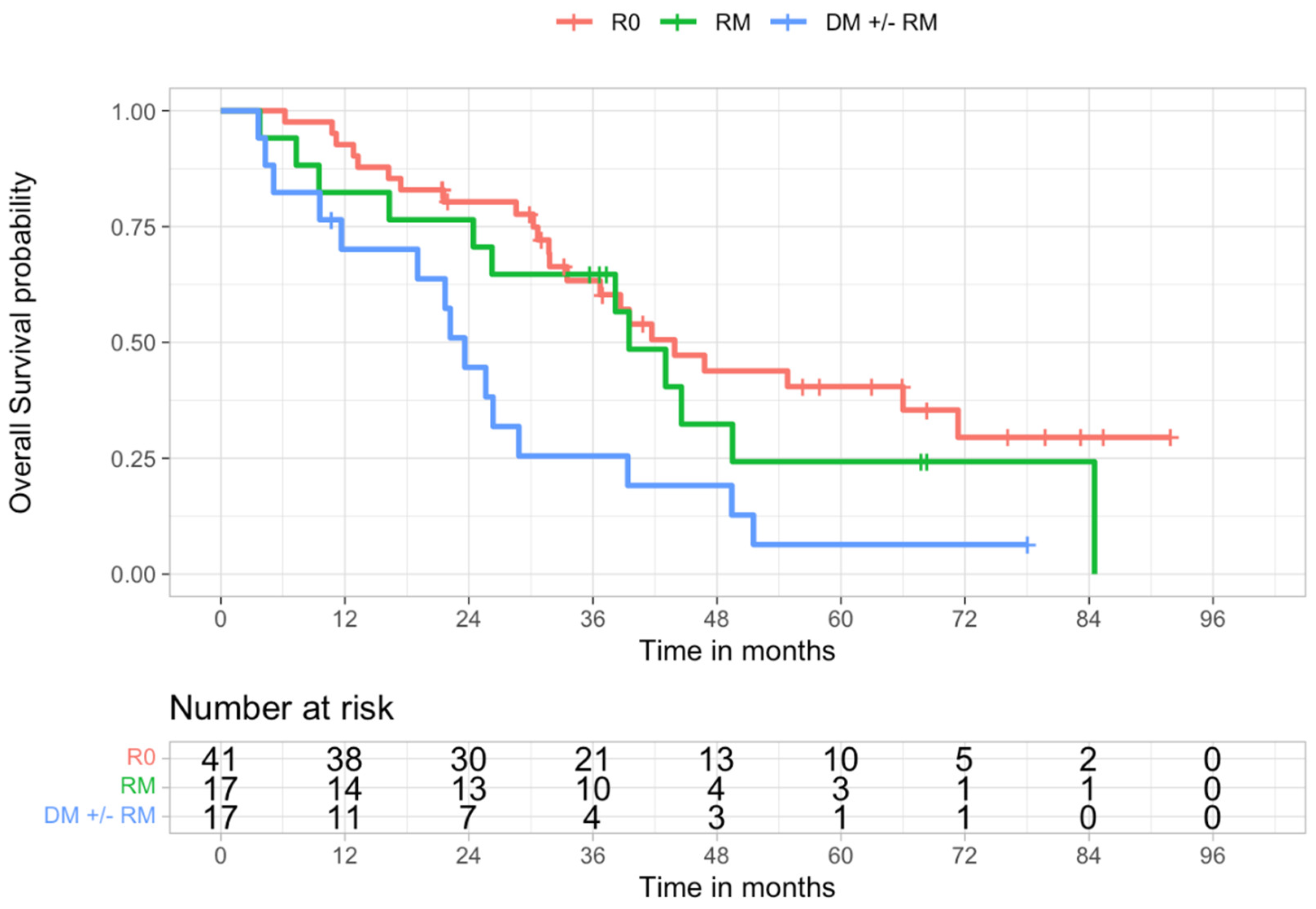
| 1 year | 92.7% | 76.4% | 82.4% | 70.1% |
| 3 years | 63.3% | 45.8% | 64.7% | 25.5% |
| 5 years | 40.5% | 15.3% | 24.3% | 6.4% |
| Median survival, months | 43.9 | 26.3 | 39.5 | 23.6 |
| HR | L 95% CI | U 95% CI | p Value | |
|---|---|---|---|---|
| RM | 1.39 | 0.69 | 2.79 | 0.358 |
| DM ± RM | 2.83 | 1.46 | 5.48 | 0.002 |
| CA19-9> 40 kU/L | 4.33 | 1.69 | 11.07 | 0.002 |
| N+ | 1.99 | 1.09 | 3.66 | 0.026 |
| Bismuth IV | 1.52 | 0.71 | 3.26 | 0.284 |
| Dimension | 1.00 | 0.99 | 1.02 | 0.545 |
| CT adj | 0.71 | 0.39 | 1.28 | 0.254 |
| Stage III–IV | 2.11 | 1.19 | 3.74 | 0.010 |
| G2 | 2.39 | 0.84 | 6.79 | 0.104 |
| G3 | 2.87 | 0.89 | 9.21 | 0.076 |
| Vascular Invasion | 2.17 | 1.04 | 4.52 | 0.038 |
| Complications | 3.94 | 1.40 | 11.04 | 0.009 |
| Recurrence | 14.78 | 3.57 | 61.23 | <0.001 |
| HR | L 95% CI | U 95% CI | p Value | |
|---|---|---|---|---|
| RM | 1.65 | 0.69 | 3.92 | 0.259 |
| DM ± RM | 2.58 | 1.02 | 6.48 | 0.044 |
| CA19-9 | 4.30 | 1.20 | 15.38 | 0.024 |
| N+ | 1.24 | 0.57 | 2.70 | 0.584 |
| Bismuth IV | 5.92 | 1.87 | 18.73 | 0.002 |
| Vascular Invasion | 2.51 | 1.05 | 6.02 | 0.039 |
| Complications | 4.47 | 0.86 | 23.22 | 0.075 |
| Recurrence | 6.91 | 1.60 | 29.83 | 0.009 |
| R0 | R1 | RM | DM ± RM | |
|---|---|---|---|---|
| 1 year | 17.9% | 37.2% | 35.3% | 39.1% |
| 3 years | 53.1% | 73.6% | 64.7% | 84.8% |
| 5 years | 65.6% | 86.4% | 73.5% | 100% |
| Median DFS, months | 30 | 16.3 | 20 | 15 |
| HR | L 95% CI | U 95% CI | p-Value | |
|---|---|---|---|---|
| RM | 1.32 | 0.66 | 2.61 | 0.432 |
| DM ± RM | 2.72 | 1.39 | 5.34 | 0.004 |
| CA19-9 >40 kU/L | 2.44 | 1.13 | 5.28 | 0.023 |
| N+ | 1.52 | 0.84 | 2.76 | 0.169 |
| Bismuth IV | 1.50 | 0.73 | 3.10 | 0.274 |
| Dimension | 1.00 | 0.99 | 1.02 | 0.547 |
| CT adj | 0.97 | 0.54 | 1.76 | 0.925 |
| Stage III-IV | 1.51 | 0.86 | 2.68 | 0.154 |
| G2 | 1.28 | 0.52 | 3.11 | 0.592 |
| G3 | 1.67 | 0.61 | 4.55 | 0.317 |
| Vascular Invasion | 1.97 | 1.00 | 3.86 | 0.049 |
| Complications | 3.00 | 1.18 | 7.65 | 0.021 |
| HR | L 95% CI | U 95% CI | p-Value | |
|---|---|---|---|---|
| RM | 1.48 | 0.66 | 3.30 | 0.340 |
| DM ± RM | 2.95 | 1.30 | 6.69 | 0.009 |
| CA19-9 | 1.80 | 0.62 | 5.26 | 0.280 |
| N+ | 1.01 | 0.50 | 2.05 | 0.973 |
| Bismuth IV | 2.07 | 0.81 | 5.32 | 0.130 |
| Vascular Invasion | 1.75 | 0.80 | 3.85 | 0.162 |
| Complications | 4.20 | 1.08 | 16.43 | 0.039 |
Publisher’s Note: MDPI stays neutral with regard to jurisdictional claims in published maps and institutional affiliations. |
© 2022 by the authors. Licensee MDPI, Basel, Switzerland. This article is an open access article distributed under the terms and conditions of the Creative Commons Attribution (CC BY) license (https://creativecommons.org/licenses/by/4.0/).
Share and Cite
D’Amico, F.E.; Mescoli, C.; Caregari, S.; Pasquale, A.; Billato, I.; Alessandris, R.; Lanari, J.; Bassi, D.; Boetto, R.; D’Amico, F.; et al. Impact of Positive Radial Margin on Recurrence and Survival in Perihilar Cholangiocarcinoma. Cancers 2022, 14, 1680. https://doi.org/10.3390/cancers14071680
D’Amico FE, Mescoli C, Caregari S, Pasquale A, Billato I, Alessandris R, Lanari J, Bassi D, Boetto R, D’Amico F, et al. Impact of Positive Radial Margin on Recurrence and Survival in Perihilar Cholangiocarcinoma. Cancers. 2022; 14(7):1680. https://doi.org/10.3390/cancers14071680
Chicago/Turabian StyleD’Amico, Francesco Enrico, Claudia Mescoli, Silvia Caregari, Alessio Pasquale, Ilaria Billato, Remo Alessandris, Jacopo Lanari, Domenico Bassi, Riccardo Boetto, Francesco D’Amico, and et al. 2022. "Impact of Positive Radial Margin on Recurrence and Survival in Perihilar Cholangiocarcinoma" Cancers 14, no. 7: 1680. https://doi.org/10.3390/cancers14071680
APA StyleD’Amico, F. E., Mescoli, C., Caregari, S., Pasquale, A., Billato, I., Alessandris, R., Lanari, J., Bassi, D., Boetto, R., D’Amico, F., Vitale, A., Lonardi, S., Gringeri, E., & Cillo, U. (2022). Impact of Positive Radial Margin on Recurrence and Survival in Perihilar Cholangiocarcinoma. Cancers, 14(7), 1680. https://doi.org/10.3390/cancers14071680