Review

Targeting Sphingolipid Metabolism as a Therapeutic Strategy in Cancer Treatment

Hollings Cancer Center, Department of Biochemistry and Molecular Biology, Medical University of South Carolina, Charleston, SC 29425, USA
*
Author to whom correspondence should be addressed.
Cancers 2022, 14(9), 2183; https://doi.org/10.3390/cancers14092183
Submission received: 18 March 2022 / Revised: 25 April 2022 / Accepted: 25 April 2022 / Published: 27 April 2022
(This article belongs to the Special Issue Integrated Management of Cancer)

Abstract

:

Simple Summary

Sphingolipids, which are important cell membrane components, have critical roles in regulating cancer cell signaling to control pro-tumoral or antitumoral functions. Ceramide, which is the central sphingolipid, facilitates cancer cell death, while sphingosine-1-phosphate (S1P) induces tumor growth/metastasis and confers resistance to chemo-, immuno-, or radiotherapies. The aim of this review is to highlight the mechanistic strategies of targeting sphingolipid metabolism for cancer therapeutics.

Abstract

Sphingolipids are bioactive molecules that have key roles in regulating tumor cell death and survival through, in part, the functional roles of ceramide accumulation and sphingosine-1-phosphate (S1P) production, respectively. Mechanistic studies using cell lines, mouse models, or human tumors have revealed crucial roles of sphingolipid metabolic signaling in regulating tumor progression in response to anticancer therapy. Specifically, studies to understand ceramide and S1P production pathways with their downstream targets have provided novel therapeutic strategies for cancer treatment. In this review, we present recent evidence of the critical roles of sphingolipids and their metabolic enzymes in regulating tumor progression via mechanisms involving cell death or survival. The roles of S1P in enabling tumor growth/metastasis and conferring cancer resistance to existing therapeutics are also highlighted. Additionally, using the publicly available transcriptomic database, we assess the prognostic values of key sphingolipid enzymes on the overall survival of patients with different malignancies and present studies that highlight their clinical implications for anticancer treatment.

1. Introduction

Sphingolipids are a class of interconvertible bioactive lipids that were first discovered in the 19th century and named after the Sphinx (a Greek mythological creature) because of the mysterious nature of their biochemical properties, having alcohol sphingosine as the common backbone [1,2,3]. Sphingolipids, such as ceramide, ceramide 1-phosphate (C1P), glucosylceramide, sphingosine, and sphingosine-1-phosphate (S1P), have long been implicated in different biological processes including cell migration, proliferation, and cell death [4,5,6,7,8]. Studies over the years have highlighted the significance of sphingolipids in human diseases [9] including but not limited to lysosomal storage diseases, autoimmune diseases, cardiovascular diseases, infectious diseases, inflammation [7,10], and cancer [8,11]. The dysregulation of sphingolipid metabolism in various human cancer types suggests that bioactive sphingolipids are vital for tumor growth and survival. Therefore, developing the most effective cancer therapeutic treatments may require the regulation and balancing of sphingolipid metabolic pathways.
Over the past few decades, there has been significant progress in elucidating the therapeutic roles of sphingolipids in cancer treatment. However, the potential benefits of targeting the sphingolipid metabolic pathway for cancer therapy have yet to be fully realized. In this review, we discuss the mechanisms of sphingolipids in tumor control based on studies from both human cancers and mouse models that have led to the recent clinical advancements of sphingolipid regulation in cancer therapy. We also evaluated the prognostic roles of sphingolipid metabolic enzymes using publicly available data sets.

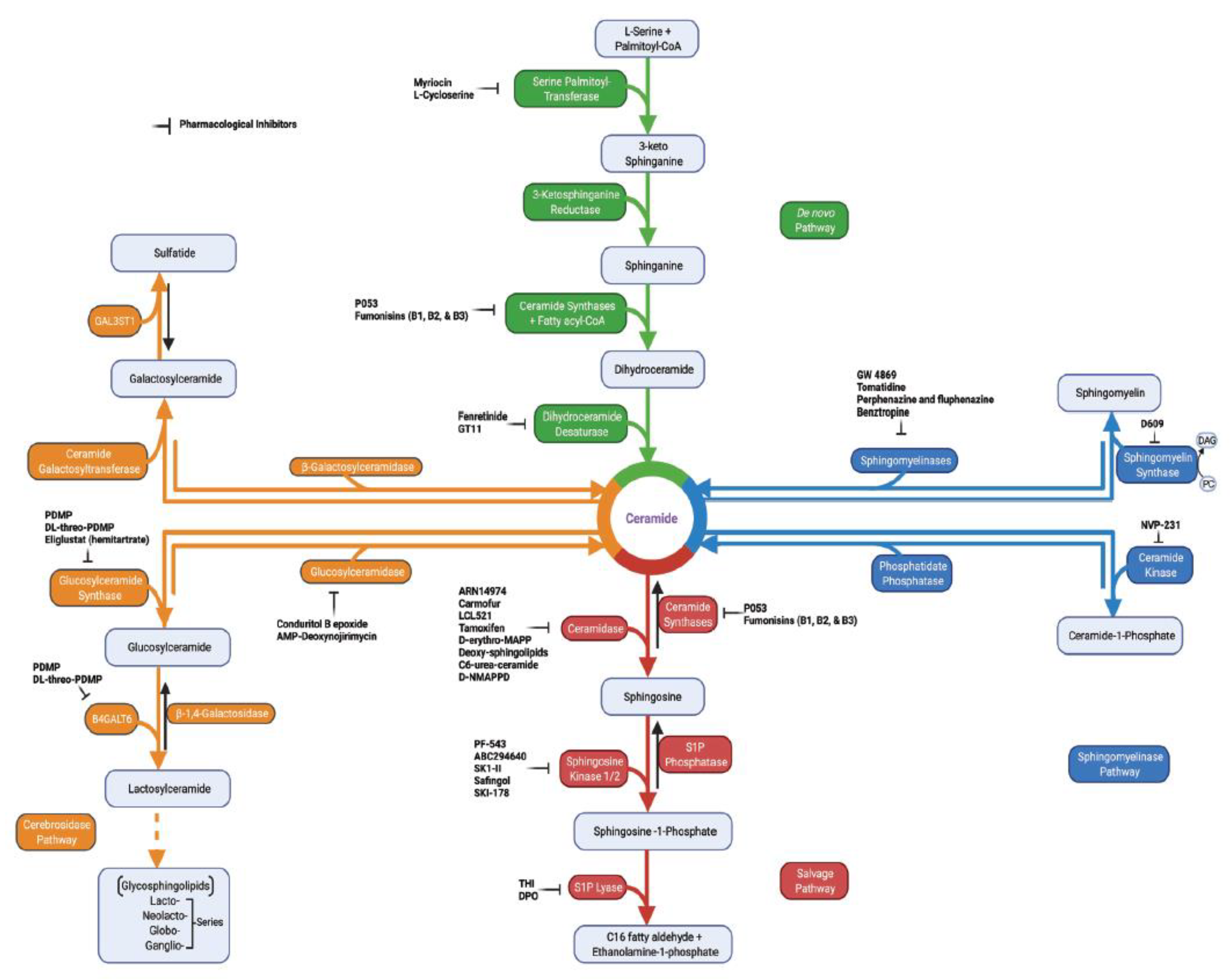
2. Sphingolipid Metabolism in Tumor Pathogenesis

In sphingolipid metabolism, the activation of de novo synthesis, sphingomyelinase, cerebrosidase, or the salvage pathways to generate ceramide and sphingosine (Figure 1) facilitates tumor cell death in response to cellular stress [8,12]. Ceramide, which is the central sphingolipid molecule, is composed of a sphingosine backbone and fatty acyl chain with variable carbon numbers (Figure 2). Interestingly, tumors with elevated levels of sphingolipid metabolic enzymes, such as sphingosine kinases, ceramide kinase, or acid ceramidase, generate crucial sphingolipids for pro-survival signaling functions [6,8]. The identification of the key enzymes in sphingolipid metabolic pathways over the years has paved the way for mechanistic investigations of the critical roles of sphingolipids in cancer progression. Moreover, there are myriads of available pharmacological inhibitors targeting sphingolipid enzymes (Figure 1) that can now be used to investigate the roles of sphingolipid metabolism in cancer pathogenesis, since many tumors have been shown to express an altered level of these sphingolipid enzymes [6,8,13].
Sphingosine(d18:1) and its derivative sphinganine(d18:0) forms the backbone of all sphingolipids. Sphingosine-1-phosphate, which is a bioactive sphingolipid, consists of a phosphate group that is attached to a sphingosine backbone at the first carbon (C1). Similarly, ceramide is composed of sphingosine backbone with 18 carbons and fatty acyl chain with variable carbon numbers (C14 to C26). Furthermore, ceramides can function as structural precursors for other sphingolipids such as sphingomyelin with a phosphocholine group and glucosylceramide with a glucose moiety. Structures were generated from https://www.lipidmaps.org (accessed on 22 April 2022).

3. Key Sphingolipid Enzymes and Their Roles in Cancer Progression

Most of the metabolic end-products in the sphingolipid pathway are regulated mainly by enzymes that are druggable (Figure 1 and Figure 2) and, thus, provide novel therapeutic targets for cancer treatment.

3.1. Sphingosine Kinases (SPHKs)

SPHKs are enzymes that catalyze the formation of S1P from sphingosine. Currently, there are two known SPHK isoenzymes that have been cloned and characterized—namely, SPHK1 and SPHK2. Both isoenzymes belong to the diacylglycerol kinase family and contain five (i.e., C1–C5) conserved domains [9,16]. SPHK1 is mainly localized in the cytosol with some intracellular organelle associations, while SPHK2 is found in nucleus, cytoplasm, and mitochondria [8,9,17]. Because of their crucial roles in sphingolipid metabolism, SPHK1 and SPHK2 both determine cell fates by regulating the balance between survival and cell death via S1P and ceramide metabolism.
Overexpression of SPHK1 in cancer cells has been shown to correlate with poor survival outcome for metastatic melanoma patients treated with an immune checkpoint inhibitor such as anti-PD-1 [18]. Interestingly, decreasing SPHK1 expression improves the efficacy of immune checkpoint inhibitors (anti-CTLA-4 and anti-PD-1 therapy) in melanoma, breast, and colon cancer mouse models [18]. Additionally, inhibiting SPHK1 enhances the metabolic activities of T cells and improves their antitumor functions against murine melanoma [19]. Moreover, anti-PD-1 and PF-543 (SPHK1 inhibitor) combinations improve the control of melanoma tumor growth [19]. SPHK1 has also been shown to play a crucial role in adipocyte-induced epithelial ovarian cancer (EOC). Adipocytes activate SPHK1 via S1PR1/3 and ERK phosphorylation to stimulate growth-promoting action leading to EOC cell proliferation [20]. SPHK1 upregulation promotes chronic inflammation and colitis-associated cancer development [21].
Similarly, increased SPHK2 expression levels correlate with augmented dihydropyrimidine dehydrogenase (DPD) in human colorectal cancer (CRC), and the inhibition of SPHK2 by SLR080811 effectively inhibits DPD expression and reverses 5-fluorouracil (5-FU) resistance in colorectal tumors of villin-Sphk2 Tg mice while also decreasing nuclear S1P concentration in tissues [22]. The colorectal tumors that were developed in Sphk2−/− mice showed great sensitivity to 5-FU therapy, indicating that high SPHK2 expression in colorectal tumors yields resistance to 5-FU chemotherapy treatment [22].
Both SPHK1 and SPHK2 have been shown to be equally responsible for follicle-stimulating hormone (FSH)-induced cell proliferation of epithelial ovarian cancer. FSH induces the phosphorylation of both SPHK1 and SPHK2 enzymes to regulate the survival and growth of ovarian cancer cells via the ERK1/2 pathway [23].
Collectively, these data suggest that both SPHK1 and SPHK2 play crucial roles in stimulating tumor growth and survival, supporting the importance of regulating S1P generation for cancer treatment.

3.2. Sphingosine-1-Phosphate Lyase 1 (SGPL1)

SGPL1 is an enzyme localized in the endoplasmic reticulum that irreversibly breaks down S1P into C16 fatty aldehyde and ethanolamine-1-phosphate [24,25]. It provides the exit point for sphingolipid metabolism. SGPL1 knockout in mouse colon tissues (T-SGPL−/−) was shown to cause immediate and extensive colon tumor formation [26]. T-SGPL−/− also stimulates cancer-induced inflammation and increased both S1P and sphingosine levels [26]. Additionally, SGPL1 knockout in mouse immune cells (I-SGPL−/−) also leads to S1P accumulation in the immune cells but causes delayed carcinogenesis compared to T-SGPL−/− cells [26]. Another study has shown that a low probability of metastasis formation is associated with high native SGPL1 expression [24]. The native form of SGPL1 expression prevents S1P-induced migration and cell-colony formation of pediatric alveolar rhabdomyosarcoma (RMA) compared to SGPL1 mutant [24]. Moreover, silencing of SGPL1 influences the tumorigenic activity of established colorectal cancer cells and partial redifferentiation of colorectal cancer [27].

3.3. Ceramide Kinase (CERK)

CERK is an enzyme that catalyzes the phosphorylation of ceramide to form C1P, and its activity is known to be regulated by Ca2+ ions [28,29]. This enzyme was first discovered in synaptic vesicles from brain cells and has been shown to have a cytosolic localization and is found in the membrane fraction as well [29,30]. CERK overexpression has been shown to promote triple-negative breast cancer (TNBC) growth and migration and confer chemotherapy resistance to breast cancer cell lines [31]. Consequently, CERK siRNA knockdown improves TNBC chemotherapy efficacy and suppresses TNBC growth, migration, and survival [31]. Another study has shown that CERK expression in breast cancer cells promotes migration and invasion via the PI3K/Akt pathway [32]. Additionally, inhibiting CERK expression with either a pharmacological inhibitor (NVP-231) or genetic tools (shRNAs) significantly reduces the migratory potential and invasiveness in breast metastatic cell lines [32]. Similarly, NVP-231 induces programmed cell death by stimulating M phase cell cycle arrest in breast and lung cancer cell proliferation [33]. Moreover, Payne et al. [34] showed that CERK is needed for mammary tumor recurrence in murine breast cancer models, following HER2/neu pathway inhibition. Consistently, in human patients, the upregulation of CERK expression is associated with an elevated risk of breast cancer recurrence in women [34].
Taken together, these findings suggest that inhibiting CERK would provide a novel therapeutic target for cancer treatment.

3.4. Ceramidases (CDases)

CDases hydrolyze ceramide, by cleaving the fatty acid moiety from ceramide, to produce sphingosine. Currently, five human CDases have been cloned and are encoded by five distinct genes, categorized into three different classes—acid ceramidase (AC), neutral ceramidase (NC), and alkaline ceramidases 1–3 (ACER1–3) [35].
AC is a lysosomal ceramidase that is overexpressed in several cancer types [36,37]. For instance, AC was reported to regulate the switch between proliferative and invasive phenotype states in melanoma cells [38]. Using both cells isolated from human melanoma biopsies and melanoma cell lines, Leclerc et al. [38] showed that melanoma cells with proliferative activity displayed increased in ASAH1 expression. Consequently, the melanoma cells developed an invasive property after the loss of ASAH1, thus losing their proliferative phenotype and acquiring enhanced motile properties [38]. Interestingly, AC inhibition in colorectal cancer cell lines using pharmacological AC inhibitors (carmofur and LCL521) or siRNA knockdown of AC enhanced X-ray radiosensitivity by increasing apoptosis [39]. Correspondingly, patient-derived organoids with decreased AC expression showed more radiosensitivity compared to the patient-derived organoids with an elevated AC expression [39]. Similarly, AC deletion in melanoma cells enhanced doxorubicin-induced apoptosis [40]. Consistently, previous studies have also suggested a role of AC in chemo- and radiotherapy failures [36,41,42].
NC is localized in the plasma membrane, Golgi apparatus, and mitochondria [43]. Inhibition of NC in colorectal cancer induces a xenograft tumor growth delay [44]. Additionally, constitutively active AKT cells of xenograft tumors are resistant to NC inhibition [44]. Consistently, pharmacological inhibition (C6 urea–ceramide) and molecular inhibition (siRNA knockdown) of NC increases ceramide and decreases cell survival via elevated cellular apoptosis in colon cancer cells [45].
ACERs (i.e., ACER1, ACER2, and ACER3) are closely related family members with distinct biological functions in regulating sphingolipid metabolism [46]. ACER1 has been shown to play a crucial role in mammalian skin homeostasis [47], and it is found to be localized in the endoplasmic reticulum [43,48]. The role of ACER1 in cancer has not been elucidated and, therefore, ACER1-specific functions in cancer progression and survival are currently unknown. However, analysis from the publicly available transcriptomic database in The Cancer Genome Atlas (TCGA) indicates that ACER1 is significantly downregulated in human skin cutaneous melanoma, head and neck squamous cell carcinoma, testicular germ cell tumors, and esophageal carcinoma primary tumors compared to normal tissues. Meanwhile, given the low expression of ACER1, TCGA data combining patients with these four different cancer types altogether had worse prognostic outcome (Figure 3A). It is particularly important to investigate the specific functions of ACER1 in melanoma (i.e., skin cancer), since ACER1 is crucial for keratinocyte differentiation [48], and there is already a known existing crosstalk between melanocytes, keratinocytes, and melanoma [49]. It would be interesting to investigate whether ACER1 has a protective function in melanoma development. ACER2 is a Golgi ceramidase [50] that has a higher affinity towards unsaturated long-chain ceramides (C18:1, C20:1, and C24:1 ceramide) [51]. ACER2 was reported to be overexpressed in hepatocellular carcinoma (HCC) tissue and to induce growth, invasion, and migration in HCC cell lines via sphingomyelin phosphodiesterase acid-like 3B (SMPDL3B) [52]. In another study, ACER2 and sphingosine levels were shown to be upregulated in DNA-damaged tumor cells [53]. The ACER2/sphingosine upregulation pathway stimulates programmed cell death in a human colorectal carcinoma cell line (i.e., HCT116 cells) by increasing reactive oxygen species (ROS) production in response to DNA damage [53]. Additionally, ACER2 regulates p53-induced autophagy and apoptosis via sphingosine and ROS generation in human non-small cell lung carcinoma cell line (i.e., H1299 cells) [54]. ACER3 is localized in both the endoplasmic reticulum and Golgi complex, and it hydrolyzes unsaturated long-chain ceramides (C20:4, C20:1, and C18:1 ceramide), phytoceramides, and dihydroceramides to produce sphingosine [55,56]. Knockdown of ACER3 suppressed tumor growth and promoted apoptosis in HCC cells [57]. Similarly, ACER3 deficiency decreased acute myeloid leukemia (AML) cell growth and increased apoptosis in the AML cells via limiting AKT signaling [58]. Interestingly, ACER3 was shown to play a vital role in regulating the expression of pro-inflammatory cytokines of the innate immune system cells via C18:1 ceramide [59]. Additionally, ACER3 and C18:1 ceramide dysregulation contribute to the pathogenesis of cancer as an inflammatory disease [59].

3.5. Ceramide Synthases 1–6 (CerS1–6)

A total of six mammalian CerS enzymes (CerS1–CerS6) have been identified, cloned, and described [60,61]. Their general function is to catalyze de novo synthesis of ceramides [62,63], which can induce apoptosis [64]. CerS1–6 enzymes are also known as the longevity assurance homologue of yeast lag1 (Lass1) [12,61], and they have been implicated in the regulation of programmed cell death [65,66].
CerS1, the first identified mammalian CerS that catabolizes the synthesis of C18 ceramide [67,68], was shown to be downregulated in oral cancer tissues and cell lines [69]. The downregulation of CerS1 promotes the aggressiveness of oral squamous cell carcinoma and chemotherapy drug (cisplatin) resistance, while CerS1 overexpression induced sensitization to cisplatin via regulating cell death [69]. Similarly, histone deacetylase 1 (HDAC1) and microRNA-574-5p axis was found to repress CerS1 and alter C18 ceramide generation in head and neck squamous cell carcinoma (HNSCC), thereby allowing tumor growth and proliferation [70]. Thus, CerS1/C18 ceramide expression inhibits HNSCC xenograft growth and induces cell death [71,72,73]. Additionally, targeting Fms-like tyrosine kinase 3 (FLT3)–internal tandem duplication (ITD) induces mitophagy, leading to AML cell death via CerS1/C18-mediated mitophagy [74]. The CerS enzyme that synthesizes very-long-chain ceramides, CerS2, was reported to have an antimetastatic gene function in ovarian cancer cells [75]. Downregulation of CerS2 in ovarian cancer cell lines stimulates in vivo metastasis and invasiveness [75]. Moreover, CerS2 alternative splicing modulates cancer cell proliferation and migration in luminal B breast cancer [76]. Interestingly, overexpression of CerS2 and C24 ceramide generation in HeLa cells partially prevents programmed cell death induced by ionizing radiation [77]. Loss of CerS3, which catabolizes C24 and long acyl chain ceramides synthesis [68], was reported to cause lethality in skin barrier disturbance [78]. Although the specific functions of CerS3 in cancer is unknown, and data from TCGA database indicates that CerS3 expression is decreased in human skin cutaneous melanoma (Figure 3B). CerS4, which catabolizes the synthesis of C18–C20 ceramides, regulates cancer cell migration and invasion [79]. Knockdown of CerS4 increases migration in A549 cells, and the restoration of CerS4 generates C18–C20 ceramides to inhibit cancer cell migration and invasion [79]. Although both CerS5 and CerS6 generate C16 ceramide, only CerS6 appears to regulate C16 ceramide in mitochondria and mitochondria-associated membranes [80]. Consequently, in activated aging T cells, the C14/C16 ceramides generated by CerS6 stimulate mitophagy and attenuate the T cells’ antitumor functions [81]. Conversely, CerS5 knockout was shown to stimulate colon cancer development in azoxymethane (AOM) and dextran sulfate sodium (DSS) colitis-associated colon cancer models [82]. Interestingly, CerS6 overexpression and C16 ceramide generation promotes cell proliferation, colony formation, and invasion via the AKT1/FOXP3 pathway in pancreatic ductal carcinoma (PDAC) cell lines [83], consistent with its proliferative roles in head and neck squamous cell carcinoma and lung cancer cell lines [71,84]. Additionally, high CerS6 expression levels predicted worse prognosis in PDAC patients and was positively correlated with disease progression [83]. Moreover, for its antiproliferative and pro-apoptotic roles, CerS6 expression was shown to increase p53 protein half-life via a positive feedback loop in polyploid giant cancer cells (PGCCs) [85]. In addition, CerS6 and p53 co-expression nullified the ability of PGCC to form offspring with a high proliferative and therapy-resistant phenotype [85]. These studies suggest that although ceramide accumulation mainly induces cancer cell death and antiproliferative signaling in many tumors, it might also have proliferative functions depending on the downstream signaling targets.

3.6. Sphingomyelinases (SMases)

SMases, or sphingomyelin phosphodiesterases, catalyze the conversion of sphingomyelin to ceramide and phosphocholine [86]. SMases are classified into three groups based on their optimum pH (i.e., alkaline, acid, and neutral) [86].
Alkaline SMase (Alk-SMase or ENPP7), which is found in intestinal mucosa, bile, and liver, was shown to reduce colon cancer progression in a mice model [87]. Alk-SMase knockout mice showed a decrease in ceramides, increased S1P levels, and resulted in enhanced colonic tumorigenesis induced by AOM/DSS treatment [87]. Acid SMase (ASMase or SMPD1) is an enzyme found in lysosomes, and its deficiency leads to an inherited lysosomal disease [88]. Interestingly, adult patients with chronic visceral (CV) ASMase deficiency (CV-ASMD) were observed to have an abnormally elevated incidence of cancers [88]. Thus, the risk of cancer was shown to be associated with CV-ASMD disease severity [88]. Furthermore, ASMase induction [89] in platelets induces B16F10 melanoma metastasis, consequently inhibiting ASMases with amitriptyline-prevented tumor metastasis by 75% [90,91]. In addition, the downregulation of neutral SMase 2 (NSMase2, also known as SMPD3) contributes to melanoma immune escape to enhance tumor progression, while the overexpression of wild-type nSMase2 enhances the efficacy of anti-PD-1 antibody therapy in both melanoma and breast cancer mouse models [92]. Moreover, SMPD3 downregulation promotes tumor progression in oral squamous cell carcinoma (OSCC) [93].

3.7. Sphingomyelin Synthase (SMS)

There are two known isoforms of SMS—SMS1 and SMS2. Both isoforms catalyze the same reaction to produce sphingomyelin and diacylglycerol [94,95]. SMS1 is localized in the trans-Golgi apparatus, while SMS2 is mainly localized in the plasma membranes [94,95]. Although the specific roles of SMS1 in cancer cell growth and survival remains to be elucidated, SMS2 has been shown to have pro-tumoral [96,97] or apoptotic [98] functions in a cancer-type dependent manner, signifying the need for further studies of SMS-specific roles in tumor control. However, it was recently shown that SMS2 but not SMS1 was upregulated in ovarian cancer tissues and cell lines and, consequently, SMS2 overexpression promoted cancer cell growth and migration [99], suggesting a therapeutic function of SMS2 inhibition in ovarian cancer treatment.
Collectively, targeting these sphingolipid enzymes while monitoring and controlling sphingolipid accumulations would be important for effective cancer therapy, since the altered expression of these enzymes regulate tumor growth/survival and cell death (Table 1).

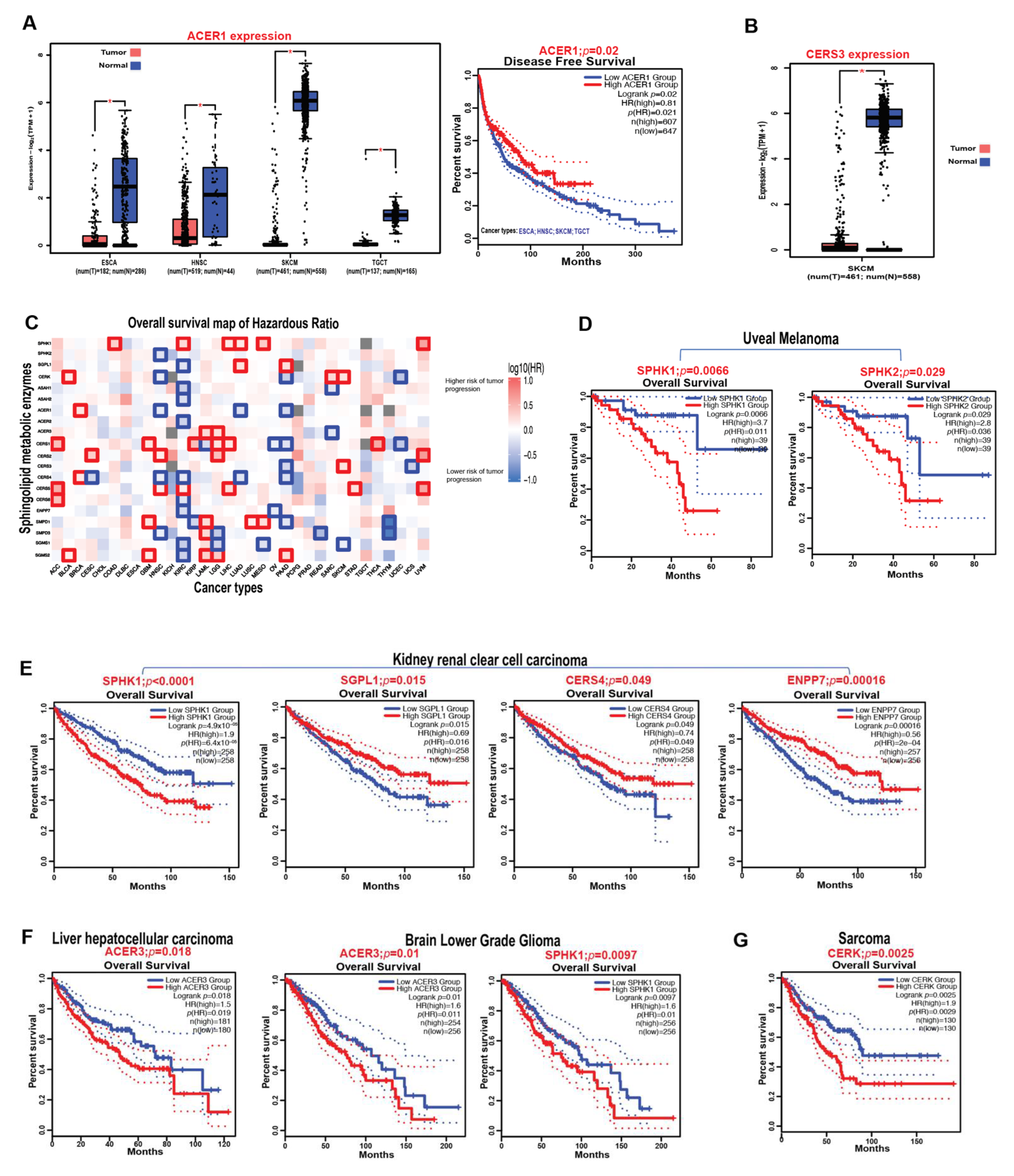
3.8. Prognostic Impact of Sphingolipid Metabolic Enzymes on the Survival of Cancer Patients

Using the GEPIA2 (Gene Expression Profiling Interactive Analysis, version 2) web server with TCGA data sets [100,101], we assessed the effects of key sphingolipid enzymes on the overall survival of patients with different types of cancer. The increased expression of these metabolic enzymes was observed to be associated with higher or lower risks of tumor progression depending on the cancer type, as indicated by the red blocks (poor prognosis) or blue blocks (good prognosis) on the heatmap, respectively (Figure 3C). For instance, SPHK1 overexpression is significantly associated with higher risks of tumor progression or worse prognostic outcome in colon adenocarcinoma (COAD), kidney renal clear cell carcinoma (KIRC), liver hepatocellular carcinoma (LIHC), lung adenocarcinoma (LUAD), mesothelioma (MESO), and uveal melanoma (UVM) as indicated by the red bold outlines on the heatmap (Figure 3C). Correspondingly, CERS4 overexpression is significantly associated with lower risks of tumor progression or better prognostic outcome in cervical squamous cell carcinoma and endocervical adenocarcinoma (CESC), head and neck squamous cell carcinoma (HNSC), KIRC, LUAD, and pancreatic adenocarcinoma (PAAD) as indicated by the blue bold outlines on the heatmap (Figure 3C). Specifically, in uveal melanoma, overexpression of both SPHK1 and SPHK2—the enzymes that catalyze the production of S1P—is associated with worse or unfavorable disease outcomes compared to the low-expression groups (Figure 3D). Similarly, in kidney renal clear cell carcinoma, worse or unfavorable prognosis is associated with high expression of SPHK1, low expression of SGPL1, CERS4, and ENPP7 (Alk-SMase) (Figure 3E), possibly due to the sustained production of S1P as indicated by high SPHK1 with low SGPL1 levels and a decrease in ceramide production to inhibit apoptosis as indicated by the low levels of CERS4 and ENPP7. Additionally, ACER3 expression, which has been shown to regulate cancer pathogenesis [57,58], was observed to have an unfavorable overall survival outcome in liver hepatocellular carcinoma and brain lower-grade glioma (Figure 3F). This observation was also consistent with the poor overall survival outcome associated with high SPHK1 expression in brain lower-grade glioma (Figure 3F). Importantly, CERK expression, which catalyzes the formation of C1P, was observed to be associated with worse overall survival in sarcoma (Figure 3G).
Overall, these observations suggest that sphingolipid metabolic enzymes play an important role in regulating tumor pathogenesis and would therefore provide therapeutic targets for cancer treatments in patients.

4. S1P Signaling in Cancer

S1P is a bioactive lipid mediator that acts as a cell signaling molecule to regulate various biological processes including cell survival, proliferation, and motility. S1P is generated intracellularly by sphingosine kinases (SPHK1/SPHK2), and the cytosolic S1P produced by SPHK1 is exported out of the cell and into the extracellular space via specific transporters, since S1P cannot freely cross the plasma membrane barrier due to the fact of its polar head group [102]. The S1P released in the extracellular space signals through S1P receptors (S1PRs) in a process called “inside-out” signaling [2], which has been shown to occur in many cancer types [102,103,104]. Although S1P produced by both SPHK1 and SPHK2 have intracellular functions [105,106,107], extracellular S1P mainly produced by SPHK1 play main roles in controlling “inside-out” signaling process [102].

4.1. S1P Transporters

The S1P generated intracellularly is exported into the extracellular space leading to inside-out signaling in the tumor microenvironment via specific S1P transporters including protein spinster homologue 2 (SPNS2), major facilitator superfamily d2b (Mfsd2b), ATP-binding cassette sub-family C member 1 (ABCC1), and ATP-binding cassette sub-family G member 2 (ABCG2) (Figure 4A).
SPNS2 belongs to the major facilitator superfamily (MFS)—the largest secondary transporter protein family with 12 putative transmembrane protein domains. It has 504 amino acid residues in zebrafish but 549 amino acid residues in human and mouse [108]. The functional roles of SPNS2 as a transporter for S1P signaling were observed in zebrafish models, where it was shown that cardia bifida (split heart abnormality or defective heart development) occurs because of a point mutation in spns2 which that inhibits S1P signaling, causing migration defect of myocardial precursors [104,109,110]. Interestingly, this defect could be rescued by exogenous S1P addition [109]. Recent studies have also shown that other MFS family members, namely, MFS domain-containing 2a and 2b (Mfsd2a and Mfsd2b) play vital roles in transporting S1P alongside SPNS2. Mfsd2a and Spns2 form a protein complex that allows for effective/sufficient S1P export from endothelial cells in the brain [111]. Circulating S1P is transported via both Mfsd2b and SPNS2. In endothelial cells, SPNS2 is the major S1P transporter, while Mfsdb2 is the key S1P transporter in erythrocytes and platelets [112,113,114,115,116,117]. Remarkably, spns2 deletion in mice, whether globally or in a lymphatic endothelial-specific manner, leads to improved tumor killing and significantly decreased metastatic burden in Spns2−/− mice as a result of increased levels of natural killer cells and effector T cells [118]. Similarly, SPNS2 has been shown to deliver S1P to S1PR2, leading to increased epidermal growth factor (EGF)-mediated cancer cell invasion [119], suggesting that SPNS2 inhibitors may be useful therapeutics for cancer treatment. Contrastingly, low SPNS2 levels are associated with worse clinical prognosis in colorectal cancer, while the ectopic expression of SPNS2 was shown to decrease migration, invasion, and metastasis in colorectal cancer cell lines via AKT signaling pathway [120]. These contrasting results could be explained by the need of SPNS2–S1P signaling to stimulate specific or opposing S1P receptors in a cancer type-specific manner.
ABCC1 and ABCG2 are ABC transporters that have been implicated in lipid signaling by transporting S1P across cell membranes [104]. ABCC1 [121] is another member of the ABC transporter family, multidrug resistance-associated protein 1 (MRP) shown to confer resistance to anticancer drugs [122,123]. ABCG2 is identified as an ABC transporter, a breast cancer resistance protein, that mediates S1P efflux from cells [123,124]. Both ABCC1 and ABCG2 were reported to mediate estradiol-induced S1P release in breast cancer cells [8,124]. Consequently, inhibiting ABCC1 or ABCG2 with pharmacological inhibitors decreases estradiol-mediated release of S1P and dihydro-S1P and ERK1/2 activation in breast cancer [124]. Additionally, decreasing ABCC1 increases ABCG2 expression levels and vice versa [124]. The presence of these ABC transporters in cancer cells may explain why most breast cancer types are resistant to chemotherapy treatment. Thus, understanding these transporters for S1P signaling could help understand cancer chemotherapy resistance.

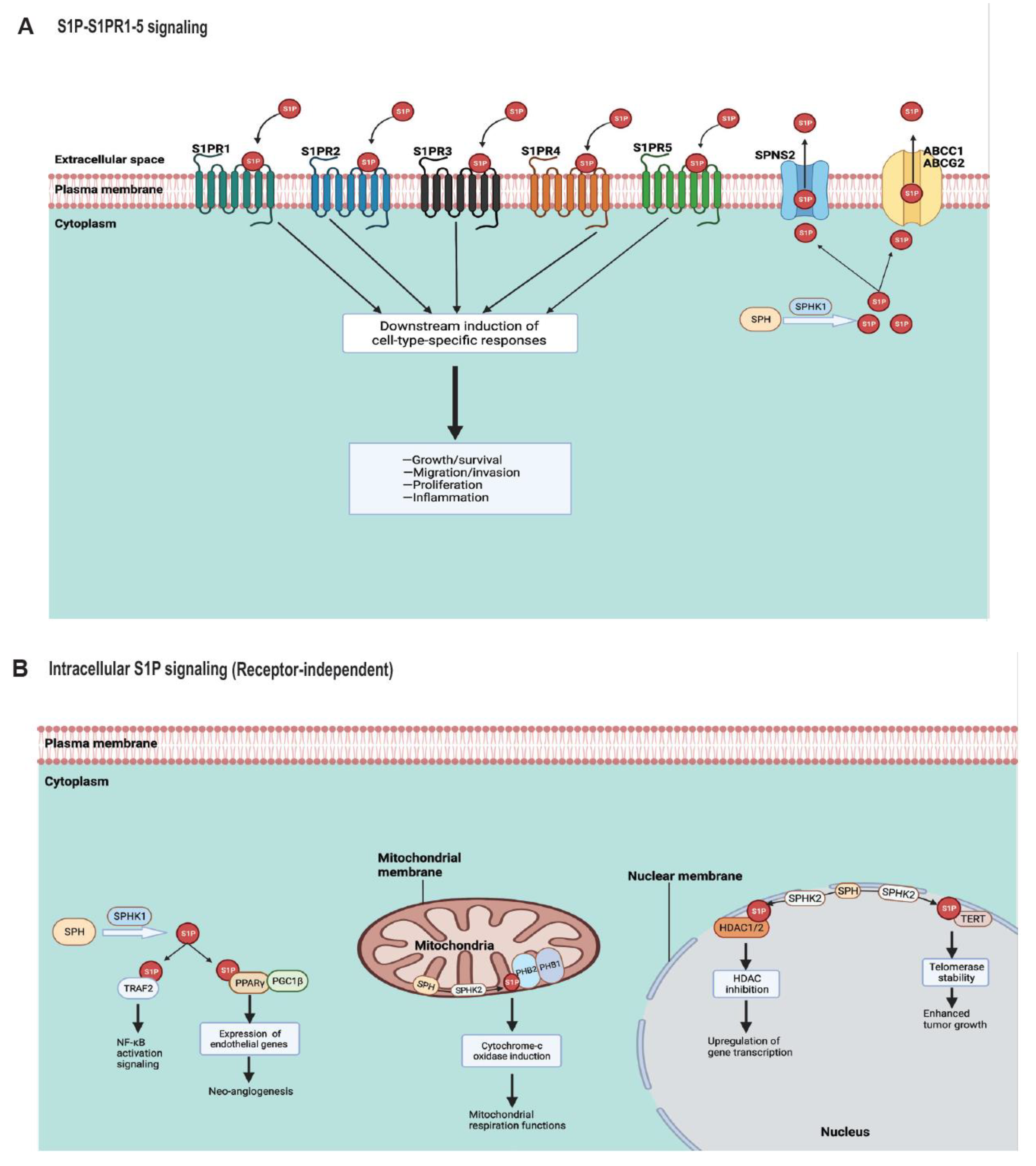
4.2. S1P Receptors (S1PR1–5)

The released S1P into the extracellular space via its transporters can engage the five known S1P-specific G protein-coupled receptors (S1PR1–S1PR5) for cellular signaling (Figure 4A). This leads to context-dependent specific functions including the proliferation, migration, and growth/survival of cancer cells. In a physiological context, circulating S1P uses protein carriers to engage its receptors. For instance, S1P could either bind to high-density lipoprotein (HDL) through apolipoprotein M (ApoM), HDL/ApoM, or albumin [125,126]. In endothelial cells, evidence showed that S1PR1 signaling is more dependent on HDL-bound S1P compared to albumin-bound S1P for their downstream activation of Akt and eNOS [9,127], which are known targets for cancer treatment. Additionally, HDL-bound S1P increases S1PR1 expression levels while decreasing its degradation rate compared to albumin-bound S1P [127]. This result is consistent with the findings that low ApoM production in aged mice reduces S1P signaling via S1PR1 in lung and kidney endothelial cells leading to maladaptive repair and fibrosis compared to young mice [128]. Moreover, S1PR1, as a pro-tumorigenic factor, has been shown to activate cancer cell signaling pathways leading to invasion, migration, and proliferation [102]. Consistently, S1P–S1PR1 signaling in tumor cells or the tumor microenvironment induced persistent STAT3 activation and IL-6 production, leading to tumor growth and metastasis [129]. Thus, inhibiting S1P–S1PR1 signaling and the STAT3 activation pathway may be a useful therapeutic strategy in treating certain cancer types [21,130]. Interestingly in mouse lung tumors, systemic loss of SPHK1 leads to an increase in S1PR1 and a decrease in S1PR2 expression levels [131].
S1PR2 mutational inactivation or deletion confers proliferative advantage in diffuse large B-cell lymphoma (DLBCL) cell lines in vitro and in vivo mouse models [132]. Conversely, the activation of S1PR2, via the (TGF-β)/TGF-βR2/SMAD1 pathway, promotes apoptosis and inhibits DLBCL cell proliferation [132]. Consistently, low S1PR2 expression is associated with worse prognosis, compared to high S1PR2 expression in patients with lymphomas—making S1PR2 a positive prognostic marker for patients with DLBCL [133]. Additionally, ectopic S1PR2 expression decreases lymphoma tumor sizes in vivo [133]. Contrastingly, in endothelial-cell-specific S1pr2 knockout (S1pr2 ECKO) mice, there was a significant decrease of B16F10 melanoma lung metastasis, compared to s1pr2 WT [134]. Consequently, tumors grown in S1pr2 ECKO mice were observed to be smaller, compared to s1pr2 WT [134]. Similarly, targeting SPHK1–S1PR2 signaling reduces acute myeloid leukemia burden and prolonged survival [135]. These data suggest that S1PR2 should be selectively targeted to provide therapeutic options for certain cancer treatments.
In cancer stem cells (CSCs) or tumor-initiating cells, SPHK1 expression enhanced tumor formation via Notch activation stimulated by S1PR3 in both in vitro and in vivo studies [136]. Conversely, tumorigenicity of CSCs was inhibited by knocking down S1PR3 or by using S1PR3 pharmacological antagonists, TY52156 and CAY10444 [136]. In support of these findings, breast cancer patient-derived CSCs were found to contain positive S1PR3/ALDH1 or SPHK1/ALDH1 cells [136]. Specifically in triple-negative breast cancer cell lines, S1P/S1PR3/Notch signaling was found to promote metastasis [137], making S1PR3 a therapeutic target for breast cancer treatment. Additionally, S1PR3 activation was shown to promote cancer progression in osteosarcoma [138] and lung adenocarcinomas [139].
The depletion of S1PR4 was shown to inhibit mammary tumor progression in vivo via CD8+ T-cell expansion, since S1PR4 signaling promotes tumor growth by inhibiting CD8+ T-cell abundance [140]. This supports the idea that targeting S1PR4 signaling could be a promising strategy to improve anti-CXCR4 cancer immunotherapy [141]. Similarly, lipopolysaccharide was shown to stimulate prostate cancer cell invasion, progression, and metastasis via SPHK1/S1PR4/matriptase signaling [142], indicating that S1PR4 signaling is crucial for the progression of prostate cancer mediated by bacterial infection.
S1PR5, which regulates T-cell infiltration and emigration from peripheral organs [143], was shown to stimulate mitotic progression in HeLa cells, suggesting S1PR5 as a possible therapeutic target for inhibiting tumor proliferation [144]. Specifically, SPNS2–S1P–S1PR5 signaling stimulates the downstream PI3K–AKT–PLK1 pathway that then regulates the metaphase-to-anaphase transition leading to mitotic progression [144].
Collectively, the signaling of all five of the S1P receptors (i.e., S1PR1–S1PR5) has proven to be critical in regulating various kinds of tumor progressions. Thus, selectively targeting these receptors depending on the cancer cell type are potential therapeutic strategies to improve cancer treatment.

4.3. Endogenous S1P Signaling Targets

Endogenous S1P generated by either SPHKI or SPHK2 can also act on intracellular targets for signaling functions without needing to engage the S1P receptors or transporters (Figure 4B). In HeLa, HEK 293, and A7 melanoma cells, it was shown that cytoplasmic intracellular S1P generated by SPHK1 is critical for the canonical NF-κB activation pathway by TNF-α—which is necessary for inflammatory immune processes and anti-apoptotic functions [145]. Specifically, endogenous S1P binds TRAF2 (TNF receptor-associated factor 2) at the N-terminal RING domain independent of S1P receptors, leading to lysine-63-linked polyubiquitination of receptor interacting protein 1 (RIP1) and NF-κB signaling activation downstream [145]. Interestingly, TRAF-interacting protein (TRIP) was shown to negatively regulate TNF-induced NF-κB activation by binding to TRAF2 and inhibiting its ubiquitination activity [146]. The TRAF2–TRIP complex formation inhibits the binding of S1P to the TRAF2 RING domain [146]. Contrastingly, in macrophages [147] and keratinocytes [148], intracellular S1P generated by sphingosine kinases was not required for NF-κB activation signaling and inflammation. Furthermore, since SPHK1 and peroxisome proliferator-activated receptor-γ (PPARγ) are known to be expressed in human cancers, PPARγ was reported as a transcription factor target for S1P generated by SPHK1, independent of the S1P receptors [149]. In endothelial cells, S1P binds and activate PPARγ, which then allows for the recruitment of peroxisome proliferator-activated receptor-γ coactivator 1β (PGC1β), forming the SlP/PPARγ/PGC1β complex, to regulate endothelial genes and neoangiogenesis [149]. Consistently, in peripheral T cells, SPHK1-generated S1P binds PPARγ for its transcriptional activation, and the inhibition of SPHK1/S1P/PPARγ signaling ameliorates antitumor immunity against mouse melanoma [19].
Furthermore, SPHK2-derived S1P in the mitochondria was shown to bind homomeric prohibitin 2 (PHB2) with great specificity and affinity without binding to prohibitin 1 (PHB1)—a closely related protein that forms complexes with PHB2 [106,150]. Thus, the S1P–PHB2 complex is critical for mitochondrial respiration functions via cytochrome c oxidase (complex IV) [106]. In the nucleus, SPHK2-derived S1P was reported to bind histone deacetylases (HDACs) 1 and 2 nuclear enzymes, inhibiting histone deacetylation in breast cancer cells [105], thus inducing epigenetic regulation of gene expression [105]. Additionally, SPHK2-generated nuclear S1P was observed to bind directly to human telomerase reverse-transcriptase (hTERT), preventing hTERT from ubiquitination and proteasomal degradation (stabilizing telomerase), leading to enhanced tumor growth [107].
Altogether, both endogenous SPHK1-derived S1P and SPHK2-derived S1P have shown to function independent of their S1P receptor signaling by binding to specific intracellular targets, thereby regulating genes involved in tumor growth/progression.

5. Sphingolipid Therapeutics in Cancer

5.1. Chemotherapy, Radiotherapy, and Immunotherapy

The combination of sorafenib (multikinase inhibitor) and vorinostat (histone deacetylase inhibitor) was reported to promote CD95 activation by inducing cytosolic Ca2+, which increases dihydroceramide levels and reactive oxygen species (ROS), to suppress the growth of gastrointestinal tumor cells and in vivo pancreatic tumors [151]. Consequently, knockdown of CerS6 abolished CD95 activation in tumor cells [151]. Thus, sorafenib plus vorinostat appears to be CerS6-ceramide-dependent, which leads to protein phosphatase 2A (PP2A) and ROS signaling [151,152]. Supportively, the anticancer drug daunorubicin was shown to induce ceramide/ceramide synthase dependent apoptosis in both human leukemia and histiocytic lymphoma cells [65]. However, since ROS production via ASMase/ceramide activation may also induce acute vascular injury [153]; its intracellular production should be controlled in order to prevent long-term side effects in cancer survivors. Additionally, Sorafenib plus vorinostat combination therapy was also shown to improve the efficacy of anti-PD-1 immunotherapy, leading to a significant reduction in pancreatic tumors in vivo [154]. Moreover, gemcitabine (antimetabolite) and doxorubicin (anthracycline) combination therapy was shown to be an effective chemotherapy for some patients with metastatic head and neck cancers [155] via caspase-9/3—dependent mitochondrial cell death by inducing CerS1/C18 ceramide in both in vitro and in vivo xenograft mouse models for head and neck cancers [156]. In the Phase II clinical trials, patients with improved response to gemcitabine plus doxorubicin also had increased in C18 ceramide serum levels [155].
Radiation-induced programmed cell death in Caenorhabditis elegans germ cells required ceramide generation via ceramide synthase activation in mitochondria [157]. Moreover, ataxia telangiectasia-mutated (ATM) kinase was shown to regulate the activation of radiation-induced ceramide synthase in the apoptotic response of intestinal crypt clonogen [158]. Interestingly, a neutralizing anti-ceramide monoclonal antibody, which binds ceramide generated for apoptotic signaling, prevented radiation gastrointestinal syndrome mortality in mice [159]. Single-dose radiotherapy/ASMase signaling was shown to ablate more than 90% of human cancers by disabling the homologous recombination of the tumor cells [160,161]. Thus, the induction and regulation of ceramide generation in tumor cells via chemotherapy and radiotherapy are vital therapeutic strategies for cancer treatment.
In addition to chemotherapy and radiotherapy, sphingolipid signaling is also critical in immunotherapy and/or tumor immunology by regulating immune cells for antitumor activities. It was shown that S1PR1 signaling in CD4+ T cells promotes breast and melanoma tumor growth via JAK/STAT3 activation in mice, limiting CD8+ T-cell recruitment [162]. Additionally, S1P–S1PR1 signaling activates STAT3 by upregulating IL-6 and JAK2 activity to promote tumor growth and metastasis [129]. Consequently, reducing S1P levels by silencing SPHK1 improves the efficacy of anti-CTLA-4 and anti-PD-1 immunotherapy, leading to significant tumor suppression and overall improved survival in mouse melanoma, breast, and colon tumor models [18]. Moreover, patients with Gaucher disease have been shown to have an elevated risk of developing malignant disorders such as multiple myeloma [163]. Interestingly, clonal immunoglobulin in Gaucher disease-associated myeloma patients and mouse models were reactive against lyso-glucosylceramide (due to the fact of a glucocerebrosidase/glucosylceramidase deficiency), which was found to be elevated in both patients and in the mouse models, indicating lyso-glucosylceramide’s involvement in Gaucher disease-associated myeloma origins [164]. Remarkably, activation of complement C5a and C5a receptor 1 (C5aR1) was shown to control glucosylceramide accumulation and inflammatory response in Gaucher disease [165]. Moreover, in a mouse model of leukemia, CerS6-derived C16 ceramide generation was required for optimum T-cell activation and cytokine production in response to alloantigen during allogeneic hematopoietic stem cell transplantation (an effective immunotherapy for hematologic malignances), leading to subsequent graft-versus-host disease (GVHD) induction [166], which is a major complication. However, silencing complement C3aR/C5aR in recipient dendritic cells stimulates lethal mitophagy, owing to ceramide generation and improved GVHD outcome while maintaining the graft-versus-leukemia effect [167]. Collectively, these data show that sphingolipid signaling is crucial in tumor immunology and for the efficacy of immune checkpoint inhibitors in cancer immunotherapy. Surprisingly, understanding the critical roles of sphingolipid signaling in complement biology could be a potential therapeutic strategy for cancer immunotherapy, since the complement components C3/C3a/C3aR and C5/C5a/C5aR signaling are now emerging as potential targets for cancer immunotherapy improvements [168,169,170,171]. Consistently, there have been interesting specific links between bioactive sphingolipids and complement activation in other diseases [172,173,174,175,176].

5.2. Anticancer Drugs Targeting Sphingolipids

There are several anticancer drugs that target sphingolipid metabolism or signaling that are being tested for cancer therapy in clinical trials (Table 2).

5.2.1. ABC294640 (Yeliva, Opaganib)

ABC294640, which prevents S1P signaling by selectively inhibiting SPHK2, has been shown to prevent tumor growth via downstream mechanisms involving the inhibition of dihydroceramide desaturase in prostate cancer cells [177], suppression of c-Myc and ribonucleoside-diphosphate reductase subunit M2 (RRM2) in pancreatic cancer cells [178], and the inhibition of telomerase stability in lung cancer cell lines [107]. Additionally, ABC294640 was reported to decrease SPHK2 expression leading to the downregulation of c-Myc and Mcl-1 to induce apoptosis in multiple myeloma [179]. A Phase I study of ABC294640 for patients with advanced solid tumors was successfully completed in which nausea, vomiting, and fatigue were reported as the common drug toxicities [180]. Currently, clinical trials are ongoing for the use of opaganib or ABC294640 to treat patients with metastatic castration-resistant prostate cancer [181] (NCT04207255) and cholangiocarcinoma (NCT03377179)(NCT03414489) (see Table 2).

5.2.2. Fingolimod (FTY720)

The FDA approved drug for multiple sclerosis, FTY720, is a structural analog of naturally occurring sphingosine that acts as a functional antagonist for S1PR1 after its phosphorylation to p-FTY720 by SPHK2 [21,182,183]. Phosphorylated FTY720 internalizes and degrades S1PRs on lymphocytes, thereby depriving them from responding to normal S1P signaling, thus preventing the egression of normal lymphocytes from lymphoid tissues [3]. Because of its mechanism of actions, FTY720 was suggested to be a suitable drug candidate for treating chronic inflammatory-related tumors, as it was shown to prevent colitis-associated cancer (CAC) progression even when given at late stages of disease development [21]. Moreover, in lung cancer [184], multiple myeloma [185], leukemias [186,187,188,189,190,191], and breast cancer stem cells [192], FTY720 mediates cancer cell death and tumor suppression via protein phosphatase 2A (PP2A)-dependent pathways. For instance, in one mechanism, FTY720 induces the formation of large ceramide-enriched membrane pores, called ceramidosomes (ceramide–myosin IIA–RIPK1 complex), leading to necroptosis and lung tumor suppression [193] by directly binding/targeting the oncoprotein I2PP2A/SET, causing PP2A activation [184]. Since FTY720 binds SET, leading to PP2A reactivation, the SET–FTY720 complex was studied using NMR spectroscopy, which revealed that FTY720 binding disrupts SET dimerization, allowing for a specific PP2A trimer activation with tumor suppressive activities [194]. However, an effective use of FTY720 for cancer therapy would be to combine it with a SPHK2 inhibitor in order to prevent the synthesis of p-FTY720, which does not seem to play a role in chronic myeloid leukemia or lung cancer cell deaths [184,188] and also causes immune suppression [8].

5.2.3. Ceramide Nanoliposomes (CNLs)

Ceramide induces programmed cell death in cancer cells, which makes it a potent tumor suppressor [195,196]. However ceramide apoptotic function is also associated with intrinsic toxicities in addition to its poor pharmacokinetics when used alone [197]. Therefore, encapsulating ceramide in nanoliposomes, which forms ceramide nanoliposomes (CNLs), selectively induces cancer cell death while also improving drug solubility and limiting toxic effects [198,199]. C6 ceramide nanoliposomes were shown to prevent tumor growth in hepatocellular cancer mouse models by enhancing their antitumor immune response [200]. Interestingly, targeting survivin with C6 ceramide nanoliposomes induces complete remission of fatal natural killer-large granular lymphocytic (NK-LGL) leukemia in rat models [201]. Moreover, CNL treatment was shown to inhibit metastatic growth in melanoma [202] and ovarian [197] cancer cells, demonstrating that CNL delivery strategy is an effective therapeutic option for cancer treatments.

5.2.4. Sonepcizumab

Sonepcizumab which is a biospecific monoclonal antibody against S1P was shown to prevent tumor progression through S1P neutralization in xenograft and allograft tumor mouse models as well as in in vitro studies using human umbilical vein endothelial cells (HUVECs) [203], making it a promising cancer therapeutic strategy. In a Phase II study, sonepcizumab was assessed in patients with metastatic renal cell carcinoma (mRCC), who had previous history of failed treatments with vascular endothelial growth factor (VEGF) and/or mammalian target of rapamycin (mTOR) inhibitors [204] (NCT01762033). However, the study was later terminated because it did not attain its two-month progression-free survival, which was the primary endpoint [204]. Nevertheless, sonepcizumab had an encouraging overall survival with a median of 21.7 months and a favorable safety profile [204]. Surprisingly, elevated serum S1P levels were observed with sonepcizumab treatment [204], which could explain its limited efficacy in the study, since the active S1P signaling may be blocking antitumor immune response. Therefore, combining sonepcizumab with inhibitors that prevent the synthesis or signaling of systemic S1P could be a much better therapeutic strategy.
There are also other sphingolipid-targeting compounds including safingol, fluphenazine, and desipramine [205] that have been assessed in the clinic for the treatment of different cancer types as shown in Table 2.

6. Conclusions and Future Directions

Sphingolipids, as complex biological molecules, have been shown to regulate various biological/cellular processes including tumor cell death and survival. The cloning of key sphingolipid enzymes has helped in understanding the mechanisms and functions of sphingolipids, such as ceramides and S1P, in regulating cancer signaling. Ceramide has emerged as a tumor suppressor by facilitating necroptosis, mitophagy, apoptosis, and lethal autophagy [74,206]. Development of compounds that lead to ceramide synthesis has proven to be a novel anticancer therapeutic strategy. However, the accumulation of ceramides also comes with intrinsic toxicities, which makes the use of CNL an effective therapeutic strategy for ceramide-based drug delivery.
S1P, which is known for its pro-tumorigenic effects and inducing tumor progression, has also emerged as a promising target for cancer treatment with few ongoing clinical trials. Sonepcizumab, the monoclonal antibody against S1P that inhibits tumor growth in xenograft models [203], was not an effective S1P blockade in clinical trials for solid tumors [204], emphasizing the need for combinatorial therapies for sphingolipid-based drugs in developing an effective anticancer treatment.
Analysis from the TCGA cancer patient panels revealed that sphingolipid metabolic enzymes are dysregulated with heterogeneity in various cancer types, which are dependent on context and cell type. For instance, although it was initially reported that PF-543 (a potent SPHK1 selective inhibitor) significantly decreased endogenous S1P levels by 10-fold in MD-1483 head and neck carcinoma cells, the data showed no effects on proliferation or survival [207]. However, recent studies show that PF-543 inhibits tumor growth in colorectal cancer cell lines and in xenograft mouse models [208]. Going forward, it is particularly important to define which cell type produces which sphingolipid enzyme in a specific cancer type, which will help in the development of effective future sphingolipid-based anticancer therapeutics.
Finally, to effectively develop sphingolipid based therapeutics for cancer treatment, we must continue to elucidate and understand specific mechanisms of sphingolipids in regulating cancer signaling. Additionally, effective sphingolipid-based drugs against cancers should be based on a combinatorial therapeutic regimen targeting different pathways to maintain sphingolipid metabolism homeostasis and avoid toxic side effects or drug resistance.

Author Contributions

Conceptualization, A.H.J. and B.O.; writing—original draft preparation, A.H.J.; writing—review and editing, A.H.J. and B.O.; visualization, A.H.J.; TCGA data analyses, A.H.J.; supervision, B.O.; project administration, B.O.; funding acquisition, B.O. All authors have read and agreed to the published version of the manuscript.

Funding

This work was supported by research funding from the National Institutes of Health (CA214461, DE016572, AG069769, and P01 CA203628 to B.O.) and a National Institute of General Medical Sciences (NIGMS) T32 training grant (T32GM132055) awarded to A.H.J.

Acknowledgments

The authors thank all the members of the Ogretmen lab, especially Mohamed Faisal Kassir and Wyatt O. Wofford, for their thoughtful discussions. The authors apologize to those investigators whose publications were not mentioned in this Review owing to space limitations.

Conflicts of Interest

The authors declare no conflict of interest.

References

  1. Weigert, A.; Olesch, C.; Brüne, B. Sphingosine-1-phosphate and macrophage biology—How the sphinx tames the big eater. Front. Immunol. 2019, 10, 1706. [Google Scholar] [CrossRef]
  2. Spiegel, S.; Milstien, S. Sphingosine-1-phosphate: An enigmatic signalling lipid. Nat. Rev. Mol. Cell Biol. 2003, 4, 397–407. [Google Scholar] [CrossRef] [PubMed]
  3. Chun, J.; Hartung, H.-P. Mechanism of action of oral fingolimod (FTY720) in multiple sclerosis. Clin. Neuropharmacol. 2010, 33, 91. [Google Scholar] [CrossRef] [PubMed] [Green Version]
  4. Presa, N.; Gomez-Larrauri, A.; Dominguez-Herrera, A.; Trueba, M.; Gomez-Munoz, A. Novel signaling aspects of ceramide 1-phosphate. Biochim. Biophys. Acta (BBA)-Mol. Cell Biol. Lipids 2020, 1865, 158630. [Google Scholar] [CrossRef] [PubMed]
  5. Cartier, A.; Hla, T. Sphingosine 1-phosphate: Lipid signaling in pathology and therapy. Science 2019, 366, eaar5551. [Google Scholar] [CrossRef] [PubMed]
  6. Pyne, N.J.; Pyne, S. Sphingosine 1-phosphate and cancer. Nat. Rev. Cancer 2010, 10, 489–503. [Google Scholar] [CrossRef] [Green Version]
  7. Wu, Y.; Liu, Y.; Gulbins, E.; Grassmé, H. The Anti-Infectious Role of Sphingosine in Microbial Diseases. Cells 2021, 10, 1105. [Google Scholar] [CrossRef]
  8. Ogretmen, B. Sphingolipid metabolism in cancer signalling and therapy. Nat. Rev. Cancer 2018, 18, 33–50. [Google Scholar] [CrossRef] [PubMed]
  9. Pyne, S.; Adams, D.R.; Pyne, N.J. Sphingosine 1-phosphate and sphingosine kinases in health and disease: Recent advances. Prog. Lipid Res. 2016, 62, 93–106. [Google Scholar] [CrossRef] [Green Version]
  10. Janneh, A.H.; Kassir, M.F.; Dwyer, C.J.; Chakraborty, P.; Pierce, J.S.; Flume, P.A.; Li, H.; Nadig, S.N.; Mehrotra, S.; Ogretmen, B. Alterations of lipid metabolism provide serologic biomarkers for the detection of asymptomatic versus symptomatic COVID-19 patients. Sci. Rep. 2021, 11, 14232. [Google Scholar] [CrossRef]
  11. Ogretmen, B.; Hannun, Y.A. Biologically active sphingolipids in cancer pathogenesis and treatment. Nat. Rev. Cancer 2004, 4, 604–616. [Google Scholar] [CrossRef]
  12. Hannun, Y.A.; Obeid, L.M. Principles of bioactive lipid signalling: Lessons from sphingolipids. Nat. Rev. Mol. Cell Biol. 2008, 9, 139–150. [Google Scholar] [CrossRef] [PubMed]
  13. Hannun, Y.A.; Obeid, L.M. Sphingolipids and their metabolism in physiology and disease. Nat. Rev. Mol. Cell Biol. 2018, 19, 175–191. [Google Scholar] [CrossRef] [PubMed]
  14. Mayo, L.; Trauger, S.A.; Blain, M.; Nadeau, M.; Patel, B.; Alvarez, J.I.; Mascanfroni, I.D.; Yeste, A.; Kivisäkk, P.; Kallas, K.; et al. Regulation of astrocyte activation by glycolipids drives chronic CNS inflammation. Nat. Med. 2014, 20, 1147–1156. [Google Scholar] [CrossRef] [Green Version]
  15. Yu, T.; Li, J.; Qiu, Y.; Sun, H. 1-phenyl-2-decanoylamino-3-morpholino-1-propanol (PDMP) facilitates curcumin-induced melanoma cell apoptosis by enhancing ceramide accumulation, JNK activation, and inhibiting PI3K/AKT activation. Mol. Cell. Biochem. 2012, 361, 47–54. [Google Scholar] [CrossRef] [PubMed]
  16. Cannavo, A.; Liccardo, D.; Komici, K.; Corbi, G.; de Lucia, C.; Femminella, G.D.; Elia, A.; Bencivenga, L.; Ferrara, N.; Koch, W.J.; et al. Sphingosine kinases and sphingosine 1-phosphate receptors: Signaling and actions in the cardiovascular system. Front. Pharmacol. 2017, 8, 556. [Google Scholar] [CrossRef] [PubMed] [Green Version]
  17. Siow, D.; Wattenberg, B. The compartmentalization and translocation of the sphingosine kinases: Mechanisms and functions in cell signaling and sphingolipid metabolism. Crit. Rev. Biochem. Mol. Biol. 2011, 46, 365–375. [Google Scholar] [CrossRef] [Green Version]
  18. Imbert, C.; Montfort, A.; Fraisse, M.; Marcheteau, E.; Gilhodes, J.; Martin, E.; Bertrand, F.; Marcellin, M.; Burlet-Schiltz, O.; de Peredo, A.G.; et al. Resistance of melanoma to immune checkpoint inhibitors is overcome by targeting the sphingosine kinase-1. Nat. Commun. 2020, 11, 437. [Google Scholar] [CrossRef]
  19. Chakraborty, P.; Vaena, S.G.; Thyagarajan, K.; Chatterjee, S.; Al-Khami, A.; Selvam, S.P.; Nguyen, H.; Kang, I.; Wyatt, M.W.; Baliga, U.; et al. Pro-survival lipid sphingosine-1-phosphate metabolically programs T cells to limit anti-tumor activity. Cell Rep. 2019, 28, 1879–1893. [Google Scholar] [CrossRef] [Green Version]
  20. Dai, L.; Wang, C.; Song, K.; Wang, W.; Di, W. Activation of SphK1 by adipocytes mediates epithelial ovarian cancer cell proliferation. J. Ovarian Res. 2021, 14, 62. [Google Scholar] [CrossRef]
  21. Liang, J.; Nagahashi, M.; Kim, E.Y.; Harikumar, K.B.; Yamada, A.; Huang, W.-C.; Hait, N.C.; Allegood, J.C.; Price, M.M.; Avni, D.; et al. Sphingosine-1-phosphate links persistent STAT3 activation, chronic intestinal inflammation, and development of colitis-associated cancer. Cancer Cell 2013, 23, 107–120. [Google Scholar] [CrossRef] [Green Version]
  22. Zhang, Y.-H.; Shi, W.-N.; Wu, S.-H.; Miao, R.-R.; Sun, S.-Y.; Luo, D.-D.; Wan, S.-B.; Guo, Z.-K.; Wang, W.-Y.; Yu, X.-F.; et al. SphK2 confers 5-fluorouracil resistance to colorectal cancer via upregulating H3K56ac-mediated DPD expression. Oncogene 2020, 39, 5214–5227. [Google Scholar] [CrossRef]
  23. Song, K.; Dai, L.; Long, X.; Wang, W.; Di, W. Follicle-stimulating hormone promotes the proliferation of epithelial ovarian cancer cells by activating sphingosine kinase. Sci. Rep. 2020, 10, 13834. [Google Scholar] [CrossRef]
  24. Adamus, A.; Engel, N.; Seitz, G. SGPL1 321 mutation: One main trigger for invasiveness of pediatric alveolar rhabdomyosarcoma. Cancer Gene Ther. 2020, 27, 571–584. [Google Scholar] [CrossRef] [PubMed] [Green Version]
  25. Lovric, S.; Goncalves, S.; Gee, H.Y.; Oskouian, B.; Srinivas, H.; Choi, W.-I.; Shril, S.; Ashraf, S.; Tan, W.; Rao, J.; et al. Mutations in sphingosine-1-phosphate lyase cause nephrosis with ichthyosis and adrenal insufficiency. J. Clin. Investig. 2017, 127, 912–928. [Google Scholar] [CrossRef] [PubMed] [Green Version]
  26. Schwiebs, A.; San Juan, M.H.; Schmidt, K.G.; Wiercinska, E.; Anlauf, M.; Ottenlinger, F.; Thomas, D.; Elwakeel, E.; Weigert, A.; Farin, H.F.; et al. Cancer-induced inflammation and inflammation-induced cancer in colon: A role for S1P lyase. Oncogene 2019, 38, 4788–4803. [Google Scholar] [CrossRef]
  27. Faqar-Uz-Zaman, W.F.; Schmidt, K.G.; Thomas, D.; Pfeilschifter, J.M.; Radeke, H.H.; Schwiebs, A. S1P Lyase siRNA Dampens Malignancy of DLD-1 Colorectal Cancer Cells. Lipids 2021, 56, 155–166. [Google Scholar] [CrossRef] [PubMed]
  28. Al-Rashed, F.; Ahmad, Z.; Snider, A.J.; Thomas, R.; Kochumon, S.; Melhem, M.; Sindhu, S.; Obeid, L.M.; Al-Mulla, F.; Hannun, Y.A.; et al. Ceramide kinase regulates TNF-α-induced immune responses in human monocytic cells. Sci. Rep. 2021, 11, 8259. [Google Scholar] [CrossRef] [PubMed]
  29. Bajjalieh, S.; Martin, T.; Floor, E. Synaptic vesicle ceramide kinase: A calcium-stimulated lipid kinase that co-purifies with brain synaptic vesicles. J. Biol. Chem. 1989, 264, 14354–14360. [Google Scholar] [CrossRef]
  30. Mitsutake, S.; Kim, T.-J.; Inagaki, Y.; Kato, M.; Yamashita, T.; Igarashi, Y. Ceramide kinase is a mediator of calcium-dependent degranulation in mast cells. J. Biol. Chem. 2004, 279, 17570–17577. [Google Scholar] [CrossRef] [Green Version]
  31. Zhu, S.; Xu, Y.; Wang, L.; Liao, S.; Wang, Y.; Shi, M.; Tu, Y.; Zhou, Y.; Wei, W. Ceramide kinase mediates intrinsic resistance and inferior response to chemotherapy in triple-negative breast cancer by upregulating Ras/ERK and PI3K/Akt pathways. Cancer Cell Int. 2021, 21, 42. [Google Scholar] [CrossRef] [PubMed]
  32. Schwalm, S.; Erhardt, M.; Römer, I.; Pfeilschifter, J.; Zangemeister-Wittke, U.; Huwiler, A. Ceramide kinase is upregulated in metastatic breast cancer cells and contributes to migration and invasion by activation of PI 3-kinase and Akt. Int. J. Mol. Sci. 2020, 21, 1396. [Google Scholar] [CrossRef] [PubMed] [Green Version]
  33. Pastukhov, O.; Schwalm, S.; Zangemeister-Wittke, U.; Fabbro, D.; Bornancin, F.; Japtok, L.; Kleuser, B.; Pfeilschifter, J.; Huwiler, A. The ceramide kinase inhibitor NVP-231 inhibits breast and lung cancer cell proliferation by inducing M phase arrest and subsequent cell death. Br. J. Pharmacol. 2014, 171, 5829–5844. [Google Scholar] [CrossRef] [PubMed] [Green Version]
  34. Payne, A.W.; Pant, D.K.; Pan, T.-C.; Chodosh, L.A. Ceramide kinase promotes tumor cell survival and mammary tumor recurrence. Cancer Res. 2014, 74, 6352–6363. [Google Scholar] [CrossRef] [PubMed] [Green Version]
  35. Mao, C.; Obeid, L.M. Ceramidases: Regulators of cellular responses mediated by ceramide, sphingosine, and sphingosine-1-phosphate. Biochim. Biophys. Acta (BBA)-Mol. Cell Biol. Lipids 2008, 1781, 424–434. [Google Scholar] [CrossRef] [PubMed] [Green Version]
  36. Govindarajah, N.; Clifford, R.; Bowden, D.; Sutton, P.; Parsons, J.; Vimalachandran, D. Sphingolipids and acid ceramidase as therapeutic targets in cancer therapy. Crit. Rev. Oncol. Hematol. 2019, 138, 104–111. [Google Scholar] [CrossRef]
  37. Mahdy, A.E.; Cheng, J.C.; Li, J.; Elojeimy, S.; Meacham, W.D.; Turner, L.S.; Bai, A.; Gault, C.R.; McPherson, A.S.; Garcia, N.; et al. Acid ceramidase upregulation in prostate cancer cells confers resistance to radiation: AC inhibition, a potential radiosensitizer. Mol. Ther. 2009, 17, 430–438. [Google Scholar] [CrossRef] [Green Version]
  38. Leclerc, J.; Garandeau, D.; Pandiani, C.; Gaudel, C.; Bille, K.; Nottet, N.; Garcia, V.; Colosetti, P.; Pagnotta, S.; Bahadoran, P.; et al. Lysosomal acid ceramidase ASAH1 controls the transition between invasive and proliferative phenotype in melanoma cells. Oncogene 2019, 38, 1282–1295. [Google Scholar] [CrossRef]
  39. Clifford, R.E.; Govindarajah, N.; Bowden, D.; Sutton, P.; Glenn, M.; Darvish-Damavandi, M.; Buczacki, S.; McDermott, U.; Szulc, Z.; Ogretmen, B.; et al. Targeting Acid Ceramidase to Improve the Radiosensitivity of Rectal Cancer. Cells 2020, 9, 2693. [Google Scholar] [CrossRef]
  40. Lai, M.; Amato, R.; La Rocca, V.; Bilgin, M.; Freer, G.; Spezia, P.; Quaranta, P.; Piomelli, D.; Pistello, M. Acid ceramidase controls apoptosis and increases autophagy in human melanoma cells treated with doxorubicin. Sci. Rep. 2021, 11, 11221. [Google Scholar] [CrossRef]
  41. Cheng, J.C.; Bai, A.; Beckham, T.H.; Marrison, S.T.; Yount, C.L.; Young, K.; Lu, P.; Bartlett, A.M.; Wu, B.X.; Keane, B.J.; et al. Radiation-induced acid ceramidase confers prostate cancer resistance and tumor relapse. J. Clin. Investig. 2013, 123, 4344–4358. [Google Scholar] [CrossRef] [Green Version]
  42. Bedia, C.; Casas, J.; Andrieu-Abadie, N.; Fabriàs, G.; Levade, T. Acid ceramidase expression modulates the sensitivity of A375 melanoma cells to dacarbazine. J. Biol. Chem. 2011, 286, 28200–28209. [Google Scholar] [CrossRef] [PubMed] [Green Version]
  43. Coant, N.; Hannun, Y.A. Neutral ceramidase: Advances in mechanisms, cell regulation, and roles in cancer. Adv. Biol. Regul. 2019, 71, 141–146. [Google Scholar] [CrossRef] [PubMed]
  44. Coant, N.; García-Barros, M.; Zhang, Q.; Obeid, L.M.; Hannun, Y.A. AKT as a key target for growth promoting functions of neutral ceramidase in colon cancer cells. Oncogene 2018, 37, 3852–3863. [Google Scholar] [CrossRef]
  45. García-Barros, M.; Coant, N.; Kawamori, T.; Wada, M.; Snider, A.J.; Truman, J.P.; Wu, B.X.; Furuya, H.; Clarke, C.J.; Bialkowska, A.B.; et al. Role of neutral ceramidase in colon cancer. FASEB J. 2016, 30, 4159–4171. [Google Scholar] [CrossRef] [PubMed] [Green Version]
  46. Xu, R.; Boasiako, P.A.; Mao, C. Alkaline ceramidase family: The first two decades. Cell. Signal. 2021, 78, 109860. [Google Scholar] [CrossRef]
  47. Liakath-Ali, K.; Vancollie, V.E.; Lelliott, C.J.; Speak, A.O.; Lafont, D.; Protheroe, H.J.; Ingvorsen, C.; Galli, A.; Green, A.; Gleeson, D.; et al. Alkaline ceramidase 1 is essential for mammalian skin homeostasis and regulating whole-body energy expenditure. J. Pathol. 2016, 239, 374–383. [Google Scholar] [CrossRef] [PubMed] [Green Version]
  48. Sun, W.; Xu, R.; Hu, W.; Jin, J.; Crellin, H.A.; Bielawski, J.; Szulc, Z.M.; Thiers, B.H.; Obeid, L.M.; Mao, C. Upregulation of the human alkaline ceramidase 1 and acid ceramidase mediates calcium-induced differentiation of epidermal keratinocytes. J. Investig. Dermatol. 2008, 128, 389–397. [Google Scholar] [CrossRef] [Green Version]
  49. Wang, J.X.; Fukunaga-Kalabis, M.; Herlyn, M. Crosstalk in skin: Melanocytes, keratinocytes, stem cells, and melanoma. J. Cell Commun. Signal. 2016, 10, 191–196. [Google Scholar] [CrossRef] [Green Version]
  50. Xu, R.; Jin, J.; Hu, W.; Sun, W.; Bielawski, J.; Szulc, Z.; Taha, T.; Obeid, L.M.; Mao, C. Golgi alkaline ceramidase regulates cell proliferation and survival by controlling levels of sphingosine and S1P. FASEB J. 2006, 20, 1813–1825. [Google Scholar] [CrossRef] [PubMed]
  51. Sun, W.; Jin, J.; Xu, R.; Hu, W.; Szulc, Z.M.; Bielawski, J.; Obeid, L.M.; Mao, C. Substrate specificity, membrane topology, and activity regulation of human alkaline ceramidase 2 (ACER2). J. Biol. Chem. 2010, 285, 8995–9007. [Google Scholar] [CrossRef] [PubMed] [Green Version]
  52. Liu, B.; Xiao, J.; Dong, M.; Qiu, Z.; Jin, J. Human alkaline ceramidase 2 promotes the growth, invasion, and migration of hepatocellular carcinoma cells via sphingomyelin phosphodiesterase acid-like 3B. Cancer Sci. 2020, 111, 2259. [Google Scholar] [CrossRef] [PubMed]
  53. Xu, R.; Wang, K.; Mileva, I.; Hannun, Y.A.; Obeid, L.M.; Mao, C. Alkaline ceramidase 2 and its bioactive product sphingosine are novel regulators of the DNA damage response. Oncotarget 2016, 7, 18440. [Google Scholar] [CrossRef] [PubMed] [Green Version]
  54. Wang, Y.; Zhang, C.; Jin, Y.; He, Q.; Liu, Z.; Ai, Q.; Lei, Y.; Li, Y.; Song, F.; Bu, Y.; et al. Alkaline ceramidase 2 is a novel direct target of p53 and induces autophagy and apoptosis through ROS generation. Sci. Rep. 2017, 7, 44573. [Google Scholar] [CrossRef] [PubMed]
  55. Hu, W.; Xu, R.; Sun, W.; Szulc, Z.M.; Bielawski, J.; Obeid, L.M.; Mao, C. Alkaline ceramidase 3 (ACER3) hydrolyzes unsaturated long-chain ceramides, and its down-regulation inhibits both cell proliferation and apoptosis. J. Biol. Chem. 2010, 285, 7964–7976. [Google Scholar] [CrossRef] [Green Version]
  56. Vasiliauskaité-Brooks, I.; Healey, R.D.; Rochaix, P.; Saint-Paul, J.; Sounier, R.; Grison, C.; Waltrich-Augusto, T.; Fortier, M.; Hoh, F.; Saied, E.M.; et al. Structure of a human intramembrane ceramidase explains enzymatic dysfunction found in leukodystrophy. Nat. Commun. 2018, 9, 5437. [Google Scholar] [CrossRef] [PubMed] [Green Version]
  57. Yin, Y.; Xu, M.; Gao, J.; Li, M. Alkaline ceramidase 3 promotes growth of hepatocellular carcinoma cells via regulating S1P/S1PR2/PI3K/AKT signaling. Pathol.-Res. Pract. 2018, 214, 1381–1387. [Google Scholar] [CrossRef]
  58. Chen, C.; Yin, Y.; Li, C.; Chen, J.; Xie, J.; Lu, Z.; Li, M.; Wang, Y.; Zhang, C.C. ACER3 supports development of acute myeloid leukemia. Biochem. Biophys. Res. Commun. 2016, 478, 33–38. [Google Scholar] [CrossRef] [Green Version]
  59. Wang, K.; Xu, R.; Snider, A.; Schrandt, J.; Li, Y.; Bialkowska, A.; Li, M.; Zhou, J.; Hannun, Y.; Obeid, L.; et al. Alkaline ceramidase 3 deficiency aggravates colitis and colitis-associated tumorigenesis in mice by hyperactivating the innate immune system. Cell Death Dis. 2016, 7, e2124. [Google Scholar] [CrossRef] [Green Version]
  60. Hannun, Y.A.; Obeid, L.M. Many ceramides. J. Biol. Chem. 2011, 286, 27855–27862. [Google Scholar] [CrossRef] [Green Version]
  61. Pewzner-Jung, Y.; Ben-Dor, S.; Futerman, A.H. When do Lasses (longevity assurance genes) become CerS (ceramide synthases)?: Insights into the regulation of ceramide synthesis. J. Biol. Chem. 2006, 281, 25001–25005. [Google Scholar] [CrossRef] [PubMed] [Green Version]
  62. Senkal, C.E.; Salama, M.F.; Snider, A.J.; Allopenna, J.J.; Rana, N.A.; Koller, A.; Hannun, Y.A.; Obeid, L.M. Ceramide is metabolized to acylceramide and stored in lipid droplets. Cell Metab. 2017, 25, 686–697. [Google Scholar] [CrossRef] [PubMed] [Green Version]
  63. Sentelle, R.D.; Senkal, C.E.; Jiang, W.; Ponnusamy, S.; Gencer, S.; Selvam, S.P.; Ramshesh, V.K.; Peterson, Y.K.; Lemasters, J.J.; Szulc, Z.M.; et al. Ceramide targets autophagosomes to mitochondria and induces lethal mitophagy. Nat. Chem. Biol. 2012, 8, 831–838. [Google Scholar] [CrossRef] [PubMed]
  64. Obeid, L.M.; Linardic, C.M.; Karolak, L.A.; Hannun, Y.A. Programmed cell death induced by ceramide. Science 1993, 259, 1769–1771. [Google Scholar] [CrossRef]
  65. Bose, R.; Verheij, M.; Haimovitz-Friedman, A.; Scotto, K.; Fuks, Z.; Kolesnick, R. Ceramide synthase mediates daunorubicin-induced apoptosis: An alternative mechanism for generating death signals. Cell 1995, 82, 405–414. [Google Scholar] [CrossRef] [Green Version]
  66. Mullen, T.D.; Hannun, Y.A.; Obeid, L.M. Ceramide synthases at the centre of sphingolipid metabolism and biology. Biochem. J. 2012, 441, 789–802. [Google Scholar] [CrossRef] [Green Version]
  67. Venkataraman, K.; Riebeling, C.; Bodennec, J.; Riezman, H.; Allegood, J.C.; Sullards, M.C.; Merrill, A.H.; Futerman, A.H. Upstream of growth and differentiation factor 1 (uog1), a mammalian homolog of the yeast Longevity Assurance Gene 1 (LAG1), regulatesN-Stearoyl-sphinganine (C18-(Dihydro) ceramide) synthesis in a fumonisin B1-independent manner in mammalian cells. J. Biol. Chem. 2002, 277, 35642–35649. [Google Scholar] [CrossRef] [Green Version]
  68. Levy, M.; Futerman, A.H. Mammalian ceramide synthases. IUBMB Life 2010, 62, 347–356. [Google Scholar] [CrossRef] [PubMed]
  69. Chen, W.; Wu, C.; Chen, Y.; Guo, Y.; Qiu, L.; Liu, Z.; Sun, H.; Chen, S.; An, Z.; Zhang, Z.; et al. Downregulation of ceramide synthase 1 promotes oral cancer through endoplasmic reticulum stress. Int. J. Oral Sci. 2021, 13, 10. [Google Scholar] [CrossRef]
  70. Meyers-Needham, M.; Ponnusamy, S.; Gencer, S.; Jiang, W.; Thomas, R.J.; Senkal, C.E.; Ogretmen, B. Concerted functions of HDAC1 and microRNA-574-5p repress alternatively spliced ceramide synthase 1 expression in human cancer cells. EMBO Mol. Med. 2012, 4, 78–92. [Google Scholar] [CrossRef]
  71. Senkal, C.E.; Ponnusamy, S.; Bielawski, J.; Hannun, Y.A.; Ogretmen, B. Antiapoptotic roles of ceramide-synthase-6-generated C16-ceramide via selective regulation of the ATF6/CHOP arm of ER-stress-response pathways. FASEB J. 2010, 24, 296–308. [Google Scholar] [CrossRef] [Green Version]
  72. Thomas, R.J.; Oleinik, N.; Panneer Selvam, S.; Vaena, S.G.; Dany, M.; Nganga, R.N.; Depalma, R.; Baron, K.D.; Kim, J.; Szulc, Z.M.; et al. HPV/E7 induces chemotherapy-mediated tumor suppression by ceramide-dependent mitophagy. EMBO Mol. Med. 2017, 9, 1030–1051. [Google Scholar] [CrossRef] [PubMed]
  73. Koybasi, S.; Senkal, C.E.; Sundararaj, K.; Spassieva, S.; Bielawski, J.; Osta, W.; Day, T.A.; Jiang, J.C.; Jazwinski, S.M.; Hannun, Y.A.; et al. Defects in cell growth regulation by C18: 0-ceramide and longevity assurance gene 1 in human head and neck squamous cell carcinomas. J. Biol. Chem. 2004, 279, 44311–44319. [Google Scholar] [CrossRef] [PubMed] [Green Version]
  74. Dany, M.; Gencer, S.; Nganga, R.; Thomas, R.J.; Oleinik, N.; Baron, K.D.; Szulc, Z.M.; Ruvolo, P.; Kornblau, S.; Andreeff, M.; et al. Targeting FLT3-ITD signaling mediates ceramide-dependent mitophagy and attenuates drug resistance in AML. Blood J. Am. Soc. Hematol. 2016, 128, 1944–1958. [Google Scholar] [CrossRef] [PubMed] [Green Version]
  75. Zhang, X.; Sakamoto, W.; Canals, D.; Ishibashi, M.; Matsuda, M.; Nishida, K.; Toyoshima, M.; Shigeta, S.; Taniguchi, M.; Senkal, C.E.; et al. Ceramide synthase 2-C24: 1-ceramide axis limits the metastatic potential of ovarian cancer cells. FASEB J. 2021, 35, e21287. [Google Scholar] [PubMed]
  76. Pani, T.; Rajput, K.; Kar, A.; Sharma, H.; Basak, R.; Medatwal, N.; Saha, S.; Dev, G.; Kumar, S.; Gupta, S.; et al. Alternative splicing of ceramide synthase 2 alters levels of specific ceramides and modulates cancer cell proliferation and migration in Luminal B breast cancer subtype. Cell Death Dis. 2021, 12, 171. [Google Scholar] [CrossRef]
  77. Mesicek, J.; Lee, H.; Feldman, T.; Jiang, X.; Skobeleva, A.; Berdyshev, E.V.; Haimovitz-Friedman, A.; Fuks, Z.; Kolesnick, R. Ceramide synthases 2, 5, and 6 confer distinct roles in radiation-induced apoptosis in HeLa cells. Cell. Signal. 2010, 22, 1300–1307. [Google Scholar] [CrossRef] [PubMed] [Green Version]
  78. Jennemann, R.; Rabionet, M.; Gorgas, K.; Epstein, S.; Dalpke, A.; Rothermel, U.; Bayerle, A.; van der Hoeven, F.; Imgrund, S.; Kirsch, J.; et al. Loss of ceramide synthase 3 causes lethal skin barrier disruption. Hum. Mol. Genet. 2012, 21, 586–608. [Google Scholar] [CrossRef] [Green Version]
  79. Gencer, S.; Oleinik, N.; Kim, J.; Panneer Selvam, S.; De Palma, R.; Dany, M.; Nganga, R.; Thomas, R.J.; Senkal, C.E.; Howe, P.H.; et al. TGF-β receptor I/II trafficking and signaling at primary cilia are inhibited by ceramide to attenuate cell migration and tumor metastasis. Sci. Signal. 2017, 10, eaam7464. [Google Scholar] [CrossRef] [Green Version]
  80. Hammerschmidt, P.; Ostkotte, D.; Nolte, H.; Gerl, M.J.; Jais, A.; Brunner, H.L.; Sprenger, H.-G.; Awazawa, M.; Nicholls, H.T.; Turpin-Nolan, S.M.; et al. CerS6-derived sphingolipids interact with Mff and promote mitochondrial fragmentation in obesity. Cell 2019, 177, 1536–1552. [Google Scholar] [CrossRef]
  81. Vaena, S.; Chakraborty, P.; Lee, H.G.; Janneh, A.H.; Kassir, M.F.; Beeson, G.; Hedley, Z.; Yalcinkaya, A.; Sofi, M.H.; Li, H.; et al. Aging-dependent mitochondrial dysfunction mediated by ceramide signaling inhibits antitumor T cell response. Cell Rep. 2021, 35, 109076. [Google Scholar] [CrossRef] [PubMed]
  82. El-Hindi, K.; Brachtendorf, S.; Hartel, J.C.; Oertel, S.; Birod, K.; Trautmann, S.; Thomas, D.; Ulshöfer, T.; Weigert, A.; Utermöhlen, O.; et al. Ceramide synthase 5 deficiency aggravates dextran sodium sulfate-induced colitis and colon carcinogenesis and impairs T-cell activation. Cancers 2020, 12, 1753. [Google Scholar] [CrossRef] [PubMed]
  83. Qi, D.; Song, X.; Xue, C.; Yao, W.; Shen, P.; Yu, H.; Zhang, Z. AKT1/FOXP3 axis-mediated expression of CerS6 promotes p53 mutant pancreatic tumorigenesis. Cancer Lett. 2021, 522, 105–118. [Google Scholar] [CrossRef] [PubMed]
  84. Senkal, C.E.; Ponnusamy, S.; Manevich, Y.; Meyers-Needham, M.; Saddoughi, S.A.; Mukhopadyay, A.; Dent, P.; Bielawski, J.; Ogretmen, B. Alteration of ceramide synthase 6/C16-ceramide induces activating transcription factor 6-mediated endoplasmic reticulum (ER) stress and apoptosis via perturbation of cellular Ca2+ and ER/Golgi membrane network. J. Biol. Chem. 2011, 286, 42446–42458. [Google Scholar] [CrossRef] [Green Version]
  85. Lu, P.; White-Gilbertson, S.; Beeson, G.; Beeson, C.; Ogretmen, B.; Norris, J.; Voelkel-Johnson, C. Ceramide Synthase 6 Maximizes p53 Function to Prevent Progeny Formation from Polyploid Giant Cancer Cells. Cancers 2021, 13, 2212. [Google Scholar] [CrossRef] [PubMed]
  86. Pavoine, C.; Pecker, F. Sphingomyelinases: Their regulation and roles in cardiovascular pathophysiology. Cardiovasc. Res. 2009, 82, 175–183. [Google Scholar] [CrossRef] [Green Version]
  87. Chen, Y.; Zhang, P.; Xu, S.-C.; Yang, L.; Voss, U.; Ekblad, E.; Wu, Y.; Min, Y.; Hertervig, E.; Nilsson, Å.; et al. Enhanced colonic tumorigenesis in alkaline sphingomyelinase (NPP7) knockout mice. Mol. Cancer Ther. 2015, 14, 259–267. [Google Scholar] [CrossRef] [Green Version]
  88. Mauhin, W.; Levade, T.; Vanier, M.T.; Froissart, R.; Lidove, O. Prevalence of Cancer in Acid Sphingomyelinase Deficiency. J. Clin. Med. 2021, 10, 5029. [Google Scholar] [CrossRef]
  89. Romiti, E.; Vasta, V.; Meacci, E.; Farnararo, M.; Linke, T.; Ferlinz, K.; Sandhoff, K.; Bruni, P. Characterization of sphingomyelinase activity released by thrombin-stimulated platelets. Mol. Cell. Biochem. 2000, 205, 75–81. [Google Scholar] [CrossRef]
  90. Carpinteiro, A.; Becker, K.A.; Japtok, L.; Hessler, G.; Keitsch, S.; Požgajovà, M.; Schmid, K.W.; Adams, C.; Müller, S.; Kleuser, B.; et al. Regulation of hematogenous tumor metastasis by acid sphingomyelinase. EMBO Mol. Med. 2015, 7, 714–734. [Google Scholar] [CrossRef] [PubMed]
  91. Hannun, Y.A.; Newcomb, B. A new twist to the emerging functions of ceramides in cancer: Novel role for platelet acid sphingomyelinase in cancer metastasis. EMBO Mol. Med. 2015, 7, 692–694. [Google Scholar] [CrossRef] [PubMed]
  92. Montfort, A.; Bertrand, F.; Rochotte, J.; Gilhodes, J.; Filleron, T.; Milhès, J.; Dufau, C.; Imbert, C.; Riond, J.; Tosolini, M.; et al. Neutral Sphingomyelinase 2 Heightens Anti-Melanoma Immune Responses and Anti–PD-1 Therapy Efficacy. Cancer Immunol. Res. 2021, 9, 568–582. [Google Scholar] [CrossRef]
  93. Jabalee, J.; Towle, R.; Lawson, J.; Dickman, C.; Garnis, C. Sphingomyelin phosphodiesterase 3 methylation and silencing in oral squamous cell carcinoma results in increased migration and invasion and altered stress response. Oncotarget 2020, 11, 523. [Google Scholar] [CrossRef] [PubMed]
  94. Huitema, K.; van den Dikkenberg, J.; Brouwers, J.F.; Holthuis, J.C. Identification of a family of animal sphingomyelin synthases. EMBO J. 2004, 23, 33–44. [Google Scholar] [CrossRef] [PubMed] [Green Version]
  95. Jiang, X.-C.; Li, Z.; Yazdanyar, A. Sphingolipids and HDL Metabolism. In The HDL Handbook; Elsevier: Amsterdam, The Netherlands, 2014; pp. 133–158. [Google Scholar]
  96. Zheng, K.; Chen, Z.; Feng, H.; Chen, Y.; Zhang, C.; Yu, J.; Luo, Y.; Zhao, L.; Jiang, X.; Shi, F. Sphingomyelin synthase 2 promotes an aggressive breast cancer phenotype by disrupting the homoeostasis of ceramide and sphingomyelin. Cell Death Dis. 2019, 10, 157. [Google Scholar] [CrossRef] [PubMed]
  97. Fernández-García, P.; Rosselló, C.A.; Rodríguez-Lorca, R.; Beteta-Göbel, R.; Fernández-Díaz, J.; Lladó, V.; Busquets, X.; Escribá, P.V. The opposing contribution of SMS1 and SMS2 to glioma progression and their value in the therapeutic response to 2OHOA. Cancers 2019, 11, 88. [Google Scholar] [CrossRef] [PubMed] [Green Version]
  98. Luo, S.; Pan, Z.; Liu, S.; Yuan, S.; Yan, N. Sphingomyelin synthase 2 overexpression promotes cisplatin-induced apoptosis of HepG2 cells. Oncol. Lett. 2018, 15, 483–488. [Google Scholar] [CrossRef] [Green Version]
  99. Jing, F.; Jing, C.; Dai, X.; Zhou, G.; Di, S.; Bi, X.; Dai, T.; Qin, T.; Hong, L. Sphingomyelin synthase 2 but not sphingomyelin synthase 1 is upregulated in ovarian cancer and involved in migration, growth and survival via different mechanisms. Am. J. Transl. Res. 2021, 13, 4412. [Google Scholar] [PubMed]
  100. Tang, Z.; Kang, B.; Li, C.; Chen, T.; Zhang, Z. GEPIA2: An enhanced web server for large-scale expression profiling and interactive analysis. Nucleic Acids Res. 2019, 47, W556–W560. [Google Scholar] [CrossRef] [Green Version]
  101. Weinstein, J.N.; Collisson, E.A.; Mills, G.B.; Shaw, K.R.; Ozenberger, B.A.; Ellrott, K.; Shmulevich, I.; Sander, C.; Stuart, J.M. The cancer genome atlas pan-cancer analysis project. Nat. Genet. 2013, 45, 1113–1120. [Google Scholar] [CrossRef]
  102. Wang, P.; Yuan, Y.; Lin, W.; Zhong, H.; Xu, K.; Qi, X. Roles of sphingosine-1-phosphate signaling in cancer. Cancer Cell Int. 2019, 19, 295. [Google Scholar] [CrossRef] [PubMed]
  103. Nagahashi, M.; Yamada, A.; Katsuta, E.; Aoyagi, T.; Huang, W.-C.; Terracina, K.P.; Hait, N.C.; Allegood, J.C.; Tsuchida, J.; Yuza, K.; et al. Targeting the SphK1/S1P/S1PR1 axis that links obesity, chronic inflammation, and breast cancer metastasis. Cancer Res. 2018, 78, 1713–1725. [Google Scholar] [CrossRef] [PubMed] [Green Version]
  104. Takabe, K.; Spiegel, S. Export of sphingosine-1-phosphate and cancer progression. J. Lipid Res. 2014, 55, 1839–1846. [Google Scholar] [CrossRef] [Green Version]
  105. Hait, N.C.; Allegood, J.; Maceyka, M.; Strub, G.M.; Harikumar, K.B.; Singh, S.K.; Luo, C.; Marmorstein, R.; Kordula, T.; Milstien, S.; et al. Regulation of histone acetylation in the nucleus by sphingosine-1-phosphate. Science 2009, 325, 1254–1257. [Google Scholar] [CrossRef] [PubMed] [Green Version]
  106. Strub, G.M.; Paillard, M.; Liang, J.; Gomez, L.; Allegood, J.C.; Hait, N.C.; Maceyka, M.; Price, M.M.; Chen, Q.; Simpson, D.C.; et al. Sphingosine-1-phosphate produced by sphingosine kinase 2 in mitochondria interacts with prohibitin 2 to regulate complex IV assembly and respiration. FASEB J. 2011, 25, 600–612. [Google Scholar] [CrossRef] [PubMed] [Green Version]
  107. Panneer Selvam, S.; De Palma, R.M.; Oaks, J.J.; Oleinik, N.; Peterson, Y.K.; Stahelin, R.V.; Skordalakes, E.; Ponnusamy, S.; Garrett-Mayer, E.; Smith, C.D.; et al. Binding of the sphingolipid S1P to hTERT stabilizes telomerase at the nuclear periphery by allosterically mimicking protein phosphorylation. Sci. Signal. 2015, 8, ra58. [Google Scholar] [CrossRef] [PubMed] [Green Version]
  108. Fang, L.; Hou, J.; Cao, Y.; Shan, J.-J.; Zhao, J. Spinster homolog 2 in cancers, its functions and mechanisms. Cell. Signal. 2021, 77, 109821. [Google Scholar] [CrossRef]
  109. Kawahara, A.; Nishi, T.; Hisano, Y.; Fukui, H.; Yamaguchi, A.; Mochizuki, N. The sphingolipid transporter spns2 functions in migration of zebrafish myocardial precursors. Science 2009, 323, 524–527. [Google Scholar] [CrossRef] [Green Version]
  110. Osborne, N.; Brand-Arzamendi, K.; Ober, E.A.; Jin, S.-W.; Verkade, H.; Holtzman, N.G.; Yelon, D.; Stainier, D.Y. The spinster homolog, two of hearts, is required for sphingosine 1-phosphate signaling in zebrafish. Curr. Biol. 2008, 18, 1882–1888. [Google Scholar] [CrossRef] [Green Version]
  111. Wang, Z.; Zheng, Y.; Wang, F.; Zhong, J.; Zhao, T.; Xie, Q.; Zhu, T.; Ma, F.; Tang, Q.; Zhou, B.; et al. Mfsd2a and Spns2 are essential for sphingosine-1-phosphate transport in the formation and maintenance of the blood-brain barrier. Sci. Adv. 2020, 6, eaay8627. [Google Scholar] [CrossRef]
  112. Spiegel, S.; Maczis, M.A.; Maceyka, M.; Milstien, S. New insights into functions of the sphingosine-1-phosphate transporter SPNS2. J. Lipid Res. 2019, 60, 484–489. [Google Scholar] [CrossRef] [PubMed] [Green Version]
  113. Vu, T.M.; Ishizu, A.-N.; Foo, J.C.; Toh, X.R.; Zhang, F.; Whee, D.M.; Torta, F.; Cazenave-Gassiot, A.; Matsumura, T.; Kim, S.; et al. Mfsd2b is essential for the sphingosine-1-phosphate export in erythrocytes and platelets. Nature 2017, 550, 524–528. [Google Scholar] [CrossRef]
  114. Fukuhara, S.; Simmons, S.; Kawamura, S.; Inoue, A.; Orba, Y.; Tokudome, T.; Sunden, Y.; Arai, Y.; Moriwaki, K.; Ishida, J.; et al. The sphingosine-1-phosphate transporter Spns2 expressed on endothelial cells regulates lymphocyte trafficking in mice. J. Clin. Investig. 2012, 122, 1416–1426. [Google Scholar] [CrossRef] [PubMed] [Green Version]
  115. Hisano, Y.; Kobayashi, N.; Yamaguchi, A.; Nishi, T. Mouse SPNS2 functions as a sphingosine-1-phosphate transporter in vascular endothelial cells. PLoS ONE 2012, 7, e38941. [Google Scholar]
  116. Chandrakanthan, M.; Nguyen, T.Q.; Hasan, Z.; Muralidharan, S.; Vu, T.M.; Li, A.W.L.; Le, U.T.N.; Thi Thuy Ha, H.; Baik, S.-H.; Tan, S.H.; et al. Deletion of Mfsd2b impairs thrombotic functions of platelets. Nat. Commun. 2021, 12, 2286. [Google Scholar] [CrossRef] [PubMed]
  117. Nguyen, T.Q.; Vu, T.M.; Tukijan, F.; Muralidharan, S.; Foo, J.C.; Chin, J.F.L.; Hasan, Z.; Torta, F.; Nguyen, L.N. Erythrocytes efficiently utilize exogenous sphingosines for S1P synthesis and export via Mfsd2b. J. Biol. Chem. 2021, 296, 100201. [Google Scholar] [CrossRef]
  118. van der Weyden, L.; Arends, M.J.; Campbell, A.D.; Bald, T.; Wardle-Jones, H.; Griggs, N.; Velasco-Herrera, M.D.C.; Tüting, T.; Sansom, O.J.; Karp, N.A. Genome-wide in vivo screen identifies novel host regulators of metastatic colonization. Nature 2017, 541, 233–236. [Google Scholar] [CrossRef] [PubMed] [Green Version]
  119. Adada, M.M.; Canals, D.; Jeong, N.; Kelkar, A.D.; Hernandez-Corbacho, M.; Pulkoski-Gross, M.J.; Donaldson, J.C.; Hannun, Y.A.; Obeid, L.M. Intracellular sphingosine kinase 2-derived sphingosine-1-phosphate mediates epidermal growth factor-induced ezrin-radixin-moesin phosphorylation and cancer cell invasion. FASEB J. 2015, 29, 4654–4669. [Google Scholar] [CrossRef] [Green Version]
  120. Lv, L.; Yi, Q.; Yan, Y.; Chao, F.; Li, M. SPNS2 downregulation induces EMT and promotes colorectal cancer metastasis via activating AKT signaling pathway. Front. Oncol. 2021, 11, 1790. [Google Scholar] [CrossRef]
  121. Cole, S.; Bhardwaj, G.; Gerlach, J.; Mackie, J.; Grant, C.; Almquist, K.; Stewart, A.; Kurz, E.; Duncan, A.; Deeley, R.G. Overexpression of a transporter gene in a multidrug-resistant human lung cancer cell line. Science 1992, 258, 1650–1654. [Google Scholar] [CrossRef]
  122. Stride, B.D.; Grant, C.E.; Loe, D.W.; Hipfner, D.R.; Cole, S.P.; Deeley, R.G. Pharmacological characterization of the murine and human orthologs of multidrug-resistance protein in transfected human embryonic kidney cells. Mol. Pharmacol. 1997, 52, 344–353. [Google Scholar] [CrossRef] [PubMed] [Green Version]
  123. Robey, R.W.; Pluchino, K.M.; Hall, M.D.; Fojo, A.T.; Bates, S.E.; Gottesman, M.M. Revisiting the role of ABC transporters in multidrug-resistant cancer. Nat. Rev. Cancer 2018, 18, 452–464. [Google Scholar] [CrossRef] [PubMed]
  124. Takabe, K.; Kim, R.H.; Allegood, J.C.; Mitra, P.; Ramachandran, S.; Nagahashi, M.; Harikumar, K.B.; Hait, N.C.; Milstien, S.; Spiegel, S. Estradiol induces export of sphingosine 1-phosphate from breast cancer cells via ABCC1 and ABCG2. J. Biol. Chem. 2010, 285, 10477–10486. [Google Scholar] [CrossRef] [PubMed] [Green Version]
  125. Pérez-Jeldres, T.; Alvarez-Lobos, M.; Rivera-Nieves, J. Targeting Sphingosine-1-Phosphate Signaling in Immune-Mediated Diseases: Beyond Multiple Sclerosis. Drugs 2021, 81, 985–1002. [Google Scholar] [CrossRef] [PubMed]
  126. Cantalupo, A.; Di Lorenzo, A. S1P signaling and de novo biosynthesis in blood pressure homeostasis. J. Pharmacol. Exp. Ther. 2016, 358, 359–370. [Google Scholar] [CrossRef] [PubMed] [Green Version]
  127. Wilkerson, B.A.; Grass, G.D.; Wing, S.B.; Argraves, W.S.; Argraves, K.M. Sphingosine 1-phosphate (S1P) carrier-dependent regulation of endothelial barrier: High density lipoprotein (HDL)-S1P prolongs endothelial barrier enhancement as compared with albumin-S1P via effects on levels, trafficking, and signaling of S1P1. J. Biol. Chem. 2012, 287, 44645–44653. [Google Scholar] [CrossRef] [PubMed] [Green Version]
  128. Ding, B.-S.; Yang, D.; Swendeman, S.L.; Christoffersen, C.; Nielsen, L.B.; Friedman, S.L.; Powell, C.A.; Hla, T.; Cao, Z. Aging suppresses sphingosine-1-phosphate chaperone ApoM in circulation resulting in maladaptive organ repair. Dev. Cell 2020, 53, 677–690. [Google Scholar] [CrossRef]
  129. Lee, H.; Deng, J.; Kujawski, M.; Yang, C.; Liu, Y.; Herrmann, A.; Kortylewski, M.; Horne, D.; Somlo, G.; Forman, S.; et al. STAT3-induced S1PR1 expression is crucial for persistent STAT3 activation in tumors. Nat. Med. 2010, 16, 1421–1428. [Google Scholar] [CrossRef] [Green Version]
  130. Liu, Y.; Deng, J.; Wang, L.; Lee, H.; Armstrong, B.; Scuto, A.; Kowolik, C.; Weiss, L.M.; Forman, S.; Yu, H. S1PR1 is an effective target to block STAT3 signaling in activated B cell–like diffuse large B-cell lymphoma. Blood J. Am. Soc. Hematol. 2012, 120, 1458–1465. [Google Scholar] [CrossRef] [PubMed]
  131. Ponnusamy, S.; Selvam, S.P.; Mehrotra, S.; Kawamori, T.; Snider, A.J.; Obeid, L.M.; Shao, Y.; Sabbadini, R.; Ogretmen, B. Communication between host organism and cancer cells is transduced by systemic sphingosine kinase 1/sphingosine 1-phosphate signalling to regulate tumour metastasis. EMBO Mol. Med. 2012, 4, 761–775. [Google Scholar] [CrossRef] [PubMed]
  132. Stelling, A.; Hashwah, H.; Bertram, K.; Manz, M.G.; Tzankov, A.; Müller, A. The tumor suppressive TGF-β/SMAD1/S1PR2 signaling axis is recurrently inactivated in diffuse large B-cell lymphoma. Blood J. Am. Soc. Hematol. 2018, 131, 2235–2246. [Google Scholar] [CrossRef] [PubMed] [Green Version]
  133. Flori, M.; Schmid, C.A.; Sumrall, E.T.; Tzankov, A.; Law, C.W.; Robinson, M.D.; Müller, A. The hematopoietic oncoprotein FOXP1 promotes tumor cell survival in diffuse large B-cell lymphoma by repressing S1PR2 signaling. Blood J. Am. Soc. Hematol. 2016, 127, 1438–1448. [Google Scholar] [CrossRef] [PubMed] [Green Version]
  134. Cartier, A.; Leigh, T.; Liu, C.H.; Hla, T. Endothelial sphingosine 1-phosphate receptors promote vascular normalization and antitumor therapy. Proc. Natl. Acad. Sci. USA 2020, 117, 3157–3166. [Google Scholar] [CrossRef] [PubMed] [Green Version]
  135. Powell, J.A.; Lewis, A.C.; Zhu, W.; Toubia, J.; Pitman, M.R.; Wallington-Beddoe, C.T.; Moretti, P.A.; Iarossi, D.; Samaraweera, S.E.; Cummings, N.; et al. Targeting sphingosine kinase 1 induces MCL1-dependent cell death in acute myeloid leukemia. Blood J. Am. Soc. Hematol. 2017, 129, 771–782. [Google Scholar] [CrossRef] [Green Version]
  136. Hirata, N.; Yamada, S.; Shoda, T.; Kurihara, M.; Sekino, Y.; Kanda, Y. Sphingosine-1-phosphate promotes expansion of cancer stem cells via S1PR3 by a ligand-independent Notch activation. Nat. Commun. 2014, 5, 4806. [Google Scholar] [CrossRef]
  137. Wang, S.; Liang, Y.; Chang, W.; Hu, B.; Zhang, Y. Triple negative breast cancer depends on sphingosine kinase 1 (SphK1)/sphingosine-1-phosphate (S1P)/sphingosine 1-phosphate receptor 3 (S1PR3)/notch signaling for metastasis. Med. Sci. Monit. Int. Med. J. Exp. Clin. Res. 2018, 24, 1912. [Google Scholar] [CrossRef]
  138. Shen, Y.; Zhao, S.; Wang, S.; Pan, X.; Zhang, Y.; Xu, J.; Jiang, Y.; Li, H.; Zhang, Q.; Gao, J.; et al. S1P/S1PR3 axis promotes aerobic glycolysis by YAP/c-MYC/PGAM1 axis in osteosarcoma. EBioMedicine 2019, 40, 210–223. [Google Scholar] [CrossRef] [Green Version]
  139. Zhao, J.; Liu, J.; Lee, J.-F.; Zhang, W.; Kandouz, M.; VanHecke, G.C.; Chen, S.; Ahn, Y.-H.; Lonardo, F.; Lee, M.-J. TGF-β/SMAD3 Pathway stimulates sphingosine-1 phosphate receptor 3 expression implication of sphingosine-1 phosphate receptor 3 in lung adenocarcinoma progression. J. Biol. Chem. 2016, 291, 27343–27353. [Google Scholar] [CrossRef] [PubMed] [Green Version]
  140. Olesch, C.; Sirait-Fischer, E.; Berkefeld, M.; Fink, A.F.; Susen, R.M.; Ritter, B.; Michels, B.E.; Steinhilber, D.; Greten, F.R.; Savai, R.; et al. S1PR4 ablation reduces tumor growth and improves chemotherapy via CD8+ T cell expansion. J. Clin. Investig. 2020, 130, 5461–5476. [Google Scholar] [CrossRef]
  141. Burkard, T.; Dreis, C.; Herrero San Juan, M.; Huhn, M.; Weigert, A.; Pfeilschifter, J.M.; Radeke, H.H. Enhanced CXCR4 Expression of Human CD8Low T Lymphocytes Is Driven by S1P4. Front. Immunol. 2021, 12, 3435. [Google Scholar] [CrossRef] [PubMed]
  142. Lee, C.-F.; Dang, A.; Hernandez, E.; Pong, R.-C.; Chen, B.; Sonavane, R.; Raj, G.; Kapur, P.; Lin, H.-Y.; Wu, S.-R.; et al. Activation of sphingosine kinase by lipopolysaccharide promotes prostate cancer cell invasion and metastasis via SphK1/S1PR4/matriptase. Oncogene 2019, 38, 5580–5598. [Google Scholar]
  143. Evrard, M.; Wynne-Jones, E.; Peng, C.; Kato, Y.; Christo, S.N.; Fonseca, R.; Park, S.L.; Burn, T.N.; Osman, M.; Devi, S.; et al. Sphingosine 1-phosphate receptor 5 (S1PR5) regulates the peripheral retention of tissue-resident lymphocytes. J. Exp. Med. 2022, 219, e20210116. [Google Scholar]
  144. Andrieu, G.; Ledoux, A.; Branka, S.; Bocquet, M.; Gilhodes, J.; Walzer, T.; Kasahara, K.; Inagaki, M.; Sabbadini, R.A.; Cuvillier, O.; et al. Sphingosine 1-phosphate signaling through its receptor S1P5 promotes chromosome segregation and mitotic progression. Sci. Signal. 2017, 10, eaah4007. [Google Scholar] [PubMed]
  145. Alvarez, S.E.; Harikumar, K.B.; Hait, N.C.; Allegood, J.; Strub, G.M.; Kim, E.Y.; Maceyka, M.; Jiang, H.; Luo, C.; Kordula, T.; et al. Sphingosine-1-phosphate is a missing cofactor for the E3 ubiquitin ligase TRAF2. Nature 2010, 465, 1084–1088. [Google Scholar] [CrossRef] [PubMed] [Green Version]
  146. Park, E.-S.; Choi, S.; Shin, B.; Yu, J.; Yu, J.; Hwang, J.-M.; Yun, H.; Chung, Y.-H.; Choi, J.-S.; Choi, Y.; et al. Tumor necrosis factor (TNF) receptor-associated factor (TRAF)-interacting protein (TRIP) negatively regulates the TRAF2 ubiquitin-dependent pathway by suppressing the TRAF2-sphingosine 1-phosphate (S1P) interaction. J. Biol. Chem. 2015, 290, 9660–9673. [Google Scholar] [CrossRef] [PubMed] [Green Version]
  147. Xiong, Y.; Lee, H.J.; Mariko, B.; Lu, Y.-C.; Dannenberg, A.J.; Haka, A.S.; Maxfield, F.R.; Camerer, E.; Proia, R.L.; Hla, T. Sphingosine kinases are not required for inflammatory responses in macrophages. J. Biol. Chem. 2013, 288, 32563–32573. [Google Scholar] [CrossRef] [PubMed] [Green Version]
  148. Etemadi, N.; Chopin, M.; Anderton, H.; Tanzer, M.C.; Rickard, J.A.; Abeysekera, W.; Hall, C.; Spall, S.K.; Wang, B.; Xiong, Y.; et al. TRAF2 regulates TNF and NF-κB signalling to suppress apoptosis and skin inflammation independently of Sphingosine kinase 1. eLife 2015, 4, e10592. [Google Scholar] [CrossRef] [PubMed] [Green Version]
  149. Parham, K.A.; Zebol, J.R.; Tooley, K.L.; Sun, W.Y.; Moldenhauer, L.M.; Cockshell, M.P.; Gliddon, B.L.; Moretti, P.A.; Tigyi, G.; Pitson, S.M.; et al. Sphingosine 1-phosphate is a ligand for peroxisome proliferator-activated receptor-γ that regulates neoangiogenesis. FASEB J. 2015, 29, 3638–3653. [Google Scholar] [PubMed]
  150. Pyne, N.J.; Pyne, S. Recent advances in the role of sphingosine 1-phosphate in cancer. FEBS Lett. 2020, 594, 3583–3601. [Google Scholar] [CrossRef]
  151. Park, M.A.; Mitchell, C.; Zhang, G.; Yacoub, A.; Allegood, J.; Häussinger, D.; Reinehr, R.; Larner, A.; Spiegel, S.; Fisher, P.B.; et al. Vorinostat and Sorafenib Increase CD95 Activation in Gastrointestinal Tumor Cells through a Ca2+-De novo Ceramide-PP2A-Reactive Oxygen Species–Dependent Signaling Pathway. Cancer Res. 2010, 70, 6313–6324. [Google Scholar] [CrossRef] [Green Version]
  152. Nganga, R.; Oleinik, N.; Ogretmen, B. Mechanisms of ceramide-dependent cancer cell death. Adv. Cancer Res. 2018, 140, 1–25. [Google Scholar] [PubMed]
  153. Mizrachi, A.; Ben-Aharon, I.; Li, H.; Bar-Joseph, H.; Bodden, C.; Hikri, E.; Popovtzer, A.; Shalgi, R.; Haimovitz-Friedman, A. Chemotherapy-induced acute vascular injury involves intracellular generation of ROS via activation of the acid sphingomyelinase pathway. Cell. Signal. 2021, 82, 109969. [Google Scholar] [CrossRef] [PubMed]
  154. Booth, L.; Roberts, J.L.; Poklepovic, A.; Dent, P. Prior exposure of pancreatic tumors to [sorafenib + vorinostat] enhances the efficacy of an anti-PD-1 antibody. Cancer Biol. Ther. 2019, 20, 109–121. [Google Scholar] [CrossRef] [PubMed] [Green Version]
  155. Saddoughi, S.A.; Garrett-Mayer, E.; Chaudhary, U.; O’Brien, P.E.; Afrin, L.B.; Day, T.A.; Gillespie, M.B.; Sharma, A.K.; Wilhoit, C.S.; Bostick, R.; et al. Results of a phase II trial of gemcitabine plus doxorubicin in patients with recurrent head and neck cancers: Serum C18-ceramide as a novel biomarker for monitoring response. Clin. Cancer Res. 2011, 17, 6097–6105. [Google Scholar] [CrossRef] [PubMed] [Green Version]
  156. Senkal, C.E.; Ponnusamy, S.; Rossi, M.J.; Bialewski, J.; Sinha, D.; Jiang, J.C.; Jazwinski, S.M.; Hannun, Y.A.; Ogretmen, B. Role of human longevity assurance gene 1 and C18-ceramide in chemotherapy-induced cell death in human head and neck squamous cell carcinomas. Mol. Cancer Ther. 2007, 6, 712–722. [Google Scholar] [CrossRef] [Green Version]
  157. Deng, X.; Yin, X.; Allan, R.; Lu, D.D.; Maurer, C.W.; Haimovitz-Friedman, A.; Fuks, Z.; Shaham, S.; Kolesnick, R. Ceramide biogenesis is required for radiation-induced apoptosis in the germ line of C. elegans. Science 2008, 322, 110–115. [Google Scholar] [CrossRef] [Green Version]
  158. Ch’ang, H.-J.; Maj, J.G.; Paris, F.; Xing, H.R.; Zhang, J.; Truman, J.-P.; Cardon-Cardo, C.; Haimovitz-Friedman, A.; Kolesnick, R.; Fuks, Z. ATM regulates target switching to escalating doses of radiation in the intestines. Nat. Med. 2005, 11, 484–490. [Google Scholar] [CrossRef] [PubMed]
  159. Rotolo, J.; Stancevic, B.; Zhang, J.; Hua, G.; Fuller, J.; Yin, X.; Haimovitz-Friedman, A.; Kim, K.; Qian, M.; Cardó-Vila, M.; et al. Anti-ceramide antibody prevents the radiation gastrointestinal syndrome in mice. J. Clin. Investig. 2012, 122, 1786–1790. [Google Scholar] [CrossRef]
  160. Bodo, S.; Campagne, C.; Thin, T.H.; Higginson, D.S.; Vargas, H.A.; Hua, G.; Fuller, J.D.; Ackerstaff, E.; Russell, J.; Zhang, Z.; et al. Single-dose radiotherapy disables tumor cell homologous recombination via ischemia/reperfusion injury. J. Clin. Investig. 2019, 129, 786–801. [Google Scholar] [CrossRef] [Green Version]
  161. Yura, Y.; Masui, A.; Hamada, M. Inhibitors of Ceramide-and Sphingosine-Metabolizing Enzymes as Sensitizers in Radiotherapy and Chemotherapy for Head and Neck Squamous Cell Carcinoma. Cancers 2020, 12, 2062. [Google Scholar] [CrossRef] [PubMed]
  162. Priceman, S.J.; Shen, S.; Wang, L.; Deng, J.; Yue, C.; Kujawski, M.; Yu, H. S1PR1 is crucial for accumulation of regulatory T cells in tumors via STAT3. Cell Rep. 2014, 6, 992–999. [Google Scholar] [CrossRef] [PubMed] [Green Version]
  163. Rosenbloom, B.E.; Weinreb, N.J.; Zimran, A.; Kacena, K.A.; Charrow, J.; Ward, E. Gaucher disease and cancer incidence: A study from the Gaucher Registry. Blood 2005, 105, 4569–4572. [Google Scholar] [CrossRef]
  164. Nair, S.; Branagan, A.R.; Liu, J.; Boddupalli, C.S.; Mistry, P.K.; Dhodapkar, M.V. Clonal immunoglobulin against lysolipids in the origin of myeloma. N. Engl. J. Med. 2016, 374, 555–561. [Google Scholar] [CrossRef] [PubMed]
  165. Pandey, M.K.; Burrow, T.A.; Rani, R.; Martin, L.J.; Witte, D.; Setchell, K.D.; Mckay, M.A.; Magnusen, A.F.; Zhang, W.; Liou, B.; et al. Complement drives glucosylceramide accumulation and tissue inflammation in Gaucher disease. Nature 2017, 543, 108–112. [Google Scholar] [CrossRef] [PubMed]
  166. Sofi, M.H.; Heinrichs, J.; Dany, M.; Nguyen, H.; Dai, M.; Bastian, D.; Schutt, S.; Wu, Y.; Daenthanasanmak, A.; Gencer, S.; et al. Ceramide synthesis regulates T cell activity and GVHD development. JCI Insight 2017, 2, e91701. [Google Scholar] [CrossRef] [Green Version]
  167. Nguyen, H.; Kuril, S.; Bastian, D.; Kim, J.; Zhang, M.; Vaena, S.G.; Dany, M.; Dai, M.; Heinrichs, J.L.; Daenthanasanmak, A.; et al. Complement C3a and C5a receptors promote GVHD by suppressing mitophagy in recipient dendritic cells. JCI Insight 2018, 3, e121697. [Google Scholar] [CrossRef] [PubMed]
  168. Wang, Y.; Zhang, H.; He, Y.-W. The complement receptors C3aR and C5aR are a new class of immune checkpoint receptor in cancer immunotherapy. Front. Immunol. 2019, 10, 1574. [Google Scholar] [CrossRef] [Green Version]
  169. Wang, Y.; Sun, S.-N.; Liu, Q.; Yu, Y.-Y.; Guo, J.; Wang, K.; Xing, B.-C.; Zheng, Q.-F.; Campa, M.J.; Patz, E.F.; et al. Autocrine complement inhibits IL10-dependent T-cell–mediated antitumor immunity to promote tumor progression. Cancer Discov. 2016, 6, 1022–1035. [Google Scholar] [CrossRef] [PubMed] [Green Version]
  170. Ajona, D.; Ortiz-Espinosa, S.; Moreno, H.; Lozano, T.; Pajares, M.J.; Agorreta, J.; Bértolo, C.; Lasarte, J.J.; Vicent, S.; Hoehlig, K.; et al. A combined PD-1/C5a blockade synergistically protects against lung cancer growth and metastasis. Cancer Discov. 2017, 7, 694–703. [Google Scholar] [CrossRef] [Green Version]
  171. Roumenina, L.T.; Daugan, M.V.; Petitprez, F.; Sautès-Fridman, C.; Fridman, W.H. Context-dependent roles of complement in cancer. Nat. Rev. Cancer 2019, 19, 698–715. [Google Scholar] [CrossRef] [Green Version]
  172. Ratajczak, M.Z.; Kim, C.; Wu, W.; Shin, D.M.; Bryndza, E.; Kucia, M.; Ratajczak, J. The role of innate immunity in trafficking of hematopoietic stem cells—an emerging link between activation of complement cascade and chemotactic gradients of bioactive sphingolipids. Curr. Top. Innate Immun. II 2012, 946, 37–54. [Google Scholar]
  173. Lei, Y.-C.; Lu, C.-L.; Chen, L.; Ge, K.; Yang, L.-L.; Li, W.; Wu, Y.-H. C5a/C5aR pathway is essential for up-regulating SphK1 expression through p38-MAPK activation in acute liver failure. World J. Gastroenterol. 2016, 22, 10148. [Google Scholar] [CrossRef] [PubMed]
  174. Bachmaier, K.; Guzman, E.; Kawamura, T.; Gao, X.; Malik, A.B. Sphingosine kinase 1 mediation of expression of the anaphylatoxin receptor C5L2 dampens the inflammatory response to endotoxin. PLoS ONE 2012, 7, e30742. [Google Scholar]
  175. Li, R.; Coulthard, L.G.; Wu, M.; Taylor, S.M.; Woodruff, T.M. C5L2: A controversial receptor of complement anaphylatoxin, C5a. FASEB J. 2013, 27, 855–864. [Google Scholar] [CrossRef] [PubMed]
  176. Liu, G.; Wang, Q.; Shi, Y.; Peng, X.; Liu, H.; Peng, Y.; He, L. Resveratrol attenuates adriamycin-induced focal segmental glomerulosclerosis through C3aR/C5aR-sphingosine kinase 1 pathway. Pharmacology 2017, 100, 253–260. [Google Scholar] [CrossRef]
  177. Venant, H.; Rahmaniyan, M.; Jones, E.E.; Lu, P.; Lilly, M.B.; Garrett-Mayer, E.; Drake, R.R.; Kraveka, J.M.; Smith, C.D.; Voelkel-Johnson, C. The sphingosine kinase 2 inhibitor ABC294640 reduces the growth of prostate cancer cells and results in accumulation of dihydroceramides in vitro and in vivo. Mol. Cancer Ther. 2015, 14, 2744–2752. [Google Scholar] [CrossRef] [Green Version]
  178. Lewis, C.S.; Voelkel-Johnson, C.; Smith, C.D. Suppression of c-Myc and RRM2 expression in pancreatic cancer cells by the sphingosine kinase-2 inhibitor ABC294640. Oncotarget 2016, 7, 60181. [Google Scholar] [CrossRef]
  179. Kummetha Venkata, J.; An, N.; Stuart, R.; Costa, L.J.; Cai, H.; Coker, W.; Song, J.H.; Gibbs, K.; Matson, T.; Garrett-Mayer, E.; et al. Inhibition of sphingosine kinase 2 downregulates the expression of c-Myc and Mcl-1 and induces apoptosis in multiple myeloma. Blood J. Am. Soc. Hematol. 2014, 124, 1915–1925. [Google Scholar] [CrossRef] [Green Version]
  180. Britten, C.D.; Garrett-Mayer, E.; Chin, S.H.; Shirai, K.; Ogretmen, B.; Bentz, T.A.; Brisendine, A.; Anderton, K.; Cusack, S.L.; Maines, L.W.; et al. A phase I study of ABC294640, a first-in-class sphingosine kinase-2 inhibitor, in patients with advanced solid tumors. Clin. Cancer Res. 2017, 23, 4642–4650. [Google Scholar] [CrossRef] [Green Version]
  181. Kucuk, O.; Smith, C.; Plasse, T.; Ogretmen, B.; Mehrotra, S.; Gourdin, T.S.; Bilen, M.A.; Carthon, B.C.; Nazha, B.; Goldman, J.; et al. Phase II Trial of Opaganib in Patients with Metastatic Castration-Resistant Prostate Cancer Progressing on Abiraterone or Enzalutamide (NCT04207255); American Society of Clinical Oncology: Alexandria, VA, USA, 2021. [Google Scholar]
  182. Cohen, J.A.; Barkhof, F.; Comi, G.; Hartung, H.-P.; Khatri, B.O.; Montalban, X.; Pelletier, J.; Capra, R.; Gallo, P.; Izquierdo, G.; et al. Oral fingolimod or intramuscular interferon for relapsing multiple sclerosis. N. Engl. J. Med. 2010, 362, 402–415. [Google Scholar] [CrossRef]
  183. Kappos, L.; Radue, E.-W.; O’Connor, P.; Polman, C.; Hohlfeld, R.; Calabresi, P.; Selmaj, K.; Agoropoulou, C.; Leyk, M.; Zhang-Auberson, L.; et al. A placebo-controlled trial of oral fingolimod in relapsing multiple sclerosis. N. Engl. J. Med. 2010, 362, 387–401. [Google Scholar] [CrossRef] [PubMed] [Green Version]
  184. Saddoughi, S.A.; Gencer, S.; Peterson, Y.K.; Ward, K.E.; Mukhopadhyay, A.; Oaks, J.; Bielawski, J.; Szulc, Z.M.; Thomas, R.J.; Selvam, S.P.; et al. Sphingosine analogue drug FTY720 targets I2PP2A/SET and mediates lung tumour suppression via activation of PP2A-RIPK1-dependent necroptosis. EMBO Mol. Med. 2013, 5, 105–121. [Google Scholar] [CrossRef] [PubMed]
  185. Zhong, Y.; Tian, F.; Ma, H.; Wang, H.; Yang, W.; Liu, Z.; Liao, A. FTY720 induces ferroptosis and autophagy via PP2A/AMPK pathway in multiple myeloma cells. Life Sci. 2020, 260, 118077. [Google Scholar] [CrossRef] [PubMed]
  186. Arriazu, E.; Pippa, R.; Odero, M.D. Protein phosphatase 2A as a therapeutic target in acute myeloid leukemia. Front. Oncol. 2016, 6, 78. [Google Scholar] [CrossRef] [Green Version]
  187. Pippa, R.; Dominguez, A.; Christensen, D.; Moreno-Miralles, I.; Blanco-Prieto, M.; Vitek, M.; Odero, M. Effect of FTY720 on the SET–PP2A complex in acute myeloid leukemia; SET binding drugs have antagonistic activity. Leukemia 2014, 28, 1915–1918. [Google Scholar] [CrossRef]
  188. Neviani, P.; Harb, J.G.; Oaks, J.J.; Santhanam, R.; Walker, C.J.; Ellis, J.J.; Ferenchak, G.; Dorrance, A.M.; Paisie, C.A.; Eiring, A.M.; et al. PP2A-activating drugs selectively eradicate TKI-resistant chronic myeloid leukemic stem cells. J. Clin. Investig. 2013, 123, 4144–4157. [Google Scholar] [CrossRef]
  189. Neviani, P.; Santhanam, R.; Oaks, J.J.; Eiring, A.M.; Notari, M.; Blaser, B.W.; Liu, S.; Trotta, R.; Muthusamy, N.; Gambacorti-Passerini, C.; et al. FTY720, a new alternative for treating blast crisis chronic myelogenous leukemia and Philadelphia chromosome–positive acute lymphocytic leukemia. J. Clin. Investig. 2007, 117, 2408–2421. [Google Scholar] [CrossRef] [Green Version]
  190. Oaks, J.J.; Santhanam, R.; Walker, C.J.; Roof, S.; Harb, J.G.; Ferenchak, G.; Eisfeld, A.-K.; Van Brocklyn, J.R.; Briesewitz, R.; Saddoughi, S.A.; et al. Antagonistic activities of the immunomodulator and PP2A-activating drug FTY720 (Fingolimod, Gilenya) in Jak2-driven hematologic malignancies. Blood J. Am. Soc. Hematol. 2013, 122, 1923–1934. [Google Scholar] [CrossRef] [Green Version]
  191. Young, M.M.; Bui, V.; Chen, C.; Wang, H.-G. FTY720 induces non-canonical phosphatidylserine externalization and cell death in acute myeloid leukemia. Cell Death Dis. 2019, 10, 847. [Google Scholar] [CrossRef] [Green Version]
  192. Hirata, N.; Yamada, S.; Yanagida, S.; Ono, A.; Kanda, Y. FTY720 Inhibits Expansion of Breast Cancer Stem Cells via PP2A Activation. Int. J. Mol. Sci. 2021, 22, 7259. [Google Scholar] [CrossRef]
  193. Nganga, R.; Oleinik, N.; Kim, J.; Selvam, S.P.; De Palma, R.; Johnson, K.A.; Parikh, R.Y.; Gangaraju, V.; Peterson, Y.; Dany, M.; et al. Receptor-interacting Ser/Thr kinase 1 (RIPK1) and myosin IIA–dependent ceramidosomes form membrane pores that mediate blebbing and necroptosis. J. Biol. Chem. 2019, 294, 502–519. [Google Scholar] [CrossRef] [Green Version]
  194. De Palma, R.M.; Parnham, S.R.; Li, Y.; Oaks, J.J.; Peterson, Y.K.; Szulc, Z.M.; Roth, B.M.; Xing, Y.; Ogretmen, B. The NMR-based characterization of the FTY720-SET complex reveals an alternative mechanism for the attenuation of the inhibitory SET-PP2A interaction. FASEB J. 2019, 33, 7647–7666. [Google Scholar] [CrossRef] [PubMed]
  195. Morad, S.A.; Cabot, M.C. Ceramide-orchestrated signalling in cancer cells. Nat. Rev. Cancer 2013, 13, 51–65. [Google Scholar] [CrossRef] [PubMed]
  196. Sheridan, M.; Ogretmen, B. The role of ceramide metabolism and signaling in the regulation of mitophagy and cancer therapy. Cancers 2021, 13, 2475. [Google Scholar] [CrossRef] [PubMed]
  197. Zhang, X.; Kitatani, K.; Toyoshima, M.; Ishibashi, M.; Usui, T.; Minato, J.; Egiz, M.; Shigeta, S.; Fox, T.; Deering, T.; et al. Ceramide nanoliposomes as a MLKL-dependent, necroptosis-inducing, chemotherapeutic reagent in ovarian cancer. Mol. Cancer Ther. 2018, 17, 50–59. [Google Scholar] [CrossRef] [PubMed] [Green Version]
  198. Companioni, O.; Mir, C.; Garcia-Mayea, Y.; LLeonart, M.E. Targeting Sphingolipids for Cancer Therapy. Front. Oncol. 2021, 11, 4295. [Google Scholar] [CrossRef] [PubMed]
  199. Ryland, L.K.; Doshi, U.A.; Shanmugavelandy, S.S.; Fox, T.E.; Aliaga, C.; Broeg, K.; Baab, K.T.; Young, M.; Khan, O.; Haakenson, J.K.; et al. C6-ceramide nanoliposomes target the Warburg effect in chronic lymphocytic leukemia. PLoS ONE 2013, 8, e84648. [Google Scholar]
  200. Li, G.; Liu, D.; Kimchi, E.T.; Kaifi, J.T.; Qi, X.; Manjunath, Y.; Liu, X.; Deering, T.; Avella, D.M.; Fox, T.; et al. Nanoliposome C6-ceramide increases the anti-tumor immune response and slows growth of liver tumors in mice. Gastroenterology 2018, 154, 1024–1036.e9. [Google Scholar] [CrossRef] [Green Version]
  201. Liu, X.; Ryland, L.; Yang, J.; Liao, A.; Aliaga, C.; Watts, R.; Tan, S.-F.; Kaiser, J.; Shanmugavelandy, S.S.; Rogers, A.; et al. Targeting of survivin by nanoliposomal ceramide induces complete remission in a rat model of NK-LGL leukemia. Blood J. Am. Soc. Hematol. 2010, 116, 4192–4201. [Google Scholar] [CrossRef] [Green Version]
  202. Zhang, P.; Fu, C.; Hu, Y.; Dong, C.; Song, Y.; Song, E. C6-ceramide nanoliposome suppresses tumor metastasis by eliciting PI3K and PKCζ tumor-suppressive activities and regulating integrin affinity modulation. Sci. Rep. 2015, 5, 9275. [Google Scholar] [CrossRef] [Green Version]
  203. Visentin, B.; Vekich, J.A.; Sibbald, B.J.; Cavalli, A.L.; Moreno, K.M.; Matteo, R.G.; Garland, W.A.; Lu, Y.; Yu, S.; Hall, H.S.; et al. Validation of an anti-sphingosine-1-phosphate antibody as a potential therapeutic in reducing growth, invasion, and angiogenesis in multiple tumor lineages. Cancer Cell 2006, 9, 225–238. [Google Scholar] [CrossRef] [PubMed] [Green Version]
  204. Pal, S.K.; Drabkin, H.A.; Reeves, J.A.; Hainsworth, J.D.; Hazel, S.E.; Paggiarino, D.A.; Wojciak, J.; Woodnutt, G.; Bhatt, R.S. A phase 2 study of the sphingosine-1-phosphate antibody sonepcizumab in patients with metastatic renal cell carcinoma. Cancer 2017, 123, 576–582. [Google Scholar] [CrossRef] [PubMed]
  205. Kroll, A.; Cho, H.E.; Kang, M.H. Antineoplastic agents targeting sphingolipid pathways. Front. Oncol. 2020, 10, 833. [Google Scholar] [CrossRef] [PubMed]
  206. Dany, M.; Ogretmen, B. Ceramide induced mitophagy and tumor suppression. Biochim. Biophys. Acta (BBA)-Mol. Cell Res. 2015, 1853, 2834–2845. [Google Scholar] [CrossRef] [PubMed] [Green Version]
  207. Schnute, M.E.; McReynolds, M.D.; Kasten, T.; Yates, M.; Jerome, G.; Rains, J.W.; Hall, T.; Chrencik, J.; Kraus, M.; Cronin, C.N.; et al. Modulation of cellular S1P levels with a novel, potent and specific inhibitor of sphingosine kinase-1. Biochem. J. 2012, 444, 79–88. [Google Scholar] [CrossRef] [Green Version]
  208. Ju, T.; Gao, D.; Fang, Z.-y. Targeting colorectal cancer cells by a novel sphingosine kinase 1 inhibitor PF-543. Biochem. Biophys. Res. Commun. 2016, 470, 728–734. [Google Scholar] [CrossRef] [PubMed]
Figure 1. Sphingolipid metabolic pathways with selected inhibitors targeting enzymes. Ceramide, which is the intermediate molecule in sphingolipid metabolic pathway, can be formed either through de novo synthesis (green), sphingomyelin hydrolysis (blue), cerebrosides (orange), or salvage pathway (red). De novo synthesis starts with the functions of serine palmitoyltransferase (generates 3-keto sphinganine), 3-ketosphinganine reductase (generates sphinganine), (dihydro)ceramide synthases (generates dihydroceramide), and dihydroceramide desaturase (generates ceramide). The hydrolysis of sphingomyelin by the functions of sphingomyelinases can also generate ceramide (blue). Glucosylceramidase and β-galactosylceramidase can break down glucosylceramide and galactosylceramide, respectively, to generate ceramide (orange path). In the salvage pathway, ceramide synthases again can convert sphingosine to ceramide. In reverse, ceramide can be metabolized by ceramidases to generate sphingosine, which can then be phosphorylated to produce sphingosine-1-phosphate (S1P) by the functions of sphingosine kinases. S1P is broken down by the actions of S1P phosphatase to restore sphingosine or by S1P lyase functions, yielding ethanolamine 1-phosphate and C16 fatty aldehyde to exit the sphingolipid metabolic pathway. Sphingomyelin synthase transfers phosphorylcholine to ceramide from phosphatidylcholine (PC) to generate sphingomyelin and, thus, releasing diacylglycerol (DAG) [8]. Additionally, ceramide kinase functions to converts ceramide into ceramide-1-phosphate, while phosphatidate phosphatase functions to restore ceramide from ceramide-1-phosphate. In the generation of complex sphingolipids from ceramide, glucosylceramide synthase and ceramide galactosyltransferase produce glucosylceramide and galactosylceramide, respectively. Generation of glycosphingolipid series requires the synthesis of lactosylceramide from glucosylceramide (orange, dotted arrows). The enzymes can be inhibited by pharmacological inhibitors to regulate the sphingolipid metabolic pathway in both in vivo and in vitro studies. B4GALT6, beta-1,4-galactosyltransferase 6 [14]; GAL3ST1, galactosylceramide sulfotransferase; PDMP, 1-phenyl-2-decanoylamino-3-morpholino-1-propanol [15]; THI, 2-acetyl-5-tetrahydroxybutyl imidazole; DPO, 4-deoxy pyridoxine.
Cancers 14 02183 g001
Figure 2. Sphingolipid structures.
Cancers 14 02183 g002
Figure 3. Expression effects of sphingolipid metabolic enzymes on the survival outcomes of cancer patients. (A) Box plots indicating the differential expression of ACER1 in ESCA, HNSC, SKCM, and TGCT patients compared to healthy controls. The Kaplan–Meier survival curve shows the overall survival impact of ACER1 expression in ESCA, HNSC, SKCM, and TGCT tumors combined. Tumor group, (T); normal group, (N). * p-Value < 0.05. The differential expression is calculated by the mean value of log2(TPM + 1). TPM, transcript per million. (B) Box plot indicating the differential expression of CERS3 in SKCM. Tumor group, (T); normal group, (N). * p-Value < 0.05. The differential expression was calculated by the mean value of log2(TPM + 1). TPM, transcript per million. (C) Heatmap representing the overall survival of hazardous ratios (HRs) predicting the risks of tumor progression in different malignancies based on the expression patterns of sphingolipid metabolic enzymes. The red colored blocks correspond to an increased risk of tumor progression when the enzyme is overexpressed, while the blue colored blocks correspond to a lower risk (protective function) when the enzyme is overexpressed. The bold outlined boxes indicate significance based on log-rank p  <  0.05. (D) Kaplan–Meier survival curves showing the overall survival impacts of SPHK1 and SPHK2 expressions in uveal melanoma. (E) Kaplan–Meier survival curves showing the overall survival impacts of SPHK1, SGPL1, CERS4, and ENPP7 expressions in kidney renal clear cell carcinoma. (F,G) Kaplan–Meier survival curves showing the overall survival impacts of ACER3 in liver hepatocellular carcinoma and brain lower-grade glioma: SPHK1 in brain lower-grade glioma (F) and CERK in sarcoma (G). Analysis was performed using the Gene Expression Profiling Interactive Analysis2 (GEPIA2) web server. ACC, adrenocortical carcinoma; BLCA, bladder urothelial carcinoma; BRCA, breast invasive; carcinoma; CESC, cervical squamous cell carcinoma and endocervical adenocarcinoma; CHOL, cholangiocarcinoma; COAD, colon adenocarcinoma; DLBC, lymphoid neoplasm diffuse large B-cell lymphoma; ESCA, esophageal carcinoma; GBM, glioblastoma multiforme; HNSC, head and neck squamous cell carcinoma; KICH, kidney chromophobe; KIRC, kidney renal clear cell carcinoma; KIRP, kidney renal papillary cell carcinoma; LAML, acute myeloid leukemia; LGG, brain lower-grade glioma; LIHC, liver hepatocellular carcinoma; LUAD, lung adenocarcinoma; LUSC, lung squamous cell carcinoma; MESO, mesothelioma; OV, ovarian serous cystadenocarcinoma; PAAD, pancreatic adenocarcinoma; PCPG, pheochromocytoma and paraganglioma; PRAD, prostate adenocarcinoma; READ, rectum adenocarcinoma; SARC, sarcoma; SKCM, skin cutaneous melanoma; STAD, stomach adenocarcinoma; TGCT, testicular germ cell tumors; THCA, thyroid carcinoma; THYM, thymoma; UCEC, uterine corpus endometrial carcinoma; UCS, uterine carcinosarcoma; UVM, uveal melanoma; SPHK1, sphingosine kinase 1; SPHK2, sphingosine kinase 2; SGPL1, sphingosine-1-phosphate lyase 1; CERK, ceramide kinase; ASAH1, acid ceramidase; ASAH2, neutral ceramidase; ACER1, alkaline ceramidase 1; ACER2, alkaline ceramidase 2; ACER3, alkaline ceramidase 3; CerS1, ceramide synthase 1; CerS2, ceramide synthase 2; CerS3, ceramide synthase 3; CerS4, ceramide synthase 4; CerS5, ceramide synthase 5; CerS6, ceramide synthase 6; ENPP7, alkaline sphingomyelinase; SMPD1, acid sphingomyelinase; SMPD3, neutral sphingomyelinase; SGMS1, sphingomyelin synthase 1; SGMS2, sphingomyelin synthase 2.
Cancers 14 02183 g003
Figure 4. S1P receptor and receptor-independent signaling. (A) SPHK1 catalyzes the synthesis of S1P from SPH in the cytoplasm. S1P then exit the cytoplasm and into the extracellular space via SPNS2, ABCC1, or ABCG2 transporters. The secreted S1P can engage the five known S1P specific G protein-coupled receptors (S1PR1–5) for cellular signaling leading to a downstream induction of cell-type-specific responses to stimulate cell growth/survival, migration/invasion, proliferation, and/or inflammation. (B) S1P can also function independent of S1PRs. In the cytoplasm, SPHK1-generated S1P can bind TRAF2 at the N-terminal RING domain, leading to NF-κB signaling activation downstream. SPHK1-derived S1P can also bind and activate PPARγ, which then allows for the recruitment of PGC1β, to form the SlP/PPARγ/PGC1β complex, inducing PPARγ-dependent genes and neo-angiogenesis. SPHK2-generated S1P in the mitochondria can bind homomeric PHB2 without binding to PHB1 to induce cytochrome c oxidase or complex IV and mitochondria respiration functions. In the nucleus, SPHK2-derived S1P can bind HDAC1 and HDAC2, inhibiting their activities to stimulate the upregulation of gene transcriptions. Additionally, SPHK2-generated S1P can also bind TERT in the nuclear membrane to stabilize telomerase and enhance tumor growth. SPH, sphingosine; SPHK1, sphingosine kinase 1; SPHK2, sphingosine kinase 2; S1P, sphingosine-1-phosphate; SPNS2, protein spinster homolog 2; ABCC1, ATP-binding cassette sub-family C member 1; ABCG2, ATP-binding cassette sub-family G member 2; S1PR, sphingosine-1-phosphate receptor; TRAF2, TNF receptor-associated factor 2; NF-κB, nuclear factor-κB; PPARγ, peroxisome proliferator-activated receptor-γ; PGC1β, PPARγ co-activator 1β; PHB1, prohibitin 1; PHB2, prohibitin 2; HDAC1, histone deacetylase 1; HDAC2, histone deacetylase 2; TERT, telomerase reverse-transcriptase.
Cancers 14 02183 g004
Table 1. Key Sphingolipid enzymes and their roles in cancer progression.
EnzymesMetabolic FunctionsRoles in CancerReferences
SPHK1S1P generationPromotes tumor growth in melanoma, ovarian, and colitis-associated cancers[18,19,20,21,22,23]
SPHK2S1P generationAugments 5-FU chemotherapy resistance in human colorectal cancer and mediates FSH-induced cell proliferation in ovarian cancer[22,23]
SGPL1Irreversibly breaks down S1PInhibits colon tumor formation and prevents S1P-induced migration and cell-colony formation in pediatric alveolar rhabdomyosarcoma[24,25,26,27]
CERKC1P generationPromotes breast cancer growth and confers chemotherapy resistance to breast cancer cell lines[31,32,33,34]
ACCleaves fatty acid moiety from ceramideOverexpressed in several cancer types and mediates the switch between proliferative and invasive phenotype states in melanoma cells. It also confers resistance to cancer cell death[36,37,38,39,40,41,42]
NCCleaves fatty acid moiety from ceramideInhibits cellular apoptosis in colon cancer cells and induces xenograft tumor growth[44,45]
ACER3Cleaves fatty acid moiety from ceramidePromotes tumor growth and inhibits apoptosis in HCC and AML cells[57,58]
CerS1Synthesis of C18 ceramideInhibits HNSCC xenograft growth and induces cancer cell death[69,70,71,72,73]
CerS2Synthesizes very-long-chain ceramidesInhibits in vivo metastasis and invasiveness of ovarian cancer cells[75]
Partially prevents programmed cell death induced by ionizing radiation in HeLa cells[77]
CerS4Synthesis of C18–C20 ceramidesInhibits A549 cancer cell migration and invasion[79]
CerS6Generates C16 ceramidePromotes cell proliferation in PDAC, HNSCC, and lung cancer cell lines[71,83,84]
Has anti-proliferative and pro-apoptotic functions in polyploid giant cancer cells[85]
Alk-SMase (ENPP7)Ceramide generationReduces colon cancer progression in a mice model[87]
ASMase (SMPD1)Ceramide generationASMase’s induction in platelets induces B16F10 melanoma metastasis[89,90,91]
NSMase2 (SMPD3)Ceramide generationEnhances the efficacy of anti-PD-1 antibody therapy in melanoma and breast cancer mouse models. It also inhibits tumor progression in oral squamous cell carcinoma[92,93]
SMS2 (SGMS2)Produces sphingomyelin and diacylglycerolPromotes ovarian cancer cell growth and migration[99]
SPHK1, sphingosine kinase 1; SPHK2, sphingosine kinase 2; SGPL1, sphingosine-1-phosphate lyase 1; CERK, ceramide kinase; AC, acid ceramidase; NC, neutral ceramidase; ACER3, alkaline ceramidase 3; CerS1, ceramide synthase 1; CerS2, ceramide synthase 2; CerS4, ceramide synthase 4; CerS6, ceramide synthase 6; Alk-SMase (ENPP7), alkaline sphingomyelinase; ASMase (SMPD1), acid sphingomyelinase; NSMase2 (SMPD3), neutral Sphingomyelinase; SMS2 (SGMS2), sphingomyelin synthase 2; S1P, sphingosine-1-phosphate; 5-FU, 5-fluorouracil; FSH, follicle-stimulating hormone; C1P, ceramide-1-phosphate; HCC, hepatocellular carcinoma; AML, acute myeloid leukemia; HNSCC, head and neck squamous cell carcinoma; PDAC, pancreatic ductal carcinoma.
Table 2. Clinical trial drugs targeting sphingolipid metabolism for cancer treatments.
NameSphingolipid TargetsCancer TypeStageClinicalTrials.gov Identifier
ABC294640 (Yeliva, opaganib)SPHK2; DESProstate Cancer Phase IINCT04207255
Multiple MyelomaPhases I and IINCT02757326
CholangiocarcinomaPhase IINCT03377179,
NCT03414489
Fingolimod (FTY720)
(FDA approved for MS)
S1PR1Breast Carcinoma (treating paclitaxel-associated neuropathy)Phase INCT03941743
Glioblastoma & Anaplastic Astrocytoma (treating severe and persistent lymphopenia in patients undergoing radiation and chemotherapy)Early Phase INCT02490930
ASONEP™ (sonepcizumab/LT1009)S1PSolid TumorsPhase INCT00661414
Ceramide NanoLiposomeCeramide inducerRenal Cell CarcinomaPhase IINCT01762033
Solid TumorsPhase INCT02834611
Acute Myeloid LeukemiaPhase INCT04716452
SafingolSPHK1Locally Advanced or Metastatic Solid TumorsPhase INCT00084812
FluphenazineASMaseMultiple Myeloma and Plasma Cell NeoplasmPhases I and IINCT00335647
Multiple MyelomaPhase INCT00821301
DesipramineACSmall Cell Lung Cancer and Neuroendocrine TumorsPhase IINCT01719861
AC, acid ceramidase; DES, dihydroceramide desaturase; S1P, sphingosine-1-phosphate; S1PR, S1P receptor; SPHK, sphingosine kinase; ASMase, acid sphingomyelinase; FDA, The United States Food and Drug Administration; MS, multiple sclerosis.
Publisher’s Note: MDPI stays neutral with regard to jurisdictional claims in published maps and institutional affiliations.

