Article

P-glycoprotein Mediates Resistance to the Anaplastic Lymphoma Kinase Inhibitor Ensartinib in Cancer Cells

1
Graduate Institute of Biomedical Sciences, College of Medicine, Chang Gung University, Taoyuan 33302, Taiwan
2
Department of Physiology and Pharmacology, College of Medicine, Chang Gung University, Taoyuan 33302, Taiwan
3
Molecular Medicine Research Center, College of Medicine, Chang Gung University, Taoyuan 33302, Taiwan
4
Department of Obstetrics and Gynecology, Taipei Chang Gung Memorial Hospital, Taipei 10507, Taiwan
5
Laboratory of Cell Biology, Center for Cancer Research, National Cancer Institute, NIH, Bethesda, MD 20892, USA
6
Department of Chemistry, Tunghai University, Taichung 40704, Taiwan
7
Department of Medicine, College of Medicine, Chang Gung University, Taoyuan 33302, Taiwan
8
Department of Obstetrics and Gynecology, Keelung Chang Gung Memorial Hospital, Keelung 204, Taiwan
9
Department of Biochemistry and Molecular Biology, College of Medicine, Chang Gung University, Taoyuan 33302, Taiwan
10
Liver Research Center, Linkou Chang Gung Memorial Hospital, Taoyuan 33302, Taiwan
*
Author to whom correspondence should be addressed.
Cancers 2022, 14(9), 2341; https://doi.org/10.3390/cancers14092341
Submission received: 22 March 2022 / Revised: 23 April 2022 / Accepted: 5 May 2022 / Published: 9 May 2022
(This article belongs to the Special Issue Mechanisms of Acquired Resistance to Targeted Therapy)

Abstract

:

Simple Summary

P-glycoprotein (P-gp; ABCB1) is the most well-characterized ATP-binding cassette (ABC) multidrug efflux transporter known to actively transport a wide variety of cytotoxic and molecularly targeted drugs out of cancer cells, causing multidrug resistance (MDR) and poor prognosis in cancer patients. In this study, we investigate whether P-gp overexpression can contribute to reduced susceptibility of cancer cells to the anaplastic lymphoma kinase (ALK) inhibitor ensartinib. We discovered that P-gp-overexpressing cancer cells and cells with ectopic expression of P-gp are significantly less sensitive to ensartinib than the respective parental cells. By blocking the drug transport activity of P-gp, the intracellular accumulation and cytotoxic activity of ensartinib were fully restored in P-gp-overexpressing cancer cells. Moreover, in silico molecular docking analysis of ensartinib with the inward-open structure of P-gp provided additional information on the potential binding orientation of ensartinib in the substrate-binding pocket of P-gp.

Abstract

Ensartinib (X-396) is a promising second-generation small-molecule inhibitor of anaplastic lymphoma kinase (ALK) that was developed for the treatment of ALK-positive non-small-cell lung cancer. Preclinical and clinical trial results for ensartinib showed superior efficacy and a favorable safety profile compared to the first-generation ALK inhibitors that have been approved by the U.S. Food and Drug Administration. Although the potential mechanisms of acquired resistance to ensartinib have not been reported, the inevitable emergence of resistance to ensartinib may limit its therapeutic application in cancer. In this work, we investigated the interaction of ensartinib with P-glycoprotein (P-gp) and ABCG2, two ATP-binding cassette (ABC) multidrug efflux transporters that are commonly associated with the development of multidrug resistance in cancer cells. Our results revealed that P-gp overexpression, but not expression of ABCG2, was associated with reduced cancer cell susceptibility to ensartinib. P-gp directly decreased the intracellular accumulation of ensartinib, and consequently reduced apoptosis and cytotoxicity induced by this drug. The cytotoxicity of ensartinib could be significantly reversed by treatment with the P-gp inhibitor tariquidar. In conclusion, we report that ensartinib is a substrate of P-gp, and provide evidence that this transporter plays a role in the development of ensartinib resistance. Further investigation is needed.

1. Introduction

Anaplastic lymphoma kinase (ALK) gene rearrangements that lead to constitutive activation of the ALK kinase are observed in approximately 5% of non–small-cell lung cancer (NSCLC) [1]. Therefore, ALK-targeted therapies are currently used in clinical practices to treat ALK-positive NSCLC patients. Ensartinib (X-396) is a potent second-generation tyrosine kinase inhibitor (TKI) of ALK [2,3]. Preclinical studies have found superior efficacy of ensartinib compared to crizotinib, ceritinib, and alectinib, which are ALK inhibitors that have been approved by the U.S. Food and Drug Administration (FDA) [4]. Clinical studies with ensartinib have been conducted to determine the pharmacokinetics of ensartinib (ClinicalTrials.gov Identifiers: NCT03804541, NCT03510611, NCT03536481, NCT03608007) and its efficacy in patients with ALK-positive NSCLC (NCT02959619, NCT04415320, NCT03753685, NCT02767804, NCT04146571, NCT03215693, NCT03737994), patients with melanoma (NCT03420508), and patients with relapsed or refractory advanced solid tumors, non-Hodgkin lymphoma or histiocytic disorders (NCT03213652, NCT03155620). Clinical studies of combination therapy with ensartinib and durvalumab in ALK-rearranged NSCLC patients (NCT02898116), and combination therapy with ensartinib, carboplatin, pemetrexed and bevacizumab for the treatment of late-stage ALK-positive NSCLC patients (NCT04837716) are also ongoing. Promising results from the first-in-human phase I/II clinical trial of ensartinib (NCT01625234) demonstrated that it is effective and generally well-tolerated in patients with ALK-positive NSCLC [5]. Despite promising results in preclinical and clinical studies, the inevitable emergence of resistance to ensartinib may present a therapeutic problem in the future. For that reason, it is important to investigate the potential mechanism of resistance to ensartinib and find the most appropriate therapeutic strategy to extend its clinical use.
P-glycoprotein (P-gp; ABCB1) and ABCG2 (BCRP; MXR) are members of the ATP-binding cassette (ABC) drug transporter family that are characteristically known for utilizing energy derived from ATP hydrolysis to actively efflux a wide range of cytotoxic and molecularly targeted anticancer agents out of cancer cells [6,7,8,9,10]. Therefore, high expression of P-gp and/or ABCG2 is frequently associated with the development of multidrug resistance (MDR) [10,11,12] and poor prognosis [13,14,15,16,17,18] in patients with several types of blood cancers [13,14,15,16,17,19,20,21,22,23] and solid tumors [18,24,25,26]. Moreover, since P-gp and ABCG2 are also highly expressed at blood-tissue barriers, such as the intestinal epithelium, blood–placenta barrier (BPB), blood-testis barrier (BTB), and the blood–brain barrier (BBB) [10,27,28], the oral absorption and tissue distribution of substrate drugs are significantly affected by these transporters [10,28,29].
In the present study, we investigated the interaction between ensartinib and two main ABC drug transporters (P-gp and ABCG2) to determine the potential impact of these multidrug efflux transporters on the susceptibility of human cancer cells to ensartinib. Our data indicated that ensartinib is a substrate for P-gp, but not for ABCG2, and the intracellular concentration of ensartinib was significantly reduced by the activity of P-gp in human cancer cells. Consequently, P-gp-overexpressing drug-resistant cancer cells were less susceptible to ensartinib than drug-sensitive parental cancer cells. In addition, ensartinib was found to inhibit P-gp-mediated efflux of other substrate drugs, but only at high concentrations. Taken together, our results suggest that the overexpression of P-gp may contribute to increased resistance to ensartinib in tumors. Further investigation is warranted.

2. Materials and Methods

2.1. Chemicals and Reagents

A Tools Cell Counting (CCK-8) kit was acquired from Biotools Co., Ltd. (Taipei, Taiwan). An Annexin V FITC-Apoptosis Detection Kit was acquired from BD Pharmingen (San Diego, CA, USA). The ALK inhibitor ensartinib (X-396) was acquired from Selleckchem (Houston, TX, USA). Dulbecco’s Modified Eagle’s medium (DMEM), Roswell Park Memorial Institute medium 1640 (RPMI-1640), and fetal calf serum (FCS) were acquired from Gibco/Thermo Fisher Scientific, Inc. (Waltham, MA, USA). All the chemicals used for this study were obtained from Sigma-Aldrich (St. Louis, MO, USA) unless stated otherwise.

2.2. Cell Culture

The KB-3-1 and KB-V1 cell lines [30], the OVCAR-8 and NCI-ADR-RES [31], and human embryonic kidney (HEK293) cells stably transfected with either empty pcDNA 3.1 vector (pcDNA 3.1-HEK293), human P-gp (MDR19-HEK293) [32] or human ABCG2 (R482-HEK293) [33] were cultured in DMEM medium. The H460 and H460-MX20 cell lines [34] and the S1 and S1-MI-80 cell lines [35] were cultured in RPMI-1640 medium. The KB-V1 cell line [36], the NCI-ADR-RES cell line [31], HEK293-transfected lines [37], the H460-MX20 cell line [38], and the S1-MI-80 cell line [35] were cultured with the addition of vinblastine (1 mg/mL), doxorubicin (0.85 μM), G418 (2 mg/mL), and mitoxantrone at 20 nM or 80 μM, respectively. Cell lines were generous gifts from Drs. Michael Gottesman and Susan Bates (NCI, NIH, Bethesda, MD, USA). All cell lines were cultured in medium supplemented with 10% FCS, 2 mM L-glutamine and 100 units of penicillin/streptomycin/mL at 37 °C in 5% CO2 humidified air and placed in drug-free medium 7 days before assay.

2.3. Cytotoxicity Assays

Cytotoxicity assays were performed as described previously [39]. Briefly, cells were seeded in 96-well flat-bottom plates at a density of 5000 cells per well in drug-free DMEM medium or RPMI 1640 medium supplemented with 10% FCS, 2 mM L-glutamine and incubated overnight at 37 °C in 5% CO2 humidified air. Cells were then treated with ensartinib or other compounds with 0.5% (v/v) final concentration of DMSO in each well for an additional 72 h, and subsequently processed using CCK-8 or MTT reagents as previously described [40].

2.4. Western Blots

Western blot analysis of P-gp protein was performed as described previously [41]. The primary antibodies C219 at 1:3000 (#517310, Merck Millipore, Burlington, MA, USA), and anti-alpha-tubulin at 1:100,000 dilution (#T6199, Sigma-Aldrich, St. Louis, MO, USA) were used to identify P-gp, with tubulin as the positive loading control. Horseradish peroxidase-conjugated goat anti-mouse immunoglobulin G (IgG) 1:100000 dilution (Abcam, Cambridge, MA, USA) was used as the secondary antibody. Signals were detected as previously described [41].

2.5. Fluorescent Calcein and Pheophorbide A Accumulation Assay

The accumulation of P-gp substrate calcein-AM and the ABCG2 substrate pheophorbide A (PhA) was determined using flow cytometry analysis as described previously [39]. Briefly, trypsinized cells were incubated in IMDM containing 5% FCS with calcein-AM or PhA with the addition of DMSO (control), 10 μM ensartinib, or a reference inhibitor for P-gp (5 μM tariquidar) or ABCG2 (5 μM Ko143), and the relative fluorescence intensity of calcein or PhA was analyzed as previously described [42].

2.6. Ultra-Performance Liquid Chromatography (UPLC)-Selected Reaction Monitoring Mass Spectrometry (SRM/MS)-Based Drug Accumulation Assay

The intracellular ensartinib was extracted and semi-quantified according to the methods reported in our previous studies [39,43]. Briefly, 2 × 10⁶ cells were incubated with 10 μM ensartinib in the absence or presence of 10 μM tariquidar at 37 °C for 60 min and processed as described previously [39,43]. Mobile phase A: 0.1% formic acid in water. Mobile phase B: 0.1% formic acid in acetonitrile. The gradient method used was: (t = 0 min, 10% B; t = 0.5 min, 10% B; t = 4.0 min, 35% B; t = 6.0 min, 60% B; t = 6.5 min, 90% B; t = 8.2 min, 90% B; t = 8.5 min, 10% B), followed by equilibration for 3.5 min (60 µL/min flow rate with a constant column temperature of 40 °C). The analysis was performed using the Waters ACQUITY UPLC system with a Waters BEH C18 Column (130 Å, 1.7 μm, 1 × 100 mm) coupled with HCT ultra (Bruker Daltonik GmbH, Bremen, Germany) by SRM in positive mode. The SRM transition of ensartinib precursor ion 561 (mass-to charge m/z) was integrated using DataAnalysis 4.2 software (Bruker Corporation, Billerica, MA, USA), and the quantitative fragment ion m/z 371 was calculated by peak area. The standard was prepared from ensartinib stock by serial dilution, and an equal matrix background of the cell lysates without ensartinib treatment was added to construct respective calibration curves for LC-SRM/MS analysis. These calibration curves were subsequently used to semi-quantify the intracellular accumulation of ensartinib in KB cells after ensartinib treatment in the presence or absence of tariquidar. The standard response curves of ensartinib were generated by analyzing four injections at each of the seven concentration levels under the same operating conditions using cell lysate extracts as background, ranging from 62.5 fmol/μL to 4 pmol/μL. The concentration of the ensartinib response curve was set, ranging from 62.5 fmol/μL to 4 pmol/μL, using the extracts of cell lysate as a background.

2.7. Apoptosis Assays

The annexin V–FITC and propidium iodide (PI) staining method for the detection of apoptosis was performed as reported by Anderson et al. [44]. Briefly, cells were treated either with DMSO (control), ensartinib (2 μM), or tariquidar (1 μM) or ensartinib in combination with tariquidar for 48 h before being processed and analyzed using a FACSort flow cytometer (Becton-Dickinson Biosciences, San Jose, CA, USA) equipped with CellQuest software as described previously [41].

2.8. Docking of Ensartinib in the Substrate-Binding Pocket of P-gp

The energy was minimized for the cryo-EM structure of the inward-open conformation of P-gp (PDBID:6QEX) [45] and ensartinib with a CHARMM force field at pH 7.4 in the Accelrys Discovery Studio 4.0 as previously described [46]. The MGLtools software package (Scripps Research Institute) was used to prepare the structures of P-gp and ensartinib [47]. The Pymol molecular graphics system, Version 1.7 (Schrödinger, LLC, New York, NY, USA) was used to analyze the docked poses.

2.9. Data Analysis

Curve fitting was performed in GraphPad Prism (GraphPad Software 3.0, La Jolla, CA, USA). Statistical data analysis (two-sided Student’s t-test) was carried out using KaleidaGraph (Synergy Software, Reading, PA, USA) software. The values were mean ± standard deviation (SD) or mean ± standard error of the mean (SEM), calculated from at least three independent experiments. The difference between mean values of experimental and control or improvement in fit was considered as “statistically significant” if the probability, p, was less than 0.05, and labeled with asterisks.

3. Results

3.1. P-gp Confers Resistance to Ensartinib

To determine if P-gp or ABCG2 expression would confer resistance to ensartinib, cytotoxicity assays were performed on several pairs of drug-sensitive cancer cell lines and the corresponding drug-resistant cell lines overexpressing either P-gp or ABCG2. Cytotoxicity assays revealed that the P-gp-overexpressing human KB-V1 epidermal cancer cell line was six-fold more resistant to ensartinib (Figure 1a), and the P-gp-overexpressing human NCI-ADR-RES ovarian cancer cell line was three-fold more resistant to ensartinib (Figure 1b) as compared to the parental KB-3-1 and OVCAR-8 cancer cell lines. In contrast, ensartinib was equally cytotoxic to the ABCG2-overexpressing human S1-MI-80 colon cancer cells and the ABCG2-overexpressing H460-MX20 human non-small-cell lung cancer (NSCLC) cells as to the parental S1 and NCI-H460 cancer cells (Table 1). The cytotoxicity of ensartinib was also examined using HEK293 cells transfected with empty vector (pcDNA3.1-HEK293), P-gp (MDR19-HEK293) or ABCG2 (R482-HEK293). The fact that the MDR19-HEK293 cell line was three-fold more resistant to ensartinib (Figure 1c) is in agreement with our observation concerning P-gp-overexpressing cancer cell lines. Since our data suggested that ensartinib resistance was mediated by P-gp, the cytotoxicity of ensartinib was determined with or without 1 μM tariquidar, a P-gp inhibitor. As shown in Table 1, tariquidar significantly reversed resistance to ensartinib in P-gp-overexpressing cell lines without affecting the parental cell lines, indicating that ensartinib was definitely a substrate for P-gp.

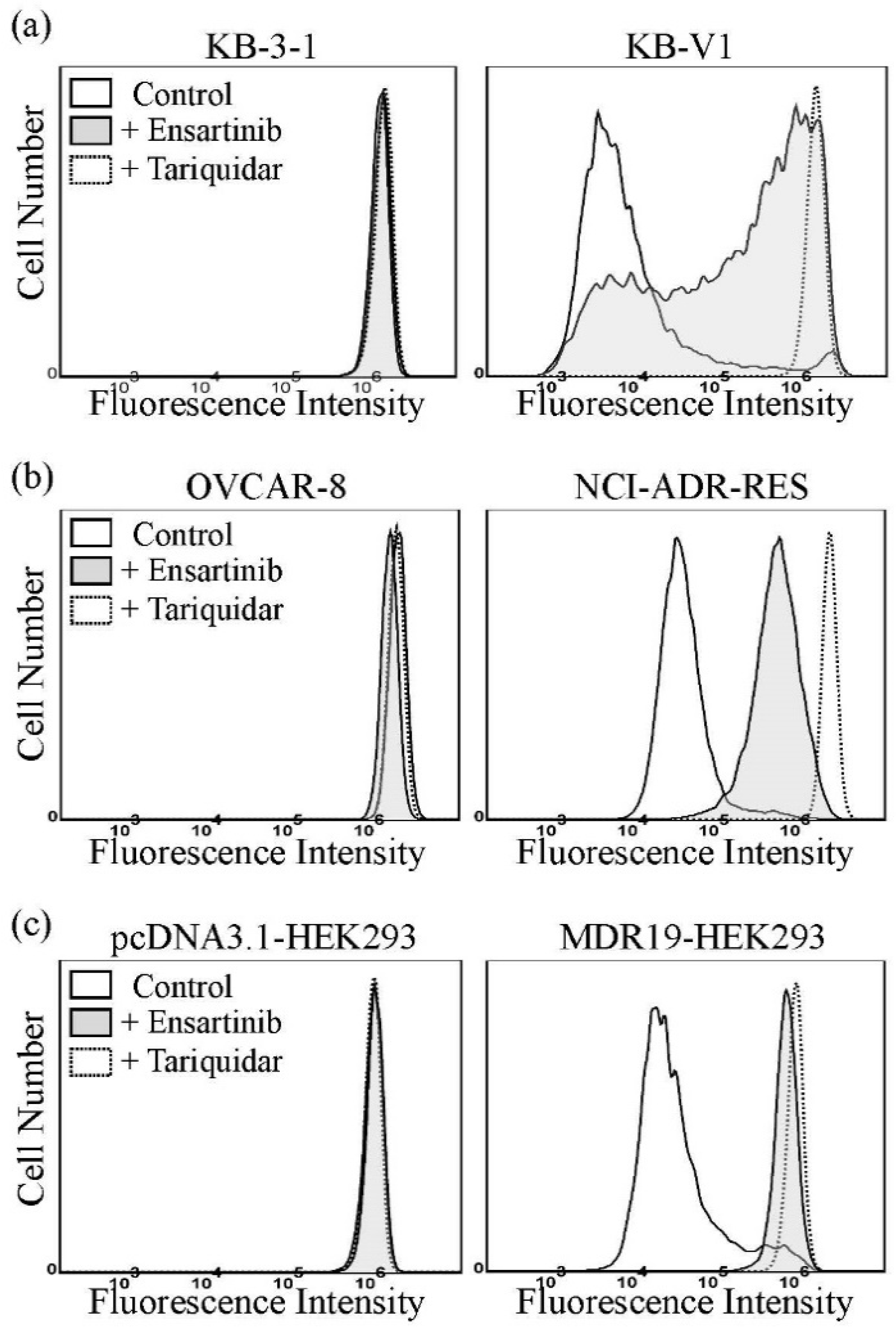
3.2. Ensartinib Attenuates the Drug Efflux Function of P-gp

Previous studies have demonstrated that, at higher concentrations, certain substrates of P-gp and/or ABCG2 can act as inhibitors of P-gp and/or ABCG2, respectively [37,48,49,50]. To this end, the ability of ensartinib to inhibit P-gp-mediated calcein-AM efflux and ABCG2-mediated PhA efflux was examined. As shown in Figure 2, the accumulation of calcein, a fluorescent product of the P-gp substrate calcein-AM [51], in KB-V1 (Figure 2a), NCI-ADR-RES (Figure 2b), and MDR19-HEK293 (Figure 2c) cells was extremely low (dotted lines). However, the intracellular fluorescence level increased significantly when these cells were incubated with calcein-AM in the presence of 10 μM ensartinib (filled solid lines), or 5 μM tariquidar (dotted lines). In contrast, the accumulation of the fluorescent ABCG2 substrate PhA [42] in H460-MX20 (Figure 2d), S1-MI-80 (Figure 2e), and R482-HEK293 (Figure 2f) cells increased substantially when these cells were incubated with PhA in the presence of 5 μM Ko143, a positive control (dotted lines), but remained low in the presence of 10 μM ensartinib (filled solid lines). These results suggest that at higher concentrations, ensartinib could compete with the transport of other substrates mediated by P-gp.
Next, the effect of ensartinib on reversing MDR mediated by P-gp and ABCG2 was determined in P-gp- and ABCG2-overexpressing multidrug-resistant cells. The extent of reversal was determined by adding a non-toxic concentration of ensartinib, tariquidar or Ko143 to the cytotoxicity assays, and presented as a fold-reversal (FR) value as described previously [48,52]. Reversal assays revealed that, at the highest non-toxic concentration of 500 nM, ensartinib had no significant effect on P-gp-mediated resistance to the P-gp substrates paclitaxel, vincristine or colchicine [53] in P-gp-overexpressing KB-V1 and NCI-ADR-RES cancer cell lines, and the P-gp-transfected MDR19-HEK293 cell line (Table 2). Similarly, ABCG2-mediated resistance to the ABCG2 drug substrates mitoxantrone, topotecan and SN-38 [54,55] was not reversed by ensartinib in ABCG2-overexpressing S1-MI-80 or H460-MX20 cancer cell lines, or in the ABCG2-transfected R482-HEK293 cell line (Table 3). Our results revealed that, although ensartinib could block P-gp-mediated drug efflux at higher concentrations, it could not resensitize P-gp- or ABCG2-overexpressing cancer cells to cytotoxic drugs at non-toxic low concentrations.

3.3. P-gp Reduces the Intracellular Accumulation of Ensartinib in Human Cancer Cells

Knowing that P-gp expression confers resistance to ensartinib, and that one of the most likely explanations for this is the reduced intracellular drug accumulation caused by P-gp-mediated drug efflux [10], we examined the effect of P-gp function on the intracellular accumulation of ensartinib in cancer cells. KB-3-1 and the P-gp-overexpressing variant KB-V1 were treated with 10 μM ensartinib in the absence or presence of 10 μM tariquidar, and the intracellular concentration of ensartinib was determined using a liquid chromatography-selected reaction monitoring mass spectrometry (LC-SRM/MS) method (Figure 3a) as described previously [39,41,43]. Human KB epidermal cancer cell lines were chosen for this study since KB-V1 showed the highest RF value among all the P-gp-overexpressing cell lines (Table 1). As shown in Figure 3b, significantly lower intracellular accumulation of ensartinib was detected in KB-V1 cancer cells (filled bars) compared to KB-3-1 cancer cells (open bars) or cells treated with ensartinib in the presence of tariquidar.
The effect of tariquidar on ensartinib-induced apoptosis was also examined. As shown in Figure 4, without inducing apoptosis in KB-V1 cells, the apoptotic KB-3-1 cell population was substantially increased by ensartinib, from an approximate 5% basal level to 26%. More importantly, the ensartinib-induced KB-V1 apoptotic cell population was significantly increased by tariquidar to a level comparable to that observed in KB-3-1 cells. Of note, tariquidar alone had no significant effect on the apoptotic cell population in either KB-3-1 or KB-V1 cell lines. These results were in agreement with the cytotoxicity data (Table 1), suggesting that P-gp mediates the efflux of ensartinib and contributes to the reduced efficacy of ensartinib in P-gp-overexpressing cancer cells.

3.4. Docking of Ensartinib in the Drug-Binding Pocket of P-gp

To further investigate the interactions between ensartinib and the substrate-binding pocket of P-gp, the in silico molecular docking analysis of ensartinib in the inward-open structure of P-gp (PDBID:6QEX) [45] was carried out. The lowest energy docking pose revealed a similar binding location for ensartinib as reported for the Taxol- and vincristine-bound cryo-EM structures. Ensartinib was shown to interact with numerous hydrophobic and aromatic residues in the transmembrane domains (TMDs) such as PHE303, ILE306, TYR310, PHE336, PHE983, MET986, and a hydrogen bond was predicted between its carbonyl group and GLN990 (Figure 5). Common interacting residues were also observed in the binding with Taxol (PHE983, MET986 and GLN990) and vincristine (ILE306, TYR310, PHE983, MET986 and GLN990) [45,56].

4. Discussion

Despite the tremendous progress made in recent years in the development of novel molecularly-targeted therapeutic agents, resistance often occurs within months, caused by resistance mechanisms including the activation of bypass tracks, secondary mutations, and amplification of resistance genes [3,56]. Nonetheless, the understanding of molecular mechanisms underlying resistance to these molecularly targeted therapeutic agents remains incomplete, which poses a therapeutic challenge. Previous studies have demonstrated that the drug efflux function of P-gp and ABCG2 could alter the bioavailability, distribution and efficacy of epidermal growth factor receptor (EGFR) inhibitors [57,58,59], vascular endothelial growth factor receptor (VEGFR) inhibitors [60], rapidly accelerated fibrosarcoma (RAF) inhibitors [61,62,63,64], PARP inhibitors [65,66,67], and ALK inhibitors [68,69,70,71]. For example, Tang and colleagues reported that the first-generation ALK inhibitor crizotinib is a good transport substrate of P-gp, but not of ABCG2 [68]. In contrast, the second-generation ALK inhibitors ceritinib [69] and brigatinib [71] are both transport substrates of P-gp and ABCG2. Moreover, Katayama and colleagues revealed that P-gp mediates resistance to both crizotinib and ceritinib in NSCLC patients [70]. In this study, we sought to determine the potential impact of P-gp and ABCG2 on the susceptibility of human cancer cells to the second-generation ALK inhibitor ensartinib.
Ensartinib is a specific ALK TKI that is cytotoxic against a wide variety of cancer cell lines, including crizotinib-resistant cancer cell lines known to harbor ALK fusions or point mutations. Other than the HepG2 hepatocellular cancel cell line, the reported IC50 values of ensartinib against the cancer cell lines tested were in the range of 15 nM to 3.0 μM [2]. In our study, the IC50 values of ensartinib for drug-sensitive cancer cell lines ranged from 0.9 μM to 3.0 μM, comparable to the IC50 values reported in previous studies [2,72]. On the other hand, we observed that the IC50 values of ensartinib for P-gp-overexpressing cancer cell lines, regardless of the tissue of origin, and HEK293 cells with ectopic expression of P-gp were significantly higher than the IC50 values in the respective drug-sensitive parental cells, which could be reversed by the P-gp inhibitor tariquidar (Table 1). Interestingly, Lovly and colleagues found that ensartinib exhibited a significantly higher IC50 value for the HepG2 cancer cell line as compared to other cell lines [2], which may be due, at least partially, to the activity of P-gp presented in the HepG2 cancer cells [73,74,75]. In addition, we discovered ensartinib could inhibit the P-gp-mediated efflux of the P-gp substrate calcein-AM from P-gp-expressing cells but had no significant effect on ABCG2-mediated efflux of its substrate PhA (Figure 2). The results of lower intracellular ensartinib accumulation (Figure 3) and reduced ensartinib-induced apoptosis (Figure 4) in P-gp-overexpressing KB-V1 cancer cells compared to the parental KB-3-1 cells correspond directly to the cytotoxicity data. Moreover, by blocking the drug-efflux function of P-gp, tariquidar was able to restore the intracellular concentration of ensartinib and ensartinib-induced apoptosis in KB-V1 cells, which is in agreement with our conclusion that P-gp reduces the efficacy of ensartinib in human cancer cells. Analysis of in silico molecular docking of ensartinib with the inward-open structure of P-gp (PDBID:6QEX) [45] revealed several common residues that interact with Taxol, vincristine and ensartinib. The docking analysis further provides support for binding of ensartinib in the drug-binding pocket that is similar to that of Taxol or vincristine (Figure 5). Collectively, these data indicate that P-gp mediates ensartinib resistance in multidrug-resistant cancer cells.
It is worth noting that Vagiannis and colleagues reported previously that ensartinib is an effective inhibitor of P-gp and ABCG2 but behaved as a substrate of P-gp in Madin-Darby canine kidney II (MDCKII) monolayer transport assays, and that ensartinib stimulated P-gp-specific ATPase activity in a concentration-dependent manner [76]. Consistent with our results, these authors found that ensartinib is not transported by ABCG2 in MDCK-II cells. However, they found that the antitumor efficiency of ensartinib was not compromised by P-gp in MDCKII, HL60, or A431 cells, but with IC50 values that were considerably higher than the IC50 values reported in other studies [2,72,76,77]. The discrepancy between results with MDCK-II and human cell lines used in our study could be due to the presence of endogenous canine P-gp and ABCG2 transporters. Another difference is that for the docking studies, Vagiannis et al. used a homology model of human P-gp based on the mouse Abcb1a structure. In addition, they docked ensartinib in the NBDs of the ATP-bound structure of the EQ mutant of human P-gp (pdb.6C0V; Figure 5). Using this structure is not logical, as the drug-binding pocket is collapsed and there is no evidence for binding of ensartinib or any other structure in the NBDs [76]. In the same study, they reported that ensartinib at 10 μM could reverse P-gp-mediated resistance to daunorubicin and ABCG2-mediated resistance to mitoxantrone in MDCKII cells transduced with P-gp or ABCG2, and in the human leukemia HL60 cell line overexpressing P-gp or ABCG2. However, considering that ensartinib at 10 μM is cytotoxic to parental MDCKII and HL60 cell lines, the chemosensitizing effect of ensartinib may be overestimated [76]. It is worth noting that, in our study, we found that at the highest non-toxic concentration of 0.5 μM, ensartinib was unable to reverse P-gp-mediated resistance to the P-gp substrates paclitaxel, vincristine, and colchicine, or ABCG2-mediated resistance to the ABCG2 substrates mitoxantrone, SN-38, and topotecan.

5. Conclusions

In summary, although experimental results from cell or animal models of multidrug-resistant cancers do not necessarily reflect clinical reality [78], our data reveal that ensartinib is a substrate for P-gp. Based on our findings, we propose that P-gp expression could contribute to the development of resistance to ensartinib resistance in cancer cells and have clinical implications in NSCLC patients receiving this drug treatment. In addition, the activity of P-gp could lead to altered pharmacokinetics and pharmacodynamics in certain patients and should be further investigated.

Author Contributions

Conceptualization, C.-P.W., Y.-S.W. and S.V.A.; methodology, C.-Y.H., M.M., C.-L.L. and Y.-H.H.; software, C.-Y.H. and C.-L.L.; validation, C.-Y.H., M.M., C.-L.L. and Y.-H.H.; formal analysis, C.-P.W., C.-Y.H., M.M. and Y.-S.W.; investigation, C.-Y.H., M.M., C.-L.L. and Y.-H.H.; resources, C.-P.W., T.-H.H., Y.-S.W., J.-S.Y. and S.V.A.; data curation, C.-Y.H., M.M., C.-L.L. and Y.-H.H.; writing—original draft preparation, C.-P.W., C.-Y.H., T.-H.H., Y.-S.W. and S.V.A.; writing—review and editing, C.-P.W., Y.-S.W. and S.V.A.; visualization, C.-Y.H., M.M., C.-L.L. and Y.-H.H.; supervision, C.-P.W., J.-S.Y. and S.V.A.; project administration, C.-P.W. and S.V.A.; funding acquisition, C.-P.W., T.-H.H., Y.-S.W., J.-S.Y. and S.V.A. All authors have read and agreed to the published version of the manuscript.

Funding

This research was funded by the Ministry of Science and Technology of Taiwan (108-2320-B-182-038-MY3 and 109-2113-M-029-007 to C.-P.W. and 109-2314-B-182A-097-MY3 to T.-H.H.), Taichung Veterans General Hospital (TCVGH-T1107807 to Y.-S.W), Chang Gung Memorial Hospital (CMRPD1K0391, CMRPD1L0051 and BMRPC17 to C.-P.W.; CMRPG1J0073, CORPG1L0051, and CORPG2L0031 to T.-H.H.; CLRPD1J0013 to J.-S.Y.), and the Intramural Research Program of the National Institutes of Health, National Cancer Institute, Center for Cancer Research (S.V.A and M.M).

Institutional Review Board Statement

Not Applicable.

Informed Consent Statement

Not Applicable.

Data Availability Statement

Not Applicable.

Acknowledgments

We thank George Leiman for editing the manuscript and the Taipei Common Laboratory of Chang Gung Memorial Hospital for providing technical assistance.

Conflicts of Interest

The authors declare no conflict of interest.

References

  1. Cameron, L.B.; Hitchen, N.; Chandran, E.; Morris, T.; Manser, R.; Solomon, B.J.; Jordan, V. Targeted therapy for advanced anaplastic lymphoma kinase (ALK)-rearranged non-small cell lung cancer. Cochrane Database Syst. Rev. 2022, 1, CD013453. [Google Scholar] [CrossRef] [PubMed]
  2. Lovly, C.M.; Heuckmann, J.M.; de Stanchina, E.; Chen, H.; Thomas, R.K.; Liang, C.; Pao, W. Insights into ALK-driven cancers revealed through development of novel ALK tyrosine kinase inhibitors. Cancer Res. 2011, 71, 4920–4931. [Google Scholar] [CrossRef] [PubMed] [Green Version]
  3. Caccese, M.; Ferrara, R.; Pilotto, S.; Carbognin, L.; Grizzi, G.; Calio, A.; Brunelli, M.; Cuppone, F.; Petraglia, S.; Scarpa, A.; et al. Current and developing therapies for the treatment of non-small cell lung cancer with ALK abnormalities: Update and perspectives for clinical practice. Expert Opin. Pharmacother. 2016, 17, 2253–2266. [Google Scholar] [CrossRef] [PubMed]
  4. Fan, J.; Fong, T.; Xia, Z.; Zhang, J.; Luo, P. The efficacy and safety of ALK inhibitors in the treatment of ALK-positive non-small cell lung cancer: A network meta-analysis. Cancer Med. 2018, 7, 4993–5005. [Google Scholar] [CrossRef] [PubMed]
  5. Horn, L.; Infante, J.R.; Reckamp, K.L.; Blumenschein, G.R.; Leal, T.A.; Waqar, S.N.; Gitlitz, B.J.; Sanborn, R.E.; Whisenant, J.G.; Du, L.; et al. Ensartinib (X-396) in ALK-Positive Non-Small Cell Lung Cancer: Results from a First-in-Human Phase I/II, Multicenter Study. Clin. Cancer Res. 2018, 24, 2771–2779. [Google Scholar] [CrossRef] [PubMed] [Green Version]
  6. Gottesman, M.M.; Fojo, T.; Bates, S.E. Multidrug resistance in cancer: Role of ATP-dependent transporters. Nat. Rev. Cancer 2002, 2, 48–58. [Google Scholar] [CrossRef] [PubMed] [Green Version]
  7. Brozik, A.; Hegedus, C.; Erdei, Z.; Hegedus, T.; Ozvegy-Laczka, C.; Szakacs, G.; Sarkadi, B. Tyrosine kinase inhibitors as modulators of ATP binding cassette multidrug transporters: Substrates, chemosensitizers or inducers of acquired multidrug resistance? Expert Opin. Drug Metab. Toxicol. 2011, 7, 623–642. [Google Scholar] [CrossRef] [PubMed]
  8. Camidge, D.R.; Pao, W.; Sequist, L.V. Acquired resistance to TKIs in solid tumours: Learning from lung cancer. Nat. Rev. Clin. Oncol. 2014, 11, 473–481. [Google Scholar] [CrossRef]
  9. Noguchi, K.; Katayama, K.; Sugimoto, Y. Human ABC transporter ABCG2/BCRP expression in chemoresistance: Basic and clinical perspectives for molecular cancer therapeutics. Pharm. Pers. Med. 2014, 7, 53–64. [Google Scholar] [CrossRef] [PubMed] [Green Version]
  10. Robey, R.W.; Pluchino, K.M.; Hall, M.D.; Fojo, A.T.; Bates, S.E.; Gottesman, M.M. Revisiting the role of ABC transporters in multidrug-resistant cancer. Nat. Rev. Cancer 2018, 18, 452–464. [Google Scholar] [CrossRef]
  11. Gillet, J.P.; Gottesman, M.M. Mechanisms of multidrug resistance in cancer. Methods Mol. Biol. 2010, 596, 47–76. [Google Scholar] [CrossRef] [PubMed]
  12. Wu, C.P.; Hsieh, C.H.; Wu, Y.S. The emergence of drug transporter-mediated multidrug resistance to cancer chemotherapy. Mol. Pharm. 2011, 8, 1996–2011. [Google Scholar] [CrossRef] [PubMed]
  13. Ross, D.D.; Karp, J.E.; Chen, T.T.; Doyle, L.A. Expression of breast cancer resistance protein in blast cells from patients with acute leukemia. Blood 2000, 96, 365–368. [Google Scholar] [CrossRef] [PubMed]
  14. Steinbach, D.; Sell, W.; Voigt, A.; Hermann, J.; Zintl, F.; Sauerbrey, A. BCRP gene expression is associated with a poor response to remission induction therapy in childhood acute myeloid leukemia. Leukemia 2002, 16, 1443–1447. [Google Scholar] [CrossRef] [PubMed]
  15. Uggla, B.; Stahl, E.; Wagsater, D.; Paul, C.; Karlsson, M.G.; Sirsjo, A.; Tidefelt, U. BCRP mRNA expression v. clinical outcome in 40 adult AML patients. Leuk. Res. 2005, 29, 141–146. [Google Scholar] [CrossRef]
  16. Matthews, C.; Catherwood, M.A.; Larkin, A.M.; Clynes, M.; Morris, T.C.; Alexander, H.D. MDR-1, but not MDR-3 gene expression, is associated with unmutated IgVH genes and poor prognosis chromosomal aberrations in chronic lymphocytic leukemia. Leuk. Lymphoma 2006, 47, 2308–2313. [Google Scholar] [CrossRef]
  17. Turner, J.G.; Gump, J.L.; Zhang, C.; Cook, J.M.; Marchion, D.; Hazlehurst, L.; Munster, P.; Schell, M.J.; Dalton, W.S.; Sullivan, D.M. ABCG2 expression, function, and promoter methylation in human multiple myeloma. Blood 2006, 108, 3881–3889. [Google Scholar] [CrossRef]
  18. Kovalev, A.A.; Tsvetaeva, D.A.; Grudinskaja, T.V. Role of ABC-cassette transporters (MDR1, MRP1, BCRP) in the development of primary and acquired multiple drug resistance in patients with early and metastatic breast cancer. Exp. Oncol. 2013, 35, 287–290. [Google Scholar]
  19. Pilarski, L.M.; Belch, A.R. Intrinsic expression of the multidrug transporter, P-glycoprotein 170, in multiple myeloma: Implications for treatment. Leuk. Lymphoma 1995, 17, 367–374. [Google Scholar] [CrossRef]
  20. Pilarski, L.M.; Szczepek, A.J.; Belch, A.R. Deficient drug transporter function of bone marrow-localized and leukemic plasma cells in multiple myeloma. Blood 1997, 90, 3751–3759. [Google Scholar] [CrossRef]
  21. Schwarzenbach, H. Expression of MDR1/P-glycoprotein, the multidrug resistance protein MRP, and the lung-resistance protein LRP in multiple myeloma. Med. Oncol. 2002, 19, 87–104. [Google Scholar] [CrossRef]
  22. Nakagawa, Y.; Abe, S.; Kurata, M.; Hasegawa, M.; Yamamoto, K.; Inoue, M.; Takemura, T.; Suzuki, K.; Kitagawa, M. IAP family protein expression correlates with poor outcome of multiple myeloma patients in association with chemotherapy-induced overexpression of multidrug resistance genes. Am. J. Hematol. 2006, 81, 824–831. [Google Scholar] [CrossRef] [PubMed]
  23. Tsubaki, M.; Satou, T.; Itoh, T.; Imano, M.; Komai, M.; Nishinobo, M.; Yamashita, M.; Yanae, M.; Yamazoe, Y.; Nishida, S. Overexpression of MDR1 and survivin, and decreased Bim expression mediate multidrug-resistance in multiple myeloma cells. Leuk. Res. 2012, 36, 1315–1322. [Google Scholar] [CrossRef] [PubMed]
  24. Tsunoda, S.; Okumura, T.; Ito, T.; Kondo, K.; Ortiz, C.; Tanaka, E.; Watanabe, G.; Itami, A.; Sakai, Y.; Shimada, Y. ABCG2 expression is an independent unfavorable prognostic factor in esophageal squamous cell carcinoma. Oncology 2006, 71, 251–258. [Google Scholar] [CrossRef]
  25. Li, F.; Zeng, H.; Ying, K. The combination of stem cell markers CD133 and ABCG2 predicts relapse in stage I non-small cell lung carcinomas. Med. Oncol. 2011, 28, 1458–1462. [Google Scholar] [CrossRef]
  26. Hang, D.; Dong, H.C.; Ning, T.; Dong, B.; Hou, D.L.; Xu, W.G. Prognostic value of the stem cell markers CD133 and ABCG2 expression in esophageal squamous cell carcinoma. Dis. Esophagus 2012, 25, 638–644. [Google Scholar] [CrossRef]
  27. Breedveld, P.; Pluim, D.; Cipriani, G.; Wielinga, P.; van Tellingen, O.; Schinkel, A.H.; Schellens, J.H. The effect of Bcrp1 (Abcg2) on the in vivo pharmacokinetics and brain penetration of imatinib mesylate (Gleevec): Implications for the use of breast cancer resistance protein and P-glycoprotein inhibitors to enable the brain penetration of imatinib in patients. Cancer Res. 2005, 65, 2577–2582. [Google Scholar]
  28. Sarkadi, B.; Homolya, L.; Szakacs, G.; Varadi, A. Human multidrug resistance ABCB and ABCG transporters: Participation in a chemoimmunity defense system. Physiol. Rev. 2006, 86, 1179–1236. [Google Scholar] [CrossRef]
  29. Paez, J.G.; Janne, P.A.; Lee, J.C.; Tracy, S.; Greulich, H.; Gabriel, S.; Herman, P.; Kaye, F.J.; Lindeman, N.; Boggon, T.J.; et al. EGFR mutations in lung cancer: Correlation with clinical response to gefitinib therapy. Science 2004, 304, 1497–1500. [Google Scholar] [CrossRef] [Green Version]
  30. Shen, D.W.; Cardarelli, C.; Hwang, J.; Cornwell, M.; Richert, N.; Ishii, S.; Pastan, I.; Gottesman, M.M. Multiple drug-resistant human KB carcinoma cells independently selected for high-level resistance to colchicine, adriamycin, or vinblastine show changes in expression of specific proteins. J. Biol. Chem. 1986, 261, 7762–7770. [Google Scholar] [CrossRef]
  31. Roschke, A.V.; Tonon, G.; Gehlhaus, K.S.; McTyre, N.; Bussey, K.J.; Lababidi, S.; Scudiero, D.A.; Weinstein, J.N.; Kirsch, I.R. Karyotypic complexity of the NCI-60 drug-screening panel. Cancer Res. 2003, 63, 8634–8647. [Google Scholar] [PubMed]
  32. Robey, R.W.; Shukla, S.; Finley, E.M.; Oldham, R.K.; Barnett, D.; Ambudkar, S.V.; Fojo, T.; Bates, S.E. Inhibition of P-glycoprotein (ABCB1)- and multidrug resistance-associated protein 1 (ABCC1)-mediated transport by the orally administered inhibitor, CBT-1((R)). Biochem. Pharmacol. 2008, 75, 1302–1312. [Google Scholar] [CrossRef] [PubMed] [Green Version]
  33. Robey, R.W.; Honjo, Y.; Morisaki, K.; Nadjem, T.A.; Runge, S.; Risbood, M.; Poruchynsky, M.S.; Bates, S.E. Mutations at amino-acid 482 in the ABCG2 gene affect substrate and antagonist specificity. Br. J. Cancer 2003, 89, 1971–1978. [Google Scholar] [CrossRef] [PubMed]
  34. Henrich, C.J.; Robey, R.W.; Bokesch, H.R.; Bates, S.E.; Shukla, S.; Ambudkar, S.V.; Dean, M.; McMahon, J.B. New inhibitors of ABCG2 identified by high-throughput screening. Mol. Cancer Ther. 2007, 6, 3271–3278. [Google Scholar] [CrossRef] [Green Version]
  35. Miyake, K.; Mickley, L.; Litman, T.; Zhan, Z.; Robey, R.; Cristensen, B.; Brangi, M.; Greenberger, L.; Dean, M.; Fojo, T.; et al. Molecular cloning of cDNAs which are highly overexpressed in mitoxantrone-resistant cells: Demonstration of homology to ABC transport genes. Cancer Res. 1999, 59, 8–13. [Google Scholar]
  36. Shen, D.W.; Fojo, A.; Chin, J.E.; Roninson, I.B.; Richert, N.; Pastan, I.; Gottesman, M.M. Human multidrug-resistant cell lines: Increased mdr1 expression can precede gene amplification. Science 1986, 232, 643–645. [Google Scholar] [CrossRef]
  37. Wu, C.P.; Shukla, S.; Calcagno, A.M.; Hall, M.D.; Gottesman, M.M.; Ambudkar, S.V. Evidence for dual mode of action of a thiosemicarbazone, NSC73306: A potent substrate of the multidrug resistance linked ABCG2 transporter. Mol. Cancer Ther. 2007, 6, 3287–3296. [Google Scholar] [CrossRef] [Green Version]
  38. Henrich, C.J.; Bokesch, H.R.; Dean, M.; Bates, S.E.; Robey, R.W.; Goncharova, E.I.; Wilson, J.A.; McMahon, J.B. A high-throughput cell-based assay for inhibitors of ABCG2 activity. J. Biomol. Screen. 2006, 11, 176–183. [Google Scholar] [CrossRef] [Green Version]
  39. Wu, C.P.; Hung, C.Y.; Lusvarghi, S.; Huang, Y.H.; Tseng, P.J.; Hung, T.H.; Yu, J.S.; Ambudkar, S.V. Overexpression of ABCB1 and ABCG2 contributes to reduced efficacy of the PI3K/mTOR inhibitor samotolisib (LY3023414) in cancer cell lines. Biochem. Pharmacol. 2020, 180, 114137. [Google Scholar] [CrossRef]
  40. Wu, C.P.; Hsiao, S.H.; Su, C.Y.; Luo, S.Y.; Li, Y.Q.; Huang, Y.H.; Hsieh, C.H.; Huang, C.W. Human ATP-Binding Cassette transporters ABCB1 and ABCG2 confer resistance to CUDC-101, a multi-acting inhibitor of histone deacetylase, epidermal growth factor receptor and human epidermal growth factor receptor 2. Biochem. Pharmacol. 2014, 92, 567–576. [Google Scholar] [CrossRef]
  41. Wu, C.P.; Hsieh, Y.J.; Murakami, M.; Vahedi, S.; Hsiao, S.H.; Yeh, N.; Chou, A.W.; Li, Y.Q.; Wu, Y.S.; Yu, J.S.; et al. Human ATP-binding cassette transporters ABCB1 and ABCG2 confer resistance to histone deacetylase 6 inhibitor ricolinostat (ACY-1215) in cancer cell lines. Biochem. Pharmacol. 2018, 155, 316–325. [Google Scholar] [CrossRef] [PubMed]
  42. Robey, R.W.; Steadman, K.; Polgar, O.; Morisaki, K.; Blayney, M.; Mistry, P.; Bates, S.E. Pheophorbide a is a specific probe for ABCG2 function and inhibition. Cancer Res. 2004, 64, 1242–1246. [Google Scholar] [CrossRef] [PubMed] [Green Version]
  43. Wu, C.P.; Hung, C.Y.; Lusvarghi, S.; Chang, Y.F.; Hsiao, S.H.; Huang, Y.H.; Hung, T.H.; Yu, J.S.; Ambudkar, S.V. Overexpression of Human ABCB1 and ABCG2 Reduces the Susceptibility of Cancer Cells to the Histone Deacetylase 6-Specific Inhibitor Citarinostat. Int. J. Mol. Sci. 2021, 22, 2592. [Google Scholar] [CrossRef] [PubMed]
  44. Anderson, H.A.; Maylock, C.A.; Williams, J.A.; Paweletz, C.P.; Shu, H.; Shacter, E. Serum-derived protein S binds to phosphatidylserine and stimulates the phagocytosis of apoptotic cells. Nat. Immunol. 2003, 4, 87–91. [Google Scholar] [CrossRef] [PubMed]
  45. Alam, A.; Kowal, J.; Broude, E.; Roninson, I.; Locher, K.P. Structural insight into substrate and inhibitor discrimination by human P-glycoprotein. Science 2019, 363, 753–756. [Google Scholar] [CrossRef] [PubMed] [Green Version]
  46. Wu, C.P.; Murakami, M.; Wu, Y.S.; Chi, Y.C.; Hsiao, S.H.; Huang, Y.H.; Hung, T.H.; Ambudkar, S.V. Branebrutinib (BMS-986195), a Bruton’s Tyrosine Kinase Inhibitor, Resensitizes P-Glycoprotein-Overexpressing Multidrug-Resistant Cancer Cells to Chemotherapeutic Agents. Front. Cell Dev. Biol. 2021, 9, 699571. [Google Scholar] [CrossRef]
  47. Sanner, M.F.; Olson, A.J.; Spehner, J.C. Reduced surface: An efficient way to compute molecular surfaces. Biopolymers 1996, 38, 305–320. [Google Scholar] [CrossRef]
  48. Dai, C.L.; Tiwari, A.K.; Wu, C.P.; Su, X.D.; Wang, S.R.; Liu, D.G.; Ashby, C.R., Jr.; Huang, Y.; Robey, R.W.; Liang, Y.J.; et al. Lapatinib (Tykerb, GW572016) reverses multidrug resistance in cancer cells by inhibiting the activity of ATP-binding cassette subfamily B member 1 and G member 2. Cancer Res. 2008, 68, 7905–7914. [Google Scholar] [CrossRef] [Green Version]
  49. Sodani, K.; Tiwari, A.K.; Singh, S.; Patel, A.; Xiao, Z.J.; Chen, J.J.; Sun, Y.L.; Talele, T.T.; Chen, Z.S. GW583340 and GW2974, human EGFR and HER-2 inhibitors, reverse ABCG2- and ABCB1-mediated drug resistance. Biochem. Pharmacol. 2012, 83, 1613–1622. [Google Scholar] [CrossRef] [Green Version]
  50. Kuang, Y.H.; Patel, J.P.; Sodani, K.; Wu, C.P.; Liao, L.Q.; Patel, A.; Tiwari, A.K.; Dai, C.L.; Chen, X.; Fu, L.W.; et al. OSI-930 analogues as novel reversal agents for ABCG2-mediated multidrug resistance. Biochem. Pharmacol. 2012, 84, 766–774. [Google Scholar] [CrossRef] [Green Version]
  51. Hollo, Z.; Homolya, L.; Davis, C.W.; Sarkadi, B. Calcein accumulation as a fluorometric functional assay of the multidrug transporter. Biochim. Biophys. Acta 1994, 1191, 384–388. [Google Scholar] [CrossRef]
  52. Wu, C.P.; Lusvarghi, S.; Wang, J.C.; Hsiao, S.H.; Huang, Y.H.; Hung, T.H.; Ambudkar, S.V. Avapritinib: A Selective Inhibitor of KIT and PDGFRalpha that Reverses ABCB1 and ABCG2-Mediated Multidrug Resistance in Cancer Cell Lines. Mol. Pharm. 2019, 16, 3040–3052. [Google Scholar] [CrossRef] [PubMed]
  53. Kartner, N.; Riordan, J.R.; Ling, V. Cell surface P-glycoprotein associated with multidrug resistance in mammalian cell lines. Science 1983, 221, 1285–1288. [Google Scholar] [CrossRef] [PubMed]
  54. Bates, S.E.; Medina-Perez, W.Y.; Kohlhagen, G.; Antony, S.; Nadjem, T.; Robey, R.W.; Pommier, Y. ABCG2 mediates differential resistance to SN-38 (7-ethyl-10-hydroxycamptothecin) and homocamptothecins. J. Pharmacol. Exp. Ther. 2004, 310, 836–842. [Google Scholar] [CrossRef] [Green Version]
  55. Maliepaard, M.; van Gastelen, M.A.; de Jong, L.A.; Pluim, D.; van Waardenburg, R.C.; Ruevekamp-Helmers, M.C.; Floot, B.G.; Schellens, J.H. Overexpression of the BCRP/MXR/ABCP gene in a topotecan-selected ovarian tumor cell line. Cancer Res. 1999, 59, 4559–4563. [Google Scholar]
  56. Nosol, K.; Romane, K.; Irobalieva, R.N.; Alam, A.; Kowal, J.; Fujita, N.; Locher, K.P. Cryo-EM structures reveal distinct mechanisms of inhibition of the human multidrug transporter ABCB1. Proc. Natl. Acad. Sci. USA 2020, 117, 26245–26253. [Google Scholar] [CrossRef]
  57. Tian, X.; Gu, T.; Lee, M.H.; Dong, Z. Challenge and countermeasures for EGFR targeted therapy in non-small cell lung cancer. Biochim. Biophys Acta Rev Cancer 2021, 1877, 188645. [Google Scholar] [CrossRef]
  58. Agarwal, S.; Sane, R.; Gallardo, J.L.; Ohlfest, J.R.; Elmquist, W.F. Distribution of gefitinib to the brain is limited by P-glycoprotein (ABCB1) and breast cancer resistance protein (ABCG2)-mediated active efflux. J. Pharmacol. Exp. Ther. 2010, 334, 147–155. [Google Scholar] [CrossRef] [Green Version]
  59. Kim, M.; Laramy, J.K.; Mohammad, A.S.; Talele, S.; Fisher, J.; Sarkaria, J.N.; Elmquist, W.F. Brain Distribution of a Panel of Epidermal Growth Factor Receptor Inhibitors Using Cassette Dosing in Wild-Type and Abcb1/Abcg2-Deficient Mice. Drug Metab. Dispos. 2019, 47, 393–404. [Google Scholar] [CrossRef] [Green Version]
  60. van Hoppe, S.; Jamalpoor, A.; Rood, J.J.M.; Wagenaar, E.; Sparidans, R.W.; Beijnen, J.H.; Schinkel, A.H. Brain accumulation of osimertinib and its active metabolite AZ5104 is restricted by ABCB1 (P-glycoprotein) and ABCG2 (breast cancer resistance protein). Pharmacol. Res. 2019, 146, 104297. [Google Scholar] [CrossRef]
  61. Wang, T.; Agarwal, S.; Elmquist, W.F. Brain distribution of cediranib is limited by active efflux at the blood-brain barrier. J Pharmacol. Exp. Ther. 2012, 341, 386–395. [Google Scholar] [CrossRef] [PubMed] [Green Version]
  62. Mittapalli, R.K.; Vaidhyanathan, S.; Sane, R.; Elmquist, W.F. Impact of P-glycoprotein (ABCB1) and breast cancer resistance protein (ABCG2) on the brain distribution of a novel BRAF inhibitor: Vemurafenib (PLX4032). J. Pharmacol. Exp. Ther. 2012, 342, 33–40. [Google Scholar] [CrossRef] [PubMed]
  63. Wu, C.P.; Sim, H.M.; Huang, Y.H.; Liu, Y.C.; Hsiao, S.H.; Cheng, H.W.; Li, Y.Q.; Ambudkar, S.V.; Hsu, S.C. Overexpression of ATP-binding cassette transporter ABCG2 as a potential mechanism of acquired resistance to vemurafenib in BRAF(V600E) mutant cancer cells. Biochem. Pharmacol. 2013, 85, 325–334. [Google Scholar] [CrossRef] [PubMed] [Green Version]
  64. Wang, J.; Gan, C.; Sparidans, R.W.; Wagenaar, E.; van Hoppe, S.; Beijnen, J.H.; Schinkel, A.H. P-glycoprotein (MDR1/ABCB1) and Breast Cancer Resistance Protein (BCRP/ABCG2) affect brain accumulation and intestinal disposition of encorafenib in mice. Pharmacol. Res. 2018, 129, 414–423. [Google Scholar] [CrossRef]
  65. Gampa, G.; Kim, M.; Mohammad, A.S.; Parrish, K.E.; Mladek, A.C.; Sarkaria, J.N.; Elmquist, W.F. Brain Distribution and Active Efflux of Three panRAF Inhibitors: Considerations in the Treatment of Melanoma Brain Metastases. J. Pharmacol. Exp. Ther. 2019, 368, 446–461. [Google Scholar] [CrossRef] [Green Version]
  66. Parrish, K.E.; Cen, L.; Murray, J.; Calligaris, D.; Kizilbash, S.; Mittapalli, R.K.; Carlson, B.L.; Schroeder, M.A.; Sludden, J.; Boddy, A.V.; et al. Efficacy of PARP Inhibitor Rucaparib in Orthotopic Glioblastoma Xenografts Is Limited by Ineffective Drug Penetration into the Central Nervous System. Mol. Cancer. Ther. 2015, 14, 2735–2743. [Google Scholar] [CrossRef] [Green Version]
  67. Durmus, S.; Sparidans, R.W.; van Esch, A.; Wagenaar, E.; Beijnen, J.H.; Schinkel, A.H. Breast cancer resistance protein (BCRP/ABCG2) and P-glycoprotein (P-GP/ABCB1) restrict oral availability and brain accumulation of the PARP inhibitor rucaparib (AG-014699). Pharm. Res. 2015, 32, 37–46. [Google Scholar] [CrossRef]
  68. de Gooijer, M.C.; Buil, L.C.M.; Citirikkaya, C.H.; Hermans, J.; Beijnen, J.H.; van Tellingen, O. ABCB1 Attenuates the Brain Penetration of the PARP Inhibitor AZD2461. Mol. Pharm. 2018, 15, 5236–5243. [Google Scholar] [CrossRef]
  69. Tang, S.C.; Nguyen, L.N.; Sparidans, R.W.; Wagenaar, E.; Beijnen, J.H.; Schinkel, A.H. Increased oral availability and brain accumulation of the ALK inhibitor crizotinib by coadministration of the P-glycoprotein (ABCB1) and breast cancer resistance protein (ABCG2) inhibitor elacridar. Int. J. Cancer 2014, 134, 1484–1494. [Google Scholar] [CrossRef]
  70. Kort, A.; Sparidans, R.W.; Wagenaar, E.; Beijnen, J.H.; Schinkel, A.H. Brain accumulation of the EML4-ALK inhibitor ceritinib is restricted by P-glycoprotein (P-GP/ABCB1) and breast cancer resistance protein (BCRP/ABCG2). Pharmacol. Res. 2015, 102, 200–207. [Google Scholar] [CrossRef]
  71. Katayama, R.; Sakashita, T.; Yanagitani, N.; Ninomiya, H.; Horiike, A.; Friboulet, L.; Gainor, J.F.; Motoi, N.; Dobashi, A.; Sakata, S.; et al. P-glycoprotein Mediates Ceritinib Resistance in Anaplastic Lymphoma Kinase-rearranged Non-small Cell Lung Cancer. EBioMedicine 2016, 3, 54–66. [Google Scholar] [CrossRef] [PubMed] [Green Version]
  72. Li, W.; Sparidans, R.W.; Wang, Y.; Lebre, M.C.; Beijnen, J.H.; Schinkel, A.H. P-glycoprotein and breast cancer resistance protein restrict brigatinib brain accumulation and toxicity, and, alongside CYP3A, limit its oral availability. Pharmacol. Res. 2018, 137, 47–55. [Google Scholar] [CrossRef] [PubMed]
  73. Di Paolo, D.; Yang, D.; Pastorino, F.; Emionite, L.; Cilli, M.; Daga, A.; Destafanis, E.; Di Fiore, A.; Piaggio, F.; Brignole, C.; et al. New therapeutic strategies in neuroblastoma: Combined targeting of a novel tyrosine kinase inhibitor and liposomal siRNAs against ALK. Oncotarget 2015, 6, 28774–28789. [Google Scholar] [CrossRef] [PubMed] [Green Version]
  74. Cheung, J.Y.; Ong, R.C.; Suen, Y.K.; Ooi, V.; Wong, H.N.; Mak, T.C.; Fung, K.P.; Yu, B.; Kong, S.K. Polyphyllin D is a potent apoptosis inducer in drug-resistant HepG2 cells. Cancer Lett. 2005, 217, 203–211. [Google Scholar] [CrossRef]
  75. Manov, I.; Bashenko, Y.; Eliaz-Wolkowicz, A.; Mizrahi, M.; Liran, O.; Iancu, T.C. High-dose acetaminophen inhibits the lethal effect of doxorubicin in HepG2 cells: The role of P-glycoprotein and mitogen-activated protein kinase p44/42 pathway. J Pharmacol. Exp. Ther. 2007, 322, 1013–1022. [Google Scholar] [CrossRef]
  76. Liu, H.; Wang, M.; Liang, N.; Guan, L. PDCD2 sensitizes HepG2 cells to sorafenib by suppressing epithelialmesenchymal transition. Mol. Med. Rep. 2019, 19, 2173–2179. [Google Scholar] [CrossRef] [Green Version]
  77. Vagiannis, D.; Novotna, E.; Skarka, A.; Kammerer, S.; Kupper, J.H.; Chen, S.; Guo, L.; Staud, F.; Hofman, J. Ensartinib (X-396) Effectively Modulates Pharmacokinetic Resistance Mediated by ABCB1 and ABCG2 Drug Efflux Transporters and CYP3A4 Biotransformation Enzyme. Cancers 2020, 12, 813. [Google Scholar] [CrossRef] [Green Version]
  78. Gillet, J.P.; Wang, J.; Calcagno, A.M.; Green, L.J.; Varma, S.; Bunkholt Elstrand, M.; Trope, C.G.; Ambudkar, S.V.; Davidson, B.; Gottesman, M.M. Clinical relevance of multidrug resistance gene expression in ovarian serous carcinoma effusions. Mol. Pharm. 2011, 8, 2080–2088. [Google Scholar] [CrossRef] [Green Version]
Figure 1. The antiproliferative effect of ensartinib is reduced in cells overexpressing P-glycoprotein. The cytotoxicity of ensartinib in (a) KB-3-1 (open circles) and the P-gp-overexpressing subline KB-V1 (filled circles); (b) OVCAR-8 (open circles) and the P-gp-overexpressing subline NCI-ADR-RES (filled circles); as well as (c) pcDNA3.1-HEK293 (open circles) and the P-gp-transfected MDR19-HEK293 (filled circles), was determined as described previously [39]. The values were mean ± SEM calculated from at least three independent experiments. Insets are representative immunoblots showing P-gp and tubulin in parental and P-gp-expressing cells.
Cancers 14 02341 g001
Figure 2. Ensartinib increases the intracellular accumulation of calcein in P-glycoprotein-overexpressing cells. The effect of ensartinib on the accumulation of the P-gp substrate drug calcein-AM (ac) or accumulation of the ABCG2 substrate pheophorbide A (df) was determined in (a) KB-3-1 cells (left panel) and P-gp-overexpressing KB-V1 cells (right panel), (b) OVCAR-8 cells (left panel) and P-gp-overexpressing NCI-ADR-RES cells (right panel), (c) pcDNA3.1-HEK293 (left panel) and P-gp-transfected MDR19-HEK293 (right panel) cells, (d) NCI-H460 (left panel) and ABCG2-overexpressing H460-MX20 cells, (e) S1 (left panel) and ABCG2-overexpressing S1-MI-80 cells (right panel), and (f) pcDNA3.1-HEK293 (left panel) and ABCG2-transfected R482-HEK293 cells (right panel). Cells were treated with DMSO (control, solid lines), 10 μM ensartinib (filled solid lines), or 5 μM tariquidar (ac, dotted lines) as a positive control for P-gp or 5 μM Ko143 (df, dotted lines) as a positive control for ABCG2. Cells were processed and analyzed as described in Materials and Methods. Representative histograms from at least three independent experiments are shown.
Cancers 14 02341 g002aCancers 14 02341 g002b
Figure 3. P-gp-mediated transport decreases the intracellular concentration of ensartinib in human cancer cells. (a) The chemical structure (precursor ion m/z 561 in positive mode) and the mass spectra of major fragment ions of ensartinib. The fragment ion m/z 371 was selected for quantitative analysis. (b) The intracellular accumulation of ensartinib in KB-3-1 (white bars) and KB-V1 cells (black bars) treated with 10 μM of ensartinib in the absence or presence of tariquidar was determined as described in Materials and Methods. Quantitative data are presented as mean values ± S.D. calculated from three independent experiments. *** p < 0.001, as compared to treatment with tariquidar.
Cancers 14 02341 g003
Figure 4. Ensartinib induces apoptosis in cancer cell lines. (a) Representative dot plots of the extent of apoptosis determined in KB-3-1 cells (upper panels) and the P-gp-overexpressing subline KB-V1 (lower panels) treated with DMSO (DMSO control), 2 µM ensartinib (+ Ensartinib), 1 µM tariquidar (+ Tariquidar), or a combination of 2 µM ensartinib and 1 µM tariquidar (+ Ensartinib + Tariquidar) for 48 h. Cells were processed and analyzed as described in Materials and Methods and as described previously [40]. (b) Quantitative data are presented as mean values ± S.D. obtained from at least three independent experiments. ** p < 0.01, versus the same treatment in the presence of tariquidar.
Cancers 14 02341 g004
Figure 5. Docking of ensartinib to P-gp. The lowest energy binding mode of ensartinib with Taxol-bound human P-gp (PDB: 6QEX) was predicted by Accelrys Discovery Studio 4.0 software as described in Materials and Methods. The molecular model of ensartinib is highlighted in yellow, and the atoms for interacting amino acid residues carbon, hydrogen, nitrogen, and oxygen are colored gray, light gray, blue, and red, respectively. Dotted lines indicate proposed interactions.
Cancers 14 02341 g005
Table 1. Cytotoxicity of ensartinib in drug-sensitive and multidrug-resistant cells overexpressing either P-glycoprotein or ABCG2.
Cell LineTypeTransporter ExpressedIC50 ± SD [μM] 1
EnsartinibEnsartinib + Tariquidar
KB-3-1Epidermal cancer-0.92 ± 0.190.77 ± 0.14
KB-V1Epidermal cancerP-gp5.86 ± 1.17 **0.88 ± 0.14 **
OVCAR-8Ovarian cancer-3.00 ± 0.573.01 ± 0.59
NCI-ADR-RESOvarian cancerP-gp9.20 ± 2.42 *5.57 ± 0.93 *
S1Colon cancer-2.84 ± 0.33NA
S1-MI-80Colon cancerABCG23.69 ± 0.63NA
NCI-H460NSCLC-1.75 ± 0.41NA
H460-MX20NSCLCABCG22.45 ± 0.51NA
pcDNA3.1-HEK293--1.30 ± 0.141.12 ± 0.09
MDR19-HEK293-P-gp4.42 ± 0.66 **1.94 ± 0.23 **
R482-HEK293-ABCG21.80 ± 0.28NA
Abbreviation: NA, not applicable. 1 IC50 values are mean ± SD calculated from at least three independent experiments as described in Materials and Methods. * p < 0.05; ** p < 0.01.
Table 2. Effect of ensartinib on reversing P-gp-mediated multidrug resistance in drug-resistant human cell lines.
CompoundsConcentration
(nM)
IC50 1 ± SD and (FR 2)
KB-3-1 (Parental)
[nM]
KB-V1 (ABCB1)
[μM]
Paclitaxel-2.45 ± 0.55 (1.0)7.08 ± 1.17 (1.0)
+ ensartinib5002.90 ± 0.70 (0.8)7.18 ± 1.19 (1.0)
+ tariquidar10003.01 ± 0.80 (0.8)2.41 ± 0.61 [nM] *** (2938)
Vincristine -2.18 ± 0.48 (1.0)3.25 ± 0.46 (1.0)
+ ensartinib5002.94 ± 0.62 (0.7)3.43 ± 0.29 (0.9)
+ tariquidar10001.76 ± 0.40 (1.2)4.00 ± 0.81 [nM] *** (813)
Colchicine-15.87 ± 5.22 (1.0)1.04 ± 0.05 (1.0)
+ ensartinib50019.85 ± 6.47 (0.8)1.38 ± 0.26 (0.8)
+ tariquidar100014.65 ± 4.87 (1.1)16.31 ± 4.19 [nM] *** (64)
OVCAR-8 (Parental)
[nM]
NCI-ADR-RES (ABCB1)
[μM]
Paclitaxel -5.10 ± 1.10 (1.0)9.81 ± 1.79 (1.0)
+ ensartinib5004.32 ± 0.95 (1.2)10.03 ± 1.71 (1.0)
+ tariquidar10004.30 ± 1.04 (1.2)7.67 ± 0.73 [nM] *** (1279)
Vincristine -9.38 ± 1.41 (1.0)5.74 ± 0.88 (1.0)
+ ensartinib5007.53 ± 1.17 (1.2)5.79 ± 0.82 (1.0)
+ tariquidar10006.74 ± 1.21 (1.4)28.56 ± 3.24 [nM] *** (201)
Colchicine -26.32 ± 7.57 (1.0)2.14 ± 0.45 (1.0)
+ ensartinib50028.26 ± 8.94 (0.9)2.33 ± 0.48 (0.9)
+ tariquidar100024.07 ± 7.29 (1.1)45.70 ± 13.54 [nM] ** (47)
pcDNA3.1-HEK293 (Parental)
[nM]
MDR19-HEK293 (ABCB1)
[nM]
Paclitaxel -2.10 ± 0.36 (1.0)1583.40 ± 212.71 (1.0)
+ ensartinib5001.73 ± 0.40 (1.2)1385.32 ± 174.42 (1.1)
+ tariquidar10002.28 ± 0.42 (0.9)3.49 ± 0.61 *** (453.70)
Vincristine -2.75 ± 0.25 (1.0)794.92 ± 123.55 (1.0)
+ ensartinib5003.33 ± 0.47 (0.8)769.60 ± 159.32 (1.0)
+ tariquidar10002.89 ± 0.44 (1.0)1.44 ± 0.29 ** (552.03)
Colchicine-12.76 ± 3.28 (1.0)195.23 ± 36.89 (1.0)
+ ensartinib50014.54 ± 2.94 (0.9)235.53 ± 66.08 (0.8)
+ tariquidar100012.66 ± 2.98 (1.0)7.69 ± 1.44 *** (25.39)
Abbreviation: FR, fold-reversal. 1 IC50 values are mean ± SD calculated from at least three independent experiments. 2 FR values were calculated by dividing the IC50 value of a known P-gp substrate drug by the IC50 value of the same substrate drug in the presence of ensartinib or tariquidar. ** p < 0.01; *** p < 0.001.
Table 3. Effect of ensartinib on reversing ABCG2-mediated multidrug resistance in drug-resistant human cell lines.
CompoundsConcentration
(nM)
IC50 1 ± SD and (FR 2)
S1 (parental)
[nM]
S1-MI-80 (ABCG2)
[μM]
Mitoxantrone -9.52 ± 2.63 (1.0)31.41 ± 6.49 (1.0)
+ ensartinib5007.08 ± 2.20 (1.3)27.51 ± 5.32 (1.1)
+ Ko14310007.52 ± 2.11 (1.3)0.94 ± 0.15 ** (33.4)
[nM][μM]
Topotecan-57.09 ± 10.95 (1.0)33.20 ± 3.08 (1.0)
+ ensartinib50058.00 ± 9.59 (1.0)27.19 ± 3.25 (1.2)
+ Ko143100060.77 ± 11.40 (0.9)1.69 ± 0.32 *** (19.6)
[nM][μM]
SN-38 -13.02 ± 2.67 (1.0)5.45 ± 1.25 (1.0)
+ ensartinib50011.97 ± 2.26 (1.1)9.04 ± 1.93 (0.6)
+ Ko143100012.56 ± 2.74 (1.0)0.12 ± 0.04 * (45.4)
NCI-H460 (parental)
[nM]
H460-MX20 (ABCG2)
[μM]
Mitoxantrone -70.76 ± 10.55 (1.0)1.07 ± 0.15 (1.0)
+ ensartinib50050.67 ± 10.42 (1.4)0.94 ± 0.19 (1.1)
+ Ko143100035.22 ± 7.44 ** (2.0)0.12 ± 0.03 *** (8.9)
[nM][nM]
Topotecan-105.58 ± 12.64 (1.0)799.88 ± 173.21 (1.0)
+ ensartinib500111.29 ± 17.03 (0.9)775.68 ± 146.16 (1.0)
+ Ko143100046.63 ± 6.32 ** (2.3)39.09 ± 10.01 ** (20.5)
[nM][nM]
SN-38 -28.66 ± 3.10 (1.0)273.60 ± 52.10 (1.0)
+ ensartinib50027.42 ± 2.86 (1.0)247.83 ± 31.09 (1.1)
+ Ko14310008.27 ± 1.76 *** (3.5)4.23 ± 1.25 *** (64.7)
pcDNA3.1-HEK293 (parental)
[μM]
R482-HEK293 (ABCG2)
[μM]
Mitoxantrone -4.99 ± 0.61 (1.0)114.63 ± 13.10 (1.0)
+ ensartinib5004.65 ± 0.40 (1.1)126.89 ± 11.12 (0.9)
+ Ko14310004.54 ± 0.46 (1.1)9.47 ± 0.86 *** (12.1)
[nM][nM]
Topotecan-31.45 ± 5.00 (1.0)669.76 ± 77.52 (1.0)
+ ensartinib50029.76 ± 5.52 (1.1)810.79 ± 94.79 (0.8)
+ Ko143100033.30 ± 6.35 (0.9)144.57 ± 27.46 *** (4.6)
[nM][nM]
SN-38-4.06 ± 0.76 (1.0)322.50 ± 33.86 (1.0)
+ ensartinib5003.94 ± 0.89 (1.0)351.15 ± 41.89 (0.9)
+ Ko14310004.00 ± 0.76 (1.0)15.48 ± 3.51 *** (20.8)
Abbreviation: FR, fold-reversal. 1 IC50 values are mean ± SD calculated from at least three independent experiments. 2 FR values were calculated by dividing the IC50 value of a known ABCG2 substrate drug by the IC50 value of the same substrate drug in the presence of ensartinib or Ko143. * p < 0.05; ** p < 0.01; *** p < 0.001.
Publisher’s Note: MDPI stays neutral with regard to jurisdictional claims in published maps and institutional affiliations.

