In the original publication [1], there was a mistake in Figure 2A as published. The upper panel was incorrectly a duplicate version of the lower panel, and included PVC VT values, instead of uncorrected VT values. In the caption, a typo in referring to legend colors (blue instead of red image color) was also observed. The corrected Figure 2 appears below. The accompanying text in the manuscript does not include these mistakes and therefore remains unchanged. The authors state that the scientific conclusions are unaffected. This correction was approved by the Academic Editor. The original publication has also been updated.
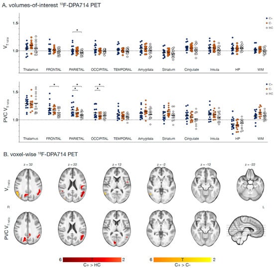
Figure 2.
Regions showing [18F]DPA714 VT-ratio differences with LGA. Fifteen chemotherapy-treated patients (C+) were assessed for [18F]DPA714 VT-ratio and compared to 15 chemotherapy-naïve patients (C−) and 15 healthy women (HC). (A) Volumes-of-interest based logan-graphical analysis (LGA) results of 11 volumes of interest are presented, showing higher VT-ratio in C+ patients compared to HC in the parietal lobe. After partial volume correction (PVC), C+ patients showed higher VT-ratio compared to C− and HC in the parietal and occipital lobe and additionally in the frontal lobe when compared to HC (* p < 0.05). (B) Voxel-based whole brain LGA results are presented, showing higher VT-ratio in C+ patients compared to HC (red) and C− patients (orange) in the occipital and parietal lobe. After PVC, only differences between C+ and HC persisted for VT-ratio images (all puncorrected < 0.005, pcluster FWE-corrected < 0.05). Section numbers refer to Montreal Neurological Institute coordinates. Abbreviations: HP = hippocampus, VT = total distribution volume, WM = white matter.
Reference
- Schroyen, G.; Blommaert, J.; van Weehaeghe, D.; Sleurs, C.; Vandenbulcke, M.; Dedoncker, N.; Hatse, S.; Goris, A.; Koole, M.; Smeets, A.; et al. Neuroinflammation and Its Association with Cognition, Neuronal Markers and Peripheral Inflammation after Chemotherapy for Breast Cancer. Cancers 2021, 13, 4198. [Google Scholar] [CrossRef] [PubMed]

Disclaimer/Publisher’s Note: The statements, opinions and data contained in all publications are solely those of the individual author(s) and contributor(s) and not of MDPI and/or the editor(s). MDPI and/or the editor(s) disclaim responsibility for any injury to people or property resulting from any ideas, methods, instructions or products referred to in the content. |
© 2023 by the authors. Licensee MDPI, Basel, Switzerland. This article is an open access article distributed under the terms and conditions of the Creative Commons Attribution (CC BY) license (https://creativecommons.org/licenses/by/4.0/).