Simple Summary
No guidelines regarding optimal treatment exist for primary cervical lymphomas. We have performed a systematic review of the literature about the management of this rare pathology. Conservative treatment with the combination of surgery and chemotherapy or surgery and radiotherapy has been reported in a few cases with apparent success. Furthermore, we have reported pregnancy outcome in patients treated with a fertility sparing approach.
Abstract
The female genital tract can be involved as a secondary manifestation of disseminated lymphomas or leukaemia but can rarely be the primary site of so-called extranodal lymphomas. Primary lymphomas of the female genital tract can affect the uterine corpus, uterine cervix, vulva, vagina, or adnexa. Only about 0.008% of all cervical tumours are primary malignant lymphomas. The most common clinical presentation of primary cervical lymphomas is a history of prolonged minor abnormal uterine bleeding, while unstoppable bleeding at presentation is rarely reported in the literature. “B” symptoms related to nodal lymphomas are usually absent. Since vaginal bleeding is a nonspecific symptom, the first diagnostic hypothesis is usually of one of the more common female genital conditions such as cervical or endometrial carcinoma or sarcoma, fibroids, adenomyosis, or endometriosis. Cervical cytology is usually negative. Preoperative diagnosis requires deep cervical biopsy. No guidelines regarding optimal treatment exists; radiotherapy, chemotherapy, and surgery are used in different combinations. Conservative treatment with the combination of surgery and chemotherapy or surgery and radiotherapy has been reported in a few cases with apparent success. With this review, we aim to understand what the best therapeutic approaches for this rare pathology in young and elderly women are. Moreover, we find favorable pregnancy outcome in patients treated with a fertility sparing approach.
1. Introduction
The female genital tract can be involved as a secondary manifestation of disseminated lymphomas or leukaemia, but rarely is the primary site of so-called extranodal lymphomas. Primary lymphomas of the female genital tract can affect the uterine corpus, uterine cervix, vulva, vagina, or adnexa [1].
From the early 1970s to the early 21st century, the incidence rates of non-Hodgkin lymphoma (NHL) nearly doubled in the United States. Although this increase can be partially explained as the effect of earlier detection resulting from improved diagnostic techniques, the rise remains mostly unexplained [2]. Currently, NHLs account for around 4% of all new cancer diagnoses [3]. Approximately one third are extranodal and can affect the gastrointestinal tract, breast, CNS, mediastinum, bone, female genital tract or testis, thyroid, skin, oropharynx, spleen, liver, and many other organs. Around 0.5–1.5% of all extranodal NHLs arise from the female genital tract and only 0.12–0.6% from the cervix. Nevertheless, only about 0.008% of all cervical tumours are primary malignant lymphomas [4,5,6,7,8,9,10,11].
In the early stages, it is quite easy to define it as a primary cervical lymphoma since it is confined to the cervix. The strictest definition of primary extranodal NHL is that of a lymphoma presenting only in extranodal sites, with no visible lymphadenopathy on imaging; thus, only I E lymphomas are included (Ann Arbor staging) [12]. Moreover, when the disease involves contiguous sites (e.g., cervix and uterine corpus or cervix and upper vagina), the site with the largest area of involvement is defined as the primary site [13]. The tumour must be confined to regional lymph nodes or neighbouring organs at diagnosis, with no bone marrow involvement, a lack of malignant cells in peripheral blood, and any distant disease must occur at least several months after the appearance of primary lesions [14,15,16].
The history of prolonged minor abnormal uterine bleeding is the most common presenting symptom of primary cervical lymphoma. Heavy bleeding at presentation is rare with only a few cases reported in the literature [17,18,19]. “B” symptoms such as fever, fatigue, weight loss, and night sweats are usually absent. Since postmenopausal vaginal bleeding or intermenstrual bleeding is a nonspecific symptom, the first consideration would be on one of the more common female genital tract pathologies [20]. A diagnostic pitfall to consider is that cervical cytology is most of the time negative. This reflects the peculiar behaviour of lymphomas which usually spread through cervical stroma leaving the epithelium intact at least in the early stages, differently from cervical adenocarcinoma. Therefore, preoperative diagnosis requires deep cervical biopsy [9]. The most frequent histological subtype is diffuse large B-cell lymphoma (DLBCL) accounting for more than 70% of all cases [21,22].
Moreover, fertility-sparing approach is increasing in the treatment of cancer in young women with satisfying overall survival end point. Even in cervical lymphoma, it has been described with a conservative approach avoiding RT and invasive surgery with optimal results.
To the best of the authors’ knowledge, the literature about primary lymphomas of the uterine cervix is limited to single case reports and case series. There are no greater studies or randomized controlled trials regarding diagnostic algorithm or treatment strategy. To date, unanimous consensus or published guidelines for the management of primary lymphomas of the uterine cervix do not exist.
We aim to analyze and group the different treatment options presented in the literature to try to provide a general common point of view and understand what the best therapeutic approaches for this rare pathology are. Particularly, we try to focus on the conservative management in fertile women who desire to preserve their fertility and we want to analyze the outcome of pregnancies following treatment.
2. Materials and Methods
This retrospective observational descriptive study was approved by our institutional review board (IRB/Burlo RC August 2020). Bibliographic search was conducted on Medline using PubMed, Scopus, and Web of Science. The aim was to find articles regarding primary diffuse large B-cell lymphoma of the uterine cervix. The search terms used were “lymphoma”, “cervix”, “B-cell”, and “Cervical cancer” with filters applied to display only articles in English from year 1995 up to 1 October 2022. A total of 166 articles were identified through Pubmed, 268 through Scopus database, and 35 through Web of Science. We decided to collect only articles published from 1995 in order to have sufficiently homogeneous data about histologic classification; in fact, at the end of 1994, the new REAL classification of lymphomas was published, and that revolutionary classification is still the base of the successive WHO classifications published in 2001, 2008, 2016, and updated again in 2017 [23,24,25,26].
We identified 471 records through databases search (n = 167 from PubMed; n = 269 from Scopus; and n = 35 from Web of Science). After removing 185 duplicate records, 286 remained to be screened. After screening, 262 records were excluded as they did not meet inclusion criteria, because they were not relevant, or for other reasons (articles with unclassified or unclear stage or unclear histologic subtype). At the end of the screening process, we included in our review thirty-four studies for a total of forty-six clinical cases.
All articles were listed by title, author, and year of publication. Following the PRISMA checklist [Figure 1], three (L.S., G.S., C.R.) independent investigators screened all the articles by title and abstract to identify those eligible [27]. Titles and abstracts were screened identifying potentially relevant articles that have been subsequently reviewed. Articles selection was concluded on 1 October 2022. Inclusion criteria were cases of primary extranodal diffuse large B-cell lymphomas arising from the uterine cervix, classified as stage IE (Ann Arbor staging), and English-language articles. We evaluated all types of articles (original article, video article, case report, review article, and metanalysis) resulting from our search on databases. Exclusion criteria were primary extra-cervical gynaecological lymphomas extending secondarily to the cervix, secondary involvement of the cervix in leukaemia or primary nodal disseminated lymphomas, cervical relapse of previously diagnosed lymphoma/leukaemia, advanced disease (stage IIE, IIIE or IVE), cervical disease in HIV-positive patients, transplanted patients or patients using immunosuppressive therapy for any reason, patients with a previous diagnosis of tumor, articles published before year 1995, articles with unclassified stage or unclear histologic subtype, and articles not written in English. The systematic review was not submitted to Prospero as only a limited number of case reports were found in the literature [28]. Three authors (L.S., G.S., C.R.) reviewed independently all identified full text papers, selecting those that met predefined eligibility criteria. Discrepancies were resolved by consensus. Another 10 articles were identified through references of the previously selected papers. The methodological quality of the included studies was assessed using the JBI Critical Appraisal Checklist for case reports [Table 1] and for case series [Table 2, Appendix A].
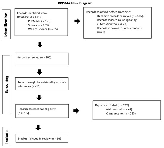
Figure 1.
Prisma flow diagram [24].

Table 1.
JBI Critical Appraisal Checklist for case reports (D1–D8 represent the eight questions about each case report).
Table 2.
JBI Critical Appraisal Checklist for case series (D1–D10 represent the ten questions about each case series).
3. Results
At the end of the systematic review, we included in our study thirty-four studies for a total of forty-six clinical cases [Table 3].
Table 3.
Case identified after systematic review.
In our analysis, the median age of women affected by primary lymphoma of the cervix uteri were 50.0 years (range 20–85 years). Regarding the age range at diagnosis, 28.2% (13/46) were under 40 years, 47.8% (22/46) were 40–60 years old, and 23.9% (11/46) were over 60 years old. More than half of the patients were known to be in post-menopause at the time of diagnosis (24/46, 52.2%), 36.9% (g) of patients were in pre-menopause, and of the remaining 5 patients (10.8%) we have no data on this.
The most frequent symptoms manifested by patients at the time of diagnosis were abnormal uterine bleeding (65%), vaginal discharge (6.1%), and pelvic pain (4.4%). A total of 6.1% of patients were asymptomatic and of 18.4% we have no data on this [Figure 2]. Referring to systemic symptoms, 88% of the patients did not present the typical “B symptoms” of lymphomas, such as mild-fever, fatigue, weight loss, and night sweats. Only one patient (2%) presented weight loss.
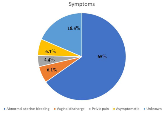
Figure 2.
Most frequent symptoms.
The median size of the mass at diagnosis was 6.6 cm, with a minimum diameter of 2 cm and a maximum of 11 cm.
The therapeutic approach was varied. The different approaches were as follows: CT + RT (36.9%), CT only (21.7%), surgery + adjuvant CT (10.9%), surgery + adjuvant RT (6.5%), neoadjuvant CT + surgery (6.5%), surgery only (4.3%), neoadjuvant CT + conization + CT (2.2%) and surgery + adjuvant CT/RT (2.2%), CT + ovarian suppression after cryopreservation (2.2%), CT + ovarian suppression (2.2%), and CT + ovarian transposition + RT (2.2%). We published the only case completely treated with a hysteroscopic approach followed by adjuvant RT.
After treatment, patients started a follow-up program. After 12 months from diagnosis, 93.5% of patients were still disease free, 2.2% had had a partial response to therapy, 2.2% relapsed, and only one patient was dead (2.2%). After 5 years from diagnosis, considering only those patients we had data about (19/45), 84% were still disease free, 6.3% relapsed, 6.3% had had a partial response, and only one patient was dead (6.3%). We have no data about the follow-up of three patients.
Even if only 28.2% of case reports are about women under 40 years old, only 5 papers reported pregnancy data. Of this, only one was spontaneously miscarried [36], while the others have reached the full term with no complications during pregnancy. Regarding the mode of delivery, 3/4 (75%) had a vaginal delivery without any complication, while 1/4 had a planned Caesarean section at 38 gestational weeks [53]. Only in 1/4 cases was the labour inducted for post-term pregnancy with Prostaglandin E2 pessary. The mean time for attempting pregnancy was 33 months after treatment (range 12–66 months). The treatment approach was different between these five women, but all of them underwent CT (CHOP ± Rituximab) [Table 4].
Table 4.
Pregnancy after treatment.
4. Discussion
4.1. Symptoms and Diagnosis
Primary cervix uteri lymphoma is a rare pathology; therefore, gynecologists should be aware when evaluating cervical lesions. Isolated and persistent vaginal bleeding without pain and without any “B symptom” should increase the level of suspicion. PAP test is usually negative, and a deep biopsy is needed to obtain a conclusive diagnosis.
The presence of a diffusely enlarged cervix or a cervical mass should trigger further investigation such as transvaginal echography and a computed tomography or magnetic resonance to better define the lesion and its extension both locally and eventually out of the pelvic. This kind of lesion can easily mimic cervical adenocarcinoma or sarcoma, but also benign conditions like fibroids, especially colliquated fibroids.
Characterization of the tumour immunophenotype is nowadays mandatory to achieve a precise diagnosis and should be made according to the latest WHO classification. Primary cervical lymphomas are usually B-cell type NHL, but there are some cases of primary cervical Hodgkin lymphoma, MALToma, and T-cell lymphoma described. The most common subtype is diffuse large B-cell lymphoma, accounting for around 70% of the cases. Older case reports do not follow the WHO classification for the characterization of tumour histology, but most reported cases are B-cell type lymphomas.
4.2. Therapy
Neither clinical trials nor international guidelines exist due to the rarity of this disease. Therefore, there is no consensus on what the best management of this disease is. In general, the cornerstone of lymphoma treatment is chemotherapy, so most reported cases are treated accordingly with chemotherapy alone or in association with radiotherapy.
RT plays a pivotal role as a treatment tool for this rare disease, as we know from literature data of other site primary extranodal NHL [58]. In fact, RT has a local effect avoiding systemic complications and can help to control symptoms in those women who are not able to address CHT or surgery. Surgery (mainly total abdominal hysterectomy with or without salpingo/oophorectomy and lymphadenectomy) is usually performed cautiously if a precise preoperative diagnosis is not possible. In a published manuscript, we have reported a case treated with hysteroscopic resection, which to date is the only one reported in the literature [56]. Hysteroscopy appears to be a very promising tool for the treatment of organ-limited utero-cervical pathology [29,30]. Rather, some reported cases of massive bleeding due to cervical lymphoma that was managed with emergent embolization of uterine arteries or emergent hysterectomy exists. Considering the age range at diagnosis of this disease, it is important to find out a conservative approach.
4.3. Fertility Sparing Treatment
To date, in young women who want to preserve their fertility, the approach has been chemotherapy (CHOP cyclophosphamide-doxorubicin-vincristine prednisone chemotherapy regime) and immunotherapy to reduce the need for radiotherapy or surgical resection. To the best of our knowledge, to date, there are no reported cases in the literature treated with cold knife or trachelectomy alone. In this review, when a fertility approach is sought, chemotherapy alone is the treatment of choice [33,37,38,46], followed by CT + ovarian transposition + RT and CT + cold knife conization [45,53]. The reported evidence regarding non-Hodgkin lymphoma in general suggests that in cases of childhood NHL, and in cases where fertility preservation is desired, current chemotherapeutic regimens are safe and can spare fertility, particularly when GnRH agonists are used in conjunction with treatment. As reported by Quaresima et al., who injected Leuprolide 11.25 mg im before therapy every 12 weeks with successful outcome [29]. Signorelli et al. reported a case series of cervical lymphoma treated with a conservative approach (CT ± surgery) and observed that almost all women resumed menses within 5 months after the end of their treatment, and only one needed hormone replacement treatment [46]. Other fertility-spearing techniques exist as ovarian transposition in those women who undergo to RT [43].
4.4. Pregnancy Outcome after Treatment
In patients who become pregnant after cervix uteri lymphoma, mode of delivery should be well discussed before childbirth, particularly if surgery or RT has been performed, even if RT does not seem to reduce the possibility of a safe vaginal delivery. In our review, 75% of pregnant patients had a vaginal delivery without any complications. This is a fact that encourages vaginal delivery in these patients regardless of the type of therapy performed.
Ferreri et al. reported a case of vaginal delivery 36 months after pelvic irradiation, without any complication during and after the delivery [45]. Parva et al. and Quaresima et al. reported two cases of safe vaginal delivery after chemotherapeutic treatment, without complication neither during pregnancy nor during the delivery [29,43]. Whereas Lorusso et al. reported a case of a planned Cesarean section at 38 gestational weeks 3 years after completing the primary treatment (R-CHOP + cold knife conization) [53].
Clearly, there are no guidelines regarding the safest mode of delivery for these patients. However, the few data available suggest that in carefully selected patients with fully treated non-Hodgkin lymphoma of the cervix with no apparent disease, it may be plausible and even prudent to allow a trial of labour with some likelihood of a successful vaginal delivery [43].
In light of our systematic review, in young women considering future pregnancy, the fertility sparing surgery as cold knife conization, trachelectomy, or hysteroscopic resection could be feasible considering the indolent course of the disease even if there are few data available and all are from case reports and case series. Moreover, Perren et al., following an extensive revision of the literature, concluded that there is no evidence that radical surgery confers more survival advantages than conservative surgery in patients with localized and low- or intermediate-grade tumors [59].
From a recent literature review by Capsa et al. emerges that the combination of CHOP CT regimen and RT as adjuvant is the most frequently chosen by clinicians with optimal results on prognosis [31].
Nevertheless, from the data of this review emerges a non-inferiority of the surgery + RT approach (considering ovarian transposition) compared to the CT ± RT approach. Furthermore, also in elderly women or in patients who are ineligible for chemotherapy, a conservative surgery and radiotherapic approach can be considered in order to reduce side effects and morbidity [56]. In conclusion, in the absence of a consensus on the best management, proper instrumental staging and detailed counselling with the patient is recommended to tailor the best treatment.
5. Conclusions
Primary cervical lymphoma is a very rare entity and affects a wide range of ages. Usually, in the early stages, primary cervical lymphoma has an excellent prognosis with any treatment combination (radiotherapy and/or chemotherapy and/or surgical resection). Considering data of follow-ups, in most cases, those patients who do not receive local therapy with RT have an excellent prognosis and this can suggest avoiding it in those women with childbearing desire. Moreover, in these cases, RT will become a tool in case of relapse. There is evidently the need for a larger study, possibly a multicentric randomized controlled trial, to define a specific classification and the best management for this rare tumour, even if due to the rarity of this pathology it would not be feasible. Particularly, it is important to look for an approach as conservative as possible considering that 30% of diagnosis occurs in women under 40 years old. Moreover, when a conservative approach is performed, we should ask if definitive surgery is necessary or recommended at the end of childbearing desire.
Author Contributions
Conceptualization, G.S., L.S., and G.R.; methodology, L.S.; software, C.R.; validation, G.R., G.S., and L.N.; formal analysis, G.S. and C.R.; investigation, C.R.; resources, L.S.; data curation, G.S., S.R., F.P.M., and C.R.; writing—original draft preparation, G.S and C.R.; writing—review and editing, G.S., S.R., and F.P.M.; visualization, L.N.; supervision, G.S. and G.R.; project administration, G.S.; funding acquisition, G.S. and G.R. All authors have read and agreed to the published version of the manuscript.
Funding
This work was supported by the Italian Ministry of Health, through the contribution given to the Institute for Maternal and Child Health IRCCS Burlo Garofolo—Trieste, Italy.
Institutional Review Board Statement
The study was conducted in accordance with the Declaration of Helsinki and approved by the Institutional Review Board of IRCCS Burlo Garofolo on 15/04/2020 (RC 08/2020).
Informed Consent Statement
Not applicable.
Data Availability Statement
The authors confirm that the data supporting the findings of this study are available within the article.
Conflicts of Interest
The authors declare no conflict of interest.
Appendix A. JBI Checklist for Case Report and Case Series
Table A1.
JBI Critical Appraisal Checklist for case reports.
Table A1.
JBI Critical Appraisal Checklist for case reports.
| D1. | Were patient’s demographic characteristics clearly described? |
| D2. | Was the patient’s history clearly described and presented as a timeline? |
| D3. | Was the current clinical condition of the patient on presentation clearly described? |
| D4. | Were diagnostic tests or assessment methods and the results clearly described? |
| D5. | Was the intervention(s) or treatment procedure(s) clearly described? |
| D6. | Was the post-intervention clinical condition clearly described? |
| D7. | Were adverse events (harms) or unanticipated events identified and described? |
| D8. | Does the case report provide takeaway lessons? |
Table A2.
JBI Critical Appraisal Checklist for case reports.
Table A2.
JBI Critical Appraisal Checklist for case reports.
| D1. | Were there clear criteria for inclusion in the case series? |
| D2. | Was the condition measured in a standard, reliable way for all participants included in the case series? |
| D3. | Were valid methods used for identification of the condition for all participants included in the case series? |
| D4. | Did the case series have consecutive inclusion of participants? |
| D5. | Did the case series have complete inclusion of participants? |
| D6. | Was there clear reporting of the demographics of the participants in the study? |
| D7. | Was there clear reporting of clinical information of the participants? |
| D8. | Were the outcomes or follow up results of cases clearly reported? |
| D9. | Was there clear reporting of the presenting site(s)/clinic(s) demographic information? |
| D10. | Was statistical analysis appropriate? |
References
- Kosari, F.; Daneshbod, Y.; Parwaresch, R.; Krams, M.; Wacker, H.H. Lymphomas of the female genital tract: A study of 186 cases and review of the literature. Am. J. Surg. Pathol. 2005, 29, 1512–1520. [Google Scholar] [CrossRef] [PubMed]
- Luo, D.; Zhou, T.; Tao, Y.; Feng, Y.; Shen, X.; Mei, S. Exposure to organochlorine pesticides and non-Hodgkin lymphoma: A meta-analysis of observational studies. Sci. Rep. 2016, 6, 25768. [Google Scholar] [CrossRef] [PubMed]
- American Cancer Society. Cancer Facts & Figures; American Cancer Society: Atlanta, GA, USA, 2019. [Google Scholar]
- Kuo, H.C.; Chou, C.Y.; Chang, C.H.; Liu, M.T.; Tzeng, C.C.; Huang, K.E. Primary malignant lymphoma of the uterine cervix shows favorable response to neoadjuvant chemotherapy. Gynecol. Oncol. 1994, 52, 408–410. [Google Scholar] [CrossRef] [PubMed]
- Baijal, G.; Vadiraja, B.M.; Fernandes, D.J.; Vidyasagar, M.S. Diffuse large B-cell lymphoma of the uterine cervix: A rare case managed novelly. J. Cancer Res. Ther. 2009, 5, 140–142. [Google Scholar]
- Cohn, D.E.; Resnick, K.E.; Eaton, L.A.; DeHart, J.; Zanagnolo, V. NonHodgkin’s lymphoma mimicking gynaecological malignancies of the vagina and cervix: A report of four cases. Int. J. Gynecol. Cancer 2007, 17, 254–293. [Google Scholar] [CrossRef]
- Korcum, A.F.; Karadogan, I.; Aksu, G.; Aralasmak, A.; Erdogan, G. Primary follicular lymphoma of the cervix uteri: A review. Ann. Hematol. 2007, 86, 623–630. [Google Scholar] [CrossRef]
- Cantu de Leon, D.; Perez Montiel, D.; Chanona Vilchis, J. Primary malignant lymphoma of uterine cervix. Int. J. Gynecol. Cancer 2006, 16, 923–927. [Google Scholar] [CrossRef]
- Ab Hamid, S.; Wastie, M.L. Primary non-Hodgkin’s lymphoma presenting as a uterine cervical mass. Singap. Med. J. 2008, 49, e73–e75. [Google Scholar]
- Stroh, E.L.; Besa, P.C.; Cox, J.D.; Fuller, L.M.; Cabanillas, F.F. Treatment of patients with lymphomas of the uterus or cervix with combination chemotherapy and radiation therapy. Cancer 1995, 75, 2392–2399. [Google Scholar] [CrossRef]
- Ferry, J.A.; Young, R.H. Malignant lymphoma, pseudolymphoma, and hematopoietic disorders of the female genital tract. Pathol. Annu. 1991, 26, 227–263. [Google Scholar]
- Rudders, R.A.; Ross, M.E.; De Lellis, R.A. Primary extranodal lymphoma: Response to treatment and factors influencing prognosis. Cancer 1978, 42, 406–416. [Google Scholar] [CrossRef] [PubMed]
- Shen, H.; Wei, Z.; Zhou, D.; Zhang, Y.; Han, X.; Wang, W.; Zhang, L.; Yang, C.; Feng, J. Primary extra-nodal diffuse large B-ell lymphoma: A prognostic analysis of 141 patients. Oncol. Lett. 2018, 16, 1602–1614. [Google Scholar] [PubMed]
- Fox, H.; Langley, F.A.; Govan, A.D.; Hill, A.S.; Bennett, M.H. Malignant lymphoma presenting as an ovarian tumour: Clinicopathological analysis of 34 cases. Br. J. Obstet. Gynaecol. 1988, 95, 386–390. [Google Scholar] [CrossRef] [PubMed]
- Kasai, M.; Ichimura, T.; Murakami, M.; Matsuda, M.; Kawamura, N.; Sumi, T. Two cases of utrine malignant lymphoma diagnosed by needle biopsy. J. Obstet. Gynaecol. Res. 2015, 41, 1664–1668. [Google Scholar] [CrossRef] [PubMed]
- Nasioudis, D.; Kampaktsis, P.N.; Frey, M.; Witkin, S.S.; Holcomb, K. Primary lymphoma of the female genital tract: An analysis of 697 cases. Gynecol. Oncol. 2017, 145, 305–309. [Google Scholar] [CrossRef]
- Bode, M.K.; Tikkakoski, T.; Jhansson, J.; Johannson, K.; Kariniemi, J.; Apaja-Sarkkinen, M. Lymphoma of the cervix: Imaging and transcatheter arterial embolization. Acta Radiol. 2002, 43, 431–432. [Google Scholar] [CrossRef]
- Abbas, M.A.; Birdwell, R.; Katz, D.S.; Chang, H.; Ostrow, K. Primary Lymphoma of the cervix in a Heart Transplant Patient. Am. J. Roentgenol. 1996, 167, 1136–1138. [Google Scholar] [CrossRef]
- Gupta, S.; Sachdev, L.; Gupta, R.; Beotra, A. Non-Hodgkin Lymphoma of the Uterine Cervix. Ann. Saudi Med. 1995, 15, 187–188. [Google Scholar] [CrossRef]
- Stabile, G.; Zinicola, G.; Romano, F.; Laganà, A.S.; Dal Pozzolo, C.; Ricci, G. Pelvic mass, ascites, hydrothorax: A malignant or benign condition? Meigs syndrome with high levels of CA 125. Prz. Menopauzalny 2021, 20, 103–107. [Google Scholar] [CrossRef]
- Goda, J.S.; Gaikwad, U.; Narayan, A.; Kurkure, D.; Yadav, S.; Khanna, N.; Jain, H.; Bagal, B.; Epari, S.; Singh, P.; et al. Primary diffuse large B cell lymphoma of Uterine Cervix: Treatment outcomes of a rare entity with literature review. Cancer Rep. 2020, 3, e1264. [Google Scholar] [CrossRef]
- Chan, J.K.; Loizzi, V.; Magistris, A. Clinicopathologic features of six cases of primary cervical lymphoma. Am. J. Obstet. Gynecol. 2005, 193, 866–872. [Google Scholar] [CrossRef] [PubMed]
- Jaffe, E.S.; Harris, N.L.; Stein, H.; Vardiman, J.W. World Health Organization Classification of Tumours: Pathology and Genetics of Tumours of Haematopoietic and Lymphoid Tissues; IARC Press: Lyon, France, 2001. [Google Scholar]
- Swerdlow, S.H.; Campo, E.; Harris, N.L.; Pileri, S.; Stein, H.; Jaffe, E.S. WHO Classification of Tumours of Haematopoietic and Lymphoid Tissues; IARC Press: Lyon, France, 2008. [Google Scholar]
- Swerdlow, S.H.; Campo, E.; Pileri, S.A.; Harris, N.L.; Stein, H.; Siebert, R.; Advani, R.; Ghielmini, M.; Salles, G.A.; Zelenetz, A.D.; et al. The 2016 revision of the World Health Organization classification of lymphoid neoplasms. Blood 2016, 127, 2375–2390. [Google Scholar] [CrossRef] [PubMed]
- Swerdlow, S.H.; Campo, E.; Harris, N.L.; Jaffe, E.S.; Pileri, S.A.; Stein, H.; Thiele, J. WHO Classification of Tumours of Haematopoietic and Lymphoid Tissues, 4th ed.; IARC Press: Lyon, France, 2017. [Google Scholar]
- Moher, D.; Liberati, A.; Tetzlaff, J.; Altman, D.G.; The PRISMA Group. Preferred Reporting Items for Systematic Reviews and Meta-Analyses: The PRISMA Statement. PLoS Med. 2009, 6, e1000097. [Google Scholar] [CrossRef] [PubMed]
- Prospero International Prospective Register of Systematic Reviews. Available online: https://www.crd.york.ac.uk/prospero/ (accessed on 3 December 2022).
- Quaresima, P.; Saccone, G.; Zuccalà, V.; Guarascio, G.; Leo, L.; Amendola, G.; Zullo, F.; Morelli, M.; Venturella, R. Successful Vaginal Delivery after Induction of Labour in a Patient Treated for Non-Hodgkin’s Lymphoma of the Cervix: A Case Report and Literature Review. Case Rep. Obstet. Gynecol. 2022, 2022, 3541046. [Google Scholar] [CrossRef]
- Sorrentino, F.; De Feo, V.; Stabile, G.; Tinelli, R.; D’Alterio, M.N.; Ricci, G.; Angioni, S.; Nappi, L. Cesarean Scar Pregnancy Treated by Artery Embolization Combined with Diode Laser: A Novel Approach for a Rare Disease. Medicina 2021, 57, 411. [Google Scholar] [CrossRef] [PubMed]
- Capsa, C.; Calustian, L.A.; Antoniu, S.A.; Bratucu, E.; Simion, L.; Prunoiu, V.M. Primary Non-Hodgkin Uterine Lymphoma of the Cervix: A Literature Review. Medicina 2022, 58, 106. [Google Scholar] [CrossRef]
- Akkour, K.; Alhulwah, M.; Alhalal, H.; Alqahtani, N.; Arafah, M. Primary extranodal diffuse large B-cell lymphoma of the uterine cervix. Malays J. Pathol. 2021, 43, 327–331. [Google Scholar]
- Gui, W.; Li, J.; Zhang, Z.; Wang, L.; Zhao, J.; Ma, L.; Su, L. Primary hematological malignancy of the uterine cervix: A case report. Oncol. Lett. 2019, 18, 3337–3341. [Google Scholar] [CrossRef]
- Boussios, S.; Zerdes, I.; Vassou, A.; Bareta, E.; Seraj, E.; Papoudou-Bai, A.; Pavlidis, N.; Batistatou, A.; Pentheroudakis, G. Extranodal diffuse large B-cell lymphomas: A retrospective case series and review of the literature. Hematol. Rep. 2018, 10, 7070. [Google Scholar] [CrossRef]
- Roberts, M.B.; Cottrill, H.M. A case of primary lymphoma in a patient with abnormal uterine bleeding. Gynecol. Oncol. Rep. 2018, 26, 105–107. [Google Scholar] [CrossRef]
- Cubo, A.M.; Soto, Z.M.; Cruz, M.Á.; Doyague, M.J.; Sancho, V.; Fraino, A.; Blanco, Ó.; Puig, N.; Alcoceba, M.; González, M.; et al. Primary diffuse large B cell lymphoma of the uterine cervix successfully treated by combined chemotherapy alone: A case report. Medicine 2017, 96, e6846. [Google Scholar] [CrossRef] [PubMed]
- Pósfai, É.; Nagy, K.; Marton, I.; Bánfalvi, A.; Kocsis, L.; Cserni, G. Incidentally discovered diffuse large B-cell lymphoma limited to the endocervical mucosa in a young female patient. Gynecol. Obstet. Investig. 2015, 80, 134–138. [Google Scholar] [CrossRef] [PubMed]
- Cao, X.X.; Li, J.; Zhang, W.; Duan, M.H.; Shen, T.; Zhou, D.B. Patients with primary diffuse large B-cell lymphoma of female genital tract have high risk of central nervous system relapse. Ann. Hematol. 2014, 93, 1001–1005. [Google Scholar] [CrossRef] [PubMed]
- Mouhajir, N.; Diakité, A.; Toulba, A.; Hemmich, M.; Saadi, I. Primary Non-Hodgkin’s Lymphoma of the Uterine Cervix: Case Report of Long-Term Survival Patient. J. Obstet. Gynecol. India 2014, 64, S145–S147. [Google Scholar] [CrossRef] [PubMed]
- Binesh, F.; Karimi Zarchi, M.; Vahedian, H.; Rajabzadeh, Y. Primary malignant lymphoma of the uterine cervix. BMJ Case Rep. 2012. [Google Scholar] [CrossRef]
- Parnis, J.; Camilleri, D.J.; Babic, D.; Degaetano, J.; Savona-Ventura, C. Lymphoma of the cervix. Case Rep Hematol. 2012, 2012, 326127. [Google Scholar] [CrossRef]
- Vasudev, D.S.; Kaler, A.K. Non-Hodgkin’s Lymphoma of the Uterine Cervix. Online J. Health Allied Sci. 2012, 11, 13. [Google Scholar]
- Parva, M.; Lamb, K.; Savior, D.C.; Gilman, P.; Belden, M. Full-term pregnancy and vaginal delivery after treatment for non-Hodgkin’s lymphoma of the cervix and lower uterine segment: A case report. J. Obstet. Gynaecol. Can. 2011, 33, 620–624. [Google Scholar] [CrossRef]
- Upanal, N.; Enjeti, A. Primary lymphoma of the uterus and cervix: Two case reports and review of the literature. Aust. N. Z. J. Obstet. Gynaecol. 2011, 51, 559–562. [Google Scholar] [CrossRef]
- Ferreri, A.J.; Verona, C.; Bolognesi, A.; Taccagni, G.; Ponzoni, M.; Ferrari, S. Successful pregnancy after chemo-immuno-radiation therapy for aggressive lymphoma of the uterus. Br. J. Haematol. 2008, 142, 141–143. [Google Scholar] [CrossRef]
- Signorelli, M.; Maneo, A.; Cammarota, S.; Isimbaldi, G.; Garcia Parra, R.; Perego, P.; Maria Pogliani, E.; Mangioni, C. Conservative management in primary genital lymphomas: The role of chemotherapy. Gynecol. Oncol. 2007, 104, 416–421. [Google Scholar] [CrossRef] [PubMed]
- Hariprasad, R.; Kumar, L.; Bhatla, D.M.; Kukreja, M.; Papaiah, S. Primary uterine lymphoma: Report of 2 cases and review of literature. Am. J. Obstet. Gynecol. 2006, 195, 308–313. [Google Scholar] [CrossRef] [PubMed]
- Semczuk, A.; Skomra, D.; Korobowicz, E.; Balon, B.; Rechberger, T. Primary non-Hodgkin’s lymphoma of the uterine cervix mimicking leiomyoma: Case report and review of the literature. Pathol. Res. Pract. 2006, 202, 61–64. [Google Scholar] [CrossRef] [PubMed]
- Dursun, P.; Gultekin, M.; Bozdag, G.; Usubutun, A.; Under, A.; Celik, N.Y.; Yuce, K.; Ayhan, A. Primary cervical lymphoma: Report of two cases and review of the literature. Gynecol. Oncol. 2005, 98, 484–489. [Google Scholar] [CrossRef]
- Lee, K.M.; Seah, E.S.; Sethi, V.K. Primary non-Hodgkin’s lymphoma of the uterine cervix: Case report of long-term survival of two patients treated with surgery and radiotherapy. Australas. Radiol. 1998, 42, 126–127. [Google Scholar] [CrossRef]
- Fratoni, S.; Abruzzese, E.; Trawinska, M.M.; Niscola, P.; de Fabritiis, P.; Santeusanio, G. Primitive “Spindle Cell Variant” (Sarcomatoid Variant) Diffuse Large B-Cell Lymphoma of the Uterine Cervix: Description and Outcome of a Rare Case. Int. J. Gynecol. Pathol. 2016, 35, 593–597. [Google Scholar] [CrossRef]
- Wuntakal, R.; Janga, D.; Satyanarayana, D.; Reynolds, K.; Hollingworth, A. An unusual cause of postmenopausal bleeding. J. Low. Genit. Tract. Dis. 2008, 12, 130–133. [Google Scholar] [CrossRef]
- Lorusso, D.; Ferrandina, G.; Pagano, L.; Gagliardi, M.L.; Scambia, G. Successful pregnancy in stage IE primary non-Hodgkin’s lymphoma of uterine cervix treated with neoadjuvant chemotherapy and conservative surgery. Oncology 2007, 72, 261–264. [Google Scholar] [CrossRef]
- González-Cejudo, C.; Martínez-Maestre, M.A.; Peregrín-Álvarez, I.; Daza-Manzano, C. Primary lymphoma of the cervix: Unusual location for a common disease. Eur. J. Obstet. Gynecol. Reprod. Biol. 2006, 125, 268–269. [Google Scholar] [CrossRef]
- Chandy, L.; Kumar, L.; Dawar, R. Non-Hodgkin’s Lymphoma Presenting as a Primary Lesion in Uterine Cervix: Case Report. J. Obstet. Gynaecol. Res. 1998, 24, 183–187. [Google Scholar] [CrossRef]
- Stabile, G.; Sancin, L.; Boschian Bailo, P.; Ripepi, C.; Romano, A.; Carlucci, S.; Ricci, G. Hysteroscopic Resection Followed by Adjuvant Radiotherapy: Report of a New Therapeutic Approach to Primary Cervical Diffuse Large B-Cell Lymphoma. Int. J. Environ. Res. Public. Health 2022, 19, 11779. [Google Scholar] [CrossRef] [PubMed]
- Nasu, K.; Yoshimatsu, J.; Urata, K.; Miyakawa, I. A case of primary non-Hodgkin’s lymphoma of the uterine cervix treated by combination chemotherapy (THP-COP). J. Obstet. Gynaecol. Res. 1998, 24, 157–160. [Google Scholar] [CrossRef] [PubMed]
- Yahalom, J.; Illidge, T.; Specht, L.; Hoppe, R.T.; Li, Y.-X.; Tsang, R.; Wirth, A. Modern Radiation Therapy for Extranodal Lymphomas: Field and Dose Guidelines from the International Lymphoma Radiation Oncology Group. Int. J. Radiat. Oncol. Biol. Phys. 2015, 92, 11–31. [Google Scholar] [CrossRef] [PubMed]
- Perren, T.; Farrant, M.; McCarthy, K.; Harper, P.; Wiltshaw, E. Lymphomas of the cervix and upper vagina: A report of five cases and a review of the literature. Gynecol. Oncol. 1992, 44, 87–95. [Google Scholar] [CrossRef] [PubMed]


Disclaimer/Publisher’s Note: The statements, opinions and data contained in all publications are solely those of the individual author(s) and contributor(s) and not of MDPI and/or the editor(s). MDPI and/or the editor(s) disclaim responsibility for any injury to people or property resulting from any ideas, methods, instructions or products referred to in the content. |
© 2023 by the authors. Licensee MDPI, Basel, Switzerland. This article is an open access article distributed under the terms and conditions of the Creative Commons Attribution (CC BY) license (https://creativecommons.org/licenses/by/4.0/).