Review

The Emerging Role of PET/CT with PSMA-Targeting Radiopharmaceuticals in Clear Cell Renal Cancer: An Updated Systematic Review

1
Department of Nuclear Medicine, Candiolo Cancer Institute, FPO–IRCCS, 10060 Turin, Italy
2
Nuclear Medicine Unit, Department of Medical Sciences, AOU Città della Salute e della Scienza, University of Turin, 10126 Turin, Italy
3
Department of Oncology, Candiolo Cancer Institute, FPO-IRCCS, 10060 Turin, Italy
4
Portsmouth Hospitals University NHS Trust, Portsmouth PO12UP, UK
5
Division of Nuclear Medicine, Università degli Studi di Brescia and ASST Spedali Civili di Brescia, 25123 Brescia, Italy
6
Unità di Medicina Nucleare, TracerGLab, Dipartimento di Diagnostica per Immagini, Radioterapia Oncologica ed Ematologia, Fondazione Policlinico Universitario A. Gemelli, IRCCS, 00168 Rome, Italy
7
Clinic of Nuclear Medicine, Imaging Institute of Southern Switzerland, Ente Ospedaliero Cantonale, 6501 Bellinzona, Switzerland
8
Faculty of Biology and Medicine, University of Lausanne, 1011 Lausanne, Switzerland
9
Faculty of Biomedical Sciences, Università della Svizzera Italiana, 6900 Lugano, Switzerland
*
Author to whom correspondence should be addressed.
Cancers 2023, 15(2), 355; https://doi.org/10.3390/cancers15020355
Submission received: 11 December 2022 / Revised: 30 December 2022 / Accepted: 3 January 2023 / Published: 5 January 2023
(This article belongs to the Special Issue Systematic Reviews and Meta-Analyses in Genitourinary Cancers)

Abstract

:

Simple Summary

Positron emission tomography/computed tomography (PET/CT) with prostate-specific membrane antigen (PSMA)-targeting radiopharmaceuticals is a relatively novel technique currently employed in the management of prostate cancer patients. These radiopharmaceuticals target PSMA, also known as carboxypeptidase type II, which is mainly expressed by prostate cancer cells. Nevertheless, its expression has been observed in the neovasculature of various kinds of cancer, including clear cell renal cell carcinoma (ccRCC). Based on this biological mechanism, several authors postulated a potential role for this diagnostic method in ccRCC patients in different clinical settings. This systematic review provides an overview of the possible applications of PET/CT with PSMA-targeting radiopharmaceuticals in ccRCC.

Abstract

Background: Recent articles proposed the employment of positron emission tomography/computed tomography (PET/CT) with prostate-specific membrane antigen (PSMA)-targeting radiopharmaceuticals in clear cell renal cell carcinoma (ccRCC). Methods: The authors performed a comprehensive literature search of studies on the performance of PET/CT with PSMA-targeting radiopharmaceuticals in ccRCC. Original articles concerning this imaging examination were included in newly diagnosed ccRCC patients and ccRCC patients with disease recurrence. Results: A total of sixteen papers concerning the diagnostic performance of PSMA-targeted PET/CT in ccRCC (331 patients) were included in this systematic review. The included articles demonstrated an excellent detection rate of PSMA-targeting PET/CT in ccRCC. Conclusions: PSMA-targeted PET/CT seems promising in detecting ccRCC lesions as well as in discriminating the presence of aggressive phenotypes. Prospective multicentric studies are warranted to strengthen the role of PSMA-targeting PET/CT in ccRCC.

1. Introduction

Kidney cancer is a common kind of tumor with an estimated incidence of approximately 400,000 new diagnoses every year worldwide [1,2]. Renal cell carcinoma (RCC) is the most common type of renal tumor [3]. Based on its histopathological features, we divide RCC into two types: clear cell RCC (ccRCC) and non-ccRCC (which categorizes at least 15 histotypes, of which the most frequent consist of papillary RCC and chromophobe RCC) [4]. However, the morphologic classification of RCC is going to become obsolete since new genomic and molecular features of these malignancies are emerging [5]. Most of our knowledge about the genetic basis of RCC comes from studies concerning its inherited forms, including von Hippel–Lindau (VHL) (ccRCC), hereditary papillary RCC (type I papillary RCC), and hereditary leiomyomatosis RCC (type II papillary RCC) [6,7]. There is a clear relationship between the prevalence of kidney cancer and VHL genotype, with a higher prevalence in patients with partial deletion of the VHL gene [8]. In contrast, no VHL gene mutations were found in patients with papillary, chromophobe, collecting duct, or medullary kidney cancers [9,10,11,12].
Due to the tumor cells’ overproduction of platelet-derived growth factor (PDGF) and vascular endothelial growth factor (VEGF), ccRCC is considered a highly vascularized tumor [4].
To date, most ccRCC diagnoses result from incidental findings as a consequence of the widespread use of noninvasive radiological techniques, including ultrasonography (US), magnetic resonance imaging (MRI), or computed tomography (CT), performed for another reason [11]. Moreover, paraneoplastic syndromes’ symptoms caused by hormones or cytokines excreted by tumor cells or by an immune response against the tumor are not uncommon in RCC, accounting for hypercalcemia, fever, and erythrocytosis [12]. Imaging examinations usually strongly suggest the diagnosis, although ccRCCs can display variable radiographic appearances [13]. Typical radiological ccRCC characteristics include exophytic growth, heterogeneity due to intratumoral necrosis or hemorrhage, and high contrast enhancement in CT and MRI examinations [14]. In current clinical practice, total-body contrast-enhanced CT is essential for optimal staging and restaging of ccRCC since it allows us to assess the primary tumor’s size and extension, lymph node involvement, and to evaluate the presence of distant metastases. MRI can also provide additional information to determine whether the tumor extends into the vasculature. Moreover, the development of radiological techniques and the availability of data derived from the Human Genome Project introduced the concept of radiogenomics. Radiogenomics, which can be applied to RCC, consists of studying the association of imaging features of a specific disease and its gene expression patterns or molecular phenotype. Its purpose is to noninvasively collect additional data for diagnosis, staging, prognostication, and the assessment of an optimal therapy based on the imaging features of the disease [15].
Nuclear medicine examinations allow physicians to detect functional abnormalities that might anticipate morphological findings in conventional imaging in oncological malignancies. With regard to nuclear medicine imaging, the most studied examination in ccRCC patients is fluorine-18 fluorodeoxyglucose ([18F]FDG) positron emission tomography (PET)/CT [16]. [18F]FDG PET/CT is not currently recommended as an imaging method in ccRCC by practice guidelines [17,18,19] and does not play a key role in managing ccRCC patients so far for several reasons. The first issue concerns the excretion of [18F]FDG and its metabolites, seeing as how their physiological renal excretion hinders the characterization of primary lesions, making the differentiation from the physiologic background challenging [16]. Furthermore, [18F]FDG PET/CT is not routinely employed to evaluate ccRCC because of its relatively low [18F]FDG uptake; the underlying mechanism for this phenomenon is partially unclear yet. Nevertheless, a recent study showed that [18F]FDG uptake reflected FBP1 expression levels in patients with ccRCC since it was higher in ccRCCs with low fructose 1,6-bisphosphatase 1 (FBP1) expression compared to patients with high FBP1 expression [20].
Carboxypeptidase type II, also known as prostate-specific membrane antigen (PSMA), is a transmembrane protein encoded by the gene FOLH1 [21,22]. Several authors observed that carboxypeptidase type II could be expressed on the surface of neovascular endothelial cells of various solid tumors other than prostate cancer [23]. In this context, different radiolabeled PSMA-targeting low-molecular-weight molecules were introduced to enhance nuclear medicine imaging and to operate as theranostic agents for patients affected by metastatic prostate cancer [24,25]. Since neovascular endothelium cells of various malignancies, including ccRCC, overexpress PSMA, and since ccRCC is considered a highly vascularized tumor, this might be a rationale to employ PET imaging with PSMA-targeting radiopharmaceuticals in tumors with variable [18F]FDG uptake, exploring different biological processes than glucose metabolism. Furthermore, since a combination of immunotherapy and antineoangiogenetic therapy are the standard of care in metastatic ccRCC [17,18,19], PSMA-targeted PET/CT might be a valuable tool to anticipate the treatment’s outcome and to assess the response to therapies in ccRCC patients.
Various recent studies evaluated the performance of PSMA-targeted PET/CT in ccRCC. This paper has the purpose of accomplishing an updated systematic review concerning PSMA-targeted PET/CT performance in patients affected by ccRCC. Furthermore, this paper aims to gather evidence to compare the diagnostic performance of PET with PSMA-radioligands to other instrumental examinations in ccRCC.

2. Materials and Methods

2.1. Protocol

This systematic review was conducted following a preconceived protocol [26] since the authors employed the “Preferred Reporting Items for a Systematic Review and Meta-Analysis” (PRISMA 2020 statement) as a touchstone in its completion [27]. The PRISMA checklist is accessible as Supplementary Materials (Table S1). This systematic review was not preregistered in any electronic database.
Firstly, a review query was established: can PSMA-targeting PET/CT help to detect ccRCC lesions?
In agreement with the Population, Intervention, Comparator, Outcomes (PICO) framework, two reviewers (G.T. and A.R.) accomplished a literature search, establishing criteria for the eligibility of the studies found in the literature search: patients with ccRCC diagnosis (Population) submitted to PET with PSMA-targeting radiopharmaceuticals (Intervention) compared or not to traditional imaging or [18F]FDG PET/CT (Comparator); the predetermined outcomes were the evaluation of PSMA-targeting radiopharmaceuticals uptake in ccRCC and PSMA-targeted PET detection rate in ccRCC.
The same authors independently carried out the comprehensive literature search, the election of the papers, and the assessment of their quality. Any discrepancy between the reviewers was solved through an online consensus meeting.

2.2. Literature Search Strategy and Information Sources

Once the review query was defined, two authors (G.T. and A.R.) independently fulfilled a literature search using two electronic bibliographic databases (Cochrane library and PubMed/MEDLINE), seeking studies assessing the diagnostic accuracy of PSMA-targeted PET in ccRCC.
The following search algorithm was employed: (A) “PSMA” AND (B) “clear cell “ OR “kidney” OR “renal” OR “ccRCC” OR “RCC” AND (C) “PET” OR “positron.” The authors did not apply any restriction concerning the publication date and language. Moreover, the authors screened the bibliography of the included studies searching for additional suitable articles to improve the research. Finally, ongoing studies were evaluated through ClinicalTrials.gov database.
The literature search was lastly updated on 24 October 2022.

2.3. Eligibility Criteria

Original papers reporting data about the use of PSMA-targeted PET in different clinical settings (characterization of ccRCC lesions, staging and restaging of ccRCC patients) were eligible for inclusion in this systematic review. Letters, comments, editorials, reviews concerning the selected topic, original papers regarding different fields of interest (including preclinical studies or studies not using PSMA-targeted PET in ccRCC), and case reports or small case series concerning the analyzed topic were excluded from the systematic review.

2.4. Selection Process

Two authors (G.T and A.R.) independently reviewed titles and abstracts of the papers obtained through the literature search and assessed if they were suitable for inclusion in the systematic review. Finally, the reviewers opted for inclusion or exclusion, stating the reason for all the screened records.

2.5. Data Collection Process and Data Extraction

Two authors (G.T. and A.R.) independently gathered all the included papers to avoid potential biases and extracted information, exploiting the data in the text, the figures, and the tables. For each included study, the following data were extracted: general study information (experimenter, publication date, nation, design of the experimentation, and eventual funding); patient characteristics (number of included patients, age, sex ratio, clinical setting, grading, and other instrumental examinations); index text characteristics (type of PSMA-targeting radiopharmaceutical administered, hybrid imaging protocol, patient preparation, injected activity, elapsed time between radiopharmaceutical administration and image acquisition, and image analysis), data concerning the detection rate of PSMA-targeted PET in ccRCC on a per-patient- and per-lesion-based analyses, and diagnostic reference standard.

2.6. Quality Assessment (Risk of Bias Assessment)

Since diagnostic accuracy studies included in systematic reviews usually report heterogeneous results due to differences in the design, the QUADAS-2, a valuable instrument to evaluate the quality of diagnostic test accuracy studies, was the selected method to assess the risk of bias and the applicability in individual studies [28]. Two reviewers (G.T and A.R) independently assessed the studies’ quality in the systematic review. Four domains (patient selection, index test, reference standard, and flow and timing) were considered concerning the risk of bias, and three fields were evaluated regarding applicability (patient selection, index test, and reference standard). Any discrepancies among the reviewers about the quality assessment were solved by online consensus.

3. Results

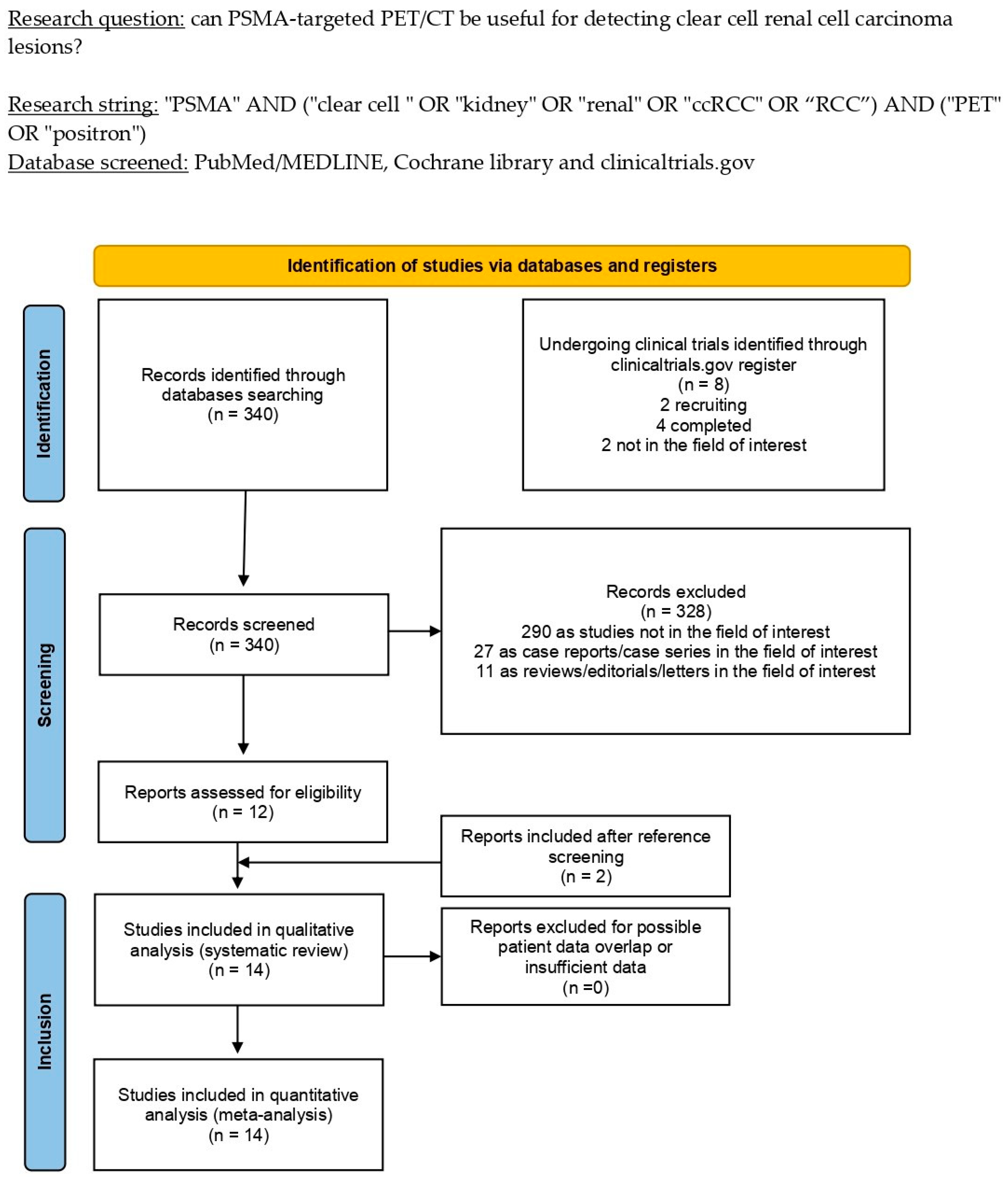
3.1. Literature Search and Study Selection

The literature search was lastly updated on 24 October 2022 and revealed a total of 340 records. Based on the inclusion and exclusion criteria mentioned above, 328 articles were excluded (290 as not in the field of interest, 27 as reviews, editorials, or letters, and 11 as case reports). Twelve remaining records were eligible for inclusion in the systematic review (qualitative synthesis) after a full-text assessment [29,30,31,32,33,34,35,36,37,38,39,40]. Two additional papers were assessed as suitable for inclusion after screening the references of these articles [41,42]. Figure 1 summarizes the study selection process.

3.2. Study Characteristics

Analyses of the characteristics of the fourteen articles eligible for the systematic review (qualitative research), including 331 ccRCC patients, are presented in Table 1, Table 2 and Table 3. With regard to the general study information (Table 1), the included papers were published between 2016 and 2022 in Europe, Asia, Australia, and the USA. Three studies had a prospective design, whereas the remaining eleven were retrospective. Among the fourteen studies included, only two were multicentric [38,39]. Only one of the revised articles declared funding in its text [42].
With regard to the patient key characteristics (Table 2), the number of ccRCC-included patients ranged from 8 to 54 patients (average age range: 57–66 years; male percentage range: 25.7–100%). PSMA-targeted PET/CT was used for staging in newly diagnosed ccRCC patients in seven studies [29,30,33,34,36,37,42], to restage ccRCC patients with suspect relapsing disease in three articles [32,35,40], and for both purposes in the remaining four articles [31,38,39,41]. Six studies classified the analyzed lesions according to the World Health Organization/International Society of Urological Pathology (WHO/ISUP) grading score [33,34,35,36,37,39], and five included an immunohistochemistry analysis to evaluate PSMA staining in the neovasculature of ccRCC lesions [33,34,35,36,42]. Concerning the comparison of PSMA-targeting PET/CT to other instrumental diagnostic examinations, four studies employed diagnostic CT [29,31,35,41], two alternated diagnostic CT and MRI [30,42], one used [18F]FDG PET/CT [32], one used only MRI [39], two employed both [18F]FDG PET/CT and diagnostic CT [38,40]; the remaining four studies did not use any comparative imaging but relied on pathology to validate their findings [33,34,36,37].
The included studies showed significant differences in the index test key characteristics (Table 3). In eight articles, the only injected tracer was [68Ga]Ga-PSMA-11 (activity range: 75–272 MBq in absolute values) [29,31,33,34,35,36,37,42]; in two studies, only [18F]F-DCFPyL was injected (mean injected activity: 333 MBq) [30,32]; in one, the only employed radiopharmaceutical was [18F]F-PSMA-1007 (activity range: 217–268 MBq); in two papers, an alternate between [68Ga]Ga-PSMA-11 and [18F]F-PSMA-1007 was reported based on radiopharmaceuticals availability [38,39]; and in the remaining study, both [68Ga]Ga-PSMA-11 and [18F]F-DCFPyL were used [40]. All the analyzed papers reported PET images coregistration with low-dose CT, and in one, a dynamic PET acquisition of the upper abdomen was performed [34]. Excluding the only study which conducted a dynamic acquisition [34], the uptake time after PSMA-targeting radiopharmaceutical administration and PET scan varied from 45 to 142 min. All the included articles performed qualitative and semiquantitative analyses within the PET imaging interpretation. Semiquantitative analyses were performed calculating the mean, maximal, and peak-standardized uptake values (SUVmean, SUVmax, and SUVpeak, respectively). The metabolic tumor volume (MTV) and total lesion PSMA uptake (TLP) were measured in one study [40]. The target-to-background uptake ratio (TBR) was assessed in four studies by dividing the lesions’ SUVmax for the background, muscle tissue, or liver SUV (SUVmax or SUVmean) [32,34,35,42]; finally, intralesional radiopharmaceutical kinetics were deepened only in one study [34].

3.3. Risk of Bias and Applicability

The overall assessment of the risk of bias and concerns about the applicability of the included papers according to QUADAS-2 is provided in Figure 2.

3.4. Results of Individual Studies (Qualitative Synthesis)

The comprehensive evaluation of PSMA-targeted PET/CT in detecting ccRCC lesions assessed an optimal diagnostic performance in all the included papers, both on a per-lesion- and per-patient-based analyses in all the evaluated clinical settings without significant differences between PET/CT scans executed for staging or restaging [31]. In this context, the reported detection rate ranged from 84% and 100% in the per-patient-based analysis [30,32,40] and from 80.5% to 100% in the per-lesion-based analysis [29,30,31,32,42], showing excellent performances in detecting metastatic lesions in lymph nodes, bone, breast, brain, pancreas, adrenal glands, liver, contralateral kidney, and lungs [29,30,35,38,40,41,42].
None of the included studies reported adverse effects after administering PSMA-targeting radiopharmaceuticals.
All the reviewed papers assessed variable PSMA-targeting radiopharmaceuticals uptake in primary and metastatic ccRCC lesions; most reports reported it was higher than the activity of the surrounding tissue. With regard to the semiquantitative data, average SUVmax reported values varied between 6.9 and 25.9 for primary lesions and ranged from 2.7 to 19.5 for metastatic lesions; the high heterogeneity observed among the included studies may be explained by the employment of three different PSMA-targeting radiopharmaceuticals in the included studies. Since every study used different background regions to calculate TBR, its variability had a poor value and was not assessed.
Most of the included studies observed that PSMA-targeting PET/CT could detect more lesions than conventional imaging, especially in metastatic lesions of the bone [29,30,31,35,38,42]. Nevertheless, in one of the included studies, PET/CT with PSMA-radioligands detected fewer lesions than diagnostic CT [40]. Finally, PSMA-targeting PET/CT showed a complementary role to MRI in detecting venous thrombi in the renal vein [39].
Two studies compared the diagnostic performances of PSMA-targeting PET/CT to [18F]FDG PET/CT [32,38]. Both reported a higher accuracy of the first over the latter, both in terms of number of highlighted lesions and of uptake entity, reporting higher SUV values in PSMA-targeting PET/CT.
Concerning the correlation between semiquantitative metrics at PSMA-targeted PET/CT and pathological characteristics of ccRCC, several studies observed higher PSMA-radioligands uptake in lesions with sarcomatoid or rhabdoid differentiation [29,33], whereas one study did not find significant differences in terms of uptake among lesions with or without this feature [40]. Furthermore, we found conflicting evidence about the correlation between semiquantitative values and the grade of PSMA staining in the immunohistochemistry analysis, since one paper reported a correlation between these two values [42]; conversely, another study did not find the same relationship [35]. Finally, several of the included studies deepened the association among PSMA-targeting radiopharmaceuticals uptake and WHO/ISUP grade, VEGFR-2/PDGFR-β expression, and HIF-2α expression, reporting that PSMA-targeting PET/CT might be a valuable instrument for discriminating the presence of these pathological features in ccRCC patients [33,36,37].
Regarding potential differences in the diagnostic performance of PSMA-targeted PET/CT among ccRCC and non-ccRCC tumors, several studies observed an inferior accuracy as well as a poorer uptake of PSMA-targeting radiopharmaceuticals in non-ccRCC lesions than ccRCC [31,42].
When reported, PET/CT with PSMA-radioligands could lead to a change in patients’ management (usually from local to systemic therapy) in all the evaluated clinical settings, ranging from 13% to 43.8% of the enrolled patients in eight studies [29,30,31,38,39,40,41,42].
Finally, one study explored the potential role of PSMA-targeting PET/CT to assess treatment response in ccRCC patients undergoing tyrosine kinase inhibitors (TKI) or immune checkpoint inhibitors (ICI) alongside conventional imaging [41], finding out discrepancies in 82% of the enrolled patients.
Since the studies that reported the PSMA-targeted PET/CT detection rate in ccRCC used different modalities for its assessment and different benchmarks to calculate it [29,30,31,32,40], a meta-analysis could not be accomplished. The results of the included papers, including semiquantitative metrics, detection rate, and percentage of change in patient management, are synthesized in Table 4.

4. Discussion

Based upon the PSMA overexpression on prostate cancer cells’ membrane, radiopharmaceuticals targeting this receptor became a relatively novel compound to improve the performance of molecular imaging and to develop new therapeutic instruments through radioligand therapy (RLT) in metastatic castration-resistant prostate cancer (although it does not substitute for laboratory examinations and MRI in its diagnosis). Furthermore, the recent literature reported that carboxypeptidase type II might be overexpressed in the neovasculature of various types of tumors other than prostate cancer, including ccRCC [43], and its expression could regulate tumor cell invasion and neoangiogenesis through the transduction of the integrin signal in the endothelium [44].
This rationale brought about several researchers to assess the diagnostic performance of PSMA-targeted PET/CT and evaluate the correlation between PSMA-targeting radiopharmaceuticals uptake and tumor histopathologic characteristics in newly diagnosed as well as previously treated ccRCC patients through the past six years [29,30,31,32,33,34,35,36,37,38,39,40,41,42]. This updated systematic review attempted to gather all the evidence concerning this emerging topic.
Despite the available evidence being quite limited and the included papers reporting different approaches to calculate the detection rate, all the authors investigating the accuracy of PET/CT with PSMA-targeting radiopharmaceuticals in ccRCC patients stated excellent performances both in per-patient- and per-lesion-based analyses in newly diagnosed patients as well as previously treated patients undergoing disease restaging without significant differences among the explored clinical settings [29,30,31,32,40].
One of the most influential prognostic factors regarding overall and progression-free survival in patients with ccRCC is the WHO/ISUP grading system based on tumor nuclear morphology, proposed by Moch et al. in 2016 [45]. In addition to this grading classification, recent shreds of evidence reported that several histopathology features, including intratumoral necrosis and the presence of rhabdoid or sarcomatoid ccRCC variants, might worsen the prognosis in ccRCC patients [46,47,48,49]. Most of the studies included in this systematic review exploring the potential of PSMA-targeting radiopharmaceuticals uptake to discriminate lesions with different WHO/ISUP grades as well as the presence of malignant histopathology features reported an SUVmax being significantly higher in ccRCC lesions with a more aggressive phenotype [29,33,40]. These results suggest that PSMA-radioligands uptake might be a valuable prognostic factor in ccRCC patients; in this context, more prospective studies are needed to assess its potential in this setting in combination with other prognostic factors as well as an independent variable.
A new diagnostic instrumental examination can change patient management once its employment brings about an up- or downstaging compared to conventional imaging and alters the previously planned treatment. Since 20–30% of patients with apparently resectable ccRCC develop metastatic disease after surgery, the current standard of care, consisting of diagnostic CT and/or MRI, might be considered insufficient [50]. When reported, PSMA-targeted PET/CT showed a superior accuracy in tumor burden characterization, highlighting more metastatic lesions than conventional imaging, reducing false positive findings, and changing patient management in a substantial percentage of cases [29,30,31,35,38,40,42]. Furthermore, in one study, PSMA-targeted PET/CT was employed to assess the presence of tumor thrombi and showed complementary results with MRI in their characterization [39]. These promising results enhance the need for more prospective clinical trials to assess how many ccRCC patients might have their treatment plan change after PSMA-targeted PET/CT and, more importantly, if its modification could actually lead to an improvement in their overall and disease-free survivals.
[18F]FDG PET imaging relies on the increased glucose demand by rapidly dividing tumor cells. Membrane glucose transporters, mainly GLUT-1, actively transport [18F]FDG into the cell, where it is converted by hexokinase into [18F]FDG-6-phosphate, which is trapped in the cytoplasm and accumulates. Tumor cells usually show increased [18F]FDG uptake due to the overexpression of GLUT-1 transporter, increased intracellular hexokinase, and low FBP1 [51]. Since FBP1 activity might be suppressed only in some ccRCCs, [18F]-FDG presents a variable uptake in ccRCC lesions; from this biological mechanism, PET/CT with [18F] FDG is not currently recommended in ccRCC management. Among the included studies, two compared the performance of [18F]FDG and PSMA-targeted PET/CT and both reported superior performance of the latter [32,38]. This result may be explained by the different biological pathways underlying the uptake of the investigated tracers since PSMA-radioligands uptake relies on neoangiogenesis, which is upregulated in ccRCC by the VHL gene mutation and the subsequent HIFs accumulation, two rate-limiting factors in ccRCC development.
Carbonic anhydrase IX (CAIX), a cell membrane antigen, has been recently used as a target for molecular imaging in ccRCC patients [52], since its expression is upregulated in hypoxic conditions [53], induced in ccRCC by the loss of the VHL complex and the subsequent accumulation of HIFs. Two papers assessed the employment of the radiolabeled anti-CAIX antibody “girentuximab” labeled with [89Zr]Zr for the imaging of ccRCC [54,55]. They observed its ability to highlight ccRCC in a localized disease as well as in ccRCC metastases and to differentiate ccRCC from non-ccRCC lesions. In this context, no articles comparing PSMA-targeted PET to [89Zr]Zr-girentuximab were found in the literature search.
Since the introduction of carboxypeptidase type II as a possible target for molecular imaging and theragnostics, an increasing number of PSMA-targeting radiopharmaceuticals has been developed, including [68Ga]Ga-PSMA-11, [18F]F-DCFPyL, and [18F]F-PSMA-1007 [56,57]. Based on the available literature data, all the above-mentioned radiopharmaceuticals were employed for detecting ccRCC lesions [29,30,31,32,33,34,35,36,37,38,39,40,41,42]. Despite the use of [18F]-PSMA-targeting radiopharmaceuticals counts on several advantages such as reduced costs through large-scale production, higher-quality images through lower positron energy and a longer half-life, none of the included studies compared the diagnostic performances between different PSMA-targeting radiopharmaceuticals.
An intriguing issue underlying the use of PSMA-radioligands in ccRCC patients is based on the overexpression of carboxypeptidase type II by intratumoral neovascular endothelial cells. Since the current standard of care in patients with unresectable or relapsing metastatic ccRCC relies on antiangiogenic therapy with tyrosine kinase inhibitors and immunotherapy with checkpoint inhibitors [11], it is possible that PSMA-targeting radiopharmaceuticals might be able to assess and predict treatment response. Among the studies included in this systematic review, one compared PSMA-targeted PET/CT in the setting of the treatment response assessment to diagnostic CT and reported a concordance between the two examinations in a minority of patients [41]. Nevertheless, since no long-term follow-up data were reported, it was not possible to comment on the meaning of the discrepancies highlighted between the examinations and if they could improve patient management. In this setting, taking into account available evidence-based data, more studies concerning the predictive value or response assessment using this novel imaging method in ccRCC patients are needed.
Considering the recent introduction of PSMA-targeting radiopharmaceuticals labeled with β and α+ emitters, such as [177Lu]Lu-PSMA-617 and [225Ac]Ac-PSMA-617, and their outstanding results in metastatic castration-resistant prostate cancer patients [24,58], another interesting aspect of PSMA overexpression in ccRCC lesions is based upon the feasibility of a PSMA-targeted radioligand therapy. Nevertheless, the comprehensive literature search performed for this systematic review did not enhance any paper reporting data about RLT in ccRCC patients. In this context, the authors warrant clinical and pre-clinical investigations to assess the feasibility of a PSMA-targeted theragnostic approach in ccRCC patients and to evaluate its safety and efficacy.
Consistently with the studies included in this systematic review, Yin et al. reported a poor accuracy of PSMA-targeted PET/CT in patients with metastatic non-ccRCC, inferior to conventional imaging with CT and/or MRI [59]. These data may be explained by the different genetic pathways guiding the oncogenesis in the different types of RCC, since only ccRCC relies on the VHL mutation, which is strictly correlated with the accumulation of HIFs and the subsequent neoangiogenesis as a rate-limiting element in its development. Moreover, a recent prospective study has evaluated the role of PSMA-targeted PET/CT in the detection of gastrointestinal and pancreatic cancers (with the latter showing similar biologic characteristics to papillary RCC). The authors reported that the low PSMA tumor expression and the high physiological uptake in organs/background hampered the clear distinction of the tumor tissue and, as a result, [18F]FDG PET/CT was superior in detecting gastrointestinal and pancreatic tumors [60].
None of the studies included in this systematic review addressed the cost-effectiveness of PET with PSMA-targeting radiopharmaceuticals in managing ccRCC patients if it was actually introduced in clinical practice as a standard of care. Since, to date, this examination is employed in a research setting, cost-effectiveness studies are needed to strengthen and refine the role of this imaging technique in ccRCC.
Two previously published reviews explored the potential role of PSMA-targeted PET/CT in RCC patients [61,62]. The current systematic review had the purpose of providing an updated literature search using PRISMA guidelines as milestone in its editing, requiring a strict methodology, appropriate criteria for the literature search as well as for the quality assessment; moreover, case reports were excluded from the analysis since they are a possible source of bias. As a result, more data were provided in the results and discussion sections.
Concerning the limitations and biases of this systematic review, a limited number of prospective studies was available and most of them enrolled a restricted number of patients. Furthermore, we clearly demonstrated a significant heterogeneity among the included studies about the study methodology, patient characteristics, index test characteristics, and reference standard or comparison used. Prospective multicentric studies are needed to strengthen the role of PSMA-targeting PET/CT in ccRCC.

5. Conclusions

The qualitative data provided by this systematic review enhanced the promising role of PSMA-targeted PET/CT in patients with ccRCC. Nevertheless, more studies are warranted to overpower these findings and deepen how PET imaging with PSMA-targeting radiopharmaceuticals could integrate with conventional imaging in different clinical settings.

Supplementary Materials

The following supporting information can be downloaded at: https://www.mdpi.com/article/10.3390/cancers15020355/s1, Table S1: PRISMA checklist, Table S2: List of excluded studies with reasons.

Author Contributions

Conceptualization: A.R. and M.R.; methodology: G.T.; writing—original draft preparation: A.R. and G.T.; writing—review and editing: A.R., M.R., P.R., S.A., S.D., G.L.B., F.D., D.A. and F.B.; supervision, G.T. All authors have read and agreed to the published version of the manuscript.

Funding

This research received no external funding.

Institutional Review Board Statement

Not applicable.

Informed Consent Statement

Not applicable.

Data Availability Statement

The data presented in this study are available on request from the corresponding author.

Conflicts of Interest

The authors declare no conflict of interest.

References

  1. Siegel, R.L.; Miller, K.D.; Fuchs, H.E.; Jemal, A. Cancer statistics, 2022. CA A Cancer J. Clin. 2022, 72, 7–33. [Google Scholar] [CrossRef] [PubMed]
  2. Bray, F.; Ferlay, J.; Soerjomataram, I.; Siegel, R.L.; Torre, L.A.; Jemal, A. Global cancer statistics 2018: GLOBOCAN estimates of incidence and mortality worldwide for 36 cancers in 185 countries. CA Cancer J. Clin. 2018, 68, 394–424. [Google Scholar] [CrossRef] [PubMed] [Green Version]
  3. Capitanio, U.; Bensalah, K.; Bex, A.; Boorjian, S.A.; Bray, F.; Coleman, J.; Gore, J.L.; Sun, M.; Wood, C.; Russo, P. Epidemiology of Renal Cell Carcinoma. Eur. Urol. 2019, 75, 74–84. [Google Scholar] [CrossRef] [PubMed]
  4. Heng, D.Y.; Xie, W.; Regan, M.M.; Warren, M.A.; Golshayan, A.R.; Sahi, C.; Eigl, B.J.; Ruether, J.D.; Cheng, T.; North, S.; et al. Prognostic Factors for Overall Survival in Patients with Metastatic Renal Cell Carcinoma Treated with Vascular Endothelial Growth Factor–Targeted Agents: Results from a Large, Multicenter Study. J. Clin. Oncol. 2009, 27, 5794–5799. [Google Scholar] [CrossRef] [PubMed]
  5. Srinivasan, R.; Ricketts, C.J.; Sourbier, C.; Linehan, W.M. New Strategies in Renal Cell Carcinoma: Targeting the Genetic and Metabolic Basis of Disease. Clin. Cancer Res. 2015, 21, 10–17. [Google Scholar] [CrossRef] [Green Version]
  6. Linehan, W.M.; Srinivasan, R.; Schmidt, L.S. The genetic basis of kidney cancer: A metabolic disease. Nat. Rev. Urol. 2010, 7, 277–285. [Google Scholar] [CrossRef]
  7. Linehan, W.M. Genetic basis of kidney cancer: Role of genomics for the development of disease-based therapeutics. Genome Res. 2012, 22, 2089–2100. [Google Scholar] [CrossRef] [Green Version]
  8. Maranchie, J.K.; Afonso, A.; Albert, P.S.; Kalyandrug, S.; Phillips, J.L.; Zhou, S.; Peterson, J.; Ghadimi, B.M.; Hurley, K.; Riss, J.; et al. Solid renal tumor severity in von Hippel Lindau disease is related to germline deletion length and location. Hum. Mutat. 2004, 23, 40–46. [Google Scholar] [CrossRef]
  9. Moore, L.E.; Nickerson, M.L.; Brennan, P.; Toro, J.R.; Jaeger, E.; Rinsky, J.; Han, S.S.; Zaridze, D.; Matveev, V.; Janout, V.; et al. Von Hippel-Lindau (VHL) Inactivation in Sporadic Clear Cell Renal Cancer: Associations with Germline VHL Polymorphisms and Etiologic Risk Factors. PLoS Genet. 2011, 7, e1002312. [Google Scholar] [CrossRef] [Green Version]
  10. Nickerson, M.L.; Jaeger, E.; Shi, Y.; Durocher, J.A.; Mahurkar, S.; Zaridze, D.; Matveev, V.; Janout, V.; Kollarova, H.; Bencko, V.; et al. Improved Identification of von Hippel-Lindau Gene Alterations in Clear Cell Renal Tumors. Clin. Cancer Res. 2008, 14, 4726–4734. [Google Scholar] [CrossRef]
  11. Hsieh, J.J.; Purdue, M.P.; Signoretti, S.; Swanton, C.; Albiges, L.; Schmidinger, M.; Heng, D.Y.; Larkin, J.; Ficarra, V. Renal cell carcinoma. Nat. Rev. Dis. Prim. 2017, 3, 17009. [Google Scholar] [CrossRef] [PubMed]
  12. Huang, D.; Ding, Y.; Zhou, M.; Rini, B.I.; Petillo, D.; Qian, C.-N.; Kahnoski, R.; Futreal, P.A.; Furge, K.A.; Teh, B.T. Interleukin-8 Mediates Resistance to Antiangiogenic Agent Sunitinib in Renal Cell Carcinoma. Cancer Res. 2010, 70, 1063–1071. [Google Scholar] [CrossRef] [Green Version]
  13. Low, G.; Huang, G.; Fu, W.; Moloo, Z.; Girgis, S. Review of renal cell carcinoma and its common subtypes in radiology. World J. Radiol. 2016, 8, 484–500. [Google Scholar] [CrossRef] [PubMed]
  14. Karlo, C.A.; Di Paolo, P.L.; Chaim, J.; Hakimi, A.A.; Ostrovnaya, I.; Russo, P.; Hricak, H.; Motzer, R.; Hsieh, J.J.; Akin, O. Radiogenomics of Clear Cell Renal Cell Carcinoma: Associations between CT Imaging Features and Mutations. Radiology 2014, 270, 464–471. [Google Scholar] [CrossRef] [PubMed]
  15. Alessandrino, F.; Krajewski, K.M.; Shinagare, A.B. Update on Radiogenomics of Clear Cell Renal Cell Carcinoma. Eur. Urol. Focus 2016, 2, 572–573. [Google Scholar] [CrossRef]
  16. Lindenberg, L.; Mena, E.; Choyke, P.L.; Bouchelouche, K. PET imaging in renal cancer. Curr. Opin. Oncol. 2019, 31, 216–221. [Google Scholar] [CrossRef]
  17. Motzer, R.J.; Jonasch, E.; Agarwal, N.; Alva, A.; Baine, M.; Beckermann, K.; Carlo, M.I.; Choueiri, T.K.; Costello, B.A.; Derweesh, I.H.; et al. Kidney Cancer, Version 3.2022, NCCN Clinical Practice Guidelines in Oncology. J. Natl. Compr. Cancer Netw. 2022, 20, 71–90. [Google Scholar] [CrossRef] [PubMed]
  18. Escudier, B.; Porta, C.; Schmidinger, M.; Rioux-Leclercq, N.; Bex, A.; Khoo, V.; Grünwald, V.; Gillessen, S.; Horwich, A.; ESMO Guidelines Committee. Renal cell carcinoma: ESMO Clinical Practice Guidelines for diagnosis, treatment and follow-up. Ann. Oncol. 2019, 30, 706–720. [Google Scholar] [CrossRef] [Green Version]
  19. Ljungberg, B.; Albiges, L.; Abu-Ghanem, Y.; Bedke, J.; Capitanio, U.; Dabestani, S.; Fernández-Pello, S.; Giles, R.H.; Hofmann, F.; Hora, M.; et al. European Association of Urology Guidelines on Renal Cell Carcinoma: The 2022 Update. Eur. Urol. 2022, 82, 399–410. [Google Scholar] [CrossRef]
  20. Chen, R.; Zhou, X.; Huang, G.; Liu, J. Fructose 1,6-Bisphosphatase 1 Expression Reduces 18F-FDG Uptake in Clear Cell Renal Cell Carcinoma. Contrast Media Mol. Imaging 2019, 2019, 9463926. [Google Scholar] [CrossRef]
  21. Israeli, R.S.; Powell, C.T.; Fair, W.R.; Heston, W.D. Molecular cloning of a complementary DNA encoding a prostate-specific membrane antigen. Cancer Res. 1993, 53, 227–230. [Google Scholar]
  22. Horoszewicz, J.S.; Kawinski, E.; Murphy, G.P. Monoclonal antibodies to a new antigenic marker in epithelial prostatic cells and serum of prostatic cancer patients. Anticancer Res. 1987, 7, 927–935. [Google Scholar] [PubMed]
  23. Wester, H.-J.; Schottelius, M. PSMA-Targeted Radiopharmaceuticals for Imaging and Therapy. Semin. Nucl. Med. 2019, 49, 302–312. [Google Scholar] [CrossRef] [PubMed]
  24. Sartor, O.; de Bono, J.; Chi, K.N.; Fizazi, K.; Herrmann, K.; Rahbar, K.; Tagawa, S.T.; Nordquist, L.T.; Vaishampayan, N.; El-Haddad, G.; et al. Lutetium-177–PSMA-617 for Metastatic Castration-Resistant Prostate Cancer. N. Engl. J. Med. 2021, 385, 1091–1103. [Google Scholar] [CrossRef] [PubMed]
  25. Jadvar, H.; Calais, J.; Fanti, S.; Feng, F.; Greene, K.L.; Gulley, J.L.; Hofman, M.; Koontz, B.F.; Lin, D.W.; Morris, M.J.; et al. Appropriate Use Criteria for Prostate-Specific Membrane Antigen PET Imaging. J. Nucl. Med. 2022, 63, 59–68. [Google Scholar] [CrossRef] [PubMed]
  26. Sadeghi, R.; Treglia, G. Systematic reviews and meta-analyses of diagnostic studies: A practical guideline. Clin. Transl. Imaging 2017, 5, 83–87. [Google Scholar] [CrossRef]
  27. Page, M.J.; McKenzie, J.E.; Bossuyt, P.M.; Boutron, I.; Hoffmann, T.C.; Mulrow, C.D.; Shamseer, L.; Tetzlaff, J.M.; Akl, E.A.; Brennan, S.E.; et al. The PRISMA 2020 Statement: An Updated Guideline for Reporting Systematic Reviews. BMJ 2021, 372, n71. [Google Scholar] [CrossRef]
  28. Whiting, P.F.; Rutjes, A.W.S.; Westwood, M.E.; Mallett, S.; Deeks, J.J.; Reitsma, J.B.; Leeflang, M.M.G.; Sterne, J.A.C.; Bossuyt, P.M.M.; QUADAS-2 Group. QUADAS-2: A Revised Tool for the Quality Assessment of Diagnostic Accuracy Studies. Ann. Intern. Med. 2011, 155, 529–536. Available online: https://www.acpjournals.org/doi/10.7326/0003-4819-155-8-201110180-00009 (accessed on 21 August 2022). [CrossRef]
  29. Rhee, H.; Blazak, J.; Tham, C.M.; Ng, K.L.; Shepherd, B.; Lawson, M.; Preston, J.; Vela, I.; Thomas, P.; Wood, S. Pilot study: Use of gallium-68 PSMA PET for detection of metastatic lesions in patients with renal tumour. EJNMMI Res. 2016, 6, 76. [Google Scholar] [CrossRef]
  30. Meyer, A.R.; Carducci, M.A.; Denmeade, S.R.; Markowski, M.C.; Pomper, M.G.; Pierorazio, P.M.; Allaf, M.E.; Rowe, S.P.; Gorin, M.A. Improved identification of patients with oligometastatic clear cell renal cell carcinoma with PSMA-targeted 18F-DCFPyL PET/CT. Ann. Nucl. Med. 2019, 33, 617–623. [Google Scholar] [CrossRef]
  31. Raveenthiran, S.; Esler, R.; Yaxley, J.; Kyle, S. The use of 68Ga-PET/CT PSMA in the staging of primary and suspected recurrent renal cell carcinoma. Eur. J. Nucl. Med. Mol. Imaging 2019, 46, 2280–2288. [Google Scholar] [CrossRef]
  32. Liu, Y.; Wang, G.; Yu, H.; Wu, Y.; Lin, M.; Gao, J.; Xu, B. Comparison of 18F-DCFPyL and 18F-FDG PET/computed tomography for the restaging of clear cell renal cell carcinoma: Preliminary results of 15 patients. Nucl. Med. Commun. 2020, 41, 1299–1305. [Google Scholar] [CrossRef]
  33. Gao, J.; Xu, Q.; Fu, Y.; He, K.; Zhang, C.; Zhang, Q.; Shi, J.; Zhao, X.; Wang, F.; Guo, H. Comprehensive evaluation of 68Ga-PSMA-11 PET/CT parameters for discriminating pathological characteristics in primary clear-cell renal cell carcinoma. Eur. J. Nucl. Med. Mol. Imaging 2021, 48, 561–569. [Google Scholar] [CrossRef] [PubMed]
  34. Golan, S.; Aviv, T.; Groshar, D.; Yakimov, M.; Zohar, Y.; Prokocimer, Y.; Nadu, A.; Baniel, J.; Domachevsky, L.; Bernstine, H. Dynamic 68Ga-PSMA-11 PET/CT for the Primary Evaluation of Localized Renal Mass: A Prospective Study. J. Nucl. Med. 2021, 62, 773–778. [Google Scholar] [CrossRef] [PubMed]
  35. Gühne, F.; Seifert, P.; Theis, B.; Steinert, M.; Freesmeyer, M.; Drescher, R. PSMA-PET/CT in Patients with Recurrent Clear Cell Renal Cell Carcinoma: Histopathological Correlations of Imaging Findings. Diagnostics 2021, 11, 1142. [Google Scholar] [CrossRef]
  36. Gao, J.; Meng, L.; Xu, Q.; Zhao, X.; Deng, Y.; Fu, Y.; Guo, S.; He, K.; Shi, J.; Wang, F.; et al. 68Ga-PSMA-11 PET/CT Parameter Correlates with Pathological VEGFR-2/PDGFR-β Expression in Renal Cell Carcinoma Patients. Mol. Imaging Biol. 2022, 24, 759–768. [Google Scholar] [CrossRef] [PubMed]
  37. Meng, L.; Zhang, S.; Gao, J.; Xu, Q.; Fu, Y.; Zhou, Y.-H.; Wang, F.; Guo, H. [68Ga]Ga-PSMA-11 PET/CT has potential application in predicting tumor HIF-2α expression and therapeutic response to HIF-2α antagonists in patients with RCC. Eur. Radiol. 2022, 32, 6545–6553. [Google Scholar] [CrossRef] [PubMed]
  38. Tariq, A.; Kwok, M.; Pearce, A.; Rhee, H.; Kyle, S.; Marsh, P.; Raveenthiran, S.; Wong, D.; McBean, R.; Westera, J.; et al. The role of dual tracer PSMA and FDG PET/CT in renal cell carcinoma (RCC) compared to conventional imaging: A multi-institutional case series with intra-individual comparison. Urol. Oncol. Semin. Orig. Investig. 2022, 40, 66.e1–66.e9. [Google Scholar] [CrossRef] [PubMed]
  39. Tariq, A.; McGeorge, S.; Pearce, A.; Rhee, H.; Wood, S.; Kyle, S.; Marsh, P.; Raveenthiran, S.; Wong, D.; McBean, R.; et al. Characterization of tumor thrombus in renal cell carcinoma with prostate specific membrane antigen (PSMA) positron emission tomography (PET)/computed tomography (CT). Urol. Oncol. Semin. Orig. Investig. 2022, 40, 276.e1–276.e9. [Google Scholar] [CrossRef] [PubMed]
  40. Udovicich, C.; Callahan, J.; Bressel, M.; Ong, W.L.; Perera, M.; Tran, B.; Azad, A.; Haran, S.; Moon, D.; Chander, S.; et al. Impact of Prostate-specific Membrane Antigen Positron Emission Tomography/Computed Tomography in the Management of Oligometastatic Renal Cell Carcinoma. Eur. Urol. Open Sci. 2022, 44, 60–68. [Google Scholar] [CrossRef] [PubMed]
  41. Mittlmeier, L.M.; Unterrainer, M.; Rodler, S.; Todica, A.; Albert, N.L.; Burgard, C.; Cyran, C.C.; Kunz, W.G.; Ricke, J.; Bartenstein, P.; et al. 18F-PSMA-1007 PET/CT for response assessment in patients with metastatic renal cell carcinoma undergoing tyrosine kinase or checkpoint inhibitor therapy: Preliminary results. Eur. J. Nucl. Med. Mol. Imaging 2021, 48, 2031–2037. [Google Scholar] [CrossRef]
  42. Li, Y.; Zheng, R.; Zhang, Y.; Huang, C.; Tian, L.; Liu, R.; Liu, Y.; Zhang, Z.; Han, H.; Zhou, F.; et al. Special issue “The advance of solid tumor research in China”: 68Ga-PSMA-11 PET/CT for evaluating primary and metastatic lesions in different histological subtypes of renal cell carcinoma. Int. J. Cancer 2022, 152, 42–50. [Google Scholar] [CrossRef]
  43. Rizzo, A.; Dall’Armellina, S.; Pizzuto, D.A.; Perotti, G.; Zagaria, L.; Lanni, V.; Treglia, G.; Racca, M.; Annunziata, S. PSMA Radioligand Uptake as a Biomarker of Neoangiogenesis in Solid Tumours: Diagnostic or Theragnostic Factor? Cancers 2022, 14, 4039. [Google Scholar] [CrossRef] [PubMed]
  44. Conway, R.E.; Rojas, C.; Alt, J.; Nováková, Z.; Richardson, S.M.; Rodrick, T.C.; Fuentes, J.L.; Richardson, N.H.; Attalla, J.; Stewart, S.; et al. Prostate-specific membrane antigen (PSMA)-mediated laminin proteolysis generates a pro-angiogenic peptide. Angiogenesis 2016, 19, 487–500. [Google Scholar] [CrossRef] [PubMed]
  45. Moch, H.; Cubilla, A.L.; Humphrey, P.A.; Reuter, V.E.; Ulbright, T.M. The 2016 WHO Classification of Tumours of the Urinary System and Male Genital Organs—Part A: Renal, Penile, and Testicular Tumours. Eur. Urol. 2016, 70, 93–105. [Google Scholar] [CrossRef]
  46. Sengupta, S.; Lohse, C.M.; Leibovich, B.C.; Frank, I.; Thompson, R.H.; Webster, W.S.; Zincke, H.; Blute, M.L.; Cheville, J.C.; Kwon, E.D. Histologic coagulative tumor necrosis as a prognostic indicator of renal cell carcinoma aggressiveness. Cancer 2005, 104, 511–520. [Google Scholar] [CrossRef]
  47. Khor, L.-Y.; Dhakal, H.P.; Jia, X.; Reynolds, J.P.; McKenney, J.K.; Rini, B.I.; Magi-Galluzzi, C.; Przybycin, C.G. Tumor Necrosis Adds Prognostically Significant Information to Grade in Clear Cell Renal Cell Carcinoma: A Study of 842 Consecutive Cases from a Single Institution. Urol. Oncol. Semin. Orig. Investig. 2016, 40, 1224–1231. [Google Scholar] [CrossRef]
  48. Pichler, R.; Compérat, E.; Klatte, T.; Pichler, M.; Loidl, W.; Lusuardi, L.; Schmidinger, M. Renal Cell Carcinoma with Sarcomatoid Features: Finally New Therapeutic Hope? Cancers 2019, 11, 422. [Google Scholar] [CrossRef] [Green Version]
  49. Przybycin, C.G.; McKenney, J.K.; Reynolds, J.P.; Campbell, S.; Zhou, M.; Karafa, M.T.; Magi-Galluzzi, C. Rhabdoid Differentiation Is Associated with Aggressive Behavior in Renal Cell Carcinoma: A Clinicopathologic Analysis of 76 Cases with Clinical Follow-Up. Am. J. Surg. Pathol. 2014, 38, 1260–1265. [Google Scholar] [CrossRef]
  50. Tyson, M.D.; Chang, S.S. Optimal Surveillance Strategies After Surgery for Renal Cell Carcinoma. J. Natl. Compr. Cancer Netw. 2017, 15, 835–840. [Google Scholar] [CrossRef] [PubMed]
  51. Liu, Y.; Ghesani, N.V.; Zuckier, L.S. Physiology and Pathophysiology of Incidental Findings Detected on FDG-PET Scintigraphy. Semin. Nucl. Med. 2010, 40, 294–315. [Google Scholar] [CrossRef] [PubMed]
  52. van Oostenbrugge, T.; Mulders, P. Targeted PET/CT imaging for clear cell renal cell carcinoma with radiolabeled antibodies: Recent developments using girentuximab. Curr. Opin. Urol. 2021, 31, 249–254. [Google Scholar] [CrossRef] [PubMed]
  53. Andreucci, E.; Peppicelli, S.; Carta, F.; Brisotto, G.; Biscontin, E.; Ruzzolini, J.; Bianchini, F.; Biagioni, A.; Supuran, C.T.; Calorini, L. Carbonic anhydrase IX inhibition affects viability of cancer cells adapted to extracellular acidosis. J. Mol. Med. 2017, 95, 1341–1353. [Google Scholar] [CrossRef] [PubMed]
  54. Hekman, M.C.H.; Rijpkema, M.; Aarntzen, E.H.; Mulder, S.F.; Langenhuijsen, J.F.; Oosterwijk, E.; Boerman, O.C.; Oyen, W.J.G.; Mulders, P.F.A. Positron Emission Tomography/Computed Tomography with 89Zr-girentuximab Can Aid in Diagnostic Dilemmas of Clear Cell Renal Cell Carcinoma Suspicion. Eur. Urol. 2018, 74, 257–260. [Google Scholar] [CrossRef]
  55. Verhoeff, S.R.; van Es, S.C.; Boon, E.; van Helden, E.; Angus, L.; Elias, S.G.; Oosting, S.F.; Aarntzen, E.H.; Brouwers, A.H.; Kwee, T.C.; et al. Lesion detection by [89Zr]Zr-DFO-girentuximab and [18F]FDG-PET/CT in patients with newly diagnosed metastatic renal cell carcinoma. Eur. J. Nucl. Med. Mol. Imaging 2019, 46, 1931–1939. [Google Scholar] [CrossRef] [PubMed] [Green Version]
  56. Chen, Y.; Lisok, A.; Chatterjee, S.; Wharram, B.; Pullambhatla, M.; Wang, Y.; Sgouros, G.; Mease, R.C.; Pomper, M.G. [18F]Fluoroethyl Triazole Substituted PSMA Inhibitor Exhibiting Rapid Normal Organ Clearance. Bioconjug. Chem. 2016, 27, 1655–1662. [Google Scholar] [CrossRef] [Green Version]
  57. Giesel, F.L.; Hadaschik, B.; Cardinale, J.; Radtke, J.; Vinsensia, M.; Lehnert, W.; Kesch, C.; Tolstov, Y.; Singer, S.; Grabe, N.; et al. F-18 labelled PSMA-1007: Biodistribution, radiation dosimetry and histopathological validation of tumor lesions in prostate cancer patients. Eur. J. Nucl. Med. Mol. Imaging 2017, 44, 678–688. [Google Scholar] [CrossRef] [Green Version]
  58. Ling, S.W.; de Blois, E.; Hooijman, E.; van der Veldt, A.; Brabander, T. Advances in 177Lu-PSMA and 225Ac-PSMA Radionuclide Therapy for Metastatic Castration-Resistant Prostate Cancer. Pharmaceutics 2022, 14, 2166. [Google Scholar] [CrossRef]
  59. Yin, Y.; Campbell, S.P.; Markowski, M.C.; Pierorazio, P.M.; Pomper, M.G.; Allaf, M.E.; Rowe, S.P.; Gorin, M.A. Inconsistent Detection of Sites of Metastatic Non-Clear Cell Renal Cell Carcinoma with PSMA-Targeted [18F]DCFPyL PET/CT. Mol. Imaging Biol. 2019, 21, 567–573. [Google Scholar] [CrossRef]
  60. Vuijk, F.A.; Kleiburg, F.; Noortman, W.A.; Heijmen, L.; Shahbazi, S.F.; van Velden, F.H.P.; Baart, V.M.; Bhairosingh, S.S.; Windhorst, B.D.; Hawinkels, L.J.A.C.; et al. Prostate-Specific Membrane Antigen Targeted Pet/CT Imaging in Patients with Colon, Gastric and Pancreatic Cancer. Cancers 2022, 14, 6209. [Google Scholar] [CrossRef]
  61. Muselaers, S.; Erdem, S.; Bertolo, R.; Ingels, A.; Kara, Ö.; Pavan, N.; Roussel, E.; Pecoraro, A.; Marchioni, M.; Carbonara, U.; et al. PSMA PET/CT in Renal Cell Carcinoma: An Overview of Current Literature. J. Clin. Med. 2022, 11, 1829. [Google Scholar] [CrossRef] [PubMed]
  62. Urso, L.; Castello, A.; Rocca, G.C.; Lancia, F.; Panareo, S.; Cittanti, C.; Uccelli, L.; Florimonte, L.; Castellani, M.; Ippolito, C.; et al. Role of PSMA-ligands imaging in Renal Cell Carcinoma management: Current status and future perspectives. J. Cancer Res. Clin. Oncol. 2022, 148, 1299–1311. [Google Scholar] [CrossRef] [PubMed]
Figure 1. Comprehensive overview of the study selection process for the systematic review.
Cancers 15 00355 g001
Figure 2. Summary of quality assessment according to the QUADAS-2 tool. The authors classified the papers included in the systematic review as a high risk or low risk of bias or applicability concerns for distinct domains listed in the ordinate axis. In contrast, the abscissa axis shows the percentage of studies. The graph indicates that about 60% of studies suffered a high risk of bias in “patient selection”, whereas a poor risk of bias was observed in “reference standard”, “index test”, and “flow and timing” domains.
Cancers 15 00355 g002
Table 1. General study information.
Authors [Ref.]YearCountryStudy Design/Number of Involved CentersFunding Sources
Rhee et al. [29]2016AustraliaProspective/monocentricNone declared
Meyer et al. [30]2019U.S.A.Prospective/monocentricNone declared
Raveenthiran [31]2019AustraliaRetrospective/monocentricNone declared
Liu et al. [32]2020ChinaRetrospective/monocentricNone declared
Gao et al. [33]2020ChinaRetrospective/monocentricNone declared
Mittlmeier et al. [41]2020GermanyRetrospective/monocentricNone declared
Golan et al. [34]2021IsraelProspective/monocentricNone declared
Gühne et al. [35]2021GermanyRetrospective/monocentricNone declared
Gao et al. [36]2022ChinaRetrospective/monocentricNone declared
Meng et al. [37]2022ChinaRetrospective/monocentricNone declared
Tariq et al. [38]2022AustraliaRetrospective/multicentricNone declared
Tariq et al. [39]2022AustraliaRetrospective/multicentricNone declared
Udovicich et al. [40]2022AustraliaRetrospective/monocentricNone declared
Li et al. [42]2022ChinaRetrospective/monocentricNational Natural Science Foundation of China
Table 2. Patient key characteristics and clinical settings.
Authors [Ref.]Sample Size (No. ccRCC Patients/All Included Patients)Mean/Median Age (Years)Gender
(Male %)
Clinical Setting
(No. Patients)
WHO/ISUP Grade
(No. Lesions)
PSMA Staining AnalysisComparative Imaging
Rhee et al. [29]8/10Median: 57100%10 stagingn.a.n.a.CT
Meyer et al. [30]14/14Median: 5964.3%14 stagingn.a.n.a.CT or MRI of abdomen + CT of the chest
Raveenthiran et al. [31]28/38Median: 6425.7%16 staging
22 restaging
n.a.n.a.CT
Liu et al. [32]15/15Mean: 57.586.6%15 restagingn.a.n.a.[18F]FDG PET/CT
Gao et al. [33]36/36Median: 6158.3%36 staging9 grade 1
12 grade 2
9 grade 3
6 grade 4
YesNo comparative exam
Mittlmeier et al. [41]8/11Mean: 59.672.7%11 staging and restaging after treatmentn.a.n.a.CT
Golan et al. [34]18/27Median: 6670%27 staging14 grade 1–2
4 grade 3
YesNo comparative exam
Gühne et al. [35]9/9Range: 52–8088%9 Restaging1 grade 1
6 grade 2
3 grade 3
1 grade 4
YesCT
Gao et al. [36]37/48Median: 5960.4%48 staging10 grade 1
13 Grade 2
11 grade 3
7 grade 4
YesNo comparative exam
Meng et al. [37]40/53Median: 5660.453 staging10 grade 1
17 grade 2
15 grade 3
3 grade 4
n.a.No comparative exam
Tariq et al. [38]10/11Mean: 65.564%4 staging
7 restaging
n.a.n.a.[18F]FDG PET/CT; CT
Tariq et al. [39]14/14Median: 6164.3%12 staging
2 restaging
3 grade 2
5 grade 3
5 grade 4
1 ungraded
n.a.MRI
Udovicich et al. [40]54/61Mean: 6556%61 restagingn.a.n.a.[18F]FDG PET/CT; CT
Li et al. [42]40/50 Median age: 5578%50 stagingn.a.YesCT or MRI
Legend: CT: computed tomography; FDG: fluorodeoxyglucose; ISUP: International Society of Urological Pathology; MRI: magnetic resonance imaging; n.a.: not available; PET: positron emission tomography; PSMA: prostate-specific membrane antigen; WHO: World Health Organization.
Table 3. Index test key characteristics.
Authors [Ref.]TracerHybrid ImagingTomographAdministered ActivityUptake Time
(Minutes)
Image Analysis
Rhee et al. [29][68Ga]Ga-PSMA-11PET/CTBiograph mCT FLOW
(Siemens ®)
150 MBq60Qualitative and semiquantative (SUVmax)
Meyer et al. [30][18F]F-DCFPyLPET/CTn.a.333 MBq60Qualitative and semiquantative (SUVmax)
Raveenthiran et al. [31][68Ga]Ga-PSMA-11PET/CTBiograph mCT (Siemens ®)150 MBq45–60Qualitative and semiquantitative (SUVmax)
Liu et al. [32][18F]F-DCFPyLPET/CTBiograph 64 (Siemens ®)0.15 Ci/kg60Qualitative and semiquantitative (SUVmax, TBR)
Gao et al. [33][68Ga]Ga-PSMA-11PET/CTuMI 780 (United Imaging Healthcare ®)n.a.45Qualitative and semiquantitative (SUVmax, SUVmean, SUVpeak)
Mittlmeier et al. [41][18F]F-PSMA-1007PET/CTBiograph mCT (Siemens ®);
Biograph 64 (Siemens ®)
217–26860Qualitative and semiquantitative (SUVmean, SUL)
Golan et al. [34][68Ga]Ga-PSMA-11Dynamic PET/CTDiscovery 710
(GE healthcare ®)
75–150 MBq0Qualitative, semiquantitative (SUVmax, SUVmean, TBR) and kinetic analysis
Gühne et al. [35][68Ga]Ga-PSMA-11PET/CTBiograph mCT 40 (Siemens ®)221–272 MBq74–103Qualitative and semiquantitative (SUVmax, SUVmean, TBR)
Gao et al. [36][68Ga]Ga-PSMA-11PET/CTuMI 780 (United Imaging Healthcare ®)n.a.45Qualitative and semiquantitative (SUVmax)
Meng et al. [37][68Ga]Ga-PSMA-11PET/CTuMI 780 (United Imaging Healthcare ®)n.a.45Qualitative and semiquantitative (SUVmax)
Tariq et al. [38][68Ga]Ga-PSMA-11;
[18F]F-PSMA-1007
PET/CTBiograph mCT (Siemens ®);
Ingenuity TF (Philips ®);
Discovery MI DR (GE ®)
124–168 MBq for [68Ga]Ga-PSMA-11;
224–244 MBq for [18F]F-PSMA-1007
45–63 for [68Ga]Ga-PSMA-11;
120–130 for [18F]F-PSMA-1007
Qualitative and semiquantitative (SUVmax)
Tariq et al. [39][68Ga]Ga-PSMA-11;
[18F]F-PSMA-1007
PET/CTBiograph mCT (Siemens ®);
Ingenuity TF (Philips ®);
Discovery MI DR (GE ®)
121–267 MBq for [68Ga]Ga-PSMA-11;
247–260 MBq for [18F]F-PSMA-1007
41–94 for [68Ga]Ga-PSMA-11;
117–142 for [18F]F-PSMA-1007
Qualitative and semiquantitative (SUVmax)
Udovicich et al. [40][68Ga]Ga-PSMA-11;
[18F]F-DCFPyL
PET/CTDiscovery 690
(GE ®);
Discovery 710
(GE ®)
2.6 MBq/kg for [68Ga]Ga-PSMA-11;
3.6 MBq/kg for [18F]F-DCFPyL
60 for [68Ga]Ga-PSMA-11;
120 for [18F]F-DCFPyL
Qualitative and semiquantitative (SUVmax, MTV, TLP)
Li et al. [42][68Ga]Ga-PSMA-11PET/CTBiograph mCT.X (Siemens ®)0.05 mCi/kg60–90Qualitative and semiquantitative (SUVmax, TBR)
Legend: DCFPyL: piflufolastat; MTV: metabolic tumor volume; n.a.: not available; PET/CT: positron emission tomography/computed tomography; PSMA: prostate-specific membrane antigen; SUL: standard uptake value corrected for lean body mass; SUV: standard uptake value; TBR: target-to-background ratio; TLP: total lesion PSMA.
Table 4. Outcomes of the included studies.
Authors [Ref.]Primitive Lesion SUVmaxMetastatic Lesions SUVmaxCorrelation of Uptake with Histology or IHCDetection RateChange of Management (No. Patients)
Rhee et al. [29]Mean: 18.0Mean: 19.5n.a.Per-lesion: 100%2 (20%)
Meyer et al. [30]Median: 9.6Median: 2.7n.a.Per-patient: 92.8%
Per-lesion: 88.9%
3 (21.4%)
Raveenthiran et al. [31]n.a.n.a.n.a.Per-lesion: 80.5%Staging: 7 (43.8%)
Restaging: 9 (40.9%)
Liu et al. [32]n.a.Soft tissue mean: 6.9
Bone mean: 8.2
n.a.Per-patient: 100% *
Per-lesion: 100% *
n.a.
Gao et al. [33]Mean: 17.78n.a.SUVmax differentiates WHO/ISUP grade and adverse pathologyn.a.n.a.
Mittlmeier et al. [41]n.a.n.a.n.a.n.a.9 (81.8%)
Golan et al. [34]Median: 9.4n.a.Cytoplasmatic PSMA staining was associated with the washout coefficient.n.a.n.a.
Gühne et al. [35]n.a.Median: 3.1No correlation between uptake and PSMA staining in IHC analysisn.a.n.a.
Gao et al. [36]n.a.n.a.SUVmax differentiates VEGFR-2 expression, PDGFR-β expression, and VEGFR-2 and PDGFR-β coexpressionn.a.n.a.
Meng et al. [37]n.a.n.a.SUVmax correlates with high HIF-2α expressionn.a.n.a.
Tariq et al. [38]Median: 3.2Median: 8.0n.a.n.a.3 (27%)
Tariq et al. [39]Mean: 25.3n.a.n.a.n.a.3 (30%)
Udovicich et al. [40]n.a.15.0n.a.Per-patient: 84%30 (49%)
Li et al. [42]Median: 18Median: 3.7–9.6
(based on location)
SUVmax values are related to pathologic subtypes and PSMA staining scoresPer-lesion: 93.6%4 (12.9%)
Legend: HIF-2α: hypoxia induced factor 2α; IHC: immunohistochemistry; ISUP: International Society of Urological Pathology; PDGFR-β: platelet-derived growth factor receptor β; PSMA: prostate-specific membrane antigen; SUVmax: maximum standard uptake value; VEGFR-2: vascular endothelial growth factor receptor 2; WHO: World Health Organization. * = only [18F]FDG PET/CT used as reference standard to calculate detection rate values.
Disclaimer/Publisher’s Note: The statements, opinions and data contained in all publications are solely those of the individual author(s) and contributor(s) and not of MDPI and/or the editor(s). MDPI and/or the editor(s) disclaim responsibility for any injury to people or property resulting from any ideas, methods, instructions or products referred to in the content.

