
The Role of MRI in Breast Cancer and Breast Conservation Therapy


Abstract
:Simple Summary
Abstract
1. Introduction
1.1. Breast Cancer Overview
1.2. Diagnostic Value of Breast MRI
2. Breast MRI Technique and Interpretation
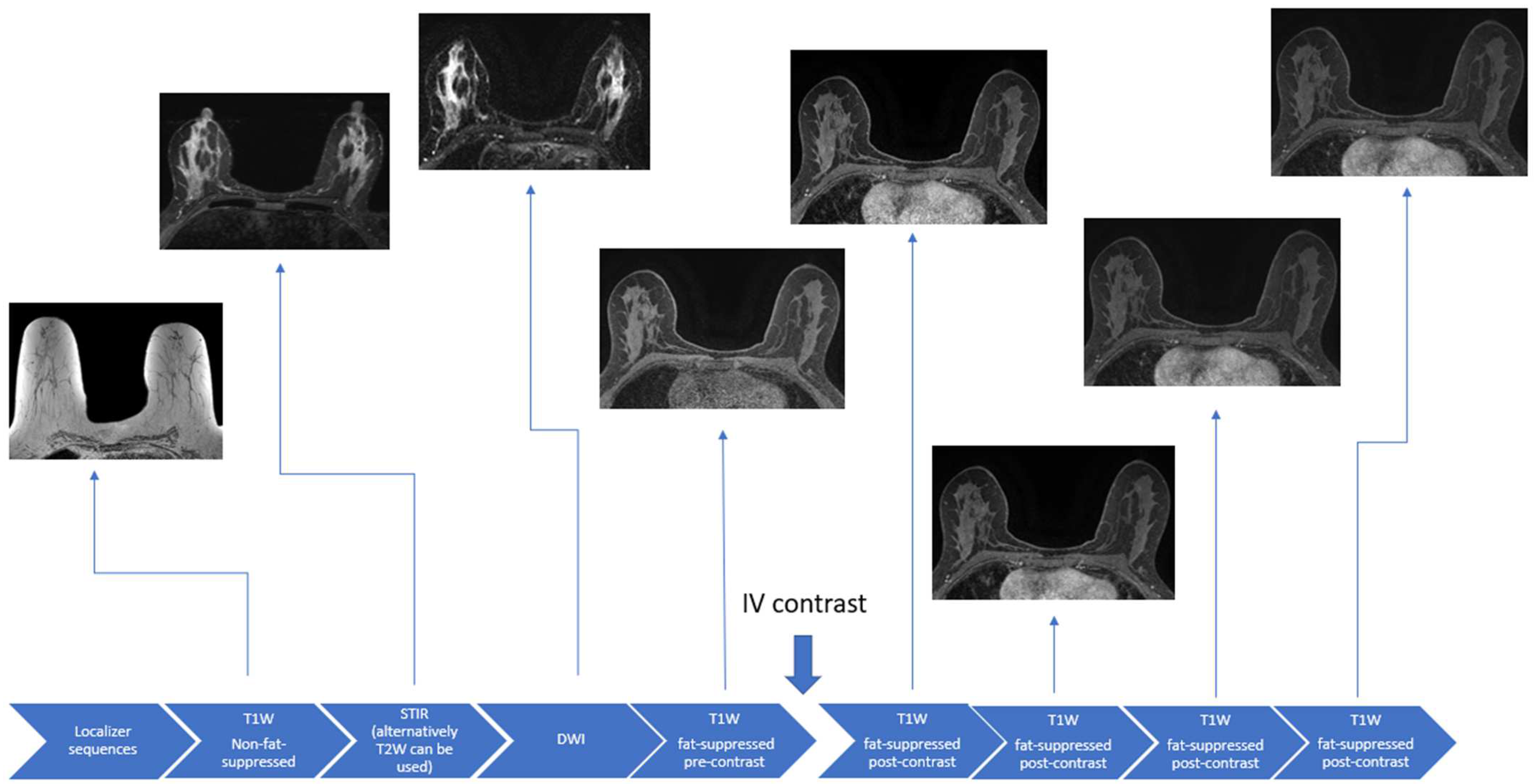
2.1. Breast MRI Protocol and Positioning
2.2. Primary Sequences for Breast MRI
2.2.1. T1-Weighted Sequences
2.2.2. T2-Weighted or STIR Sequences
2.2.3. Diffusion Weighted Imaging (DWI)
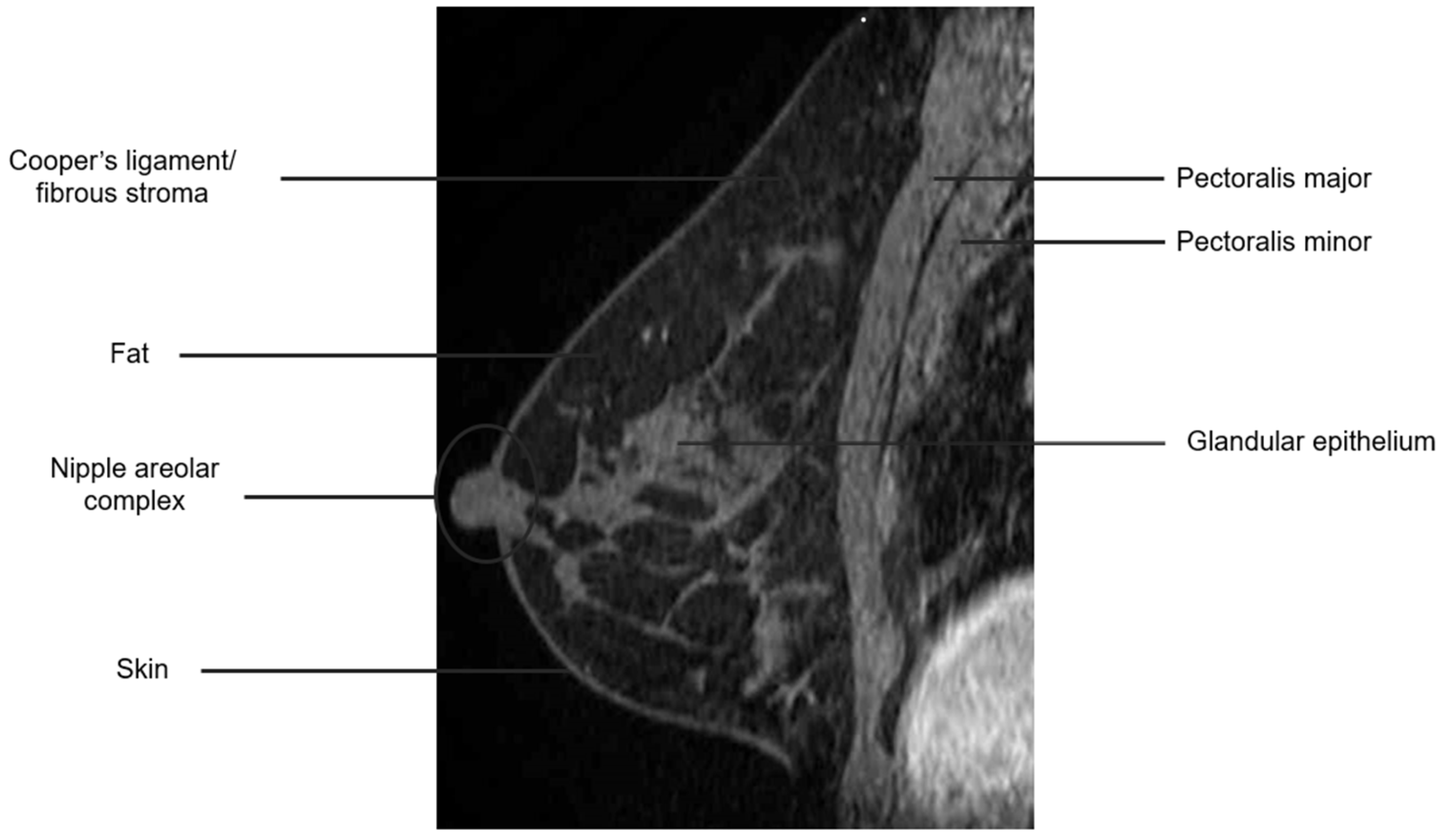
2.3. Breast Anatomy with MRI
2.4. Findings in the Breast on MRI
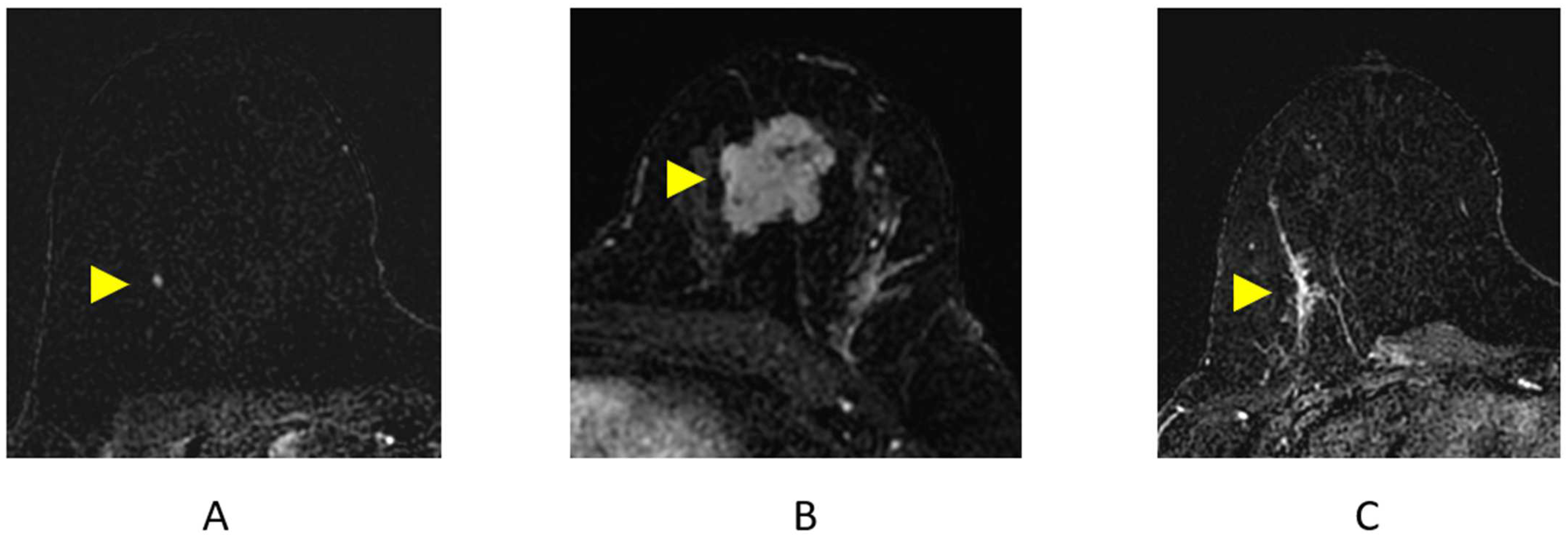
2.4.1. Focus
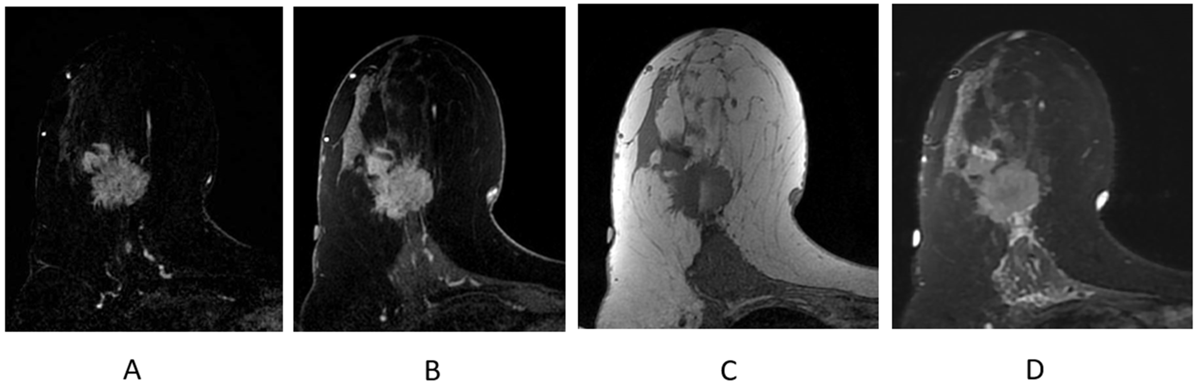
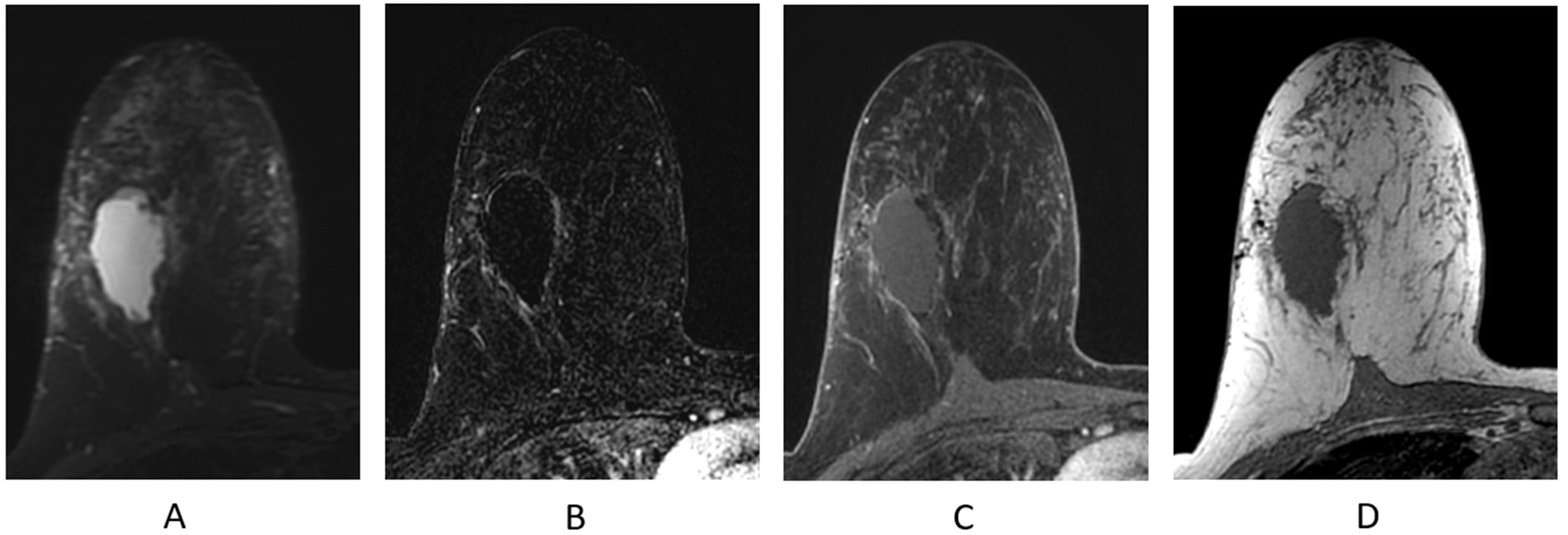
2.4.2. Mass (Figure 7B)
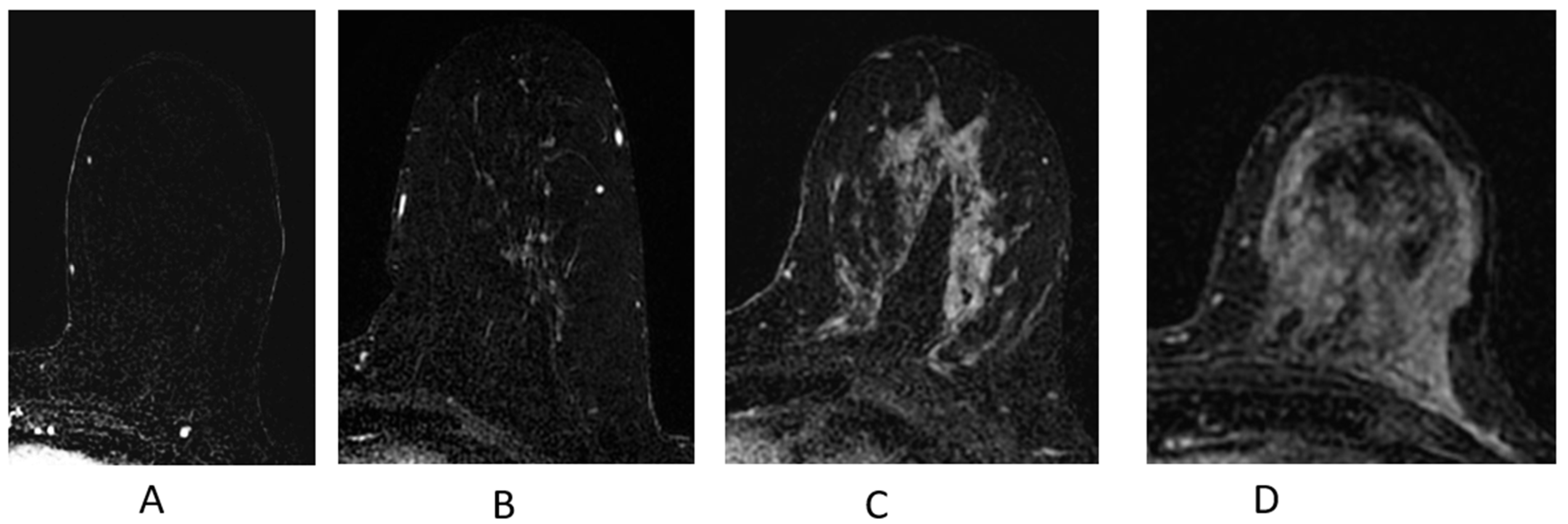
2.4.3. Non-Mass Enhancement (NME)
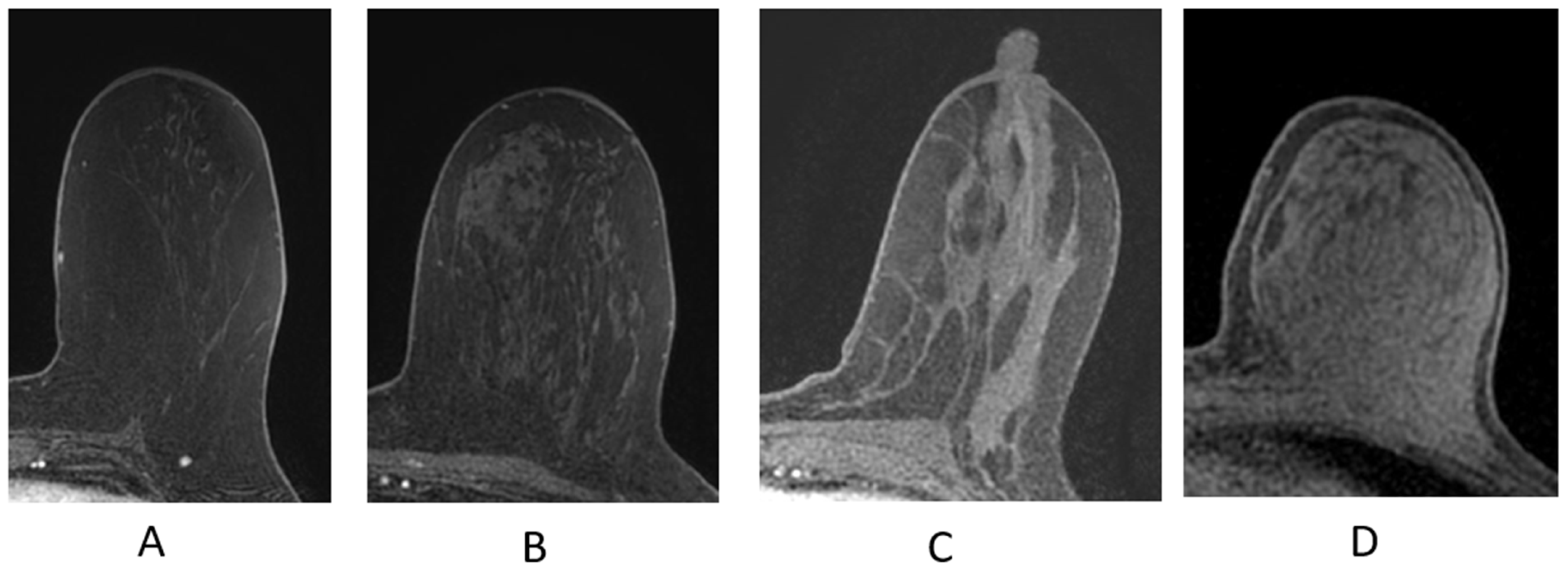
2.4.4. Background Parenchymal Enhancement (BPE)
2.4.5. Kinetic Curve Assessment
2.4.6. Identifying Cancers
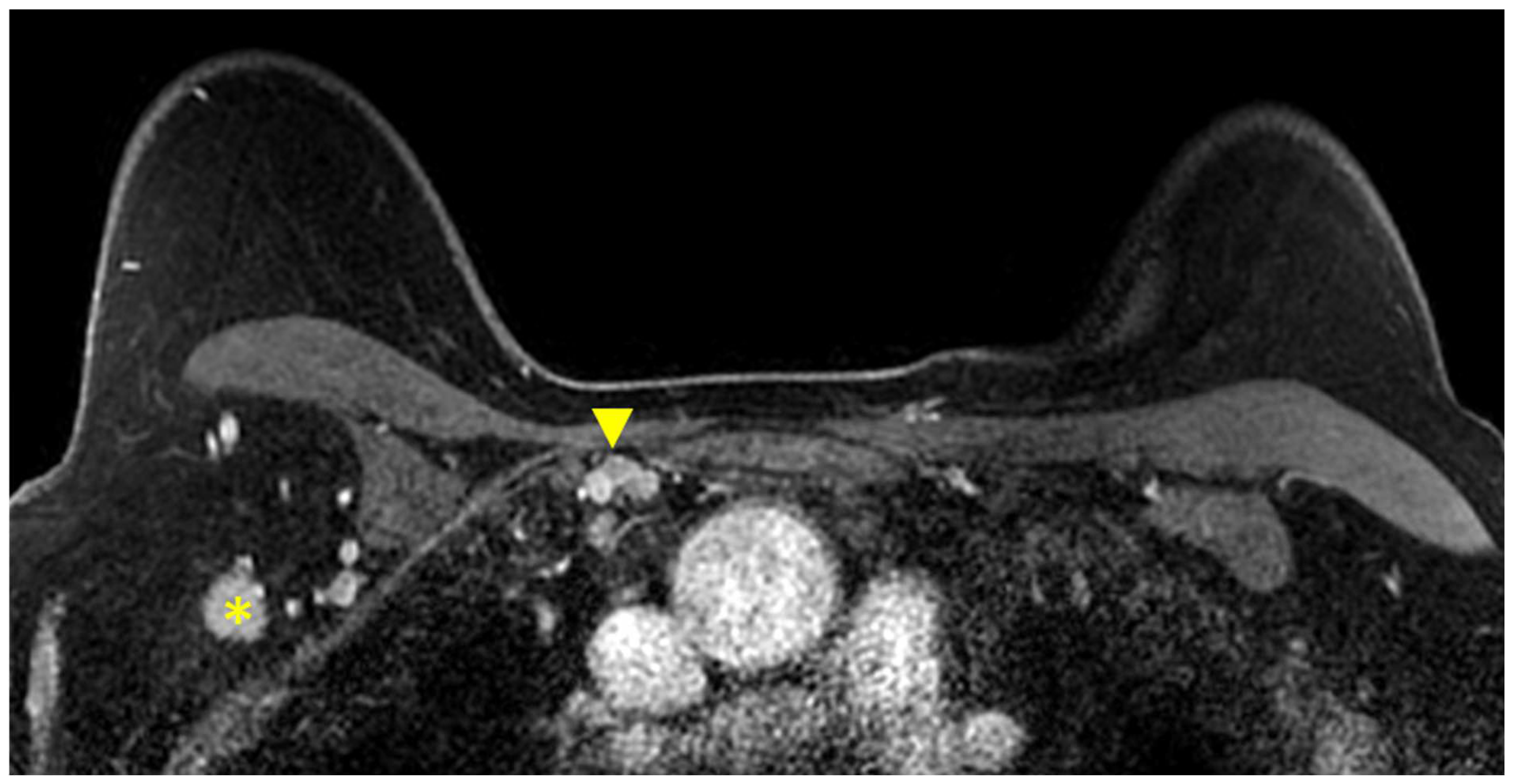
2.4.7. Identifying Lymph Nodes
2.5. Breast Imaging Reporting and Data System
3. Clinical Utility of MRI for Breast Cancer
3.1. Identification of Cancer in the Breast and Axilla on MRI
3.1.1. Tumor Size Estimation and Factors Contributing to MRI-Pathology Concordance
3.1.2. Evaluation of the Pectoralis Musculature, Chest Wall, and Nipple–Areolar Complex
3.1.3. Multifocal/Multicentric Disease
3.1.4. Contralateral Disease
3.1.5. Evaluation of Axillary Lymph Nodes
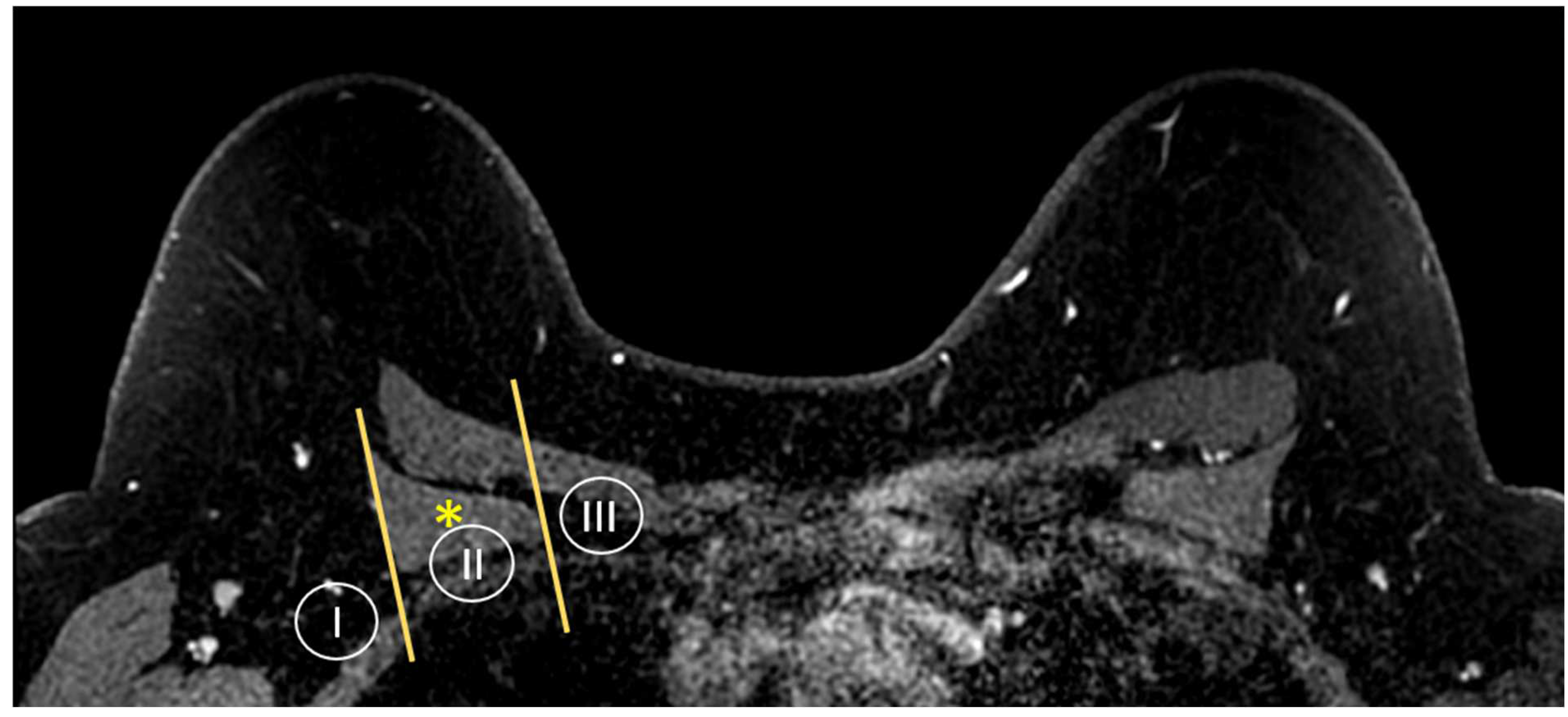
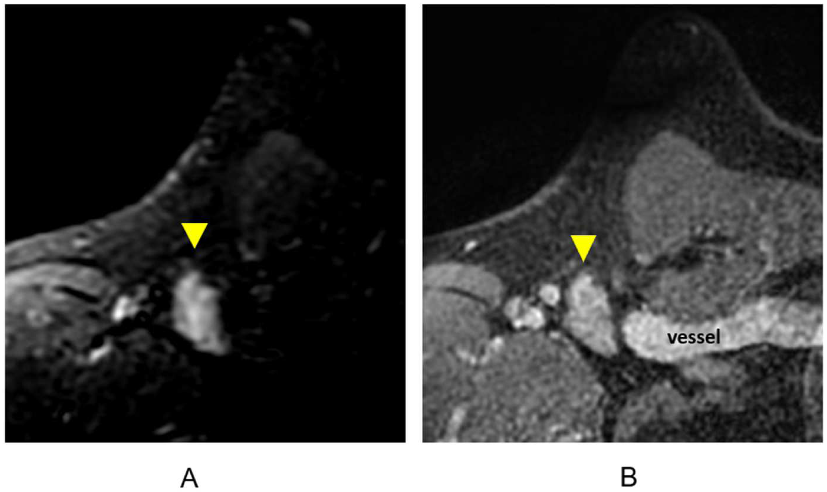
3.1.6. Evaluation of Internal Mammary Lymph Nodes (IMLNs)
3.2. The Impact of MRI on Decisions for Breast Conservation Therapy
3.2.1. Breast Conservation Therapy
3.2.2. The Use of MRI for Preoperative Staging and Surgical Planning
3.2.3. Assessment of Tumor Response after Neoadjuvant Therapy
3.2.4. MR Imaging Response by Molecular Subtype
3.2.5. MR Imaging Response by Imaging Phenotype
3.2.6. MRI in Assessing Nodal Response to NAT
3.2.7. Systemic Therapy
3.2.8. MRI vs. FDG PET/CT in Assessment of NAT Response
3.3. The Role of MRI in Assisting with APBI Candidacy
MRI-Based Eligibility for APBI
3.4. Use of MRI for Post Breast Conservation Therapy Evaluation

| Strengths | Limitations |
|---|---|
| High sensitivity [5,6,7,8,9,10] | Cost |
| Tumor size estimation [55,56,57,58,59] | Accessibility |
| Defining extent of disease | Claustophobia |
| Pectoralis major/minor invasion [64,65] | IV gadolinium contrast allergy (rare) |
| Invasion into the nipple–areolar complex [66] | Presence of a MRI incompatible implantable device |
| Multicentric/multifocal disease [68] | |
| Mammographically occult disease in contralateral breast [17,69,70,71,72,73,74] | |
| Metastatic involvement of axillary or IMN nodes [36,52,77,78,79,91,96,97] | |
| Tumor response to NAT [57,58,59,70,120,121,122,123,127,128,129,130] | |
| Post treatment changes in the breast | |
| Non-ionizing radiation |
4. Breast MRI for Radiation Treatment Planning
4.1. Identification and Sequences for Cavity and Clips
4.1.1. Identifying Seromas and Surgical Cavities
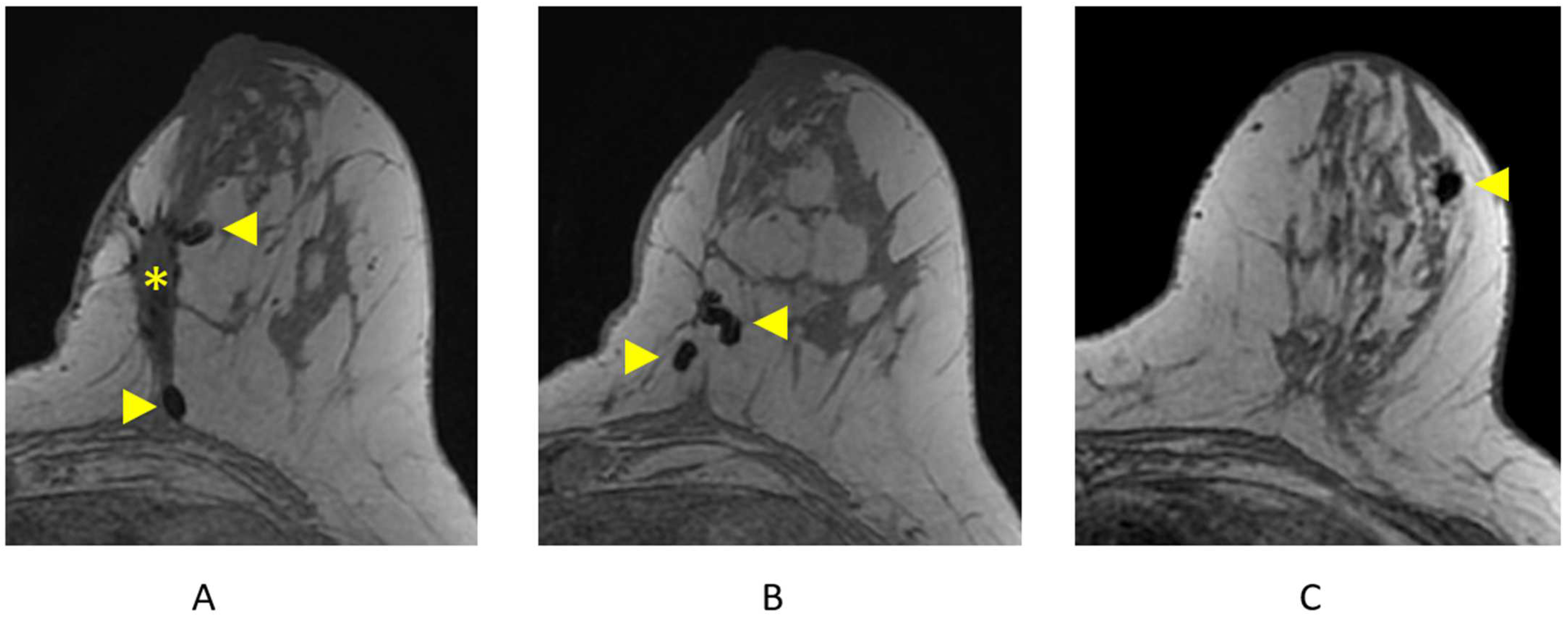
4.1.2. Identifying Surgical/Biopsy Clips
4.2. Treatment Planning Position and Co-Registration
4.2.1. CT Simulation Position
4.2.2. CT-MRI Co-Registration
4.3. Radiation Therapy Target Volumes
4.3.1. Lumpectomy Cavity
4.3.2. CTV and PTV Expansions
4.3.3. The Impact of MRI on Whole-Breast Radiotherapy Planning
4.3.4. Preoperative APBI
5. MRI-Guided Breast Radiotherapy
5.1. MRI-Linac Systems
5.2. Implications of Intrafraction Monitoring
5.3. Dosimetric Impact of the Magnetic Field on Breast Radiotherapy
5.4. Patient Position Considerations
6. Conclusions and Future Directions
Author Contributions
Funding
Conflicts of Interest
Abbreviations
| ACR | American College of Radiology |
| ADC | apparent diffusion coefficient |
| AJCC | American Joint Committee on Cancer |
| APBI | accelerated partial breast irradiation |
| BCS | breast conserving surgery |
| BCT | breast conservation therapy |
| BI-RADS | Breast Imaging Reporting and Data System |
| BPE | background parenchymal enhancement |
| CTV | clinical target volume |
| DCE | dynamic contrast-enhanced |
| DCIS | ductal carcinoma in situ |
| DWI | diffusion weighted imaging |
| ERE | electron return effect |
| ESE | electron stream effect |
| FDG | fludeoxyglucose |
| FGT | fibroglandular tissue |
| GRE | gradient echo sequence |
| ILC | invasive lobular carcinoma |
| IMLN | internal mammary lymph node |
| IOV | interobserver variability |
| LC | lumpectomy cavity |
| LR | local recurrence |
| MBD | mammographic breast density |
| MIP | maximum intensity projection |
| MRI | Magnetic resonance imaging |
| MRL | MR-linac |
| NAC | neoadjuvant chemotherapy |
| NME | non-mass enhancement |
| NPV | negative predictive value |
| OAR | organ at risk |
| pCR | pathologic complete response |
| PET/CT | positron emission tomography–computed tomography |
| PPV | positive predictive value |
| PTV | planning target volume |
| RECIST | response evaluation criteria in solid tumors |
| RFS | recurrence-free survival |
| SBRT/SABR | stereotactic body radiotherapy |
| SNR | signal-to-noise ratio |
| STIR | Short-tau Inversion Recovery |
| T | Tesla |
| US | Ultrasound |
| WLE | wide local excision |
References
- American Cancer Society. American Cancer Society: Cancer Facts & Figures 2019; American Cancer Society: Atlanta, GA, USA, 2019. [Google Scholar]
- Oeffinger, K.C.; Fontham, E.T.H.; Etzioni, R.; Herzig, A.; Michaelson, J.S.; Shih, Y.-C.T.; Walter, L.C.; Church, T.R.; Flowers, C.R.; LaMonte, S.J.; et al. Breast Cancer Screening for Women at Average Risk: 2015 Guideline Update From the American Cancer Society. JAMA 2015, 314, 1599–1614. [Google Scholar] [CrossRef] [PubMed]
- Pisano, E.D.; Hendrick, R.E.; Yaffe, M.J.; Baum, J.K.; Acharyya, S.; Cormack, J.B.; Hanna, L.A.; Conant, E.F.; Fajardo, L.L.; Bassett, L.W.; et al. Diagnostic accuracy of digital versus film mammography: Exploratory analysis of selected population subgroups in DMIST. Radiology 2008, 246, 376–383. [Google Scholar] [CrossRef] [PubMed]
- Berg, W.A.; Zhang, Z.; Lehrer, D.; Jong, R.A.; Pisano, E.D.; Barr, R.G.; Bohm-Velez, M.; Mahoney, M.C.; Evans, W.P., 3rd; Larsen, L.H.; et al. Detection of breast cancer with addition of annual screening ultrasound or a single screening MRI to mammography in women with elevated breast cancer risk. JAMA 2012, 307, 1394–1404. [Google Scholar] [CrossRef] [PubMed]
- Sardanelli, F.; Podo, F.; Santoro, F.; Manoukian, S.; Bergonzi, S.; Trecate, G.; Vergnaghi, D.; Federico, M.; Cortesi, L.; Corcione, S.; et al. Multicenter surveillance of women at high genetic breast cancer risk using mammography, ultrasonography, and contrast-enhanced magnetic resonance imaging (the high breast cancer risk italian 1 study): Final results. Investig. Radiol. 2011, 46, 94–105. [Google Scholar] [CrossRef] [PubMed]
- Riedl, C.C.; Luft, N.; Bernhart, C.; Weber, M.; Bernathova, M.; Tea, M.K.; Rudas, M.; Singer, C.F.; Helbich, T.H. Triple-modality screening trial for familial breast cancer underlines the importance of magnetic resonance imaging and questions the role of mammography and ultrasound regardless of patient mutation status, age, and breast density. J. Clin. Oncol. 2015, 33, 1128–1135. [Google Scholar] [CrossRef] [PubMed]
- Kuhl, C.K.; Schrading, S.; Leutner, C.C.; Morakkabati-Spitz, N.; Wardelmann, E.; Fimmers, R.; Kuhn, W.; Schild, H.H. Mammography, breast ultrasound, and magnetic resonance imaging for surveillance of women at high familial risk for breast cancer. J. Clin. Oncol. 2005, 23, 8469–8476. [Google Scholar] [CrossRef] [PubMed]
- Kuhl, C.; Weigel, S.; Schrading, S.; Arand, B.; Bieling, H.; Konig, R.; Tombach, B.; Leutner, C.; Rieber-Brambs, A.; Nordhoff, D.; et al. Prospective multicenter cohort study to refine management recommendations for women at elevated familial risk of breast cancer: The EVA trial. J. Clin. Oncol. 2010, 28, 1450–1457. [Google Scholar] [CrossRef] [PubMed]
- Lehman, C.D.; Blume, J.D.; Weatherall, P.; Thickman, D.; Hylton, N.; Warner, E.; Pisano, E.; Schnitt, S.J.; Gatsonis, C.; Schnall, M.; et al. Screening women at high risk for breast cancer with mammography and magnetic resonance imaging. Cancer 2005, 103, 1898–1905. [Google Scholar] [CrossRef] [PubMed]
- Morris, E.A.; Liberman, L.; Ballon, D.J.; Robson, M.; Abramson, A.F.; Heerdt, A.; Dershaw, D.D. MRI of occult breast carcinoma in a high-risk population. AJR Am. J. Roentgenol. 2003, 181, 619–626. [Google Scholar] [CrossRef]
- Kriege, M.; Brekelmans, C.T.; Boetes, C.; Besnard, P.E.; Zonderland, H.M.; Obdeijn, I.M.; Manoliu, R.A.; Kok, T.; Peterse, H.; Tilanus-Linthorst, M.M.; et al. Efficacy of MRI and mammography for breast-cancer screening in women with a familial or genetic predisposition. N. Engl. J. Med. 2004, 351, 427–437. [Google Scholar] [CrossRef]
- Leach, M.O.; Boggis, C.R.; Dixon, A.K.; Easton, D.F.; Eeles, R.A.; Evans, D.G.; Gilbert, F.J.; Griebsch, I.; Hoff, R.J.; Kessar, P.; et al. Screening with magnetic resonance imaging and mammography of a UK population at high familial risk of breast cancer: A prospective multicentre cohort study (MARIBS). Lancet 2005, 365, 1769–1778. [Google Scholar] [CrossRef]
- Warner, E.; Plewes, D.B.; Hill, K.A.; Causer, P.A.; Zubovits, J.T.; Jong, R.A.; Cutrara, M.R.; DeBoer, G.; Yaffe, M.J.; Messner, S.J.; et al. Surveillance of BRCA1 and BRCA2 mutation carriers with magnetic resonance imaging, ultrasound, mammography, and clinical breast examination. JAMA 2004, 292, 1317–1325. [Google Scholar] [CrossRef]
- Weinstein, S.P.; Localio, A.R.; Conant, E.F.; Rosen, M.; Thomas, K.M.; Schnall, M.D. Multimodality screening of high-risk women: A prospective cohort study. J. Clin. Oncol. 2009, 27, 6124–6128. [Google Scholar] [CrossRef] [PubMed]
- Lehman, C.D.; Isaacs, C.; Schnall, M.D.; Pisano, E.D.; Ascher, S.M.; Weatherall, P.T.; Bluemke, D.A.; Bowen, D.J.; Marcom, P.K.; Armstrong, D.K.; et al. Cancer yield of mammography, MR, and US in high-risk women: Prospective multi-institution breast cancer screening study. Radiology 2007, 244, 381–388. [Google Scholar] [CrossRef] [PubMed]
- Raikhlin, A.; Curpen, B.; Warner, E.; Betel, C.; Wright, B.; Jong, R. Breast MRI as an adjunct to mammography for breast cancer screening in high-risk patients: Retrospective review. AJR Am. J. Roentgenol. 2015, 204, 889–897. [Google Scholar] [CrossRef]
- Lehman, C.D.; Gatsonis, C.; Kuhl, C.K.; Hendrick, R.E.; Pisano, E.D.; Hanna, L.; Peacock, S.; Smazal, S.F.; Maki, D.D.; Julian, T.B.; et al. MRI evaluation of the contralateral breast in women with recently diagnosed breast cancer. N. Engl. J. Med. 2007, 356, 1295–1303. [Google Scholar] [CrossRef]
- Niell, B.L.; Gavenonis, S.C.; Motazedi, T.; Chubiz, J.C.; Halpern, E.P.; Rafferty, E.A.; Lee, J.M. Auditing a breast MRI practice: Performance measures for screening and diagnostic breast MRI. J. Am. Coll. Radiol. 2014, 11, 883–889. [Google Scholar] [CrossRef]
- Kuhl, C.K.; Keulers, A.; Strobel, K.; Schneider, H.; Gaisa, N.; Schrading, S. Not all false positive diagnoses are equal: On the prognostic implications of false-positive diagnoses made in breast MRI versus in mammography/digital tomosynthesis screening. Breast Cancer Res. 2018, 20, 13. [Google Scholar] [CrossRef]
- Sung, J.S.; Stamler, S.; Brooks, J.; Kaplan, J.; Huang, T.; Dershaw, D.D.; Lee, C.H.; Morris, E.A.; Comstock, C.E. Breast Cancers Detected at Screening MR Imaging and Mammography in Patients at High Risk: Method of Detection Reflects Tumor Histopathologic Results. Radiology 2016, 280, 716–722. [Google Scholar] [CrossRef]
- Kuhl, C.K.; Strobel, K.; Bieling, H.; Leutner, C.; Schild, H.H.; Schrading, S. Supplemental Breast MR Imaging Screening of Women with Average Risk of Breast Cancer. Radiology 2017, 283, 361–370. [Google Scholar] [CrossRef]
- Turnbull, L.; Brown, S.; Harvey, I.; Olivier, C.; Drew, P.; Napp, V.; Hanby, A.; Brown, J. Comparative effectiveness of MRI in breast cancer (COMICE) trial: A randomised controlled trial. Lancet 2010, 375, 563–571. [Google Scholar] [CrossRef] [PubMed]
- Houssami, N.; Turner, R.; Morrow, M. Preoperative Magnetic Resonance Imaging in Breast Cancer: Meta-Analysis of Surgical Outcomes. Ann. Surg. 2013, 257, 249–255. [Google Scholar] [CrossRef] [PubMed]
- Mann, R.M.; Cho, N.; Moy, L. Breast MRI: State of the Art. Radiology 2019, 292, 520–536. [Google Scholar] [CrossRef] [PubMed]
- Siegler, P.; Holloway, C.M.; Causer, P.; Thevathasan, G.; Plewes, D.B. Supine breast MRI. J. Magn. Reson. Imaging 2011, 34, 1212–1217. [Google Scholar] [CrossRef]
- Kuhl, C.K.; Schrading, S.; Strobel, K.; Schild, H.H.; Hilgers, R.D.; Bieling, H.B. Abbreviated breast magnetic resonance imaging (MRI): First postcontrast subtracted images and maximum-intensity projection-a novel approach to breast cancer screening with MRI. J. Clin. Oncol. 2014, 32, 2304–2310. [Google Scholar] [CrossRef] [PubMed]
- Grimm, L.E.M.; Ghate, S. Solitary, Well-Circumscribed, T2 Hyperintense Masses on MRI Have Very Low Malignancy Rates. J. Breast Imaging 2019, 1, 37–42. [Google Scholar] [CrossRef] [PubMed]
- Ballesio, L.; Savelli, S.; Angeletti, M.; Porfiri, L.M.; D’Ambrosio, I.; Maggi, C.; Castro, E.D.; Bennati, P.; Fanelli, G.P.; Vestri, A.R.; et al. Breast MRI: Are T2 IR sequences useful in the evaluation of breast lesions? Eur. J. Radiol. 2009, 71, 96–101. [Google Scholar] [CrossRef] [PubMed]
- Arponen, O.; Masarwah, A.; Sutela, A.; Taina, M.; Kononen, M.; Sironen, R.; Hakumaki, J.; Vanninen, R.; Sudah, M. Incidentally detected enhancing lesions found in breast MRI: Analysis of apparent diffusion coefficient and T2 signal intensity significantly improves specificity. Eur. Radiol. 2016, 26, 4361–4370. [Google Scholar] [CrossRef] [PubMed]
- Cheon, H.; Kim, H.J.; Kim, T.H.; Ryeom, H.K.; Lee, J.; Kim, G.C.; Yuk, J.S.; Kim, W.H. Invasive Breast Cancer: Prognostic Value of Peritumoral Edema Identified at Preoperative MR Imaging. Radiology 2018, 287, 68–75. [Google Scholar] [CrossRef]
- Uematsu, T.; Kasami, M.; Watanabe, J. Can T2-weighted 3-T breast MRI predict clinically occult inflammatory breast cancer before pathological examination? A single-center experience. Breast Cancer 2014, 21, 115–121. [Google Scholar] [CrossRef]
- Uematsu, T.; Kasami, M.; Watanabe, J. Is evaluation of the presence of prepectoral edema on T2-weighted with fat-suppression 3 T breast MRI a simple and readily available noninvasive technique for estimation of prognosis in patients with breast cancer? Breast Cancer 2014, 21, 684–692. [Google Scholar] [CrossRef] [PubMed]
- Yadav, P.; Chauhan, S. Effectivity of combined diffusion-weighted imaging and contrast-enhanced MRI in malignant and benign breast lesions. Pol. J. Radiol. 2018, 83, e82–e93. [Google Scholar] [CrossRef] [PubMed]
- Baltzer, P.; Mann, R.M.; Iima, M.; Sigmund, E.E.; Clauser, P.; Gilbert, F.J.; Martincich, L.; Partridge, S.C.; Patterson, A.; Pinker, K.; et al. Diffusion-weighted imaging of the breast-a consensus and mission statement from the EUSOBI International Breast Diffusion-Weighted Imaging working group. Eur. Radiol. 2019, 30, 1436–1450. [Google Scholar] [CrossRef]
- Morris, E.A.; Comstock, C.E.; Lee, C.H. ACR BI-RADS® Magnetic Resonance Imaging. In ACR BI-RADS® Atlas, Breast Imaging Reporting and Data System; American College of Radiology: Reston, VA, USA, 2013. [Google Scholar]
- Ecanow, J.S.; Abe, H.; Newstead, G.M.; Ecanow, D.B.; Jeske, J.M. Axillary staging of breast cancer: What the radiologist should know. RadioGraphics 2013, 33, 1589–1612. [Google Scholar] [CrossRef] [PubMed]
- Ha, R.; Sung, J.; Lee, C.; Comstock, C.; Wynn, R.; Morris, E. Characteristics and outcome of enhancing foci followed on breast MRI with management implications. Clin. Radiol. 2014, 69, 715–720. [Google Scholar] [CrossRef] [PubMed]
- Liberman, L.; Morris, E.A.; Lee, M.J.; Kaplan, J.B.; LaTrenta, L.R.; Menell, J.H.; Abramson, A.F.; Dashnaw, S.M.; Ballon, D.J.; Dershaw, D.D. Breast lesions detected on MR imaging: Features and positive predictive value. AJR Am. J. Roentgenol. 2002, 179, 171–178. [Google Scholar] [CrossRef] [PubMed]
- Baltzer, P.A.; Benndorf, M.; Dietzel, M.; Gajda, M.; Runnebaum, I.B.; Kaiser, W.A. False-positive findings at contrast-enhanced breast MRI: A BI-RADS descriptor study. AJR Am. J. Roentgenol. 2010, 194, 1658–1663. [Google Scholar] [CrossRef] [PubMed]
- Nunes, L.W.; Schnall, M.D.; Orel, S.G. Update of breast MR imaging architectural interpretation model. Radiology 2001, 219, 484–494. [Google Scholar] [CrossRef]
- Giess, C.S.; Raza, S.; Birdwell, R.L. Patterns of nonmasslike enhancement at screening breast MR imaging of high-risk premenopausal women. RadioGraphics 2013, 33, 1343–1360. [Google Scholar] [CrossRef]
- Gity, M.; Ghazi Moghadam, K.; Jalali, A.H.; Shakiba, M. Association of Different MRI BIRADS Descriptors With Malignancy in Non Mass-Like Breast Lesions. Iran. Red. Crescent Med. J. 2014, 16, e26040. [Google Scholar] [CrossRef]
- Tozaki, M.; Fukuda, K. High-spatial-resolution MRI of non-masslike breast lesions: Interpretation model based on BI-RADS MRI descriptors. AJR Am. J. Roentgenol. 2006, 187, 330–337. [Google Scholar] [CrossRef] [PubMed]
- Uematsu, T.; Kasami, M. High-spatial-resolution 3-T breast MRI of nonmasslike enhancement lesions: An analysis of their features as significant predictors of malignancy. AJR Am. J. Roentgenol. 2012, 198, 1223–1230. [Google Scholar] [CrossRef] [PubMed]
- Tozaki, M.; Igarashi, T.; Fukuda, K. Breast MRI using the VIBE sequence: Clustered ring enhancement in the differential diagnosis of lesions showing non-masslike enhancement. AJR Am. J. Roentgenol. 2006, 187, 313–321. [Google Scholar] [CrossRef] [PubMed]
- Giess, C.S.; Yeh, E.D.; Raza, S.; Birdwell, R.L. Background parenchymal enhancement at breast MR imaging: Normal patterns, diagnostic challenges, and potential for false-positive and false-negative interpretation. RadioGraphics 2014, 34, 234–247. [Google Scholar] [CrossRef] [PubMed]
- Li, J.; Dershaw, D.D.; Lee, C.H.; Joo, S.; Morris, E.A. Breast MRI after conservation therapy: Usual findings in routine follow-up examinations. AJR Am. J. Roentgenol. 2010, 195, 799–807. [Google Scholar] [CrossRef] [PubMed]
- Baek, J.E.; Kim, S.H.; Lee, A.W. Background parenchymal enhancement in breast MRIs of breast cancer patients: Impact on tumor size estimation. Eur. J. Radiol. 2014, 83, 1356–1362. [Google Scholar] [CrossRef] [PubMed]
- Preibsch, H.; Beckmann, J.; Pawlowski, J.; Kloth, C.; Hahn, M.; Staebler, A.; Wietek, B.M.; Nikolaou, K.; Wiesinger, B. Accuracy of Breast Magnetic Resonance Imaging Compared to Mammography in the Preoperative Detection and Measurement of Pure Ductal Carcinoma In Situ: A Retrospective Analysis. Acad. Radiol. 2019, 26, 760–765. [Google Scholar] [CrossRef] [PubMed]
- Schnall, M.D.; Rosten, S.; Englander, S.; Orel, S.G.; Nunes, L.W. A combined architectural and kinetic interpretation model for breast MR images. Acad. Radiol. 2001, 8, 591–597. [Google Scholar] [CrossRef] [PubMed]
- Murray, A.D.; Staff, R.T.; Redpath, T.W.; Gilbert, F.J.; Ah-See, A.K.; Brookes, J.A.; Miller, I.D.; Payne, S. Dynamic contrast enhanced MRI of the axilla in women with breast cancer: Comparison with pathology of excised nodes. Br. J. Radiol. 2002, 75, 220–228. [Google Scholar] [CrossRef]
- Mortellaro, V.E.; Marshall, J.; Singer, L.; Hochwald, S.N.; Chang, M.; Copeland, E.M.; Grobmyer, S.R. Magnetic resonance imaging for axillary staging in patients with breast cancer. J. Magn. Reson. Imaging 2009, 30, 309–312. [Google Scholar] [CrossRef]
- Kvistad, K.A.; Rydland, J.; Smethurst, H.B.; Lundgren, S.; Fjosne, H.E.; Haraldseth, O. Axillary lymph node metastases in breast cancer: Preoperative detection with dynamic contrast-enhanced MRI. Eur. Radiol. 2000, 10, 1464–1471. [Google Scholar] [CrossRef]
- D’Orsi, C.J.; Sickles, E.A.; Mendelson, E.B.; Morris, E.A. ACR BI-RADS® Atlas, Breast Imaging Re-porting and Data System; American College of Radiology: Reston, VA, USA, 2013. [Google Scholar]
- Berg, W.A.; Gutierrez, L.; NessAiver, M.S.; Carter, W.B.; Bhargavan, M.; Lewis, R.S.; Ioffe, O.B. Diagnostic accuracy of mammography, clinical examination, US, and MR imaging in preoperative assessment of breast cancer. Radiology 2004, 233, 830–849. [Google Scholar] [CrossRef]
- Boetes, C.; Mus, R.D.; Holland, R.; Barentsz, J.O.; Strijk, S.P.; Wobbes, T.; Hendriks, J.H.; Ruys, S.H. Breast tumors: Comparative accuracy of MR imaging relative to mammography and US for demonstrating extent. Radiology 1995, 197, 743–747. [Google Scholar] [CrossRef]
- Lobbes, M.B.; Prevos, R.; Smidt, M.; Tjan-Heijnen, V.C.; van Goethem, M.; Schipper, R.; Beets-Tan, R.G.; Wildberger, J.E. The role of magnetic resonance imaging in assessing residual disease and pathologic complete response in breast cancer patients receiving neoadjuvant chemotherapy: A systematic review. Insights Imaging 2013, 4, 163–175. [Google Scholar] [CrossRef]
- Marinovich, M.L.; Houssami, N.; Macaskill, P.; Sardanelli, F.; Irwig, L.; Mamounas, E.P.; von Minckwitz, G.; Brennan, M.E.; Ciatto, S. Meta-analysis of magnetic resonance imaging in detecting residual breast cancer after neoadjuvant therapy. J. Natl. Cancer Inst. 2013, 105, 321–333. [Google Scholar] [CrossRef]
- Yeh, E.; Slanetz, P.; Kopans, D.B.; Rafferty, E.; Georgian-Smith, D.; Moy, L.; Halpern, E.; Moore, R.; Kuter, I.; Taghian, A. Prospective comparison of mammography, sonography, and MRI in patients undergoing neoadjuvant chemotherapy for palpable breast cancer. AJR Am. J. Roentgenol. 2005, 184, 868–877. [Google Scholar] [CrossRef]
- Jethava, A.; Ali, S.; Wakefield, D.; Crowell, R.; Sporn, J.; Vrendenburgh, J. Diagnostic Accuracy of MRI in Predicting Breast Tumor Size: Comparative Analysis of MRI vs Histopathological Assessed Breast Tumor Size. Conn. Med. 2015, 79, 261–267. [Google Scholar]
- Mennella, S.; Garlaschi, A.; Paparo, F.; Perillo, M.; Celenza, M.; Massa, T.; Rollandi, G.A.; Garlaschi, G. Magnetic resonance imaging of breast cancer: Factors affecting the accuracy of preoperative lesion sizing. Acta Radiol. 2015, 56, 260–268. [Google Scholar] [CrossRef] [PubMed]
- Rominger, M.; Berg, D.; Frauenfelder, T.; Ramaswamy, A.; Timmesfeld, N. Which factors influence MRI-pathology concordance of tumour size measurements in breast cancer? Eur. Radiol. 2016, 26, 1457–1465. [Google Scholar] [CrossRef] [PubMed]
- Expert Panel on Breast Imaging; Slanetz, P.J.; Moy, L.; Baron, P.; di Florio, R.M.; Green, E.D.; Heller, S.L.; Holbrook, A.I.; Lee, S.J.; Lewin, A.A.; et al. ACR Appropriateness Criteria((R)) Monitoring Response to Neoadjuvant Systemic Therapy for Breast Cancer. J. Am. Coll. Radiol. 2017, 14, S462–S475. [Google Scholar] [CrossRef]
- Kazama, T.; Nakamura, S.; Doi, O.; Suzuki, K.; Hirose, M.; Ito, H. Prospective evaluation of pectoralis muscle invasion of breast cancer by MR imaging. Breast Cancer 2005, 12, 312–316. [Google Scholar] [CrossRef]
- Morris, E.A.; Schwartz, L.H.; Drotman, M.B.; Kim, S.J.; Tan, L.K.; Liberman, L.; Abramson, A.F.; Van Zee, K.J.; Dershaw, D.D. Evaluation of pectoralis major muscle in patients with posterior breast tumors on breast MR images: Early experience. Radiology 2000, 214, 67–72. [Google Scholar] [CrossRef]
- Moon, J.Y.; Chang, Y.W.; Lee, E.H.; Seo, D.Y. Malignant invasion of the nipple-areolar complex of the breast: Usefulness of breast MRI. AJR Am. J. Roentgenol. 2013, 201, 448–455. [Google Scholar] [CrossRef]
- Tardivon, A.A.; Ollivier, L.; El Khoury, C.; Thibault, F. Monitoring therapeutic efficacy in breast carcinomas. Eur. Radiol. 2006, 16, 2549–2558. [Google Scholar] [CrossRef]
- Houssami, N.; Ciatto, S.; Macaskill, P.; Lord, S.J.; Warren, R.M.; Dixon, J.M.; Irwig, L. Accuracy and surgical impact of magnetic resonance imaging in breast cancer staging: Systematic review and meta-analysis in detection of multifocal and multicentric cancer. J. Clin. Oncol. 2008, 26, 3248–3258. [Google Scholar] [CrossRef]
- Fischer, U.; Kopka, L.; Grabbe, E. Breast carcinoma: Effect of preoperative contrast-enhanced MR imaging on the therapeutic approach. Radiology 1999, 213, 881–888. [Google Scholar] [CrossRef]
- Hollingsworth, A.B.; Stough, R.G.; O’Dell, C.A.; Brekke, C.E. Breast magnetic resonance imaging for preoperative locoregional staging. Am. J. Surg. 2008, 196, 389–397. [Google Scholar] [CrossRef]
- Liberman, L.; Morris, E.A.; Kim, C.M.; Kaplan, J.B.; Abramson, A.F.; Menell, J.H.; Van Zee, K.J.; Dershaw, D.D. MR imaging findings in the contralateral breast of women with recently diagnosed breast cancer. AJR Am. J. Roentgenol. 2003, 180, 333–341. [Google Scholar] [CrossRef]
- Lee, S.G.; Orel, S.G.; Woo, I.J.; Cruz-Jove, E.; Putt, M.E.; Solin, L.J.; Czerniecki, B.J.; Schnall, M.D. MR imaging screening of the contralateral breast in patients with newly diagnosed breast cancer: Preliminary results. Radiology 2003, 226, 773–778. [Google Scholar] [CrossRef]
- Lehman, C.D.; Blume, J.D.; Thickman, D.; Bluemke, D.A.; Pisano, E.; Kuhl, C.; Julian, T.B.; Hylton, N.; Weatherall, P.; O’Loughlin, M.; et al. Added cancer yield of MRI in screening the contralateral breast of women recently diagnosed with breast cancer: Results from the International Breast Magnetic Resonance Consortium (IBMC) trial. J. Surg. Oncol. 2005, 92, 9–15. [Google Scholar] [CrossRef] [PubMed]
- Faermann, R.W.J.; Chepelev, L.; Kendal, W.; Verma, R.; Scott-Moncrieff, A.; Peddle, S.; Doherty, G.; Lau, J.; Ramsay, T.; Arnaout, A.; et al. Outcomes after Surgery for Early Stage Breast Cancer in Women Staged With Preoperative Breast Magnetic Resonance Imaging According to Breast Tissue Density. J. Breast Imaging 2019, 1, 115–121. [Google Scholar] [CrossRef]
- Khan, A.; Sabel, M.S.; Nees, A.; Diehl, K.M.; Cimmino, V.M.; Kleer, C.G.; Schott, A.F.; Hayes, D.F.; Chang, A.E.; Newman, L.A. Comprehensive axillary evaluation in neoadjuvant chemotherapy patients with ultrasonography and sentinel lymph node biopsy. Ann. Surg. Oncol. 2005, 12, 697–704. [Google Scholar] [CrossRef]
- Henry-Tillman, R.; Glover-Collins, K.; Preston, M.; Gallagher, K.; Tummel, E.; Robertson, Y.V.; Ochoa, D.; Korourian, S.; Westbrook, K.; Klimberg, V.S. The SAVE review: Sonographic analysis versus excision for axillary staging in breast cancer. J. Am. Coll. Surg. 2015, 220, 560–567. [Google Scholar] [CrossRef]
- Baltzer, P.A.; Dietzel, M.; Burmeister, H.P.; Zoubi, R.; Gajda, M.; Camara, O.; Kaiser, W.A. Application of MR mammography beyond local staging: Is there a potential to accurately assess axillary lymph nodes? evaluation of an extended protocol in an initial prospective study. AJR Am. J. Roentgenol. 2011, 196, W641–W647. [Google Scholar] [CrossRef]
- Korteweg, M.A.; Zwanenburg, J.J.; Hoogduin, J.M.; van den Bosch, M.A.; van Diest, P.J.; van Hillegersberg, R.; Eijkemans, M.J.; Mali, W.P.; Luijten, P.R.; Veldhuis, W.B. Dissected sentinel lymph nodes of breast cancer patients: Characterization with high-spatial-resolution 7-T MR imaging. Radiology 2011, 261, 127–135. [Google Scholar] [CrossRef]
- Memarsadeghi, M.; Riedl, C.C.; Kaneider, A.; Galid, A.; Rudas, M.; Matzek, W.; Helbich, T.H. Axillary lymph node metastases in patients with breast carcinomas: Assessment with nonenhanced versus uspio-enhanced MR imaging. Radiology 2006, 241, 367–377. [Google Scholar] [CrossRef] [PubMed]
- Abe, H.; Schmidt, R.A.; Kulkarni, K.; Sennett, C.A.; Mueller, J.S.; Newstead, G.M. Axillary lymph nodes suspicious for breast cancer metastasis: Sampling with US-guided 14-gauge core-needle biopsy--clinical experience in 100 patients. Radiology 2009, 250, 41–49. [Google Scholar] [CrossRef]
- Alvarez, S.; Anorbe, E.; Alcorta, P.; Lopez, F.; Alonso, I.; Cortes, J. Role of sonography in the diagnosis of axillary lymph node metastases in breast cancer: A systematic review. AJR Am. J. Roentgenol. 2006, 186, 1342–1348. [Google Scholar] [CrossRef]
- Bedi, D.G.; Krishnamurthy, R.; Krishnamurthy, S.; Edeiken, B.S.; Le-Petross, H.; Fornage, B.D.; Bassett, R.L., Jr.; Hunt, K.K. Cortical morphologic features of axillary lymph nodes as a predictor of metastasis in breast cancer: In vitro sonographic study. AJR Am. J. Roentgenol. 2008, 191, 646–652. [Google Scholar] [CrossRef] [PubMed]
- Neal, C.H.; Daly, C.P.; Nees, A.V.; Helvie, M.A. Can preoperative axillary US help exclude N2 and N3 metastatic breast cancer? Radiology 2010, 257, 335–341. [Google Scholar] [CrossRef] [PubMed]
- Assing, M.A.; Patel, B.K.; Karamsadkar, N.; Weinfurtner, J.; Usmani, O.; Kiluk, J.V.; Drukteinis, J.S. A comparison of the diagnostic accuracy of magnetic resonance imaging to axillary ultrasound in the detection of axillary nodal metastases in newly diagnosed breast cancer. Breast J. 2017, 23, 647–655. [Google Scholar] [CrossRef] [PubMed]
- Almerey, T.; Villacreses, D.; Li, Z.; Patel, B.; McDonough, M.; Gibson, T.; Maimone, S.; Gray, R.; McLaughlin, S.A. Value of Axillary Ultrasound after Negative Axillary MRI for Evaluating Nodal Status in High-Risk Breast Cancer. J. Am. Coll. Surg. 2019, 228, 792–797. [Google Scholar] [CrossRef] [PubMed]
- Lee, S.C.; Jain, P.A.; Jethwa, S.C.; Tripathy, D.; Yamashita, M.W. Radiologist’s role in breast cancer staging: Providing key information for clinicians. RadioGraphics 2014, 34, 330–342. [Google Scholar] [CrossRef] [PubMed]
- Scatarige, J.C.; Boxen, I.; Smathers, R.L. Internal mammary lymphadenopathy: Imaging of a vital lymphatic pathway in breast cancer. RadioGraphics 1990, 10, 857–870. [Google Scholar] [CrossRef] [PubMed]
- Singh, S.; Ramani, S.K.; Rastogi, A.; Thakur, M.H. Incidence of internal mammary node in locally advanced breast cancer and its correlation with metastatic disease: A retrospective observational study. Br. J. Radiol. 2019, 92, 20190098. [Google Scholar] [CrossRef]
- Wang, K.; Zhang, X.; Zheng, K.; Yin, X.D.; Xing, L.; Zhang, A.J.; Shi, Y.; Kong, L.Q.; Li, F.; Ma, B.L.; et al. Predictors of internal mammary lymph nodes (IMLN) metastasis and disease-free survival comparison between IMLN-positive and IMLN-negative breast cancer patients: Results from Western China Clinical Cooperation Group (WCCCG) database (CONSORT). Medicine 2018, 97, e11296. [Google Scholar] [CrossRef]
- Chen, L.; Gu, Y.; Leaw, S.; Wang, Z.; Wang, P.; Hu, X.; Chen, J.; Lu, J.; Shao, Z. Internal mammary lymph node recurrence: Rare but characteristic metastasis site in breast cancer. BMC Cancer 2010, 10, 479. [Google Scholar] [CrossRef]
- Behzadi, S.T.; Moser, R.; Kiesl, S.; Nano, J.; Peeken, J.C.; Fischer, J.C.; Fallenberg, E.M.; Huber, T.; Haller, B.; Klein, E.; et al. Tumor Contact With Internal Mammary Perforator Vessels as Risk Factor for Gross Internal Mammary Lymph Node Involvement in Patients With Breast Cancer. Int. J. Radiat. Oncol. Biol. Phys. 2024in press. [CrossRef] [PubMed]
- Whelan, T.J.; Olivotto, I.A.; Parulekar, W.R.; Ackerman, I.; Chua, B.H.; Nabid, A.; Vallis, K.A.; White, J.R.; Rousseau, P.; Fortin, A.; et al. Regional Nodal Irradiation in Early-Stage Breast Cancer. N. Engl. J. Med. 2015, 373, 307–316. [Google Scholar] [CrossRef]
- Poortmans, P.M.; Collette, S.; Kirkove, C.; Van Limbergen, E.; Budach, V.; Struikmans, H.; Collette, L.; Fourquet, A.; Maingon, P.; Valli, M.; et al. Internal Mammary and Medial Supraclavicular Irradiation in Breast Cancer. N. Engl. J. Med. 2015, 373, 317–327. [Google Scholar] [CrossRef]
- Mack, M.; Chetlen, A.; Liao, J. Incidental Internal Mammary Lymph Nodes Visualized on Screening Breast MRI. AJR Am. J. Roentgenol. 2015, 205, 209–214. [Google Scholar] [CrossRef] [PubMed]
- Ray, K.M.; Munir, R.; Wisner, D.J.; Azziz, A.; Holland, B.C.; Kornak, J.; Joe, B.N. Internal mammary lymph nodes as incidental findings at screening breast MRI. Clin. Imaging 2015, 39, 791–793. [Google Scholar] [CrossRef] [PubMed]
- Patel, S.; Delikat, A.; Liao, J.; Chetlen, A.L. Pre- and post-magnetic resonance imaging features of suspicious internal mammary lymph nodes in breast cancer patients receiving neo-adjuvant therapy: Are any imaging features predictive of malignancy? Breast J. 2018, 24, 997–1000. [Google Scholar] [CrossRef] [PubMed]
- Kinoshita, T.; Odagiri, K.; Andoh, K.; Doiuchi, T.; Sugimura, K.; Shiotani, S.; Asaga, T. Evaluation of small internal mammary lymph node metastases in breast cancer by MRI. Radiat. Med. 1999, 17, 189–193. [Google Scholar] [PubMed]
- Seo, M.J.; Lee, J.J.; Kim, H.O.; Chae, S.Y.; Park, S.H.; Ryu, J.S.; Ahn, S.H.; Lee, J.W.; Son, B.H.; Gong, G.Y.; et al. Detection of internal mammary lymph node metastasis with (18)F-fluorodeoxyglucose positron emission tomography/computed tomography in patients with stage III breast cancer. Eur. J. Nucl. Med. Mol. Imaging 2014, 41, 438–445. [Google Scholar] [CrossRef] [PubMed]
- Eubank, W.B.; Mankoff, D.A.; Takasugi, J.; Vesselle, H.; Eary, J.F.; Shanley, T.J.; Gralow, J.R.; Charlop, A.; Ellis, G.K.; Lindsley, K.L.; et al. 18fluorodeoxyglucose positron emission tomography to detect mediastinal or internal mammary metastases in breast cancer. J. Clin. Oncol. 2001, 19, 3516–3523. [Google Scholar] [CrossRef] [PubMed]
- Segaert, I.; Mottaghy, F.; Ceyssens, S.; De Wever, W.; Stroobants, S.; Van Ongeval, C.; Van Limbergen, E.; Wildiers, H.; Paridaens, R.; Vergote, I.; et al. Additional value of PET-CT in staging of clinical stage IIB and III breast cancer. Breast J. 2010, 16, 617–624. [Google Scholar] [CrossRef] [PubMed]
- An, Y.Y.; Kim, S.H.; Kang, B.J.; Lee, A.W. Comparisons of Positron Emission Tomography/Computed Tomography and Ultrasound Imaging for Detection of Internal Mammary Lymph Node Metastases in Patients With Breast Cancer and Pathologic Correlation by Ultrasound-Guided Biopsy Procedures. J. Ultrasound Med. 2015, 34, 1385–1394. [Google Scholar] [CrossRef] [PubMed]
- Darby, S.; McGale, P.; Correa, C.; Taylor, C.; Arriagada, R.; Clarke, M.; Cutter, D.; Davies, C.; Ewertz, M.; Godwin, J.; et al. Effect of radiotherapy after breast-conserving surgery on 10-year recurrence and 15-year breast cancer death: Meta-analysis of individual patient data for 10,801 women in 17 randomised trials. Lancet 2011, 378, 1707–1716. [Google Scholar] [CrossRef]
- Fisher, B.; Anderson, S.; Bryant, J.; Margolese, R.G.; Deutsch, M.; Fisher, E.R.; Jeong, J.H.; Wolmark, N. Twenty-year follow-up of a randomized trial comparing total mastectomy, lumpectomy, and lumpectomy plus irradiation for the treatment of invasive breast cancer. N. Engl. J. Med. 2002, 347, 1233–1241. [Google Scholar] [CrossRef]
- Veronesi, U.; Cascinelli, N.; Mariani, L.; Greco, M.; Saccozzi, R.; Luini, A.; Aguilar, M.; Marubini, E. Twenty-year follow-up of a randomized study comparing breast-conserving surgery with radical mastectomy for early breast cancer. N. Engl. J. Med. 2002, 347, 1227–1232. [Google Scholar] [CrossRef] [PubMed]
- Almahariq, M.F.; Quinn, T.J.; Siddiqui, Z.; Jawad, M.S.; Chen, P.Y.; Gustafson, G.S.; Dilworth, J.T. Breast conserving therapy is associated with improved overall survival compared to mastectomy in early-stage, lymph node-negative breast cancer. Radiother. Oncol. 2020, 142, 186–194. [Google Scholar] [CrossRef]
- Christiansen, P.; Mele, M.; Bodilsen, A.; Rocco, N.; Zachariae, R. Breast-Conserving Surgery or Mastectomy?: Impact on Survival. Ann. Surg. Open 2022, 3, e205. [Google Scholar] [CrossRef]
- Haviland, J.S.; Owen, J.R.; Dewar, J.A.; Agrawal, R.K.; Barrett, J.; Barrett-Lee, P.J.; Dobbs, H.J.; Hopwood, P.; Lawton, P.A.; Magee, B.J.; et al. The UK Standardisation of Breast Radiotherapy (START) trials of radiotherapy hypofractionation for treatment of early breast cancer: 10-year follow-up results of two randomised controlled trials. Lancet Oncol. 2013, 14, 1086–1094. [Google Scholar] [CrossRef] [PubMed]
- Hahn, C.; Kavanagh, B.; Bhatnagar, A.; Jacobson, G.; Lutz, S.; Patton, C.; Potters, L.; Steinberg, M. Choosing wisely: The American Society for Radiation Oncology’s top 5 list. Pract. Radiat. Oncol. 2014, 4, 349–355. [Google Scholar] [CrossRef]
- Fisher, E.R.; Dignam, J.; Tan-Chiu, E.; Costantino, J.; Fisher, B.; Paik, S.; Wolmark, N. Pathologic findings from the National Surgical Adjuvant Breast Project (NSABP) eight-year update of Protocol B-17: Intraductal carcinoma. Cancer 1999, 86, 429–438. [Google Scholar] [CrossRef]
- Whelan, T.J.; Julian, J.A.; Berrang, T.S.; Kim, D.H.; Germain, I.; Nichol, A.M.; Akra, M.; Lavertu, S.; Germain, F.; Fyles, A.; et al. External beam accelerated partial breast irradiation versus whole breast irradiation after breast conserving surgery in women with ductal carcinoma in situ and node-negative breast cancer (RAPID): A randomised controlled trial. Lancet 2019, 394, 2165–2172. [Google Scholar] [CrossRef] [PubMed]
- Vicini, F.A.; Cecchini, R.S.; White, J.R.; Arthur, D.W.; Julian, T.B.; Rabinovitch, R.A.; Kuske, R.R.; Ganz, P.A.; Parda, D.S.; Scheier, M.F.; et al. Long-term primary results of accelerated partial breast irradiation after breast-conserving surgery for early-stage breast cancer: A randomised, phase 3, equivalence trial. Lancet 2019, 394, 2155–2164. [Google Scholar] [CrossRef]
- Houssami, N.; Hayes, D.F. Review of Preoperative Magnetic Resonance Imaging (MRI) in Breast Cancer: Should MRI Be Performed on All Women with Newly Diagnosed, Early Stage Breast Cancer? CA Cancer J. Clin. 2009, 59, 290–302. [Google Scholar] [CrossRef]
- Scomersi, S.; Urbani, M.; Tonutti, M.; Zanconati, F.; Bortul, M. Role of magnetic resonance imaging in managing selected women with newly diagnosed breast cancer. Breast 2010, 19, 115–119. [Google Scholar] [CrossRef]
- Peters, N.H.; van Esser, S.; van den Bosch, M.A.; Storm, R.K.; Plaisier, P.W.; van Dalen, T.; Diepstraten, S.C.; Weits, T.; Westenend, P.J.; Stapper, G.; et al. Preoperative MRI and surgical management in patients with nonpalpable breast cancer: The MONET–randomised controlled trial. Eur. J. Cancer 2011, 47, 879–886. [Google Scholar] [CrossRef] [PubMed]
- Houssami, N.; Turner, R.; Macaskill, P.; Turnbull, L.W.; McCready, D.R.; Tuttle, T.M.; Vapiwala, N.; Solin, L.J. An individual person data meta-analysis of preoperative magnetic resonance imaging and breast cancer recurrence. J. Clin. Oncol. 2014, 32, 392–401. [Google Scholar] [CrossRef] [PubMed]
- Morrow, M.; Waters, J.; Morris, E. MRI for breast cancer screening, diagnosis, and treatment. Lancet 2011, 378, 1804–1811. [Google Scholar] [CrossRef] [PubMed]
- Kuhl, C.K.; Strobel, K.; Bieling, H.; Wardelmann, E.; Kuhn, W.; Maass, N.; Schrading, S. Impact of Preoperative Breast MR Imaging and MR-guided Surgery on Diagnosis and Surgical Outcome of Women with Invasive Breast Cancer with and without DCIS Component. Radiology 2017, 284, 645–655. [Google Scholar] [CrossRef]
- Vos, E.L.; Voogd, A.C.; Verhoef, C.; Siesling, S.; Obdeijn, I.M.; Koppert, L.B. Benefits of preoperative MRI in breast cancer surgery studied in a large population-based cancer registry. Br. J. Surg. 2015, 102, 1649–1657. [Google Scholar] [CrossRef]
- Obdeijn, I.M.; Tilanus-Linthorst, M.M.; Spronk, S.; van Deurzen, C.H.; de Monye, C.; Hunink, M.G.; Menke, M.B. Preoperative breast MRI can reduce the rate of tumor-positive resection margins and reoperations in patients undergoing breast-conserving surgery. AJR Am. J. Roentgenol. 2013, 200, 304–310. [Google Scholar] [CrossRef] [PubMed]
- Fisher, B.; Brown, A.; Mamounas, E.; Wieand, S.; Robidoux, A.; Margolese, R.G.; Cruz, A.B., Jr.; Fisher, E.R.; Wickerham, D.L.; Wolmark, N.; et al. Effect of preoperative chemotherapy on local-regional disease in women with operable breast cancer: Findings from National Surgical Adjuvant Breast and Bowel Project B-18. J. Clin. Oncol. 1997, 15, 2483–2493. [Google Scholar] [CrossRef] [PubMed]
- Tinterri, C.; Barbieri, E.; Sagona, A.; Bottini, A.; Canavese, G.; Gentile, D. De-Escalation Surgery in cT3-4 Breast Cancer Patients after Neoadjuvant Therapy: Predictors of Breast Conservation and Comparison of Long-Term Oncological Outcomes with Mastectomy. Cancers 2024, 16, 1169. [Google Scholar] [CrossRef] [PubMed]
- Croshaw, R.; Shapiro-Wright, H.; Svensson, E.; Erb, K.; Julian, T. Accuracy of clinical examination, digital mammogram, ultrasound, and MRI in determining postneoadjuvant pathologic tumor response in operable breast cancer patients. Ann. Surg. Oncol. 2011, 18, 3160–3163. [Google Scholar] [CrossRef]
- Yuan, Y.; Chen, X.S.; Liu, S.Y.; Shen, K.W. Accuracy of MRI in prediction of pathologic complete remission in breast cancer after preoperative therapy: A meta-analysis. AJR Am. J. Roentgenol. 2010, 195, 260–268. [Google Scholar] [CrossRef]
- Wu, L.M.; Hu, J.N.; Gu, H.Y.; Hua, J.; Chen, J.; Xu, J.R. Can diffusion-weighted MR imaging and contrast-enhanced MR imaging precisely evaluate and predict pathological response to neoadjuvant chemotherapy in patients with breast cancer? Breast Cancer Res. Treat. 2012, 135, 17–28. [Google Scholar] [CrossRef]
- Sheikhbahaei, S.; Trahan, T.J.; Xiao, J.; Taghipour, M.; Mena, E.; Connolly, R.M.; Subramaniam, R.M. FDG-PET/CT and MRI for Evaluation of Pathologic Response to Neoadjuvant Chemotherapy in Patients with Breast Cancer: A Meta-Analysis of Diagnostic Accuracy Studies. Oncologist 2016, 21, 931–939. [Google Scholar] [CrossRef]
- Palta, M.; Yoo, S.; Adamson, J.D.; Prosnitz, L.R.; Horton, J.K. Preoperative single fraction partial breast radiotherapy for early-stage breast cancer. Int. J. Radiat. Oncol. Biol. Phys. 2012, 82, 37–42. [Google Scholar] [CrossRef]
- Mann, R.M.; Kuhl, C.K.; Kinkel, K.; Boetes, C. Breast MRI: Guidelines from the European Society of Breast Imaging. Eur. Radiol. 2008, 18, 1307–1318. [Google Scholar] [CrossRef]
- Bevers, T.B.; Helvie, M.; Bonaccio, E.; Calhoun, K.E.; Daly, M.B.; Farrar, W.B.; Garber, J.E.; Gray, R.; Greenberg, C.C.; Greenup, R.; et al. Breast Cancer Screening and Diagnosis, Version 3.2018, NCCN Clinical Practice Guidelines in Oncology. J. Natl. Compr. Cancer Netw. 2018, 16, 1362–1389. [Google Scholar] [CrossRef] [PubMed]
- Hylton, N.M.; Blume, J.D.; Bernreuter, W.K.; Pisano, E.D.; Rosen, M.A.; Morris, E.A.; Weatherall, P.T.; Lehman, C.D.; Newstead, G.M.; Polin, S.; et al. Locally advanced breast cancer: MR imaging for prediction of response to neoadjuvant chemotherapy—Results from ACRIN 6657/I-SPY TRIAL. Radiology 2012, 263, 663–672. [Google Scholar] [CrossRef] [PubMed]
- Chagpar, A.B.; Middleton, L.P.; Sahin, A.A.; Dempsey, P.; Buzdar, A.U.; Mirza, A.N.; Ames, F.C.; Babiera, G.V.; Feig, B.W.; Hunt, K.K.; et al. Accuracy of physical examination, ultrasonography, and mammography in predicting residual pathologic tumor size in patients treated with neoadjuvant chemotherapy. Ann. Surg. 2006, 243, 257–264. [Google Scholar] [CrossRef]
- Belli, P.; Costantini, M.; Malaspina, C.; Magistrelli, A.; LaTorre, G.; Bonomo, L. MRI accuracy in residual disease evaluation in breast cancer patients treated with neoadjuvant chemotherapy. Clin. Radiol. 2006, 61, 946–953. [Google Scholar] [CrossRef] [PubMed]
- Semiglazov, V. RECIST for Response (Clinical and Imaging) in Neoadjuvant Clinical Trials in Operable Breast Cancer. J. Natl. Cancer Inst. Monogr. 2015, 2015, 21–23. [Google Scholar] [CrossRef] [PubMed]
- Dialani, V.; Chadashvili, T.; Slanetz, P.J. Role of imaging in neoadjuvant therapy for breast cancer. Ann. Surg. Oncol. 2015, 22, 1416–1424. [Google Scholar] [CrossRef]
- Andrade, W.P.; Lima, E.N.; Osorio, C.A.; do Socorro Maciel, M.; Baiocchi, G.; Bitencourt, A.G.; Fanelli, M.F.; Damascena, A.S.; Soares, F.A. Can FDG-PET/CT predict early response to neoadjuvant chemotherapy in breast cancer? Eur. J. Surg. Oncol. 2013, 39, 1358–1363. [Google Scholar] [CrossRef]
- Liedtke, C.; Mazouni, C.; Hess, K.R.; Andre, F.; Tordai, A.; Mejia, J.A.; Symmans, W.F.; Gonzalez-Angulo, A.M.; Hennessy, B.; Green, M.; et al. Response to neoadjuvant therapy and long-term survival in patients with triple-negative breast cancer. J. Clin. Oncol. 2008, 26, 1275–1281. [Google Scholar] [CrossRef] [PubMed]
- von Minckwitz, G.; Untch, M.; Blohmer, J.U.; Costa, S.D.; Eidtmann, H.; Fasching, P.A.; Gerber, B.; Eiermann, W.; Hilfrich, J.; Huober, J.; et al. Definition and impact of pathologic complete response on prognosis after neoadjuvant chemotherapy in various intrinsic breast cancer subtypes. J. Clin. Oncol. 2012, 30, 1796–1804. [Google Scholar] [CrossRef] [PubMed]
- Cortazar, P.; Zhang, L.; Untch, M.; Mehta, K.; Costantino, J.P.; Wolmark, N.; Bonnefoi, H.; Cameron, D.; Gianni, L.; Valagussa, P.; et al. Pathological complete response and long-term clinical benefit in breast cancer: The CTNeoBC pooled analysis. Lancet 2014, 384, 164–172. [Google Scholar] [CrossRef]
- Houssami, N.; Macaskill, P.; von Minckwitz, G.; Marinovich, M.L.; Mamounas, E. Meta-analysis of the association of breast cancer subtype and pathologic complete response to neoadjuvant chemotherapy. Eur. J. Cancer 2012, 48, 3342–3354. [Google Scholar] [CrossRef] [PubMed]
- Petrelli, F.; Borgonovo, K.; Cabiddu, M.; Ghilardi, M.; Barni, S. Neoadjuvant chemotherapy and concomitant trastuzumab in breast cancer: A pooled analysis of two randomized trials. Anticancer Drugs 2011, 22, 128–135. [Google Scholar] [CrossRef] [PubMed]
- Mukhtar, R.A.; Yau, C.; Rosen, M.; Tandon, V.J.; I-Spy, T.; Investigators, A.; Hylton, N.; Esserman, L.J. Clinically meaningful tumor reduction rates vary by prechemotherapy MRI phenotype and tumor subtype in the I-SPY 1 TRIAL (CALGB 150007/150012; ACRIN 6657). Ann. Surg. Oncol. 2013, 20, 3823–3830. [Google Scholar] [CrossRef] [PubMed]
- McGuire, K.P.; Toro-Burguete, J.; Dang, H.; Young, J.; Soran, A.; Zuley, M.; Bhargava, R.; Bonaventura, M.; Johnson, R.; Ahrendt, G. MRI staging after neoadjuvant chemotherapy for breast cancer: Does tumor biology affect accuracy? Ann. Surg. Oncol. 2011, 18, 3149–3154. [Google Scholar] [CrossRef] [PubMed]
- Chen, J.H.; Bahri, S.; Mehta, R.S.; Kuzucan, A.; Yu, H.J.; Carpenter, P.M.; Feig, S.A.; Lin, M.; Hsiang, D.J.; Lane, K.T.; et al. Breast cancer: Evaluation of response to neoadjuvant chemotherapy with 3.0-T MR imaging. Radiology 2011, 261, 735–743. [Google Scholar] [CrossRef]
- Delille, J.P.; Slanetz, P.J.; Yeh, E.D.; Halpern, E.F.; Kopans, D.B.; Garrido, L. Invasive ductal breast carcinoma response to neoadjuvant chemotherapy: Noninvasive monitoring with functional MR imaging pilot study. Radiology 2003, 228, 63–69. [Google Scholar] [CrossRef]
- Marinovich, M.L.; Sardanelli, F.; Ciatto, S.; Mamounas, E.; Brennan, M.; Macaskill, P.; Irwig, L.; von Minckwitz, G.; Houssami, N. Early prediction of pathologic response to neoadjuvant therapy in breast cancer: Systematic review of the accuracy of MRI. Breast 2012, 21, 669–677. [Google Scholar] [CrossRef] [PubMed]
- Padhani, A.R.; Hayes, C.; Assersohn, L.; Powles, T.; Makris, A.; Suckling, J.; Leach, M.O.; Husband, J.E. Prediction of clinicopathologic response of breast cancer to primary chemotherapy at contrast-enhanced MR imaging: Initial clinical results. Radiology 2006, 239, 361–374. [Google Scholar] [CrossRef] [PubMed]
- Bahri, S.; Chen, J.H.; Mehta, R.S.; Carpenter, P.M.; Nie, K.; Kwon, S.Y.; Yu, H.J.; Nalcioglu, O.; Su, M.Y. Residual breast cancer diagnosed by MRI in patients receiving neoadjuvant chemotherapy with and without bevacizumab. Ann. Surg. Oncol. 2009, 16, 1619–1628. [Google Scholar] [CrossRef] [PubMed]
- Kim, H.J.; Im, Y.H.; Han, B.K.; Choi, N.; Lee, J.; Kim, J.H.; Choi, Y.L.; Ahn, J.S.; Nam, S.J.; Park, Y.S.; et al. Accuracy of MRI for estimating residual tumor size after neoadjuvant chemotherapy in locally advanced breast cancer: Relation to response patterns on MRI. Acta Oncol. 2007, 46, 996–1003. [Google Scholar] [CrossRef] [PubMed]
- Ko, E.S.; Han, B.K.; Kim, R.B.; Ko, E.Y.; Shin, J.H.; Hahn, S.Y.; Nam, S.J.; Lee, J.E.; Lee, S.K.; Im, Y.H.; et al. Analysis of factors that influence the accuracy of magnetic resonance imaging for predicting response after neoadjuvant chemotherapy in locally advanced breast cancer. Ann. Surg. Oncol. 2013, 20, 2562–2568. [Google Scholar] [CrossRef] [PubMed]
- Ko, E.S.; Han, H.; Han, B.K.; Kim, S.M.; Kim, R.B.; Lee, G.W.; Park, Y.H.; Nam, S.J. Prognostic Significance of a Complete Response on Breast MRI in Patients Who Received Neoadjuvant Chemotherapy According to the Molecular Subtype. Korean J. Radiol. 2015, 16, 986–995. [Google Scholar] [CrossRef] [PubMed]
- Wasser, K.; Sinn, H.P.; Fink, C.; Klein, S.K.; Junkermann, H.; Lüdemann, H.P.; Zuna, I.; Delorme, S. Accuracy of tumor size measurement in breast cancer using MRI is influenced by histological regression induced by neoadjuvant chemotherapy. Eur. Radiol. 2003, 13, 1213–1223. [Google Scholar] [CrossRef] [PubMed]
- Denis, F.; Desbiez-Bourcier, A.V.; Chapiron, C.; Arbion, F.; Body, G.; Brunereau, L. Contrast enhanced magnetic resonance imaging underestimates residual disease following neoadjuvant docetaxel based chemotherapy for breast cancer. Eur. J. Surg. Oncol. 2004, 30, 1069–1076. [Google Scholar] [CrossRef] [PubMed]
- Hieken, T.J.; Boughey, J.C.; Jones, K.N.; Shah, S.S.; Glazebrook, K.N. Imaging response and residual metastatic axillary lymph node disease after neoadjuvant chemotherapy for primary breast cancer. Ann. Surg. Oncol. 2013, 20, 3199–3204. [Google Scholar] [CrossRef]
- Javid, S.; Segara, D.; Lotfi, P.; Raza, S.; Golshan, M. Can breast MRI predict axillary lymph node metastasis in women undergoing neoadjuvant chemotherapy. Ann. Surg. Oncol. 2010, 17, 1841–1846. [Google Scholar] [CrossRef]
- Alvarado, R.; Yi, M.; Le-Petross, H.; Gilcrease, M.; Mittendorf, E.A.; Bedrosian, I.; Hwang, R.F.; Caudle, A.S.; Babiera, G.V.; Akins, J.S.; et al. The role for sentinel lymph node dissection after neoadjuvant chemotherapy in patients who present with node-positive breast cancer. Ann. Surg. Oncol. 2012, 19, 3177–3184. [Google Scholar] [CrossRef] [PubMed]
- Di Leo, G.; Trimboli, R.M.; Benedek, A.; Jereczek-Fossa, B.A.; Fossati, P.; Leonardi, M.C.; Carbonaro, L.A.; Orecchia, R.; Sardanelli, F. MR Imaging for Selection of Patients for Partial Breast Irradiation: A Systematic Review and Meta-Analysis. Radiology 2015, 277, 716–726. [Google Scholar] [CrossRef] [PubMed]
- Dorn, P.L.; Al-Hallaq, H.A.; Haq, F.; Goldberg, M.; Abe, H.; Hasan, Y.; Chmura, S.J. A prospective study of the utility of magnetic resonance imaging in determining candidacy for partial breast irradiation. Int. J. Radiat. Oncol. Biol. Phys. 2013, 85, 615–622. [Google Scholar] [CrossRef] [PubMed]
- Godinez, J.; Gombos, E.C.; Chikarmane, S.A.; Griffin, G.K.; Birdwell, R.L. Breast MRI in the evaluation of eligibility for accelerated partial breast irradiation. AJR Am. J. Roentgenol. 2008, 191, 272–277. [Google Scholar] [CrossRef] [PubMed]
- Chin, C.; Consul, N.; Jadeja, P.; Kwak, E.; Patel, S.; Wynn, R.; Hershman, D.; Connolly, E.P.; Ha, R. Utility of preoperative breast MRI in patient selection for accelerated partial breast irradiation by different consensus guidelines. Breast J. 2019, 25, 160–162. [Google Scholar] [CrossRef] [PubMed]
- Drukteinis, J.S.; Gombos, E.C.; Raza, S.; Chikarmane, S.A.; Swami, A.; Birdwell, R.L. MR imaging assessment of the breast after breast conservation therapy: Distinguishing benign from malignant lesions. RadioGraphics 2012, 32, 219–234. [Google Scholar] [CrossRef] [PubMed]
- Batumalai, V.; Liney, G.; Delaney, G.P.; Rai, R.; Boxer, M.; Min, M.; Berry, M.; Pham, T.; Phan, P.; Choong, C.; et al. Assessment of MRI image quality for various setup positions used in breast radiotherapy planning. Radiother. Oncol. 2016, 119, 57–60. [Google Scholar] [CrossRef] [PubMed]
- Janssen, N.N.Y.; Ter Beek, L.C.; Loo, C.E.; Winter-Warnars, G.; Lange, C.A.H.; van Loveren, M.; Alderliesten, T.; Sonke, J.J.; Nijkamp, J. Supine Breast MRI Using Respiratory Triggering. Acad. Radiol. 2017, 24, 818–825. [Google Scholar] [CrossRef] [PubMed]
- Pierce, L.J.; Feng, M.; Griffith, K.A.; Jagsi, R.; Boike, T.; Dryden, D.; Gustafson, G.S.; Benedetti, L.; Matuszak, M.M.; Nurushev, T.S.; et al. Recent Time Trends and Predictors of Heart Dose From Breast Radiation Therapy in a Large Quality Consortium of Radiation Oncology Practices. Int. J. Radiat. Oncol. Biol. Phys. 2017, 99, 1154–1161. [Google Scholar] [CrossRef]
- Formenti, S.C.; DeWyngaert, J.K.; Jozsef, G.; Goldberg, J.D. Prone vs supine positioning for breast cancer radiotherapy. JAMA 2012, 308, 861–863. [Google Scholar] [CrossRef]
- Buijsen, J.; Jager, J.J.; Bovendeerd, J.; Voncken, R.; Borger, J.H.; Boersma, L.J.; Murrer, L.H.; Lambin, P. Prone breast irradiation for pendulous breasts. Radiother. Oncol. 2007, 82, 337–340. [Google Scholar] [CrossRef] [PubMed]
- Krengli, M.; Masini, L.; Caltavuturo, T.; Pisani, C.; Apicella, G.; Negri, E.; Deantonio, L.; Brambilla, M.; Gambaro, G. Prone versus supine position for adjuvant breast radiotherapy: A prospective study in patients with pendulous breasts. Radiat. Oncol. 2013, 8, 232. [Google Scholar] [CrossRef] [PubMed]
- Griem, K.L.; Fetherston, P.; Kuznetsova, M.; Foster, G.S.; Shott, S.; Chu, J. Three-dimensional photon dosimetry: A comparison of treatment of the intact breast in the supine and prone position. Int. J. Radiat. Oncol. Biol. Phys. 2003, 57, 891–899. [Google Scholar] [CrossRef] [PubMed]
- Merchant, T.E.; McCormick, B. Prone position breast irradiation. Int. J. Radiat. Oncol. Biol. Phys. 1994, 30, 197–203. [Google Scholar] [CrossRef] [PubMed]
- Yeh, E.D.; Georgian-Smith, D.; Raza, S.; Bussolari, L.; Pawlisz-Hoff, J.; Birdwell, R.L. Positioning in breast MR imaging to optimize image quality. RadioGraphics 2014, 34, E1–E17. [Google Scholar] [CrossRef] [PubMed]
- Landis, D.M.; Luo, W.; Song, J.; Bellon, J.R.; Punglia, R.S.; Wong, J.S.; Killoran, J.H.; Gelman, R.; Harris, J.R. Variability among breast radiation oncologists in delineation of the postsurgical lumpectomy cavity. Int. J. Radiat. Oncol. Biol. Phys. 2007, 67, 1299–1308. [Google Scholar] [CrossRef]
- Major, T.; Gutiérrez, C.; Guix, B.; Mózsa, E.; Hannoun-Levi, J.-M.; Lössl, K.; Niehoff, P.; Resch, A.; van Limbergen, E.; Polgár, C. Interobserver variations of target volume delineation in multicatheter partial breast brachytherapy after open cavity surgery. Brachytherapy 2015, 14, 925–932. [Google Scholar] [CrossRef]
- Petersen, R.P.; Truong, P.T.; Kader, H.A.; Berthelet, E.; Lee, J.C.; Hilts, M.L.; Kader, A.S.; Beckham, W.A.; Olivotto, I.A. Target volume delineation for partial breast radiotherapy planning: Clinical characteristics associated with low interobserver concordance. Int. J. Radiat. Oncol. Biol. Phys. 2007, 69, 41–48. [Google Scholar] [CrossRef]
- Coles, C.E.; Wilson, C.B.; Cumming, J.; Benson, J.R.; Forouhi, P.; Wilkinson, J.S.; Jena, R.; Wishart, G.C. Titanium clip placement to allow accurate tumour bed localisation following breast conserving surgery: Audit on behalf of the IMPORT Trial Management Group. Eur. J. Surg. Oncol. 2009, 35, 578–582. [Google Scholar] [CrossRef]
- Kirby, A.M.; Yarnold, J.R.; Evans, P.M.; Morgan, V.A.; Schmidt, M.A.; Scurr, E.D.; Desouza, N.M. Tumor bed delineation for partial breast and breast boost radiotherapy planned in the prone position: What does MRI add to X-ray CT localization of titanium clips placed in the excision cavity wall? Int. J. Radiat. Oncol. Biol. Phys. 2009, 74, 1276–1282. [Google Scholar] [CrossRef]
- Al-Hammadi, N.; Caparrotti, P.; Divakar, S.; Riyas, M.; Chandramouli, S.H.; Hammoud, R.; Hayes, J.; McGarry, M.; Paloor, S.P.; Petric, P. MRI Reduces Variation of Contouring for Boost Clinical Target Volume in Breast Cancer Patients Without Surgical Clips in the Tumour Bed. Radiol. Oncol. 2017, 51, 160–168. [Google Scholar] [CrossRef]
- Jolicoeur, M.; Racine, M.L.; Trop, I.; Hathout, L.; Nguyen, D.; Derashodian, T.; David, S. Localization of the surgical bed using supine magnetic resonance and computed tomography scan fusion for planification of breast interstitial brachytherapy. Radiother. Oncol. 2011, 100, 480–484. [Google Scholar] [CrossRef]
- Jacobson, G.; Zamba, G.; Betts, V.; Muruganandham, M.; Buechler-Price, J. Image-Based Treatment Planning of the Post-Lumpectomy Breast Utilizing CT and 3TMRI. Int. J. Breast Cancer 2011, 2011, 246265. [Google Scholar] [CrossRef] [PubMed]
- den Hartogh, M.D.; van den Bongard, H.J.; Davidson, M.T.; Kotte, A.N.; Verkooijen, H.M.; Philippens, M.E.; van Vulpen, M.; van Asselen, B.; Pignol, J.P. Full-thickness closure in breast-conserving surgery: The impact on radiotherapy target definition for boost and partial breast irradiation. A multimodality image evaluation. Ann. Surg. Oncol. 2014, 21, 3774–3779. [Google Scholar] [CrossRef]
- Pogson, E.M.; Delaney, G.P.; Ahern, V.; Boxer, M.M.; Chan, C.; David, S.; Dimigen, M.; Harvey, J.A.; Koh, E.S.; Lim, K.; et al. Comparison of Magnetic Resonance Imaging and Computed Tomography for Breast Target Volume Delineation in Prone and Supine Positions. Int. J. Radiat. Oncol. Biol. Phys. 2016, 96, 905–912. [Google Scholar] [CrossRef] [PubMed]
- Mast, M.; Coerkamp, E.; Heijenbrok, M.; Scholten, A.; Jansen, W.; Kouwenhoven, E.; Nijkamp, J.; de Waard, S.; Petoukhova, A.; Struikmans, H. Target volume delineation in breast conserving radiotherapy: Are co-registered CT and MR images of added value? Radiat. Oncol. 2014, 9, 65. [Google Scholar] [CrossRef]
- Den Hartogh, M.D.; Philippens, M.E.P.; Van Dam, I.E.; Kleynen, C.E.; Tersteeg, R.J.H.A.; Kotte, A.N.T.J.; Van Vulpen, M.; Van Asselen, B.; Van Den Bongard, D.H.J.G. Post-lumpectomy CT-guided tumor bed delineation for breast boost and partial breast irradiation: Can additional pre- and postoperative imaging reduce interobserver variability? Oncol. Lett. 2015, 10, 2795–2801. [Google Scholar] [CrossRef]
- Giezen, M.; Kouwenhoven, E.; Scholten, A.N.; Coerkamp, E.G.; Heijenbrok, M.; Jansen, W.P.; Mast, M.E.; Petoukhova, A.L.; Struikmans, H. MRI- versus CT-based volume delineation of lumpectomy cavity in supine position in breast-conserving therapy: An exploratory study. Int. J. Radiat. Oncol. Biol. Phys. 2012, 82, 1332–1340. [Google Scholar] [CrossRef] [PubMed]
- Smitt, M.C.; Birdwell, R.L.; Goffinet, D.R. Breast Electron Boost Planning: Comparison of CT and US. Radiology 2001, 219, 203–206. [Google Scholar] [CrossRef]
- Hurkmans, C.; Admiraal, M.; van der Sangen, M.; Dijkmans, I. Significance of breast boost volume changes during radiotherapy in relation to current clinical interobserver variations. Radiother. Oncol. 2009, 90, 60–65. [Google Scholar] [CrossRef]
- Whipp, E.C.; Halliwell, M. Magnetic resonance imaging appearances in the postoperative breast: The clinical target volume-tumor and its relationship to the chest wall. Int. J. Radiat. Oncol. Biol. Phys. 2008, 72, 49–57. [Google Scholar] [CrossRef] [PubMed]
- Giezen, M.; Kouwenhoven, E.; Scholten, A.N.; Coerkamp, E.G.; Heijenbrok, M.; Jansen, W.P.; Mast, M.E.; Petoukhova, A.L.; Struikmans, H. Magnetic resonance imaging- versus computed tomography-based target volume delineation of the glandular breast tissue (clinical target volume breast) in breast-conserving therapy: An exploratory study. Int. J. Radiat. Oncol. Biol. Phys. 2011, 81, 804–811. [Google Scholar] [CrossRef] [PubMed]
- Dundas, K.; Pogson, E.M.; Batumalai, V.; Delaney, G.P.; Boxer, M.M.; Yap, M.L.; Ahern, V.; Chan, C.; David, S.; Dimigen, M.; et al. The impact of imaging modality (CT vs MRI) and patient position (supine vs prone) on tangential whole breast radiation therapy planning. Pract. Radiat. Oncol. 2018, 8, e87–e97. [Google Scholar] [CrossRef] [PubMed]
- Jagsi, R.; Ben-David, M.A.; Moran, J.M.; Marsh, R.B.; Griffith, K.A.; Hayman, J.A.; Pierce, L.J. Unacceptable cosmesis in a protocol investigating intensity-modulated radiotherapy with active breathing control for accelerated partial-breast irradiation. Int. J. Radiat. Oncol. Biol. Phys. 2010, 76, 71–78. [Google Scholar] [CrossRef] [PubMed]
- Hepel, J.T.; Tokita, M.; MacAusland, S.G.; Evans, S.B.; Hiatt, J.R.; Price, L.L.; DiPetrillo, T.; Wazer, D.E. Toxicity of three-dimensional conformal radiotherapy for accelerated partial breast irradiation. Int. J. Radiat. Oncol. Biol. Phys. 2009, 75, 1290–1296. [Google Scholar] [CrossRef] [PubMed]
- Schmitz, A.C.; van den Bosch, M.A.; Loo, C.E.; Mali, W.P.; Bartelink, H.; Gertenbach, M.; Holland, R.; Peterse, J.L.; Rutgers, E.J.; Gilhuijs, K.G. Precise correlation between MRI and histopathology–Exploring treatment margins for MRI-guided localized breast cancer therapy. Radiother. Oncol. 2010, 97, 225–232. [Google Scholar] [CrossRef]
- Charaghvandi, R.K.; Yoo, S.; van Asselen, B.; Rodrigues, A.; van den Bongard, D.; Horton, J.K. Treatment constraints for single dose external beam preoperative partial breast irradiation in early-stage breast cancer. Clin. Transl. Radiat. Oncol. 2017, 6, 7–14. [Google Scholar] [CrossRef] [PubMed]
- Horton, J.K.; Blitzblau, R.C.; Yoo, S.; Geradts, J.; Chang, Z.; Baker, J.A.; Georgiade, G.S.; Chen, W.; Siamakpour-Reihani, S.; Wang, C.; et al. Preoperative Single-Fraction Partial Breast Radiation Therapy: A Novel Phase 1, Dose-Escalation Protocol With Radiation Response Biomarkers. Int. J. Radiat. Oncol. Biol. Phys. 2015, 92, 846–855. [Google Scholar] [CrossRef]
- Blitzblau, R.C.; Arya, R.; Yoo, S.; Baker, J.A.; Chang, Z.; Palta, M.; Duffy, E.; Horton, J.K. A phase 1 trial of preoperative partial breast radiation therapy: Patient selection, target delineation, and dose delivery. Pract. Radiat. Oncol. 2015, 5, e513–e520. [Google Scholar] [CrossRef]
- Bondiau, P.Y.; Courdi, A.; Bahadoran, P.; Chamorey, E.; Queille-Roussel, C.; Lallement, M.; Birtwisle-Peyrottes, I.; Chapellier, C.; Pacquelet-Cheli, S.; Ferrero, J.M. Phase 1 clinical trial of stereotactic body radiation therapy concomitant with neoadjuvant chemotherapy for breast cancer. Int. J. Radiat. Oncol. Biol. Phys. 2013, 85, 1193–1199. [Google Scholar] [CrossRef]
- Vasmel, J.E.; Charaghvandi, R.K.; Houweling, A.C.; Philippens, M.E.P.; van Asselen, B.; Vreuls, C.P.H.; van Diest, P.J.; van Leeuwen, A.M.G.; van Gorp, J.; Witkamp, A.J.; et al. Tumor Response After Neoadjuvant Magnetic Resonance Guided Single Ablative Dose Partial Breast Irradiation. Int. J. Radiat. Oncol. Biol. Phys. 2020, 106, 821–829. [Google Scholar] [CrossRef] [PubMed]
- van der Leij, F.; Bosma, S.C.; van de Vijver, M.J.; Wesseling, J.; Vreeswijk, S.; Rivera, S.; Bourgier, C.; Garbay, J.R.; Foukakis, T.; Lekberg, T.; et al. First results of the preoperative accelerated partial breast irradiation (PAPBI) trial. Radiother. Oncol. 2015, 114, 322–327. [Google Scholar] [CrossRef] [PubMed]
- Nichols, E.; Kesmodel, S.B.; Bellavance, E.; Drogula, C.; Tkaczuk, K.; Cohen, R.J.; Citron, W.; Morgan, M.; Staats, P.; Feigenberg, S.; et al. Preoperative Accelerated Partial Breast Irradiation for Early-Stage Breast Cancer: Preliminary Results of a Prospective, Phase 2 Trial. Int. J. Radiat. Oncol. Biol. Phys. 2017, 97, 747–753. [Google Scholar] [CrossRef]
- Charaghvandi, R.K.; den Hartogh, M.D.; van Ommen, A.M.; de Vries, W.J.; Scholten, V.; Moerland, M.A.; Philippens, M.E.; Schokker, R.I.; van Vulpen, M.; van Asselen, B.; et al. MRI-guided single fraction ablative radiotherapy for early-stage breast cancer: A brachytherapy versus volumetric modulated arc therapy dosimetry study. Radiother. Oncol. 2015, 117, 477–482. [Google Scholar] [CrossRef] [PubMed]
- den Hartogh, M.D.; Philippens, M.E.; van Dam, I.E.; Kleynen, C.E.; Tersteeg, R.J.; Pijnappel, R.M.; Kotte, A.N.; Verkooijen, H.M.; van den Bosch, M.A.; van Vulpen, M.; et al. MRI and CT imaging for preoperative target volume delineation in breast-conserving therapy. Radiat. Oncol. 2014, 9, 63. [Google Scholar] [CrossRef] [PubMed]
- Vasmel, J.E.; Groot Koerkamp, M.L.; Kirby, A.M.; Russell, N.S.; Shaitelman, S.F.; Vesprini, D.; Anandadas, C.N.; Currey, A.; Keller, B.M.; Braunstein, L.Z.; et al. Consensus on Contouring Primary Breast Tumors on MRI in the Setting of Neoadjuvant Partial Breast Irradiation in Trials. Pract. Radiat. Oncol. 2020, 10, e466–e474. [Google Scholar] [CrossRef] [PubMed]
- Civil, Y.A.; Jonker, L.W.; Groot Koerkamp, M.P.M.; Duvivier, K.M.; de Vries, R.; Oei, A.L.; Slotman, B.J.; van der Velde, S.; van den Bongard, H. Preoperative Partial Breast Irradiation in Patients with Low-Risk Breast Cancer: A Systematic Review of Literature. Ann. Surg. Oncol. 2023, 30, 3263–3279. [Google Scholar] [CrossRef] [PubMed]
- Meattini, I.; Francolini, G.; Di Cataldo, V.; Visani, L.; Becherini, C.; Scoccimarro, E.; Salvestrini, V.; Bellini, C.; Masi, L.; Doro, R.; et al. Preoperative robotic radiosurgery for early breast cancer: Results of the phase II ROCK trial (NCT03520894). Clin. Transl. Radiat. Oncol. 2022, 37, 94–100. [Google Scholar] [CrossRef] [PubMed]
- Barry, A.; Fyles, A. Establishing the Role of Stereotactic Ablative Body Radiotherapy in Early-Stage Breast Cancer. Int. J. Breast Cancer 2018, 2018, 2734820. [Google Scholar] [CrossRef]
- Raaymakers, B.W.; Lagendijk, J.J.; Overweg, J.; Kok, J.G.; Raaijmakers, A.J.; Kerkhof, E.M.; van der Put, R.W.; Meijsing, I.; Crijns, S.P.; Benedosso, F.; et al. Integrating a 1.5 T MRI scanner with a 6 MV accelerator: Proof of concept. Phys. Med. Biol. 2009, 54, N229–N237. [Google Scholar] [CrossRef]
- Crijns, S.P.; Kok, J.G.; Lagendijk, J.J.; Raaymakers, B.W. Towards MRI-guided linear accelerator control: Gating on an MRI accelerator. Phys. Med. Biol. 2011, 56, 4815–4825. [Google Scholar] [CrossRef]
- Bjerre, T.; Crijns, S.; af Rosenschold, P.M.; Aznar, M.; Specht, L.; Larsen, R.; Keall, P. Three-dimensional MRI-linac intra-fraction guidance using multiple orthogonal cine-MRI planes. Phys. Med. Biol. 2013, 58, 4943–4950. [Google Scholar] [CrossRef] [PubMed]
- Crijns, S.P.; Raaymakers, B.W.; Lagendijk, J.J. Proof of concept of MRI-guided tracked radiation delivery: Tracking one-dimensional motion. Phys. Med. Biol. 2012, 57, 7863–7872. [Google Scholar] [CrossRef] [PubMed]
- van Heijst, T.C.; Philippens, M.E.; Charaghvandi, R.K.; den Hartogh, M.D.; Lagendijk, J.J.; van den Bongard, H.J.; van Asselen, B. Quantification of intra-fraction motion in breast radiotherapy using supine magnetic resonance imaging. Phys. Med. Biol. 2016, 61, 1352–1370. [Google Scholar] [CrossRef]
- Henke, L.E.; Contreras, J.A.; Green, O.L.; Cai, B.; Kim, H.; Roach, M.C.; Olsen, J.R.; Fischer-Valuck, B.; Mullen, D.F.; Kashani, R.; et al. Magnetic Resonance Image-Guided Radiotherapy (MRIgRT): A 4.5-Year Clinical Experience. Clin. Oncol. 2018, 30, 720–727. [Google Scholar] [CrossRef] [PubMed]
- Acharya, S.; Fischer-Valuck, B.W.; Mazur, T.R.; Curcuru, A.; Sona, K.; Kashani, R.; Green, O.; Ochoa, L.; Mutic, S.; Zoberi, I.; et al. Magnetic Resonance Image Guided Radiation Therapy for External Beam Accelerated Partial-Breast Irradiation: Evaluation of Delivered Dose and Intrafractional Cavity Motion. Int. J. Radiat. Oncol. Biol. Phys. 2016, 96, 785–792. [Google Scholar] [CrossRef] [PubMed]
- Ng, J.; Pennell, R.; Formenti, S.C. The initial experience of MRI-guided precision prone breast irradiation with daily adaptive planning in treating early stage breast cancer patients. Front. Oncol. 2022, 12, 1048512. [Google Scholar] [CrossRef]
- Raaijmakers, A.J.; Raaymakers, B.W.; van der Meer, S.; Lagendijk, J.J. Integrating a MRI scanner with a 6 MV radiotherapy accelerator: Impact of the surface orientation on the entrance and exit dose due to the transverse magnetic field. Phys. Med. Biol. 2007, 52, 929–939. [Google Scholar] [CrossRef] [PubMed]
- Raaijmakers, A.J.; Raaymakers, B.W.; Lagendijk, J.J. Integrating a MRI scanner with a 6 MV radiotherapy accelerator: Dose increase at tissue-air interfaces in a lateral magnetic field due to returning electrons. Phys. Med. Biol. 2005, 50, 1363–1376. [Google Scholar] [CrossRef]
- Raaijmakers, A.J.; Raaymakers, B.W.; Lagendijk, J.J. Magnetic-field-induced dose effects in MR-guided radiotherapy systems: Dependence on the magnetic field strength. Phys. Med. Biol. 2008, 53, 909–923. [Google Scholar] [CrossRef]
- Park, J.M.; Shin, K.H.; Kim, J.I.; Park, S.Y.; Jeon, S.H.; Choi, N.; Kim, J.H.; Wu, H.G. Air-electron stream interactions during magnetic resonance IGRT: Skin irradiation outside the treatment field during accelerated partial breast irradiation. Strahlenther. Onkol. 2018, 194, 50–59. [Google Scholar] [CrossRef] [PubMed]
- van Heijst, T.C.; den Hartogh, M.D.; Lagendijk, J.J.; van den Bongard, H.J.; van Asselen, B. MR-guided breast radiotherapy: Feasibility and magnetic-field impact on skin dose. Phys. Med. Biol. 2013, 58, 5917–5930. [Google Scholar] [CrossRef] [PubMed]
- Kim, A.; Lim-Reinders, S.; McCann, C.; Ahmad, S.B.; Sahgal, A.; Lee, J.; Keller, B.M. Magnetic field dose effects on different radiation beam geometries for hypofractionated partial breast irradiation. J. Appl. Clin. Med. Phys. 2017, 18, 62–70. [Google Scholar] [CrossRef] [PubMed]
- Thibault, I.; Chiang, A.; Erler, D.; Yeung, L.; Poon, I.; Kim, A.; Keller, B.; Lochray, F.; Jain, S.; Soliman, H.; et al. Predictors of Chest Wall Toxicity after Lung Stereotactic Ablative Radiotherapy. Clin. Oncol. 2016, 28, 28–35. [Google Scholar] [CrossRef]
- Groot Koerkamp, M.L.; Vasmel, J.E.; Russell, N.S.; Shaitelman, S.F.; Anandadas, C.N.; Currey, A.; Vesprini, D.; Keller, B.M.; De-Colle, C.; Han, K.; et al. Optimizing MR-Guided Radiotherapy for Breast Cancer Patients. Front. Oncol. 2020, 10, 1107. [Google Scholar] [CrossRef]

| Indication Type | Evaluation of/for: |
|---|---|
| Screening |
|
| Extent of Disease |
|
| Additional Evaluation of Imaging or Clinical Findings |
|
| Category | Assessment | Risk of Malignancy |
|---|---|---|
| 0 | Incomplete–Need Additional Imaging Evaluation | N/A |
| 1 | Negative | Essentially 0% |
| 2 | Benign | Essentially 0% |
| 3 | Probably Benign | >0% but ≤2% |
| 4 | Suspicious | >2% but <95% |
| 5 | Highly Suggestive of Malignancy | ≥95% |
| 6 | Known Biopsy-Proven Malignancy | N/A |
Disclaimer/Publisher’s Note: The statements, opinions and data contained in all publications are solely those of the individual author(s) and contributor(s) and not of MDPI and/or the editor(s). MDPI and/or the editor(s) disclaim responsibility for any injury to people or property resulting from any ideas, methods, instructions or products referred to in the content. |
© 2024 by the authors. Licensee MDPI, Basel, Switzerland. This article is an open access article distributed under the terms and conditions of the Creative Commons Attribution (CC BY) license (https://creativecommons.org/licenses/by/4.0/).
Share and Cite
Washington, I.; Palm, R.F.; White, J.; Rosenberg, S.A.; Ataya, D. The Role of MRI in Breast Cancer and Breast Conservation Therapy. Cancers 2024, 16, 2122. https://doi.org/10.3390/cancers16112122
Washington I, Palm RF, White J, Rosenberg SA, Ataya D. The Role of MRI in Breast Cancer and Breast Conservation Therapy. Cancers. 2024; 16(11):2122. https://doi.org/10.3390/cancers16112122
Chicago/Turabian StyleWashington, Iman, Russell F. Palm, Julia White, Stephen A. Rosenberg, and Dana Ataya. 2024. "The Role of MRI in Breast Cancer and Breast Conservation Therapy" Cancers 16, no. 11: 2122. https://doi.org/10.3390/cancers16112122