Article

The Prognostic Impact of HER2-Low and Menopausal Status in Triple-Negative Breast Cancer

1
Division of Breast Surgery, Department of Surgery, Samsung Medical Center, Sungkyunkwan University School of Medicine, Seoul 06351, Republic of Korea
2
Department of Health Sciences and Technology, Samsung Advanced Institute for Health Science & Technology, Sungkyunkwan University, Seoul 06351, Republic of Korea
*
Author to whom correspondence should be addressed.
Cancers 2024, 16(14), 2566; https://doi.org/10.3390/cancers16142566
Submission received: 5 June 2024 / Revised: 7 July 2024 / Accepted: 16 July 2024 / Published: 17 July 2024
(This article belongs to the Section Cancer Epidemiology and Prevention)

Abstract

:

Simple Summary

Triple-negative breast cancer (TNBC) is a challenging form of breast cancer known for its aggressive nature and limited treatment options. The DESTINY-Breast 04 trial has demonstrated improved survival outcomes in metastatic human epithelial growth factor receptor 2 (HER2)-low breast cancer patients treated with trastuzumab-deruxtecan (T-Dxd). Since the potential proportion of breast cancer patients that may benefit from these novel agents are reported to be around 39% to 79%, investigating the role of HER2-low has been a topic of interest. By analyzing the outcomes of over 2500 patients, we discovered that those with HER2-low TNBC typically experience better survival rates than HER2-0 TNBC. This difference was especially significant in postmenopausal women. These findings suggest a potential re-evaluation of how TNBC is classified and treated, highlighting the importance of identifying HER2-low expression for improving personalized treatment strategies and patient care.

Abstract

TNBC is noted for its aggressive behavior and poor prognosis. Recently developed HER2 target agents have shown potential benefit even in HER2-low expressing breast cancers. This study retrospectively analyzed 2542 non-metastatic TNBC patients from 2008 to 2020, revealing that 26.0% were HER2-low. Data on demographics, tumor characteristics, pathologic complete response (pCR) rates and disease-free survival (DFS), distant metastasis-free survival (DMFS), overall survival (OS), and breast cancer-specific survival (BCSS) were analyzed. The HER2-low group, compared to the HER2-0 group, showed significantly better DFS, DMFS, OS, BCSS (p = 0.0072, p = 0.0096, p = 0.0180, and p = 0.0001, respectively) with older age and higher rates of postmenopausal status (p < 0.0001). No significant differences in pCR rates were observed. Multivariate analyses identified HER2 status as a significant prognostic factor for DFS (p = 0.048), DMFS (p = 0.018), OS (p = 0.049), and BCSS (p = 0.008). Subgroup analysis revealed that these effects varied with menopausal status, showing more pronounced benefits in postmenopausal women. Our findings suggest that HER2-low TNBC patients exhibit a distinct clinical profile and improved survival compared to HER2-0 TNBC patients, especially in postmenopausal patients. Further research on estrogen and HER2 interaction is needed.

1. Introduction

Breast cancer, the most common cancer in women worldwide, is classified into four subtypes with distinct features. TNBC is a subtype with a lack of expression of the estrogen receptor (ER), progesterone receptor (PR), and HER2 [1]. Because of the expression of these markers, ER/PR positive breast cancers and HER2-positive breast cancers can be targeted with endocrine therapy and HER2 target agents [2,3]. On the other hand, TNBC is known to have more aggressive features, and the lack of ER/PR/HER2 expression makes it difficult to target TNBC, therefore presenting a poorer prognosis compared to hormone receptor positive breast cancer [4,5].
On the other hand, the development of HER2 targeting agents have remarkably improved prognosis in HER2 positive breast cancer, and novel agents such as antibody–drug conjugates (ADCs) are showing improved results [3,6,7]. Recent interest has focused on the efficacy of ADCs in treating HER2-low breast cancer, defined as a HER2 immunohistochemistry (IHC) score of 1+ or 2+ with negative in situ hybridization (ISH). Recently, researchers of the DESTINY-Breast 04 trial have reported improved survival outcomes in metastatic HER2-low breast cancer patients treated with T-Dxd [8]. Since the potential proportion of breast cancer patients that may benefit from these novel agents are reported to be around 39% to 79%, investigating the role of HER2-low has been a topic of interest [9,10,11].
Even though many studies have investigated the impact of HER2-low expression on oncologic outcomes, its significance is still unclear. While several studies showed improved survival in patients with HER2-low breast cancer compared to patients with HER2-0 breast cancer [10,12,13,14,15,16], other studies showed no significant difference in survival between the two groups [9,17,18,19].
Previously, we have reported the impact of HER2-low expression in hormone-receptor positive breast cancer. Although HER2 status was a significant prognostic factor, the impact differed among premenopausal and postmenopausal patients [20]. Thus, this study was conducted to verify whether similar results would be obtained in TNBC. The main objective of this study was to compare the oncologic outcomes according to HER2 status and analyze the prognostic impact of HER2-low. Furthermore, we aimed to investigate whether menopausal status affected the prognostic impact of HER2-low. Additionally, we also aimed to investigate whether the response to neoadjuvant chemotherapy (NAC) differed between the two groups.

2. Materials and Methods

2.1. Study Population

We retrospectively analyzed non-metastatic TNBC patients treated from January 2008 to December 2020 at Samsung Medical Center (SMC). Data were retrieved from our institution’s prospectively collected database. Primary invasive breast cancer cases, negative for ER/PR and HER2, were included. Patients diagnosed with in situ carcinomas, malignancies other than primary invasive breast cancers, stage IV disease at initial diagnosis, and male patients were excluded. Additionally, patients with a follow-up period of less than 12 months after surgery were excluded. However, patients who deceased or experienced recurrence or metastasis within 12 months were included. Overall, a total of 2542 patients were included for analysis.

2.2. Data Collection

Data on baseline characteristics, including age at diagnosis, initial menopausal status, BRCA1/2 mutation status, multiplicity of the tumor, tumor size, lymph node metastasis status, nuclear grade (NG), histologic grade (HG), lymphovascular invasion (LVI) status, ER, PR status, HER2 status, and treatment applied such as type of breast/axillary surgery, neoadjuvant and adjuvant chemotherapy, and radiation therapy. Additionally, response to NAC was also recorded.
To interpret ER, PR, HER2 status, anti-ER, anti-PR, and anti-HER2, monoclonal antibody assays performed on 10% formalin fixed and paraffin-embedded tissue were used. Staining of less than 1% of cells was considered negative for ER and PR [21]. TNBC was immunohistochemically defined as negative for ER/PR and HER2. HER2 status was assessed by the pathologists in the SMC pathology department according to the 2007, 2013, and 2018 American Society of Clinical Oncology (ASCO)/College of American Pathologist (CAP)guidelines. Patients with an IHC score of 0 were defined as HER2-0, and patients with an IHC score of 1+ or 2+ (ISH-negative) were defined as HER2-low [20]. Due to the update in HER2 interpreting guidelines, patients classified as a HER2 IHC score 1+ according to the 2007 ASCO/CAP guidelines would have been classified as HER2 IHC score 0 since the implementation of the 2013 and 2018 ASCO/CAP guidelines. Therefore, to maintain the homogeneity of HER2 classification, these patients were re-classified as HER2-0 in our study. In short, the definition used for interpreting HER2 IHC score 0, 1+, and 2+ was as follows: no staining or incomplete and faint/barely perceptible membrane staining in ≤10% of tumor cells (IHC score 0), incomplete and faint/barely perceptible membrane staining in >10% of tumor cells (IHC score 1+), and weak to moderate complete membrane staining in >10% of tumor cells (IHC score 2+) (Figure 1) [22].
Chemotherapy regimens were administered according to the National Comprehensive Cancer Network (NCCN) guidelines and Korean national health insurance policies [23]. In Korea, ADCs are approved for use in HER2-positive breast cancer patients, and the usage of pembrolizumab in treating TNBC patients was approved in July 2022. Therefore, none of the patients in our study received treatment with ADCs or pembrolizumab.
The methods and definitions used for cancer staging and menopause were as in the previous study [20]. Menopause was defined as the absence of menstruation for at least 12 consecutive months, and a serum follicle stimulating hormone (FSH) level of 30 mIU/mL or higher. Patient’s recurrent or distant metastasis status and survival data were also collected. All cases were staged according to the 6th or 7th edition of TNM classification by the American Joint Committee on Cancer (AJCC). pCR to NAC was defined as ypTisN0 or ypT0N0. DFS was defined as the interval from date of diagnosis to locoregional recurrence or distant metastasis or censored at the last follow-up date if no events occurred. DMFS was defined as the interval from date of diagnosis to distant metastasis or censored at the last follow-up date if no events occurred. OS was defined as the interval from date of diagnosis to death from any cause or censored at the last follow-up date if no events occurred. BCSS was defined as the interval from date of diagnosis to death caused by breast cancer progression or censored at the last follow-up date if no events occurred.

2.3. Statistical Analysis

The Chi-square test or Fisher’s exact test were used for analyzing categorical variables, and we assessed continuous variables using the Mann–Whitney U test. Survival analyses were performed using the Kaplan–Meier method with the p values of the log-rank test to assess statistical significance. The Cox regression method was used for univariate and multivariate analyses. Variables showing statistical significance (p value < 0.05) in the univariate model were included in the multivariate analysis. All p values were two-sided and a p value < 0.05 was considered statistically significant. All data analyses were performed with SPSS statistical software program, version 28.0 (SPSS, Chicago, IL, USA) and R statistics, version 4.3.2 (Vienna, Austria; http://www.R-project.org (accessed on 6 July 2024)).

2.4. Ethics

This study was approved on 16 January 2024 by the Institutional Review Board (IRB) of our institution (IRB no. SMC 2024-01-057).

3. Results

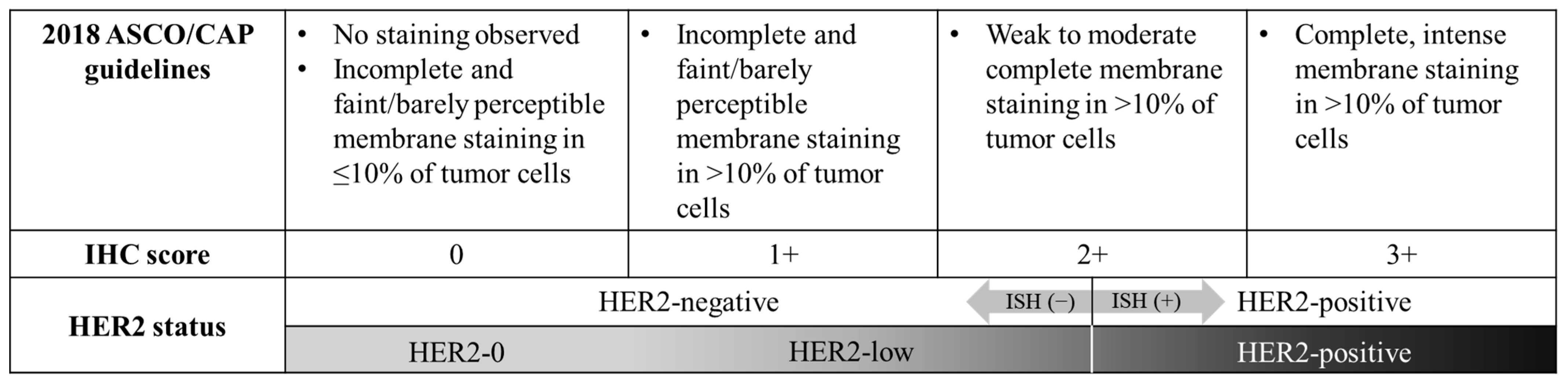
3.1. Baseline Characteristics According to HER2 Status

This study evaluated 2542 TNBC patients. A total of 1882 patients (74.0%) were HER2-0, and 660 patients (26.0%) were HER2-low. Table 1 summarized the baseline characteristics of HER2-0 and HER2-low patients. Patients in the HER2-low group were significantly older (median age (interquartile range (IQR)) 48 (41–56) vs. 52 (44–60), p < 0.001) and more often postmenopausal (56.4% vs. 46.8%, p < 0.001) compared to those in the HER2-0 group. Tumor multiplicity was more common in the HER2-low group, with 19.7% of patients presenting multiple tumors, compared to 15.1% in the HER2-0 group (p = 0.005). Significant differences were observed in surgical treatment types; the HER2-low group had higher mastectomy rates (30.8% vs. 22.6%, p < 0.001) and lower axillary lymph node dissection (ALND) rates (26.1% vs. 34.4%, p < 0.001) compared to the HER2-0 group. The HER2-low group also had a higher rate of patients receiving NAC (39.4% vs. 29.6%, p < 0.001) and a lower rate receiving adjuvant chemotherapy (63.2% vs. 73.4%, p < 0.001). Radiation therapy was less commonly administered to the HER2-low group (79.8% vs. 86.0%, p < 0.001). Pathologically, the HER2-low group exhibited a lower proportion of histologic and nuclear grade III tumors (61.4% vs. 68.8%, p < 0.001 and 69.5% vs. 75.2%, p = 0.001, respectively), as well as a lower incidence of LVI (18.9% vs. 24.3%, p < 0.001).

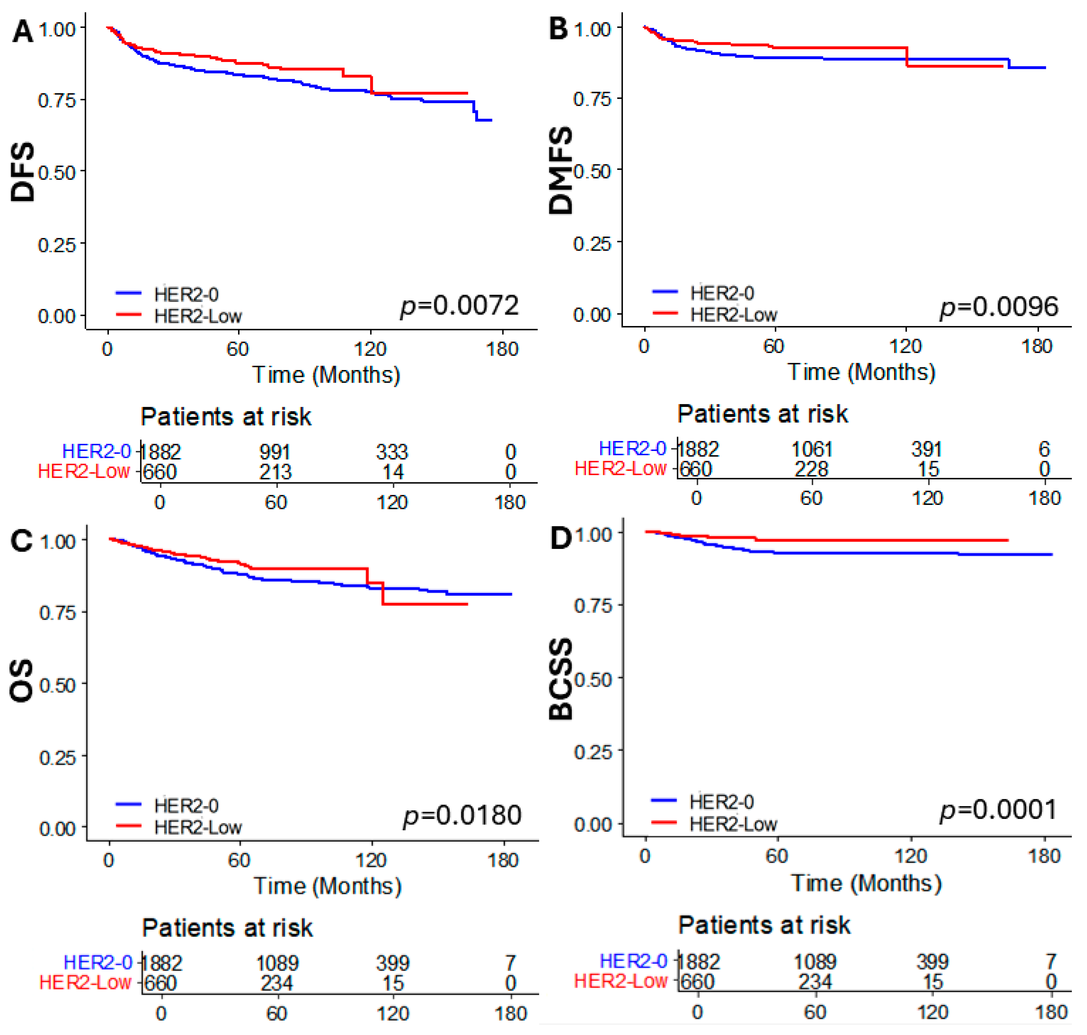
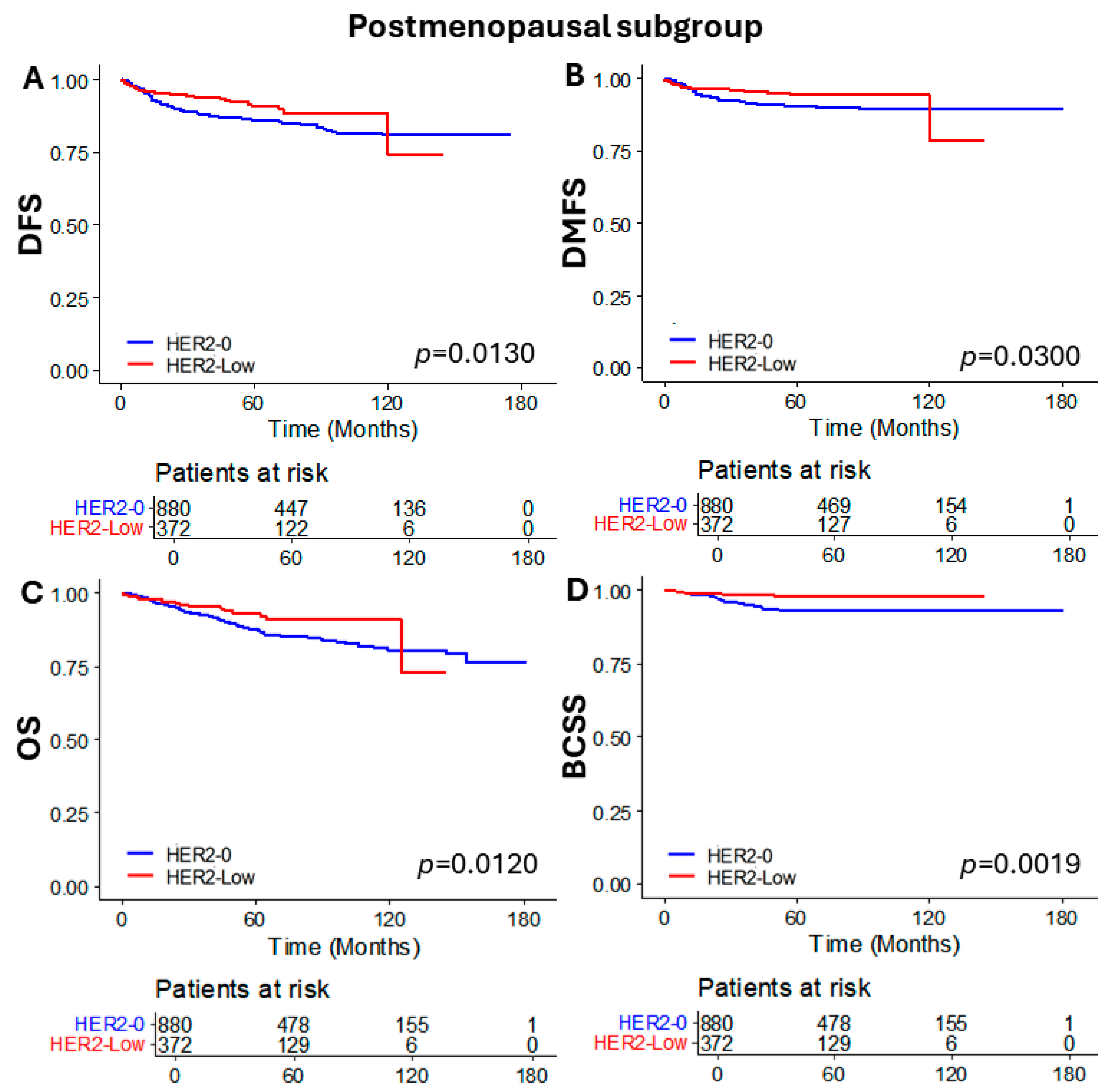
3.2. Oncologic Outcomes According to HER2 Status

The median follow-up time of the total study population was 62 months (IQR: 42–105), 70 months (IQR 45–113) for the HER2-0 group, and 52 months (IQR 37–71) for the HER2-low group. There were significant differences in DFS, DMFS, OS, and BCSS between the HER2-low and HER2-0 group, with better survival outcomes observed in the HER2-low group (p = 0.0072, p = 0.0096, p = 0.0180, and p = 0.0001, respectively) (Figure 2).
To evaluate whether these differences were consistent in both premenopausal and postmenopausal patient groups, we analyzed the oncologic outcomes based on HER2 status in each menopausal subgroup. In the premenopausal subgroup, DFS, DMFS, and OS were not significantly different according to HER2 status. However, significantly better BCSS was observed in the HER2-low group compared to the HER2-0 group (p = 0.0290) (Figure 3). On the other hand, significant differences were observed in the postmenopausal subgroup. The HER2-low group demonstrated superior outcomes for DFS (p = 0.0130), DMFS (p = 0.0300), OS (p = 0.0120), and particularly for BCSS, which showed a markedly significant difference (p = 0.0019) (Figure 4).

3.3. Factors Associated with Oncologic Outcomes

Univariate and multivariate analysis was performed to find out the significant factors associated with DFS, DMFS, OS, and BCSS (Table 2 and Table 3). The number of patients that experienced recurrence, distant metastasis, and death are described in the Supplementary Table S1. In summary, a total of 436 recurrences (190 locoregional recurrences, 172 distant metastases, and 74 both locoregional and distant metastases), and 308 mortalities (139 breast cancer related mortalities and 169 mortalities of other causes) were observed.
The analysis of DFS revealed several factors significantly associated with outcomes both in univariate and multivariate models. Factors such as HER2 status, age, breast surgery type, axilla surgery type, NAC, tumor size and LVI showed significant associations. HER2-low (HR = 0.761, p = 0.048) and age (HR = 0.980, p = 0.009) were protective factors in the multivariate model. A mastectomy, ALND, and NAC were associated with poorer DFS (HR = 1.573, p < 0.001, HR = 2.099, p < 0.001, and HR = 1.872, p < 0.001, respectively), likely indicating it was performed on more advanced cases. LVI and larger tumor size (>2 cm) also negatively impacted DFS (HR = 2.244, p < 0.001 and HR = 1.393, p = 0.029, respectively).
Similar results were notable for DMFS. As in DFS, HER2-low (HR = 0.647, p = 0.018) was a protective factor, and mastectomy (HR = 2.149, p < 0.001), ALND (HR = 2.904, p < 0.001), and NAC (HR = 2.030, p < 0.001) were significantly associated with worse DMFS. Additionally, larger tumor size (HR = 2.019, p = 0.004) and LVI (HR = 1.925, p < 0.001) had a negative impact on DMFS. Compared to the DFS model, age was not a significant factor associated with DMFS.
OS was significantly influenced by HER2 status, the type of breast and axillary surgeries performed, tumor size, LVI, and adjuvant chemotherapy. HER2-low status and receiving adjuvant chemotherapy improved survival outcomes in the multivariate model (HR = 0.725, p = 0.049 and HR = 0.566, p = 0.002). Mastectomy, ALND, larger tumor size, and LVI were linked to worse OS.
BCSS analyses revealed strong associations with surgical treatment type, LVI, and tumor size. HER2-low status again showed a significant protective effect (HR = 0.476, p = 0.008). Extensive surgeries like mastectomy and especially ALND correlated with poorer outcomes (HR = 1.494, p = 0.050 and HR = 3.437, p < 0.001). The presence of LVI and larger tumor sizes also worsened BCSS.
Overall, the presence of LVI, larger tumor size, and receiving mastectomy and ALND were factors of poorer survival along all survival analyses. On the other hand, HER2-low status consistently emerged as a significant protective factor in all survival outcomes.

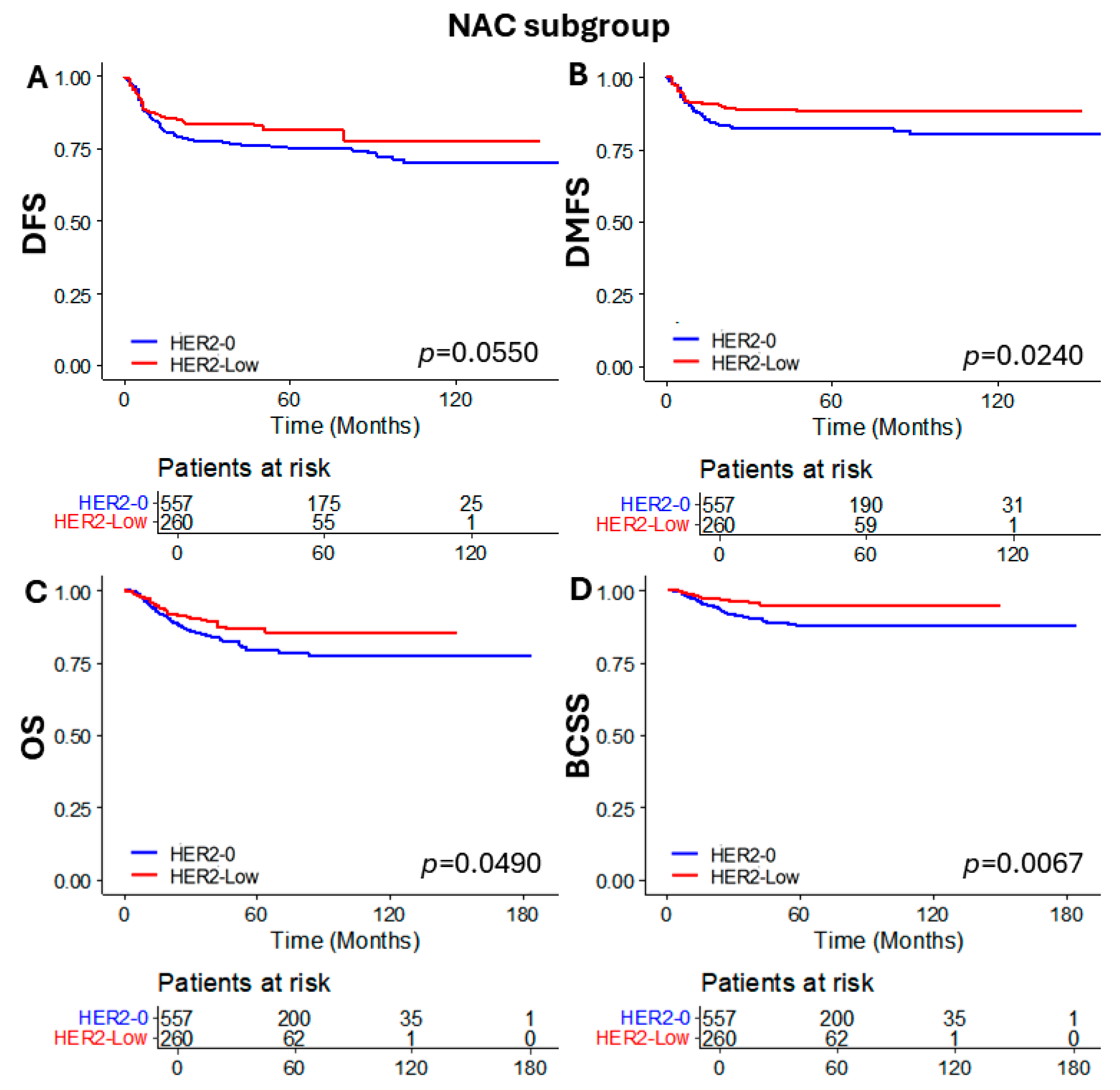
3.4. Subgroup Analysis of Patients Who Received NAC

To compare oncologic outcomes and response to NAC, we performed a subgroup analysis for patients who received NAC according to HER2 status. The chemotherapy regimens were administered according to the NCCN guidelines and Korean national health insurance policies (for example, dose-dense AC (doxorubicin/cyclophosphamide) preceded by weekly paclitaxel, AC followed by docetaxel, TC (docetaxel and cyclophosphamide), TAC (docetaxel/doxorubicin/cyclophosphamide), paclitaxel + carboplatin, or docetaxel + carboplatin) [23]. Additionally, none of the patients in this study received pembrolizumab because its use in TNBC was approved in 2022. When comparing the baseline characteristics, only age and incidence of LVI were different between the two groups (Table 4). Other than these factors, the two groups showed similar characteristics. Additionally, no significant difference in pCR rate was observed (26.9% vs. 29.1%, p = 0.523). However, when comparing the oncologic outcomes, DMFS, OS, and BCSS were significantly better in the HER2-low group (p = 0.0240, p = 0.0490, and p = 0.0067, respectively). Difference in DFS was not statistically significant (p = 0.0550) (Figure 5).

4. Discussion

In our previous study of hormone-receptor-positive breast cancer patients, we reported significantly improved oncologic outcomes in the HER2-low group compared to the HER2-0 group (median follow-up period of 64 months). However, these findings differed according to menopausal status. Also, HER2 status was an independent predictor of DFS, OS, and BCSS [20]. With a median follow-up period of 62 months, this study demonstrated that similar results were consistent in the TNBC patients. Improved oncologic outcomes were observed in the HER2-low group, and HER2 status was an independent predictor of DFS, DMFS, OS, and BCSS. The impact of HER2-low differed according to menopausal status. Although significant differences in oncologic outcomes according to HER2 status were consistent in the NAC subgroup, there were no differences in pCR rates based on HER2 status.
Previous studies have reported varying proportions of HER2-low in TNBC, ranging from 17.6% to 66.3% [9,10,12,24,25]. In our study, HER2-low proportion was 26.0%.
Some notable differences were noted in baseline characteristics between the HER2-low group and HER2-0 group. The HER2-low group had a higher proportion of postmenopausal patients and older age compared to the HER2-0 group. In addition, patients presenting with multiple tumors were more frequent in the HER2-low group. This may have been one of the factors that affected the higher mastectomy rate in the HER2-low group. Furthermore, due to the higher rate of mastectomy and SLNB, fewer patients received adjuvant radiation therapy in the HER2-low group. While there were no differences in tumor size and lymph node metastasis, the HER2-low group had fewer tumors with high grade and LVI. These characteristics were comparable to other previous studies that investigated HER2-low TNBC patients. In a retrospective study of 296 non-metastatic TNBC patients, HER2-low patients were older, had a lower HG, and had an apocrine-like molecular feature compared to the HER2-0 group [26]. Within the Korean nationwide study, HER2-low TNBC patients were older and had tumors with a lower Ki-67 index [10]. Similarly, older age and smaller proportion of T3 tumors were observed in a retrospective study that included 466 TNBC patients [9].
The impact of HER2-low on oncologic outcomes was significant in our study, with favorable results in the HER2-low group. Several past studies, including a retrospective study of 4918 breast cancer patients, described that HER2-low status had no prognostic impact on oncologic outcomes [9,17,26,27,28]. On the contrary, other studies showed that HER2-low was a significant prognostic factor [10,11,12,13,14,15,16,19]. Furthermore, discordance in the expression of HER2 status were observed between the primary tumor and recurred or metastatic tissue [29]. Considering these findings, whether HER2-low status truly has a prognostic value seems unclear. Nevertheless, it seems important to reassess the tumor subtype including HER2 status in the recurrent/metastatic situation because the HER2 status may have dynamically changed, and the patient could benefit from targetable agent such as T-Dxd.
Currently, there are no concrete explanations on why HER2-low breast cancer has better outcomes compared to HER2-0 breast cancer. Using single-cell RNA sequencing, Hu et al. revealed that HER2-0 TNBC had a more active immune microenvironment and higher levels of immunotherapeutic biomarkers compared to HER2-low TNBC [25]. Tan et al. reported that ERBB2 copy number variant (CNV) score was an independent prognostic factor for recurrence free survival [12]. Also, Agostinetto et al. demonstrated that ERBB2 expression correlated with HER2 IHC scores [17]. An international multicenter cohort study of 28,280 non-metastatic breast cancer patients described that better recurrence free survival and OS of HER2-low did not correlate with HER2 IHC score and mainly resulted from the HER2 IHC1+ subgroup [12]. A study that re-evaluated HER2 staining status in 281 breast cancer cases identified inter-observer variations in evaluating HER2 IHC staining, specifically for scores IHC 0 and IHC 1+ [24]. This poor reproducibility of HER2 IHC evaluation is a big hurdle in clearly defining HER2-low status, with inter-observer variations up to 75% reported when reassessed [30]. Therefore, setting the boundary (especially the lower limit of HER2-low) for selecting potential patients that may benefit from treatment with T-Dxd is challenging. Further dividing HER2-0 into HER2-null and IHC > 0 to <1+ has been suggested, and clinical trials are ongoing to see the efficacy of T-Dxd on this population [31,32,33]. Future studies on researching the tumor microenvironment and biomarkers of HER2-low breast cancer are necessary. In summary, new assays for a more precise evaluation of HER2 status seems necessary. Furthermore, as we await the results of ongoing clinical trials, re-defining the lower-limit of HER2-low may be needed in the future.
In addition to the improvement and development of tailored systemic treatment, there had been efforts to investigate the feasibility of tailored surgical treatment according to different subtypes in recurrent cases. Gentile et al. revealed that a second breast conserving surgery may be feasible in recurrent small-sized HER2-positive cases or TNBCs [34]. When combined with the novel target agents, this may improve both survival and the quality of life of selected patients.
Achieving pCR after NAC is an important prognostic factor in breast cancer treatment [35]. Therefore, pCR rate is measured as one of the primary outcomes in many studies including those regarding the HER2-low group [11,18,36,37,38,39,40]. The pCR rate in the HER2-low group ranged from 11.86% to 51%, while the HER2-0 group ranged from 15.2% to 52% [11,36,38,39,40]. In a retrospective nationwide study including 20,029 TNBC patients, Li, H. et al. reported significantly lower pCR rates in the HER2-low group compared to the HER2-0 group [11]. On the contrary, Denkert et al., conducted a pooled analysis of four prospective neoadjuvant clinical trials and found no difference in the pCR rates according to HER2 status in TNBC patients [15]. Interestingly, better oncologic outcomes were observed in the HER2-low TNBC group in both studies. In our study, the pCR rate did not differ between HER2-low and HER2-0 group (26.9% vs. 29.1%, respectively). Jacot et al. reported that HER2-low TNBC expressed more apocrine-like molecular features with lower HG compared to HER2-0 TNBC [26]. These relatively less-aggressive tumor characteristics of HER2-low breast cancer may be the reason why HER2-low breast cancer has favorable prognosis even though the response to systemic treatment is not different compared to HER2-0 breast cancer.
Our study has limitations. Due to the retrospective nature of this study, incomplete patient data were included. Therefore, the possibility of other confounding variables of measured or unmeasured factors exists. Furthermore, since it was a single center study, we only included Korean TNBC patients. Therefore, further multi-national study results are necessary to generalize our results on a global basis. We included patients from 2008 to 2020. Between this time interval, guidelines on treatment and management have changed, and thus may have affected the oncologic outcomes.
To our knowledge, this is the largest single center study examining the impact of HER2-low status on oncologic outcomes in TNBC patients. Moreover, data were extracted from our prospectively collected database, ensuring the homogeneity of the treatment applied. Another strength of our study is the reclassification of patients from HER2 IHC score 1+ to HER2-0 according to the updated ASCO/CAP guidelines, ensuring homogeneity in HER2 status classification. Compared to the other studies, our previous study and current study revealed that the impact of HER2-low on oncologic outcomes differed according to menopausal status [20]. In a study that analyzed the effect of menopausal status in different breast cancer subtypes, the authors reported a higher proportion of postmenopausal patients in the TNBC subtype and a tendency to have favorable outcomes compared to premenopausal patients [41]. Our study demonstrated a higher proportion of postmenopausal patients in the HER2-low TNBC group. Also, the expression of HER2-low seemed to have a positive association with ER expression [42]. Considering the lower estrogen level and favorable prognosis of postmenopausal patients compared to premenopausal women, future studies are needed to investigate the possible crosstalk between estrogen and HER2 expression.

5. Conclusions

Despite similar rates of pCR, our study revealed that HER2-low status was an independent predictor of improved DFS, DMFS, OS, and BCSS compared to HER2-0 in TNBC. However, the prognostic impact differed between menopausal status, especially benefitting postmenopausal patients. Therefore, assessing HER2 status along with menopausal status may help predict prognosis in TNBC patients. Updates in interpreting HER2 expression seem necessary to precisely define the boundaries of HER2-low expression. Future studies to discover the crosstalk between hormone levels and HER2 expression are required.

Supplementary Materials

The following supporting information can be downloaded at: https://www.mdpi.com/article/10.3390/cancers16142566/s1. Table S1. Number of recurrences and deaths occurred according to HER2 status.

Author Contributions

Concept and design, W.K.P., S.J.N., S.W.K., J.E.L., J.Y., S.K.L., J.M.R. and B.J.C.; Acquisition, analysis, or interpretation of data, W.K.P., J.Y., J.E.L. and B.J.C.; Drafting of the manuscript, W.K.P.; Critical revision of the manuscript for important intellectual content, W.K.P., S.J.N., S.W.K., J.E.L., J.Y., S.K.L., J.M.R. and B.J.C.; Statistical analysis, W.K.P.; Obtained funding, B.J.C.; Administrative, technical, or material support, W.K.P., S.J.N., S.W.K., J.E.L., J.Y., S.K.L., J.M.R. and B.J.C.; Supervision, B.J.C.; W.K.P. had full access to all the data in this study and takes responsibility for the integrity of the data and the accuracy of the data analysis. All authors have read and agreed to the published version of the manuscript.

Funding

This study was supported by the Future Medicine 20*30 Project of the Samsung Medical Center [#SMO1230021].

Institutional Review Board Statement

This study adhered to the ethical tenets of the Declaration of Helsinki and was approved on 16 January 2024 by the Institutional Review Board of SMC in Seoul, Korea (IRB no. SMC 2024-01-057).

Informed Consent Statement

The need for informed consent was waived because of the low risk posed by this investigation.

Data Availability Statement

The datasets used and/or analyzed during the current study are available from the corresponding author on reasonable request.

Conflicts of Interest

The authors declare no conflicts of interest.

Abbreviations

ADCs, antibody–drug conjugates; AJCC, American Joint Committee on Cancer; ALND, axillary lymph node dissection; ASCO, American Society of Clinical Oncology; BCSS, Breast cancer specific survival; CAP, College of American Pathologist; CNV, copy number variant; DFS, disease free survival; DMFS, distant metastasis free survival; ER, estrogen receptor; FSH, follicle-stimulating hormone; HER2, human epithelial growth factor receptor 2; HG, histologic grade; IHC, immunohistochemistry; IRB, Institutional Review Board; ISH, in situ hybridization; LVI, lymphovascular invasion; NAC, neoadjuvant chemotherapy; NCCN, National Comprehensive Cancer Network; NG, nuclear grade; OS, overall survival; pCR, pathologic complete response; PR, progesterone receptor; SMC, Samsung Medical Center; T-Dxd, trastuzumab-deruxtecan; TNBC, triple-negative breast cancer.

References

  1. Dai, X.; Li, T.; Bai, Z.; Yang, Y.; Liu, X.; Zhan, J.; Shi, B. Breast cancer intrinsic subtype classification, clinical use and future trends. Am. J. Cancer Res. 2015, 5, 2929–2943. [Google Scholar] [PubMed]
  2. Alves, C.L.; Ehmsen, S.; Terp, M.G.; Portman, N.; Tuttolomondo, M.; Gammelgaard, O.L.; Hundebol, M.F.; Kaminska, K.; Johansen, L.E.; Bak, M.; et al. Co-targeting CDK4/6 and AKT with endocrine therapy prevents progression in CDK4/6 inhibitor and endocrine therapy-resistant breast cancer. Nat. Commun. 2021, 12, 5112. [Google Scholar] [CrossRef] [PubMed]
  3. Slamon, D.J.; Leyland-Jones, B.; Shak, S.; Fuchs, H.; Paton, V.; Bajamonde, A.; Fleming, T.; Eiermann, W.; Wolter, J.; Pegram, M.; et al. Use of chemotherapy plus a monoclonal antibody against HER2 for metastatic breast cancer that overexpresses HER2. N. Engl. J. Med. 2001, 344, 783–792. [Google Scholar] [CrossRef] [PubMed]
  4. Foulkes, W.D.; Smith, I.E.; Reis-Filho, J.S. Triple-negative breast cancer. N. Engl. J. Med. 2010, 363, 1938–1948. [Google Scholar] [CrossRef]
  5. Lin, N.U.; Vanderplas, A.; Hughes, M.E.; Theriault, R.L.; Edge, S.B.; Wong, Y.N.; Blayney, D.W.; Niland, J.C.; Winer, E.P.; Weeks, J.C. Clinicopathologic features, patterns of recurrence, and survival among women with triple-negative breast cancer in the National Comprehensive Cancer Network. Cancer 2012, 118, 5463–5472. [Google Scholar] [CrossRef] [PubMed]
  6. Swain, S.M.; Baselga, J.; Kim, S.B.; Ro, J.; Semiglazov, V.; Campone, M.; Ciruelos, E.; Ferrero, J.M.; Schneeweiss, A.; Heeson, S.; et al. Pertuzumab, trastuzumab, and docetaxel in HER2-positive metastatic breast cancer. N. Engl. J. Med. 2015, 372, 724–734. [Google Scholar] [CrossRef] [PubMed]
  7. Swain, S.M.; Kim, S.B.; Cortes, J.; Ro, J.; Semiglazov, V.; Campone, M.; Ciruelos, E.; Ferrero, J.M.; Schneeweiss, A.; Knott, A.; et al. Pertuzumab, trastuzumab, and docetaxel for HER2-positive metastatic breast cancer (CLEOPATRA study): Overall survival results from a randomised, double-blind, placebo-controlled, phase 3 study. Lancet Oncol. 2013, 14, 461–471. [Google Scholar] [CrossRef] [PubMed]
  8. Modi, S.; Jacot, W.; Yamashita, T.; Sohn, J.; Vidal, M.; Tokunaga, E.; Tsurutani, J.; Ueno, N.T.; Prat, A.; Chae, Y.S.; et al. Trastuzumab Deruxtecan in Previously Treated HER2-Low Advanced Breast Cancer. N. Engl. J. Med. 2022, 387, 9–20. [Google Scholar] [CrossRef] [PubMed]
  9. Horisawa, N.; Adachi, Y.; Takatsuka, D.; Nozawa, K.; Endo, Y.; Ozaki, Y.; Sugino, K.; Kataoka, A.; Kotani, H.; Yoshimura, A.; et al. The frequency of low HER2 expression in breast cancer and a comparison of prognosis between patients with HER2-low and HER2-negative breast cancer by HR status. Breast Cancer 2022, 29, 234–241. [Google Scholar] [CrossRef]
  10. Won, H.S.; Ahn, J.; Kim, Y.; Kim, J.S.; Song, J.Y.; Kim, H.K.; Lee, J.; Park, H.K.; Kim, Y.S. Clinical significance of HER2-low expression in early breast cancer: A nationwide study from the Korean Breast Cancer Society. Breast Cancer Res. 2022, 24, 22. [Google Scholar] [CrossRef]
  11. Li, H.; Plichta, J.K.; Li, K.; Jin, Y.; Thomas, S.M.; Ma, F.; Tang, L.; Wei, Q.; He, Y.W.; Chen, Q.; et al. Impact of HER2-low status for patients with early-stage breast cancer and non-pCR after neoadjuvant chemotherapy: A National Cancer Database Analysis. Breast Cancer Res. Treat. 2024, 204, 89–105. [Google Scholar] [CrossRef] [PubMed]
  12. Tan, R.; Ong, W.S.; Lee, K.H.; Lim, A.H.; Park, S.; Park, Y.H.; Lin, C.H.; Lu, Y.S.; Ono, M.; Ueno, T.; et al. HER2 expression, copy number variation and survival outcomes in HER2-low non-metastatic breast cancer: An international multicentre cohort study and TCGA-METABRIC analysis. BMC Med. 2022, 20, 105. [Google Scholar] [CrossRef] [PubMed]
  13. Rosso, C.; Voutsadakis, I.A. Characteristics, Clinical Differences and Outcomes of Breast Cancer Patients with Negative or Low HER2 Expression. Clin. Breast Cancer 2022, 22, 391–397. [Google Scholar] [CrossRef] [PubMed]
  14. Ergun, Y.; Ucar, G.; Akagunduz, B. Comparison of HER2-zero and HER2-low in terms of clinicopathological factors and survival in early-stage breast cancer: A systematic review and meta-analysis. Cancer Treat. Rev. 2023, 115, 102538. [Google Scholar] [CrossRef] [PubMed]
  15. Denkert, C.; Seither, F.; Schneeweiss, A.; Link, T.; Blohmer, J.U.; Just, M.; Wimberger, P.; Forberger, A.; Tesch, H.; Jackisch, C.; et al. Clinical and molecular characteristics of HER2-low-positive breast cancer: Pooled analysis of individual patient data from four prospective, neoadjuvant clinical trials. Lancet Oncol. 2021, 22, 1151–1161. [Google Scholar] [CrossRef]
  16. Almstedt, K.; Heimes, A.S.; Kappenberg, F.; Battista, M.J.; Lehr, H.A.; Krajnak, S.; Lebrecht, A.; Gehrmann, M.; Stewen, K.; Brenner, W.; et al. Long-term prognostic significance of HER2-low and HER2-zero in node-negative breast cancer. Eur. J. Cancer 2022, 173, 10–19. [Google Scholar] [CrossRef]
  17. Agostinetto, E.; Rediti, M.; Fimereli, D.; Debien, V.; Piccart, M.; Aftimos, P.; Sotiriou, C.; de Azambuja, E. HER2-Low Breast Cancer: Molecular Characteristics and Prognosis. Cancers 2021, 13, 2824. [Google Scholar] [CrossRef]
  18. Douganiotis, G.; Kontovinis, L.; Markopoulou, E.; Ainali, A.; Zarampoukas, T.; Natsiopoulos, I.; Papazisis, K. Prognostic Significance of Low HER2 Expression in Patients With Early Hormone Receptor Positive Breast Cancer. Cancer Diagn. Progn. 2022, 2, 316–323. [Google Scholar] [CrossRef]
  19. Xu, B.; Shen, J.; Shen, J.; Wang, L. Prognostic impact of HER2-low expression in HER2-negative breast cancer under different hormone receptor status. Int. J. Clin. Oncol. 2023, 28, 543–549. [Google Scholar] [CrossRef]
  20. Park, W.K.; Nam, S.J.; Kim, S.W.; Lee, J.E.; Yu, J.; Ryu, J.M.; Chae, B.J. The Impact of HER2-Low Expression on Oncologic Outcomes in Hormone Receptor-Positive Breast Cancer. Cancers 2023, 15, 5361. [Google Scholar] [CrossRef]
  21. Hammond, M.E.; Hayes, D.F.; Dowsett, M.; Allred, D.C.; Hagerty, K.L.; Badve, S.; Fitzgibbons, P.L.; Francis, G.; Goldstein, N.S.; Hayes, M.; et al. American Society of Clinical Oncology/College Of American Pathologists guideline recommendations for immunohistochemical testing of estrogen and progesterone receptors in breast cancer. J. Clin. Oncol. 2010, 28, 2784–2795. [Google Scholar] [CrossRef] [PubMed]
  22. Wolff, A.C.; Hammond, M.E.H.; Allison, K.H.; Harvey, B.E.; Mangu, P.B.; Bartlett, J.M.S.; Bilous, M.; Ellis, I.O.; Fitzgibbons, P.; Hanna, W.; et al. Human Epidermal Growth Factor Receptor 2 Testing in Breast Cancer: American Society of Clinical Oncology/College of American Pathologists Clinical Practice Guideline Focused Update. J. Clin. Oncol. 2018, 36, 2105–2122. [Google Scholar] [CrossRef] [PubMed]
  23. National Comprehensive Cancer Network. NCCN Guidelines: Breast Cancer. 2024. Available online: https://www.nccn.org/guidelines/guidelines-detail?category=1&id=1419 (accessed on 3 July 2024).
  24. Zhang, H.; Katerji, H.; Turner, B.M.; Audeh, W.; Hicks, D.G. HER2-low breast cancers: Incidence, HER2 staining patterns, clinicopathologic features, MammaPrint and BluePrint genomic profiles. Mod. Pathol. 2022, 35, 1075–1082. [Google Scholar] [CrossRef] [PubMed]
  25. Hu, X.E.; Yang, P.; Chen, S.; Wei, G.; Yuan, L.; Yang, Z.; Gong, L.; He, L.; Yang, L.; Peng, S.; et al. Clinical and biological heterogeneities in triple-negative breast cancer reveals a non-negligible role of HER2-low. Breast Cancer Res. 2023, 25, 34. [Google Scholar] [CrossRef] [PubMed]
  26. Jacot, W.; Maran-Gonzalez, A.; Massol, O.; Sorbs, C.; Mollevi, C.; Guiu, S.; Boissiere-Michot, F.; Ramos, J. Prognostic Value of HER2-Low Expression in Non-Metastatic Triple-Negative Breast Cancer and Correlation with Other Biomarkers. Cancers 2021, 13, 6059. [Google Scholar] [CrossRef] [PubMed]
  27. Gampenrieder, S.P.; Rinnerthaler, G.; Tinchon, C.; Petzer, A.; Balic, M.; Heibl, S.; Schmitt, C.; Zabernigg, A.F.; Egle, D.; Sandholzer, M.; et al. Landscape of HER2-low metastatic breast cancer (MBC): Results from the Austrian AGMT_MBC-Registry. Breast Cancer Res. 2021, 23, 112. [Google Scholar] [CrossRef] [PubMed]
  28. Schettini, F.; Chic, N.; Braso-Maristany, F.; Pare, L.; Pascual, T.; Conte, B.; Martinez-Saez, O.; Adamo, B.; Vidal, M.; Barnadas, E.; et al. Clinical, pathological, and PAM50 gene expression features of HER2-low breast cancer. NPJ Breast Cancer 2021, 7, 1. [Google Scholar] [CrossRef] [PubMed]
  29. Tarantino, P.; Gandini, S.; Nicolo, E.; Trillo, P.; Giugliano, F.; Zagami, P.; Vivanet, G.; Bellerba, F.; Trapani, D.; Marra, A.; et al. Evolution of low HER2 expression between early and advanced-stage breast cancer. Eur. J. Cancer 2022, 163, 35–43. [Google Scholar] [CrossRef] [PubMed]
  30. Lambein, K.; Van Bockstal, M.; Vandemaele, L.; Geenen, S.; Rottiers, I.; Nuyts, A.; Matthys, B.; Praet, M.; Denys, H.; Libbrecht, L. Distinguishing score 0 from score 1+ in HER2 immunohistochemistry-negative breast cancer: Clinical and pathobiological relevance. Am. J. Clin. Pathol. 2013, 140, 561–566. [Google Scholar] [CrossRef]
  31. Curigliano, G.; Dent, R.; Earle, H.; Modi, S.; Tarantino, P.; Viale, G.; Tolaney, S.M. Open questions, current challenges, and future perspectives in targeting human epidermal growth factor receptor 2-low breast cancer. ESMO Open 2024, 9, 102989. [Google Scholar] [CrossRef]
  32. Mosele, F.; Deluche, E.; Lusque, A.; Le Bescond, L.; Filleron, T.; Pradat, Y.; Ducoulombier, A.; Pistilli, B.; Bachelot, T.; Viret, F.; et al. Trastuzumab deruxtecan in metastatic breast cancer with variable HER2 expression: The phase 2 DAISY trial. Nat. Med. 2023, 29, 2110–2120. [Google Scholar] [CrossRef] [PubMed]
  33. ClinicalTrials.gov. Study of Trastuzumab Deruxtecan (T-DXd) vs Investigator’s Choice Chemotherapy in HER2-low, Hormone Receptor Positive, Metastatic Breast Cancer (DB-06). 2024. Available online: https://clinicaltrials.gov/study/NCT04494425 (accessed on 12 April 2024).
  34. Gentile, D.; Sagona, A.; Spoto, R.; Franceschini, D.; Vaccari, S.; Vinci, V.; Biondi, E.; Scardina, L.; Tinterri, C. Salvage Mastectomy Is not the Treatment of Choice for Aggressive Subtypes of Ipsilateral Breast Cancer Recurrence: A Single-Institution Retrospective Study. Eur. J. Breast Health 2022, 18, 315–322. [Google Scholar] [CrossRef] [PubMed]
  35. Von Minckwitz, G.; Untch, M.; Blohmer, J.U.; Costa, S.D.; Eidtmann, H.; Fasching, P.A.; Gerber, B.; Eiermann, W.; Hilfrich, J.; Huober, J.; et al. Definition and impact of pathologic complete response on prognosis after neoadjuvant chemotherapy in various intrinsic breast cancer subtypes. J. Clin. Oncol. 2012, 30, 1796–1804. [Google Scholar] [CrossRef] [PubMed]
  36. De Moura Leite, L.; Cesca, M.G.; Tavares, M.C.; Santana, D.M.; Saldanha, E.F.; Guimaraes, P.T.; Sa, D.D.S.; Simoes, M.F.E.; Viana, R.L.; Rocha, F.G.; et al. HER2-low status and response to neoadjuvant chemotherapy in HER2 negative early breast cancer. Breast Cancer Res. Treat. 2021, 190, 155–163. [Google Scholar] [CrossRef] [PubMed]
  37. Shao, Y.; Yu, Y.; Luo, Z.; Guan, H.; Zhu, F.; He, Y.; Chen, Q.; Liu, C.; Nie, B.; Liu, H. Clinical, Pathological Complete Response, and Prognosis Characteristics of HER2-Low Breast Cancer in the Neoadjuvant Chemotherapy Setting: A Retrospective Analysis. Ann. Surg. Oncol. 2022, 29, 8026–8034. [Google Scholar] [CrossRef]
  38. Li, J.J.; Yu, Y.; Ge, J. HER2-low-positive and response to NACT and prognosis in HER2-negative non-metastatic BC. Breast Cancer 2023, 30, 364–378. [Google Scholar] [CrossRef] [PubMed]
  39. Wang, W.; Zhu, T.; Chen, H.; Yao, Y. The impact of HER2-low status on response to neoadjuvant chemotherapy in clinically HER2-negative breast cancer. Clin. Transl. Oncol. 2023, 25, 1673–1681. [Google Scholar] [CrossRef]
  40. Xu, W.; Jiang, Y.; Xu, L.; Li, C.; Wang, J.; Liu, Z.; Xue, D.; Gu, Y.; Zhong, Z.; He, S.; et al. HER2-low status may predict poor neoadjuvant chemotherapy response in HR-negative breast cancer: A real-world multicenter study. Jpn. J. Clin. Oncol. 2023, 53, 463–471. [Google Scholar] [CrossRef] [PubMed]
  41. Nishimura, R.; Osako, T.; Okumura, Y.; Nakano, M.; Otsuka, H.; Fujisue, M.; Arima, N. Triple Negative Breast Cancer: An Analysis of the Subtypes and the Effects of Menopausal Status on Invasive Breast Cancer. J. Clin. Med. 2022, 11, 2331. [Google Scholar] [CrossRef]
  42. Tarantino, P.; Jin, Q.; Tayob, N.; Jeselsohn, R.M.; Schnitt, S.J.; Vincuilla, J.; Parker, T.; Tyekucheva, S.; Li, T.; Lin, N.U.; et al. Prognostic and Biologic Significance of ERBB2-Low Expression in Early-Stage Breast Cancer. JAMA Oncol. 2022, 8, 1177–1183. [Google Scholar] [CrossRef]
Figure 1. Definition and flowchart of HER2 status. ASCO American Society of Clinical Oncology, CAP College of American Pathologist, IHC immunohistochemistry, ISH in situ hybridization.
Cancers 16 02566 g001
Figure 2. Kaplan–Meier curves of (A) DFS, (B) DMFS, (C) OS, and (D) BCSS according to HER2 status. DFS, disease free survival; DMFS, distant metastasis free survival; OS, overall survival; and BCSS, breast cancer specific survival.
Cancers 16 02566 g002
Figure 3. Kaplan–Meier curves of (A) DFS, (B) DMFS, (C) OS, and (D) BCSS according to HER2 status in the premenopausal subgroup. DFS, disease free survival; DMFS, distant metastasis free survival; OS, overall survival; and BCSS, breast cancer specific survival.
Cancers 16 02566 g003
Figure 4. Kaplan–Meier curves of (A) DFS, (B) DMFS, (C) OS, and (D) BCSS according to HER2 status in the postmenopausal subgroup. DFS, disease free survival; DMFS, distant metastasis free survival; OS, overall survival; and BCSS, breast cancer specific survival.
Cancers 16 02566 g004
Figure 5. Kaplan–Meier curves of (A) DFS, (B) DMFS, (C) OS, and (D) BCSS according to HER2 status in the NAC subgroup. DFS, disease free survival; DMFS, distant metastasis free survival; OS, overall survival; BCSS, breast cancer specific survival; and NAC, neoadjuvant chemotherapy.
Cancers 16 02566 g005
Table 1. Baseline characteristics according to HER2 expression.
HER2-0HER2-Lowp-Value
Clinical variables
Total no. (%)1882 (74.0)660 (26.0)
Age
 Median (IQR)48 (41–56)52 (44–60)<0.001
Menopausal status, no. (%) <0.001
 Pre1002 (53.2)288 (43.6)
 Post880 (46.8)372 (56.4)
BRCA 1/2 mutation, no. (%) 0.510
 Not detected1736 (92.2)614 (93.0)
 Detected146 (7.8)46 (7.0)
Laterality 0.888
 Unilateral cancer1866 (99.1)654 (99.1)
 Bilateral cancer16 (0.9)6 (0.9)
Multiplicity 0.005
 No1567 (83.3)514 (77.9)
 Yes285 (15.1)130 (19.7)
 Unknown30 (1.6)16 (2.4)
Breast surgery, no. (%) <0.001
 BCS1457 (77.4)457 (69.2)
 Mastectomy425 (22.6)203 (30.8)
Axilla surgery, no. (%) <0.001
 SLNB1235 (65.6)488 (73.9)
 ALND647 (34.4)172 (26.1)
Neoadjuvant chemotherapy, no. (%) <0.001
 No1325 (70.4)400 (60.6)
 Yes557 (29.6)260 (39.4)
Adjuvant chemotherapy, no. (%) <0.001
 No501 (26.6)243 (36.8)
 Yes1381 (73.4)417 (63.2)
Radiation therapy, no. (%) <0.001
 No264 (14.0)133 (20.2)
 Yes1618 (86.0)527 (79.8)
Pathologic variables
Tumor size 0.109
 ≤2 cm712 (37.8)273 (41.4)
 >2 cm1170 (62.2)387 (58.6)
Lymph node metastasis 0.504
 Negative1069 (56.8)365 (55.3)
 Positive813 (43.2)295 (44.7)
Histologic grade <0.001
 ≤II420 (22.3)190 (28.8)
 >II1295 (68.8)405 (61.4)
 Unknown167 (8.9)65 (9.8)
Nuclear grade 0.001
 ≤II313 (16.6)147 (22.3)
 >II1416 (75.2)459 (69.5)
 Unknown153 (8.2)54 (8.2)
Lymphovascular invasion <0.001
 No881 (46.8)444 (67.3)
 Yes457 (24.3)125 (18.9)
 Unknown544 (28.9)91 (13.8)
IQR, interquartile range; BCS, breast conserving surgery; SLNB, sentinel lymph node biopsy; ALND, axillary lymph node dissection.
Table 2. Univariate and multivariate Cox models regarding DFS and DMFS.
Univariate Modelp-ValueMultivariate Modelp-Value
HR (95% CI)HR (95% CI)
Disease Free Survival
 HER-2 status (0 vs. low)0.715 (0.559–0.915)0.0080.761 (0.581–0.997)0.048
 Age0.977 (0.968–0.985)<0.0010.980 (0.965–0.995)0.009
 Menopausal status (pre- vs. post-)0.628 (0.518–0.763)<0.0010.909 (0.649–1.272)0.577
BRCA1/2 mutation status (No vs. Yes)1.302 (0.950–1.784)0.100
 Laterality (uni- vs. bilateral)1.373 (0.568–3.316)0.481
 Multiplicity (single vs. multiple)1.290 (1.017–1.636)0.0360.830 (0.629–1.094)0.186
 Breast Surgery (BCS vs. Mastectomy)2.040 (1.677–2.481)<0.0011.573 (1.236–2.002)<0.001
 Axilla Surgery (SLNB vs. ALND)2.794 (2.311–3.377)<0.0012.099 (1.553–2.836)<0.001
 NAC (No vs. Yes)2.070 (1.705–2.513)<0.0011.872 (1.327–2.640)<0.001
 Adjuvant CTx (No vs. Yes)0.602 (0.494–0.734)<0.0010.778 (0.570–1.060)0.112
 Adjuvant RT (No vs. Yes)0.861 (0.670–1.108)0.245
 Tumor size (≤2 cm vs. >2 cm)2.017 (1.673–2.573)<0.0011.393 (1.035–1.875)0.029
 LN metastasis (No vs. Yes)2.115 (1.747–2.559)<0.0010.777 (0.566–1.066)0.118
 HG (≤GrII vs. >GrII)1.073 (0.860–1.339)0.532
 NG (≤GrII vs. >GrII)1.128 (0.879–1.448)0.345
 LVI (No vs. Yes)3.141 (2.529–3.901)<0.0012.244 (1.746–2.884)<0.001
Distant Metastasis Free Survival
 HER-2 status (0 vs. low)0.652 (0.470–0.903)0.0100.647 (0.451–0.929)0.018
 Age0.984 (0.972–0.995)0.0050.989 (0.970–1.009)0.281
 Menopausal status (pre- vs. post-)0.721 (0.559–0.930)0.0120.861 (0.559–1.326)0.497
BRCA1/2 mutation status (No vs. Yes)0.775 (0.460–1.306)0.339
 Laterality (uni- vs. bilateral)1.475 (0.472–4.608)0.503
 Multiplicity (single vs. multiple)1.069 (0.766–1.491)0.696
 Breast Surgery (BCS vs. Mastectomy)3.258 (2.536–4.185)<0.0012.149 (1.586–2.913)<0.001
 Axilla Surgery (SLNB vs. ALND)5.113 (3.901–6.702)<0.0012.904 (1.912–4.410)<0.001
 NAC (No vs. Yes)2.779 (2.159–3.575)<0.0012.030 (1.334–3.088)<0.001
 Adjuvant CTx (No vs. Yes)0.515 (0.399–0.664)<0.0010.942 (0.647–1.372)0.757
 Adjuvant RT (No vs. Yes)0.849 (0.611–1.180)0.331
 Tumor size (≤2 cm vs. >2 cm)3.569 (2.544–5.009)<0.0012.019 (1.259–3.238)0.004
 LN metastasis (No vs. Yes)3.623 (2.745–4.781)<0.0010.929 (0.595–1.452)0.748
 HG (≤GrII vs. >GrII)0.968 (0.725–1.293)0.826
 NG (≤GrII vs. >GrII)0.913 (0.668–1.248)0.568
 LVI (No vs. Yes)3.855 (2.891–5.142)<0.0011.925 (1.382–2.681)<0.001
DFS, disease free survival; DMFS, distant metastasis free survival; BCS, breast conserving surgery; SLNB, sentinel lymph node biopsy; ALND, axillary lymph node dissection; NAC, neoadjuvant chemotherapy; CTx, chemotherapy; RT, radiation therapy; LN, lymph node; HG, histologic grade; NG, nuclear grade; LVI, lymphovascular invasion.
Table 3. Univariate and multivariate Cox models regarding OS and BCSS.
Univariate Modelp-ValueMultivariate Modelp-Value
HR (95% CI)HR (95% CI)
Overall Survival
 HER-2 status (0 vs. low)0.698 (0.517–0.943)0.0190.725 (0.526–0.999)0.049
 Age1.006 (0.996–1.016)0.271
 Menopausal status (pre- vs. post-)1.052 (0.841–1.316)0.656
BRCA1/2 mutation status (No vs. Yes)0.805 (0.512–1.267)0.349
 Laterality (uni- vs. bilateral)1.953 (0.807–4.728)0.138
 Multiplicity (single vs. multiple)1.116 (0.834–1.494)0.461
 Breast Surgery (BCS vs. Mastectomy)2.706 (2.158–3.392)<0.0011.458 (1.074–1.978)0.016
 Axilla Surgery (SLNB vs. ALND)4.304 (3.391–5.462)<0.0012.697 (1.858–3.914)<0.001
 NAC (No vs. Yes)2.262 (1.799–2.843)<0.0011.440 (0.953–2.174)0.083
 Adjuvant CTx (No vs. Yes)0.468 (0.372–0.587)<0.0010.566 (0.395–0.812)0.002
 Adjuvant RT (No vs. Yes)0.706 (0.535–0.933)0.0140.726 (0.501–1.054)0.092
 Tumor size (≤2 cm vs. >2 cm)3.075 (2.311–4.093)<0.0012.070 (1.407–3.045)<0.001
 LN metastasis (No vs. Yes)2.942 (2.322–3.729)<0.0010.880 (0.598–1.294)0.516
 HG (≤GrII vs. >GrII)1.052 (0.962–1.152)0.266
 NG (≤GrII vs. >GrII)1.077 (0.971–1.193)0.161
 LVI (No vs. Yes)3.575 (2.768–4.618)<0.0012.229 (1.657–2.997)<0.001
Breast cancer Specific Survival
 HER-2 status (0 vs. low)0.368 (0.215–0.630)<0.0010.476 (0.274–0.827)0.008
 Age0.982 (0.967–0.997)0.0210.983 (0.966–1.001)0.065
 Menopausal status (pre- vs. post-)0.757 (0.540–1.060)0.105
BRCA1/2 mutation status (No vs. Yes)0.626 (0.293–1.339)0.227
 Laterality (uni- vs. bilateral)1.710 (0.423–6.907)0.451
 Multiplicity (single vs. multiple)1.117 (0.724–1.722)0.617
 Breast Surgery (BCS vs. Mastectomy)2.640 (1.887–3.694)<0.0011.494 (0.999–2.234)0.05
 Axilla Surgery (SLNB vs. ALND)5.907 (4.067–8.578)<0.0013.437 (1.911–6.179)<0.001
 NAC (No vs. Yes)2.448 (1.752–3.419)<0.0011.551 (0.861–2.795)0.144
 Adjuvant CTx (No vs. Yes)0.492 (0.351–0.688)<0.0010.652 (0.378–1.122)0.123
 Adjuvant RT (No vs. Yes)1.009 (0.634–1.605)0.971
 Tumor size (≤2 cm vs. >2 cm)4.981 (2.999–8.274)<0.0013.107 (1.549–6.230)0.001
 LN metastasis (No vs. Yes)3.299 (2.296–4.739)<0.0010.797 (0.441–1.440)0.452
 HG (≤GrII vs. >GrII)1.117 (0.972–1.283)0.118
 NG (≤GrII vs. >GrII)1.109 (0.950–1.295)0.191
 LVI (No vs. Yes)4.948 (3.320–7.375)<0.0012.527 (1.609–3.970)<0.001
OS, overall survival; BCSS, breast cancer specific survival; BCS, breast conserving surgery; SLNB, sentinel lymph node biopsy; ALND, axillary lymph node dissection; NAC, neoadjuvant chemotherapy; CTx, chemotherapy; RT, radiation therapy; LN, lymph node; HG, histologic grade; NG, nuclear grade; LVI, lymphovascular invasion.
Table 4. Baseline characteristics according to HER2 status in the NAC subgroup.
HER2-0HER2-Lowp-Value
Clinical variables
Total no.557 (68.2)260 (31.8)
Age 0.014
 Median (IQR)46.00 (38–53)48.00 (40–55.75)
Menopausal status, no. (%) 0.066
 Pre menopause342 (61.4)142 (54.6)
 Post menopause215 (38.6)118 (45.4)
BRCA 1/2 mutation, no. (%) 0.906
 Not detected507 (91.0)236 (90.8)
 Detected50 (9.0)24 (9.2)
Laterality 1.000
 Unilateral cancer554 (99.5)259 (99.6)
 Bilateral cancer3 (0.5)1 (0.4)
Multiplicity 0.490
 No442 (83.7)209 (85.7)
 Yes86 (16.3)35 (14.3)
 Unknown
Breast surgery, no. (%) 0.647
 BCS368 (66.1)176 (67.7)
 Mastectomy189 (33.9)84 (32.3)
Axilla surgery, no. (%) 0.245
 SLNB295 (53.0)149 (57.3)
 ALND262 (47.0)111 (42.7)
Adjuvant chemotherapy, no. (%) 0.339
 No385 (69.1)171 (65.8)
 Yes172 (30.9)89 (34.2)
Radiation therapy, no. (%) 0.395
 No53 (9.5)20 (7.7)
 Yes504 (90.5)240 (92.3)
Pathologic variables
Clinical T stage 0.071
 139 (7.0)20 (7.7)
 2377 (67.7)190 (73.1)
 3102 (18.3)43 916.5)
 439 (7.0)7 (2.7)
Clinical N stage 0.537
 081 (14.5)43 (16.5)
 1212 (38.1)108 (41.5)
 2162 (29.1)67 (25.8)
 3102 (18.3)42 (16.2)
Histologic grade 0.760
 ≤II126 (31.0)65 (32.2)
 >II281 (69.0)137 (67.8)
 Unknown
Nuclear grade 0.738
 ≤II69 (16.5)32 (15.5)
 >II349 (83.5)175 (84.5(
 Unknown
Lymphovascular invasion 0.007
 No236 (42.4)144 (55.4)
 Yes132 (23.7)47 (18.1)
 Unknown189 (33.9)69 (26.5)
Pathologic response 0.523
 pCR162 (29.1)70 (26.9)
 non-pCR395 (70.9)190 (73.1)
IQR, interquartile range; NAC, neoadjuvant chemotherapy; BCS, breast conserving surgery; SLNB, sentinel lymph node biopsy; ALND, axillary lymph node dissection; pCR, pathologic complete response.
Disclaimer/Publisher’s Note: The statements, opinions and data contained in all publications are solely those of the individual author(s) and contributor(s) and not of MDPI and/or the editor(s). MDPI and/or the editor(s) disclaim responsibility for any injury to people or property resulting from any ideas, methods, instructions or products referred to in the content.

