Review

TP53 Mutation-Mediated Immune Evasion in Cancer: Mechanisms and Therapeutic Implications

1
Department of Pharmacy & Pharmaceutical Sciences, National University of Singapore, Singapore 117559, Singapore
2
Duke-NUS Medical School, Singapore 169857, Singapore
*
Author to whom correspondence should be addressed.
These authors contributed equally to the work.
Cancers 2024, 16(17), 3069; https://doi.org/10.3390/cancers16173069
Submission received: 1 August 2024 / Revised: 29 August 2024 / Accepted: 2 September 2024 / Published: 3 September 2024
(This article belongs to the Special Issue Targeting Tumor Microenvironment in Cancer: Promises and Challenges)

Abstract

:

Simple Summary

p53 mutations are prevalent across a variety of human cancers and are regarded as a major obstacle in cancer therapy. These mutations can confer resistance to apoptosis and cell cycle arrest, contributing to multidrug resistance in tumors. Recent studies have uncovered important immunomodulatory functions of p53, but these functions are still underappreciated compared to its other well-known roles. This review aims to summarize the latest literature on immune evasion in p53-mutant tumors and explore the potential of targeting p53 to enhance anti-tumor immunity.

Abstract

Mutation in p53 is the most frequent event in cancer development and a leading cause of cancer therapy resistance due to evasion of the apoptosis cascade. Beyond chemotherapies and radiation therapies, growing evidence indicates that p53-mutant tumors are resistant to a broad range of immune-based therapies, such as immune checkpoint inhibitors, chimeric antigen receptor (CAR) T, and hematopoietic stem cell transplantation (HSCT). This highlights the role of p53 mutations in driving immune evasion of tumor cells. In this review, we first summarize recent studies revealing mechanisms by which p53-mutant tumors evade immune surveillance from T cells, natural killer (NK) cells, and macrophages. We then review how these mutant tumor cells reshape the tumor microenvironment (TME), modulating bystander cells such as macrophages, neutrophils, and regulatory T (Treg) cells to foster immunosuppression. Additionally, we review clinical observations indicative of immune evasion associated with p53 loss or mutations. Finally, we discuss therapeutic strategies to enhance immune response in p53 wild-type (WT) or mutant tumors.

1. Introduction

TP53 is a gene located on chromosome 17 that provides instructions for making the p53 protein. This protein plays a key role in regulating the cell cycle, maintaining genomic stability, and suppressing oncogenesis (Figure 1). The TP53 gene is one of the most frequently mutated genes in human tumors. Estimates suggest that mutations in TP53 are found in over 50% of all human tumors. The frequency of TP53 mutations can also vary depending on the type of cancer, but its high mutation rate is a common characteristic across many cancer types, spanning both solid tumors and hematological malignancies [1,2,3]. The p53 protein functions as a “guardian of the genome” by monitoring DNA integrity, initiating cellular responses to DNA damage, and functioning as a tumor suppressor by triggering apoptosis in cells with damaged DNA or other abnormalities [4,5]. Upon activation by stress stimuli, the p53 protein is released from the MDM-2/4-p53 complex, accumulates in the cell, and regulates the transcription of over 500 genes directly and indirectly, thus affecting numerous cellular functions. Critically, p53 seizes cell cycle progression through the upregulation of p21 and induces apoptosis in irreparably damaged cells via the direct transcriptional activation of pro-apoptotic BH3-only proteins, such as PUMA and NOXA [6,7]. Therefore, a loss of function in p53 due to mutations leads to selective clonal advantage to bypass checkpoints that prevent uncontrolled proliferation of abnormal cells [8]. Certain p53 mutations predominantly manifest dominant-negative effects over its wild-type (WT) counterpart, suppressing any residual regulatory function of the latter [9]. Some mutations in TP53 not only disrupt its normal functions but also endow the p53 protein with new, potentially harmful properties. These gain-of-function (GOF) mutations may account for 30% of all missense mutations of the TP53 gene and can contribute to increased cancer progression, metastasis, and resistance to therapies [10,11].
Given p53’s critical role as a tumor suppressor, p53 alterations often correlate with poor clinical outcomes, frequently heralding a dismal prognosis [12]. These mutations lead to the expression of a structurally and conformationally abnormal p53 protein that is less effective at canonical tumor suppressive functions, coupled with the gain of new functions that further drive tumorigenesis. Specifically, it loses the ability to initiate apoptosis in cancer cells with mutated or damaged DNA, allowing these cells to survive and proliferate despite DNA damage induced by chemotherapeutic agents [13]. The loss of functional p53 also disrupts regulation of the cell cycle, particularly at the G1/S checkpoint, enabling cells with damaged DNA to proliferate. Furthermore, it can also bypass the induction of cellular senescence, a state of permanent cell cycle arrest, allowing cancer cells to evade the cytotoxic effects of chemotherapy. p53 also influences the tumor microenvironment, potentially creating a supportive microenvironment for tumor growth and resistance to chemotherapies [14]. By impairing these critical cellular processes, mutant p53 allows cancer cells to evade the cytotoxic effects of chemotherapy, thus accounting for poor clinical outcomes.
While p53 mutations have been extensively studied in the context of genomic instability and chemotherapy resistance, their role in modulating the immune response remains incompletely understood. Immunotherapy has revolutionized cancer treatment by leveraging the body’s immune system to target and eliminate tumor cells. However, p53-mutant cancers present unique challenges to this approach due to several factors contributing to immunotherapy resistance. Emerging evidence suggests that p53-mutant cancer cells exhibit unique immune evasion mechanisms that underscore the multifaceted role of p53 in modulating immune responses and shaping the TME [15,16,17]. Therefore, in this review, we delve into the complex interplay between p53 mutations in cancers and their consequent capacity for immune evasion. By shedding light on the influence of p53 mutations on the tumor microenvironment, we can better understand the emerging strategies designed to overcome the immune evasive capacity of p53-mutant cancers. We aim to summarize known evidence surrounding the clinical response of p53-mutant cancers to immunotherapy and elucidate the mechanistic underpinnings driving immune dysregulation in p53-mutant cancers. This exploration will provide valuable insights into the development of more effective cancer immunotherapies and potential avenues for improving clinical outcomes in patients with p53-mutant tumors.

2. TP53 Gene Structure, Mutation Spectrum, and Etiology

The TP53 gene is located on the short arm of chromosome 17 (17p13.1) and spans approximately 20 kilobases of DNA [18]. Structurally, TP53 comprises 11 exons and 10 introns and encodes the p53 protein, which is a phosphoprotein consisting of 393 amino acids [19]. The exons are responsible for encoding the various functional domains of the p53 protein, including the transactivation domain, the DNA-binding domain, and the tetramerization domain. The sequence of exons in TP53 includes several functional domains:
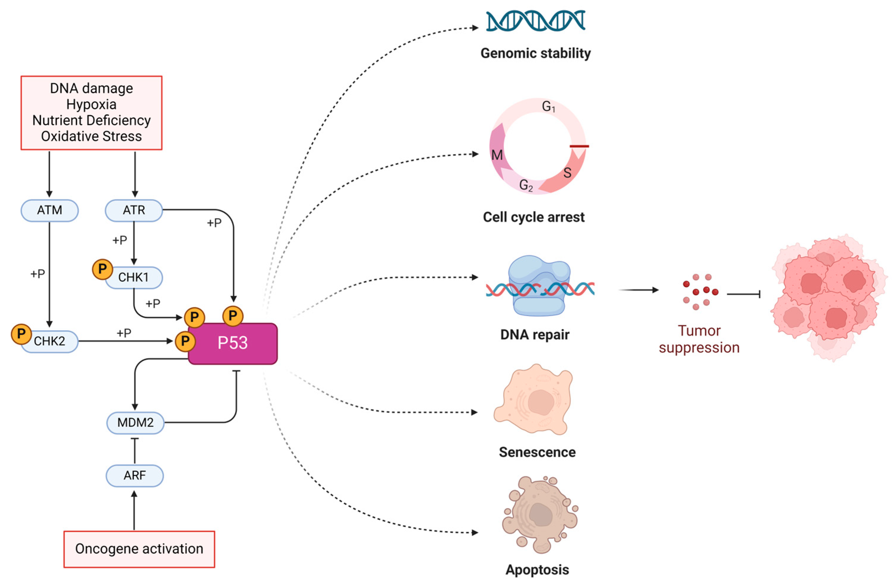
  • Exon 1–2: Encodes the transactivation domain, which is responsible for activating the transcription of target genes.
  • Exon 3–4: Contains sequences that contribute to the proline-rich region, which is important for the apoptotic function of p53.
  • Exon 5–8: Encodes the DNA-binding domain, a crucial region that allows the p53 protein to bind to specific DNA sequences and regulate gene expression. This region is also the most frequently mutated in cancers.
  • Exon 9: Encodes part of the oligomerization domain, which is essential for the tetramer formation of the p53 protein.
  • Exon 10–11: Encodes the remaining part of the oligomerization domain and the C-terminal regulatory domain, which is involved in the regulation of p53’s activity.
Approximately 80% of TP53 mutations are missense mutations that are primarily found in exons 5–8, which encode the DNA-binding domain. According to the Catalogue of Somatic Mutations in Cancer (COSMIC) database, the most frequently mutated sites across all tumor types include R175, G245, R248, R249, R273, and R282 [20]. In contrast, the introns play roles in gene regulation, mRNA stability, and alternative splicing, which can influence the protein’s functionality. Less commonly, TP53 intronic polymorphisms were also discovered, which may regulate gene expression and affect susceptibility to cancer. For instance, earlier studies have indicated that a polymorphism in intron 6 is significantly linked to an increased risk of various cancers, such as gastrointestinal tumors, breast cancer, thyroid cancer, and ovarian cancer, and is associated with the notorious Li–Fraumeni syndrome. Common hotspot mutations in p53 and their respective phenotypes are summarized in Table 1.
The etiology of TP53 gene mutations is influenced by a combination of genomic instability, environmental factors, and inherited genetic predispositions [31]. Genomic instability, often resulting from errors in DNA replication or repair, can lead to mutations in TP53. Environmental carcinogens, such as tobacco smoke, UV radiation, and certain chemicals, can further exacerbate this instability by directly causing DNA damage and introducing mutations into the TP53 gene [32]. Germline mutations in TP53, as observed in disorders like the Li–Fraumeni syndrome, can predispose individuals to a higher risk of developing multiple cancers from birth [33]. Additionally, infections, like those caused by human papillomavirus (HPV), can interfere with p53 function and increase mutation rates [34]. Moreover, recent studies have revealed that p53 activity can be regulated at the post-translational level [35].

3. Mechanisms of Immune Evasion Caused by TP53 Mutations

Accumulating evidence from basic research suggests that p53-mutant tumors are more immune evasive compared to p53-WT tumors. In this section, we summarize the mechanisms of immune evasion from both the adaptive and innate immune systems driven by TP53 mutations/deletion (Figure 2). The immunomodulatory effects of different p53 mutations are summarized in Table 2.

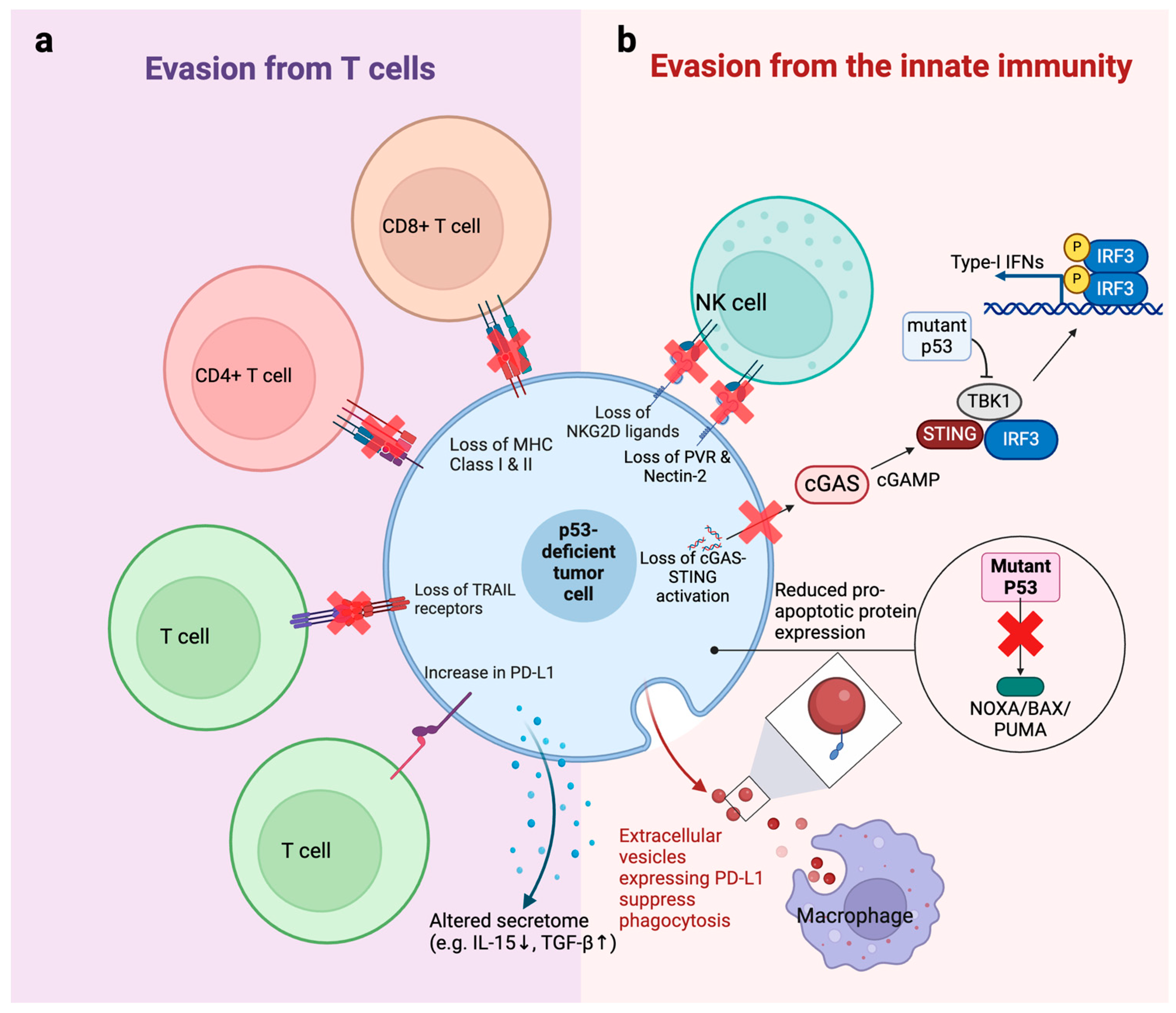
3.1. Evasion from T Cells

T cells are the most critical immune cells in surveilling and eliminating malignant transformed cells. In vivo CRISPR screening identified TP53 as a top tumor suppressor gene in immunocompetent WT mice but not in SCID immunodeficient mice using the 4T1 breast tumor syngeneic model, suggesting that loss of TP53 may contribute to evasion from the adaptive immune system [45]. Tumor cells present peptides derived from tumor-associated antigens or tumor-specific antigens on their surface in the context of major histocompatibility complex (MHC) class I molecules, which T cells recognize through their T cell receptors (TCRs). It has been shown that tumor cells could develop resistance to T cells by alterations in MHC molecules, making them less recognizable to cytotoxic T lymphocytes [46]. They can also upregulate immune checkpoints and soluble immunosuppressive molecules to contribute to evasion from T cells [47]. Evidence suggests that p53 mutations can cause T cell immune evasion by similar mechanisms (Figure 2a).
p53 can regulate immune-activating ligands on tumor cells. p53 has been shown to regulate MHC class I and II expression in various cancer types via diverse mechanisms. p53 can transcriptionally activate endoplasmic reticulum aminopeptidase 1 (ERAP1) expression [36]. ERAP1 is responsible for the final trimming of antigen precursors before loading into MHC class I proteins. Defects in ERAP1 result in changes in MHC class I expression and impaired T cell responses. Therefore, p53-null tumor cells have less MHC class I expression, highlighting the important role of p53 in the immune response [36]. The transporter associated with antigen processing (TAP) gene plays an essential role in MHC class I antigen presentation. Zhu et al. found that p53 induced surface MHC class 1 expression by upregulating TAP1 [48], which was also observed by another study [36]. Beyond MHC I, p53 was also shown to regulate MHC class II. Activating p53 by mouse double minute 2 (MDM2) inhibitors, a class of p53-activating agents, could upregulate MHC class II in p53-WT but not p53-knockdown (KD) acute myeloid leukemia (AML) cells, suggesting that the regulation of MHC II by MDM2 inhibitor is p53 dependent [49]. Apoptosis-inducing ligand receptor-1 and -2 (TRAIL-R1/2) plays a central role in the graft versus leukemia (GVL) effect during hematopoietic stem cell transplantation therapy [50]. p53 was reported to be a transcription factor for TRAIL-R1/2, which partially explains the immune evasion of p53-mutant AML cells [49].
In addition to immune-activating molecules, p53 can regulate immunosuppressive molecules in tumor cells, such as the immune checkpoint PD-L1 (CD274). Cortez et al. showed that WT p53 downregulated PD-L1 expression by increasing levels of miR-34 microRNA [37]. Another study demonstrated a similar regulatory mechanism via miR-320a along with miR-200a and miR-34a [51]. These findings suggest that p53 mutations could cause PD-L1 upregulation and, therefore, immune evasion.
Beyond the cell surface molecules mentioned above, TP53 may regulate the secretome of tumor cells, which mediates immune activation or suppression. One study found that p53 induction by MDM2 inhibitor upregulated IL-15, which serves as an activator of anti-tumor CD8+ T cells and NK cells [52]. Another study showed that an AML cell line with TP53 KD secreted more TGF-β to suppress T cell proliferation and cytotoxicity [53]. Whether other soluble factors are also regulated by p53 remains to be determined.

3.2. Evasion from the Innate Immune System

p53 mutation can drive immune evasion by disrupting immunosurveillance mechanisms within the innate immune system (Figure 2b). As an important member of the innate immune system, natural killer (NK) cells supplement the function of T cells by killing MHC-defective tumor cells [54]. Unlike T cells, NK cells do not require MHC. They recognize abnormal cells by a variety of activating and inhibitory receptors [55]. Studies showed that p53 is important in NK immune surveillance. p53 transcriptionally regulates ULBP1 and ULBP2 in tumor cells, which serve ligands for the NKG2D receptor on NK cells [42]. Inducing WT p53 but not mutant p53 in tumor cells increased recognition by NK cells characterized by higher IFN-γ expression and degranulation in NK cells, highlighting the importance of WT p53 in NK recognition. In addition to NKG2D ligands, ligands for DNAM1 are also modulated by p53 [56]. Veneziani et al. showed that p53 could activate the transcription of PVR and Nectin-2, which are ligands for DNAM1. Restoring p53 by MDM2 inhibitor Nutlin-3a increased PVR and Nectin-2 expression and sensitized neuroblastoma cells to NK adoptive immunotherapy [56]. On top of reduced NK recognition, tumor cells might acquire intrinsic resistance to NK cytotoxicity by reducing their sensitivity to apoptosis. A study showed that the mitochondrial apoptosis of tumor cells is vital for NK-mediated killing [57]. Mitochondrial apoptosis is regulated by a series of pro-apoptotic and anti-apoptotic proteins in the mitochondria. p53 is known to induce the expression of pro-apoptotic proteins Noxa [58], Bax [59], and Puma [6], thus making tumor cells more primed to mitochondrial apoptosis. Therefore, loss of p53 might also cause resistance to NK cells by reducing the sensitivity of cancer cells to apoptosis.
Macrophages serve as a part of the body’s first line of defense against pathogens but have a complex role in cancers. In antibody-based therapies, macrophages are extremely important anti-tumor effectors. They exert their anti-tumor effect via antibody-dependent cellular phagocytosis (ADCP) [60]. p53-null B cell malignancies were shown to have reduced phagocytosis by macrophages [38]. Mechanistically, p53-null cells produce more extracellular vesicles (EV) compared to p53-WT cells. These EV from p53-null cells carry higher PD-L1, which inhibits ADCP by macrophages. This study demonstrated that TP53 mutations may cause resistance to ADCP and that PD-1/PD-L1 checkpoint inhibitors may be a good partner with therapeutic antibodies in treating p53-deficient B cell malignancies.
Additionally, the loss and mutation of p53 have also been reported to interfere with the function of the cGAS-STING cytosolic DNA sensing pathway. The cGAS-STING pathway is a critical mechanism of the innate immune system that detects cytosolic DNA, a signal often associated with viral infections or cellular damage [14,61]. Upon sensing DNA, the enzyme cGAS produces cyclic GMP-AMP (cGAMP), which activates the STING protein located in the endoplasmic reticulum. This activation triggers a signaling cascade involving TBK1 and IRF3, leading to the production of type I interferons and other cytokines that orchestrate the immune response and enhance the body’s defense against pathogens. Ghosh et al. has recently found that TP53 promotes the degradation of cytosolic DNA exonuclease TREX1, causing cytoplasmic DNA accumulation and activation of the cGAS-STING pathway [39]. Consequently, the deletion of TP53 results in a direct loss of cGAS-STING pathway activation, while mutant p53 proteins but not wild-type p53 have been found to bind to TANK-binding protein kinase 1 (TBK1) [44]. This abrogates the formation of a critical trimeric complex between TBK1, STING, and IRF3, which is necessary for the activation of the innate immune response via the cGAS-STING-TBK1-IRF3 pathway. As such, it is postulated that the loss or mutation of TP53 results in escape from the innate immune system.

4. p53-Mutant Tumor Cells Create an Immunosuppressive TME to Promote Immune Evasion

The crosstalk between TP53-mutated cancer cells and the tumor microenvironment, particularly involving myeloid cells and Treg cells, plays a crucial role in tumor progression and immune evasion. Myeloid cells, including macrophages and neutrophils, exhibit phenotypic and functional alterations in the presence of TP53-mutated cancer cells [62]. These alterations often lead to an immunosuppressive microenvironment that facilitates tumor growth and metastasis (Figure 3).

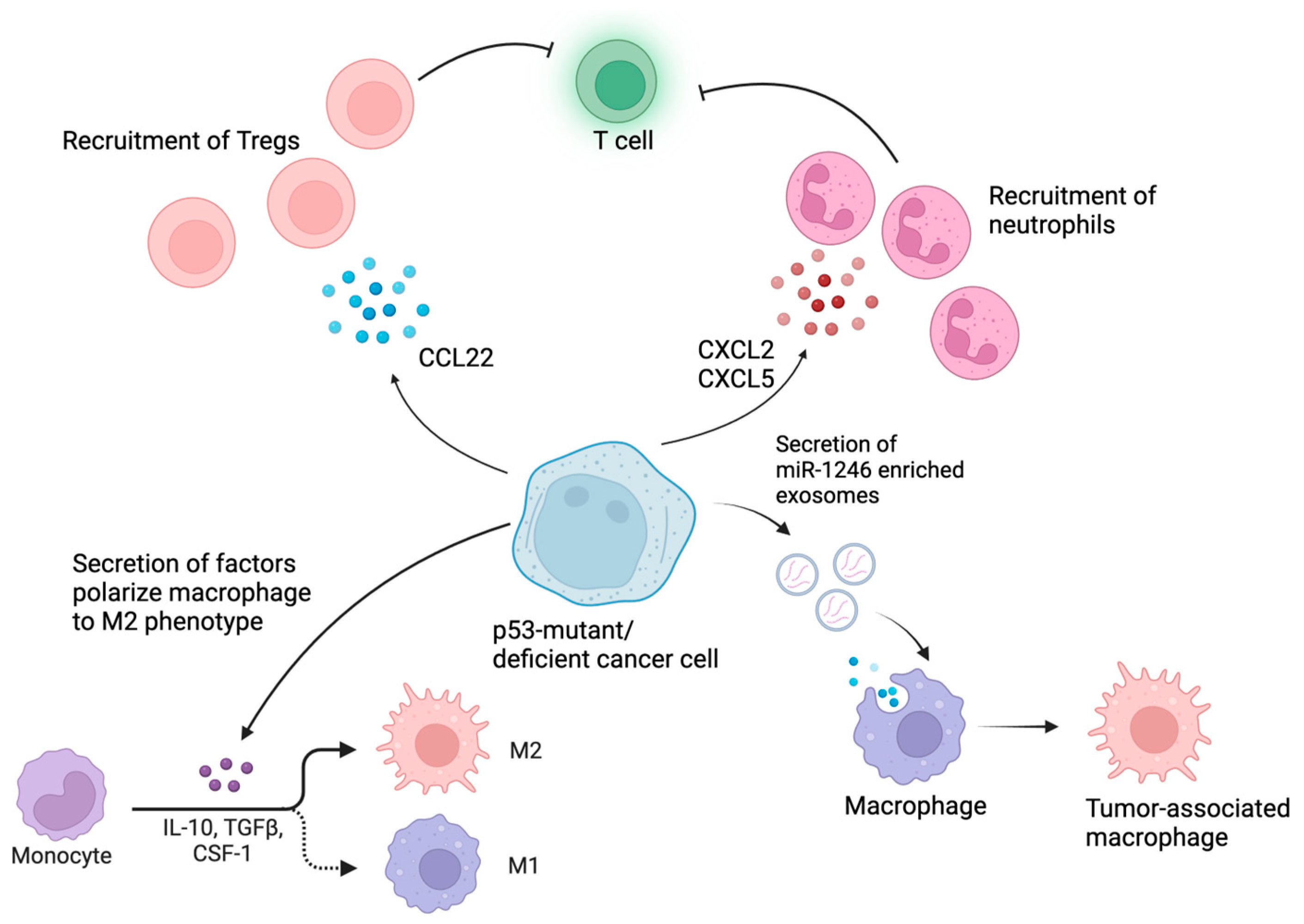
4.1. p53 Mutations Reprogram Myeloid Cells in the TME into Immunosuppressive Phenotypes

Within the TME, macrophages can adopt different functional states, broadly categorized as M1-like (classically activated) or M2-like (alternatively activated). M2-like macrophages are often found to be correlated with immunosuppression and failure of immunotherapies in various types of cancer [63,64]. p53 mutations within cancer cells have been shown to skew the polarization of macrophages towards an M2-like phenotype [43,65]. This shift in polarization is often associated with an immunosuppressive microenvironment that promotes tumor growth, invasion, and metastasis. p53-mutated cancer cells may also secrete factors such as CSF-1 (colony-stimulating factor 1) [40], IL-10 (interleukin-10) [43], and TGF-β (transforming growth factor-beta) [53], which are known to polarize macrophages towards the M2 phenotype. Li et al. have also demonstrated that p53 suppresses M2 polarization through the inhibition of c-myc, a transcription factor that promotes M2 macrophage polarization [66]. Beyond disrupting the tumor-suppressive functions of WT p53, certain missense mutations confer GOF activities to mutant p53 (mutp53) proteins [11]. These GOF activities profoundly alter tumor cell behavior by influencing protein interactions and modulating transcriptional programs. As such, it has been shown that p53-mutant cancer cells can actively reprogram macrophages into a tumor-supportive and anti-inflammatory state. Specifically, colon cancer cells carrying GOF mutp53 selectively release exosomes enriched with miR-1246 [43]. Upon uptake by neighboring macrophages, these exosomes induce miR-1246-dependent reprogramming that favors cancer progression. The mutp53-reprogrammed tumor-associated macrophages (TAMs) exhibit enhanced immunosuppressive activity, characterized by increased levels of TGF-β. Another study showed that the GOF TP53 R172H mutation drives an increase in CD11b+Ly6G+ neutrophils, accompanied by a reduction in CD3+ T cells, CD8+ T cells, and CD4+ T helper 1 cells [41].

4.2. p53-Mutant Cancers Recruit Tregs to Cause Immunosuppression

Tregs are a key component of the immunosuppressive network within the TME, helping cancer cells evade the immune system and promoting tumor progression [67]. They can be recruited to the tumor microenvironment by various chemokines, and these Tregs promote tumor growth via inhibition of anti-tumor cells such as cytotoxic CD8+ T cells. One study showed that p53 deficiency in prostate, ovarian, and pancreatic cancers increased the frequency of Tregs in the TME. This was demonstrated to be via the suppression of miR-34a, causing enhanced production of CCL22 and enhancing Treg recruitment to the TME [68]. When Tregs are recruited by CCL22, they have also been shown to be selectively activated in the lymphoid infiltrates surrounding breast tumors, which is associated with an adverse clinical outcome in primary breast cancer patients [69]. Blagih et al. found that p53-null tumor cells had an accumulation of Tregs, accompanied by a lower CD4+ T helper 1 (Th1) and CD8+ T cell response in mouse models [62]. Tregs from AML patients with TP53 mutations have also been shown to exhibit distinct metabolic characteristics. These Tregs display an enrichment of gene sets linked to glycolysis, fatty acid metabolism, and oxidative phosphorylation relative to controls [70]. This indicates that p53-mutant cancers possibly influence Tregs within the tumor microenvironment to enhance their energy production using both glucose and fatty acids in the context of AML.
Overall, the crosstalk between p53-mutant cancer cells and the tumor microenvironment, particularly involving myeloid cells and Treg cells, creates a conducive immunosuppressive niche that promotes tumor growth, invasion, and resistance to immunotherapy. Understanding these interactions is essential for developing novel therapeutic strategies to target p53-mutant tumors and overcome immune evasion mechanisms.

5. Clinical Evidence of Immune Evasion in p53-Mutant Cancers

Mutations in p53 are significant predictors of poor responses to various immunotherapy-based treatment options (Table 3). Clinically, p53-mutant diffuse large B cell lymphoma (DLBCL) demonstrates significantly worse outcomes when treated with anti-CD19 CAR-T therapy, showing a complete response (CR) rate of 34%, markedly lower than the 65% CR rate observed in p53-WT patients [71]. Moreover, the 1-year overall survival (OS) rate for patients with TP53 alterations is 44% compared to 76% for those without such alterations. Progression-free survival (PFS) is also shorter in the p53-altered group. Transcriptomic analysis showed alterations in interferon and apoptosis pathways, which are crucial for the cytotoxic effects of CAR-T cells [71].
p53 mutation is also a poor prognostic marker of allogeneic hematopoietic stem cell transplantation (allo-HSCT). HSCT remains the only curative treatment in many blood malignancies and the first therapeutic option for high-risk leukemia patients. HSCT is known to eliminate leukemia cells by the GvL effect by which reconstituted donor immune cells kill host leukemia cells. Relapse after HSCT is more common in p53-mutant patients, with a three-year OS rate as low as 10% in AML patients after HSCT [72,73]. TP53 status is also an independent predictor of poor survival for myelodysplastic syndrome (MDS) patients treated with stem cell transplantation [74].
p53 mutations may significantly influence the response to immune checkpoint blockade (ICB) therapies. In gastric cancer, patients treated with nivolumab exhibit higher objective response rates in p53-WT patients compared to those with p53 mutations [75]. Similarly, worse survival outcomes are observed in p53-mutant patients treated with atezolizumab in non-small cell lung cancer (NSCLC) compared to patients without such mutations [76,77]. This impact of ICB is also evident in patients treated with pembrolizumab, where those with p53 truncating mutations have a shorter median survival compared to p53-WT patients, while those with p53 missense mutations show less clear but potentially negative impacts on survival [78]. Additionally, Kim et al. reported the poor immunotherapy response in p53-mutant metastatic solid tumors [79].
Contradictory reports, however, showed that mutant p53 can be more immunogenic compared to WT p53. For instance, the CD123 × CD3 bispecific antibody flotetuzumab demonstrated higher objective response rates in p53-mutant cases than in p53-WT cases, attributed to higher immune infiltration and IFN-gamma responses [80].

6. Strategies Enhancing the Efficacy of Immune Response by Targeting p53

Given the role of p53 in regulating immune response, activating or restoring p53 functions is expected to enhance the anti-tumor immunity in cancers. In fact, p53-activating/restoring agents combined with various immunotherapies have been shown to achieve a synergy in treating multiple cancers in preclinical and clinical studies. One class of p53-activating agents is MDM2 inhibitors. Under normal conditions, p53 levels are kept low by MDM2, which binds to p53, promoting its ubiquitination and proteasomal degradation [81]. By inhibiting the interaction between MDM2 and p53, MDM2 inhibitors prevent the ubiquitination and subsequent degradation of p53, leading to the stabilization and accumulation of p53 protein in the cell. MDM2 inhibitors are being studied in combination with other therapies, such as chemotherapy and immune therapies, to enhance their efficacy and overcome resistance mechanisms. However, despite their promise in p53-WT cancers, most MDM2 inhibitors are not able to restore the functions of mutant p53. Therefore, some small molecules have been developed in recent years to restore the WT-like functions and structure in mutant p53, serving as promising candidates for treating p53-mutant cancers. It is noteworthy that, over the past decade, these small molecule p53-targeted therapies have experienced the most rapid growth among different p53-targeted therapies [82]. Despite this, other p53-targeted therapies, such as gene therapies and immune-based therapies, also gained huge interest in overcoming p53 mutation or deletion. In this section, we attempt to summarize the progress in targeting MDM2 to potentiate immunotherapies and discuss the potential of using STING agonists, p53-restoring agents, gene therapies, and immune-based therapies in targeting immune evasion in p53-mutant cancers (Figure 4).

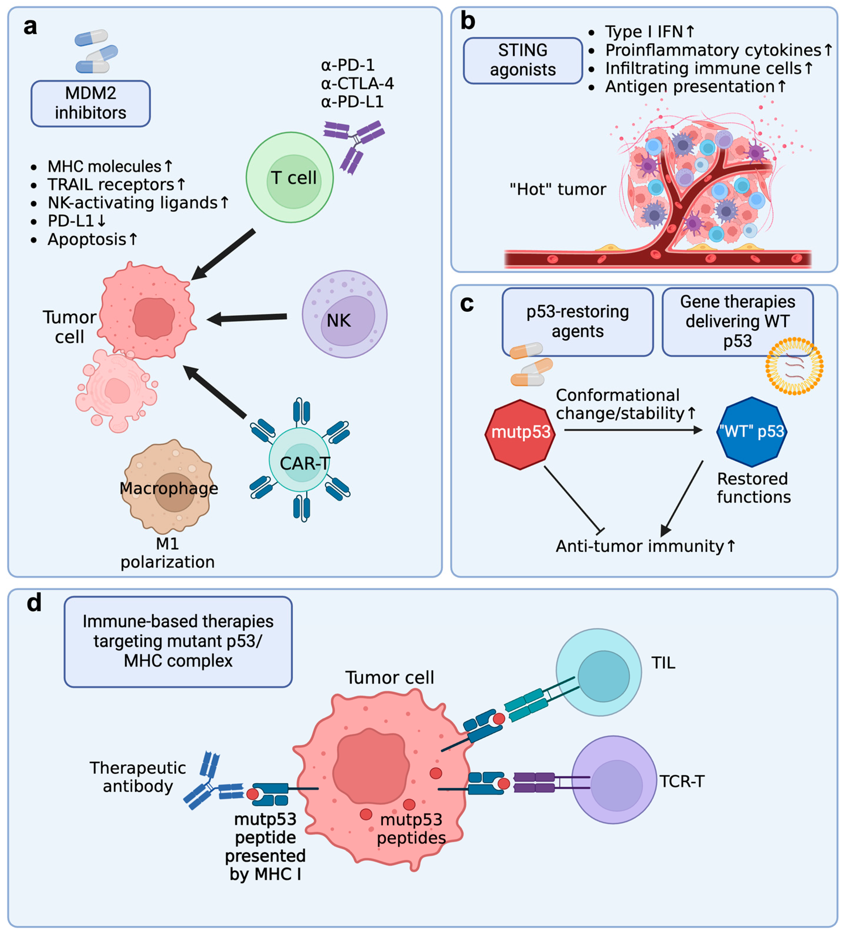
6.1. MDM2 Inhibitor + Immune Checkpoint Blockade (ICB)

ICB has achieved success in treating multiple malignancies, such as lung cancer, triple-negative breast cancer, melanoma, and gastrointestinal cancers [83]. However, the response rate of ICB remains low. Multiple studies have shown that MDM2 inhibitors achieve a synergy with ICB in treating cancers (Figure 4a). In a pre-clinical study using syngeneic mouse lung cancer models, Zhu et al. found that tumor immunogenicity and response to ICB are dependent on p53 status, partially due to reduced antigen presentation [78]. Stabilizing p53 by MDM2 inhibitor Nutlin-3a increased T cell cytotoxicity and the efficacy of ICB. Another MDM2 inhibitor, APG-115, was also shown to enhance the anti-tumor activity of anti-PD-1 therapy in syngeneic mouse models [84]. Surprisingly, in the same study, APG-115 also enhanced anti-PD-1 therapy in treating p53-mutant and p53-null tumors. This is unexpected as MDM2 inhibitors are thought to induce apoptosis only in p53-WT tumors. This suggests that APG-115 might have tumor-independent effects, such as impacts on the tumor microenvironment. Mechanistic studies showed that the drug reprogramed immunosuppressive M2 macrophages into the proinflammatory M1 phenotype and increased cytotoxic CD8+ T cells in the tumors. Consistent with this finding, Guo et al. showed that Nutlin-3a reversed immunosuppression in the TME to induce systemic anti-tumor immunity and immune regression [58]. In another study, MDM2 inhibitor APR-246 was shown to counteract GOF immune evasive mechanisms by regulating IFN expression, promoting CD4+ T cell infiltration, and slowing T cell exhaustion in the TME [13]. These pre-clinical findings provide rationale for moving the combination therapy into clinical trials. A phase II clinical trial (NCT03611868) showed that APG-115 in combination with pembrolizumab was well-tolerated and had preliminary anti-tumor activity in multiple tumor types [85].

6.2. MDM2 Inhibitor + Immune Cell Adoptive Transfer Therapy

We have introduced that p53 can upregulate the expression of ligands for NK activating receptors, such as ULBP1/2, PVR, and Nectin-2 [42,56]. Therefore, MDM2 inhibitors might have a synergy with NK adoptive transfer therapy. Indeed, MDM2 inhibitors RITA and Nutlin-3a were shown to increase NK cytotoxicity in vitro and in vivo [42,56], which might be used for NK cell-based immunotherapy. Another study showed that Nutlin-3a effectively sensitized neuroblastoma cells to DNAM-1 CAR-NK cells by upregulating DNAM-1 ligands [86].

6.3. MDM2 Inhibitors + HSCT

Many patients suffer from relapse after HSCT [87]. Targeting p53 might be a potential way to prevent relapse after HSCT by activating anti-tumor immunity. In mouse models, MDM2 inhibitors successfully prolonged the survival of mice after HSCT by three converging mechanisms: MHC-II upregulation, TRAIL-R1/2, and direct activation of T cells [49]. This study suggested that giving MDM2 inhibitors to AML patients after HSCT might be a promising strategy to enhance anti-leukemia immunity and prevent relapse.

6.4. STING Agonists + Immunotherapies

Due to the importance of the STING pathway in anti-tumor immunity, various STING agonists have been developed to treat cancers [88]. Accumulating evidence has shown that p53-mutant tumors have reduced STING signaling and are immunologically “cold”, which underlies the immune evasion of the p53-mutant tumors [39,44]. Therefore, reactivating the STING pathway may be a potential way to restore the anti-tumor immunity against p53-mutant tumors. Putting STING back to p53-mutant blood cancers was shown to have a synergy with BH3 mimetics in p53-mutant cells and prolong the survival of tumor-bearing mice [89]. Therefore, it is also possible that STING agonists can enhance the apoptotic priming and immunogenicity of p53-mutant tumors, which make the malignant cells more responsive to immunotherapies such as ICB and CAR-T (Figure 4b).

6.5. Targeting Immune Evasion of p53-Mutant Cancers by p53-Restoring Agents

Mutant p53 had long been considered undruggable. Although MDM2 inhibitors can induce apoptosis in p53-WT cancers, their effect in p53-mutant cancers is limited. However, small molecules that restore the structure and functionality of mutant p53 give hope to enhance the immune response to p53-mutant cancers (Figure 4c). In a phase 1b clinical study, Eprenetapopt (APR-246) was combined with pembrolizumab in patients with advanced/metastatic solid tumors (NCT04383938) [90]. Eprenetapopt is a first-in-class p53-reactivating small molecule that covalently modifies cysteine residues in mutant p53, leading to restoration of WT p53 protein function. Overall, the combination of eprenetapopt and pembrolizumab was well-tolerated and showed clinical activity in heavily treated patients. Further clinical studies are needed to evaluate the anti-tumor activity of this therapy and determine responsive tumor subsets.

6.6. Restoring Function of Mutant p53 with Gene Therapies

The effectiveness of small molecules in reactivating mutant p53 is questionable [91]. An alternative approach to restoring p53 function involves delivering the WT TP53 gene into tumor cells (Figure 4c). Various vectors have been explored for this purpose. One promising example is SGT-53, a cationic liposome complex encapsulating WT p53 cDNA. In a pre-clinical study, SGT-53 was shown to restore anti-tumor immunity and overcome tumor resistance to a checkpoint inhibitor [92]. In a phase II trial, SGT-53 combined with gemcitabine/nab-paclitaxel demonstrated encouraging results in treating metastatic pancreatic cancer [93]. mRNA nanoparticles have emerged as a promising platform for gene therapies, particularly following their success in COVID-19 vaccines. Synthetic mRNA nanoparticles could restore p53 expression and sensitize p53-deficient cancers to mTOR inhibition [94].
Although the concept of gene therapies seems promising, several challenges remain. (1) Can the gene delivery systems reach out to all tumor cells? If not, some p53-mutant or -null cells can remain resistant to anti-tumor immunity and therapies. (2) Mutant p53 can interfere with WT p53 in a dominant-negative manner [9]. Therefore, reintroduced WT p53 may not fully regain its function in cells harboring p53 mutations.

6.7. Targeting Mutant p53 by Immune-Based Therapies

Mutant p53 is an intracellular protein and is out of reach for immune cells and antibodies. However, it can be degraded into peptides and presented by MHC on the cell surface, making it targetable by T cells. Thus, missense mutant p53 may become a neoantigen and a potential target for immune-based therapies (Figure 4d). Tumor-infiltrating lymphocytes (TILs) isolated from p53-mutant tumors have been shown to recognize mutant p53 as a neoantigen, suggesting that mutant p53 is immunogenic [20,95]. It is, therefore, possible to use TCR-T or TIL therapies to treat p53-mutant cancers.
Kim et al. successfully generated a library of 39 TCRs that recognize tumor cells in a TP53 mutation- and human leucocyte antigen (HLA)-specific manner [96]. They then treated 12 patients with TILs that were naturally reactive against p53 mutations. However, the efficacy was modest, with only two partial responses among twelve patients. They also treated one patient with TCR-T comprising autologous T cells transduced with an allogeneic HLA-A_02–restricted TCR specific for p53R175H, resulting in a tumor regression (−55%) over 6 months.
Bispecific T cell engagers have also become more and more popular in recent years. Hsiue et al. developed a bispecific antibody that targets p53R175H presented by HLA-A*02:01 on one arm and CD3 on the other arm to engage T cells to kill tumor cells [97]. This strategy was effective even for killing tumor cells with low antigen density on their surface.
However, there are limitations to targeting p53 neoantigens. Although T cells can recognize tumor cells carrying p53 mutations, they may be unable to kill the tumor cells due to the immunosuppressive mechanisms of mutant p53, as discussed in previous sections. This was observed in p53-mutant large B cell lymphomas, which express CD19 antigen but remain insensitive to CD19-CAR-T therapy [71]. Combining TCR-T, TIL, or bispecific antibodies with ICB or other therapies could potentially improve outcomes by counteracting the immune-evasive mechanisms of p53-mutant tumors.

7. Discussion

Despite advancements in cancer treatment over the past decades, the effective treatment of p53-mutant tumors remains a formidable challenge. TP53 mutation or deletion, which renders the tumor suppressor p53 inactive, was once considered completely undruggable due to the immense difficulty of targeting cancer cells carrying these p53 mutations or deletions. Historically, the primary focus of p53 has been centered on its role in cell cycle arrest, apoptosis, and senescence, all of which are critical for tumor suppression. However, the significance of p53 in regulating the immune response has been underappreciated. In this review, we have summarized up-to-date evidence from both basic research and clinical studies, which demonstrate the immune evasive properties of p53-mutant cancers. We have also reviewed the potential mechanisms by which TP53 mutations facilitate immune evasion. These mechanisms include alterations in antigen presentation, modulation of immune checkpoints, alterations in the cancer cell secretome, modulation of NK cell activation, suppression of phagocytosis by macrophages, and changes in the tumor microenvironment that foster immunosuppression. The studies about the mechanisms are crucial for developing new therapeutic strategies.
Based on these findings, combinatorial therapies are currently under investigation to enhance current immunotherapies or to overcome the immune evasion mechanisms associated with TP53 mutations. These therapies include the use of p53-reactivating agents in combination with ICB, HSCT, and cellular immunotherapies. Given the suppression of the cGAS-STING pathway by mutant p53, we believe that STING agonists should be further evaluated for the potential to restore the immune response in p53-mutant cancers. Future studies are also needed to rigorously assess the efficacy and safety of these combinatorial therapies.
Although this review mainly discusses the alterations of p53 in cancer cells, the p53 status of immune cells may also modulate their respective immune functions. Activating or reactivating p53 in non-tumor cells in the TME also represents a compelling strategy to counteract immunosuppression [98]. p53 mutations are identified in clonal hematopoiesis, which means these mutations can occur in hematopoietic stem cells and immune cells differentiated from these stem cells without overt malignancy [99]. However, whether p53 mutations in the immune compartment are associated with specific functional changes and disease outcome remains to be determined. Additionally, whether p53 mutations in pre-malignant clones promote malignant transformation via immune evasion remains to be studied.

8. Conclusions

Despite continuous advances in treating p53-mutant cancers, significant challenges remain, and further efforts are needed to develop novel compounds targeting these malignancies. p53-targeted drugs are unlikely to be effective as monotherapies, making it crucial to explore combination strategies that include p53-targeted therapies alongside immunotherapies, targeted therapies (e.g., BCL-2 inhibitors, SMAC mimetics, STING agonists), and others. In our view, genome-wide CRISPR screening offers a powerful approach for identifying mechanisms of immune evasion and potential therapeutic targets in p53-mutant tumors. Unlike transcriptomics and proteomics, CRISPR screens provide direct functional insights by loss-of-function or gain-of-function genetic manipulation in cells. These screens can be applied to both tumor and immune cell compartments, allowing for complete gene disruption, leading to clearer phenotypic outcomes. Additionally, drug screening is another strategy for discovering molecules that can be combined with immunotherapies to target p53-mutant cells.
In summary, while the role of p53 in cancer immunity is complex, the insights gained from recent studies provide a foundation for developing new therapeutic strategies. Translating these findings into effective treatments for patients with p53-mutant cancers remains a critical goal. Ongoing studies and clinical trials will be pivotal in bringing these innovative therapies from the laboratory to the clinic, ultimately improving outcomes and providing new hope for patients battling these challenging forms of cancer.

Author Contributions

Conceptualization, C.W. and J.Y.M.T.; investigation, C.W., J.Y.M.T. and N.C.; resources, C.W., J.Y.M.T. and N.C.; data curation, C.W., J.Y.M.T. and N.C.; writing—original draft preparation, C.W., J.Y.M.T. and N.C.; writing—review and editing, C.W. and J.Y.M.T.; visualization, C.W. and J.Y.M.T.; supervision, S.B.; project administration, C.W.; funding acquisition, S.B. All authors have read and agreed to the published version of the manuscript.

Funding

S.B. acknowledges funding from the Paris-NUS Call for Proposals 2024 (ANR-18-IDEX-0001). C.W. acknowledges support from the Paris-NUS call for PhD mobility 2024 (ANR-18-IDEX-0001).

Conflicts of Interest

The authors declare no conflicts of interest.

References

  1. Ge, T.; Gu, X.; Jia, R.; Ge, S.; Chai, P.; Zhuang, A.; Fan, X. Crosstalk between metabolic reprogramming and epigenetics in cancer: Updates on mechanisms and therapeutic opportunities. Cancer Commun. 2022, 42, 1049–1082. [Google Scholar] [CrossRef]
  2. Zhang, Y.; Xiong, S.; Liu, B.; Pant, V.; Celii, F.; Chau, G.; Elizondo-Fraire, A.C.; Yang, P.; You, M.J.; El-Naggar, A.K.; et al. Somatic Trp53 mutations differentially drive breast cancer and evolution of metastases. Nat. Commun. 2018, 9, 3953. [Google Scholar] [CrossRef]
  3. Olivier, M.; Langerød, A.; Carrieri, P.; Bergh, J.; Klaar, S.; Eyfjord, J.; Theillet, C.; Rodriguez, C.; Lidereau, R.; BièChe, I.; et al. The clinical value of somatic TP53 gene mutations in 1,794 patients with breast cancer. Clin. Cancer Res. 2006, 12, 1157–1167. [Google Scholar] [CrossRef]
  4. Lane, D.P. p53, guardian of the genome. Nature 1992, 358, 15–16. [Google Scholar] [CrossRef]
  5. Vogelstein, B.; Lane, D.; Levine, A.J. Surfing the p53 network. Nature 2000, 408, 307–310. [Google Scholar] [CrossRef] [PubMed]
  6. Nakano, K.; Vousden, K.H. PUMA, a novel proapoptotic gene, is induced by p53. Mol. Cell 2001, 7, 683–694. [Google Scholar] [CrossRef] [PubMed]
  7. Brady, C.A.; Jiang, D.; Mello, S.S.; Johnson, T.M.; Jarvis, L.A.; Kozak, M.M.; Broz, D.K.; Basak, S.; Park, E.J.; McLaughlin, M.E.; et al. Distinct p53 transcriptional programs dictate acute DNA-damage responses and tumor suppression. Cell 2011, 145, 571–583. [Google Scholar] [CrossRef] [PubMed]
  8. Rivlin, N.; Brosh, R.; Oren, M.; Rotter, V. Mutations in the p53 Tumor Suppressor Gene: Important Milestones at the Various Steps of Tumorigenesis. Genes Cancer 2011, 2, 466–474. [Google Scholar] [CrossRef]
  9. Boettcher, S.; Miller, P.G.; Sharma, R.; McConkey, M.; Leventhal, M.; Krivtsov, A.V.; Giacomelli, A.O.; Wong, W.; Kim, J.; Chao, S.; et al. A dominant-negative effect drives selection of TP53 missense mutations in myeloid malignancies. Science 2019, 365, 599–604. [Google Scholar] [CrossRef]
  10. Roszkowska, K.A.; Piecuch, A.; Sady, M.; Gajewski, Z.; Flis, S. Gain of Function (GOF) Mutant p53 in Cancer-Current Therapeutic Approaches. Int. J. Mol. Sci. 2022, 23, 13287. [Google Scholar] [CrossRef]
  11. Alvarado-Ortiz, E.; de la Cruz-López, K.G.; Becerril-Rico, J.; Sarabia-Sánchez, M.A.; Ortiz-Sánchez, E.; García-Carrancá, A. Mutant p53 Gain-of-Function: Role in Cancer Development, Progression, and Therapeutic Approaches. Front. Cell Dev. Biol. 2021, 8, 607670. [Google Scholar] [CrossRef] [PubMed]
  12. Olivier, M.; Hollstein, M.; Hainaut, P. TP53 mutations in human cancers: Origins, consequences, and clinical use. Cold Spring Harb. Perspect. Biol. 2009, 2, a001008. [Google Scholar] [CrossRef] [PubMed]
  13. Geske, F.J.; Nelson, A.C.; Lieberman, R.; Strange, R.; Sun, T.; E Gerschenson, L. DNA repair is activated in early stages of p53-induced apoptosis. Cell Death Differ. 2000, 7, 393–401. [Google Scholar] [CrossRef] [PubMed]
  14. McCubrey, J.A.; Yang, L.V.; Abrams, S.L.; Steelman, L.S.; Follo, M.Y.; Cocco, L.; Ratti, S.; Martelli, A.M.; Augello, G.; Cervello, M. Effects of TP53 Mutations and miRs on Immune Responses in the Tumor Microenvironment Important in Pancreatic Cancer Progression. Cells 2022, 11, 2155. [Google Scholar] [CrossRef]
  15. Zhu, K.-L.; Su, F.; Yang, J.-R.; Xiao, R.-W.; Wu, R.-Y.; Cao, M.-Y.; Ling, X.-L.; Zhang, T. TP53 to mediate immune escape in tumor microenvironment: An overview of the research progress. Mol. Biol. Rep. 2024, 51, 205. [Google Scholar] [CrossRef]
  16. Zhou, X.; Santos, G.S.; Zhan, Y.; Oliveira, M.M.S.; Rezaei, S.; Singh, M.; Peuget, S.; Westerberg, L.S.; Johnsen, J.I.; Selivanova, G. Mutant p53 gain of function mediates cancer immune escape that is counteracted by APR-246. Br. J. Cancer 2022, 127, 2060–2071. [Google Scholar] [CrossRef]
  17. Wang, H.; Guo, M.; Wei, H.; Chen, Y. Targeting p53 pathways: Mechanisms, structures, and advances in therapy. Signal Transduct. Target. Ther. 2023, 8, 92. [Google Scholar] [CrossRef]
  18. Jeffrey, P.D.; Gorina, S.; Pavletich, N.P. Crystal structure of the tetramerization domain of the p53 tumor suppressor at 1.7 angstroms. Science 1995, 267, 1498–1502. [Google Scholar] [CrossRef]
  19. Saha, M.N.; Qiu, L.; Chang, H. Targeting p53 by small molecules in hematological malignancies. J. Hematol. Oncol. 2013, 6, 23. [Google Scholar] [CrossRef]
  20. Malekzadeh, P.; Pasetto, A.; Robbins, P.F.; Parkhurst, M.R.; Paria, B.C.; Jia, L.; Gartner, J.J.; Hill, V.; Yu, Z.; Restifo, N.P.; et al. Neoantigen screening identifies broad TP53 mutant immunogenicity in patients with epithelial cancers. J. Clin. Investig. 2019, 129, 1109–1114. [Google Scholar] [CrossRef]
  21. Hingorani, S.R.; Wang, L.; Multani, A.S.; Combs, C.; Deramaudt, T.B.; Hruban, R.H.; Rustgi, A.K.; Chang, S.; Tuveson, D.A. Trp53R172H and KrasG12D cooperate to promote chromosomal instability and widely metastatic pancreatic ductal adenocarcinoma in mice. Cancer Cell 2005, 7, 469–483. [Google Scholar] [CrossRef] [PubMed]
  22. Joerger, A.C.; Fersht, A.R. Structural biology of the tumor suppressor p53. Annu. Rev. Biochem. 2008, 77, 557–582. [Google Scholar] [CrossRef] [PubMed]
  23. Lepre, M.G.; Omar, S.I.; Grasso, G.; Morbiducci, U.; Deriu, M.A.; Tuszynski, J.A. Insights into the Effect of the G245S Single Point Mutation on the Structure of p53 and the Binding of the Protein to DNA. Molecules 2017, 22, 1358. [Google Scholar] [CrossRef] [PubMed]
  24. Pintus, S.; Ivanisenko, N.; Demenkov, P.; Ivanisenko, T.; Ramachandran, S.; Kolchanov, N.; Ivanisenko, V. The substitutions G245C and G245D in the Zn(2+)-binding pocket of the p53 protein result in differences of conformational flexibility of the DNA-binding domain. J. Biomol. Struct. Dyn. 2013, 31, 78–86. [Google Scholar] [CrossRef] [PubMed]
  25. Balasundaram, A.; Doss, C.G.P. Unraveling the Structural Changes in the DNA-Binding Region of Tumor Protein p53 (TP53) upon Hotspot Mutation p53 Arg248 by Comparative Computational Approach. Int. J. Mol. Sci. 2022, 23, 15499. [Google Scholar] [CrossRef] [PubMed]
  26. Klemke, L.; Fehlau, C.F.; Winkler, N.; Toboll, F.; Singh, S.K.; Moll, U.M.; Schulz-Heddergott, R. The Gain-of-Function p53 R248W Mutant Promotes Migration by STAT3 Deregulation in Human Pancreatic Cancer Cells. Front. Oncol. 2021, 11, 642603. [Google Scholar] [CrossRef] [PubMed]
  27. Wang, H.; Liao, P.; Zeng, S.X.; Lu, H. It takes a team: A gain-of-function story of p53-R249S. J. Mol. Cell Biol. 2019, 11, 277–283. [Google Scholar] [CrossRef]
  28. Zhang, J.; Liu, M.; Fang, Y.; Li, J.; Chen, Y.; Jiao, S. TP53 R273C Mutation Is Associated with Poor Prognosis in LGG Patients. Front. Genet. 2022, 13, 720651. [Google Scholar] [CrossRef]
  29. Garg, A.; Hazra, J.P.; Sannigrahi, M.K.; Rakshit, S.; Sinha, S. Variable Mutations at the p53-R273 Oncogenic Hotspot Position Leads to Altered Properties. Biophys. J. 2019, 118, 720–728. [Google Scholar] [CrossRef]
  30. Calhoun, S.; Daggett, V. Structural effects of the L145Q, V157F, and R282W cancer-associated mutations in the p53 DNA-binding core domain. Biochemistry 2011, 50, 5345–5353. [Google Scholar] [CrossRef]
  31. Zhang, M.; Zhuang, G.; Sun, X.; Shen, Y.; Wang, W.; Li, Q.; Di, W. TP53 mutation-mediated genomic instability induces the evolution of chemoresistance and recurrence in epithelial ovarian cancer. Diagn. Pathol. 2017, 12, 16. [Google Scholar] [CrossRef]
  32. Kucab, J.E.; Phillips, D.H.; Arlt, V.M. Linking environmental carcinogen exposure to TP53 mutations in human tumours using the human TP53 knock-in (Hupki) mouse model. FEBS J. 2010, 277, 2567–2583. [Google Scholar]
  33. Guha, T.; Malkin, D. Inherited TP53 Mutations and the Li-Fraumeni Syndrome. Cold Spring Harb. Perspect. Med. 2017, 7, a026187. [Google Scholar] [CrossRef]
  34. Travé, G.; Zanier, K. HPV-mediated inactivation of tumor suppressor p53. Cell Cycle 2016, 15, 2231–2232. [Google Scholar] [CrossRef]
  35. Zong, Z.; Xie, F.; Wang, S.; Wu, X.; Zhang, Z.; Yang, B.; Zhou, F. Alanyl-tRNA synthetase, AARS1, is a lactate sensor and lactyltransferase that lactylates p53 and contributes to tumorigenesis. Cell 2024, 187, 2375–2392.e33. [Google Scholar] [CrossRef] [PubMed]
  36. Wang, B.; Niu, D.; Lai, L.; Ren, E.C. p53 increases MHC class I expression by upregulating the endoplasmic reticulum aminopeptidase ERAP1. Nat. Commun. 2013, 4, 2359. [Google Scholar] [CrossRef]
  37. Cortez, M.A.; Ivan, C.; Valdecanas, D.; Wang, X.; Peltier, H.J.; Ye, Y.; Araujo, L.; Carbone, D.P.; Shilo, K.; Giri, D.K.; et al. PDL1 Regulation by p53 via miR-34. J. Natl. Cancer Inst. 2016, 108, djv303. [Google Scholar] [CrossRef] [PubMed]
  38. Izquierdo, E.; Vorholt, D.; Blakemore, S.J.; Sackey, B.; Nolte, J.L.; Barbarino, V.; Schmitz, J.H.; Nickel, N.; Bachurski, D.; Lobastova, L.; et al. Extracellular vesicles and PDL1 suppress macrophages inducing therapy resistance in TP53-deficient B-cell malignancies. Blood 2022, 139, 3617–3629. [Google Scholar] [CrossRef] [PubMed]
  39. Ghosh, M.; Saha, S.; Li, J.; Montrose, D.C.; Martinez, L.A. p53 engages the cGAS/STING cytosolic DNA sensing pathway for tumor suppression. Mol. Cell 2023, 83, 266–280.e6. [Google Scholar] [CrossRef]
  40. Efe, G.; Dunbar, K.J.; Sugiura, K.; Cunningham, K.; Carcamo, S.; Karaiskos, S.; Tang, Q.; Cruz-Acuña, R.; Resnick-Silverman, L.; Peura, J.; et al. p53 Gain-of-Function Mutation Induces Metastasis via BRD4-Dependent CSF-1 Expression. Cancer Discov. 2023, 13, 2632–2651. [Google Scholar] [CrossRef]
  41. Siolas, D.; Vucic, E.; Kurz, E.; Hajdu, C.; Bar-Sagi, D. Gain-of-function p53(R172H) mutation drives accumulation of neutrophils in pancreatic tumors, promoting resistance to immunotherapy. Cell Rep. 2021, 36, 109578. [Google Scholar] [CrossRef] [PubMed]
  42. Textor, S.; Fiegler, N.; Arnold, A.; Porgador, A.; Hofmann, T.G.; Cerwenka, A. Human NK cells are alerted to induction of p53 in cancer cells by upregulation of the NKG2D ligands ULBP1 and ULBP2. Cancer Res. 2011, 71, 5998–6009. [Google Scholar] [CrossRef]
  43. Cooks, T.; Pateras, I.S.; Jenkins, L.M.; Patel, K.M.; Robles, A.I.; Morris, J.; Forshew, T.; Appella, E.; Gorgoulis, V.G.; Harris, C.C. Mutant p53 cancers reprogram macrophages to tumor supporting macrophages via exosomal miR-1246. Nat. Commun. 2018, 9, 771. [Google Scholar] [CrossRef]
  44. Ghosh, M.; Saha, S.; Bettke, J.; Nagar, R.; Parrales, A.; Iwakuma, T.; van der Velden, A.W.M.; Martinez, L.A. Mutant p53 suppresses innate immune signaling to promote tumorigenesis. Cancer Cell 2021, 39, 494–508.e5. [Google Scholar] [CrossRef] [PubMed]
  45. Martin, T.D.; Patel, R.S.; Cook, D.R.; Choi, M.Y.; Patil, A.; Liang, A.C.; Li, M.Z.; Haigis, K.M.; Elledge, S.J. The adaptive immune system is a major driver of selection for tumor suppressor gene inactivation. Science 2021, 373, 1327–1335. [Google Scholar] [CrossRef] [PubMed]
  46. Aptsiauri, N.; Garrido, F. The Challenges of HLA Class I Loss in Cancer Immunotherapy: Facts and Hopes. Clin. Cancer Res. 2022, 28, 5021–5029. [Google Scholar] [CrossRef] [PubMed]
  47. Vinay, D.S.; Ryan, E.P.; Pawelec, G.; Talib, W.H.; Stagg, J.; Elkord, E.; Lichtor, T.; Decker, W.K.; Whelan, R.L.; Kumara, H.M.C.S.; et al. Immune evasion in cancer: Mechanistic basis and therapeutic strategies. Semin. Cancer Biol. 2015, 35, S185–S198. [Google Scholar] [CrossRef]
  48. Zhu, K.; Wang, J.; Zhu, J.; Jiang, J.; Shou, J.; Chen, X. p53 induces TAP1 and enhances the transport of MHC class I peptides. Oncogene 1999, 18, 7740–7747. [Google Scholar] [CrossRef] [PubMed]
  49. Ho, J.; Schmidt, D.; Lowinus, T.; Ryoo, J.; Dopfer, E.P.; Nunez, N.G.; Costa-Pereira, S.; Toffalori, C.; Punta, M.; Fetsch, V.; et al. Targeting MDM2 enhances antileukemia immunity after allogeneic transplantation via MHC-II and TRAIL-R1/2 upregulation. Blood 2022, 140, 1167–1181. [Google Scholar] [CrossRef] [PubMed]
  50. Schmaltz, C.; Alpdogan, O.; Kappel, B.J.; Muriglan, S.J.; Rotolo, J.A.; Ongchin, J.; Willis, L.M.; Greenberg, A.S.; Eng, J.M.; Crawford, J.M.; et al. T cells require TRAIL for optimal graft-versus-tumor activity. Nat. Med. 2002, 8, 1433–1437. [Google Scholar] [CrossRef]
  51. Costa, C.; Indovina, P.; Mattioli, E.; Forte, I.M.; Iannuzzi, C.A.; Luzzi, L.; Bellan, C.; De Summa, S.; Bucci, E.; Di Marzo, D.; et al. P53-regulated miR-320a targets PDL1 and is downregulated in malignant mesothelioma. Cell Death Dis. 2020, 11, 748. [Google Scholar] [CrossRef] [PubMed]
  52. Langenbach, M.; Giesler, S.; Richtsfeld, S.; Costa-Pereira, S.; Rindlisbacher, L.; Wertheimer, T.; Braun, L.M.; Andrieux, G.; Duquesne, S.; Pfeifer, D.; et al. MDM2 Inhibition Enhances Immune Checkpoint Inhibitor Efficacy by Increasing IL15 and MHC Class II Production. Mol. Cancer Res. 2023, 21, 849–864. [Google Scholar] [CrossRef] [PubMed]
  53. Winter, L.; Pawlowsky, L.; Marcinek, A.; Brauchle, B.; Muth, A.; Kazerani, M.; Petrera, A.; Kischel, R.; Emhardt, A.J.; Rothenberg-Thurley, M.; et al. The Battle within: AML´s p53 Strategies to Evade T-Cell Attack. Blood 2023, 142 (Suppl. S1), 1411. [Google Scholar] [CrossRef]
  54. Liao, N.-S.; Bix, M.; Zijlstra, M.; Jaenisch, R.; Raulet, D. MHC Class I Deficiency: Susceptibility to Natural Killer (NK) Cells and Impaired NK Activity. Science 1991, 253, 199–202. [Google Scholar] [CrossRef] [PubMed]
  55. Wolf, N.K.; Kissiov, D.U.; Raulet, D.H. Roles of natural killer cells in immunity to cancer, and applications to immunotherapy. Nat. Rev. Immunol. 2022, 23, 90–105. [Google Scholar] [CrossRef]
  56. Veneziani, I.; Infante, P.; Ferretti, E.; Melaiu, O.; Battistelli, C.; Lucarini, V.; Compagnone, M.; Nicoletti, C.; Castellano, A.; Petrini, S.; et al. Nutlin-3a Enhances Natural Killer Cell-Mediated Killing of Neuroblastoma by Restoring p53-Dependent Expression of Ligands for NKG2D and DNAM-1 Receptors. Cancer Immunol. Res. 2021, 9, 170–183. [Google Scholar] [CrossRef]
  57. Pan, R.; Ryan, J.; Pan, D.; Wucherpfennig, K.W.; Letai, A. Augmenting NK cell-based immunotherapy by targeting mitochondrial apoptosis. Cell 2022, 185, 1521–1538.e18. [Google Scholar] [CrossRef]
  58. Oda, E.; Ohki, R.; Murasawa, H.; Nemoto, J.; Shibue, T.; Yamashita, T.; Tokino, T.; Taniguchi, T.; Tanaka, N. Noxa, a BH3-only member of the Bcl-2 family and candidate mediator of p53-induced apoptosis. Science 2000, 288, 1053–1058. [Google Scholar] [CrossRef]
  59. Chipuk, J.E.; Kuwana, T.; Bouchier-Hayes, L.; Droin, N.M.; Newmeyer, D.D.; Schuler, M.; Green, D.R. Direct activation of Bax by p53 mediates mitochondrial membrane permeabilization and apoptosis. Science 2004, 303, 1010–1014. [Google Scholar] [CrossRef]
  60. Weiskopf, K.; Weissman, I.L. Macrophages are critical effectors of antibody therapies for cancer. mAbs 2015, 7, 303–310. [Google Scholar] [CrossRef]
  61. Zhang, X.; Wu, J.; Du, F.; Xu, H.; Sun, L.; Chen, Z.; Brautigam, C.A.; Zhang, X.; Chen, Z.J. The cytosolic DNA sensor cGAS forms an oligomeric complex with DNA and undergoes switch-like conformational changes in the activation loop. Cell Rep. 2014, 6, 421–430. [Google Scholar] [CrossRef]
  62. Blagih, J.; Zani, F.; Chakravarty, P.; Hennequart, M.; Pilley, S.; Hobor, S.; Hock, A.K.; Walton, J.B.; Morton, J.P.; Gronroos, E.; et al. Cancer-Specific Loss of p53 Leads to a Modulation of Myeloid and T Cell Responses. Cell Rep. 2020, 30, 481–496.e6. [Google Scholar] [CrossRef] [PubMed]
  63. Onkar, S.S.; Carleton, N.M.; Lucas, P.C.; Bruno, T.C.; Lee, A.V.; Vignali, D.A.; Oesterreich, S. The Great Immune Escape: Understanding the Divergent Immune Response in Breast Cancer Subtypes. Cancer Discov. 2022, 13, 23–40. [Google Scholar] [CrossRef]
  64. Christofides, A.; Strauss, L.; Yeo, A.; Cao, C.; Charest, A.; Boussiotis, V.A. The complex role of tumor-infiltrating macrophages. Nat. Immunol. 2022, 23, 1148–1156. [Google Scholar] [CrossRef] [PubMed]
  65. Quail, D.F.; Joyce, J.A. Microenvironmental regulation of tumor progression and metastasis. Nat. Med. 2013, 19, 1423–1437. [Google Scholar] [CrossRef] [PubMed]
  66. Li, L.; Ng, D.S.W.; Mah, W.-C.; Almeida, F.F.; A Rahmat, S.; Rao, V.K.; Leow, S.C.; Laudisi, F.; Peh, M.T.; Goh, A.M.; et al. A unique role for p53 in the regulation of M2 macrophage polarization. Cell Death Differ. 2014, 22, 1081–1093. [Google Scholar] [CrossRef]
  67. Paluskievicz, C.M.; Cao, X.; Abdi, R.; Zheng, P.; Liu, Y.; Bromberg, J.S. T Regulatory Cells and Priming the Suppressive Tumor Microenvironment. Front. Immunol. 2019, 10, 2453. [Google Scholar] [CrossRef]
  68. Yang, P.; Li, Q.J.; Feng, Y.; Zhang, Y.; Markowitz, G.J.; Ning, S.; Deng, Y.; Zhao, J.; Jiang, S.; Yuan, Y.; et al. TGF-β-miR-34a-CCL22 signaling-induced Treg cell recruitment promotes venous metastases of HBV-positive hepatocellular carcinoma. Cancer Cell 2012, 22, 291–303. [Google Scholar] [CrossRef]
  69. Gobert, M.; Treilleux, I.; Bendriss-Vermare, N.; Bachelot, T.; Goddard-Leon, S.; Arfi, V.; Biota, C.; Doffin, A.C.; Durand, I.; Olive, D.; et al. Regulatory T cells recruited through CCL22/CCR4 are selectively activated in lymphoid infiltrates surrounding primary breast tumors and lead to an adverse clinical outcome. Cancer Res. 2009, 69, 2000–2009. [Google Scholar] [CrossRef]
  70. Abolhalaj, M.; Sincic, V.; Lilljebjörn, H.; Sandén, C.; Aab, A.; Hägerbrand, K.; Ellmark, P.; Borrebaeck, C.A.K.; Fioretos, T.; Lundberg, K. Transcriptional profiling demonstrates altered characteristics of CD8(+) cytotoxic T-cells and regulatory T-cells in TP53-mutated acute myeloid leukemia. Cancer Med. 2022, 11, 3023–3032. [Google Scholar] [CrossRef]
  71. Shouval, R.; Tomas, A.A.; Fein, J.A.; Flynn, J.R.; Markovits, E.; Mayer, S.; Afuye, A.O.; Alperovich, A.; Anagnostou, T.; Besser, M.J.; et al. Impact of TP53 Genomic Alterations in Large B-Cell Lymphoma Treated With CD19-Chimeric Antigen Receptor T-Cell Therapy. J. Clin. Oncol. 2022, 40, 369–381. [Google Scholar] [CrossRef]
  72. Zhang, R.; Wang, L.; Chen, P.; Gao, X.; Wang, S.; Li, F.; Dou, L.; Gao, C.; Li, Y.; Liu, D. Haematologic malignancies with unfavourable gene mutations benefit from donor lymphocyte infusion with/without decitabine for prophylaxis of relapse after allogeneic HSCT: A pilot study. Cancer Med. 2021, 10, 3165–3176. [Google Scholar] [CrossRef]
  73. Middeke, J.M.; Herold, S.; Rücker-Braun, E.; Berdel, W.E.; Stelljes, M.; Kaufmann, M.; Schäfer-Eckart, K.; Baldus, C.D.; Stuhlmann, R.; Ho, A.D.; et al. TP53 mutation in patients with high-risk acute myeloid leukaemia treated with allogeneic haematopoietic stem cell transplantation. Br. J. Haematol. 2016, 172, 914–922. [Google Scholar] [CrossRef] [PubMed]
  74. Lindsley, R.C.; Saber, W.; Mar, B.G.; Redd, R.; Wang, T.; Haagenson, M.D.; Grauman, P.V.; Hu, Z.-H.; Spellman, S.R.; Lee, S.J.; et al. Prognostic Mutations in Myelodysplastic Syndrome after Stem-Cell Transplantation. N. Engl. J. Med. 2017, 376, 536–547. [Google Scholar] [CrossRef] [PubMed]
  75. Ando, K.; Nakamura, Y.; Kitao, H.; Shimokawa, M.; Kotani, D.; Bando, H.; Nishina, T.; Yamada, T.; Yuki, S.; Narita, Y.; et al. Mutational spectrum of TP53 gene correlates with nivolumab treatment efficacy in advanced gastric cancer (TP53MUT study). Br. J. Cancer 2023, 129, 1032–1039. [Google Scholar] [CrossRef] [PubMed]
  76. Wang, H.; Shan, Q.; Guo, J.; Han, X.; Zhao, C.; Li, H.; Wang, Z. PDL1 high expression without TP53, KEAP1 and EPHA5 mutations could better predict survival for patients with NSCLC receiving atezolizumab. Lung Cancer 2021, 151, 76–83. [Google Scholar] [CrossRef] [PubMed]
  77. Carlisle, J.W.; Nho, N.T.; Kim, C.; Chen, Z.; Li, S.; Hill, C.; Ramalingam, S.S.; Sica, G.; Owonikoko, T.K. Impact of TP53 mutations on efficacy of PD-1 targeted immunotherapy in non-small cell lung cancer (NSCLC). J. Clin. Oncol. 2018, 36 (Suppl. S15), e21090. [Google Scholar] [CrossRef]
  78. Zhu, M.; Kim, J.; Deng, Q.; Ricciuti, B.; Alessi, J.V.; Eglenen-Polat, B.; Bender, M.E.; Huang, H.-C.; Kowash, R.R.; Cuevas, I.; et al. Loss of p53 and mutational heterogeneity drives immune resistance in an autochthonous mouse lung cancer model with high tumor mutational burden. Cancer Cell 2023, 41, 1731–1748.e8. [Google Scholar] [CrossRef]
  79. Kim, J.; Jung, J.; Kim, K.; Lee, J.; Im, Y. TP53 mutations predict poor response to immunotherapy in patients with metastatic solid tumors. Cancer Med. 2023, 12, 12438–12451. [Google Scholar] [CrossRef]
  80. Vadakekolathu, J.; Lai, C.; Reeder, S.; Church, S.E.; Hood, T.; Lourdusamy, A.; Rettig, M.P.; Aldoss, I.; Advani, A.S.; Godwin, J.; et al. TP53 abnormalities correlate with immune infiltration and associate with response to flotetuzumab immunotherapy in AML. Blood Adv. 2020, 4, 5011–5024. [Google Scholar] [CrossRef]
  81. Nag, S.; Qin, J.; Srivenugopal, K.S.; Wang, M.; Zhang, R. The MDM2-p53 pathway revisited. J. Biomed. Res. 2013, 27, 254–271. [Google Scholar]
  82. Hassin, O.; Oren, M. Drugging p53 in cancer: One protein, many targets. Nat. Rev. Drug Discov. 2022, 22, 127–144. [Google Scholar] [CrossRef] [PubMed]
  83. Topalian, S.L.; Forde, P.M.; Emens, L.A.; Yarchoan, M.; Smith, K.N.; Pardoll, D.M. Neoadjuvant immune checkpoint blockade: A window of opportunity to advance cancer immunotherapy. Cancer Cell 2023, 41, 1551–1566. [Google Scholar] [CrossRef] [PubMed]
  84. Fang, D.D.; Tang, Q.; Kong, Y.; Wang, Q.; Gu, J.; Fang, X.; Zou, P.; Rong, T.; Wang, J.; Yang, D.; et al. MDM2 inhibitor APG-115 synergizes with PD-1 blockade through enhancing antitumor immunity in the tumor microenvironment. J. Immunother. Cancer 2019, 7, 327. [Google Scholar] [CrossRef]
  85. McKean, M.; Tolcher, A.W.; Reeves, J.A.; Chmielowski, B.; Shaheen, M.F.; Beck, J.T.; Orloff, M.M.; Somaiah, N.; Van Tine, B.A.; Drabick, J.J.; et al. Newly updated activity results of alrizomadlin (APG-115), a novel MDM2/p53 inhibitor, plus pembrolizumab: Phase 2 study in adults and children with various solid tumors. J. Clin. Oncol. 2022, 40, 9517. [Google Scholar] [CrossRef]
  86. Focaccetti, C.; Benvenuto, M.; Pighi, C.; Vitelli, A.; Napolitano, F.; Cotugno, N.; Fruci, D.; Palma, P.; Rossi, P.; Bei, R.; et al. DNAM-1-chimeric receptor-engineered NK cells, combined with Nutlin-3a, more effectively fight neuroblastoma cells in vitro: A proof-of-concept study. Front. Immunol. 2022, 13, 886319. [Google Scholar] [CrossRef] [PubMed]
  87. Roshandel, E.; Tavakoli, F.; Parkhideh, S.; Akhlaghi, S.S.; Ardakani, M.T.; Soleimani, M. Post-hematopoietic stem cell transplantation relapse: Role of checkpoint inhibitors. Health Sci. Rep. 2022, 5, e536. [Google Scholar] [CrossRef] [PubMed]
  88. Amouzegar, A.; Chelvanambi, M.; Filderman, J.N.; Storkus, W.J.; Luke, J.J. STING Agonists as Cancer Therapeutics. Cancers 2021, 13, 2695. [Google Scholar] [CrossRef] [PubMed]
  89. Diepstraten, S.T.; Yuan, Y.; La Marca, J.E.; Young, S.; Chang, C.; Whelan, L.; Ross, A.M.; Fischer, K.C.; Pomilio, G.; Morris, R.; et al. Putting the STING back into BH3-mimetic drugs for TP53-mutant blood cancers. Cancer Cell 2024, 42, 850–868. [Google Scholar] [CrossRef]
  90. Park, H.; Shapiro, G.; Gao, X.; Mahipal, A.; Starr, J.; Furqan, M.; Singh, P.; Ahrorov, A.; Gandhi, L.; Ghosh, A.; et al. Phase Ib study of eprenetapopt (APR-246) in combination with pembrolizumab in patients with advanced or metastatic solid tumors. ESMO Open 2022, 7, 100573. [Google Scholar] [CrossRef]
  91. Xiao, S.; Shi, F.; Song, H.; Cui, J.; Zheng, D.; Zhang, H.; Tan, K.; Wu, J.; Chen, X.; Wu, J.; et al. Characterization of the generic mutant p53-rescue compounds in a broad range of assays. Cancer Cell 2024, 42, 325–327. [Google Scholar] [CrossRef] [PubMed]
  92. Kim, S.-S.; Harford, J.B.; Moghe, M.; Rait, A.; Chang, E.H. Combination with SGT-53 overcomes tumor resistance to a checkpoint inhibitor. OncoImmunology 2018, 7, e1484982. [Google Scholar] [CrossRef]
  93. Leung, C.P.; Barve, M.A.; Wu, M.-S.; Pirollo, K.F.; Strauss, J.F.; Liao, W.-C.; Yang, S.-H.; Nunan, R.A.; Adams, J.; Harford, J.B.; et al. A phase II trial combining tumor-targeting TP53 gene therapy with gemcitabine/nab-paclitaxel as a second-line treatment for metastatic pancreatic cancer. J. Clin. Oncol. 2021, 39 (Suppl. S15), 413. [Google Scholar] [CrossRef]
  94. Kong, N.; Tao, W.; Ling, X.; Wang, J.; Xiao, Y.; Shi, S.; Ji, X.; Shajii, A.; Gan, S.T.; Kim, N.Y.; et al. Synthetic mRNA nanoparticle-mediated restoration of p53 tumor suppressor sensitizes p53-deficient cancers to mTOR inhibition. Sci. Transl. Med. 2019, 11, eaaw1565. [Google Scholar] [CrossRef]
  95. Lo, W.; Parkhurst, M.; Robbins, P.F.; Tran, E.; Lu, Y.-C.; Jia, L.; Gartner, J.J.; Pasetto, A.; Deniger, D.; Malekzadeh, P.; et al. Immunologic Recognition of a Shared p53 Mutated Neoantigen in a Patient with Metastatic Colorectal Cancer. Cancer Immunol. Res. 2019, 7, 534–543. [Google Scholar] [CrossRef] [PubMed]
  96. Kim, S.P.; Vale, N.R.; Zacharakis, N.; Krishna, S.; Yu, Z.; Gasmi, B.; Gartner, J.J.; Sindiri, S.; Malekzadeh, P.; Deniger, D.C.; et al. Adoptive Cellular Therapy with Autologous Tumor-Infiltrating Lymphocytes and T-cell Receptor-Engineered T Cells Targeting Common p53 Neoantigens in Human Solid Tumors. Cancer Immunol. Res. 2022, 10, 932–946. [Google Scholar] [CrossRef] [PubMed]
  97. Hsiue, E.H.-C.; Wright, K.M.; Douglass, J.; Hwang, M.S.; Mog, B.J.; Pearlman, A.H.; Paul, S.; DiNapoli, S.R.; Konig, M.F.; Wang, Q.; et al. Targeting a neoantigen derived from a common TP53 mutation. Science 2021, 371, 1009. [Google Scholar] [CrossRef]
  98. Cui, Y.; Guo, G. Immunomodulatory Function of the Tumor Suppressor p53 in Host Immune Response and the Tumor Microenvironment. Int. J. Mol. Sci. 2016, 17, 1942. [Google Scholar] [CrossRef]
  99. Jaiswal, S. Clonal hematopoiesis and nonhematologic disorders. Blood 2020, 136, 1606–1614. [Google Scholar]
Figure 1. Molecular functions of p53. p53 is activated by cytotoxic stressors such as DNA damage, hypoxia, nutrient deficiency, oxidative stress, or oncogene activation via the inhibition of the MDM-2 pathway. p53 acts as a tumor suppressor, ensuring genomic stability within the cell by inducing cell cycle arrest, DNA repair mechanisms, senescence, or apoptosis, which consequently leads to effective tumor suppression.
Cancers 16 03069 g001
Figure 2. Mechanisms of immune evasion mediated by p53 mutation or deletion. (a) p53-deficient tumor cells employ multiple strategies to evade T cell-mediated killing, including downregulation of MHC Class I and II molecules, loss of TRAIL receptors, and upregulation of PD-L1 expression. (b) p53 mutations hinder the anti-tumor functions of the innate immunity. p53-mutant cells are evasive to NK cells by downregulation of NK-activating ligands (e.g., NKG2D ligands, PVR, and Nectin-2) and suppressing the cGAS-STING pathway. Additionally, p53 mutation leads to reduced expression of pro-apoptotic protein (NOXA/BAX/PUMA), which makes the tumor cells resistant to NK-mediated apoptosis. p53-mutant cells also release extracellular vesicles expressing PD-L1 to inhibit phagocytosis by macrophages, contributing to the immune evasion. Alterations in cancer cell secretome (e.g., reduced IL-15 and increased TGF-β) further suppress overall immune response.
Cancers 16 03069 g002
Figure 3. p53-mutant/deficient tumor cells reshape the TME to cause immunosuppression. p53-mutant tumor cells can release cytokines and exosomes to reprogram macrophages into immunosuppressive phenotypes. These mutant tumor cells can also recruit neutrophils and Tregs into the TME to suppress T cell functions.
Cancers 16 03069 g003
Figure 4. Strategies to enhance immune response against p53-WT and -mutant tumors. (a) MDM2 inhibitors can synergize with immunotherapies, such as ICB, immune cell adoptive transfer, and HSCT, by increasing the immunogenicity of tumor cells and polarizing macrophages in the TME into the proinflammatory M1 phenotype. (b) STING agonists have the potential to boost anti-tumor immunity against p53-mutant tumors and sensitize them to immunotherapies. (c) Novel p53-restoring small molecules and gene therapies delivering WT p53 can restore the function of mutant p53, making tumors susceptible to immune attack. (d) Immune-based therapies, such as TIL, TCR-T, and therapeutic antibodies, can kill p53-mutant tumor cells by recognizing mutant p53 peptides presented by MHC I on the tumor surface.
Cancers 16 03069 g004
Table 1. Common hotspot mutations in p53 and their respective effects.
Mutation TypeLocationAmino Acid ChangePhenotypic EffectLOF/GOF *Examples of Associated CancersCitations
MissenseExon 5R175HImpaired DNA-binding ability, induced genetic instabilityLOFBreast, Lung, Ovarian[21]
MissenseExon 6Y220CAltered protein conformation and DNA-binding abilityLOFBreast, Lung, Head and Neck[20,22]
MissenseExon 7G245SAltered DNA-binding domainLOFOvarian, Breast, Lung[23]
MissenseExon 7G245DDisrupted DNA-binding domainLOFColorectal, Breast, Ovarian[24]
MissenseExon 8R248LReduced DNA-binding capacityLOFVarious Cancers[25]
MissenseExon 8R248QReduced DNA-binding capacityLOFOvarian, Esophageal, Colorectal[9]
MissenseExon 8R248WReduced DNA-binding capacityLOF and GOFBreast, Colorectal, Pancreatic[26]
MissenseExon 8R249SReduced DNA-binding capacityLOF and GOFLiver, Ovarian, Lung[27]
MissenseExon 8R273CDisrupted DNA-binding domainLOFBladder, Lung, Colorectal[28]
MissenseExon 8R273LAlters DNA-binding domainLOF and GOFVarious Cancers[29]
MissenseExon 10R282WDisruption of tetramerization and DNA-bindingLOFSarcoma, Colon, Brain[30]
* LOF: loss of function; GOF: gain of function.
Table 2. TP53 mutation types and their immunoregulatory effects.
Mutation TypeImmunoregulatory EffectsAffected Immune CellsTumor TypeCell Line or Primary TumorReferences
DeletionReduced ERAP1 and MHC 1T cellsColon cancerHCT116[36]
Upregulation of PD-L1T cellsColon cancer, lung cancerHCT116, H1299[37]
Increased release of extracellular vesicles carrying PD-L1MacrophagesB cell malignanciesPatient primary tumors, Eμ-TCL1[38]
Reduced cGAS/STING activationVarious immune cellsLung cancer, colon cancerA549, H1299, CT26[39]
R172H (mouse)Increased M2 polarization of macrophages caused by upregulation of CSF1 in mutant tumor cellsMacrophagesEsophageal squamous cell carcinomaChemically induced primary tumor in mice[40]
Increased release of CXCL2, which causes neutrophil infiltrationNeutrophilsPancreatic ductal adenocarcinomaPrimary murine tumor[41]
R175HReduced ERAP1 and MHC 1T cellsColon cancerHCT116[36]
Reduced ligands for NKG2DNK cellsLung cancerH1299[42]
G245DReduced ERAP1 and MHC 1T cellsColon cancerHCT116[36]
R248WReprogramming of macrophages into M2 phenotypeTMEColon cancerHCT116[43]
R249S (mouse)Reduced cGAS/STING activationVarious immune cellsLung cancer4T1[44]
R273CReduced ERAP1 and MHC 1T cellsColon cancerHCT116[36]
R280TReduced ERAP1 and MHC 1T cellsColon cancerHCT116[36]
Table 3. A summary of clinical studies that showed correlation between p53 mutations and response to immunotherapies.
Cancer TypeImmunotherapyFindingsReferences
DLBCLAnti-CD19 CAR-Tp53 alterations are associated with lower OR rate, shorter OS, and shorter PFS[71]
AML, MDSHematopoietic stem cell transplantation (HSCT)p53 mutations associated with lower 3-year RFS[72,73,74]
Solid tumors (gastric cancer, colorectal cancer, breast cancer, NSCLC, etc.)Immune checkpoint blockade (ICB)p53 mutations correlate with poor efficacy of ICB[75,76,77,78,79]
AMLFlotetuzumab (CD123 × CD3 bispecific)Higher objective response rates in TP53-mutated cases[80]
Disclaimer/Publisher’s Note: The statements, opinions and data contained in all publications are solely those of the individual author(s) and contributor(s) and not of MDPI and/or the editor(s). MDPI and/or the editor(s) disclaim responsibility for any injury to people or property resulting from any ideas, methods, instructions or products referred to in the content.

