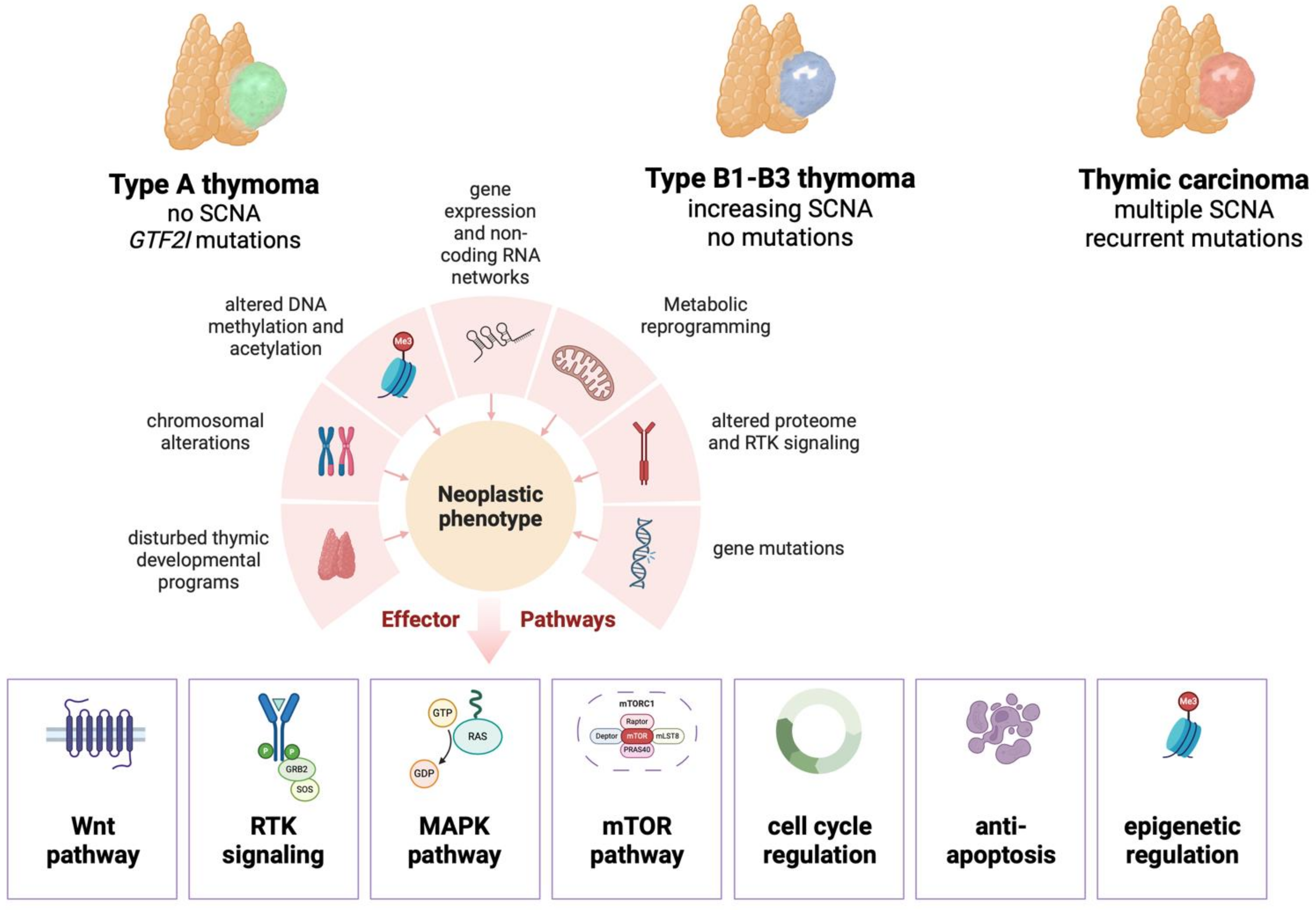
Non-Mutational Key Features in the Biology of Thymomas
Abstract
Simple Summary
Abstract
1. Introduction
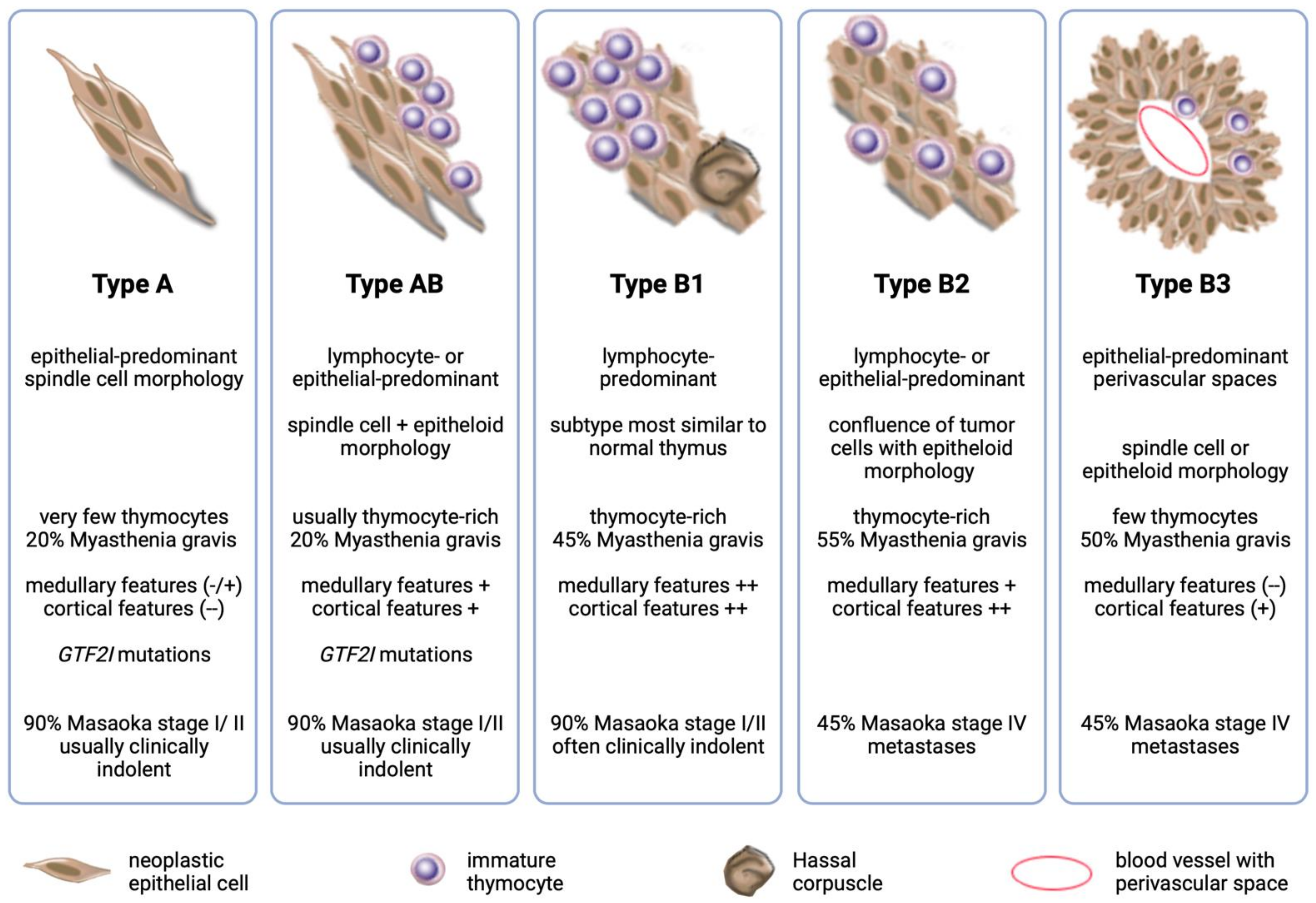
2. Disturbed Thymic Developmental Programs in Thymomas
3. Functional Implications of Chromosomal Alterations in Thymomas
4. Altered DNA Methylation and Acetylation in Thymomas
5. Altered Gene Expression and Non-Coding RNA Networks in Thymomas
6. Metabolic Reprogramming in Thymomas
7. Proteomics and Altered Tyrosine Kinase Signaling
8. Conclusions and Future Directions
Author Contributions
Funding
Conflicts of Interest
References
- Buckley, C.; Douek, D.; Newsom-Davis, J.; Vincent, A.; Willcox, N. Mature, long-lived CD4+ and CD8+ T cells are generated by the thymoma in myasthenia gravis. Ann. Neurol. 2001, 50, 64–72. [Google Scholar] [CrossRef] [PubMed]
- Marx, A.; Detterbeck, F.; Marom, E.M.; Ströbel, P.; Rajan, A. Tumors of the thymus: Introduction. In WHO Classification of Tumours. Thoracic Tumours, 5th ed.; Marx, A., Chan, J.K.C., Eds.; WHO Classification of Tumours; International Agency for Research on Cancer: Lyon, France, 2021; Volume 5. [Google Scholar]
- Marx, A.; Porubsky, S.; Belharazem, D.; Saruhan-Direskeneli, G.; Schalke, B.; Strobel, P.; Weis, C.A. Thymoma related myasthenia gravis in humans and potential animal models. Exp. Neurol. 2015, 270, 55–65. [Google Scholar] [CrossRef] [PubMed]
- Radovich, M.; Pickering, C.R.; Felau, I.; Ha, G.; Zhang, H.; Jo, H.; Hoadley, K.A.; Anur, P.; Zhang, J.; McLellan, M.; et al. The Integrated Genomic Landscape of Thymic Epithelial Tumors. Cancer Cell 2018, 33, 244–258.e10. [Google Scholar] [CrossRef] [PubMed]
- Venuta, F.; Rendina, E.A.; Pescarmona, E.O.; De Giacomo, T.; Vegna, M.L.; Fazi, P.; Flaishman, I.; Guarino, E.; Ricci, C. Multimodality treatment of thymoma: A prospective study. Ann. Thorac. Surg. 1997, 64, 1585–1591, discussion 1591–1582. [Google Scholar] [CrossRef] [PubMed]
- Petrini, I.; Meltzer, P.S.; Kim, I.K.; Lucchi, M.; Park, K.S.; Fontanini, G.; Gao, J.; Zucali, P.A.; Calabrese, F.; Favaretto, A.; et al. A specific missense mutation in GTF2I occurs at high frequency in thymic epithelial tumors. Nat. Genet. 2014, 46, 844–849. [Google Scholar] [CrossRef] [PubMed]
- Strobel, P.; Hartmann, E.; Rosenwald, A.; Kalla, J.; Ott, G.; Friedel, G.; Schalke, B.; Kasahara, M.; Tomaru, U.; Marx, A. Corticomedullary differentiation and maturational arrest in thymomas. Histopathology 2014, 64, 557–566. [Google Scholar] [CrossRef]
- Giorgetti, O.B.; Nusser, A.; Boehm, T. Human thymoma-associated mutation of the GTF2I transcription factor impairs thymic epithelial progenitor differentiation in mice. Commun. Biol. 2022, 5, 1037. [Google Scholar] [CrossRef]
- Strobel, P.; Murumagi, A.; Klein, R.; Luster, M.; Lahti, M.; Krohn, K.; Schalke, B.; Nix, W.; Gold, R.; Rieckmann, P.; et al. Deficiency of the autoimmune regulator AIRE in thymomas is insufficient to elicit autoimmune polyendocrinopathy syndrome type 1 (APS-1). J. Pathol. 2007, 211, 563–571. [Google Scholar] [CrossRef]
- Bleul, C.C.; Corbeaux, T.; Reuter, A.; Fisch, P.; Monting, J.S.; Boehm, T. Formation of a functional thymus initiated by a postnatal epithelial progenitor cell. Nature 2006, 441, 992–996. [Google Scholar] [CrossRef]
- Rossi, S.W.; Jenkinson, W.E.; Anderson, G.; Jenkinson, E.J. Clonal analysis reveals a common progenitor for thymic cortical and medullary epithelium. Nature 2006, 441, 988–991. [Google Scholar] [CrossRef]
- Bautista, J.L.; Cramer, N.T.; Miller, C.N.; Chavez, J.; Berrios, D.I.; Byrnes, L.E.; Germino, J.; Ntranos, V.; Sneddon, J.B.; Burt, T.D.; et al. Single-cell transcriptional profiling of human thymic stroma uncovers novel cellular heterogeneity in the thymic medulla. Nat. Commun. 2021, 12, 1096. [Google Scholar] [CrossRef] [PubMed]
- Lepletier, A.; Hun, M.L.; Hammett, M.V.; Wong, K.; Naeem, H.; Hedger, M.; Loveland, K.; Chidgey, A.P. Interplay between Follistatin, Activin A, and BMP4 Signaling Regulates Postnatal Thymic Epithelial Progenitor Cell Differentiation during Aging. Cell Rep. 2019, 27, 3887–3901.e3884. [Google Scholar] [CrossRef] [PubMed]
- Balciunaite, G.; Keller, M.P.; Balciunaite, E.; Piali, L.; Zuklys, S.; Mathieu, Y.D.; Gill, J.; Boyd, R.; Sussman, D.J.; Hollander, G.A. Wnt glycoproteins regulate the expression of FoxN1, the gene defective in nude mice. Nat. Immunol. 2002, 3, 1102–1108. [Google Scholar] [CrossRef] [PubMed]
- Gordon, J.; Patel, S.R.; Mishina, Y.; Manley, N.R. Evidence for an early role for BMP4 signaling in thymus and parathyroid morphogenesis. Dev. Biol. 2010, 339, 141–154. [Google Scholar] [CrossRef] [PubMed]
- Bleul, C.C.; Boehm, T. BMP signaling is required for normal thymus development. J. Immunol. 2005, 175, 5213–5221. [Google Scholar] [CrossRef] [PubMed]
- Wei, T.; Zhang, N.; Guo, Z.; Chi, F.; Song, Y.; Zhu, X. Wnt4 signaling is associated with the decrease of proliferation and increase of apoptosis during age-related thymic involution. Mol. Med. Rep. 2015, 12, 7568–7576. [Google Scholar] [CrossRef] [PubMed]
- Heinonen, K.M.; Vanegas, J.R.; Brochu, S.; Shan, J.; Vainio, S.J.; Perreault, C. Wnt4 regulates thymic cellularity through the expansion of thymic epithelial cells and early thymic progenitors. Blood 2011, 118, 5163–5173. [Google Scholar] [CrossRef] [PubMed]
- Varecza, Z.; Kvell, K.; Talaber, G.; Miskei, G.; Csongei, V.; Bartis, D.; Anderson, G.; Jenkinson, E.J.; Pongracz, J.E. Multiple suppression pathways of canonical Wnt signalling control thymic epithelial senescence. Mech. Ageing Dev. 2011, 132, 249–256. [Google Scholar] [CrossRef]
- Kvell, K.; Varecza, Z.; Bartis, D.; Hesse, S.; Parnell, S.; Anderson, G.; Jenkinson, E.J.; Pongracz, J.E. Wnt4 and LAP2alpha as pacemakers of thymic epithelial senescence. PLoS ONE 2010, 5, e10701. [Google Scholar] [CrossRef]
- Zhang, X.; Schalke, B.; Kvell, K.; Kriegsmann, K.; Kriegsmann, M.; Graeter, T.; Preissler, G.; Ott, G.; Kurz, K.; Bulut, E.; et al. WNT4 overexpression and secretion in thymic epithelial tumors drive an autocrine loop in tumor cells in vitro. Front. Oncol. 2022, 12, 920871. [Google Scholar] [CrossRef]
- Chen, Y.; Zhang, P.; Tang, P.; Lv, P.; Li, X.; Wang, Y.; Lv, Y.; Liu, Y. Wnt4 overexpression promotes thymoma development through a JNK-mediated planar cell polarity-like pathway. Oncol. Lett. 2018, 15, 83–90. [Google Scholar] [CrossRef]
- Nehls, M.; Kyewski, B.; Messerle, M.; Waldschutz, R.; Schuddekopf, K.; Smith, A.J.; Boehm, T. Two genetically separable steps in the differentiation of thymic epithelium. Science 1996, 272, 886–889. [Google Scholar] [CrossRef]
- Corbeaux, T.; Hess, I.; Swann, J.B.; Kanzler, B.; Haas-Assenbaum, A.; Boehm, T. Thymopoiesis in mice depends on a Foxn1-positive thymic epithelial cell lineage. Proc. Natl. Acad. Sci. USA 2010, 107, 16613–16618. [Google Scholar] [CrossRef]
- Li, J.; Wachsmuth, L.P.; Xiao, S.; Condie, B.G.; Manley, N.R. Foxn1 overexpression promotes thymic epithelial progenitor cell proliferation and mTEC maintenance, but does not prevent thymic involution. Development 2023, 150, dev200995. [Google Scholar] [CrossRef] [PubMed]
- Zhang, H.; Zhang, P.; Liu, Y.; Lv, P.; Wang, Y.; Chen, Y. In vitro study of the effect of small interfering ribonucleic acid on the expression of FOXN1 and B cell-attracting chemokine 1 in thymoma cell lines. Thorac. Cancer 2015, 6, 172–179. [Google Scholar] [CrossRef] [PubMed]
- Chen, Y.; Liu, X.; Liu, Y.; Wang, Y.; Wang, H.; Lu, C.; Zhang, P. Decreased Wnt4 expression inhibits thymoma development through downregulation of FoxN1. J. Thorac. Dis. 2017, 9, 1574–1583. [Google Scholar] [CrossRef] [PubMed]
- Nonaka, D.; Henley, J.D.; Chiriboga, L.; Yee, H. Diagnostic utility of thymic epithelial markers CD205 (DEC205) and Foxn1 in thymic epithelial neoplasms. Am. J. Surg. Pathol. 2007, 31, 1038–1044. [Google Scholar] [CrossRef] [PubMed]
- Pardini, E.; Cucchiara, F.; Palumbo, S.; Tarrini, G.; Di Vita, A.; Coppede, F.; Nicoli, V.; Guida, M.; Maestri, M.; Ricciardi, R.; et al. Somatic mutations of thymic epithelial tumors with myasthenia gravis. Front. Oncol. 2023, 13, 1224491. [Google Scholar] [CrossRef] [PubMed]
- Higuchi, R.; Goto, T.; Hirotsu, Y.; Yokoyama, Y.; Nakagomi, T.; Otake, S.; Amemiya, K.; Oyama, T.; Mochizuki, H.; Omata, M. Primary Driver Mutations in GTF2I Specific to the Development of Thymomas. Cancers 2020, 12, 2032. [Google Scholar] [CrossRef] [PubMed]
- He, Y.; Kim, I.K.; Bian, J.; Polyzos, A.; Di Giammartino, D.C.; Zhang, Y.W.; Luo, J.; Hernandez, M.O.; Kedei, N.; Cam, M.; et al. A Knock-In Mouse Model of Thymoma With the GTF2I L424H Mutation. J. Thorac. Oncol. 2022, 17, 1375–1386. [Google Scholar] [CrossRef] [PubMed]
- Kim, I.K.; Rao, G.; Zhao, X.; Fan, R.; Avantaggiati, M.L.; Wang, Y.; Zhang, Y.W.; Giaccone, G. Mutant GTF2I induces cell transformation and metabolic alterations in thymic epithelial cells. Cell Death Differ. 2020, 27, 2263–2279. [Google Scholar] [CrossRef]
- Cavallo, F.; Troglio, F.; Faga, G.; Fancelli, D.; Shyti, R.; Trattaro, S.; Zanella, M.; D’Agostino, G.; Hughes, J.M.; Cera, M.R.; et al. High-throughput screening identifies histone deacetylase inhibitors that modulate GTF2I expression in 7q11.23 microduplication autism spectrum disorder patient-derived cortical neurons. Mol. Autism 2020, 11, 88. [Google Scholar] [CrossRef]
- Adamo, A.; Atashpaz, S.; Germain, P.L.; Zanella, M.; D’Agostino, G.; Albertin, V.; Chenoweth, J.; Micale, L.; Fusco, C.; Unger, C.; et al. 7q11.23 dosage-dependent dysregulation in human pluripotent stem cells affects transcriptional programs in disease-relevant lineages. Nat. Genet. 2015, 47, 132–141. [Google Scholar] [CrossRef]
- Zettl, A.; Strobel, P.; Wagner, K.; Katzenberger, T.; Ott, G.; Rosenwald, A.; Peters, K.; Krein, A.; Semik, M.; Muller-Hermelink, H.K.; et al. Recurrent genetic aberrations in thymoma and thymic carcinoma. Am. J. Pathol. 2000, 157, 257–266. [Google Scholar] [CrossRef] [PubMed]
- Petrini, I.; Meltzer, P.S.; Zucali, P.A.; Luo, J.; Lee, C.; Santoro, A.; Lee, H.S.; Killian, K.J.; Wang, Y.; Tsokos, M.; et al. Copy number aberrations of BCL2 and CDKN2A/B identified by array-CGH in thymic epithelial tumors. Cell Death Dis. 2012, 3, e351. [Google Scholar] [CrossRef] [PubMed]
- Sherr, C.J. The INK4a/ARF network in tumour suppression. Nat. Rev. Mol. Cell Biol. 2001, 2, 731–737. [Google Scholar] [CrossRef] [PubMed]
- Girard, N.; Basse, C.; Schrock, A.; Ramkissoon, S.; Killian, K.; Ross, J.S. Comprehensive Genomic Profiling of 274 Thymic Epithelial Tumors Unveils Oncogenic Pathways and Predictive Biomarkers. Oncologist 2022, 27, 919–929. [Google Scholar] [CrossRef] [PubMed]
- Kurokawa, K.; Shukuya, T.; Greenstein, R.A.; Kaplan, B.G.; Wakelee, H.; Ross, J.S.; Miura, K.; Furuta, K.; Kato, S.; Suh, J.; et al. Genomic characterization of thymic epithelial tumors in a real-world dataset. ESMO Open 2023, 8, 101627. [Google Scholar] [CrossRef] [PubMed]
- Rubin, S.M. Deciphering the retinoblastoma protein phosphorylation code. Trends Biochem. Sci. 2013, 38, 12–19. [Google Scholar] [CrossRef] [PubMed]
- Jiang, Y.Z.; Ma, D.; Suo, C.; Shi, J.; Xue, M.; Hu, X.; Xiao, Y.; Yu, K.D.; Liu, Y.R.; Yu, Y.; et al. Genomic and Transcriptomic Landscape of Triple-Negative Breast Cancers: Subtypes and Treatment Strategies. Cancer Cell 2019, 35, 428–440.e425. [Google Scholar] [CrossRef] [PubMed]
- Hirose, Y.; Kondo, K.; Takizawa, H.; Nagao, T.; Nakagawa, Y.; Fujino, H.; Toba, H.; Kenzaki, K.; Sakiyama, S.; Tangoku, A. Aberrant methylation of tumour-related genes in thymic epithelial tumours. Lung Cancer 2009, 64, 155–159. [Google Scholar] [CrossRef]
- Hirabayashi, H.; Fujii, Y.; Sakaguchi, M.; Tanaka, H.; Yoon, H.E.; Komoto, Y.; Inoue, M.; Miyoshi, S.; Matsuda, H. p16INK4, pRB, p53 and cyclin D1 expression and hypermethylation of CDKN2 gene in thymoma and thymic carcinoma. Int. J. Cancer 1997, 73, 639–644. [Google Scholar] [CrossRef]
- Lal, A.; Kim, H.H.; Abdelmohsen, K.; Kuwano, Y.; Pullmann, R., Jr.; Srikantan, S.; Subrahmanyam, R.; Martindale, J.L.; Yang, X.; Ahmed, F.; et al. p16(INK4a) translation suppressed by miR-24. PLoS ONE 2008, 3, e1864. [Google Scholar] [CrossRef]
- Aesif, S.W.; Aubry, M.C.; Yi, E.S.; Kloft-Nelson, S.M.; Jenkins, S.M.; Spears, G.M.; Greipp, P.T.; Sukov, W.R.; Roden, A.C. Loss of p16(INK4A) Expression and Homozygous CDKN2A Deletion Are Associated with Worse Outcome and Younger Age in Thymic Carcinomas. J. Thorac. Oncol. 2017, 12, 860–871. [Google Scholar] [CrossRef]
- Stefanaki, K.; Rontogianni, D.; Kouvidou, C.H.; Bolioti, S.; Delides, G.; Pantelidaki, A.; Sotsiou, F.; Kanavaros, P. Expression of p53, mdm2, p21/waf1 and bcl-2 proteins in thymomas. Histopathology 1997, 30, 549–555. [Google Scholar] [CrossRef]
- Pan, C.C.; Chen, P.C.; Wang, L.S.; Lee, J.Y.; Chiang, H. Expression of apoptosis-related markers and HER-2/neu in thymic epithelial tumours. Histopathology 2003, 43, 165–172. [Google Scholar] [CrossRef] [PubMed]
- Khoury, T.; Arshad, A.; Bogner, P.; Ramnath, N.; Zhang, S.; Chandrasekhar, R.; Wilding, G.; Alrawi, S.; Tan, D. Apoptosis-related (survivin, Bcl-2), tumor suppressor gene (p53), proliferation (Ki-67), and non-receptor tyrosine kinase (Src) markers expression and correlation with clinicopathologic variables in 60 thymic neoplasms. Chest 2009, 136, 220–228. [Google Scholar] [CrossRef] [PubMed]
- Lozada-Nur, F. HIV: Common early oral markers. J. Calif. Dent. Assoc. 1989, 17, 36–48. [Google Scholar] [PubMed]
- Chen, F.F.; Yan, J.J.; Chang, K.C.; Lai, W.W.; Chen, R.M.; Jin, Y.T. Immunohistochemical localization of Mcl-1 and bcl-2 proteins in thymic epithelial tumours. Histopathology 1996, 29, 541–547. [Google Scholar] [CrossRef] [PubMed]
- Huang, B.; Belharazem, D.; Li, L.; Kneitz, S.; Schnabel, P.A.; Rieker, R.J.; Korner, D.; Nix, W.; Schalke, B.; Muller-Hermelink, H.K.; et al. Anti-Apoptotic Signature in Thymic Squamous Cell Carcinomas-Functional Relevance of Anti-Apoptotic BIRC3 Expression in the Thymic Carcinoma Cell Line 1889c. Front. Oncol. 2013, 3, 316. [Google Scholar] [CrossRef] [PubMed]
- Belharazem, D.; Grass, A.; Paul, C.; Vitacolonna, M.; Schalke, B.; Rieker, R.J.; Korner, D.; Jungebluth, P.; Simon-Keller, K.; Hohenberger, P.; et al. Increased cFLIP expression in thymic epithelial tumors blocks autophagy via NF-kappaB signalling. Oncotarget 2017, 8, 89580–89594. [Google Scholar] [CrossRef]
- Muller, D.; Mazzeo, P.; Koch, R.; Bosherz, M.S.; Welter, S.; von Hammerstein-Equord, A.; Hinterthaner, M.; Cordes, L.; Belharazem, D.; Marx, A.; et al. Functional apoptosis profiling identifies MCL-1 and BCL-xL as prognostic markers and therapeutic targets in advanced thymomas and thymic carcinomas. BMC Med. 2021, 19, 300. [Google Scholar] [CrossRef]
- Ji, G.; Ren, R.; Fang, X. Identification and Characterization of Non-Coding RNAs in Thymoma. Med. Sci. Monit. 2021, 27, e929727. [Google Scholar] [CrossRef]
- Massoth, L.R.; Hung, Y.P.; Dias-Santagata, D.; Onozato, M.; Shah, N.; Severson, E.; Duncan, D.; Gillespie, B.J.; Williams, N.F.; Ross, J.S.; et al. Pan-Cancer Landscape Analysis Reveals Recurrent KMT2A-MAML2 Gene Fusion in Aggressive Histologic Subtypes of Thymoma. JCO Precis. Oncol. 2020, 4, 109–115. [Google Scholar] [CrossRef]
- Milne, T.A.; Briggs, S.D.; Brock, H.W.; Martin, M.E.; Gibbs, D.; Allis, C.D.; Hess, J.L. MLL targets SET domain methyltransferase activity to Hox gene promoters. Mol. Cell 2002, 10, 1107–1117. [Google Scholar] [CrossRef]
- Wachter, K.; Kowarz, E.; Marschalek, R. Functional characterisation of different MLL fusion proteins by using inducible Sleeping Beauty vectors. Cancer Lett. 2014, 352, 196–202. [Google Scholar] [CrossRef][Green Version]
- Nemoto, N.; Suzukawa, K.; Shimizu, S.; Shinagawa, A.; Takei, N.; Taki, T.; Hayashi, Y.; Kojima, H.; Kawakami, Y.; Nagasawa, T. Identification of a novel fusion gene MLL-MAML2 in secondary acute myelogenous leukemia and myelodysplastic syndrome with inv(11)(q21q23). Genes Chromosomes Cancer 2007, 46, 813–819. [Google Scholar] [CrossRef] [PubMed]
- Salame, H.; McKey, R.; Ballout, M.; Saad, W. The First Reported Case of Neurotrophic Tyrosine Receptor Kinase Fusion-Positive Thymoma Treated Successfully With Entrectinib. Cureus 2021, 13, e20588. [Google Scholar] [CrossRef] [PubMed]
- Chen, C.; Yin, N.; Yin, B.; Lu, Q. DNA methylation in thoracic neoplasms. Cancer Lett. 2011, 301, 7–16. [Google Scholar] [CrossRef] [PubMed]
- Nicoli, V.; Coppede, F. Epigenetics of Thymic Epithelial Tumors. Cancers 2023, 15, 360. [Google Scholar] [CrossRef] [PubMed]
- Psilopatis, I.; Pergaris, A.; Vrettou, K.; Theocharis, S.; Troungos, C. Thymic Epithelial Neoplasms: Focusing on the Epigenetic Alterations. Int. J. Mol. Sci. 2022, 23, 4045. [Google Scholar] [CrossRef]
- Gaiser, T.; Hirsch, D.; Porth, I.; Sahm, F.; Strobel, P.; von Deimling, A.; Marx, A. DNA-Methylation Analysis as a Tool for Thymoma Classification. Cancers 2022, 14, 5876. [Google Scholar] [CrossRef] [PubMed]
- Bi, Y.; Meng, Y.; Niu, Y.; Li, S.; Liu, H.; He, J.; Zhang, Y.; Liang, N.; Liu, L.; Mao, X.; et al. Genome-wide DNA methylation profile of thymomas and potential epigenetic regulation of thymoma subtypes. Oncol. Rep. 2019, 41, 2762–2774. [Google Scholar] [CrossRef] [PubMed]
- Li, S.; Yuan, Y.; Xiao, H.; Dai, J.; Ye, Y.; Zhang, Q.; Zhang, Z.; Jiang, Y.; Luo, J.; Hu, J.; et al. Discovery and validation of DNA methylation markers for overall survival prognosis in patients with thymic epithelial tumors. Clin. Epigenetics 2019, 11, 38. [Google Scholar] [CrossRef] [PubMed]
- Chen, C.; Yin, B.; Wei, Q.; Li, D.; Hu, J.; Yu, F.; Lu, Q. Aberrant DNA methylation in thymic epithelial tumors. Cancer Investig. 2009, 27, 582–591. [Google Scholar] [CrossRef] [PubMed]
- Shvedunova, M.; Akhtar, A. Modulation of cellular processes by histone and non-histone protein acetylation. Nat. Rev. Mol. Cell Biol. 2022, 23, 329–349. [Google Scholar] [CrossRef]
- Palamaris, K.; Tzimou, L.M.; Levidou, G.; Masaoutis, C.; Theochari, I.; Rontogianni, D.; Theocharis, S. Histone Deacetylases (HDACs): Promising Biomarkers and Potential Therapeutic Targets in Thymic Epithelial Tumors. Int. J. Mol. Sci. 2023, 24, 4263. [Google Scholar] [CrossRef]
- Arjonen, A.; Makela, R.; Harma, V.; Rintanen, N.; Kuopio, T.; Kononen, J.; Rantala, J.K. Image-based ex vivo drug screen to assess targeted therapies in recurrent thymoma. Lung Cancer 2020, 145, 27–32. [Google Scholar] [CrossRef] [PubMed]
- Giaccone, G.; Rajan, A.; Berman, A.; Kelly, R.J.; Szabo, E.; Lopez-Chavez, A.; Trepel, J.; Lee, M.J.; Cao, L.; Espinoza-Delgado, I.; et al. Phase II study of belinostat in patients with recurrent or refractory advanced thymic epithelial tumors. J. Clin. Oncol. 2011, 29, 2052–2059. [Google Scholar] [CrossRef]
- Thomas, A.; Rajan, A.; Szabo, E.; Tomita, Y.; Carter, C.A.; Scepura, B.; Lopez-Chavez, A.; Lee, M.J.; Redon, C.E.; Frosch, A.; et al. A phase I/II trial of belinostat in combination with cisplatin, doxorubicin, and cyclophosphamide in thymic epithelial tumors: A clinical and translational study. Clin. Cancer Res. 2014, 20, 5392–5402. [Google Scholar] [CrossRef]
- Badve, S.; Goswami, C.; Gokmen-Polar, Y.; Nelson, R.P., Jr.; Henley, J.; Miller, N.; Zaheer, N.A.; Sledge, G.W., Jr.; Li, L.; Kesler, K.A.; et al. Molecular analysis of thymoma. PLoS ONE 2012, 7, e42669. [Google Scholar] [CrossRef]
- Gokmen-Polar, Y.; Cook, R.W.; Goswami, C.P.; Wilkinson, J.; Maetzold, D.; Stone, J.F.; Oelschlager, K.M.; Vladislav, I.T.; Shirar, K.L.; Kesler, K.A.; et al. A gene signature to determine metastatic behavior in thymomas. PLoS ONE 2013, 8, e66047. [Google Scholar] [CrossRef]
- Girard, N.; Shen, R.; Guo, T.; Zakowski, M.F.; Heguy, A.; Riely, G.J.; Huang, J.; Lau, C.; Lash, A.E.; Ladanyi, M.; et al. Comprehensive genomic analysis reveals clinically relevant molecular distinctions between thymic carcinomas and thymomas. Clin. Cancer Res. 2009, 15, 6790–6799. [Google Scholar] [CrossRef]
- Sasaki, H.; Ide, N.; Fukai, I.; Kiriyama, M.; Yamakawa, Y.; Fujii, Y. Gene expression analysis of human thymoma correlates with tumor stage. Int. J. Cancer 2002, 101, 342–347. [Google Scholar] [CrossRef]
- Liang, N.; Liu, L.; Huang, C.; Liu, H.; Guo, C.; Li, J.; Wang, W.; Li, N.; Lin, R.; Wang, T.; et al. Transcriptomic and Mutational Analysis Discovering Distinct Molecular Characteristics Among Chinese Thymic Epithelial Tumor Patients. Front. Oncol. 2021, 11, 647512. [Google Scholar] [CrossRef]
- Radovich, M.; Solzak, J.P.; Hancock, B.A.; Conces, M.L.; Atale, R.; Porter, R.F.; Zhu, J.; Glasscock, J.; Kesler, K.A.; Badve, S.S.; et al. A large microRNA cluster on chromosome 19 is a transcriptional hallmark of WHO type A and AB thymomas. Br. J. Cancer 2016, 114, 477–484. [Google Scholar] [CrossRef]
- Liu, D.; Zhang, P.; Zhao, J.; Yang, L.; Wang, W. Identification of Molecular Characteristics and New Prognostic Targets for Thymoma by Multiomics Analysis. Biomed. Res. Int. 2021, 2021, 5587441. [Google Scholar] [CrossRef]
- Wang, X.D.; Lin, P.; Li, Y.X.; Chen, G.; Yang, H.; He, Y.; Li, Q.; Liu, R.C. Identification of potential agents for thymoma by integrated analyses of differentially expressed tumour-associated genes and molecular docking experiments. Exp. Ther. Med. 2019, 18, 2001–2014. [Google Scholar] [CrossRef] [PubMed]
- Meng, F.J.; Wang, S.; Zhang, J.; Yan, Y.J.; Wang, C.Y.; Yang, C.R.; Guan, Z.Y.; Wang, C.L. Alteration in gene expression profiles of thymoma: Genetic differences and potential novel targets. Thorac. Cancer 2019, 10, 1129–1135. [Google Scholar] [CrossRef] [PubMed]
- Yu, L.; Ke, J.; Du, X.; Yu, Z.; Gao, D. Genetic characterization of thymoma. Sci. Rep. 2019, 9, 2369. [Google Scholar] [CrossRef] [PubMed]
- Lee, H.S.; Jang, H.J.; Shah, R.; Yoon, D.; Hamaji, M.; Wald, O.; Lee, J.S.; Sugarbaker, D.J.; Burt, B.M. Genomic Analysis of Thymic Epithelial Tumors Identifies Novel Subtypes Associated with Distinct Clinical Features. Clin. Cancer Res. 2017, 23, 4855–4864. [Google Scholar] [CrossRef] [PubMed]
- Iaiza, A.; Tito, C.; Ganci, F.; Sacconi, A.; Gallo, E.; Masciarelli, S.; Fontemaggi, G.; Fatica, A.; Melis, E.; Petrozza, V.; et al. Long Non-Coding RNAs in the Cell Fate Determination of Neoplastic Thymic Epithelial Cells. Front. Immunol. 2022, 13, 867181. [Google Scholar] [CrossRef] [PubMed]
- Grillone, K.; Riillo, C.; Scionti, F.; Rocca, R.; Tradigo, G.; Guzzi, P.H.; Alcaro, S.; Di Martino, M.T.; Tagliaferri, P.; Tassone, P. Non-coding RNAs in cancer: Platforms and strategies for investigating the genomic “dark matter”. J. Exp. Clin. Cancer Res. 2020, 39, 117. [Google Scholar] [CrossRef] [PubMed]
- Zhang, P.; Wu, W.; Chen, Q.; Chen, M. Non-Coding RNAs and their Integrated Networks. J. Integr. Bioinform. 2019, 16, 20190027. [Google Scholar] [CrossRef] [PubMed]
- Anastasiadou, E.; Jacob, L.S.; Slack, F.J. Non-coding RNA networks in cancer. Nat Rev Cancer 2018, 18, 5–18. [Google Scholar] [CrossRef] [PubMed]
- Kristensen, L.S.; Andersen, M.S.; Stagsted, L.V.W.; Ebbesen, K.K.; Hansen, T.B.; Kjems, J. The biogenesis, biology and characterization of circular RNAs. Nat. Rev. Genet. 2019, 20, 675–691. [Google Scholar] [CrossRef]
- Wu, Q.; Luo, X.; Li, H.; Zhang, L.; Su, F.; Hou, S.; Yin, J.; Zhang, W.; Zou, L. Identification of differentially expressed circular RNAs associated with thymoma. Thorac. Cancer 2021, 12, 1312–1319. [Google Scholar] [CrossRef]
- Su, Y.; Chen, Y.; Tian, Z.; Lu, C.; Chen, L.; Ma, X. lncRNAs classifier to accurately predict the recurrence of thymic epithelial tumors. Thorac. Cancer 2020, 11, 1773–1783. [Google Scholar] [CrossRef]
- Gong, J.; Jin, S.; Pan, X.; Wang, G.; Ye, L.; Tao, H.; Wen, H.; Liu, Y.; Xie, Q. Identification of Long Non-Coding RNAs for Predicting Prognosis Among Patients with Thymoma. Clin. Lab. 2018, 64, 1193–1198. [Google Scholar] [CrossRef]
- Yang, J.; Zhang, Y.; Song, H. A disparate role of RP11-424C20.2/UHRF1 axis through control of tumor immune escape in liver hepatocellular carcinoma and thymoma. Aging 2019, 11, 6422–6439. [Google Scholar] [CrossRef]
- Obata, Y.; Furusawa, Y.; Endo, T.A.; Sharif, J.; Takahashi, D.; Atarashi, K.; Nakayama, M.; Onawa, S.; Fujimura, Y.; Takahashi, M.; et al. The epigenetic regulator Uhrf1 facilitates the proliferation and maturation of colonic regulatory T cells. Nat. Immunol. 2014, 15, 571–579. [Google Scholar] [CrossRef]
- Lu, T.X.; Rothenberg, M.E. MicroRNA. J. Allergy Clin. Immunol. 2018, 141, 1202–1207. [Google Scholar] [CrossRef]
- Malumbres, M. miRNAs and cancer: An epigenetics view. Mol. Aspects Med. 2013, 34, 863–874. [Google Scholar] [CrossRef]
- Enkner, F.; Pichlhofer, B.; Zaharie, A.T.; Krunic, M.; Holper, T.M.; Janik, S.; Moser, B.; Schlangen, K.; Neudert, B.; Walter, K.; et al. Molecular Profiling of Thymoma and Thymic Carcinoma: Genetic Differences and Potential Novel Therapeutic Targets. Pathol. Oncol. Res. 2017, 23, 551–564. [Google Scholar] [CrossRef] [PubMed]
- Wang, J.; Huang, H.; Zhang, X.; Ma, H. LOXL1-AS1 promotes thymoma and thymic carcinoma progression by regulating miR-525-5p-HSPA9. Oncol. Rep. 2021, 45, 117. [Google Scholar] [CrossRef]
- Tito, C.; Ganci, F.; Sacconi, A.; Masciarelli, S.; Fontemaggi, G.; Pulito, C.; Gallo, E.; Laquintana, V.; Iaiza, A.; De Angelis, L.; et al. LINC00174 is a novel prognostic factor in thymic epithelial tumors involved in cell migration and lipid metabolism. Cell Death Dis. 2020, 11, 959. [Google Scholar] [CrossRef]
- Ganci, F.; Vico, C.; Korita, E.; Sacconi, A.; Gallo, E.; Mori, F.; Cambria, A.; Russo, E.; Anile, M.; Vitolo, D.; et al. MicroRNA expression profiling of thymic epithelial tumors. Lung Cancer 2014, 85, 197–204. [Google Scholar] [CrossRef]
- Tan, S.; Chen, J. si-MALAT1 attenuates thymic cancer cell proliferation and promotes apoptosis via the miR-145-5p/HMGA2 pathway. Oncol. Lett. 2021, 22, 585. [Google Scholar] [CrossRef] [PubMed]
- Iaiza, A.; Tito, C.; Ianniello, Z.; Ganci, F.; Laquintana, V.; Gallo, E.; Sacconi, A.; Masciarelli, S.; De Angelis, L.; Aversa, S.; et al. METTL3-dependent MALAT1 delocalization drives c-Myc induction in thymic epithelial tumors. Clin. Epigenetics 2021, 13, 173. [Google Scholar] [CrossRef]
- Damron, B.L. The relationship of maximum or intermediate coccidiostat levels to broiler chick water intake. Poult. Sci. 1994, 73, 33–36. [Google Scholar] [CrossRef] [PubMed]
- Miller, J.W.; Faubert, B.M.; Mathews, T.P.; Waters, J.K.; DeBerardinis, R.J.; Kernstine, K.H. Metabolic Signatures of Thymomas: Potential Biomarkers and Treatment Targets. Eur. J. Cardiothorac. Surg. 2023, 65, ezad394. [Google Scholar] [CrossRef]
- Ying, J.; Huang, Y.; Ye, X.; Zhang, Y.; Yao, Q.; Wang, J.; Yang, X.; Yu, C.; Guo, Y.; Zhang, X.; et al. Comprehensive study of clinicopathological and immune cell infiltration and lactate dehydrogenase expression in patients with thymic epithelial tumours. Int. Immunopharmacol. 2024, 126, 111205. [Google Scholar] [CrossRef]
- Alwahsh, M.; Knitsch, R.; Marchan, R.; Lambert, J.; Hoerner, C.; Zhang, X.; Schalke, B.; Lee, D.H.; Bulut, E.; Graeter, T.; et al. Metabolic Profiling of Thymic Epithelial Tumors Hints to a Strong Warburg Effect, Glutaminolysis and Precarious Redox Homeostasis as Potential Therapeutic Targets. Cancers 2022, 14, 1564. [Google Scholar] [CrossRef]
- Zhang, X.; Zeng, B.; Zhu, H.; Ma, R.; Yuan, P.; Chen, Z.; Su, C.; Liu, Z.; Yao, X.; Lawrence, A.; et al. Role of glycosphingolipid biosynthesis coregulators in malignant progression of thymoma. Int J Biol Sci 2023, 19, 4442–4456. [Google Scholar] [CrossRef] [PubMed]
- Tang, E.; Zhou, Y.; Liu, S.; Zhang, Z.; Zhang, R.; Huang, D.; Gao, T.; Zhang, T.; Xu, G. Metabolomic and Transcriptomic Profiling Identified Significant Genes in Thymic Epithelial Tumor. Metabolites 2022, 12, 567. [Google Scholar] [CrossRef] [PubMed]
- Regina Todeschini, A.; Hakomori, S.I. Functional role of glycosphingolipids and gangliosides in control of cell adhesion, motility, and growth, through glycosynaptic microdomains. Biochim. Biophys. Acta 2008, 1780, 421–433. [Google Scholar] [CrossRef] [PubMed]
- Hakomori, S.I. Glycosynaptic microdomains controlling tumor cell phenotype through alteration of cell growth, adhesion, and motility. FEBS Lett. 2010, 584, 1901–1906. [Google Scholar] [CrossRef]
- Li, J.; Long, Y.; Sun, J.; Wu, J.; He, X.; Wang, S.; Wang, X.; Miao, X.; Huang, R.; Yan, J. Comprehensive landscape of the ST3GAL family reveals the significance of ST3GAL6-AS1/ST3GAL6 axis on EGFR signaling in lung adenocarcinoma cell invasion. Front. Cell Dev. Biol. 2022, 10, 931132. [Google Scholar] [CrossRef]
- Bremmer, F.; Bohnenberger, H.; Findeisen, P.; Welter, S.; von Hammerstein-Equord, A.; Hinterthaner, M.; Muller, D.; Kuffer, S.; Okada, S.; Marx, A.; et al. Proteomic analysis identifies argininosuccinate synthetase 1 and special AT-rich sequence binding protein 1 as reliable markers for the immunohistochemical distinction between WHO types A and B3 thymomas. Histopathology 2023, 83, 607–616. [Google Scholar] [CrossRef]
- Delage, B.; Fennell, D.A.; Nicholson, L.; McNeish, I.; Lemoine, N.R.; Crook, T.; Szlosarek, P.W. Arginine deprivation and argininosuccinate synthetase expression in the treatment of cancer. Int. J. Cancer 2010, 126, 2762–2772. [Google Scholar] [CrossRef]
- Zhao, T.; Wu, J.; Liu, X.; Zhang, L.; Chen, G.; Lu, H. Diagnosis of thymic epithelial tumor subtypes by a quantitative proteomic approach. Analyst 2018, 143, 2491–2500. [Google Scholar] [CrossRef] [PubMed]
- Wang, L.; Branson, O.E.; Shilo, K.; Hitchcock, C.L.; Freitas, M.A. Proteomic Signatures of Thymomas. PLoS ONE 2016, 11, e0166494. [Google Scholar] [CrossRef] [PubMed]
- Sun, Q.L.; Fang, W.T.; Feng, J.; Zhang, J.; Yang, X.H.; Gu, Z.T.; Zhu, L.; Sha, H.F. Proteome analysis and tissue array for profiling protein markers associated with type B thymoma subclassification. Chin. Med. J. 2012, 125, 2811–2818. [Google Scholar] [PubMed]
- Lai, L.C.; Sun, Q.L.; Chen, Y.A.; Hsiao, Y.W.; Lu, T.P.; Tsai, M.H.; Zhu, L.; Chuang, E.Y.; Fang, W. Using proteomic profiling to characterize protein signatures of different thymoma subtypes. BMC Cancer 2019, 19, 796. [Google Scholar] [CrossRef] [PubMed]
- Jin, M.; Liu, P.; Qi, G. Exploring Potential Biomarkers of Early Thymoma based on Serum Proteomics. Protein Pept. Lett. 2023, 31, 74–83. [Google Scholar] [CrossRef] [PubMed]
- Li, J.; Qi, G.; Liu, Y. Proteomics analysis of serum from thymoma patients. Sci. Rep. 2023, 13, 5117. [Google Scholar] [CrossRef] [PubMed]
- Yang, H.; Qi, G.; Dong, H.; Liu, Z.; Ma, M.; Liu, P. Identification of Potential Serum Protein Biomarkers in Thymoma with Myasthenia Gravis After Docetaxel Treatment. Neurol. Ther. 2023, 12, 559–570. [Google Scholar] [CrossRef] [PubMed]
- Tong, T.; Zhang, J.; Jia, L.; Liang, P.; Wang, N. Integrated proteomics and metabolomics analysis reveals hubs protein and network alterations in myasthenia gravis. Aging 2022, 14, 5417–5426. [Google Scholar] [CrossRef]
- Hayashi, Y.; Ishii, N.; Obayashi, C.; Jinnai, K.; Hanioka, K.; Imai, Y.; Itoh, H. Thymoma: Tumour type related to expression of epidermal growth factor (EGF), EGF-receptor, p53, v-erb B and ras p21. Virchows Arch. 1995, 426, 43–50. [Google Scholar] [CrossRef]
- Ionescu, D.N.; Sasatomi, E.; Cieply, K.; Nola, M.; Dacic, S. Protein expression and gene amplification of epidermal growth factor receptor in thymomas. Cancer 2005, 103, 630–636. [Google Scholar] [CrossRef]
- Gilhus, N.E.; Jones, M.; Turley, H.; Gatter, K.C.; Nagvekar, N.; Newsom-Davis, J.; Willcox, N. Oncogene proteins and proliferation antigens in thymomas: Increased expression of epidermal growth factor receptor and Ki67 antigen. J. Clin. Pathol. 1995, 48, 447–455. [Google Scholar] [CrossRef]
- Pescarmona, E.; Pisacane, A.; Pignatelli, E.; Baroni, C.D. Expression of epidermal and nerve growth factor receptors in human thymus and thymomas. Histopathology 1993, 23, 39–44. [Google Scholar] [CrossRef]
- Henley, J.D.; Koukoulis, G.K.; Loehrer, P.J., Sr. Epidermal growth factor receptor expression in invasive thymoma. J. Cancer Res. Clin. Oncol. 2002, 128, 167–170. [Google Scholar] [CrossRef] [PubMed]
- Remon, J.; Abedallaa, N.; Taranchon-Clermont, E.; Bluthgen, V.; Lindsay, C.R.; Besse, B.; Thomas de Montpreville, V. CD52, CD22, CD26, EG5 and IGF-1R expression in thymic malignancies. Lung Cancer 2017, 108, 168–172. [Google Scholar] [CrossRef] [PubMed]
- Girard, N.; Teruya-Feldstein, J.; Payabyab, E.C.; Riely, G.J.; Rusch, V.W.; Kris, M.G.; Zakowski, M.F. Insulin-like growth factor-1 receptor expression in thymic malignancies. J. Thorac. Oncol. 2010, 5, 1439–1446. [Google Scholar] [CrossRef]
- Zucali, P.A.; Petrini, I.; Lorenzi, E.; Merino, M.; Cao, L.; Di Tommaso, L.; Lee, H.S.; Incarbone, M.; Walter, B.A.; Simonelli, M.; et al. Insulin-like growth factor-1 receptor and phosphorylated AKT-serine 473 expression in 132 resected thymomas and thymic carcinomas. Cancer 2010, 116, 4686–4695. [Google Scholar] [CrossRef] [PubMed]
- Festenstein, H. Molecular features of the H-2 class I and Qa antigens expressed on Gross virus induced AKR leukaemias. J. Immunogenet. 1989, 16, 329–333. [Google Scholar] [CrossRef] [PubMed]
- Avci, N.; Cecener, G.; Deligonul, A.; Erturk, E.; Tunca, B.; Egeli, U.; Tezcan, G.; Akyildiz, E.U.; Bayram, A.S.; Gebitekin, C.; et al. Molecular markers for patients with thymic malignancies: Not feasible at present? Asian Pac. J. Cancer Prev. 2014, 15, 3457–3460. [Google Scholar] [CrossRef] [PubMed]
- Pfister, F.; Hussain, H.; Belharazem, D.; Busch, S.; Simon-Keller, K.; Becker, D.; Pfister, E.; Rieker, R.; Strobel, P.; Marx, A. Vascular architecture as a diagnostic marker for differentiation of World Health Organization thymoma subtypes and thymic carcinoma. Histopathology 2017, 70, 693–703. [Google Scholar] [CrossRef]
- Lattanzio, R.; La Sorda, R.; Facciolo, F.; Sioletic, S.; Lauriola, L.; Martucci, R.; Gallo, E.; Palmieri, G.; Evoli, A.; Alessandrini, G.; et al. Thymic epithelial tumors express vascular endothelial growth factors and their receptors as potential targets of antiangiogenic therapy: A tissue micro array-based multicenter study. Lung Cancer 2014, 85, 191–196. [Google Scholar] [CrossRef]
- Angirekula, M.; Chang, S.Y.; Jenkins, S.M.; Greipp, P.T.; Sukov, W.R.; Marks, R.S.; Olivier, K.R.; Cassivi, S.D.; Roden, A.C. CD117, BAP1, MTAP, and TdT Is a Useful Immunohistochemical Panel to Distinguish Thymoma from Thymic Carcinoma. Cancers 2022, 14, 2299. [Google Scholar] [CrossRef]
- Kornstein, M.J.; Rosai, J. CD5 labeling of thymic carcinomas and other nonlymphoid neoplasms. Am. J. Clin. Pathol. 1998, 109, 722–726. [Google Scholar] [CrossRef] [PubMed]
- Jeong, J.H.; Pyo, J.S.; Kim, N.Y.; Kang, D.W. Diagnostic Roles of Immunohistochemistry in Thymic Tumors: Differentiation between Thymic Carcinoma and Thymoma. Diagnostics 2020, 10, 460. [Google Scholar] [CrossRef]
- Tiseo, M.; Damato, A.; Longo, L.; Barbieri, F.; Bertolini, F.; Stefani, A.; Migaldi, M.; Gnetti, L.; Camisa, R.; Bordi, P.; et al. Analysis of a panel of druggable gene mutations and of ALK and PD-L1 expression in a series of thymic epithelial tumors (TETs). Lung Cancer 2017, 104, 24–30. [Google Scholar] [CrossRef]
- Aisner, S.C.; Dahlberg, S.; Hameed, M.R.; Ettinger, D.S.; Schiller, J.H.; Johnson, D.H.; Aisner, J.; Loehrer, P.J. Epidermal growth factor receptor, C-kit, and Her2/neu immunostaining in advanced or recurrent thymic epithelial neoplasms staged according to the 2004 World Health Organization in patients treated with octreotide and prednisone: An Eastern Cooperative Oncology Group study. J. Thorac. Oncol. 2010, 5, 885–892. [Google Scholar] [CrossRef] [PubMed]
- Mimae, T.; Tsuta, K.; Kondo, T.; Nitta, H.; Grogan, T.M.; Okada, M.; Asamura, H.; Tsuda, H. Protein expression and gene copy number changes of receptor tyrosine kinase in thymomas and thymic carcinomas. Ann. Oncol. 2012, 23, 3129–3137. [Google Scholar] [CrossRef] [PubMed]
- Kuffer, S.; Grabowski, J.; Okada, S.; Sojka, N.; Welter, S.; von Hammerstein-Equord, A.; Hinterthaner, M.; Cordes, L.; von Hahn, X.; Muller, D.; et al. Phosphoproteomic Analysis Identifies TYRO3 as a Mediator of Sunitinib Resistance in Metastatic Thymomas. Cancers 2022, 14, 4762. [Google Scholar] [CrossRef]
- Yang, Z.; Liu, S.; Wang, Y.; Chen, Y.; Zhang, P.; Liu, Y.; Zhang, H.; Zhang, P.; Tao, Z.; Xiong, K. High expression of KITLG is a new hallmark activating the MAPK pathway in type A and AB thymoma. Thorac. Cancer 2020, 11, 1944–1954. [Google Scholar] [CrossRef]


| Pathway | Summary | References |
|---|---|---|
 |
| [14,17,18,19] [17,18,19] [21,22] [78,82] [82] |
 |
| [13,14,15,16] [4] [121,122,123,124,125,126,127,128,129,130,131,132] [138] [139] |
 |
| [105] [4] [139] |
 |
| [77] [4] [77] |
 |
| [39,40,62,63] [4,72,79,81] [8,31,32] [72] |
 |
| [36,47,48,49,50,51,52] [36,53] [100,101] [29] |
 |
| [4,63,64,65] [60,68] [70,71] |
Disclaimer/Publisher’s Note: The statements, opinions and data contained in all publications are solely those of the individual author(s) and contributor(s) and not of MDPI and/or the editor(s). MDPI and/or the editor(s) disclaim responsibility for any injury to people or property resulting from any ideas, methods, instructions or products referred to in the content. |
© 2024 by the authors. Licensee MDPI, Basel, Switzerland. This article is an open access article distributed under the terms and conditions of the Creative Commons Attribution (CC BY) license (https://creativecommons.org/licenses/by/4.0/).
Share and Cite
Küffer, S.; Müller, D.; Marx, A.; Ströbel, P. Non-Mutational Key Features in the Biology of Thymomas. Cancers 2024, 16, 942. https://doi.org/10.3390/cancers16050942
Küffer S, Müller D, Marx A, Ströbel P. Non-Mutational Key Features in the Biology of Thymomas. Cancers. 2024; 16(5):942. https://doi.org/10.3390/cancers16050942
Chicago/Turabian StyleKüffer, Stefan, Denise Müller, Alexander Marx, and Philipp Ströbel. 2024. "Non-Mutational Key Features in the Biology of Thymomas" Cancers 16, no. 5: 942. https://doi.org/10.3390/cancers16050942
APA StyleKüffer, S., Müller, D., Marx, A., & Ströbel, P. (2024). Non-Mutational Key Features in the Biology of Thymomas. Cancers, 16(5), 942. https://doi.org/10.3390/cancers16050942


