Simple Summary
Prostate cancer screening has traditionally been accomplished by a blood test for prostate serum antigen (PSA) followed by biopsy. MRI is very accurate at finding cancer, but ultrasound provides real-time guidance for biopsy. Using magnetic resonance imaging (MRI) to guide the biopsy under ultrasound improves detection and, in the case that only non-aggressive cancer is found, confidence in avoiding treating cancers that are unlikely to be deadly. Different approaches can be used with many different MRI–ultrasound fusion techniques, including “cognitive” fusion using only the practitioner’s reading of the scans, but also software fusion using mechanical or even pure software-aided matching of the information from MRI and ultrasound.
Abstract
The use of MRI–ultrasound image fusion targeted biopsy of the prostate in the face of an elevated serum PSA is now recommended by multiple societies, and results in improved detection of clinically significant cancer and, potentially, decreased detection of indolent disease. This combines the excellent sensitivity of MRI for clinically significant prostate cancer and the real-time biopsy guidance and confirmation of ultrasound. Both transperineal and transrectal approaches can be implemented using cognitive fusion, mechanical fusion with an articulated arm and electromagnetic registration, or pure software registration. The performance has been shown comparable to in-bore MRI biopsy performance. However, a number of factors influence the performance of this technique, including the quality and interpretation of the MRI, the approach used for biopsy, and experience of the practitioner, with most studies showing comparable performance of MRI–ultrasound fusion to in-bore targeted biopsy. Future improvements including artificial intelligence promise to refine the performance of all approaches.
1. Introduction
Prostate cancer (PCa) is the most common cancer diagnosis and the second leading cause of cancer-related death in men in the United States (US), with similar prevalence and mortality in the European Union (EU) [1,2]. This prevalence is largely due to increased detection due to advances in PCa screening practices. However, many of these cancers are indolent and of low metastatic potential, thus considered clinically insignificant PCa (cisPCa) [3].
Systematic transrectal ultrasound (TRUS)-guided biopsy was previously the standard of care for diagnosing PCa and is associated with high false negative rates and underdiagnosis of clinically significant PCa (csPCa) [4,5]. The advent of multiparametric magnetic resonance imaging (mpMRI) of the prostate and its incorporation into targeting lesions for prostate biopsy has considerably changed prostate cancer evaluation and diagnosis through improved detection of csPCa and a reduction in detection of clinically insignificant cancers [6,7,8,9,10]. mpMRI-guided biopsy has gained widespread use due to improved detection and accuracy of csPCa over systematic biopsy alone and is now recommended in major national and international PCa guidelines [4,11].
This review aims to provide an overview of how mpMRI-guided biopsy is performed, including mpMRI and the identification of target lesions, current indications, and an overview of mpMRI-guided techniques and their comparative effectiveness, limitations, and future directions.
2. mpMRI and Identification of Lesions
Utilization of mpMRI for PCa evaluation has substantially increased over the past decade and plays an integral role in the prostate biopsy decision-making and targeting process [12,13]. Current mpMRI technology utilizes four sequences, T1- and T2-weighted images, diffusion-weighted images (DWI), and dynamic contrast-enhanced imaging (DCEI), to depict detailed anatomy and identify suspicious lesions. T2-weighted and DWI sequences, in combination, are the most prominent in identifying suspicious lesions [14].
To standardize radiologic evaluation of lesions, the Prostate Imaging Reporting and Data System (PI-RADS) scoring system was established in 2012 by the European Society of Urogenital Radiology (ESUR). PI-RADS utilizes standardized criteria to evaluate suspicious lesions on mpMRI, resulting in an assigned score from 1 to 5 (very low to very high suspicion). It has since been updated, first with v2.0 in 2014, which established a dominant sequence for evaluation with DWI for the peripheral zone and T2-weighted imaging for the transition zone, decreased the importance of DCEI in evaluation, and removed magnetic resonance spectroscopic imaging (MRSI), resulting in improved sensitivity of detecting csPCa when compared to v1 in head-to-head trials, but no difference in specificity [15].
In 2019, PI-RADS v2.1 further refined the scoring system via changes including additional T2-weighted images, a clarified role of b value interpretation for DWI apparent diffusion coefficient (ADC) map use, and revisions in criteria for DWI scores 2 and 3 as well as an evaluation of lesions in the central zone and anterior fibromuscular stroma [16,17]. Current recommendations for mpMRI for fusion biopsy include use of a 1.5 Tesla (T) or higher field strength scanner (3 T when available and feasible) with an endorectal coil when available. They should at minimum include T2-weighted imaging, DWI with ADC mapping with at least one high b value ≥ 1400 s/mm2, one intermediate b value ~800 s/mm2, and one low b value ≤ 100 s/mm2 to ensure optimal contrast, and DCEI [16,17]. With these changes, a 2021 systematic review and meta-analysis of PI-RADS v2.1, the pooled sensitivity and specificity for detecting csPCa were 87% (95% CI, 82–91%) and 74% (63–82%), respectively, with no statistically significant difference from v2.0 [18].
3. Current Indications
Most current national and international guidelines define an abnormal mpMRI as PI-RADS ≥ 3, with caveats for significantly abnormal mpMRI to PI-RADS 4 or 5 based on local expertise. The Prostate MR Imaging Study (PROMIS) was a multicenter, paired-cohort study comparing mpMRI via template mapping biopsy to TRUS biopsy. The investigators found a significantly higher sensitivity for mpMRI, 93% (95% CI 88–96%) versus 48% (95% CI 42–55, p < 0.0001), as well as a high negative predictive value of 89% (95% CI 83–94%) for PI-RADS lesions < 3, and thus the ability to avoid biopsy in these cases [8]. A 2020 systematic review and meta-analysis of prospective studies showed pooled detection rates of csPCa sequentially increased for each PI-RADS v2 category [4% (95% CI 2–8) for 1–2, 17% (95% CI 13–21) for 3, 46% (95% CI 38–55) for 4, and 75% (95% CI 73–78) for 5] [19]. Because of the equivocal nature of PI-RADS 3 lesions and lower demonstrated yield of csPCa in these lesions, clinical correlation with other clinical factors, including PSA density, age, biopsy-naïve status, and prior negative biopsy to assess risk for csPCa, is recommended [20,21].
mpMRI-guided biopsy techniques are currently recommended or provided as an option by major guideline societies, including the American Urological Association (AUA), Society of Urologic Oncology (SUO), National Comprehensive Cancer Network (NCCN), and European Association of Urology (EAU), predominantly for biopsy-naïve men and men with prior negative biopsy [22,23,24]. AUA guidelines provide a conditional recommendation that clinicians may use MRI prior to initial biopsy to increase detection of csPCa, and moderately recommend targeted biopsy in biopsy-naïve patients with a suspicious lesion on MRI. In patients with an indication for repeat biopsy without prior prostate mpMRI, obtaining mpMRI is currently strongly recommended with subsequent targeted biopsy if abnormal, with an optional additional systematic biopsy. In EAU guidelines, mpMRI with subsequent biopsy if positive is strongly recommended in biopsy-naïve patients. Additionally, patients with prior negative biopsies with an indication for repeat biopsy are recommended to undergo mpMRI with targeted biopsy alone if abnormal. In the NCCN Prostate Cancer Early Detection guidelines, mpMRI-guided techniques are strongly recommended to be employed routinely. A list of guidelines and recommendations from major US and EU associations is displayed in Table 1.

Table 1.
Current guideline recommendations and indications for mpMRI-guided biopsy. “mpMRI” and “MRI” are used interchangeably, as both terminologies are used across different guidelines.
4. Overview of mpMRI-Guided Biopsy Techniques
After an mpMRI is obtained and read using the PI-RADS v2.1 scoring system, identified regions of interest must be segmented for targeting. T2-weighted imaging is most commonly utilized for fusion due to its spatial resolution and decreased vulnerability to susceptibility artifacts and geometric distortion compared to DWI [16,25,26].
Three predominant techniques for mpMRI targeting are commonly utilized: cognitive fusion, mpMRI-TRUS image fusion, and MRI in-bore/in-gantry techniques. Initially, lesions were targeted via cognitive fusion, i.e., visual estimation of lesion location on TRUS based on mpMRI images. The subsequent development of image fusion technologies, as well as in-scanner, MRI-guided techniques, has allowed for more precise identification of lesions. mpMRI–TRUS image fusion biopsy utilizes mpMRI images and TRUS to identify and target lesions via the transrectal (TR) or transperineal (TP) approach. MRI direct in-bore or in-gantry targeting technologies allow for in-scanner identification and targeting of lesions. A list of some available systems is provided in Table 2.
Table 2.
List of some current platforms for MRI–ultrasound fusion biopsy.
4.1. Cognitive Fusion
Cognitive fusion was the original method of fusion biopsy in which the operator reviews mpMRI and visually registers and targets the lesion via anatomic positioning on TRUS. Advantages of cognitive fusion include its relatively low cost, as it requires no additional software or equipment, low relative procedural time, as there is no need for software segmentation pre- or intraprocedurally, and ease of concurrent systematic biopsy [27]. It can be performed via TR or TP approach. Cognitive fusion biopsy is limited by operator dependence, which can be negatively affected by inexperience with either mpMRI or TRUS-guided biopsy [27].
4.2. mpMRI–TRUS Fusion
mpMRI–TRUS image fusion techniques utilize software to register mpMRI target lesion(s) to a corresponding anatomic location on TRUS and provides a guide for the biopsy needle. mpMRI images are segmented prior to or during the biopsy procedure. mpMRI and TRUS images are then registered by one of two types of registration, rigid or elastic. Rigid registration aligns mpMRI and TRUS images without altering their shapes and only accounts for rotational or translational differences, i.e., the operator must manually correct for distortions during the procedure. Elastic registration adjusts for intraprocedural changes in US images, such as alterations due to mass effect by the TRUS probe or prostate deformation from adjacent structures. Despite the perceived advantages of elastic registration, a 2016 meta-analysis comparing the two methods demonstrated no difference in the detection of PCa [28]. Continuous tracking of the real-time position of the US probe and needle intra-procedurally is performed via several methods, including electromagnetic tracking (e.g., UroNav, Philips Healthcare), position-encoded sensors in smart robotic arms (e.g., Artemis, Eigen), and image-based software tracking (e.g., Trinity, Koelis). Additionally, targeting can be prospective, in which the software displays the biopsy needle tract before biopsy, or retrospective, in which a scan is taken after the biopsy needle is deployed to confirm positioning in the target lesion.
Similar to cognitive fusion, image fusion allows for ease of concurrent systematic biopsy and is widely available. Disadvantages in comparison to cognitive fusion include increased cost due to required software/hardware and increased relative procedural time due to image registration. This technique can also be performed via TR or TP approach. One single-center study demonstrated that the learning curve in terms of timing, csPCa detection, and pain for operators for mpMRI targeted biopsy is a minimum of 50 cases [29]. Another study demonstrated improving csPCa detection rates with experience but showed no difference in detection rates by operator seniority [30].
4.3. Direct In-Bore/In-Gantry
MRI direct in-bore, or in-gantry, targeting is performed in the MRI scanner (e.g., DynaTRIM, Philips Healthcare). The patient is positioned head first and prone, and a needle guide is rectally or transperineally placed. The operator then obtains axial T2-weighted imaging and DWI sequences to identify the ROI. A fast, steady-state free precession image is then obtained, and the biopsy needle sequentially advanced with its position confirmed through repeated scans [31]. It can be performed via TR or TP approach. This method is the least commonly used due to its increased costs and procedural time (30–60 min), compared to both cognitive and image fusion techniques [3,31,32,33].
5. Comparative Effectiveness
5.1. mpMRI-Guided versus Systematic TRUS Biopsy
As noted previously, all three mpMRI-guided biopsy techniques have demonstrated advantages in diagnosing csPCa and reducing the diagnosis of cisPCa over systematic biopsy. The 2018 PRECISION trial was a multicenter, randomized, controlled noninferiority trial comparing mpMRI-targeted biopsy versus systematic TRUS-guided biopsy in 500 biopsy-naïve men. In this study, mpMRI-targeted biopsy had a significantly higher rate of detection for csPCa, defined as Gleason ≥ 3 + 4, at 38% versus 26% for TRUS biopsy (p < 005), and a significantly lower rate of cisPCa (9% versus 22%, p < 0.001) [7].
A 2019 Cochrane Systematic Review was consistent with these findings; in the mpMRI pathway with targeted biopsy in men with prior negative biopsy or who were biopsy-naïve, there was a pooled detection ratio (mpMRI pathway detection rate:systematic TRUS biopsy pathway rate) of 1.12 (95% CI, 1.02–1.23, 25 studies) for csPCa. When evaluated separately, the detection ratio was higher for men with prior negative biopsy than biopsy-naïve men, with the latter just below statistical significance in its meta-analysis. They concluded the MRI pathway has the most favorable diagnostic accuracy in detection of csPCA, while also decreasing the detection of cisPCa. [6]
Regarding cognitive fusion biopsy alone, several studies have demonstrated improved detection of csPCa and greater concordance with final histopathology compared to systematic biopsy [34,35,36,37,38]. Another study showed TP cognitive biopsy had similar csPCa detection rates to TP template biopsy, although the detection rate of clinically insignificant cancer was lower [39].
For in-bore techniques, a 2018 large, prospective, multicenter head-to-head study of in-bore biopsy versus systematic TRUS biopsy in 626 men found similar rates of detection of csPCa, but a significantly lower rate of detection for cisPCa for the in-bore technique, reducing the number of men requiring biopsy by 49% [10].
A more recent 2022 meta-analysis found a significantly higher csPCa detection rate for all mpMRI-guided biopsy techniques versus TRUS-guided biopsy with a pooled relative cancer detection rate of 1.24 (95% CI, 1.03–1.50, p = 0.02), as well as a significantly lower cisPCa yield with a pooled relative yield of 0.58 (95% CI, 0.46–0.74) compared to TRUS-guided biopsy, in line with other previous meta-analyses [9,38,40].
Given the data demonstrating the superior performance of mpMRI-guided biopsy in detecting csPCa, there is ongoing debate regarding the necessity of concurrent systematic TRUS biopsy. However, several studies have shown an improved diagnostic yield of csPCA, with reported rates as much as 20% higher when systematic TRUS biopsy is performed in addition to mpMRI-guided biopsy [10,41,42,43,44,45,46,47,48]. A prospective National Cancer Institute (NCI) study of 2103 patients who underwent both mpMRI-targeted and systematic biopsy showed improved csPCa detection rates for mpMRI-targeted biopsy. However, when analyzing MRI-targeting alone, 8.8% of csPCa (grade group ≥ 3 in this study) were misclassified, and 8.7% upgraded on histopathological analysis at radical prostatectomy in comparison to combined biopsy (3.5%) [45]. The multicenter, paired diagnostic MRI-FIRST study of 251 biopsy-naïve men who underwent MRI-targeted TR biopsy and systematic TRUS biopsy also demonstrated combined improvement in detecting csPCa [45]. Similarly, in the PAIREDCAP, a paired diagnostic trial of 248 biopsy-naïve men who underwent systematic biopsy plus cognitive and image fusion biopsy, the combined approach yielded an additional 11% of csPCa [43]. Furthermore, inaccurate co-registration and targeting have been shown to introduce error while utilizing fusion technology [49,50]. Thus, the combination of mpMRI-targeted plus systematic TRUS biopsy remains included in international guideline statements with differing strengths of recommendation and consideration (Table 1).
5.2. Comparison across mpMRI-Guided Biopsy Techniques
Based on current evidence, there is no consensus for the best mpMRI-guided technique for prostate biopsy. The 2018 multicenter, randomized, controlled FUTURE Trial prospectively compared all three techniques in 665 patients. The investigators found no significant difference in PCa or csPCa detection rates across cognitive fusion, image fusion, or in-bore techniques. The study was noted to be limited by a low rate of PIRADS ≥ 3 lesions on mpMRI, potentially underpowering their findings. [51]
Image fusion biopsy techniques have demonstrated improved accuracy over cognitive fusion in several single-institution studies [52,53,54]. In recent meta-analyses and numerous single institutional studies, however, there have been no demonstrated significant differences between cognitive biopsy and image fusion techniques in terms of csPCa detection rates [36,38,55,56,57,58,59]. Despite similar detection rates, image fusion biopsy has been shown to have improved detection rates of anterior and transition zone lesions, while cognitive fusion has been shown to have improved detection of lesions at the base, suggesting a potential complementary effect of these techniques [27,36,56,57].
Micro-ultrasound technology is a recent advancement in US imaging that utilizes high-frequency imaging at 29 MHz, allowing for visualization of MRI lesions in real time. Prospective trials have demonstrated improved sensitivity over mpMRI and improved detection of csPCa for TR cognitive biopsy with micro-ultrasound compared to transperineal image fusion biopsy [60]. Furthermore, micro-ultrasound technology is low cost and can be performed in a single session, necessitating a larger-scale study of its potential in improving cognitive biopsy [61,62].
Regarding in-bore or in-gantry biopsy, several retrospective studies and one small prospective study have demonstrated improved detection of overall PCa and csPCa than cognitive or image fusion methods [51,63,64,65]. This is in contrast to large, randomized studies, which to date have demonstrated no significant difference in csPCa detection [51,66].
Perhaps most notably, updated systematic reviews and meta-analyses have consistently demonstrated similar detection rates across all three techniques across indications [38,40]. Thus, no mpMRI-guided technique has received a preferential recommendation in national and international guidelines (Table 1).
5.3. TP vs. TR Approach
All three mpMRI-guided biopsy techniques can be performed via TP and TR approaches. The TR approach has historically been advantageous in terms of cost and time but disadvantaged by increased risk of sepsis up to 7%. The TP approach offers reduced risk of infection, as well as improved anterior and apical sampling, but is limited by the requirement of general anesthesia [67]. The introduction of the freehand technique (e.g., PrecisionPoint Transperineal Access System, Perineologic) paired with effective local anesthesia has decreased the procedural time allowed for office-based practice, thus rapidly increasing adaptation of the TP approach [68,69].
CsPCa detection rates between the two techniques have been similar, with a few caveats. A 2022 systematic review and meta-analysis of 11 studies demonstrated no significant difference in the overall detection of csPCa; however, TP demonstrated significantly higher detection rates in apical (OR 1.86; 95% CI, 1.14–3.03; p = 0.01) and anterior (OR 2.17, 95% CI 1.46–3.22; p < 0.001) lesions, as well as significantly higher rates of detection for PI-RADS 4 lesions, with no difference for PI-RADS 3 and 5 lesions. This review was, however, limited by the retrospective design of most of the included studies [70]. Recently, the 2023 multicenter, randomized, controlled PREVENT Trial compared outcomes for mpMRI-guided and systematic biopsy via TP approach without antibiotic prophylaxis versus TR with targeted prophylaxis in 658 patients. The rates of infectious complications were 0% and 1.4% for TP and TR, respectively, just outside of statistical significance (p = 0.059). CsPCa detection rates were similar, 53% for TP versus 50% for TR [71]. Further data on csPCa detection rates are expected in 2024; the TRANSLATE trial, a multicenter, randomized controlled trial comparing TP versus TR mpMRI-guided plus systematic biopsy in 1042 patients, remains in the accrual phase [72]
5.4. Case Presentation
The following is a case presentation and description of an mpMRI-guided biopsy via the TP approach performed at our institution. A 62-year-old male presented with a rise in prostate specific antigen (PSA) from baseline ~2 to ~7. The patient underwent 3.0 T mpMRI, which demonstrated a PI-RADS 4 lesion in the left posterior mid-gland that can be seen on T2-weighted (Figure 1, left) and ADC images (Figure 1, right).
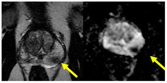
Figure 1.
Axial T2-weighted (left) and axial ADC images (right) with arrow identifying the ROI.
After counseling on the risks and benefits of different biopsy options, the patient elected to undergo TP biopsy. The performing author (T.M.) at our institution predominantly utilizes mpMRI software fusion biopsy, specifically the UroNav system (Philips Healthcare) for TP biopsy [73]. This system was updated in 2018 to include a TP stepper with a grid for ease of TP biopsy targeting.
This software requires pre-biopsy segmentation of the prostate and targeted lesion via manual (most commonly) or automated methods. Our institution offers TP biopsy under local anesthesia or sedation per patient preference. As mentioned previously, UroNav works via electromagnetic tracking through a field generator and US probe manipulated via a robotic arm with multiple degrees of freedom. The UroNav cart and ultrasound machine is placed alongside the patient. The patient is given an enema prior to the procedure. The patient is placed in the dorsal lithotomy position. The scrotum is then elevated and secured superiorly. The field generator is positioned over the patient’s pelvis. The perineum is then prepped with chlorhexidine, sterilely draped, and 1% lidocaine is injected to provide local anesthesia. Antibiotics are then administered perioperatively. The US probe is covered with an endocavity balloon containing ultrasound gel. A TP stepper with a grid is then connected to the US probe for tracking. The US probe is then inserted into the rectum and the stepper aligned.
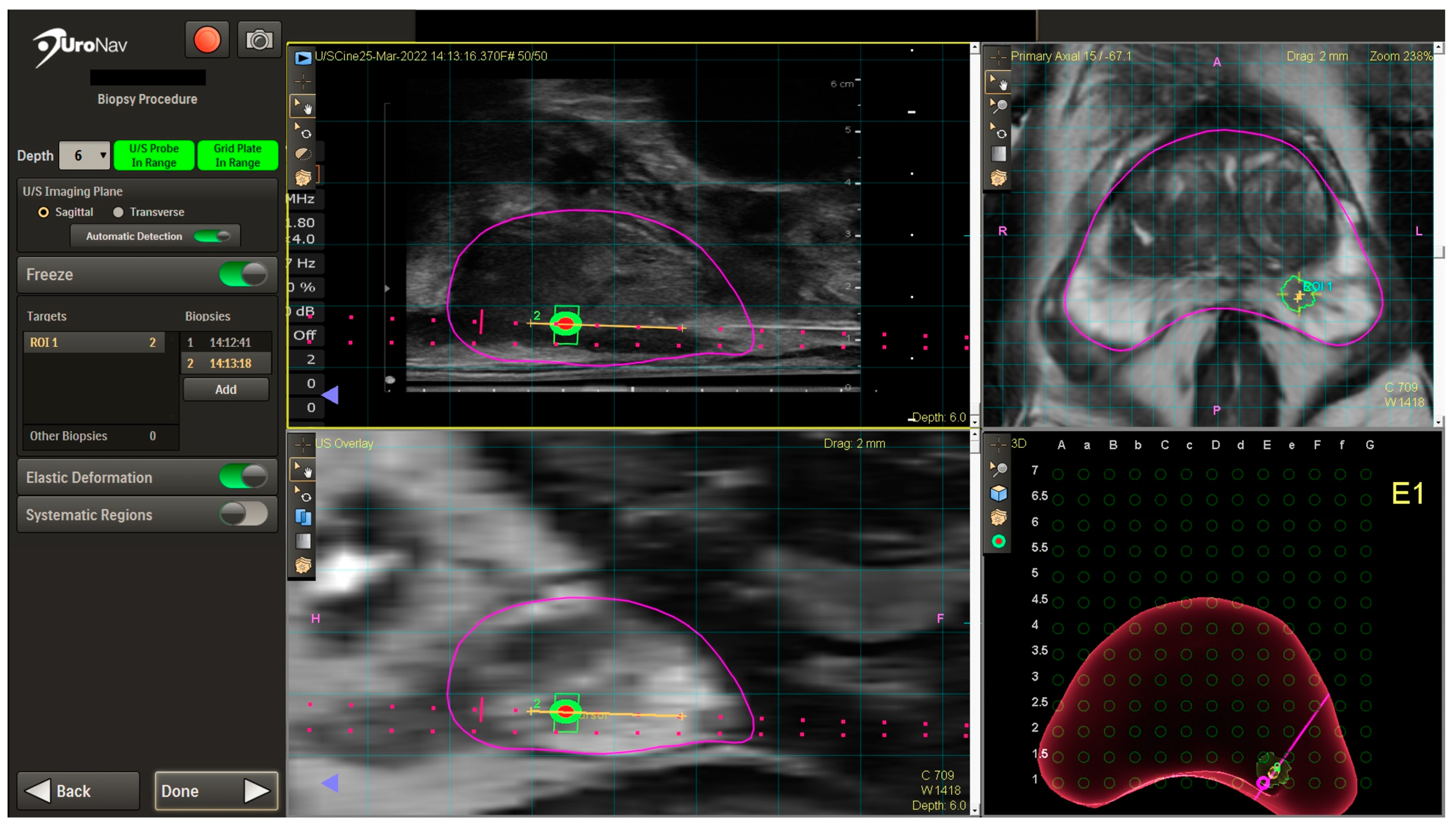
A TRUS sagittal sweep of the prostate is then performed to obtain TRUS images and dimensions, which are then fused with mpMRI images in real time on the UroNav system screen (Figure 2). After fusion, the UroNav display includes the identified ROI in the left posterior mid-gland with sagittal views (left upper and lower quadrants), axial view (right upper quadrant), and 3D rendered image with grid overlay for targeting (right lower quadrant). Biopsies are then obtained using grid holes corresponding to the location of the ROI. Cores are then taken from the ROI (four cores in this case). A systematic biopsy is then generally performed in biopsy-naïve patients. The UroNav system stores the biopsy needle trajectory for potential future use should the patient need another biopsy. At the conclusion of the procedure, the US probe is removed, perineum cleaned, and bacitracin applied. No post-procedure antibiotics or pain medications are prescribed at our institution.

Figure 2.
UroNav TP biopsy display images with ROI highlighted. The magenta outline defines the prostate. The orange line shows the biopsy trajectory. The green trapezoid is the radiologist-defined region of interest, whereas the green circle with orange center is the ideal “target”.
At follow up, pathology from the ROI demonstrated prostatic adenocarcinoma, Grade Group 3 (Gleason score 4 + 3 = 7), involving 70%, 50%, and 30% (5 mm, 5 mm, 3.5 mm) of 3/4 cores. Gleason pattern 4 constituted 50–60% of the total tumor volume. At our institution TP biopsy is a collaboration with Urology and Interventional Radiology, with ~500 TP biopsies performed since 2018 and no reported cases of sepsis. Through this collaboration we have also begun to offer same day mpMRI, consultation, and potential biopsy.
5.5. Future Directions
Application of deep learning models (DLMs) to mpMRI lesion detection has shown promise in improving detection of csPCa. A 2024 study comparing a DLM integrating three individual mpMRI sequences using neural networks to clinical PI-RADS score in classification of csPCa and cisPCa demonstrated improved sensitivity, specificity, and accuracy over PI-RADS score [74]. A similar 2021 study of a texture-based DLM developed using T2-weighted and ADC images in comparison to PI-RADS classification also showed improved specificity, as well as overall area under the ROC curve (AUC), with greatest improvements in detection of peripheral zone and solitary tumor lesions on sub-analyses [75]. Incorporation of DLMs into clinical practice represents an emerging area of investigation to enhance classification of csPCa and cisPCa [76].
Micro-ultrasound offers the ability to detect lesions seen on mpMRI in real time at a low cost and demonstrates similar rates of detection for mpMRI-guided biopsy. It is not yet known whether use alone or in conjunction with mpMRI-guided methods is optimal for detection of csPCa. The ongoing three-armed randomized-controlled OPTIMUM trial comparing csPCa detection rates of micro-ultrasound alone versus mpMRI-US fusion biopsy versus mpMRI/micro-ultrasound with micro-ultrasound-based fusion device aims to address these questions [77,78].
6. Conclusions
mpMRI-guided prostate biopsy techniques have transformed the diagnosis, staging, and treatment of PCa through improved detection of csPCa and reduced diagnosis of cisPCa. This has led to recommended use in multiple national and international guidelines, predominantly for patients who are biopsy-naïve or with a prior negative biopsy. Cognitive fusion, image fusion, and in-bore/in-gantry techniques have similar rates of csPCa detection, and are all suitable targeting techniques that can be employed based on local circumstances. However, current evidence suggests combination with systematic biopsy results in the highest csPCa detection rates. TP and TR approaches to biopsy have similar csPCa detection rates. However, the TP approach is advantageous in the detection of csPCa in apical and anterior lesions, as well as in lower infectious risk and antibiotic stewardship. Lastly, the advent of DLMs for mpMRI classification and micro-ultrasound technology for real-time detection of lesions may further augment biopsy practice. Randomized controlled trials evaluating optimal mpMRI-guided biopsy techniques are ongoing.
Author Contributions
Conceptualization, All authors; methodology, All authors; software, All authors; validation, All authors; formal analysis, All authors; investigation, All authors; resources, All authors; data curation, All authors; writing—original draft preparation, All authors; writing—review and editing, All authors; visualization, All authors; supervision, T.D.M. and D.J.A.M.; project administration, All authors. All authors have read and agreed to the published version of the manuscript.
Funding
This research received no external funding.
Institutional Review Board Statement
The study did not require ethical approval.
Informed Consent Statement
Not applicable.
Data Availability Statement
Data from case description is unavailable due to privacy or ethical restrictions.
Conflicts of Interest
The authors declare no conflicts of interest.
References
- Siegel, R.L.; Giaquinto, A.N.; Jemal, A. Cancer statistics, 2024. CA Cancer J. Clin. 2024, 74, 12–49. [Google Scholar] [CrossRef] [PubMed]
- Sung, H.; Ferlay, J.; Siegel, R.L.; Laversanne, M.; Soerjomataram, I.; Jemal, A.; Bray, F. Global cancer statistics 2020: GLOBOCAN estimates of incidence and mortality worldwide for 36 cancers in 185 countries. CA Cancer J. Clin. 2021, 71, 209–249. [Google Scholar] [CrossRef] [PubMed]
- Connor, M.J.; Gorin, M.A.; Eldred-Evans, D.; Bass, E.J.; Desai, A.; Dudderidge, T.; Winkler, M.; Ahmed, H.U. Landmarks in the evolution of prostate biopsy. Nat. Rev. Urol. 2023, 20, 241–258. [Google Scholar] [CrossRef] [PubMed]
- Siddiqui, M.M.; Rais-Bahrami, S.; Truong, H.; Stamatakis, L.; Vourganti, S.; Nix, J.; Hoang, A.N.; Walton-Diaz, A.; Shuch, B.; Weintraub, M.; et al. Magnetic resonance imaging/ultrasound-fusion biopsy significantly upgrades prostate cancer versus systematic 12-core transrectal ultrasound biopsy. Eur. Urol. 2013, 64, 713–719. [Google Scholar] [CrossRef] [PubMed]
- Serefoglu, E.C.; Altinova, S.; Ugras, N.S.; Akincioglu, E.; Asil, E.; Balbay, M.D. How reliable is 12-core prostate biopsy procedure in the detection of prostate cancer? Can. Urol. Assoc. J. 2013, 7, E293–E298. [Google Scholar] [CrossRef] [PubMed]
- Drost, F.-J.H.; Osses, D.F.; Nieboer, D.; Steyerberg, E.W.; Bangma, C.H.; Roobol, M.J.; Schoots, I.G. Prostate MRI, with or without MRI-targeted biopsy, and systematic biopsy for detecting prostate cancer. Cochrane Database Syst. Rev. 2019, 4, CD012663. [Google Scholar] [CrossRef] [PubMed]
- Kasivisvanathan, V.; Rannikko, A.S.; Borghi, M.; Panebianco, V.; Mynderse, L.A.; Vaarala, M.H.; Briganti, A.; Budäus, L.; Hellawell, G.; Hindley, R.G.; et al. PRECISION Study Group Collaborators MRI-Targeted or Standard Biopsy for Prostate-Cancer Diagnosis. N. Engl. J. Med. 2018, 378, 1767–1777. [Google Scholar] [CrossRef] [PubMed]
- Ahmed, H.U.; El-Shater Bosaily, A.; Brown, L.C.; Gabe, R.; Kaplan, R.; Parmar, M.K.; Collaco-Moraes, Y.; Ward, K.; Hindley, R.G.; Freeman, A.; et al. PROMIS study group Diagnostic accuracy of multi-parametric MRI and TRUS biopsy in prostate cancer (PROMIS): A paired validating confirmatory study. Lancet 2017, 389, 815–822. [Google Scholar] [CrossRef] [PubMed]
- Schoots, I.G.; Roobol, M.J.; Nieboer, D.; Bangma, C.H.; Steyerberg, E.W.; Hunink, M.G.M. Magnetic resonance imaging-targeted biopsy may enhance the diagnostic accuracy of significant prostate cancer detection compared to standard transrectal ultrasound-guided biopsy: A systematic review and meta-analysis. Eur. Urol. 2015, 68, 438–450. [Google Scholar] [CrossRef]
- van der Leest, M.; Cornel, E.; Israël, B.; Hendriks, R.; Padhani, A.R.; Hoogenboom, M.; Zamecnik, P.; Bakker, D.; Setiasti, A.Y.; Veltman, J.; et al. Head-to-head Comparison of Transrectal Ultrasound-guided Prostate Biopsy Versus Multiparametric Prostate Resonance Imaging with Subsequent Magnetic Resonance-guided Biopsy in Biopsy-naïve Men with Elevated Prostate-specific Antigen: A Large Prospective Multicenter Clinical Study. Eur. Urol. 2019, 75, 570–578. [Google Scholar] [CrossRef]
- Kvåle, R.; Møller, B.; Wahlqvist, R.; Fosså, S.D.; Berner, A.; Busch, C.; Kyrdalen, A.E.; Svindland, A.; Viset, T.; Halvorsen, O.J. Concordance between Gleason scores of needle biopsies and radical prostatectomy specimens: A population-based study. BJU Int. 2009, 103, 1647–1654. [Google Scholar] [CrossRef] [PubMed]
- Siddiqui, M.R.; Ansbro, B.; Shah, P.V.; Aguiar, J.A.; Li, E.V.; Rich, J.M.; Mahenthiran, A.K.; Moataz, S.A.S.; Keeter, M.-K.; Mai, Q.; et al. Real-world use of MRI for risk stratification prior to prostate biopsy. Prostate Cancer Prostatic Dis. 2023, 26, 353–359. [Google Scholar] [CrossRef] [PubMed]
- Robinson, D.; Abdulkareem, R.; Nasrollah, D.; Ljung, A.; Hintze, P.; Wallby, S.; Ståhlbrandt, H.; Frennvall, T.; Styrke, J.; Stattin, P.; et al. Frequency of biopsy and tumor grade before vs after introduction of prostate magnetic resonance imaging. JAMA Netw. Open 2023, 6, e2330233. [Google Scholar] [CrossRef]
- Stabile, A.; Giganti, F.; Rosenkrantz, A.B.; Taneja, S.S.; Villeirs, G.; Gill, I.S.; Allen, C.; Emberton, M.; Moore, C.M.; Kasivisvanathan, V. Multiparametric MRI for prostate cancer diagnosis: Current status and future directions. Nat. Rev. Urol. 2020, 17, 41–61. [Google Scholar] [CrossRef] [PubMed]
- Woo, S.; Suh, C.H.; Kim, S.Y.; Cho, J.Y.; Kim, S.H. Diagnostic Performance of Prostate Imaging Reporting and Data System Version 2 for Detection of Prostate Cancer: A Systematic Review and Diagnostic Meta-analysis. Eur. Urol. 2017, 72, 177–188. [Google Scholar] [CrossRef] [PubMed]
- Scott, R.; Misser, S.K.; Cioni, D.; Neri, E. PI-RADS v2.1: What has changed and how to report. SA J. Radiol. 2021, 25, 2062. [Google Scholar] [CrossRef] [PubMed]
- PI-RADS|American College of Radiology. Available online: https://www.acr.org/Clinical-Resources/Reporting-and-Data-Systems/PI-RADS (accessed on 12 February 2024).
- Park, K.J.; Choi, S.H.; Kim, M.-H.; Kim, J.K.; Jeong, I.G. Performance of Prostate Imaging Reporting and Data System Version 2.1 for Diagnosis of Prostate Cancer: A Systematic Review and Meta-Analysis. J. Magn. Reson. Imaging 2021, 54, 103–112. [Google Scholar] [CrossRef] [PubMed]
- Park, K.J.; Choi, S.H.; Lee, J.S.; Kim, J.K.; Kim, M.-H.; Jeong, I.G. Risk Stratification of Prostate Cancer According to PI-RADS® Version 2 Categories: Meta-Analysis for Prospective Studies. J. Urol. 2020, 204, 1141–1149. [Google Scholar] [CrossRef] [PubMed]
- Camacho, A.; Salah, F.; Bay, C.P.; Waring, J.; Umeton, R.; Hirsch, M.S.; Cole, A.P.; Kibel, A.S.; Loda, M.; Tempany, C.M.; et al. PI-RADS 3 score: A retrospective experience of clinically significant prostate cancer detection. BJUI Compass 2023, 4, 473–481. [Google Scholar] [CrossRef] [PubMed]
- Fang, A.M.; Shumaker, L.A.; Martin, K.D.; Jackson, J.C.; Fan, R.E.; Khajir, G.; Patel, H.D.; Soodana-Prakash, N.; Vourganti, S.; Filson, C.P.; et al. Multi-institutional analysis of clinical and imaging risk factors for detecting clinically significant prostate cancer in men with PI-RADS 3 lesions. Cancer 2022, 128, 3287–3296. [Google Scholar] [CrossRef]
- Mottet, N.; van den Bergh, R.C.N.; Briers, E.; Van den Broeck, T.; Cumberbatch, M.G.; De Santis, M.; Fanti, S.; Fossati, N.; Gandaglia, G.; Gillessen, S.; et al. EAU-EANM-ESTRO-ESUR-SIOG Guidelines on Prostate Cancer-2020 Update. Part 1: Screening, Diagnosis, and Local Treatment with Curative Intent. Eur. Urol. 2021, 79, 243–262. [Google Scholar] [CrossRef]
- Early Detection of Prostate Cancer: AUA/SUO Guideline (2023)—American Urological Association. Available online: https://www.auanet.org/guidelines-and-quality/guidelines/early-detection-of-prostate-cancer-guidelines (accessed on 13 February 2024).
- Wei, J.T.; Barocas, D.; Carlsson, S.; Coakley, F.; Eggener, S.; Etzioni, R.; Fine, S.W.; Han, M.; Kim, S.K.; Kirkby, E.; et al. Early detection of prostate cancer: AUA/SUO guideline part I: Prostate cancer screening. J. Urol. 2023, 210, 46–53. [Google Scholar] [CrossRef]
- Fedorov, A.; Khallaghi, S.; Sánchez, C.A.; Lasso, A.; Fels, S.; Tuncali, K.; Sugar, E.N.; Kapur, T.; Zhang, C.; Wells, W.; et al. Open-source image registration for MRI-TRUS fusion-guided prostate interventions. Int. J. Comput. Assist. Radiol. Surg. 2015, 10, 925–934. [Google Scholar] [CrossRef]
- Sparks, R.; Bloch, B.N.; Feleppa, E.; Barratt, D.; Madabhushi, A. Fully automated prostate magnetic resonance imaging and transrectal ultrasound fusion via a probabilistic registration metric. Proc. SPIE 2013, 8671, 72–85. [Google Scholar] [CrossRef]
- Chang, S.D.; Ghai, S.; Kim, C.K.; Oto, A.; Giganti, F.; Moore, C.M. MRI targeted prostate biopsy techniques: AJR expert panel narrative review. AJR Am. J. Roentgenol. 2021, 217, 1263–1281. [Google Scholar] [CrossRef]
- Venderink, W.; de Rooij, M.; Sedelaar, J.P.M.; Huisman, H.J.; Fütterer, J.J. Elastic Versus Rigid Image Registration in Magnetic Resonance Imaging-transrectal Ultrasound Fusion Prostate Biopsy: A Systematic Review and Meta-analysis. Eur. Urol. Focus 2018, 4, 219–227. [Google Scholar] [CrossRef]
- Lenfant, L.; Beitone, C.; Troccaz, J.; Rouprêt, M.; Seisen, T.; Voros, S.; Mozer, P.C. Learning curve for fusion magnetic resonance imaging targeted prostate biopsy and three-dimensional transrectal ultrasonography segmentation. BJU Int. 2024; early view. [Google Scholar] [CrossRef]
- Taha, F.; Larre, S.; Branchu, B.; ReSurg. Surgeon seniority and experience have no effect on CaP detection rates using MRI/TRUS fusion-guided targeted biopsies. Urol. Oncol. 2024, 42, 67.e1–67.e7. [Google Scholar] [CrossRef]
- Overduin, C.G.; Fütterer, J.J.; Barentsz, J.O. MRI-guided biopsy for prostate cancer detection: A systematic review of current clinical results. Curr. Urol. Rep. 2013, 14, 209–213. [Google Scholar] [CrossRef]
- Beyersdorff, D.; Winkel, A.; Hamm, B.; Lenk, S.; Loening, S.A.; Taupitz, M. MR imaging-guided prostate biopsy with a closed MR unit at 1.5 T: Initial results. Radiology 2005, 234, 576–581. [Google Scholar] [CrossRef]
- Venderink, W.; Govers, T.M.; de Rooij, M.; Fütterer, J.J.; Sedelaar, J.P.M. Cost-Effectiveness Comparison of Imaging-Guided Prostate Biopsy Techniques: Systematic Transrectal Ultrasound, Direct In-Bore MRI, and Image Fusion. AJR Am. J. Roentgenol. 2017, 208, 1058–1063. [Google Scholar] [CrossRef]
- Goldberg, H.; Ahmad, A.E.; Chandrasekar, T.; Klotz, L.; Emberton, M.; Haider, M.A.; Taneja, S.S.; Arora, K.; Fleshner, N.; Finelli, A.; et al. Comparison of Magnetic Resonance Imaging and Transrectal Ultrasound Informed Prostate Biopsy for Prostate Cancer Diagnosis in Biopsy Naïve Men: A Systematic Review and Meta-Analysis. J. Urol. 2020, 203, 1085–1093. [Google Scholar] [CrossRef]
- Rouse, P.; Shaw, G.; Ahmed, H.U.; Freeman, A.; Allen, C.; Emberton, M. Multi-parametric magnetic resonance imaging to rule-in and rule-out clinically important prostate cancer in men at risk: A cohort study. Urol. Int. 2011, 87, 49–53. [Google Scholar] [CrossRef]
- Puech, P.; Rouvière, O.; Renard-Penna, R.; Villers, A.; Devos, P.; Colombel, M.; Bitker, M.-O.; Leroy, X.; Mège-Lechevallier, F.; Comperat, E.; et al. Prostate cancer diagnosis: Multiparametric MR-targeted biopsy with cognitive and transrectal US-MR fusion guidance versus systematic biopsy--prospective multicenter study. Radiology 2013, 268, 461–469. [Google Scholar] [CrossRef]
- Zheng, T.; Bi, K.; Tang, Y.; Zeng, Y.; Wang, J.; Yan, L. Cognitive fusion-targeted biopsy versus transrectal ultrasonography-guided systematic biopsy: Comparison and analysis of the risk of Gleason score upgrading. Int. Urol. Nephrol. 2024, 56, 981–988. [Google Scholar] [CrossRef]
- Bass, E.J.; Pantovic, A.; Connor, M.J.; Loeb, S.; Rastinehad, A.R.; Winkler, M.; Gabe, R.; Ahmed, H.U. Diagnostic accuracy of magnetic resonance imaging targeted biopsy techniques compared to transrectal ultrasound guided biopsy of the prostate: A systematic review and meta-analysis. Prostate Cancer Prostatic Dis. 2022, 25, 174–179. [Google Scholar] [CrossRef]
- Kasivisvanathan, V.; Dufour, R.; Moore, C.M.; Ahmed, H.U.; Abd-Alazeez, M.; Charman, S.C.; Freeman, A.; Allen, C.; Kirkham, A.; van der Meulen, J.; et al. Transperineal magnetic resonance image targeted prostate biopsy versus transperineal template prostate biopsy in the detection of clinically significant prostate cancer. J. Urol. 2013, 189, 860–866. [Google Scholar] [CrossRef]
- Wegelin, O.; van Melick, H.H.E.; Hooft, L.; Bosch, J.L.H.R.; Reitsma, H.B.; Barentsz, J.O.; Somford, D.M. Comparing Three Different Techniques for Magnetic Resonance Imaging-targeted Prostate Biopsies: A Systematic Review of In-bore versus Magnetic Resonance Imaging-transrectal Ultrasound fusion versus Cognitive Registration. Is There a Preferred Technique? Eur. Urol. 2017, 71, 517–531. [Google Scholar] [CrossRef]
- Connor, M.J.; Eldred-Evans, D.; van Son, M.; Hosking-Jervis, F.; Bertoncelli Tanaka, M.; Reddy, D.; Bass, E.J.; Powell, L.; Ahmad, S.; Pegers, E.; et al. A Multicenter Study of the Clinical Utility of Nontargeted Systematic Transperineal Prostate Biopsies in Patients Undergoing Pre-Biopsy Multiparametric Magnetic Resonance Imaging. J. Urol. 2020, 204, 1195–1201. [Google Scholar] [CrossRef]
- Miah, S.; Hosking-Jervis, F.; Connor, M.J.; Eldred-Evans, D.; Shah, T.T.; Arya, M.; Barber, N.; Bhardwa, J.; Bott, S.; Burke, D.; et al. A Multicentre Analysis of the Detection of Clinically Significant Prostate Cancer Following Transperineal Image-fusion Targeted and Nontargeted Systematic Prostate Biopsy in Men at Risk. Eur. Urol. Oncol 2020, 3, 262–269. [Google Scholar] [CrossRef]
- Elkhoury, F.F.; Felker, E.R.; Kwan, L.; Sisk, A.E.; Delfin, M.; Natarajan, S.; Marks, L.S. Comparison of targeted vs systematic prostate biopsy in men who are biopsy naive: The prospective assessment of image registration in the diagnosis of prostate cancer (PAIREDCAP) study. JAMA Surg. 2019, 154, 811–818. [Google Scholar] [CrossRef]
- Rouvière, O.; Puech, P.; Renard-Penna, R.; Claudon, M.; Roy, C.; Mège-Lechevallier, F.; Decaussin-Petrucci, M.; Dubreuil-Chambardel, M.; Magaud, L.; Remontet, L.; et al. MRI-FIRST Investigators Use of prostate systematic and targeted biopsy on the basis of multiparametric MRI in biopsy-naive patients (MRI-FIRST): A prospective, multicentre, paired diagnostic study. Lancet Oncol. 2019, 20, 100–109. [Google Scholar] [CrossRef]
- Ahdoot, M.; Wilbur, A.R.; Reese, S.E.; Lebastchi, A.H.; Mehralivand, S.; Gomella, P.T.; Bloom, J.; Gurram, S.; Siddiqui, M.; Pinsky, P.; et al. MRI-Targeted, Systematic, and Combined Biopsy for Prostate Cancer Diagnosis. N. Engl. J. Med. 2020, 382, 917–928. [Google Scholar] [CrossRef]
- Wei, C.; Szewczyk-Bieda, M.; Bates, A.S.; Donnan, P.T.; Rauchhaus, P.; Gandy, S.; Ragupathy, S.K.A.; Singh, P.; Coll, K.; Serhan, J.; et al. Multicenter Randomized Trial Assessing MRI and Image-guided Biopsy for Suspected Prostate Cancer: The MULTIPROS Study. Radiology 2023, 308, e221428. [Google Scholar] [CrossRef]
- Thompson, A.; Eguru, V.; Moosa, S.; Ng, Y. Do concomitant systematic biopsies add to fusion targeted biopsies in the diagnosis and management of clinically significant prostate cancer? Urol. Res. Pract. 2023, 49, 169–177. [Google Scholar] [CrossRef]
- Novara, G.; Zattoni, F.; Zecchini, G.; Aceti, A.; Pellizzari, A.; Ferraioli, G.; Cobacchini, C.; Taverna, A.; Sattin, F.; Carletti, F.; et al. Role of targeted biopsy, perilesional biopsy, random biopsy, and their combination in the detection of clinically significant prostate cancer by mpMRI/transrectal ultrasonography fusion biopsy in confirmatory biopsy during active surveillance program. Prostate Cancer Prostatic Dis. 2024, 27, 129–135. [Google Scholar] [CrossRef]
- Brisbane, W.G.; Priester, A.M.; Ballon, J.; Kwan, L.; Delfin, M.K.; Felker, E.R.; Sisk, A.E.; Hu, J.C.; Marks, L.S. Targeted prostate biopsy: Umbra, penumbra, and value of perilesional sampling. Eur. Urol. 2022, 82, 303–310. [Google Scholar] [CrossRef]
- Williams, C.; Ahdoot, M.; Daneshvar, M.A.; Hague, C.; Wilbur, A.R.; Gomella, P.T.; Shih, J.; Khondakar, N.; Yerram, N.; Mehralivand, S.; et al. Why Does Magnetic Resonance Imaging-Targeted Biopsy Miss Clinically Significant Cancer? J. Urol. 2022, 207, 95–107. [Google Scholar] [CrossRef]
- Wegelin, O.; Exterkate, L.; van der Leest, M.; Kummer, J.A.; Vreuls, W.; de Bruin, P.C.; Bosch, J.L.H.R.; Barentsz, J.O.; Somford, D.M.; van Melick, H.H.E. The FUTURE Trial: A Multicenter Randomised Controlled Trial on Target Biopsy Techniques Based on Magnetic Resonance Imaging in the Diagnosis of Prostate Cancer in Patients with Prior Negative Biopsies. Eur. Urol. 2019, 75, 582–590. [Google Scholar] [CrossRef]
- Osses, D.F.; van Asten, J.J.; Tijsterman, J.D. Cognitive-Targeted versus Magnetic Resonance Imaging-Guided Prostate Biopsy in Prostate Cancer Detection. Curr. Urol. 2018, 11, 182–188. [Google Scholar] [CrossRef]
- Cool, D.W.; Zhang, X.; Romagnoli, C.; Izawa, J.I.; Romano, W.M.; Fenster, A. Evaluation of MRI-TRUS fusion versus cognitive registration accuracy for MRI-targeted, TRUS-guided prostate biopsy. AJR Am. J. Roentgenol. 2015, 204, 83–91. [Google Scholar] [CrossRef]
- Delongchamps, N.B.; Peyromaure, M.; Schull, A.; Beuvon, F.; Bouazza, N.; Flam, T.; Zerbib, M.; Muradyan, N.; Legman, P.; Cornud, F. Prebiopsy magnetic resonance imaging and prostate cancer detection: Comparison of random and targeted biopsies. J. Urol. 2013, 189, 493–499. [Google Scholar] [CrossRef]
- Watts, K.L.; Frechette, L.; Muller, B.; Ilinksy, D.; Kovac, E.; Sankin, A.; Aboumohamed, A. Systematic review and meta-analysis comparing cognitive vs. image-guided fusion prostate biopsy for the detection of prostate cancer. Urol. Oncol. 2020, 38, 734.e19. [Google Scholar] [CrossRef]
- Wysock, J.S.; Rosenkrantz, A.B.; Huang, W.C.; Stifelman, M.D.; Lepor, H.; Deng, F.-M.; Melamed, J.; Taneja, S.S. A prospective, blinded comparison of magnetic resonance (MR) imaging-ultrasound fusion and visual estimation in the performance of MR-targeted prostate biopsy: The PROFUS trial. Eur. Urol. 2014, 66, 343–351. [Google Scholar] [CrossRef]
- Lee, D.J.; Recabal, P.; Sjoberg, D.D.; Thong, A.; Lee, J.K.; Eastham, J.A.; Scardino, P.T.; Vargas, H.A.; Coleman, J.; Ehdaie, B. Comparative effectiveness of targeted prostate biopsy using magnetic resonance imaging ultrasound fusion software and visual targeting: A prospective study. J. Urol. 2016, 196, 697–702. [Google Scholar] [CrossRef]
- Ho, K.; Zhu, D.; Gupta, K.; Loloi, J.; Abramson, M.; Watts, K.; Agalliu, I.; Sankin, A. Performance of cognitive vs. image-guided fusion biopsy for detection of overall and clinically significant prostate cancer in a multiethnic population. Urol. Oncol. 2024, 42, e1–e29. [Google Scholar] [CrossRef]
- Pirola, G.M.; Castellani, D.; Orecchia, L.; Giulioni, C.; Gubbiotti, M.; Rubilotta, E.; Maggi, M.; Teoh, J.Y.-C.; Gauhar, V.; Naselli, A. Transperineal US-MRI Fusion-Guided Biopsy for the Detection of Clinical Significant Prostate Cancer: A Systematic Review and Meta-Analysis Comparing Cognitive and Software-Assisted Technique. Cancers 2023, 15, 3443. [Google Scholar] [CrossRef]
- Claros, O.R.; Tourinho-Barbosa, R.R.; Fregeville, A.; Gallardo, A.C.; Muttin, F.; Carneiro, A.; Stabile, A.; Moschini, M.; Macek, P.; Cathala, N.; et al. Comparison of Initial Experience with Transrectal Magnetic Resonance Imaging Cognitive Guided Micro-Ultrasound Biopsies versus Established Transperineal Robotic Ultrasound Magnetic Resonance Imaging Fusion Biopsies for Prostate Cancer. J. Urol. 2020, 203, 918–925. [Google Scholar] [CrossRef]
- Klotz, L.; Lughezzani, G.; Maffei, D.; Sánchez, A.; Pereira, J.G.; Staerman, F.; Cash, H.; Luger, F.; Lopez, L.; Sanchez-Salas, R.; et al. Comparison of micro-ultrasound and multiparametric magnetic resonance imaging for prostate cancer: A multicenter, prospective analysis. Can. Urol. Assoc. J. 2021, 15, E11–E16. [Google Scholar] [CrossRef]
- Calace, F.P.; Napolitano, L.; Arcaniolo, D.; Stizzo, M.; Barone, B.; Crocetto, F.; Olivetta, M.; Amicuzi, U.; Cirillo, L.; Rubinacci, A.; et al. Micro-Ultrasound in the Diagnosis and Staging of Prostate and Bladder Cancer: A Comprehensive Review. Medicina 2022, 58, 1624. [Google Scholar] [CrossRef]
- Prince, M.; Foster, B.R.; Kaempf, A.; Liu, J.-J.; Amling, C.L.; Isharwal, S.; Chen, Y.; Coakley, F.V. In-Bore Versus Fusion MRI-Targeted Biopsy of PI-RADS Category 4 and 5 Lesions: A Retrospective Comparative Analysis Using Propensity Score Weighting. AJR Am. J. Roentgenol. 2021, 217, 1123–1130. [Google Scholar] [CrossRef]
- Venderink, W.; van der Leest, M.; van Luijtelaar, A.; van de Ven, W.J.M.; Fütterer, J.J.; Sedelaar, J.P.M.; Huisman, H.J. Retrospective comparison of direct in-bore magnetic resonance imaging (MRI)-guided biopsy and fusion-guided biopsy in patients with MRI lesions which are likely or highly likely to be clinically significant prostate cancer. World J. Urol. 2017, 35, 1849–1855. [Google Scholar] [CrossRef]
- Kaufmann, S.; Russo, G.I.; Bamberg, F.; Löwe, L.; Morgia, G.; Nikolaou, K.; Stenzl, A.; Kruck, S.; Bedke, J. Prostate cancer detection in patients with prior negative biopsy undergoing cognitive-, robotic- or in-bore MRI target biopsy. World J. Urol. 2018, 36, 761–768. [Google Scholar] [CrossRef]
- Arsov, C.; Rabenalt, R.; Blondin, D.; Quentin, M.; Hiester, A.; Godehardt, E.; Gabbert, H.E.; Becker, N.; Antoch, G.; Albers, P.; et al. Prospective randomized trial comparing magnetic resonance imaging (MRI)-guided in-bore biopsy to MRI-ultrasound fusion and transrectal ultrasound-guided prostate biopsy in patients with prior negative biopsies. Eur. Urol. 2015, 68, 713–720. [Google Scholar] [CrossRef]
- Chung, Y.; Hong, S.K. Shifting to transperineal prostate biopsy: A narrative review. Prostate Int. 2023, 12, 10–14. [Google Scholar] [CrossRef]
- Tzeng, M.; Basourakos, S.P.; Patel, H.D.; Allaway, M.J.; Hu, J.C.; Gorin, M.A. Pooled outcomes of performing freehand transperineal prostate biopsy with the PrecisionPoint Transperineal Access System. BJUI Compass 2022, 3, 434–442. [Google Scholar] [CrossRef]
- Gereta, S.; Hung, M.; Alexanderani, M.K.; Robinson, B.D.; Hu, J.C. Evaluating the Learning Curve for In-office Freehand Cognitive Fusion Transperineal Prostate Biopsy. Urology 2023, 181, 31–37. [Google Scholar] [CrossRef]
- Uleri, A.; Baboudjian, M.; Tedde, A.; Gallioli, A.; Long-Depaquit, T.; Palou, J.; Basile, G.; Gaya, J.M.; Sanguedolce, F.; Lughezzani, G.; et al. Is There an Impact of Transperineal Versus Transrectal Magnetic Resonance Imaging-targeted Biopsy in Clinically Significant Prostate Cancer Detection Rate? A Systematic Review and Meta-analysis. Eur. Urol. Oncol 2023, 6, 621–628. [Google Scholar] [CrossRef]
- Hu, J.C.; Assel, M.; Allaf, M.E.; Ehdaie, B.; Vickers, A.J.; Cohen, A.J.; Ristau, B.T.; Green, D.A.; Han, M.; Rezaee, M.E.; et al. Transperineal Versus Transrectal Magnetic Resonance Imaging-targeted and Systematic Prostate Biopsy to Prevent Infectious Complications: The PREVENT Randomized Trial. Eur. Urol. 2024, in press. [Google Scholar] [CrossRef]
- Bryant, R.J.; Yamamoto, H.; Eddy, B.; Kommu, S.; Narahari, K.; Omer, A.; Leslie, T.; Catto, J.W.F.; Rosario, D.J.; Good, D.W.; et al. Protocol for the TRANSLATE prospective, multicentre, randomised clinical trial of prostate biopsy technique. BJU Int. 2023, 131, 694–704. [Google Scholar] [CrossRef]
- Kosarek, C.D.; Mahmoud, A.M.; Eyzaguirre, E.J.; Shan, Y.; Walser, E.M.; Horn, G.L.; Williams, S.B. Initial series of magnetic resonance imaging (MRI)-fusion targeted prostate biopsy using the first transperineal targeted platform available in the USA. BJU Int. 2018, 122, 909–912. [Google Scholar] [CrossRef]
- Yang, C.; Li, B.; Luan, Y.; Wang, S.; Bian, Y.; Zhang, J.; Wang, Z.; Liu, B.; Chen, X.; Hacker, M. Deep learning model for the detection of prostate cancer and classification of clinically significant disease using multiparametric MRI in comparison to PI-RADs score. Urol. Oncol. 2024, in press. [Google Scholar] [CrossRef] [PubMed]
- Liu, Y.; Zheng, H.; Liang, Z.; Miao, Q.; Brisbane, W.G.; Marks, L.S.; Raman, S.S.; Reiter, R.E.; Yang, G.; Sung, K. Textured-Based Deep Learning in Prostate Cancer Classification with 3T Multiparametric MRI: Comparison with PI-RADS-Based Classification. Diagnostics 2021, 11, 1785. [Google Scholar] [CrossRef] [PubMed]
- Sushentsev, N.; Moreira Da Silva, N.; Yeung, M.; Barrett, T.; Sala, E.; Roberts, M.; Rundo, L. Comparative performance of fully-automated and semi-automated artificial intelligence methods for the detection of clinically significant prostate cancer on MRI: A systematic review. Insights Imaging 2022, 13, 59. [Google Scholar] [CrossRef] [PubMed]
- Basso Dias, A.; Ghai, S. Micro-Ultrasound: Current Role in Prostate Cancer Diagnosis and Future Possibilities. Cancers 2023, 15, 1280. [Google Scholar] [CrossRef]
- Klotz, L.; Andriole, G.; Cash, H.; Cooperberg, M.; Crawford, E.D.; Emberton, M.; Gomez-Sancha, F.; Klein, E.; Lughezzani, G.; Marks, L.; et al. Optimization of prostate biopsy—Micro-Ultrasound versus MRI (OPTIMUM): A 3-arm randomized controlled trial evaluating the role of 29 MHz micro-ultrasound in guiding prostate biopsy in men with clinical suspicion of prostate cancer. Contemp. Clin. Trials 2022, 112, 106618. [Google Scholar] [CrossRef]

Disclaimer/Publisher’s Note: The statements, opinions and data contained in all publications are solely those of the individual author(s) and contributor(s) and not of MDPI and/or the editor(s). MDPI and/or the editor(s) disclaim responsibility for any injury to people or property resulting from any ideas, methods, instructions or products referred to in the content. |
© 2024 by the authors. Licensee MDPI, Basel, Switzerland. This article is an open access article distributed under the terms and conditions of the Creative Commons Attribution (CC BY) license (https://creativecommons.org/licenses/by/4.0/).