
Exploring the Link between BMI and Aggressive Histopathological Subtypes in Differentiated Thyroid Carcinoma—Insights from a Multicentre Retrospective Study
 , ,
, ,  ,
, 
 ,
, 

Abstract
Simple Summary
Abstract
1. Introduction
2. Materials and Methods
2.1. Study Design and Patient Selection
2.2. Data Collection
2.3. Statistical Analysis
3. Results
4. Discussion
5. Conclusions
Author Contributions
Funding
Institutional Review Board Statement
Informed Consent Statement
Data Availability Statement
Conflicts of Interest
References
- Rahib, L.; Smith, B.D.; Aizenberg, R.; Rosenzweig, A.B.; Fleshman, J.M.; Matrisian, L.M. Projecting Cancer Incidence and Deaths to 2030: The Unexpected Burden of Thyroid, Liver, and Pancreas Cancers in the United States. Cancer Res. 2014, 74, 2913–2921. [Google Scholar] [CrossRef] [PubMed]
- Li, Y.; Huang, Y.; He, X.; Han, C.; Wu, W.; Shen, H.; Xu, Y.; Liu, Y.; Zhu, Z. The Global Burden of Thyroid Cancer in High-Income Asia-Pacific: A Systematic Analysis of the Global Burden of Disease Study. Ther. Adv. Endocrinol. Metab. 2022, 13, 20420188221090012. [Google Scholar] [CrossRef] [PubMed]
- Sanabria, A.; Kowalski, L.P.; Shah, J.P.; Nixon, I.J.; Angelos, P.; Williams, M.D.; Rinaldo, A.; Ferlito, A. Growing Incidence of Thyroid Carcinoma in Recent Years: Factors Underlying Overdiagnosis. Head Neck 2018, 40, 855–866. [Google Scholar] [CrossRef] [PubMed]
- Pizzato, M.; Li, M.; Vignat, J.; Laversanne, M.; Singh, D.; Vecchia, C.L.; Vaccarella, S. The Epidemiological Landscape of Thyroid Cancer Worldwide: GLOBOCAN Estimates for Incidence and Mortality Rates in 2020. Lancet Diabetes Endocrinol. 2022, 10, 264–272. [Google Scholar] [CrossRef] [PubMed]
- Malandrino, P.; Russo, M.; Gianì, F.; Pellegriti, G.; Vigneri, P.; Belfiore, A.; Rizzarelli, E.; Vigneri, R. Increased Thyroid Cancer Incidence in Volcanic Areas: A Role of Increased Heavy Metals in the Environment? Int. J. Mol. Sci. 2020, 21, 3425. [Google Scholar] [CrossRef] [PubMed]
- Mazonakis, M.; Tzedakis, A.; Damilakis, J.; Gourtsoyiannis, N. Thyroid Dose from Common Head and Neck CT Examinations in Children: Is There an Excess Risk for Thyroid Cancer Induction? Eur. Radiol. 2007, 17, 1352–1357. [Google Scholar] [CrossRef]
- Boutari, C.; Mantzoros, C.S. A 2022 Update on the Epidemiology of Obesity and a Call to Action: As Its Twin COVID-19 Pandemic Appears to Be Receding, the Obesity and Dysmetabolism Pandemic Continues to Rage On. Metabolism 2022, 133, 155217. [Google Scholar] [CrossRef] [PubMed]
- Pati, S.; Irfan, W.; Jameel, A.; Ahmed, S.; Shahid, R.K. Obesity and Cancer: A Current Overview of Epidemiology, Pathogenesis, Outcomes, and Management. Cancers 2023, 15, 485. [Google Scholar] [CrossRef] [PubMed]
- Mitchelson, K.A.J.; O’Connell, F.; O’Sullivan, J.; Roche, H.M. Obesity, Dietary Fats, and Gastrointestinal Cancer Risk-Potential Mechanisms Relating to Lipid Metabolism and Inflammation. Metabolites 2024, 14, 42. [Google Scholar] [CrossRef]
- Patel, A.V.; Patel, K.S.; Teras, L.R. Excess Body Fatness and Cancer Risk: A Summary of the Epidemiologic Evidence. Surg. Obes. Relat. Dis. 2023, 19, 742–745. [Google Scholar] [CrossRef]
- Kitahara, C.M.; Platz, E.A.; Park, Y.; Hollenbeck, A.R.; Schatzkin, A.; Berrington de González, A. Body Fat Distribution, Weight Change during Adulthood, and Thyroid Cancer Risk in the NIH-AARP Diet and Health Study. Int. J. Cancer 2012, 130, 1411–1419. [Google Scholar] [CrossRef] [PubMed]
- Harikrishna, A.; Ishak, A.; Ellinides, A.; Saad, R.; Christodoulou, H.; Spartalis, E.; Paschou, S.A. The Impact of Obesity and Insulin Resistance on Thyroid Cancer: A Systematic Review. Maturitas 2019, 125, 45–49. [Google Scholar] [CrossRef] [PubMed]
- Li, L.-R.; Song, J.-L.; Liu, H.-Q.; Chen, C. Metabolic Syndrome and Thyroid Cancer: Risk, Prognosis, and Mechanism. Discov. Oncol. 2023, 14, 23. [Google Scholar] [CrossRef] [PubMed]
- Paes, J.E.; Hua, K.; Nagy, R.; Kloos, R.T.; Jarjoura, D.; Ringel, M.D. The Relationship between Body Mass Index and Thyroid Cancer Pathology Features and Outcomes: A Clinicopathological Cohort Study. J. Clin. Endocrinol. Metab. 2010, 95, 4244–4250. [Google Scholar] [CrossRef] [PubMed]
- Economides, A.; Giannakou, K.; Mamais, I.; Economides, P.A.; Papageorgis, P. Association Between Aggressive Clinicopathologic Features of Papillary Thyroid Carcinoma and Body Mass Index: A Systematic Review and Meta-Analysis. Front. Endocrinol. 2021, 12, 692879. [Google Scholar] [CrossRef] [PubMed]
- Kaliszewski, K.; Diakowska, D.; Rzeszutko, M.; Rudnicki, J. Obesity and Overweight Are Associated with Minimal Extrathyroidal Extension, Multifocality and Bilaterality of Papillary Thyroid Cancer. J. Clin. Med. 2021, 10, 970. [Google Scholar] [CrossRef]
- Shin, A.; Cho, S.; Jang, D.; Abe, S.K.; Saito, E.; Rahman, M.S.; Islam, M.R.; Sawada, N.; Shu, X.-O.; Koh, W.-P.; et al. Body Mass Index and Thyroid Cancer Risk: A Pooled Analysis of Half a Million Men and Women in the Asia Cohort Consortium. Thyroid 2022, 32, 306–314. [Google Scholar] [CrossRef] [PubMed]
- Kwon, H.; Chang, Y.; Cho, A.; Ahn, J.; Park, S.E.; Park, C.-Y.; Lee, W.-Y.; Oh, K.-W.; Park, S.-W.; Shin, H.; et al. Metabolic Obesity Phenotypes and Thyroid Cancer Risk: A Cohort Study. Thyroid 2019, 29, 349–358. [Google Scholar] [CrossRef] [PubMed]
- Haugen, B.R.; Alexander, E.K.; Bible, K.C.; Doherty, G.M.; Mandel, S.J.; Nikiforov, Y.E.; Pacini, F.; Randolph, G.W.; Sawka, A.M.; Schlumberger, M.; et al. 2015 American Thyroid Association Management Guidelines for Adult Patients with Thyroid Nodules and Differentiated Thyroid Cancer: The American Thyroid Association Guidelines Task Force on Thyroid Nodules and Differentiated Thyroid Cancer. Thyroid 2016, 26, 1–133. [Google Scholar] [CrossRef]
- Obesity: Preventing and Managing the Global Epidemic. Report of a WHO Consultation; WHO Technical Report Series 894; World Health Organization: Geneva, Switzerland, 2000; pp. 1–253.
- Baloch, Z.W.; Asa, S.L.; Barletta, J.A.; Ghossein, R.A.; Juhlin, C.C.; Jung, C.K.; LiVolsi, V.A.; Papotti, M.G.; Sobrinho-Simões, M.; Tallini, G.; et al. Overview of the 2022 WHO Classification of Thyroid Neoplasms. Endocr. Pathol. 2022, 33, 27–63. [Google Scholar] [CrossRef]
- Kwon, H.; Han, K.-D.; Park, C.-Y. Weight Change Is Significantly Associated with Risk of Thyroid Cancer: A Nationwide Population-Based Cohort Study. Sci. Rep. 2019, 9, 1546. [Google Scholar] [CrossRef] [PubMed]
- Pellegriti, G.; Frasca, F.; Regalbuto, C.; Squatrito, S.; Vigneri, R. Worldwide Increasing Incidence of Thyroid Cancer: Update on Epidemiology and Risk Factors. J. Cancer Epidemiol. 2013, 2013, 965212. [Google Scholar] [CrossRef] [PubMed]
- Hoang, T.; Song, D.; Lee, J.; Lee, E.K.; Hwangbo, Y.; Kim, J. Association among Body Mass Index, Genetic Variants of FTO, and Thyroid Cancer Risk: A Hospital-Based Case-Control Study of the Cancer Screenee Cohort in Korea. Cancer Res. Treat. 2021, 53, 857–873. [Google Scholar] [CrossRef] [PubMed]
- Kwan, M.L.; Kroenke, C.H.; Sweeney, C.; Bernard, P.S.; Weltzien, E.K.; Castillo, A.; Factor, R.E.; Maxfield, K.S.; Stijleman, I.J.; Kushi, L.H.; et al. Association of High Obesity with PAM50 Breast Cancer Intrinsic Subtypes and Gene Expression. BMC Cancer 2015, 15, 278. [Google Scholar] [CrossRef] [PubMed]
- Torres-de la Roche, L.A.; Steljes, I.; Janni, W.; Friedl, T.W.P.; De Wilde, R.L. The Association between Obesity and Premenopausal Breast Cancer According to Intrinsic Subtypes—A Systematic Review. Geburtshilfe Frauenheilkd. 2020, 80, 601–610. [Google Scholar] [CrossRef] [PubMed]
- Olsen, C.M.; Nagle, C.M.; Whiteman, D.C.; Ness, R.; Pearce, C.L.; Pike, M.C.; Rossing, M.A.; Terry, K.L.; Wu, A.H.; Risch, H.A.; et al. Obesity and Risk of Ovarian Cancer Subtypes: Evidence from the Ovarian Cancer Association Consortium. Endocr. Relat. Cancer 2013, 20, 251–262. [Google Scholar] [CrossRef] [PubMed]
- Grani, G.; Lamartina, L.; Montesano, T.; Ronga, G.; Maggisano, V.; Falcone, R.; Ramundo, V.; Giacomelli, L.; Durante, C.; Russo, D.; et al. Lack of Association between Obesity and Aggressiveness of Differentiated Thyroid Cancer. J. Endocrinol. Investig. 2019, 42, 85–90. [Google Scholar] [CrossRef]
- Kim, M.; Kang, Y.E.; Park, Y.J.; Koo, B.S.; Ku, E.J.; Choi, J.Y.; Lee, E.K.; Kim, B.H. Potential Impact of Obesity on the Aggressiveness of Low- to Intermediate-Risk Papillary Thyroid Carcinoma: Results from a MASTER Cohort Study. Endocrine 2023, 82, 134–142. [Google Scholar] [CrossRef]
- Basolo, A.; Poma, A.M.; Giannini, R.; Ceccarini, G.; Pelosini, C.; Fierabracci, P.; Castany, M.U.; Bechi Genzano, S.; Ambrosini, C.E.; Materazzi, G.; et al. Histological Pattern and Gene Expression Profiling of Thyroid Tissue in Subjects with Obesity. J. Endocrinol. Investig. 2022, 45, 413–423. [Google Scholar] [CrossRef]
- Kim, W.G.; Park, J.W.; Willingham, M.C.; Cheng, S. Diet-Induced Obesity Increases Tumor Growth and Promotes Anaplastic Change in Thyroid Cancer in a Mouse Model. Endocrinology 2013, 154, 2936–2947. [Google Scholar] [CrossRef]


| Variable | N (%); Median (IQR) | |
|---|---|---|
| Age at Surgery, years | 50 (38–60) | |
| BMI, kg/m2 | 25 (22–28) | |
| BMI, kg/m2 | <25 | 1778 (46%) |
| 25–29.9 | 1333 (34.5%) | |
| >29.9 | 757 (19.6%) | |
| Gender | Female | 2765 (71.5%) |
| Male | 1103 (28.5%) | |
| Hyperthyroidism | No | 3486 (90.1%) |
| Yes | 382 (9.9%) | |
| Preoperative Diagnosis | Basedow | 141 (3.6%) |
| Indeterminate nodule | 1026 (26.5%) | |
| Malignancy | 1806 (46.7%) | |
| N/MNG | 885 (22.9%) | |
| Plummer | 10 (0.3%) | |
| Substernal Goiter | No | 3765 (97.3%) |
| Yes | 103 (2.7%) | |
| Type of Surgery | Completion Thyroidectomy | 29 (0.7%) |
| Lobectomy | 492 (12.7%) | |
| Lobectomy + Completion Thyroidectomy | 77 (2%) | |
| Total Thyroidectomy | 3270 (84.5%) | |
| Monolateral Central Compartment lymphadenectomy | No | 3847 (99.5%) |
| Yes | 21 (0.5%) | |
| Bilateral Central Compartment lymphadenectomy | No | 3115 (80.5%) |
| Yes | 753 (19.5%) | |
| Monolateral Lateral Compartment lymphadenectomy | No | 3586 (92.7%) |
| Yes | 282 (7.3%) | |
| Bilateral Lateral Compartment lymphadenectomy | No | 3835 (99.1%) |
| Yes | 33 (0.9%) | |
| Variable | N (%); | |
|---|---|---|
| Median (IQR) | ||
| Chronic Thyroiditis | No | 2407 (62.2%) |
| Yes | 1461 (37.8%) | |
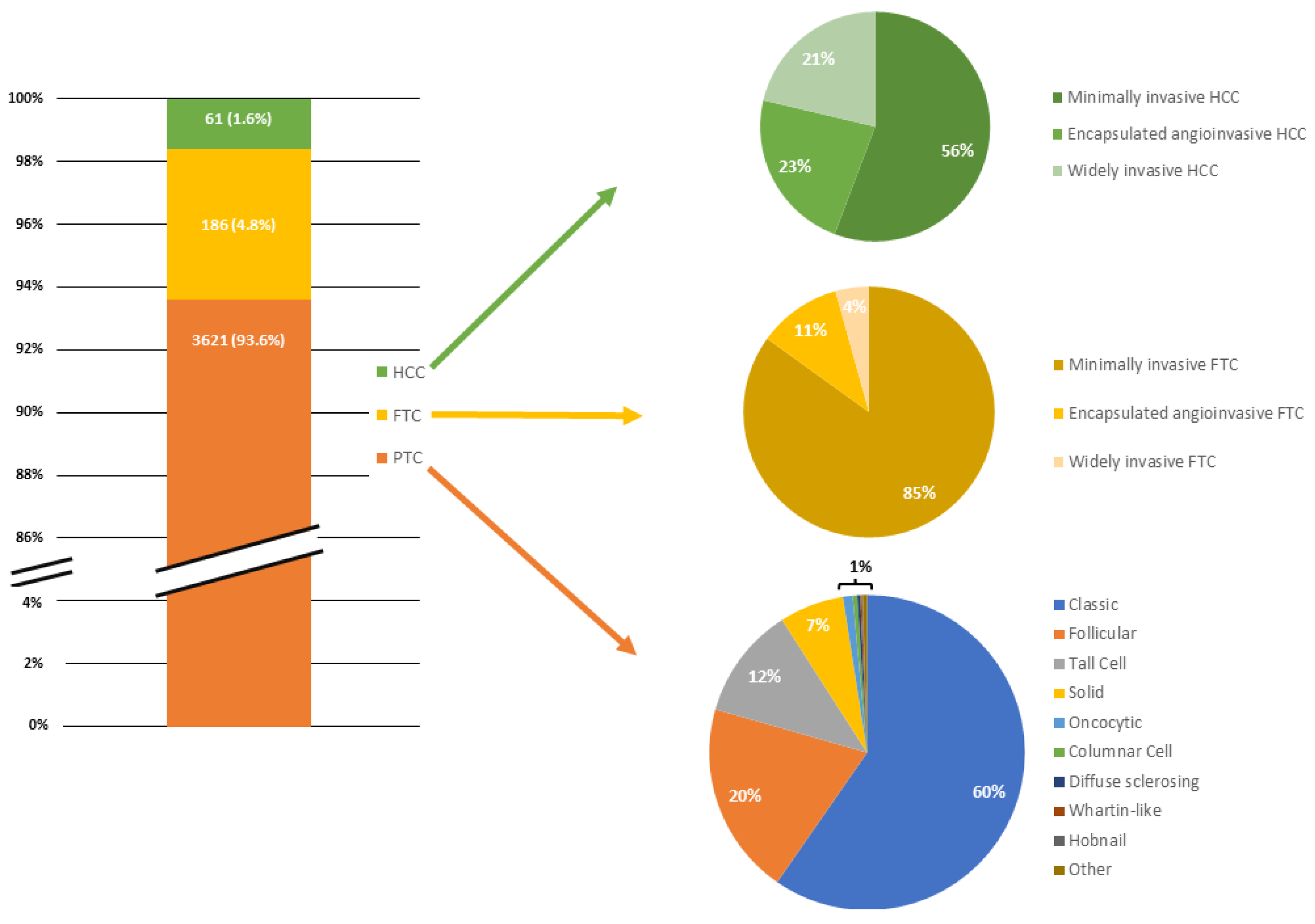
| Histopathology | FTC | 186 (4.8%) |
| HCC | 61 (1.6%) | |
| PTC | 3621 (93.6%) | |
| Max Cancer Diameter, mm | 11 (5–19) | |
| N° microfoci | 2 (1–2) | |
| Lymph Node Metastasis | No | 655 (47.6%) |
| Yes | 720 (52.4%) | |
| CC Pathological Lymph Nodes | No | 689 (51.3%) |
| Yes | 654 (48.7%) | |
| CC N lymph nodes excised | 5 (2–9) | |
| CC N Pathological Lymph Nodes | 0 (0–3) | |
| LC Pathological Lymph Nodes | No | 308 (51.2%) |
| Yes | 294 (48.8%) | |
| LC N lymph nodes excised | 23 (16–31) | |
| LC N Pathological Lymph Nodes | 0 (0–3) | |
| Pathological lymph node max dimension, mm | 8 (3–16) | |
| Extranodal infiltration | No | 3094 (97.7%) |
| Yes | 72 (2.3%) | |
| Aggressive Variant | No | 3151 (81.5%) |
| Yes | 717 (18.5%) | |
| Multifocal | No | 2164 (55.9%) |
| Yes | 1704 (44.1%) | |
| Bilateral | No | 2734 (72.9%) |
| Yes | 1016 (27.1%) | |
| Aggressive Variant on Microfoci | No | 1648 (90.6%) |
| Yes | 171 (9.4%) | |
| Surgical Margin Infiltration | No | 3828 (99%) |
| Yes | 40 (1%) | |
| Extrathyroid Microscopic infiltration | No | 3095 (80%) |
| Yes | 773 (20%) | |
| Extrathyroid Macroscopic Infiltration | No | 3785 (97.9%) |
| Yes | 83 (2.1%) | |
| Vascular-Lymphatic infiltration | No | 3249 (84%) |
| Yes | 619 (16%) | |
| Metastasis | No | 3606 (99.9%) |
| Yes | 1 (0.1%) | |
| pT | 1A | 1822 (47.1%) |
| 1B | 1147 (29.7%) | |
| 2 | 597 (15.4%) | |
| 3A | 222 (5.7%) | |
| 3B | 57 (1.5%) | |
| 4A | 21 (0.5%) | |
| pN | 0 | 655 (47.6%) |
| 1A | 426 (31%) | |
| 1B | 294 (21.4%) | |
| pM | 0 | 371 (99.7%) |
| 1 | 1 (0.3%) | |
| ATA Risk stratification system | High | 395 (10.2%) |
| Intermediate | 1386 (35.8%) | |
| Low | 2087 (54%) | |
| BMI, kg/m2 | |||||
|---|---|---|---|---|---|
| <25 | 25–29.9 | >29.9 | |||
| N (%); Median (IQR) | N (%); Median (IQR) | N (%); Median (IQR) | p Value | ||
| Age at Surgery, years | 46 (35–57) | 52 (41–62) | 53 (42–61) | 0.001 | |
| Gender | Female | 1330 (79.9%) | 779 (62.7%) | 488 (68.4%) | 0.001 |
| Male | 335 (20.1%) | 464 (37.3%) | 225 (31.6%) | ||
| Hyperthyroidism | No | 1505 (90.4%) | 1109 (89.2%) | 639 (89.6%) | 0.570 |
| Yes | 160 (9.6%) | 134 (10.8%) | 74 (10.4%) | ||
| Preoperative Diagnosis | Basedow | 67 (4%) | 46 (3.7%) | 24 (3.4%) | 0.001 |
| Indeterminate nodule | 394 (23.7%) | 307 (24.7%) | 167 (23.4%) | ||
| Malignancy | 904 (54.3%) | 569 (45.8%) | 307 (43.1%) | ||
| Nodular or multinodular Goiter | 295 (17.7%) | 321 (25.8%) | 210 (29.5%) | ||
| Plummer | 5 (0.3%) | - | 5 (0.7%) | ||
| Substernal Goiter | No | 1639 (98.4%) | 1212 (97.5%) | 684 (95.9%) | 0.001 |
| Yes | 26 (1.6%) | 31 (2.5%) | 29 (4.1%) | ||
| Type of Surgery | Completion Thyroidectomy | 8 (0.5%) | 14 (1.1%) | 3 (0.4%) | 0.870 |
| Lobectomy | 220 (13.2%) | 137 (11%) | 75 (10.5%) | ||
| Lobectomy + Completion Thyroidectomy | 37 (2.2%) | 17 (1.4%) | 15 (2.1%) | ||
| Total Thyroidectomy | 1400 (84.1%) | 1075 (86.5%) | 620 (87%) | ||
| Monolateral Central Compartment lymphadenectomy | No | 1655 (99.4%) | 1237 (99.5%) | 710 (99.6%) | 0.820 |
| Yes | 10 (0.6%) | 6 (0.5%) | 3 (0.4%) | ||
| Bilateral Central Compartment lymphadenectomy | No | 1297 (77.9%) | 1007 (81%) | 572 (80.2%) | 0.100 |
| Yes | 368 (22.1%) | 236 (19%) | 141 (19.8%) | ||
| Monolateral Lateral Compartment lymphadenectomy | No | 1535 (92.2%) | 1143 (92%) | 662 (92.8%) | 0.770 |
| Yes | 130 (7.8%) | 100 (8%) | 51 (7.2%) | ||
| Bilateral Lateral Compartment lymphadenectomy | No | 1653 (99.3%) | 1230 (99%) | 706 (99%) | 0.610 |
| Yes | 12 (0.7%) | 13 (1%) | 7 (1%) | ||
| Chronic Thyroiditis | No | 986 (59.2%) | 777 (62.5%) | 463 (64.9%) | 0.021 |
| Yes | 679 (40.8%) | 466 (37.5%) | 250 (35.1%) | ||
| Variant | Classic | 1026 (61.6%) | 731 (58.8%) | 403 (56.5%) | 0.012 |
| Columnar Cell | 3 (0.2%) | 5 (0.4%) | 9 (1.3%) | ||
| Diffuse sclerosing | 5 (0.3%) | 2 (0.2%) | 4 (0.6%) | ||
| Follicular | 301 (18.1%) | 272 (21.9%) | 143 (20.1%) | ||
| Hobnail | 3 (0.2%) | - | - | ||
| Oncocytic | 19 (1.1%) | 12 (1%) | 5 (0.7%) | ||
| Other | 8 (0.5%) | 7 (0.6%) | 1 (0.1%) | ||
| Solid | 115 (6.9%) | 71 (5.7%) | 53 (7.4%) | ||
| Tall Cell | 183 (11%) | 141 (11.3%) | 93 (13%) | ||
| Whartin-like | 2 (0.1%) | 2 (0.2%) | 2 (0.3%) | ||
| Aggressive Variant | No | 1356 (81.4%) | 1024 (82.4%) | 554 (77.7%) | 0.033 |
| Yes | 309 (18.6%) | 219 (17.6%) | 159 (22.3%) | ||
| Aggressive Variant on Microfoci | No | 703 (90.6%) | 558 (93.2%) | 328 (87.2%) | 0.008 |
| Yes | 73 (9.4%) | 41 (6.8%) | 48 (12.8%) | ||
| Max Cancer Diameter | 11 (6–18) | 10 (4–17) | 11 (5–17) | 0.378 | |
| Multifocal Tumor | No | 1195 (71.8%) | 849 (68.3%) | 482 (67.6%) | 0.049 |
| Yes | 470 (28.2%) | 394 (31.7%) | 231 (32.4%) | ||
| N microfoci | 1 (1–2) | 2 (1–3) | 2 (1–3) | 0.011 | |
| AHS on main tumor OR on microfoci | No | 1333 (80.1%) | 1015 (81.7%) | 539 (75.6%) | 0.005 |
| Yes | 332 (19.9%) | 228 (18.3%) | 174 (24.4%) | ||
| Bilateral | No | 1199 (74.4%) | 856 (70.6%) | 477 (69.1%) | 0.014 |
| Yes | 413 (25.6%) | 357 (29.4%) | 213 (30.9%) | ||
| Lymph Node Metastasis | No | 287 (44.6%) | 185 (46.3%) | 120 (46%) | 0.840 |
| Yes | 357 (55.4%) | 215 (53.8%) | 141 (54%) | ||
| CC Pathological Lymph Nodes | No | 300 (47.8%) | 198 (50.5%) | 127 (50.2%) | 0.640 |
| Yes | 328 (52.2%) | 194 (49.5%) | 126 (49.8%) | ||
| CC N lymph nodes excised | 5 (3–9) | 6 (2–10) | 5 (2–9) | 0.313 | |
| CC N Pathological Lymph Nodes | 1 (0–3) | 0 (0–3) | 0 (0–3) | 0.778 | |
| LC Pathological Lymph Nodes | No | 122 (47.8%) | 71 (40.8%) | 51 (47.7%) | 0.310 |
| Yes | 133 (52.2%) | 103 (59.2%) | 56 (52.3%) | ||
| LC N lymph nodes excised | 23 (17–31) | 23 (16–32) | 24 (17–35) | 0.556 | |
| LC N Pathological Lymph Nodes | 1 (0–4) | 1 (0–4) | 1 (0–3) | 0.211 | |
| Pathological lymph node max dimension | 6 (3–15) | 10 (3.5–20) | 8 (2–15.5) | 0.017 | |
| Extranodal infiltration | No | 1326 (97.5%) | 988 (97.7%) | 574 (97.5%) | 0.920 |
| Yes | 34 (2.5%) | 23 (2.3%) | 15 (2.5%) | ||
| Surgical Margin Infiltration | No | 1649 (99%) | 1226 (98.6%) | 706 (99%) | 0.540 |
| Yes | 16 (1%) | 17 (1.4%) | 7 (1%) | ||
| Extrathyroid Microscopic infiltration | No | 1317 (79.1%) | 980 (78.8%) | 560 (78.5%) | 0.950 |
| Yes | 348 (20.9%) | 263 (21.2%) | 153 (21.5%) | ||
| Extrathyroid Macroscopic Infiltration | No | 1632 (98%) | 1216 (97.8%) | 694 (97.3%) | 0.570 |
| Yes | 33 (2%) | 27 (2.2%) | 19 (2.7%) | ||
| Vascular-Lymphatic infiltration | No | 1404 (84.3%) | 1066 (85.8%) | 619 (86.8%) | 0.240 |
| Yes | 261 (15.7%) | 177 (14.2%) | 94 (13.2%) | ||
| Metastasis | No | 1563 (100%) | 1164 (100%) | 642 (99.8%) | 0.120 |
| Yes | - | - | 1 (0.2%) | ||
| pT | 1A | 819 (49.2%) | 625 (50.3%) | 357 (50.1%) | 0.160 |
| 1B | 512 (30.8%) | 368 (29.6%) | 207 (29%) | ||
| 2 | 247 (14.8%) | 167 (13.4%) | 88 (12.3%) | ||
| 3A | 56 (3.4%) | 57 (4.6%) | 42 (5.9%) | ||
| 3B | 22 (1.3%) | 19 (1.5%) | 12 (1.7%) | ||
| 4A | 8 (0.5%) | 6 (0.5%) | 7 (1%) | ||
| pT | pT1 or pT2 | 1578 (94.8%) | 1160 (93.3%) | 652 (91.4%) | 0.008 |
| pT3 or pT4 | 87 (5.2%) | 83 (6.7%) | 61 (8.6%) | ||
| pN | 0 | 287 (44.6%) | 185 (46.3%) | 120 (46%) | 0.150 |
| 1A | 224 (34.8%) | 112 (28%) | 85 (32.6%) | ||
| 1B | 133 (20.7%) | 103 (25.8%) | 56 (21.5%) | ||
| M | 0 | 154 (100%) | 83 (100%) | 54 (98.2%) | 0.115 |
| 1 | - | - | 1 (1.8%) | ||
| ATA Risk stratification system | High | 175 (10.5%) | 122 (9.8%) | 71 (10%) | 0.042 |
| Intermediate | 638 (38.3%) | 413 (33.2%) | 258 (36.2%) | ||
| Low | 852 (51.2%) | 708 (57%) | 384 (53.9%) | ||
| BMI, kg/m2 | |||||
|---|---|---|---|---|---|
| <25 | 25–29.9 | >29.9 | |||
| N (%); Median (IQR) | N (%); Median (IQR) | N (%); Median (IQR) | p Value | ||
| Age at Surgery, years | 50 (36–63) | 54 (46–67) | 53.5 (44–60.5) | 0.280 | |
| Gender | Female | 70 (78.7%) | 42 (60.9%) | 16 (57.1%) | 0.020 |
| Male | 19 (21.3%) | 27 (39.1%) | 12 (42.9%) | ||
| Hyperthyroidism | No | 83 (93.3%) | 62 (89.9%) | 28 (100%) | 0.205 |
| Yes | 6 (6.7%) | 7 (10.1%) | - | ||
| Preoperative Diagnosis | Basedow | 3 (3.4%) | 1 (1.4%) | - | 0.583 |
| Indeterminate nodule | 61 (68.5%) | 40 (58%) | 16 (57.1%) | ||
| Malignancy | 5 (5.6%) | 6 (8.7%) | 3 (10.7%) | ||
| N/MNG | 20 (22.5%) | 22 (31.9%) | 9 (32.1%) | ||
| Plummer | - | - | - | ||
| Substernal Goiter | No | 86 (96.6%) | 60 (87%) | 25 (89.3%) | 0.070 |
| Yes | 3 (3.4%) | 9 (13%) | 3 (10.7%) | ||
| Type of Surgery | Completion Thyroidectomy | 2 (2.2%) | 1 (1.4%) | - | 0.639 |
| Lobectomy | 28 (31.5%) | 14 (20.3%) | 7 (25%) | ||
| Lobectomy + Completion Thyroidectomy | 3 (3.4%) | 1 (1.4%) | 1 (3.6%) | ||
| Total Thyroidectomy | 56 (62.9%) | 53 (76.8%) | 20 (71.4%) | ||
| Monolateral Central Compartment lymphadenectomy | No | 89 (100%) | 68 (98.6%) | 28 (100%) | 0.426 |
| Yes | - | 1 (1.4%) | - | ||
| Bilateral Central Compartment lymphadenectomy | No | 87 (97.8%) | 67 (97.1%) | 28 (100%) | 0.669 |
| Yes | 2 (2.2%) | 2 (2.9%) | - | ||
| Monolateral Lateral Compartment lymphadenectomy | No | 89 (100%) | 69 (100%) | 28 (100%) | - |
| Yes | - | - | - | ||
| Bilateral Lateral Compartment lymphadenectomy | No | 89 (100%) | 69 (100%) | 28 (100%) | - |
| Yes | - | - | - | ||
| Chronic Thyroiditis | No | 62 (69.7%) | 51 (73.9%) | 21 (75%) | 0.780 |
| Yes | 27 (30.3%) | 18 (26.1%) | 7 (25%) | ||
| Variant | Minimally invasive FTC | 75 (84.3%) | 60 (87%) | 23 (82.1%) | 0.950 |
| Encapsulated angioinvasive FTC | 10 (11.2%) | 6 (8.7%) | 4 (14.3%) | ||
| Widely invasive FTC | 4 (4.5%) | 3 (4.3%) | 1 (3.6%) | ||
| Aggressive Variant | No | 85 (95.5%) | 63 (91.3%) | 25 (89.3%) | 0.415 |
| Yes | 4 (4.5%) | 6 (8.7%) | 3 (10.7%) | ||
| Aggressive Variant on Microfoci | No | 24 (96%) | 16 (72.7%) | 5 (71.4%) | 0.068 |
| Yes | 1 (4%) | 6 (27.3%) | 2 (28.6%) | ||
| AHS on main tumor OR on microfoci | No | 85 (95.5%) | 58 (84.1%) | 23 (82.1%) | 0.030 |
| Yes | 4 (4.5%) | 11 (15.9%) | 5 (17.9%) | ||
| Max Cancer Diameter, mm | 22 (16–38) | 30 (20–40) | 40 (23.5–54) | 0.030 | |
| N microfoci | 1.5 (1–2.5) | 1 (1–2) | 2 (1–3.5) | 0.717 | |
| Bilateral | No | 73 (84.9%) | 57 (89.1%) | 21 (84%) | 0.710 |
| Yes | 13 (15.1%) | 7 (10.9%) | 4 (16%) | ||
| Multifocal | No | 66 (74.2%) | 51 (73.9%) | 21 (75%) | 0.990 |
| Yes | 23 (25.8%) | 18 (26.1%) | 7 (25%) | ||
| Lymph Node Metastasis | No | 27 (96.4%) | 21 (95.5%) | 2 (66.7%) | 0.101 |
| Yes | 1 (3.6%) | 1 (4.5%) | 1 (33.3%) | ||
| CC Pathological Lymph Nodes | No | 27 (96.4%) | 21 (95.5%) | 2 (66.7%) | 0.101 |
| Yes | 1 (3.6%) | 1 (4.5%) | 1 (33.3%) | ||
| CC N lymph nodes excised | 2 (1–3) | 2 (1–4) | 3 (2–4) | 0.437 | |
| CC N Pathological Lymph Nodes | 0 (0–0) | 0 (0–0) | 0 (0–1) | 0.147 | |
| LC Pathological Lymph Nodes | No | 31 (100%) | 14 (100%) | 7 (100%) | - |
| Yes | - | - | - | ||
| LC N lymph nodes excised | 0 (0–0) | 0 (0–0) | 0 (0–0) | 0.317 | |
| LC N Pathological Lymph Nodes | 0 (0–0) | 0 (0–0) | 0 (0–0) | 0.718 | |
| Extranodal infiltration | No | 76 (100%) | 56 (100%) | 21 (100%) | |
| Yes | - | - | - | ||
| Pathological lymph node max dimension | 0 (0–0) | 0 (0–0) | 0 (0–0) | 0.317 | |
| Surgical Margin Infiltration | No | 89 (100%) | 69 (100%) | 28 (100%) | - |
| Yes | - | - | - | ||
| Extrathyroid Microscopic infiltration | No | 87 (97.8%) | 68 (98.6%) | 28 (100%) | 0.706 |
| Yes | 2 (2.2%) | 1 (1.4%) | - | ||
| Extrathyroid Macroscopic Infiltration | No | 88 (98.9%) | 69 (100%) | 28 (100%) | 0.578 |
| Yes | 1 (1.1%) | - | - | ||
| Vascular-Lymphatic infiltration | No | 63 (70.8%) | 51 (73.9%) | 16 (57.1%) | 0.250 |
| Yes | 26 (29.2%) | 18 (26.1%) | 12 (42.9%) | ||
| pT | 1A | 7 (7.9%) | 8 (11.6%) | 2 (7.1%) | 0.033 |
| 1B | 32 (36%) | 11 (15.9%) | 3 (10.7%) | ||
| 2 | 31 (34.8%) | 33 (47.8%) | 11 (39.3%) | ||
| 3A | 18 (20.2%) | 17 (24.6%) | 12 (42.9%) | ||
| 3B | 1 (1.1%) | - | - | ||
| 4A | - | - | - | ||
| pT | pT1 or pT2 | 70 (78.7%) | 52 (75.4%) | 16 (57.1%) | 0.070 |
| pT3 or pT4 | 19 (21.3%) | 17 (24.6%) | 12 (42.9%) | ||
| pN | 0 | 27 (96.4%) | 21 (95.5%) | 2 (66.7%) | 0.101 |
| 1A | 1 (3.6%) | 1 (4.5%) | 1 (33.3%) | ||
| 1B | - | - | - | ||
| Metastasis | No | 86 (100%) | 65 (100%) | 27 (100%) | - |
| Yes | - | - | - | ||
| ATA Risk stratification system | High | 7 (7.9%) | 5 (7.2%) | 2 (7.1%) | 0.750 |
| Intermediate | 23 (25.8%) | 20 (29%) | 11 (39.3%) | ||
| Low | 59 (66.3%) | 44 (63.8%) | 15 (53.6%) | ||
| BMI, kg/m2 | |||||
|---|---|---|---|---|---|
| <25 | 25–29.9 | >29.9 | |||
| N (%); Median (IQR) | N (%); Median (IQR) | N (%); Median (IQR) | p Value | ||
| Age at Surgery, years | 50 (42–66.5) | 56 (51–62) | 62 (50.5–73) | 0.158 | |
| Gender | Female | 19 (79.2%) | 14 (66.7%) | 7 (43.8%) | 0.060 |
| Male | 5 (20.8%) | 7 (33.3%) | 9 (56.3%) | ||
| Hyperthyroidism | No | 23 (95.8%) | 21 (100%) | 16 (100%) | 0.457 |
| Yes | 1 (4.2%) | - | - | ||
| Preoperative Diagnosis | Basedow | - | - | - | 0.751 |
| Indeterminate nodule | 17 (70.8%) | 12 (57.1%) | 12 (75%) | ||
| Malignancy | 4 (16.7%) | 6 (28.6%) | 2 (12.5%) | ||
| N/MNG | 3 (12.5%) | 3 (14.3%) | 2 (12.5%) | ||
| Plummer | - | - | - | ||
| Substernal Goiter | No | 24 (100%) | 20 (95.2%) | 15 (93.8%) | 0.495 |
| Yes | - | 1 (4.8%) | 1 (6.3%) | ||
| Type of Surgery | Completion Thyroidectomy | - | 1 (4.8%) | - | 0.897 |
| Lobectomy | 5 (20.8%) | 3 (14.3%) | 3 (18.8%) | ||
| Lobectomy + Completion Thyroidectomy | 1 (4.2%) | 1 (4.8%) | 1 (6.3%) | ||
| Total Thyroidectomy | 18 (75%) | 16 (76.2%) | 12 (75%) | ||
| Monolateral Central Compartment lymphadenectomy | No | 24 (100%) | 20 (95.2%) | 16 (100%) | 0.380 |
| Yes | - | 1 (4.8%) | - | ||
| Bilateral Central Compartment lymphadenectomy | No | 23 (95.8%) | 19 (90.5%) | 15 (93.8%) | 0.768 |
| Yes | 1 (4.2%) | 2 (9.5%) | 1 (6.3%) | ||
| Monolateral Lateral Compartment lymphadenectomy | No | 24 (100%) | 20 (95.2%) | 16 (100%) | 0.380 |
| Yes | - | 1 (4.8%) | - | ||
| Bilateral Lateral Compartment lymphadenectomy | No | 23 (95.8%) | 21 (100%) | 16 (100%) | 0.457 |
| Yes | 1 (4.2%) | - | - | ||
| Chronic Thyroiditis | No | 16 (66.7%) | 17 (81%) | 14 (87.5%) | 0.268 |
| Yes | 8 (33.3%) | 4 (19%) | 2 (12.5%) | ||
| Variant | Encapsulated angioinvasive HCC | 6 (25%) | 3 (14.3%) | 5 (31.3%) | 0.208 |
| Minimally invasive HCC | 15 (62.5%) | 10 (47.6%) | 9 (56.3%) | ||
| Widely invasive HCC | 3 (12.5%) | 8 (38.1%) | 2 (12.5%) | ||
| Aggressive Variant | No | 20 (83.3%) | 12 (57.1%) | 12 (75%) | 0.140 |
| Yes | 4 (16.7%) | 9 (42.9%) | 4 (25%) | ||
| Aggressive Variant on Microfoci | No | 6 (100%) | 6 (100%) | 2 (100%) | - |
| Yes | - | - | - | ||
| Max Cancer Diameter, mm | 30 (19.5–45) | 35 (20–45) | 34 (21.5–44.5) | 0.910 | |
| N microfoci | 1 (1–2) | 1.5 (1–2.5) | 0 (0–0) | 0.717 | |
| Bilateral | No | 19 (79.2%) | 17 (81%) | 15 (100%) | 0.169 |
| Yes | 5 (20.8%) | 4 (19%) | - | ||
| Multifocal | No | 18 (75%) | 15 (71.4%) | 15 (93.8%) | 0.221 |
| Yes | 6 (25%) | 6 (28.6%) | 1 (6.3%) | ||
| Lymph Node Metastasis | No | 4 (80%) | 5 (62.5%) | 4 (100%) | 0.344 |
| Yes | 1 (20%) | 3 (37.5%) | - | ||
| CC Pathological Lymph Nodes | No | 5 (100%) | 5 (62.5%) | 4 (100%) | 0.129 |
| Yes | - | 3 (37.5%) | - | ||
| CC N lymph nodes excised | 2 (1–3) | 3 (2–7) | 2.5 (1–5) | 0.437 | |
| CC N Pathological Lymph Nodes | 0 (0–0) | 0 (0–1) | 0 (0–0) | 0.147 | |
| LC Pathological Lymph Nodes | No | 5 (83.3%) | 4 (80%) | 3 (100%) | 0.719 |
| Yes | 1 (16.7%) | 1 (20%) | - | ||
| LC N lymph nodes excised | 21 (21–21) | 12 (12–12) | 0 (0–0) | 0.317 | |
| LC N Pathological Lymph Nodes | 0 (0–0) | 0 (0–0) | 0 (0–0) | 0.718 | |
| Pathological lymph node max dimension | 0 (0–0) | 4 (3–21) | 0 (0–0) | 0.317 | |
| Extranodal infiltration | No | 22 (100%) | 18 (100%) | 13 (100%) | - |
| Yes | - | - | - | ||
| Surgical Margin Infiltration | No | 24 (100%) | 21 (100%) | 16 (100%) | - |
| Yes | - | - | - | ||
| Extrathyroid Microscopic infiltration | No | 22 (91.7%) | 17 (81%) | 16 (100%) | 0.148 |
| Yes | 2 (8.3%) | 4 (19%) | - | ||
| Extrathyroid Macroscopic Infiltration | No | 23 (95.8%) | 20 (95.2%) | 15 (93.8%) | 0.956 |
| Yes | 1 (4.2%) | 1 (4.8%) | 1 (6.3%) | ||
| Vascular-Lymphatic infiltration | No | 11 (45.8%) | 10 (47.6%) | 9 (56.3%) | 0.790 |
| Yes | 13 (54.2%) | 11 (52.4%) | 7 (43.8%) | ||
| pT | 1A | 1 (4.2%) | 3 (14.3%) | - | 0.752 |
| 1B | 6 (25%) | 4 (19%) | 4 (25%) | ||
| 2 | 8 (33.3%) | 5 (23.8%) | 7 (43.8%) | ||
| 3A | 8 (33.3%) | 8 (38.1%) | 4 (25%) | ||
| 3B | 1 (4.2%) | 1 (4.8%) | 1 (6.3%) | ||
| 4A | - | - | - | ||
| pT | pT1 or pT2 | 15 (62.5%) | 12 (57.1%) | 11 (68.8%) | 0.770 |
| pT3 or pT4 | 9 (37.5%) | 9 (42.9%) | 5 (31.3%) | ||
| pN | 0 | 4 (80%) | 5 (62.5%) | 4 (100%) | 0.476 |
| 1A | - | 2 (25%) | - | ||
| 1B | 1 (20%) | 1 (12.5%) | - | ||
| Metastasis | No | 24 (100%) | 19 (100%) | 16 (100%) | - |
| Yes | - | - | - | ||
| ATA Risk stratification system | High | 3 (12.5%) | 8 (38.1%) | 2 (12.5%) | 0.220 |
| Intermediate | 11 (45.8%) | 6 (28.6%) | 6 (37.5%) | ||
| Low | 10 (41.7%) | 7 (33.3%) | 8 (50%) | ||
| Univariate | Multivariate | |||||
|---|---|---|---|---|---|---|
| OR | 95% C.I. | p | OR | 95% C.I. | p | |
| BMI | 1.016 | 1.00–1.03 | 0.05 | 1.018 | 1.01–1.03 | 0.028 |
| Age at Surgery | 1.001 | 0.99–1.00 | n.s. | 1.001 | 0.995–1.01 | n.s. |
| Female Gender | 0.816 | 0.67–0.98 | 0.036 | 0.795 | 0.66–0.96 | 0.019 |
Disclaimer/Publisher’s Note: The statements, opinions and data contained in all publications are solely those of the individual author(s) and contributor(s) and not of MDPI and/or the editor(s). MDPI and/or the editor(s) disclaim responsibility for any injury to people or property resulting from any ideas, methods, instructions or products referred to in the content. |
© 2024 by the authors. Licensee MDPI, Basel, Switzerland. This article is an open access article distributed under the terms and conditions of the Creative Commons Attribution (CC BY) license (https://creativecommons.org/licenses/by/4.0/).
Share and Cite
Di Filippo, G.; Canu, G.L.; Lazzari, G.; Serbusca, D.; Morelli, E.; Brazzarola, P.; Rossi, L.; Gjeloshi, B.; Caradonna, M.; Kotsovolis, G.; et al. Exploring the Link between BMI and Aggressive Histopathological Subtypes in Differentiated Thyroid Carcinoma—Insights from a Multicentre Retrospective Study. Cancers 2024, 16, 1429. https://doi.org/10.3390/cancers16071429
Di Filippo G, Canu GL, Lazzari G, Serbusca D, Morelli E, Brazzarola P, Rossi L, Gjeloshi B, Caradonna M, Kotsovolis G, et al. Exploring the Link between BMI and Aggressive Histopathological Subtypes in Differentiated Thyroid Carcinoma—Insights from a Multicentre Retrospective Study. Cancers. 2024; 16(7):1429. https://doi.org/10.3390/cancers16071429
Chicago/Turabian StyleDi Filippo, Giacomo, Gian Luigi Canu, Giovanni Lazzari, Dorin Serbusca, Eleonora Morelli, Paolo Brazzarola, Leonardo Rossi, Benard Gjeloshi, Mariangela Caradonna, George Kotsovolis, and et al. 2024. "Exploring the Link between BMI and Aggressive Histopathological Subtypes in Differentiated Thyroid Carcinoma—Insights from a Multicentre Retrospective Study" Cancers 16, no. 7: 1429. https://doi.org/10.3390/cancers16071429
APA StyleDi Filippo, G., Canu, G. L., Lazzari, G., Serbusca, D., Morelli, E., Brazzarola, P., Rossi, L., Gjeloshi, B., Caradonna, M., Kotsovolis, G., Pliakos, I., Poulios, E., Papavramidis, T., Cappellacci, F., Nocini, P. F., Calò, P. G., Materazzi, G., & Medas, F. (2024). Exploring the Link between BMI and Aggressive Histopathological Subtypes in Differentiated Thyroid Carcinoma—Insights from a Multicentre Retrospective Study. Cancers, 16(7), 1429. https://doi.org/10.3390/cancers16071429