Review

Recent Developments in Differentiation Therapy of Acute Myeloid Leukemia

Department of Oncology, Istituto Superiore di Sanità, 00161 Rome, Italy
*
Author to whom correspondence should be addressed.
Cancers 2025, 17(7), 1141; https://doi.org/10.3390/cancers17071141
Submission received: 7 February 2025 / Revised: 13 March 2025 / Accepted: 23 March 2025 / Published: 28 March 2025
(This article belongs to the Section Cancer Therapy)

Simple Summary

Acute myeloid leukemia (AML) is a heterogenous disease caused by a combination of molecular events in hematopoietic stem cells that drive proliferation and block differentiation. The traditional approach to treating AMLs consists of inducing the large-scale elimination of leukemic blasts with chemotherapeutic agents. An alternative approach consists of inducing the differentiation of leukemic cells and has been successfully developed in the treatment of acute promyelocytic leukemia. For many years, the extension of differentiation therapy to other AML subtypes has remained challenging. However, the development of studies on the molecular characterization of AMLs has led to the identification of genetic abnormalities that can be specifically targeted by some drugs, such as inhibitors of isocitrate dehydrogenase (IDH), lysine demethylase 1 (LSD1), and Menin, capable of inducing AML differentiation. This review will discuss the clinical evolution of these inhibitors, outcome data from the latest clinical studies, the development of resistance mechanisms, and strategies to improve outcomes and future directions.

Abstract

Acute myeloid leukemia (AML) is characterized by the clonal expansion of myeloid progenitors blocked at various stages of their differentiation process, and drugs that bypass this differentiation block are therapeutically efficient, as shown by retinoic acid and arsenic trioxide in acute promyelocytic leukemia. However, the successful application of differentiation therapy in APL has not translated into clinical benefit for other non-APL subtypes of AML, in which intensive chemotherapy regimens represent the standard of care. However, the development of molecular studies has led to the identification of therapeutic targets (such as mutated proteins and deregulated pathways) and has led to the generation of a new category of specific pharmacologic agents. Some of these agents, such as inhibitors of mutant isocitrate dehydrogenase (IDH1 and IDH2), lysine-specific demethylase-1 (LSD1), and Menin, have shown the capacity to induce leukemic cell differentiation and with significant therapeutic efficacy.

1. Introduction

A hallmark of acute myeloid leukemia (AML) is represented by a differentiation block of myeloid progenitors/precursors that accumulate in bone marrow and in the blood.
One AML subtype, acute promyelocytic leukemia (APL), displays a unique sensitivity to retinoic acid, promoting the differentiation of leukemic promyelocytes to mature neutrophils [1]. This discovery has provided the rationale for initial clinical studies based on the use of all-trans retinoic acid (ATRA) in APL, which has supported the capacity of this drug to promote the in vivo differentiation of leukemic cells [2,3,4]. Subsequent studies have clarified the molecular basis of the high sensitivity of APL to ATRA; in fact, APL is characterized by a 15;17 chromosome translocation with breakpoints within the retinoic acid receptor α (RARA) gene on chromosome 17 and the PML gene, which encodes a transcription factor on chromosome 15. A PML-RARA fusion protein is formed because of the translocation. The specific targeting of this protein represents the molecular basis of the marked sensitivity of APL to ATRA [5]. The PML-RARA protein is responsible for all of the unique features of APL cells, such as the sensitivity to ATRA, block of cell differentiation at the promyelocyte stage, and increased proliferation due to diminished apoptotic cell death [6]. This specific molecular vulnerability of APL has been exploited and allowed for the development of a curative dual differentiation therapy based on the association of ATRA and arsenic trioxide (ATO), leading to more than 90% of patients being cured from this leukemia without the use of cytotoxic chemotherapy [7].
Although differentiation therapy has completely revolutionized APL treatment, changing the outcome of this leukemic subtype from highly fatal to curable, significant challenges have been observed in attempting to develop differentiation-based treatments for other AML subtypes. However, some recent developments of targeted therapies have led to the identification of newer targets for differentiation therapy. Particularly, in this context, the targeting of some molecules in non-APL AMLs, such as isocitrate dehydrogenase 1 and 2 (IDH1 and IDH2), lysine-specific demethylase 1 (LSD1), and Menin, is associated with the induction of leukemic cell differentiation and significant therapeutic effects.
This review will analyze recent developments in the therapy of AML based on IDH, LSD1, and Menin inhibitors evaluated in clinical trials (Table 1).

2. IDH Inhibitors

2.1. Biology and Mechanisms of IDH Mutations

Isocitrate dehydrogenase (IDH) is an enzyme that catalyzes the oxidative decarboxylation of isocitrate, producing α-ketoglutarate and CO2. In humans, three isoforms of IDH exist: IDH3 catalyzes the third step of the citric cycle, while converting NAD to NADH in the mitochondria; the isoforms 1 and 2 (IDH1 and IDH2) catalyze the same reaction outside of the citric acid cycle and use NADP as cofactor.
Mutations of IDH are frequently observed in AML and their prevalence increases with age. While WT-IDH1 and IDH2 catalyze the decarboxylation of isocitrate to α-ketoglutarate, mutant IDH1 and IDH2 enzymes catalyze the conversion of α-KG to 2-hydroxyglutarate (2-HG), an oncometabolite responsible for the oncogenic activity of IDH1 and IDH2 mutations [8]. Cancer-associated IDH mutations occur at the level of distinct arginine residues in the enzyme active sites. IDH1/IDH2 mutations occur in about 20% of adult AML patients, with a higher frequency of IDH2 (8–19%) than IDH1 (6–16%) mutations. IDH1 is most frequently mutated at the level of the Arg residue (R132), with R132H and R132C being the most recurrent IDH1 mutations and R132L and R132S less commonly observed [8]. At the level of the IDH2 gene, the most recurrent mutations occur at the level of Arg140 (R140Q) and more rarely at the level of R172 (R172K) [8].
Inhibitors of IDH are small molecules that bind within the IDH enzymatic active site, blocking aberrant 2-HG production and inducing myeloid differentiation.
A large study involving the molecular characterization of 1023 older AML patients reported a frequency of 9.7% for IDH1MUT and 18.9% for IDH2MUT, including 1% for double-mutant IDH1/IDH2; IDH1MUT significantly co-occurred with DNMT3AMUT (42.4%), while IDH2MUT was associated with DNMT3AMUT (35.8%), NPM1MUT (31.1%), and SRFSF2MUT (38.3%) [9]. IDH1MUT was less frequently associated with TET2MUT (6.3%) and TP53MUT (9.3%) [9]. A normal karyotype was more frequently observed in patients with IDH1MUT and IDH2MUT, while a complex karyotype was less frequent in patients with IDH1MUT and IDH2MUT AMLs [9]. Patients with IDH2MUT AMLs exhibited improved survival when treated with low-intensity treatments compared to those with IDHWT AMLs.
The co-mutation profile significantly differed in patients with IDH1R132C and IDH1R132H AMLs in that in the former ones, there was a significantly higher frequency of SRSF2, ASXL1, and RUNX1 mutations and a lower frequency of NPM1 mutations than in the latter ones [10].
The frequency of both IDH1 and IDH2 mutations in AML is strongly influenced by age. IDH1 + IDH2 mutations increased from 3.4%, 11.3%, and 17.7% up to 21% in groups of pediatric (0–12 yrs), 13–39 yr old, 40–59 yr old, and >60 yr old patients, respectively [11]. ASXL1 and RUNX1 mutations were markedly increased in older AML patients [11].
Experimental studies have, in part, elucidated the mechanisms through which IDH mutations exert a leukemogenic effect. Studies in mouse models have shown that IDH mutations alone cannot induce the development of the full leukemic process in vivo but need to cooperate with additional genetic lesions to initiate leukemia [12,13]. More recently, transgenic IDH2MUT zebrafish models have confirmed these findings and have reported, in transgenic embryos co-expressing IDH2 mutations and FLT3ITD, the development of a leukemic process recapitulating features of human IDHMUT AML [14]. A single-cell transcriptomic analysis showed increased myeloid skewing, differentiation blockade, and leukemia-associated gene signatures [14].
In vitro models of IDH mutations have shown a consistent pattern of DNA hypermethylation, similar to that observed in primary IDHMUT AML cells [15]. Conditional knock-in experiments have allowed for the exploration of the effects of DNA hypermethylation in hematopoietic progenitor cells (HPCs); using this approach, an IDH1 mutation (IDH1R132H) was inserted into the endogenous murine IDH1 locus and was expressed in all hematopoietic cells or specifically in cells of myeloid cell lineage [15]. These mutant mice showed increased numbers of HPCs and self-renewal capacity [12]. The expansion of HPCs and increased self-renewal were observed also in a model of transgenic mice expressing IDH2R140Q in the hematopoietic system; in addition, a block of cell differentiation was also observed [13].
In addition to hematopoietic cells, IDH mutations impair histone demethylation and inhibit cell differentiation in normal astrocytes [16].
Additional studies have shown that the IDH1R132H mutant promotes cytokine independence and blocks differentiation in hematopoietic cells and these effects can be recapitulated by 2-HG [17].
The key role of IDH mutations in blocking HPC differentiation and promoting leukemic growth was further and more strongly supported by the observation that a small molecule (AGI-6780) acting as a potent and specific inhibitor of mutant IDH2 suppressed the growth of patient-derived leukemia cells and induced cell differentiation of IDH2MUT leukemic cells [18].
A recent study has shown that the expression of IDH1R132H and IDHR140Q mutants into human CD34+ cells via lentiviral transduction markedly impaired the colony forming capacity of erythroid, monocytic, and granulocytic lineage cells [19]. In line with this observation, CD34+ cells isolated from an IDH2MUT AML patient undergoing treatment with Enasidenib, an IDH2 inhibitor, showed a progressive improvement in their differentiation capacity, as supported by the in vitro generation of colonies of mature hematopoietic cells [19].
Finally, a recent study has additionally supported the role of IDH mutations in blocking hematopoietic differentiation. Landberg et al. generated CD34+ cells edited by CRISPR/Cas9 and AAV6-mediated homology-directed repair to express IDH1R132H or IDHR140Q mutations; the mutant CD34+ cells displayed a pronounced decrease in their colony and differentiation capacity. The block of cell differentiation of IDH1R132H-edited cells was rescued by Ivosidenib [20,21].
All of these observations support a key role of IDH mutations in blocking the cell differentiation of IDHMUT AMLs.
It is important to underline that the effects of IDH mutations on cell differentiation are largely dependent on the methylation changes induced by these mutations. The effects induced by IDH mutations on DNA methylation are largely mediated by 2-HG: 2-HG competitively binds and inhibits α-KG-dependent enzymes such as ten-eleven translocation 2 (TET2), an enzyme converting 5-methylcytosine (5-mC) to 5-hydroxymethylcytosine (5hmC). 2-HG-mediated block of TET2 leads to the hypermethylation of the HSC genome [15] (Figure 1). IDH1 and IDH2 mutations in AML are associated with increased DNA methylation. The effects of these DNA methylation changes are not widespread but occur at the level of thousands of focal regions that are specifically hypermethylated compared to normal CD34+ cells and AMLs without IDH mutations [22]. The methylation profile observed at these focal regions is different compared to that observed at the level of loci commonly hypermethylated in AML [22]. The profile of hypermethylated loci is highly comparable in IDH1- and IDH2-mutant AMLs, but DNA hypermethylation is more pronounced in IDH1MUT AMLs than in IDH2MUT AMLs [22]. AMLs with biallelic inactivating TET2 mutations had a less dramatic methylation phenotype, but many of the hypermethylated regions correspond to those observed in IDHMUT AMLs [22]. The 5 hydroxy methyl cytosine (5hmC) levels in hypermethylated DNA regions are significantly lower in IDHMUT and TET2MUT AMLs, thus providing evidence that both mutations lead to increased DNA methylation by blocking TET2-mediated demethylation (TET2 promotes DNA demethylation through the hydroxylation of 5mC) [22]. DNA hypermethylation in IDHMUT AML cells requires the activity of the DNMT3A metyltransferase. Importantly, IDHMUT-specific DNA hypermethylated regions are enriched at the level of enhancers involved in the interaction with genes involved in normal hematopoiesis and AML [22].

2.2. Small Molecule Inhibitors of IDH1: Ivosidenib

Ivosidenib (AG-120) is a first-in-class inhibitor of IDH1MUT that exhibits marked 2-HG reduction in IDH1MUT AMLs and the induction of cell differentiation [23]. Particularly, Ivosidenib inhibits R132 IDH1 mutants with an IC50 in the range of 10–12 nM and, at higher concentrations, inhibits IDH1-WT but not IDH2 [23].

2.3. Clinical Studies with Ivosidenib

Several clinical studies have explored the safety and efficacy of Ivosidenib alone or in association with other antileukemic drugs in IDH1-mutant AMLs (Table 2). A pivotal study by Di Nardo and coworkers reported a rate of complete remission (CR) or complete remission with partial hematological recovery (CRi) of 30.4%, with an overall response rate (ORR) of 41.6% and a median duration of response (mDR) of 8.2 months [24]. Bone marrow studies showed that Ivosidenib induced myeloid differentiation and hematopoietic recovery without an intervening period of bone marrow aplasia, a finding consistent with a mechanism of action involving the induction of myeloid cell differentiation [24]. Differentiation syndrome was observed in 11% of the treated patients and all of these patients showed different degrees of response to treatment [24].
Seventeen patients enrolled in this study responded to Ivosidenib treatment and subsequently underwent allogeneic HSCT [25]. A proportion of 12% of these patients had relapsed following prior transplant before starting Ivosidenib therapy, and 76% were refractory to initial therapy; the most common baseline co-mutations were NPM1 (29%), DNMT3A (24%), and SRFSF2 (18%) [24]. The prior responses to Ivosidenib before allo-HSCT were CR in 10/17, CRi in 4/7, and MLFS (morphologic leukemia-free state) in 3/17 [25]. The post-HSCT mOS was 7.7 months, with survival rates of 76.5% and 47.1% at 6 and 12 months, respectively; the mRFS was 7.3 months, with RFS rates of 58.8% and 47.1% at 6 and 12 months [23]. In all of the patients whose best OR was CR following Ivosidenib therapy and who underwent HSCT, the median OS was not reached [25].
In newly diagnosed IDH1MUT AML patients ineligible for standard chemotherapy induction, Ivosidenib treatment achieved a CR/CRi rate of 42.4%; 18% of the treated patients developed a DS [26].
The phase III trial AG120-C-009 evaluated newly diagnosed IDH1MUT AML patients ineligible for intensive chemotherapy receiving Ivosidenib and Azacitidine, a hypomettylating agent (IA), or placebo and Azacitidine (PA): the event-free survival was significantly longer in patients receiving IA than those receiving PA; the median OS was 24 months with IA and 7.9 months with PA [27]. The IA group had a favorable toxicity profile compared to the PA group [27]. DS was observed in 14% of the patients receiving IA and 8% in those treated with PA [27]. On May 2022, on the basis of the results of this trial, the FDA approved a supplemental application for Ivosidenib, extending the indication in patients with newly diagnosed IDH1-mutated AML patients in older adults or those with comorbidities to include the combination with azacitidine [28].
Several studies combining Ivosidenib- and Ventoclax-based regimens are under evaluation. In a phase IB trial, Ivosidenib and Venetoclax (IVO + VEN) with or without Azacitidine (AZA) were evaluated in 31 patients with IDH1-mutant AML. The CR + CRi rate was 90% in the patients treated with the triplet combination, compared with 83% in those treated with IVO + VEN alone [29]. A proportion of 63% of the patients attained an MRD-negative CR [29]. An updated analysis of this study, including 25 additional patients in phase II (all treated with the triplet drug combination) was recently reported, showing an ORR of 94% with a 93% CR + CRi; the 3-year OS was 70.5%; the patients who received HSCT had a 3-year OS of 94.7% compared to 52.8% in those who did not [30]. A retrospective study analyzed 283 newly diagnosed AML patients ineligible for intensive chemotherapy treated with Ivosidenib plus a hypomethylating agent (IVO + HMA) or VEN + HMA: the CRRs including CR + CRi were 63.3% and 49.5% for IVO + HMA and VEN + HMA, respectively; the 6-month EFS was 56% vs. 39.6% for IVO + HMA compared to VEN + HMA, respectively; and 11.5% of the patients with IVO + HMA compared to 5.0% of the patients treated with VEN + HMA were bridged to HSCT [31].
A phase I clinical study evaluated the safety and efficacy of Ivosidenib in association with induction chemotherapy in newly diagnosed IDH1MUT AML patients, resulting in a CR + CRi rate of 72% [32]. In this study, Ivosidenib was administered during induction, consolidation, and maintenance therapy; thus, it is difficult to understand the relative impact of Ivosidenib at each treatment stage. In this clinical setting, the frequency of DS was low [32]. A more careful examination of these patients at the level of the BM morphology and flow cytometric phenotype at day 14 and 21 showed three different patterns of response: (i) aplasia pattern of <10% cellular, <5% blasts; (ii) >10% cellular, <5% blasts, with morphologic and flow cytometric evidence of blast cell differentiation; (iii) and persistence of leukemic blasts [33]. A differentiation response was observed in 20% of the patients treated with Ivosidenib and chemotherapy [33].
Fathi et al. reported the results of a multicenter phase I trial involving post-HSCT treatment with Ivosidenib for IDH1-mutated AML [34]. In total, 16 patients were enrolled and 8 discontinued maintenance with Ivosidenib. The two-year PFS was 81% and the two-year OS was 88% [34].
The analysis of the primary response and relapse after Ivosidenib treatment allowed the researchers to define the mechanisms of resistance and relapse in R/R IDH1MUT AML patients. Clinical response to Ivosidenib was not predicted by the position of IDH1MUT within the clonal hierarchy: IDH1MUT was subclonal in 28% of cases and clonal in 72% of cases and there was no association between IDH1MUT subclonal or clonal status and the response to Ivosidenib [35]. Baseline RTK mutations (NRAS, KRAS, FLT3-ITD, and FLT3-TKD) are associated with primary treatment resistance [35]. Relapse is characterized by multiple mechanisms, including the emergence of RTK pathway mutations and IDH mutations conferring resistance to Ivosidenib [35].
The TARGET-seq+ method was used for single-cell genotyping to explore sequential bone marrow samples from eight patients (six relapsed and two in sustained remission), six treated with Ivosidenib/Venetoclax and two with Ivosidenib/Venetoclax/Azacitidine [32]. Relapse was associated with either genetic clonal evolution or impaired differentiation of pre-existing clones fully differentiated into mature myeloid cells prior to treatment within HSPC/precursor cell compartments known to demonstrate LSC potential [36]. In the two patients that remained in sustained remission, therapy eradicated all leukemic clones within three cycles of treatment [36].
In conclusion, clinical studies carried out using Ivosidenib have supported its target-specific antileukemic efficacy, associated with a good safety profile. However, several important questions remain to be answered in future studies: the optimal sequencing of Ivosidenib during the various stages of AML disease is unclear; the IVO + AZA regimen must be compared to the NEN + AZA regimen in prospective, randomized clinical trials; the triple therapy regimen (IVO + AZA + VEN) must be evaluated in randomized clinical trials with adequate controls; and the optimal combination therapy in R/R AML patients needs to be established.

2.4. Small Molecule Inhibitors of IDH1: Olutasidenib

Olutasidenib is an allosteric, highly selective, potent, orally administered IDH1MUT inhibitor that binds at the level of the hydrophobic pocket located near the IDH1 heterodimer interfaces and determines the stabilization of the mutant IDH1 molecule in an inactive conformation, thus blocking its neomorphic enzymatic activity. At the structural level, Olutasidenib is a quinoline derivative [37]. In contrast to Ivosidenib, Olutasidenib binds mutated IDH1 in a 2:1 stechiometric ratio, thus offering a potential mechanism to overcome some mutations associated with Ivosidenib resistance [37]. Given these inhibitory properties, Olutasidenib inhibits 2-HG production and promotes granulo-monocytic differentiation in primary IDH1MUT AML cells and inhibits the growth of IDH1MUT AML xenograft models [38].

2.5. Clinical Studies with Olutasidenib

A phase I/II clinical study evaluated the safety and the efficacy of Olutasidenib in IDH1MUT AML and MDS patients. Phase I of the study evaluated the safety and efficacy of Olutasidenib as a single agent and in combination with azacitidine in patients with IDH1MUT AML and MDS; Olutasidenib monotherapy in R/R patients and Olutasidenib as monotherapy or in combination with Azacitidine was evaluated. Grade 3–4 hematologic adverse events were more common with combination therapy than with monotherapy; differentiation syndrome was observed in both the monotherapy and combination treatment [39]. The ORR was 41% in the patients receiving monotherapy and 46% in the patients treated with combination therapy; for treatment-naïve AML patients, the ORR was 25% in monotherapy and 77% in combination therapy [39]. For the R/R IDH1MUT AML patients, the median OS was 8.7 months for monotherapy and 12.1 months for combination therapy [39].
The phase II study involving monotherapy treatment enrolled 153 R/R IDH1MUT AML patients, with a median age of 71 years and a median of two prior therapy regimens; the CR + CRi was 35% and the ORR was 48%; the median duration of the CR + CRi was 25.9 months; the median OS was 11.6 months; and the response rates were similar in the patients who did and did not receive prior Venetoclax treatment [40]. OS occurred in 22% of the patients and 9% of the patients were grade ≥3, with one fatal case [40]. The final analysis at the 5-year follow-up showed a CR + CRi rate of 35% with a median duration of CR + CRi of 25.3 months; the ORR was 48%, with a median duration of 15.5 months; the mOS was 11.6 months and in the patients R/R to prior Venetoclax, the mOS was 16.2 months [41]. Transfusion independence from red blood cells and platelets was achieved in 39% and 41% of the patients, respectively [41]. The analysis restricted to elderly patients (≥75 years) showed that Olutasedinib was well tolerated and 31% of these patients achieved CR + CRi, with a median time to CR + CRi of 15 months and with a median duration of CR of 25.9 months [42].
Phase II of the combination of Olutasidenib and Azacitidine was based on a study involving four cohorts of patients: treatment-naïve patients with AML; R/R AML and MDS patients with no prior exposure to HMA or IDH1 inhibitors; patients R/R to HMA; and patients with prior exposure to IDH1 inhibitors [43]. In an initial report, 72 patients with AML/MDS were reported (20 R/R without prior HMA/HD1 inhibitor therapy; 21 R/R with prior HMA therapy; 20 R/R with prior HD1 therapy; and 11 treatment-naïve AML patients) [40]. In the treatment-naïve AML patients, the CR + CRi rates were 45% and in the R/R setting, the CR + CRi rates were 47% in those without prior HMA/HD1 inhibitor therapy, 38% with prior HMA/HD1 inhibitor therapy, and 30% with prior IDH1 inhibitor therapy [40]. The duration of CR + CRi was longest among the treatment-naïve patients [43].
More recently, a pooled analysis of R/R IDH1-mutant AML patients included in this trial was presented: 43% were refractory and 57% relapsed after a prior treatment; 83% of these patients had at least two prior treatment regimens, including HMA (40%), an IDH1 inhibitor (31%), and HSCT (10%); CR + CRi was observed in 31% of the patients, with a median duration of 15 months; CR was achieved in 27% of the patients with a median duration of 20 months; the ORR was 51%; the median OS was 13 months for all of the patients, being 24 months in the overall responders, 30.6 months in the patients with CR + CRi, and not reached in the patients with CR [44,45].
A multicenter, investigator-initiated phase I/II study will evaluate the safety and efficacy of a triplet regimen based on Olutasidenib in combination with Decitabine and Venetoclax [46]. Interestingly, Cortes et al. showed the efficacy and the safety of Olutasidenib administered to 18 patients with IDH1-mutant AML who relapsed or were refractory to a Venetoclax regimen; 31.3% of these patients displayed CR + CRi [47].
The outcomes in Olutasidenib-treated patients from the 2102-HEM-101 single-arm trial were compared to a real-world external control arm of Ivosidenib, suggestive of favorable effectiveness of Olutasidenib for patients with IDH1-mutant AML who are R/R to a Venetoclax-based regimen [48].
A recent study reported a comparative analysis of Olutasidenib and Ivosidenib. Both these drugs have been approved by the FDA for the treatment of IDH1-mutant AML. In the absence of head-to-head studies, this analysis compared the outcomes observed in studies carried out in R/R AML patients: the rates of CR + CRi observed with Olutasidenib and Ivosidenib were similar; however, the duration of the response observed in the patients treated with Olutasidenib was longer than that observed in patients treated with Ivosidenib (25.9 months vs. 8.2 months, respectively). Another notable difference is that Olutasidenib is structurally smaller with a lower molecular weight than Ivosidenib; thus, it occupies less space in the binding pocket of IDH1 dimers, making it resistant to displacement by a second IDH1 mutation [49]. In biochemical studies, Olutasidenib was found to inhibit mutant IDH1 but not wild-type IDH1 [49].
In conclusion, clinical studies have supported a favorable safety profile and significant antileukemic activity of Olutasidenib. As outlined above, only indirect comparisons with Ivosidenib have suggested that Olutasidenib could induce a longer duration of remission than Ivosidenib. Furthermore, future studies are required to evaluate Olutasidenib as a salvage therapy in patients failing after Ivosidenib treatment. Finally, combination treatments in regimens including Venetoclax have to be explored both in newly diagnosed and R/R AML patients.

2.6. Small Molecule Inhibitor of IDH2: Enasidenib

Enasidenib (AG-221) is a specific IDH2-mutant inhibitor developed by Agios Pharmaceuticals/Celgene, reducing 2HG levels and promoting cell differentiation [50]. Particularly, Enasidenib binds to an allosteric site within the enzyme dimer interface, stabilizing an open conformation of the IDH2-mutant enzyme and inhibiting the conversion of α-KG to 2HG [50]. Enasidenib potently inhibited 2HG production by IDH2R140Q homodimer and IDH2R140Q/WT heterodimer and displayed favorable pharmaceutical properties such as solubulity, slow clearance and oral bioavailability [50].

2.7. Clinical Studies with Enasidenib

Several clinical trials have explored the safety and the efficacy of Enasidenib alone or in association with other antileukemic drugs in IDH2-mutant AML patients (Table 3).
A pivotal study by Stein and coworkers in relapsed or refractory IDH2 mutant AML patients reported an overall response rate of 40.3%, with a median OS of 9.3 months (19.7 months in patients who attained a CR); responses were associated with the cellular differentiation of leukemic blasts and cytotoxicity was not the main driver of the antileukemic activity of Enasidenib [51]. Enasidenib was also explored in older AML patients who were unfit for intensive chemotherapy, showing an ORR of 30.8%, with 18% achieving CR and with an mOS of 11.3 months [52]. The analysis of R/R AML patients treated with Enasidenib showed that Enasidenib induced a marked decrease in the 2-HG levels, preceding the clinical response; 2-HG suppression by Enasidenib did not predict response since most non-responding patients also exhibited 2-HG suppression; complete remission and the normalization of stem and progenitor compartments were associated with the emergence of functional neutrophils bearing IDH2 mutations; in a subset of responding patients, the IDH2MUT burden decreased and remained undetectable during the response; co-mutations in NRAS and MAPK pathway effectors were enriched in the patients resistant to treatment [53].
A randomized phase 3 clinical trial explored Enasidenib vs. conventional care in a population of 319 older IDHMUT R/R AML patients who received two to three prior AML-directed therapies [48]. Enasidenib improved the EFS and hematological parameters but failed to improve the OS compared to standard treatment; the OS could be confounded by early dropout and the use of subsequent AML therapies. [54].
In a phase Ib/II clinical trial, Enasidenib was evaluated in 101 newly diagnosed IDH2-mutated AML patients not eligible for standard induction chemotherapy, the treatment based on Enasidenib plus Azacitidine (two-thirds of patients), or Azacitidine alone (one-third of patients) [55]. Significant differences were observed between the two groups of patients concerning the ORR (48% vs. 14% in Ena + AZA vs. AZA alone, respectively) and CRR + CRRi (43% vs. 8% in Ena + AZA vs. AZA alone, respectively), but not in the median OS [55]. About one-third of the patients in the group receiving Azacitidine alone received Enasidenib at progression as a post-protocol therapy and this may have confounded the survival analysis [55].
Explorative correlative studies performed in patients treated with Enasidenib and Azacitidine showed that responding patients displayed a decrease in blast cell population markers and an increase in differentiated myeloid markers, such as CD11b, and these changes were paralleled by a decrease in the VAF of IDH2 mutations or NPM1 mutations up to low/very low levels; no patients with NRAS, KRAS, or PTPN11 mutations achieved CR [56].
A recent study explored Enasidenib combined with Venetoclax in a population of patients with R/R AML or other myeloid malignancies, showing that this drug combination induced an ORR higher than that observed with Enasidenib alone: the ORR was significantly higher in the IDH2R172- than in the IDH2R140-mutant patients (83% vs. 55%, respectively) [57]. In the whole treated population, the ORR was 70% and the CRR was 57%; the median duration of response was 16.6 months, and three patients proceeded to HSCT following CR [57].
The combination of Enasidenib with intensive induction and consolidation chemotherapy was explored in 93 fit IDH2MUT AML patients, showing that it was well tolerated and was associated with a CR + CRi rate of 77%, with 23% of the patients achieving IDH2 mutation clearance [32].
A recent pilot clinical trial (NCT 03728335) assessed the use of Enasidenib as a post-HSCT maintenance therapy for 15 patients with IDH2-mutated AML: the patients received 24 cycles of Enasidenib therapy with leukemia-free survival rates of 100%, chromic GVHD-free survival rates of 93%, and relapse-free survival rates of 87% [58]. At the safety level, the treatment was well tolerated, with adverse events easily managed [58]. The results were considered highly promising and the enrollment of 20 additional patients was scheduled [58].
Concurrent RAS-signaling mutations are a great challenge in the treatment of IDH-mutant AML patients in that they infer resistance to IDH inhibitors. Thus, as supported by preclinical studies, a phase Ib study was proposed based on the administration of Enasidenib and MEK inhibitor Cobimetinib in R/R AML patients who have co-occurring IDH2 and RAS signaling mutations [59].
Interestingly, a recent study reported an ORR of 43% in high-risk IDH2-mutant MDS treated with Enasidenib monotherapy, with an OS of 14.9 and 25.5 months in R/R and first-line treated patients, respectively [60]. Durable responses were observed in >50% of the low-risk MDS patients [60].
In conclusion, Enasidenib showed a good safety profile and displayed a significant antileukemic efficacy. Although Enasidenib was approved for the treatment of R/R AML patients, a randomized phase III clinical study failed to show an improvement in the OS compared to that with the standard chemotherapy treatment. Similarly, Anasidenib in association with Azacitidine failed to improve the OS of newly diagnosed AML patients compared to Azacitidine alone. Ongoing clinical studies have shown promising preliminary results when Enasidenib was associated with standard chemotherapy or with Venetoclax; however, randomization studies are required to assess the real impact of Enasidenib in these clinical settings.

2.8. Differentiation Syndrome in Patients Treated with IDH Inhibitors

Treatment with IDH inhibitors was associated with the development of differentiation syndrome (DS), a potentially lethal adverse reaction triggered by agents that induce myeloid differentiation.
DS was initially described in acute promyelocytic leukemia (APL) treated with agents inducing myeloid differentiation, such as all-trans retinoic acid (ATRA) and arsenic trioxide (ATO) [61]. DS is a relatively common and potentially severe complication observed in APL patients treated with ATRA and/or ATO; this syndrome is characterized by the association of a number of symptoms, including unexplained fever, weight gain, dyspnea with pulmonary infiltrates, pleuropericardial effusion, hypertension, and renal failure [61,62]. This syndrome may occur in a mild and in a severe form. The mechanisms responsible for the development of DS remain not fully understood but seem to be related to the induction of the differentiation of leukemic blasts, resulting in the massive release of cytokines (“cytokine storm”) and chemokines, and a subsequent systemic inflammatory response and increased expression of adhesion molecules on the surface of differentiated leukemic cells, mediating their adhesion to vascular endothelium and then their migration into tissues [63].
The prevalence, risk factors, and clinical outcomes of IDH-mutant AML patients treated with IDH inhibitors were not prospectively evaluated. A retrospective analysis performed by an independent differentiation review committee, composed of investigators who have participated in the development of Enasidenib, showed that DS was observed in 11.7% of R/R AML patients treated with Enasidenib and that the most frequent symptoms were dyspnea, fever, pulmonary infiltrates, and hypoxia; in 395 of cases, DS was associated with concomitant leukocytosis [64].
The Food and Drug Administration performed a systematic analysis of DS observed in 393 AML patients treated with Ivosidenib or Enasidenib [60]. According to this analysis, DS was identified in 19% of the patients; the predominant symptoms observed in these patients are pulmonary (dyspnea, pulmonary infiltrates, and effusions) [65]. In these patients, the onset of symptoms occurred at a median of 19–20 days after treatment [61]. The predictors of DS were represented by a higher leukemic burden in the bone marrow (≥48% of blasts) and in the blood (≥15–25% blasts); furthermore, concurrent mutations in TET2 and SRFSF2 were associated with a higher risk of developing DS, and the CR + CRi rates were lower in the patients with versus those without DS (Ivosidenib 18% vs. 36%; Enasidenib 18% vs. 25%) [65].
Montesinos and coworkers performed a pooled analysis from four clinical trials involving Enasidenib in AML patients, either as a monotherapy or in combination with Azacitidine or with chemotherapy [66]. The highest incidence of DS was observed in the patients who received Enasidenib plus Azacitidine (17.6%) and the lowest incidence was seen in the patients who received Enasidenib plus chemotherapy (2.2%) [66]. The most common symptoms were dyspnea/hypoxia (80.6%) and pulmonary infiltrates (73.1%) [66]. The baseline risk factors for developing DS were represented by higher levels of bone marrow blasts and lactate dehydrogenase [66].

3. Inhibitors of Lysine-Specific Demethylase 1 (LSD1 or KMD1A)

3.1. Role of LSD1 Inhibitors in HSC Differentiation and in Myeloid Leukemia

LSD1 is a member of the flavin adenine dinucleotide-dependent (FAD-dependent) amine oxidase family of demethylase, participating in the formation of various chromatin multiprotein complexes involved in gene regulation, such as rest co-repressor (CoREST) and nucleosome and remodeling and deacetylase (NuRD) [67,68]. LSD1 acts by demethylating lysine 4 and 9 on histone 3 and also on non-histone substrates, such as p53 and, through these effects, it represses gene expression [67]. LSD1 is essential for the function of HSCs and its knockout results in a reduction in granulopoiesis, thrombocytopoiesis, and erythropoiesis, associated with an expansion of granulo-monocytic, megakaryocytic, and erythroid progenitors, thus supporting a physiological role for LSD1 in myeloid differentiation and maturation [69]. LSD1 is overexpressed in many cancer types, including AML [70].

3.2. Small Molecule Inhibitors of LSD1

A number of compounds with amine oxidase inhibitory activity (trancylcypromine, paragyline, and phenelzine) and SP-2509 and GSK-LSD1 have been utilized in initial studies aiming to explore their anticancer activity. New LDS1 inhibitors suitable for clinical studies, such as ORY-1001 (Iadademstat), SP-2577 (Seclidemstat), IMG-7289 (Bodemstat), and CC-90011 (Pulodenstat), have been evaluated in clinical studies [67,68]. These compounds act as irreversible or reversible LSD1 inhibitors with various degrees of specificity and display in vitro efficacy in AML with the inhibition of cell proliferation and induction of cell differentiation [67,68].
Experimental studies in suitable models have supported the clinical use of LSD1 inhibitors as antileukemic drugs. A pivotal study by Lynch and coworkers provided evidence that pharmacological inhibitors of LSD1 promote the differentiation of myeloid leukemic cells through a mechanism independent of the inhibition of LSD1 enzymatic activity (histone demethylation) but dependent of the interaction between LSD1 and transcription factor GFI1, essential to maintain the differentiation block in AML [71]. Particularly, LSD1 inhibitors disrupt the interaction of LSD1 and RCOR1 with the transcription repressor GFI1, which is bound to a set of enhancers located close to transcription factor genes involved in the regulation of myeloid cell differentiation; the inactivation of GFI1 leads to increased enhancer histone acetylation and consequent gene activation [72]. LSD1 inhibitors interfere with the GFI-mediated repression of PU.1 and C/EBPα target genes and induce the differentiation of AML cells [73,74] (Figure 2). The stabilization of the binding of LSD1 on chromatin at GFI1 binding sites requires the interaction of the HMG-box protein HMG 20B with LSD1 [75] (Figure 2).
Cai et al. showed that the cell of origin of leukemic cell transformation seems to be a determinant of their sensitivity to LSD1 inhibitors [76]. Particularly, they showed that leukemias initiating in HSCs highly express the transcription factor EVI1, have attenuated p53 transcriptional output, are less sensitive to chemotherapy, and are resistant to LSD1 inhibitors [76]. P53 loss of function in progenitor-derived leukemias expressing low EVI1 levels induces resistance to LSD1 inhibition [76]. Interestingly, EVI1high leukemias are sensitized to LSD1 by pretreatment with Venetoclax [76].
The LSD1/CoREST/HDCA2 complex is recruited by the SNAG domain of GFI1 [72]. In addition to GFI1, GFI1B is another repressor containing a SNAG domain and mediating the recruitment of LSD1/CoREST/HDCA2 complexes [77]. These molecular complexes involving LSD1 act as epigenetic regulators of hematopoietic differentiation [77]. GFI1 plays an important role in the control of granulo-monocytic differentiation, while GFI1B plays a role in erythroid and megakaryocytic differentiation [78].
The N-terminal intrinsically disordered region (IDR) is required for the regulation of LSD1–transcription factor interactions, controlling enhancer activation that is necessary for AML cell differentiation [79].

3.3. Clinical Trials with LSD1 Inhibitors
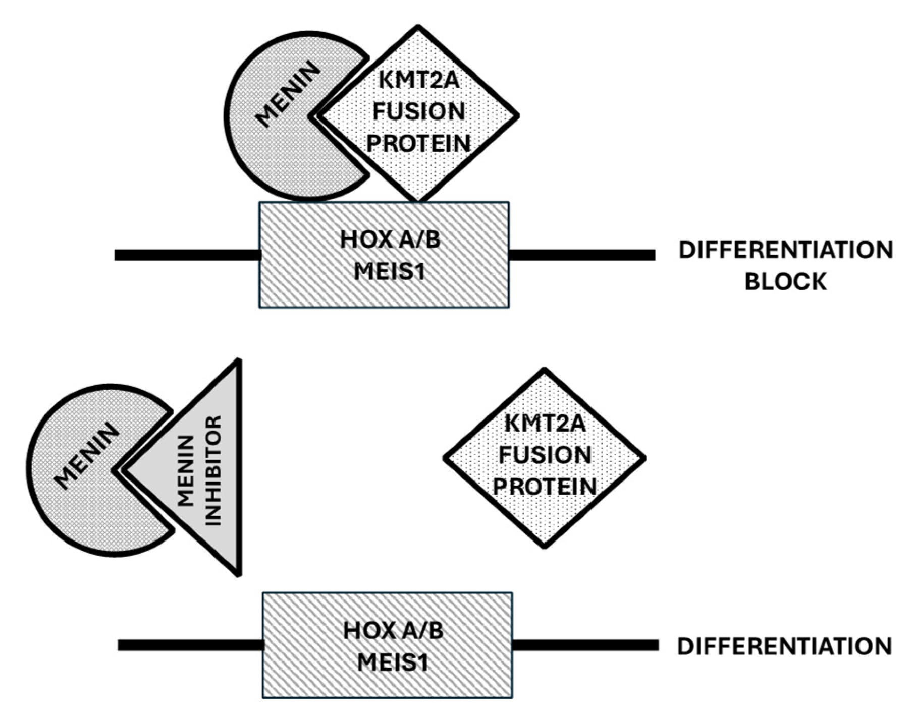
ORY-1001 is a potent and selective LSD1 inhibitor that induces H3K4me2 accumulation on LSD1 target genes, differentiation of leukemic cells, and inhibition of leukemic stem cell activity in AML models [80]. ORY-1001 synergizes with other antileukemic drugs currently used in the treatment of AML and, as seen in a patient-derived xenograft model of T-ALL, significantly reduces leukemia development and improves survival [80]. These observations have supported the clinical evaluation of ORY-1001 (Iadademstat) in AML patients (Table 4).
A phase I clinical study evaluated Iadademstat in relapsed or refractory AML patients: the dose-escalation section of the study carried out in 27 patients showed that the recommended dose of Iadademstat for an extension cohort was 140 μg/m2 administered as a single agent from day 1 to 5 of 28-day cycles; in the extension cohort, 14 additional patients were treated, including 5 patients with MLL/KMT2A-rearranged AMLs [81]. A reduction in the blood and bone marrow percentages of blasts was observed, with one patient achieving CR; the inhibition of blast cell differentiation was frequently observed, particularly in patients with MLL translocations: cell differentiation was observed in 80% of these patients and was particularly pronounced in two patients, with one of these patients developing a rapid and acute differentiation syndrome [81]. Preclinical studies have shown a synergism between Azacitidine and Iadademstat, thus supporting the clinical evaluation of this drug combination. Thus, the phase II ALICE clinical study evaluated 36 newly diagnosed AML patients with intermediate or advanced-risk disease, with a median follow-up of 22 months; dose–response studies showed that the optimal dose of Iadademstat in combination with Azacitidine was 90 μg/m2 [82]. In 27 patients, the ORR was 81% with 14/27 CR + CRi (11/27 MRD−) [82]. Three patients displayed DS and one patient had a fatal grade 5 intracranial hemorrhage (Table 4).
Iadademstat is also being evaluated in combination with Venetoclax and Azacitidine in newly diagnosed AML patients in an investigator-initiated phase I clinical trial at Oregon Health & Science University Knight Cancer Institute (NCT 06357182) and in a company-sponsored phase Ib clinical trial in combination with Gilteritinib in patients with R/R AML harboring FLT3 mutations (NC 05546580).
Particularly, concerning the association of Iadademstat with Gilteritinib, the FRIDA study aims to establish the safety, tolerability, and recommended phase II dose in R/R FLT3-mutant AML patients [83]. This trial was supported by preclinical studies showing a strong synergy of Iadademstat with FLT3 inhibitors and particularly with Gilteritinib in FLT3-mutant AML cells [83]. The FRIDA study was based on a classical design for phase I/II studies, with an escalation phase (from 75 to 150 μg/orally of Iadademstat) and an expansion phase at the selected safe and pharmacologically active dose [83]. The preliminary results for the first 13 patients of the FRIDA trial were presented at the EHA 2024 Meeting: the combination of Iadademstat and Gilteritinib appeared to be safe and well tolerated, with no dose-limiting toxicities at the initial dose (75 μg) and DL1 (100 μg) of Iadademstat; encouraging antileukemic activity was shown, with 5 out of 13 patients (38%) achieving CR + CRi and 9 out of 13 patients (69%) achieving bone marrow blast clearance in the first cycle of treatment [84].
Two other LSD1 inhibitors, SP-2577 (Seclidemstat) and CC-90011 (Purodemstat), are under evaluation in patients with hematological malignancies, but their evaluation in AML patients is, at the moment, very limited.
In conclusion, the clinical studies carried out using Iadademstat in R/R AML patients have shown that this inhibitor has limited efficacy when used in monotherapy. However, the clinical studies performed in FLT3-mutated R/R AML patients using Iadademstat in association with Gilteritinib have preliminarily shown promising results that need to be extended and confirmed in randomized clinical trials with an appropriate control group. In newly diagnosed Aml patients, promising results were observed when Iadademstat was used in association with Azacitidine that need to be confirmed and validated in terms of their clinical impact through randomized clinical studies.

4. Menin Inhibitors

4.1. Role of Menin in the Control of Normal and Leukemic Hematopoiesis

The Menin protein is encoded by the MEN 1 gene, whose germline mutations are the causation of sporadic or autosomal dominant hereditary cancer syndromes affecting the endocrine system; Menin plays a key role as an epigenetic regulator of gene expression for its role as a scaffold protein able to interact with various partners; thus, Menin acts as an adaptor protein between H3K4 (histone 3 protein with the lysine amino acid at position 4) methyltransferase KMT2A (also known as MLL), and LEDGF (lens epithelium derived growth factor). Menin interacts with both the wild-type and rearranged KMT2A, regardless of the fusion partner. Menin was found to be crucial for KMT2A activity and the maintenance of HOXA expression but not essential for normal hematopoiesis. Normal regulation of KMT2A activity is required for the maintenance of expression of the HOX family cluster genes in tissues.
Upregulated HOXA/MEIS1 expression is observed in AMLs characterized by the presence of NPM1 mutations or by rearrangements of the MLL gene [78,79,80]. In NPM1-mutant AMLs (20–30% of total AMLs), the nucleolar protein is mutated and is usually delocalized in the cytoplasm; however, a part of the mutated NPM1 protein resides in the nucleus, where it interacts and is co-localized with KMT2A at the level of the HOXA locus [85,86,87]. The mutant NPM1c protein amplifies the function of KMT2A [79] and also inhibits the activity of histone deacetylases [87] to maintain the active transcription of HOXA/B cluster genes and MEIS1. Mutant NPM1 upregulates the expression of HOXA/B gene clusters also through another mechanism dependent upon the interaction between mutant NPM1c protein and FOXM1 protein with its consequent inactivation and delocalization in the cytoplasm: the transcriptional inactivation of FOXM1 by cytoplasmic sequestration induces the de-repression of HOX A/B cluster genes [88].
KMT2A-rearranged AMLs represent 5–10% of adult AMLs, but are much more frequent in younger patients; in these leukemias, the rearrangement of the KMT2A gene upregulates HOX-MEIS1 expression [89]. The KMT2A protein directly binds to and uses its SET domain H3 (Lys4) methyltransferase to regulate HOX gene promoters. A third AML subset associated with increased HOX A/B expression is represented by the partial tandem duplication (PTD) of the KMT2A gene, occurring in 3–11% of adult de novo AML and associated with adverse outcomes. At the level of gene expression, KMT2A-PTD is characterized by a peculiar profile, showing the increased expression of several HOX genes, including HOX-B5, HOX-B7, HOX-B8, and HOX-B9. A comparative analysis of the profile of HOX-A/B genes deregulated in these AMLs overexpressing HOX genes showed a different profile in KMT2A-PTD- and KMT2A-rearranged AMLs and a similar profile to that of KMT2A-PTD- and NPM1-mutant AMLs [90]. The analysis of the whole gene expression profile allowed for the definition of three gene signatures along the HOX-targeted gene axis (HOX primitive, HOX transient, and HOX committed profiles); the KMT2A-rearranged AMLs were mostly classified as HOX-committed, while the KMT2A-PTD as HOX-primitive/transient and NPM1-mutant AMLs were distributed along the three HOX profiles [90].
A recent study provided an analysis of pediatric AMLs subdivided according to the HOX family gene expression. No expression or very low expression of the HOXA and HOXB genes was observed in normal bone marrow cells, while 25% of pediatric AML had significant upregulation of HOX A/B gene expression. The cluster of AMLs with the highest HOX A expression contained 88% of the KMT2A-rearranged AMLs and these patients displayed a significantly worse outcome compared to those with low/moderate HOX expression; patients without KMT2A rearrangements that clustered within the group at the highest HOX A expression had a dismal outcome [91]. Patients with high HOXB expression mainly have FLT3-ITD or NUP98-NSD1 gene alterations; NUP-98-rearranged AML also expresses high HOXA levels [91]. AMLs with HOX-A expression display a clear co-expression of MEIS-1 [91]. NUP98 gene rearrangements are associated with increased HOX gene expression, related to a direct activation effect exerted by NUP98 fusion proteins [88,92].
In addition to NUP98, NUP214 is another nucleoporin involved in translocations with two chromatin remodeling proteins, SET and DEK, resulting in fusion proteins that influence HOX gene expression [88,92].

4.2. Menin Inhibitors

By applying high-throughput screening, Grembecka and coworkers identified lead compounds targeting and inhibiting the Menin–MLL interaction; some of these compounds were then optimized by medicinal chemistry to develop inhibitors with nanomolar affinities [93,94]. Preclinical studies have shown that these compounds block proliferation, inducing apoptosis and differentiation in leukemic cells bearing MLL translocations; furthermore, these inhibitors reverse MLL fusion protein-mediated leukemic transformation though the downregulation of the expression of target genes involved in MLL fusion protein oncogenic activity [94] (Figure 3). Preclinical studies have supported the efficacy of Menin inhibitors in both MLL-rearranged and NPM1-mutated models of leukemia. Thus, Krivstov et al. showed that the highly selective Menin inhibitor VTP50469 displaced Menin from protein complexes and inhibited chromatin occupancy by MLL of selected genes in leukemic cells bearing MLL rearrangements, resulting in the induction of cell differentiation and apoptosis [94]. Patient-derived xenograft models derived from MLL-rearranged AML cells showed dramatic reductions in leukemia when treated with VTP50469 [95]. Klossowski et al. confirmed that the Menin-specific inhibitor MI-3454 inhibited cell proliferation and induced differentiation in primary patient samples with MLL translocations or NPM1 mutations and induced the remission or regression of leukemia in mouse models of MLL-rearranged or NPM1-mutated leukemia [96]. Menin inhibitors silence both a canonical HOX- and MEIS-1-dependent oncogenic gene expression program and a noncanonical transcriptional program involving tumor suppressor genes and both these events are required to achieve a good therapeutic response [97].
In vitro studies in NPM1-mutant or MLL-rearranged leukemic cell lines or primary AML cells using the Menin inhibitor DS-1594b showed that the primary effect was the induction of cell differentiation and not of apoptosis; when the Menin inhibitor was associated with the BCL-2 inhibitor Venetoclax, the apoptotic effect was predominant [98].

4.3. Clinical Studies with Menin Inhibitors

4.3.1. Revumenib

Revumenib is a potent oral small molecule inhibitor of the Menin–KMT2A interaction. This compound was evaluated in monotherapy in R/R AML patients with KMT2A rearrangements or NPM1 mutations. The AUGMENT-101 trial (NCT 04065399) is a phase I clinical study evaluating the safety and efficacy of Revumenib in patients with R/R heavily pretreated NPM1-mutant or KMT2A-rearranged AMLs [88]. Since Revumenib is a substrate of CYP3A4, the study was subdivided into two cohorts: cohort A, with patients receiving a CYP3A4 inhibitor, and cohort B, with patients not receiving a CYP3A4 inhibitor [85]. The first evaluation of this study was based on 60 patients (46 with KMT2A-rearranged AMLs and 14 with NPM1-mutant AMLs); the ORR was 59% in the KMT2A-rearranged AMLs and 36% in the NPM1-mutant AMLs; the rate of CR + CRi was 30%; with a median follow-up of 11.9 months, in the patients who achieved morphologic CR + CRi, the median duration of response was 9.1 months; the mOS was 14.3 months; 56% of the responder patients achieved MRD negativity and 38% underwent allogeneic hematopoietic stem cell transplantation [99] (Table 5). Interestingly, in many patients with KMT2A rearrangements achieving morphological remission after one cycle of treatment, there was continued evidence of KMT2A fusions, a phenomenon seemingly related to the induction of leukemic blast differentiation elicited by Revumenib. Differentiation syndrome was observed in 16% of the patients, with all cases classified as grade 2 [99]. An additional eight AML patients with NPM1-WT and KMT2A-WT did not respond to Revumenib [99]. An analysis of the pharmacodynamic effects of Revumenib assessed through the transcriptional analysis of the RNA expression of bone marrow cells showed that Menin inhibition resulted in the downregulation of HOXA9 and MEIS1, associated with an increase in the expression of genes related to differentiation, such as CD11b and CD14 [99]. Single-cell studies carried out on four patients treated with Revumenib showed a differentiation continuum that started with immature AML blasts (CD34+/c-kit+), progressed through intermediate blast cells (CD68+, CD11b, and CD14), and ended with more differentiated monocytic cells (CD68+, CD11b+, and CD14+); intermediate AML blasts and monocytic cells were enriched in post-treatment samples [100].
A second study reported the results of phase II, carried out in 94 patients with R/R KMT2A-rearranged acute leukemia (78 with AML and 14 with ALL); 13% of the patients discontinued treatment for adverse events; DS was observed in 26% of the patients (14.9% had grade 3 and 1% grade 4) [101]. The CR + CRi rate was 22.8%, with an ORR of 63.2%; the median duration of response was 6.4 months; the mOS was 8.0 months; and among the responding patients, 38.9% received allo-HSCT [101]. Transcriptional changes in the bone marrow cells showed a decrease in MEIS1 and HOXA expression and an increase in the differentiation-related genes CD11b and CD14 [101]. An updated evaluation of the KMT2A-rearranged AML patients enrolled in the AUGMENT-001 trial extended to a total of 116 patients showed that 23% of the patients achieved CR + CRi, with an ORR of 64% and with a rate of 5.8% for MRD negativity among the patients achieving CR + CRi; 34% of the patients who achieved ORR proceeded to HSCT [102].
Recently, Syndex Pharmaceuticals announced the results of phase II of the AUGMENT-001 study evaluating the efficacy of Revumenib in 64 R/R heavily pretreated (75% relapsing after Ventoclax therapy) NPM1-mutated AML patients: 23% of the patients achieved CR + CRi; the ORR was 47%; the MRD negativity was 64% among the patients who achieved CR + CRi; and 17% of the responding patients underwent HSCT [103].
The ALLG AMLM26 INTERCEPT multiarm study was designed to obtain proof-of-concept for novel therapies targeting MRD or early relapse in AML [104]. In the context of this study, a preliminary analysis on nine NPM1-mutant AML patients with MRD-positivity showed an NPM1 mutant ≥ 1 log10 MRD reduction in 62.5% of the patients, with 37.5% of the patients achieving MRD negativity [104].
A phase I/II study evaluated the combination of Revumenib with decitabine/cedazuridine and Venetoclax in R/R AML patients (NPM1-mutant, KMT2A-rearranged, and NUP98-rearranged), reporting an ORR of 88%, with a CR + CRi of 58%, and with a rate of MRD negativity according to flow cytometry of 93% among the patients with CR + CRi [105].
In pediatric patients, KMT2A rearrangements are frequently associated with RAS pathway mutations (51% of cases) and with a worse prognosis [106]. A recent preclinical study showed that the combination of a Menin inhibitor (VTP-50469, an analog of Revumenib) and Selutenib (a MEK 1–2 inhibitor) exerted a synergistic antitumor effect in vitro and in PDX models of KMT2A-rearranged AML cells bearing RAS pathway mutations [106].
About 40% of patients treated with Revumenib develop MEN1 mutations and some acquire resistance to Menin inhibitors without MEN1 mutations. Particularly, Perner et al. identified Menin gene mutations that were not present at diagnosis and developed with Revumenib treatment; the clonal expansion of these mutations was observed in about 39% of evaluable patients who had undergone at least two cycles of treatment [107]. These mutations were found at residues M327, G331, or T349 which do not impact the interaction between KMT2A and Menin or its oncogenic properties but decrease the binding affinity of Revumenib to KMT2A and mediate therapeutic resistance [107,108]. A recent study characterized in detail the consequences of Menin mutations in the interaction with Menin inihbitors [93]. The crystal structure of the Menin mutants T349M, M327I, G331R, and G331D and the N-terminal of MLL1 showed that drug-resistant mutations in Menin occur at a site located in proximity of the MLL1 binding site, but do not affect MLL1 binding to Menin. All of these point mutations in Menin generate a steric collision with Menin inhibitors; the G331D mutant shows a particularly slow dissociation of MLL1 from Menin and thus seems to be particularly difficult to inhibit with small molecule inhibitory drugs [109].
The identification of MEN1 mutations in AML patients undergoing treatment with Revumenib is important because it may offer opportunities for patients with some MEN1 mutations to derive benefit from treatment with some new Menin inhibitors.

4.3.2. Bleximenib

JNJ-75276617 (Bleximenib) is a novel potent inhibitor of the protein–protein interaction between Menin and KMT2A; in KMT2A-rearranged and NPM1-mutant leukemia cells, this compound inhibited the interaction of the Menin–KMT2A complex with chromatin at the level of target genes, resulting in the reduced expression of several target genes such as MEIS1 and FLT3 and exerted potent antiproliferative activity [110]. Bleximenib displayed synergistic antileukemic activity with Gilteritinib, Venetoclax, and Azacitidine [110]. Interestingly, Bleximenib displayed strong antiproliferative activity in leukemic cells bearing mutations (MEN1M371 or MEN1T349M) observed in patients refractory to Revumenib [110]. In these mutant AMLs, Bleximenib was still able to displace KMT2A and prevent its interaction with Menin despite the presence of MEN1 mutations that block the activity of other Menin inhibitors.
A first-in-human phase I clinical study evaluated the safety and, preliminary, the efficacy of Bleximenib in 58 R/R acute leukemia patients (56 AML and 2 ALL), showing an acceptable safety profile and preliminary evidence of antileukemic efficacy and biologic activity [111].
A phase I clinical study (NCT 04811560) carried out in 121 R/R acute leukemia (mostly AML, at 108) patients identified the optimal dose of Bleximenib (100 mg) in monotherapy to be used in phase II studies; at this dose, the ORR was 50% and the CR + CRi was 40% [112]. The enrolled patients had either KMT2A rearrangements or NPM1 mutations. Another recent phase I clinical study explored Bleximenib in association with standard chemotherapy in a group of 22 newly diagnosed AML patients (11 KMT2A-rearranged and 11 NPM1-mutated), showing an acceptable safety profile with no DS or dose-limiting toxicities [113]. The ORR was 93% (83% in KMT2A-rearranged AML and 100% in NPM1-mutated AML); six patients proceeded to HSCT [113]. A phase Ib (NCT 05453903) clinical study explored the safety and efficacy of Bleximenib in combination with Venetoclax and Azacitidine in R/R AML patients with KMT2A rearrangements or NPM1 mutations [98]. In the safety dataset, 45 patients received the triplet combination treatment, with a median of two prior lines of treatment, including prior Venetoclax treatment in 56% of cases and allo-HSCT in 27% of cases [113]. The safety profile was acceptable and no patient developed DS or tumor lysis syndrome [114]. In the efficacy dataset, the ORR was 86% and the rate of CR + CRi was 48%; for patients with prior Venetoclax exposure, the ORR was 82% and the rate of CR + CRi was 36% [114]. Nine responder patients discontinued treatment and proceeded to allo-HSCT [114] (Table 5).
Hogeling et al. recently reported a large in vitro screening of primary AML cells incubated in the presence of Bleximenib [115]. This study showed that AML cells bearing NPM1 mutations or KMT2A rearrangements are sensitive to the antiproliferative and differentiation-inducing effects of Bleximenib [115]. Concerning NPM1-mutant AMLs, those bearing both NPM1 and DNMT3A mutations are those markedly sensitive to the induction of differentiation [104]. In addition to these AML subtypes, AML characterized by a granulo-monocyte progenitor (GMP)-like phenotype also displayed sensitivity to Bleximenib; these cases included leukemias bearing CEBPA mutations [115]. Gene expression studies have shown that AMLs responding to Bleximenib display some epigenetic alterations inducing a striking upregulation of HLA class I and class II expression through a mechanism independent of MEIS 1 loss [115]. These epigenetic changes resulted in an enhanced sensitivity of leukemic cells to T cell-mediated cytotoxicity in allogeneic and autologous settings [115].
In line with this observation, previous studies have shown that CEBPA is an essential collaborator in HOXA9/MEIS 1-mediated leukemogenesis [116]; CEBPA and KMT2A are co-localized on chromatin and CEBPA-mutated HPCs are hypersensitive to the pharmacologic targeting of the KMT2A complex using Menin inhibitors [117].

4.3.3. DSPP-5336 (Enzomenib)

Enzomenib is a potent, orally bioavailable inhibitor of the Menin–MLL interaction, exhibiting antiproliferative activity in vitro in cell lines, primary leukemic blasts, and patient-derived xenograft mouse models of KMT2A-rearranged or NPM1-mutant AMLs [118]. Enzomenib directly binds to Menin (Kd 6.0 nM) and inhibits the Menin–KMT2A interaction (IC50 1.4 nM) [118].
A phase I/II clinical study evaluated 81 patients in Arm A (without CYP3A4 inhibitor) and in Arm B (with CYP inhibitor) for the safety and efficacy of a treatment based on Enzamenib monotherapy [119]. In the phase I portion of the study, it was found that the active doses of Enzomenib corresponded to ≥ 140 mg [120]. In total, 35 patients were treated at the active Enzomenib dose: 22 patients had KMT2A-rearranged AML (20 AML and 2 ALL) with an ORR of 59% and a CR + CRi of 22.7%; 13 NPM1-mutant AML patients had an ORR of 53.8% and a CR + CRi of 23% [120]. The pharmacodynamic changes supported the induction of cell differentiation with a decrease in stemness markers (HOXA9 and MEIS1) and an increase in differentiation markers (CD11b) [120]. DS was observed in 11% of the treated patients (Table 5).

4.3.4. KO-539 (Ziftomenib)

Ziftomenib is an oral Menin inhibitor targeting the Menin–KMT2A protein–protein interaction; Ziftomenib is metabolized into two metabolites with comparable activity to Ziftomenib itself. Preclinical studies have supported the antileukemic activity of Ziftomenib in models of NPM1-mutant and KMT2A-rearranged AMLs. Particularly, Ziftomenib induced Menin protein degradation through the ubiquitin–proteasome pathway, reduces the MEIS1, FLT3, CDK6, and BCL2 protein levels, in association with the induction of differentiation and the induction of cell apoptosis of leukemic cells harboring MLL-rearrangements or NPM1 mutations [121]. Furthermore, co-treatment with Ziftomenib and BCL-2, CDK6, or BET inhibitors induced synergistic lethality in MLL-rearranged or NPM1-mutant leukemic cells [121]. These findings were confirmed and extended in a second study showing that Ziftomenib had marked anti-proliferative activity in combination with drugs from various classes, including those targeting chromatin regulation and DNA damage as well as apoptosis and cell cycle block [122]. Particularly pronounced was the synergistic interaction between Ziftomenib and Venetoclax, resulting in pronounced anti-proliferative activity in MLL-rearranged and NPM1-mutant leukemic cells [122].
The KOMET-001 multicenter, open-label, multicohort phase I/II clinical trial evaluated Ziftomenib in adult R/R AML patients; the study was subdivided into two phases: a phase Ia (dose–response), in which the patients received Ziftomenib (from 50 to 1000 mg) orally once daily in 28-day cycles; and a phase Ib, in which patients with KMT2A rearrangements or NPM1 mutations were randomly assigned to two parallel dose cohorts (200 and 600 mg Ziftomenib) [123]. In phase Ib, no responses were observed in the patients treated at 200 mg of Ziftomenib; at the recommended dose for phase II (600 mg), the rate of CR + CRi was 12.5% (2/16) in the KMT2A-rearranged and 35% (7/20) in the NPM1-mutant AML patients; the ORR in the KMT2A-rearranged AML patients was 17% and 45% in the NPM1-mutant AML patients; the median OS was 6.0 and 5.6 months, respectively, in the KMT2A-rearranged and NPM1-mutant AMLs [123].
The KOMET-007 phase I clinical study evaluated the safety and the efficacy of Ziftomenib combined with standard chemotherapy in 34 newly diagnosed AML patients with KMT2A rearrangements or NPM1 mutations; the patients were treated either with 200 or 400 mg of Ziftomenib [124]. For NPM1-mutant AML patients, the CR + CRi rate was 100% at 200 mg and 86% at 400 mg, with MRD negativity among the responders of 100% and 80%, respectively; for the KMT2A-rearranged patients, the CR + CRi rates were 90% at 200 mg and 63% at 400 mg, with an MRD negativity rate among the responders of 83% and 100%, respectively [124].
The ongoing KOMET-008 trial is an open-label dose-escalation and expansion study aiming to determine the safety, tolerability, and preliminary efficacy of Ziftomenib in association with standard-of-care regimens for the treatment of either R/R NPM1-mutant AML (arm A) or KMT2A-rearranged AML (arm B). Arm A is subdivided into three cohorts: cohort A-1 (Ziftomeniob + FLAG-IDA), cohort A-2 (Ziftomenib plus LDAC), and cohort A-3 (Ziftomenib plus Gilteritinib in NPM1/FLT3 double mutant AML). Arm B is subdivided into two cohorts: cohort B-1 (Ziftomenib plus FLAG-IDA) and cohort B-2 (Ziftomenib plus LDAC) [125].

4.3.5. BMF-219 (Icovamenib)

BMF-219 is the only covalent Menin inhibitor in clinical development under evaluation in multiple hematologic malignancies, solid tumors, and diabetes mellitus. Preclinical studies have shown that BMF-219 shows sustained and marked inhibition of Menin-dependent oncogenic signaling.
The phase I COVALENT-101 phase I dose-escalation and dose-expansion study evaluated BMF-219 in R/R acute leukemia (cohort 1), DLBCL (cohort 2), multiple myeloma (Cohort 3), and CLL (cohort 4) [115]. A recent report analyzed the results observed in 26 R/R AL patients (24 AML and 2 ALL) enrolled in two parallel arms, with or without a CYPP314 inhibitor [126]. BMF-219 was usually well tolerated, with no dose-limiting toxicities; DS was observed in 13% of the cases [115]. Five patients were evaluable for response and two achieved CR (one CR and one CRi) [126].

4.3.6. Menin Inhibitors in AML Patients with NUP98 Rearrangements

Recurrent chromosomal rearrangements involving the Nucleoporin 98 (NUP98) gene, detected in 5–10% of pediatric AML cases and in 2–4% of adult AMLs, are classified as high-risk AMLs [116]. In NUP98 rearrangements, the NUP98 gene is fused with various partners, the most frequent being NSD1 and KDM5A [127]. NUP98 fusions elicit leukemogenesis through interactions with histone-modifying complexes: particularly, NUP98 fusion proteins interact with KMT2A complexes and are co-localized with KMT2A at the level of HOX A/B genes [128]. NUP98-rearranged AMLs are Menin-dependent, as supported by the observation that the Menin inhibitor VTP50469 induced antiproliferative effects and prolongation of survival in mouse AML models driven by NUP98 fusion proteins [129].
Another study showed that the Menin inhibitor Revumenib inhibited the proliferation and survival of primary NUP08 fusion protein-positive AML cells and inhibited numerous NUP fusion protein target genes, such as MEIS1 and CDK6 [130].
In order to exert their leukemogenetic activity, NUP98 fusion proteins and KMT2A–Menin antagonize the noncanonical polycomb repressive complex 1.1 (PRC1.1) [131].
Interestingly, Carraway et al. reported the case of a patient with NUP98 fusion who relapsed after prior treatments, including allo-HSCT; the patient was treated with BMF-219 and achieved CR after a few cycles of treatment; unfortunately, after 5 months of treatment, the patient relapsed with NUP98-NSD1-positive AML [132].
The findings of this case report suggest that Menin inhibitor monotherapy is not sufficient to obtain complete eradication of leukemic clones in NUP98 fusion-positive AML patients and that the cooperation between Menin inhibition and kinase inhibitors targeting either CDK6 or FLT3 strongly cooperate in NUP98-rearranged primary AML cells and in PDX models [133].
In conclusion, the studies carried out using Menin inhibitors have supported their efficacy in monotherapy in NMP1-mutant and KMT2A-rearranged AMLs. Given the preliminary stage of development of clinical studies with Menin inhibitors, Revumenib, Bleximenib, Enzomenib, and Ziftomenib, at the moment, it is difficult to perform a comparison of their clinical efficacy. Preliminary data have shown the promising efficacy of either Revumenib or Bleximenib in association with an HMA and Venetoclax in R/R NMP1-mutant and KMT2A-rearranged AMLs; these findings need to be confirmed in a large number of patients and compared to an appropriate control representing a standard of care for these patients.

5. Conclusions

In AML, different genetic events determine a block in the differentiation of leukemic cells at various stages of the differentiation/maturation process. Targeted therapies developed in the late twentieth century have introduced a new therapeutic approach based on the targeting of a specific abnormality present in a subset of AML cells. One of the aims of target therapy consists of forcing leukemic cells to differentiate. A prototype of successful differentiation therapy was represented by the treatment of APL using the differentiation-inducing agents ATRA and ATO that have completely revolutionized the therapy of this AML subtype, transforming its outcome from the deadliest to the most curable.
However, ATRA and ATO resulted as not effective in AML outside APL, thus providing evidence that the differentiation block observed in various AML subtypes does not have a uniform underlying mechanism causing differentiation impairment. Thus, the idea that a strategy to induce differentiation in other AML subtypes through the targeting of specific alterations during the leukemic process emerged. This view was strongly supported by recent studies showing that IDH inhibitors, LSD1 inhibitors, and Menin inhibitors induce the differentiation of some AML subtypes. Although these agents have shown a marked capacity to induce leukemic cell differentiation associated with antileukemic effects, therapeutic success was clearly lower than that observed using ATRA + ATO in APL. This lower efficacy of differentiation therapy based on IDH, LSD1, and Menin inhibitors compared to ATRA + ATO in APL is seemingly related to a higher genetic complexity and heterogeneity of IDH-mutant, KMT2A-rearranged, and NPM1-mutant AMLs compared to APL cells, in which all the pathogenic events are driven by PML-RARA fusion protein.
However, in spite of these difficulties, these agents represent an important therapeutic tool for improving the outcomes of IDH-mutant, NPM1-mutant, and KMT2A-rearranged AMLs and possibly also of other AML subtypes. Optimal responses induced by IDH, LSD1, and Menin inhibitors may be significantly improved by strategic partnering with other therapies: ongoing and future clinical studies will clarify the impact of these association therapies, appropriately extended to AML patients at various disease stages. In this context, the introduction of some of these agents into the first-line treatment of elderly non-fit patients using triplet drug combinations is promising.
Future studies will be required to evaluate the impact of these agents on the overall survival in the context of various therapeutic settings involving R/R and first-line patients. Future studies will also assess the potential therapeutic impact of these drugs as maintenance therapy or treatment for the eradication of MRD.

Author Contributions

Conceptualization, U.T., G.C. and E.P; writing—original draft preparation, U.T., G.C. and E.P; writing—review and editing, U.T., G.C. and E.P. All authors have read and agreed to the published version of the manuscript.

Funding

This research received no external funding.

Conflicts of Interest

The authors declare no conflicts of interest.

References

  1. Breitman, T.R.; Collins, S.J.; Keene, B.R. Terminal differentiation of human promyelocytic leukemic cells in primary culture in response to retinoic acid. Blood 1981, 57, 100–1004. [Google Scholar] [CrossRef]
  2. Huang, M.E.; Ye, Y.C.; Chen, S.R.; Chai, J.R.; Lu, J.X.; Zhou, L.; Gu, L.J.; Zhen, Y. Use of all-trans retinoic acid in the treatment of acute promyelocytic leukemia. Blood 1988, 72, 567–572. [Google Scholar] [CrossRef] [PubMed]
  3. Degos, L.; Chomienne, C.; Daniel, M.T.; Berger, R.; Dombret, H.; Fenaux, P.; Castaigne, S. Treatment of first relapse in acute promyelocytic leukemia with all-trans retinoic acid. Lancet 1990, 336, 1440–1441. [Google Scholar] [CrossRef] [PubMed]
  4. Castaigne, S.; Chomienne, C.; Daniel, M.T.; Ballerini, P.; Berger, R.; Fenaux, P.; Degos, L. All-trans retinoic acid as a differentiation therapy for acute promyelocytic leukemia, I: Clinical results. Blood 1990, 76, 1704–1709. [Google Scholar] [CrossRef] [PubMed]
  5. De Thé, H.; Chomienne, C.; Lanotte, M.; Degos, L.; Dejan, A. The t(15;17) translocation of acute promyelocytic leukemia fuses the retinoic acid receptor alpha geneto a novel transcribed locus. Nature 1990, 347, 558–561. [Google Scholar] [CrossRef]
  6. Grignani, F.; Ferrucci, P.F.; Testa, U.; Talamo, G.; Fagioli, M.; Alacalay, M.; Mencarelli, A.; Grignani, F.; Peschle, C.; Nicoletti, I.; et al. The acute promyelocytic leukemia-specific PML-RARα fusion protein inhibits differentiation and promotes survival of myeloid precursor cells. Cell 1993, 74, 423–431. [Google Scholar] [CrossRef]
  7. Lo-Coco, F.; Avvisati, G.; Vignetti, M.; Thiede, C.; Orlando, S.M.; Iacobelli, S.; Ferrara, F.; Fazzi, P.; Cicconi, L.; Di Bona, E.; et al. Retinoic acid and arsenic trioxide for acute promyelocytic leukemia. N. Engl. J. Med. 2013, 369, 111–121. [Google Scholar] [CrossRef]
  8. Testa, U.; Castelli, G.; Pelosi, E. Isocitrate dehydrogenase mutations in myelodysplastic syndromes and in acute myeloid leukemia. Cancers 2020, 12, 2427. [Google Scholar] [CrossRef]
  9. Hoff, F.W.; Huang, Y.; Welkie, R.L.; Swords, R.T.; Traer, E.; Stein, E.M.; Lin, T.L.; Patel, P.A.; Collins, R.H.; Baer, M.R.; et al. IDH2 mutation is associated with favorable outcome among older adults with newly diagnosed acute myeloid leukemia treated with lower-intensity therapy. Blood 2024, 144 (Suppl. S1), 4325. [Google Scholar] [CrossRef]
  10. Falini, B.; Spinelli, O.; Meggendorfer, M.; Martelli, M.P.; Bigerna, B.; Ascani, S.; Stein, H.; Rambaldi, A.; Haferlach, T. IDH1-R132 changes vary according to NPM1 and other mutations status in AML. Leukemia 2019, 33, 1043–1047. [Google Scholar] [CrossRef]
  11. Zamegar-Lumley, S.; Alonzo, T.A.; Gerbing, R.B.; Othus, M.; Sun, Z.; Reis, R.E.; Wang, J.; Leonti, A.; Kutny, M.A.; Ostronoff, F.; et al. Characteristics and prognostic impact of IDH mutations in AML: A COG, SWOG and ECOG analysis. Blood Adv. 2023, 7, 5941–5953. [Google Scholar] [CrossRef] [PubMed]
  12. Sasaki, M.; Knobbe, C.B.; Munger, J.C.; Lind, E.F.; Brenner, D.; Brustle, A.; Harris, I.S.; Holmes, R.; Wakeham, A.; Heigth, J.; et al. IDH1(R132H) mutation increase murine hematopoietic progenitors and alters epigenetics. Nature 2012, 488, 656–659. [Google Scholar]
  13. Kats, L.M.; Reschke, M.; Taulli, R.; Pozdnyakova, O.; Buegess, K.; Bhagarva, P.; Straley, K.; Karnik, R.; Meissner, A.; Small, D.; et al. Proto-oncogenic role of mutant Idh2 in leukemia initiation and maintenance. Cell Stem Cell 2014, 14, 329–341. [Google Scholar]
  14. Wang, D.; Zheng, L.; Cheng, B.Y.L.; Sin, C.F.; Li, R.; Tsui, S.P.; Yi, X.; Ma, A.C.H.; He, B.L.; Leung, A.Y.H.; et al. Transgenic IDH2R172K and IDH2R140Q zebrafish model recapitulated features of human acute myeloid leukemia. Oncogene 2023, 42, 1272–1281. [Google Scholar] [PubMed]
  15. Figueroa, M.E.; Abdel-Wahab, O.; Lu, C.; Ward, P.S.; Patel, J.; Shih, A.; Li, Y.; Bhagvat, N.; Vasannthakumar, A.; Fernandez, H.F.; et al. Leukemic Idh1 and Idh2 mutations result in a hypermethylation phenotype, disrupt Tet2 function, and impair hematopoietic differentiation. Cancer Cell 2010, 18, 553–567. [Google Scholar] [CrossRef] [PubMed]
  16. Lu, C.; Ward, P.S.; Kapoor, G.S.; Rohe, D.; Turcan, S.; Abdel-Wahab, O.; Edwards, C.R.; Khanin, R.; Figueroa, M.E.; Melnick, A.; et al. IDH mutation impairs histone demethylation and results in a block of cell differentiation. Nature 2012, 483, 474–478. [Google Scholar]
  17. Losman, J.A.; Looper, R.E.; Koivunen, P.; Lee, P.; Schneider, R.K.; McMahon, R.K.; Cowley, G.S.; Root, D.E.; Ebert, B.L.; Kaelin, W.G. (8R)-2-hydroxyglutarate is sufficient to promote leukemogenesis and its effects are reversible. Science 2013, 339, 1621–1625. [Google Scholar]
  18. Wang, F.; Trafins, J.; DeLa Barre, B.; Penard-Lacronique, V.; Schalm, V.; Hansen, E.; Starley, K.; Kewrnytsky, A.; Liu, W.; Gliser, C.; et al. Targeted inhibition of mutant Idh2 in leukemia cells induces cellular differentiation. Science 2013, 340, 622–626. [Google Scholar]
  19. Pierangeli, S.; Donnini, S.; Ciaurro, V.; Milano, F.; Cardinali, V.; Sciabolacci, S.; Cimino, G.; Gianfriddo, I.; Ranieri, R.; Cipriani, S.; et al. The leukemic isocitrate dehydrogenase (IDH) 1/2 mutations impair myeloid and erythroid cell differentiation of primary human hematopoietic stem and progenitor cells (HSPCs). Cancers 2024, 16, 2675. [Google Scholar] [CrossRef]
  20. Landberg, N.; Koehnke, T.; Nakauchi, Y.; Fan, A.; Linde, M.H.; Karigane, D.; Thomas, D.; Majeti, R. Targeting Idh1-mutated pre-leukemic hematopoietic stem cells in myeloid disease, including CCUS and AML. Blood 2022, 140 (Suppl. S1), 2234–2235. [Google Scholar] [CrossRef]
  21. Landberg, N.; Köhnke, T.; Feng, Y.; Nakauchi, Y.; Fan, A.C.; Linde, M.H.; Karigane, D.; Lim, K.; Sinha, R.; Malcovati, L.; et al. Idh1-mutant preleukemic hematopoietic stem cells can be eliminated by inhibition of oxidative phosphorylation. Blood Cancer Discov. 2024, 5, 114–131. [Google Scholar]
  22. Wilson, E.R.; Helton, E.M.; Heath, S.E.; Fulton, S.R.; Payton, J.E.; Welch, J.S.; Walter, M.J.; Westervelt, P.; DiPersio, J.F.; Link, D.C.; et al. Focal disruption of DNA methylation dynamics at enhancers in IDH-Mutant AML cells. Leukemia 2022, 36, 935–945. [Google Scholar] [CrossRef] [PubMed]
  23. Popovici-Muller, J.; Lemieux, R.M.; Artin, E.; Saunders, J.O.; Salituro, F.G.; Travins, J.; Cianchetta, G.; Cai, Z.; Zhou, D.; Cui, D.; et al. Discovery of AG-120 (Ivosidenib): A first-in-class mutant IDH1 inhibitor for the treatment of IDH1 mutant cancers. ACS Med. Chem. Lett. 2018, 9, 300–305. [Google Scholar] [CrossRef]
  24. Di Nardo, C.D.; Stein, E.M.; de Botton, S.; Roboz, J.K.; Altman, A.S.; Mims, A.S.; Swords, R.; Collins, R.H.; Mannis, G.N.; Pollyea, D.A.; et al. Durable remissions with ivodisenib in IDH1-mutated relapsed or refractory AML. N. Engl. J. Med. 2018, 378, 2386–2398. [Google Scholar] [PubMed]
  25. Di Nardo, C.D.; Stein, E.M.; Pigneux, A.; Altman, J.K.; Collins, R.; Erba, E.P.; Watts, J.M.; Uy, G.L.; Winkler, T.; Wang, H.; et al. Outcomes of patients with IDH1-mutant relapsed or refractory acute myeloid leukemia receiving ivosidenib who proceeded to hematopoietic stem cell transplant. Leukemia 2021, 35, 3278–3281. [Google Scholar] [CrossRef]
  26. Roboz, G.J.; DiNardo, C.D.; Stein, E.M.; de Botton, S.; Mims, A.S.; Prince, G.T.; Altman, J.K.; Arellano, M.L.; Donnellan, W.; Erba, H.P.; et al. Ivosidenib induces deep durable remissions in patients with newly diagnosed IDH1-mutant acute myeloid leukemia. Blood 2020, 135, 463–471. [Google Scholar] [CrossRef]
  27. Montesinos, P.; Recher, C.; Vives, S.; Zarzycha, E.; Wang, J.; Bertani, G.; Heuser, M.; Calado, R.T.; Schuh, A.C.; Yeh, S.P.; et al. Ivosidenib and azacitidine in IDH1-mutated acute myeloid leukemia. N. Engl. J. Med. 2022, 386, 1519–1531. [Google Scholar]
  28. Woods, A.; Nosworthy, K.J.; Wang, X.; Vallejo, J.; Chow, E.C.Y.; Li, R.J.; Sun, J.; Charlab, R.; Jiang, X.; Pazdur, R.; et al. FDA approval summary: Ivosidenib in combination with azacitidine for treatment of patients with newly diagnosed acute myeloid leukemia with an IDH1 mutation. Clin. Cancer Res. 2024, 30, 1226–1231. [Google Scholar]
  29. Lachowiez, C.A.; Loghavi, S.; Zeng, Z.; Tanaka, T.; Kim, Y.J.; Uryu, H.; Turkalj, S.; Jakobsen, N.A.; Luskin, M.R.; Duose, D.Y.; et al. A phase Ib/II study of ivosidenib with venetoclax ± azacitidine in IDH1-mutated myeloid maliugnancies. Blood Cancer J. 2023, 4, 276–293. [Google Scholar] [CrossRef]
  30. Marvin-Peek, J.; Garcia, J.S.; Borthakur, G.; Garcia-Manero, G.; Short, N.J.; Kadia, T.M.; Loghavi, S.; Masarova, L.; Daver, N.A.; Mati, A. A phase Ib/II study of Ivosidenib with Venetoclax ± Azacitidine in IDH1-mutated hematologic malignancies: A 2024 update. Blood 2024, 144 (Suppl. S1), 219–221. [Google Scholar] [CrossRef]
  31. Smith, B.D.; Lachowiez, C.A.; Ambinder, A.J.; Binder, G.; Angiolillo, A.; Vestin, A.; Paglia, R.; Potluri, R.; Papademnetriou, E.; LeBlanc, T.W. A comparison of acute myeloid leukemia (AML) regimens: Hypomethylating agents combined with ivosidenib or venetoclax in newly diagnosed patients with IDH1 mutations: A real-world evidence study. Blood 2023, 142 (Suppl. S1), 971. [Google Scholar]
  32. Stein, E.M.; DiNardo, C.D.; Fathi, A.T.; Mims, A.S.; Pratz, K.W.; Savona, M.R.; Stein, A.S.; Stone, R.M.; Winer, E.S.; Seet, C.S.; et al. Ivosidenib or enasidenib combined with intensive chemotherapy in patients with newly diagnosed AML: A phase I study. Blood 2021, 137, 1792–1803. [Google Scholar]
  33. Mason, E.F.; Pozdynakova, O.; Roshal, M.; Fathi, A.T.; Stein, E.M.; Ferrell, P.B.; Shaver, A.C.; Frattini, M.; Wang, H.; Hua, L.; et al. A novel differentiation response with combination IDH inhibitor and intensive induction therapy for AML. Blood Adv. 2021, 5, 2279–2283. [Google Scholar] [PubMed]
  34. Fathi, A.T.; Kim, H.T.; Soiffer, R.J.; Levis, M.J.; Li, S.; Kim, A.S.; DeFilipp, Z.; El-Jewari, A.; MCAfee, S.; Brunner, A.M.; et al. Multicenter phase I trial of ivosidenib as maintenance treatment following as maintenance treatment following allogeneic hematopoietic cell transplantation for IDH1-mutated acute myeloid leukemia. Clin. Cancer Res. 2023, 29, 2034–2042. [Google Scholar] [PubMed]
  35. Choe, S.; Wang, H.; Di Nardo, C.D.; Stein, E.M.; de Botton, S.; Roboz, G.J.; Altman, J.K.; Mims, A.S.; Watts, J.M.; Pollyea, D.A.; et al. Molecular mechanisms mediating relapse following Ivosidenib monotherapy in IDH1-mutant relapsed or refractory AML. Blood Adv. 2020, 4, 1894–1905. [Google Scholar]
  36. Turkalj, S.; Stoilova, B.; Groom, A.J.; Radtke, F.E.; Mecklenbrauck, R.; Jakobsen, N.A.; Lachowiez, C.A.; Metzner, M.; Usukhbayar, B.; Salazar, M.A.; et al. Clonal basis of resistance and response to ivosidenib combination therapies is established early during treatment in IDH1-mutated myeloid malignancies. Blood 2024, 144, 642–644. [Google Scholar]
  37. Caravella, J.A.; Lin, J.; Diebold, R.B.; Campbell, A.M.; Ericsson, A.; Gustafsson, G.; Wang, Z.; Castro, J.; Clarke, A.; Gofur, D.; et al. Structure-based design and identification of FT-2102 (Olutasidenib), a potent mutant-selective IDH1 inhibitor. J. Med. Chem. 2020, 63, 1612–1623. [Google Scholar]
  38. Lin, J.; Lu, W.; Caravella, J.A.; Campbell, A.M.; Diebold, F.B.; Ericsson, A.; Fritzen, E.; Gustafson, G.R.; Lancia, D.R.; Shelekhin, T.; et al. Discovery and optimization of quinolone derivatives as potent, selective, and orally bioavailable mutant ioscitreate dehydrogenase 1 (mIDH1) inhibitors. J. Med. Chem. 2019, 62, 6575–6592. [Google Scholar]
  39. Watts, J.M.; Baer, M.R.; Yang, J.; Prebet, T.; Lee, S.; Schiller, G.J.; Dinner, S.N.; Pigneux, A.; Montesinos, P.; Wang, A.S.; et al. Olutasedinib alone or with azicitidine in IDH1-mutated acute myeloid leukemia and myelodysplastic syndrome: Phase 1 results fo a phase 1-2 trial. Lancet Haematol. 2023, 10, e46–e58. [Google Scholar]
  40. De Botton, S.; Fenaux, P.; Yee, K.; Récher, C.; Wei, A.H.; Montesinos, P.; Taussig, D.C.; Pigneux, A.; Braun, T.; Curti, A.; et al. Olutasedinib (FT-2102) induces durable complete remissions in patients with relapsed or refractory IDH1-muatted AML. Blood Adv. 2023, 7, 3117–3127. [Google Scholar]
  41. Cortes, J.E.; Jonas, B.A.; Watts, J.M.; Chao, M.M.; De Botton, S. Olutasidenib for mutated IDH1 acute myeloid leukemia: Final five-year results from the phase 2 pivotal cohort. J. Clin. Oncol. 2024, 42 (Suppl. S16), 6528. [Google Scholar]
  42. De Botton, S.; Jonas, B.A.; Ferrell, B.; Choa, M.M.; Mims, A.S. Safety and efficacy of olutasidenib treatment in elderly patients with relapsed/refractory mIDH1 acute myeloid leukemia. J. Clin. Oncol. 2024, 42 (Suppl. S16), 6527. [Google Scholar]
  43. Cortes, J.E.; Esteve, J.; Bajel, A.; Yee, K.; Braun, T.; De Botton, S.; Peterlin, P.; Recher, C.; Thomas, X.; Watts, J.; et al. Olutasidenib (FT-2102) in combination with azacitidine induces durable complete remissions in patients with mIDH1 acute myeloid leukemia. Blood 2021, 138, 698–701. [Google Scholar]
  44. Cortes, J.E.; Roboz, G.J.; Watts, J.; Baer, M.R.; Jonas, B.A.; Schiller, G.J.; Yee, K.; Ferrell, B.; Yang, J.; Wang, E.S.; et al. Combination of olutasidenib and azacitidine induces durable complete remissions in mIDH1 acute myeloid leukemia: A multicohort open-label phase 1-2 trial. Blood 2024, 144 (Suppl. S1), 2886. [Google Scholar]
  45. Cortes, J.E.; Roboz, G.J.; Watts, J.; Baer, M.R.; Jonas, B.A.; Schiller, G.J.; Yee, K.; Ferrell, B.; Yang, J.; Wang, E.S.; et al. Olutasidenib in combination with azacitidine induces durable complete remissions in patients with relapsed or refractory mIDH1 acute myeloid leukemia: A multicohort open-label phase 1-2 trial. J. Hematol. Oncol. 2025, 18, 7. [Google Scholar]
  46. DiNardo, C.D.; Chien, K.S.; Mullin, J.; Hammond, D.; Ramdial, J.; Kadia, T.M.; Haddad, F.G.; Yilmaz, M.; Sasaki, K.; Issa, G.C.; et al. Phase ½ study of decitabine and venetoclax in combination with the targeted mutant IDH1 inhibitor olutasidenib for patients with relapsed/refractory AML, high risk MDS, or newly diagnosed AML not eligible for chemotherapy with an IDH1 mutation. Blood 2024, 144 (Suppl. S1), 617. [Google Scholar]
  47. Cortes, J.; Jonas, B.A.; Schiller, G.; Mims, A.; Roboz, G.J.; Wei, A.H.; Montesinos, P.; Ferrell, P.B.; Yee, K.; Fenaux, P.; et al. Olutasidenib in post-venetoclax patients with mutant isocitrate dehydrogenase 1 (IDH1) acute myeloid leukemia (AML). Leuk. Lymphoma 2024, 65, 1145–1152. [Google Scholar]
  48. Lai, C.E.; Leahy, T.P.; Turner, A.; Thomassen, A.; Wang, L.; Sheppard, A.; Cortes, J.E. Effectiveness of olutasidenib versus ivosidenib in patients with mutated isocitrate dehydrogenase 1 acute myeloid leukemia who are relapsed or refractory to venetoclax: The 2102-HEM-101 trial versus a US electronic health record-based external control arm. Blood 2024, 144, 1525. [Google Scholar]
  49. Watts, J.M.; Shaw, S.J.; Jona, B.A. Looking beyond the surface: Olutasidenib and ivosidenib for treatment of mIDH1 acute myeloid leukemia. Curr. Treat. Options Oncol. 2024, 25, 1345–1353. [Google Scholar]
  50. Yen, K.; Travens, J.; Wang, F.; David, M.D.; Artin, E.; Straley, K.; Padyana, A.; Gross, S.; DeLa Barre, B.; Tobin, E.; et al. AG-221, a first-in-class therapy targeting acute myeloid leukemia harboring oncogenic IDH2 mutations. Cancer Discov. 2017, 7, 478–493. [Google Scholar]
  51. Stein, E.M.; Di Nardo, C.D.; Pollyea, D.A.; Fathi, A.T.; Roboz, G.J.; Altman, J.K.; Stone, R.M.; De Angelo, D.J.; Levine, R.L.; Finn, J.W.; et al. Enasidenib in mutant IDH2 relapsed or refractory acute myeloid leukemia. Blood 2017, 130, 722–731. [Google Scholar] [CrossRef] [PubMed]
  52. Pollyea, D.A.; Tallman, M.S.; De Botton, S.; Komborjian, A.M.; Collins, R.; Stein, A.S.; Frattini, M.G.; Xu, Q.; Tosolini, A.; See, W.L.; et al. Enasidenib, an inhibitor of mutant IDH2 proteins, induces durable remissions in older patients with newly diagnosed acute myeloid leukemia. Leukemia 2019, 33, 2575–2584. [Google Scholar] [CrossRef] [PubMed]
  53. Amatangelo, M.D.; Quek, L.; Shih, A.; Stein, E.M.; Roshal, M.; David, M.D.; Marteyn, B.; Farnoud, N.R.; de Botton, S.; Bernard, O.A.; et al. Enasidenib induces acute myeloid leukemia cell differentiation to promote clinical response. Blood 2017, 130, 732–741. [Google Scholar] [CrossRef] [PubMed]
  54. De Botton, S.; Montesinos, P.; Schuh, A.C.; Papayannidis, C.; Vyes, P.; Wei, A.H.; Ommen, H.; Semochkin, S.; Kim, H.J.; Larsom, R.A.; et al. Enasidenib versus conventional care in older patients with late-stage mutant IDH2-relapsed/refractory AML: A randomized phase 3 trial. Blood 2023, 141, 156–164. [Google Scholar] [CrossRef]
  55. DiNardo, C.D.; Schuh, A.C.; Stein, E.M.; Montesinos, P.; Wei, A.H.; de Botton, S.; Ziedan, A.M.; Fathi, A.T.; Kantarjian, H.M.; Bennett, J.M.; et al. Enasidenib plus azacitidine versus azacitidine alone in patients with newly diagnosed, mutant-IDH2 acute myeloid leukemia (AG221-AML-005): A single-arm, phase 1b and randomized, phase 2 trial. Lancet Oncol. 2021, 22, 1597–1608. [Google Scholar] [CrossRef]
  56. Cai, S.F.; Huang, Y.; Lance, J.R.; Mao, H.C.; Dunbar, A.J.; McNulty, S.N.; Druley, T.; Li, Y.; Baer, M.R.; Stock, W.; et al. A study to assess the efficacy of enasidenib and risk-adapted addition of azacitidine in newly diagnosed IDH2-mutant AML. Blood Adv. 2024, 8, 429–440. [Google Scholar] [CrossRef]
  57. Richard-Carpentier, G.; Gupta, G.; Cameron, C.; Chatelin, S.; Bankar, A.; Davidson, M.B.; Gupta, V.; Maze, D.C.; Minden, M.D.; Murphy, T.; et al. Final results of the phase Ib/II study evaluating enasidenib in combination with venetoclax in patients with IDH2-mutated relapsed/refractory myeloid malignancies. Blood 2023, 142 (Suppl. S1), 159–161. [Google Scholar] [CrossRef]
  58. Salhotra, A.; Bejanyan, N.; Yang, D. Multicenter pilot clinical trial of enasidenib as maintenance therapy after allogeneic hematopoietic cell transplantation (allo-HCT) in patients with acute myeloid leukemia (AML) carrying IDH2 mutations. In Proceedings of the Transplantation and Cellular Therapies Meeting, Sant Antonio, TX, USA, 21–24 February 2024. abst 10. [Google Scholar]
  59. Ball, B.J.; Zhang, J.; Afkhami, M.; Robbins, M.; Chang, L.; Humpal, S.; Porras, V.; Liu, Y.; Synold, T.; Farzinkhou, S.; et al. Study of IDH inhibition with Enasidenib and MEK inhibition with Cobimetinib in patients with relapsed or refractory acute myeloid leukemia who have co-occurring IDH2 and RAS signling gene mutations. Blood 2024, 144 (Suppl. S1), 6043–6044. [Google Scholar] [CrossRef]
  60. Sebert, M.; Chevrer, S.; Dimicoli-Salazar, S.; Cluzeau, T.; Rauzy, O.; Bastard, A.S.; Lribi, K.; Fosard, G.; Thépot, S.; Gloaguen, S.; et al. Enasidenib (ENA) monotherapy in patients with IDH2 muatted myelodysplastic syndrome (MDS), the ideal phase 2 study by the GFM and Emsco groups. Blood 2024, 144 (Suppl. S1), 1839–1840. [Google Scholar] [CrossRef]
  61. Montesinos, P.; Bergua, J.M.; Vellenga, E.; Rayon, C.; Parody, R.; de la Serna, J.; Leon, A.; Esteve, J.; Milone, G.; Debén, G.; et al. Differentiation syndrome in patients with acute promyelocytic leukemia treated with all-trans retinoic acid and anthracycline chemotherapy: Characteristics, outcome, and prognostic factors. Blood 2009, 113, 775–783. [Google Scholar] [CrossRef]
  62. Luesink, M.; Pennings, J.; Wissink, W.; Linssen, P.; Muus, P.; mPfundt, R.; de Witte, T.; van der Reijden, B.; Jansen, J.H. Chemokine induction by all-trans retinoic acid and arsenic trioxide in acute promyelocytic leukemia: Triggering the differentiation syndrome. Blood 2009, 114, 5512–5521. [Google Scholar] [CrossRef] [PubMed]
  63. Sanz, M.; Montesinos, P. How we prevent and treat differentiation syndrome in patients with acute promyelocytic leukemia. Blood 2014, 123, 2777–2782. [Google Scholar] [CrossRef] [PubMed]
  64. Fathi, A.T.; DiNardo, C.D.; Kline, I.; Kenvin, L.; Gupta, I.; Attar, E.C.; Stein, E.M.; de Botton, S.; AG221-C-001 Study Investigators. Differentiation syndrome associated with enasidenib, a selective inhibitor of mutant isocitrate dehydrogenase 2: Analysis of a phase 1–2 study. JAMA Oncol. 2018, 4, 1106–1110. [Google Scholar] [CrossRef]
  65. Nosworthy, K.J.; Mulkey, F.; Scott, E.C.; Ward, A.F.; Przepiorka, D.; Charlab, R.; Dorff, S.E.; Deisseroth, A.; Kazandjian, D.; Sridhara, R.; et al. Differentiation syndrome with ivosidenib and enasidenib treatment in patients with relapsed or refractory IDH-mutated AML: A U.S. food and drug administration systematic analysis. Clin. Cancer Res. 2020, 26, 4280–4288. [Google Scholar] [CrossRef]
  66. Montesinos, P.; Fathi, A.T.; de Botton, S.; Stein, E.M.; Zeidan, A.M.; Zhu, Y.; Prtebet, T.; Vigil, C.E.; Bluemmert, I.; Yu, X.; et al. Differentiation syndrome associated with treatment with IDH2 inhibitor enasidenib: Pooled analysis from clinical trials. Blood Adv. 2024, 6, 2509–2520. [Google Scholar] [CrossRef]
  67. Sacilotto, N.; Dessanti, P.; Lufino, M.M.P.; Ortega, A.; Rodriguiez-Gimeno, A.; Salas, J.; Maes, T.; Bues, C.; Mascarò, C.; Soliva, R. Comprehensive in vitro characterization of the LSD1 small molecule inhibitor class in oncology. ACS Pharmacol. Transl. Sci. 2021, 4, 1818–1834. [Google Scholar] [CrossRef] [PubMed]
  68. Baby, S.; Shinde, S.D.; Kulkarni, N.; Sahu, B. Lysine-specific demethylase 1 (LSD1) inhibitors: Peptides as emerging class of therapeutics. ACS Chem. Biol. 2023, 18, 2144–2155. [Google Scholar] [CrossRef]
  69. Sprussel, A.; Schulte, J.H.; Weber, S.; Necke, M.; Handschke, K.; Thor, T.; Pajtler, K.W.; Schramm, A.; Konig, K.; Diehl, L.; et al. Lysine-specific demethylase 1 restricts hematopoietic progenitor proliferation and is essential for terminal differentiation. Leukemia 2012, 26, 2039–2051. [Google Scholar] [CrossRef]
  70. Cai, W.; Xia, C.; Fan, T.; Deng, Z.; Wang, D.; Liu, Y.; Li, C.; He, J. Targeting LSD1 in cancer: Molecular elucidation and recent advances. Cancer Lett. 2024, 598, 217093. [Google Scholar] [CrossRef]
  71. Lynch, J.T.; Spence, G.J.; Harris, W.J.; Maiques-Diaz, A.; Ciceri, F.; Huang, X.; Somervaille, T. Pharmacological inhibitors of LSD1 promote differentiation of myeloid leukemia cells through a mechanism independent of histone demethylation. Blood Adv. 2014, 124 (Suppl. S1), 267. [Google Scholar] [CrossRef]
  72. Maiques-Diaz, A.; Spencer, G.J.; Lynch, J.T.; Ciceri, F.; Williams, E.L.; Amaral, F.; Wiseman, D.H.; Harris, W.J.; Li, Y.; Sahoo, S.; et al. Enhancer activation by pharmacologic displacement of LSD1 from GFI1 induces differentiation in acute myeloid leukemia. Cell Rep. 2018, 22, 3641–3659. [Google Scholar] [CrossRef] [PubMed]
  73. Barth, J.; Abou-El-Ardat, K.; Dalic, D.; Kurrle, N.; Maier, A.M.; Mohr, S.; Schutte, J.; Vassen, L.; Greve, G.; Schulz-Fincke, J.; et al. Lsd1 inhibition by tranylcypromine derivatives interferes with GFI1.mediated repression of PU.1 target genes and induces differentiation in AML. Leukemia 2019, 33, 1411–1426. [Google Scholar]
  74. Cusan, M.; Cai, S.F.; Mohammad, H.P.; Krivstov, A.; Chramiec, A.; Loizou, E. LSD1 inhibition exerts in antileukemic effect by recommissioning PU.1 and C/EBPα-dependent enhancers in AML. Blood 2018, 131, 1730–1742. [Google Scholar] [CrossRef]
  75. Maiques-Diaz, A.; Nicosia, L.; Bosma, N.J.; Romero-Camarero, I.; Camera, F.; Spencer, G.J.; Amad, F.; Sineom, F.; Wingelhafer, B.; Williamson, A.; et al. HMG20B stabilizes association of LSD1 with GFI1 on chromatin to confer transcription repression and leukemia differentiation block. Oncogene 2022, 44, 4841–4854. [Google Scholar]
  76. Cai, S.F.; Chu, S.H.; Goldberg, A.D.; Parvin, S.; Koche, R.P.; Glass, J.L.; Tallman, M.S.; Sen, F.; Famulare, C.A.; Cusen, M.; et al. Leukemic cell of origin influences apoptotic priming and sensitivity to LSD1 inhibition. Cancer Discov. 2020, 10, 1500–1513. [Google Scholar]
  77. Saleque, S.; Kim, J.; Rooke, H.M.; Orkin, S.H. Epigenetic regulation of hematopoietic differentiation by GFI1 and GFI1B is mediated by the cofactors CoREST and LSD1. Mol. Cell 2007, 27, 562–572. [Google Scholar] [PubMed]
  78. Moroy, T.; Vassen, L.; Wilkes, B.; Khandanpour, C. From cytopenia to leukemia. The role of GFI1 and GFI1B in blood formation. Blood 2015, 126, 2561–2569. [Google Scholar]
  79. Waterbury, A.L.; Kwok, H.S.; Lee, C.; Narducci, D.N.; Freedy, A.M.; Su, C.; Raval, S.; Reiter, A.H.; Hawkins, W.; Lee, K. An autoinhibitory switch of the LSD1 disordered region controls enhancer silencing. Mol. Cell 2024, 84, 2238–2254. [Google Scholar] [PubMed]
  80. Maes, T.; Mascarò, C.; Tirapu, I.; Estiarte, A.; Ciceri, F.; Lunardi, S.; Guibourt, N.; Perdone, A.; Lufino, M.M.; Somervaille, T.; et al. ORY-1001, a potent and selective covalent KMD1A inhibitor, for the treatment of acute leukemia. Cancer Cell 2018, 33, 495–511. [Google Scholar] [CrossRef]
  81. Salamero, O.; Montesinos, P.; Willekens, C.; Perez-Simon, J.A.; Pigneux, A.; Rècher, C.; Popat, R.; Carpo, C.; Molinero, C.; Mascaro, C.; et al. First-in-human phase I study of Iadademstat (ORY-1001): A first-in-class lysine-specific histone demethylase 1A inhibitor, in relapsed or refractory acute myeloid leukemia. J. Clin. Oncol. 2020, 38, 4260–4273. [Google Scholar]
  82. Salamero, O.; Molero, A.; Perez-Simon, J.A.; Arnan, M.; Cokli, R.; Garcia-Avila, S.; Acuna-Criz, E.; Cano, I.; Somervaille, T.; Gutierrez, S.; et al. Iadademstat in combination with azacitidine in patients with newly diagnosed acute myeloid leukemia (ALICE): An open-label, phase 2a dose-finding study. Lancet Hematol. 2024, 11, e487–e498. [Google Scholar]
  83. Fathi, A.; Braun, T.P.; Abinder, A.J.; Borthakur, G.; Redner, R.L.; Arevalo, M.; Gutierrez, S.; Limon, A.; Faller, D.V. Iadademstat and gilteritinib for the treatment of FLT3-mutated relapsed/refractory acute myeloid leukemia: The Frida study. Blood 2023, 142 (Suppl. S1), 5974–5975. [Google Scholar]
  84. Fathi, A.; Braun, T.P.; Abinder, A.J.; Palmisano, N.; Khurana, S.; Strickland, S.; Murthy, G.; Venugopal, S.; Feld, J.; Sanchez, E.; et al. Preliminary Results of the FRIDA Study: Iadademstat and Gilteritinib in FLT3-Mutated R/R AML; EHA: Hague, The Netherlands, 2024. [Google Scholar]
  85. Brunett, L.; Gundry, M.C.; Sorcini, D.; Guzman, A.G.; Huang, Y.H.; Ramabradan, R.; Gionfriddo, I.; Mezzasoma, F.; Milano, F.; Nabet, B.; et al. Mutant NPM1 maintains the leukemic state through HOX expression. Cancer Cell 2018, 34, 499–512. [Google Scholar]
  86. Uckelmann, H.J.; Haaer, E.L.; Takeda, R.; Wong, E.M.; Hatton, C.; Marinaccio, C.; Perner, F.; Rajput, M.; Antonissen, N.J.C.; Wen, Y.; et al. Mutant NPM1 directly regulates oncogenic transcription in acute myeloid leukemia. Cancer Discov. 2023, 13, 746–765. [Google Scholar] [PubMed]
  87. Wang, X.Q.D.; Fan, D.; Han, Q.; Liu, Y.; Miao, H.; Wang, X.; Li, Q.; Chen, D.; Gore, H.; Himadewi, P.; et al. Mutant NPM1 hijacks transcriptional hubs to maintain pathogenic gene programs in acute myeloid leukemia. Cancer Discov. 2023, 13, 724–745. [Google Scholar]
  88. Khan, I.; Amin, M.A.; Eklund, E.A.; Gartel, A.L. Regulation of HOX gene expression in AML. Blood Cancer J. 2024, 14, 42. [Google Scholar]
  89. Kubota, Y.; Reynold, M.; Williams, N.D.; Kawashima, N.; Bravo-Perez, C.; Guarnera, L.; Haddad, C.; Mandala, A.; Guarnari, C.; Durmaz, A.; et al. Genomic analyses unveil the pahtogenesis and inform on therapeutic targeting in KMT2A-PTD AML. Blood 2023, 142 (Suppl. S1), 5696. [Google Scholar] [CrossRef]
  90. Yokoyama, A.; SKhan Somervaille, T.C.P.; Smith, K.S.; Rozenblatt-Rosen, O.; Meyerson, M.; Cleary, M.L. The menin tumor suppressor protein is an essential oncogenic cofactor for MLL-associated leukemogenesis. Cell 2005, 123, 207–218. [Google Scholar] [CrossRef]
  91. Wallace, L.K.; Peplinski, J.H.; Ries, R.E.; Kirkey, D.C.; Meshinchi, S. The AML HOX-ome: The landscape of developmental transcription factors across pediatric acute myeloid leukemia. Blood 2024, 144 (Suppl. S1), 618. [Google Scholar] [CrossRef]
  92. Dhillon, V.; Aguilar, J.; Kim, P.; Padmanabhan, D.; Dhiman, S.; Dyson, G.; Maciejewski, J.; Balasubramanian, S.K. Clinical significance and Meis gene expression profiling in acute myeloid leukemia. Blood 2024, 144 (Suppl. S1), 6162–6163. [Google Scholar] [CrossRef]
  93. Grembecka, J.; He, S.; Shi, A.; Purohit, T.; Muntean, A.G.; Sorenson, R.J.; Showalter, H.D.; Muari, M.J.; Belcher, A.M.; Hartley, T.; et al. Meinin-MLL inhibitors reverse oncogenic activity of MLL fusion proteins in leukemia. Nat. Chem. Biol. 2012, 8, 277–284. [Google Scholar] [PubMed]
  94. Borkin, D.; He, S.; Miao, H.; Kempinska, K.; Pollock, J.; Chase, J.; Purohit, T.; Malik, B.; Zhao, T.; Wang, J.; et al. Pharmacologic inhibition of the menin-MLL interaction blocks progression of MLL leukemia in vivo. Cancer Cell 2015, 27, 589–602. [Google Scholar] [PubMed]
  95. Krivstov, A.V.; Evans, K.; Gadrey, J.Y.; Eschle, B.K.; Hatton, C.; Uckelmann, H.J.; Ross, K.N.; Perner, F.; Olsen, S.N.; Pritchard, T.; et al. A menin-MLL inhibitor induces specific chromatin changes and eradicates disease in models of MLL-rearranged lkeukemia. Cancer Cell 2019, 36, 660–673. [Google Scholar]
  96. Klossowski, S.; Miao, H.; Kempinska, K.; Wu, T.; Purohit, T.; Kim, E.; Linhares, B.; Chen, D.; Jih, G.; Perkey, E.; et al. Menin inhibitor MI-3454 induces remission in MLL1-rearranged and NPM1-mutated models of leukemia. J. Clin. InvestIG. 2020, 130, 981–997. [Google Scholar]
  97. Soto-Feliciano, Y.; Sanchez-Rovera, F.; Perner, F.; Barrows, D.; Kastenhuber, E.; Ho, Y.J.; Carool, T.; Xiong, Y.; Aand, D.; Soshnev, A.A.; et al. A molecular switch between mammalian MLL complexes dictates response to menin-MLL inhibition. Cancer Discov. 2023, 13, 146–169. [Google Scholar]
  98. Ciaurro, V.; Skwarska, A.; Daver, N.; Konopleva, M. Menin inhibtor DS-1594b drives differentiation and induces synergistic lethality in combination with venetoclax in AML cells with MLL-rearranged and NPM1 mutation. Blood 2022, 140 (Suppl. S1), 3082–3083. [Google Scholar]
  99. Issa, C.; Aldoss, I.; DiPersio, J.; Cuglievan, B.; Stone, R.; Arellano, M.; Thirman, M.J.; Patel, M.R.; Dickens, D.S.; Shenoy, S.; et al. The menin inhibitor revumenib in KMT2A-rearranged or NPM1-mutant leukemia. Nature 2023, 615, 920–926. [Google Scholar] [PubMed]
  100. Muftuoglu, M.; Basyal, M.; Ayoub, E.; Lv, J.; Bedoy, A.; Patsilevas, T.; Bidikian, A.; Issa, G.S.; Andreef, M. Single-cell proteome analysis reveal menin inhibition-induced proteomic alterations in AML patients treated with Revumenib. Blood 2023, 142 (Suppl. S1), 2933. [Google Scholar]
  101. Issa, G.C.; Aldoss, I.; Thirman, M.J.; DiPersio, J.; Arellano, M.; Blachly, J.S.; Mannis, G.N.; Perl, A.; Dickens, D.S.; McMahon, C.M.; et al. Menin inhibition with Revumenib for KMT2A-rearranged relapsed or refractory acue leukemia (AUGMANY-101). J. Clin. Oncol. 2024, 43, 75–84. [Google Scholar]
  102. Aldoss, I.; Issa, G.C.; Blachly, J.; Thirman, M.J.; Mannis, G.N.; Arellano, M.; DiPersio, J.; Traer, E.; Zwaan, M.; Shukla, N.; et al. Updated results and longer follow-up from AUGMENT-101 phase 2 study of remuvenib in all patients with relapsed or refractory (R/R) KMT2A-Ar acute leukemia. Blood 2024, 144 (Suppl. S1), 211. [Google Scholar]
  103. Syndax Pharmaceuticals Announced Pivotal Topline Results from Relapsed or Refractory AML Cohort in AUGMAENT-101 Trial of Revumenib; New release; Syndax Pharmaceuticals: Waltham, MA, USA, 2024.
  104. Loo, S.; Iland, H.; Tiong, I.S.; Westerman, D.; Othman, J.; Masrlton, P.; Chua, C.C.; Putill, D.; Rose, H.; Fleming, S.; et al. Revumenib as pre-emptive therapy for measurable residual disease in NPM1 mutated or KMT2A-rearranged acute myeloid leukemia: A domain of the multi-arm ALLG AMLM26 intercept platform trial. Blood 2024, 144, 223–225. [Google Scholar] [CrossRef]
  105. Issa, G.C.; Cuglievan, B.; Daver, N.; DiNardo, C.; Farhat, A.; Short, N.J.; McCall, D.; Pike, A.; Tan, S.; Kamerer, B.; et al. Phase I/II stud of the all-oral combination of remivenib (SNDX-5613) with decitabine/cedazuridine (ASTX727) and venetoclax (SAVE) in R/R AML. Blood 2024, 144 (Suppl. S1), 216. [Google Scholar] [CrossRef]
  106. Scheidegger, N.; Alexa, G.; Khalid, D.; Ries, R.E.; Wang, J.; Alonzo, T.A.; Perr, J.; Armstrong, S.A.; Meshinchi, S.; Pikman, Y.; et al. Combining menin and MEK inhibition to target poor prognostic KMT2A-rearranged RAS pathway-mutant acute leukemia. Blood 2023, 142 (Suppl. S1), 166–167. [Google Scholar] [CrossRef]
  107. Perner, F.; Stein, E.M.; Wenge, D.V.; Singh, S.; Kim, J.; Apazidis, A.; Rahnamoun, H.; Anand, D.; Marinaccio, C.; Hatton, C.; et al. MEN1 mutations mediate clinical resistance to menin inhibition. Nature 2023, 615, 913–919. [Google Scholar] [CrossRef]
  108. Perner, F.; Cai, S.F.; Wenge, D.V.; Kim, J.; Cutler, J.; Nowak, R.; Cassel, J.; Singh, S.; Bijpuria, S.; Miller, W.H.; et al. Characterization of acquired resistance mutations to Menin inhibitors. Cancer Res. 2023, 83 (Suppl. S7), 3457. [Google Scholar]
  109. Ray, J.; Clegg, B.; Grembecka, J.; Cierpicki, T. Drug-resistant menin variants retain high binding affinity and interactions with MLL1. J. Biol. Chem. 2024, 300, 107777. [Google Scholar]
  110. Kwon, M.C.; Thuring, J.W.; Querolle, O.; Dai, X.; Verhulst, T.; Pande, V.; Marien, A.; Goffin, D.; Wenge, D.V.; Yue, H.; et al. Preclinical efficacy of the potent, selective menin-KMT2A inhibitor JNJ-75276617 (bleximenib) in KMT2A- and NPM1-altered leukemias. Blood 2024, 144, 1206–1214. [Google Scholar] [CrossRef]
  111. Jabbour, E.; Searle, E.; Abdul-Hay, M.; Abedin, S.; Aldoss, I.; Pierola, A.A.; Alonso-Dominguez, J.M.; Chevallier, P.; Cost, C.; Diskalakis, N.; et al. A first-in-human phase 1 stud of the menin-KMT2A (MLL-1) inhibitor JNJ-75276617 in adult patients relapsed/refractory acute leukemia harboring KMT2A or NPM1 alterations. Blood 2023, 142 (Suppl. S1), 57–60. [Google Scholar] [CrossRef]
  112. Searle, E.; Recher, C.; Abdul-Hay, M.; Abedin, S.; Aldoss, I.; Pierole, A.; Aolnso-Dominguiez, J.; Chevallier, P.; Cost, C.; Daskalakis, N.; et al. Bleximenib dose optimization and determination of RP2D from a phase 1 stud in relapsed/refractor acute leukemia patients with KMt2A and NPM1 alterations. Blood 2024, 144 (Suppl. S1), 212. [Google Scholar] [CrossRef]
  113. Recher, C.; O’Nions, J.; Aldoss, I.; Pierola, A.A.; Allred, A.; Alonso-Dominguez, J.M.; Barreyro, L.; Bories, P.; Curtis, M.; Daskalakis, N.; et al. Phase Ib study of menin-KMT2A inhibitor Blixemenib in combination with intensive chemotherapy in newly diagnosed acute myeloid leukemia with KMT2A or NPM1 alterations. Blood 2024, 144 (Suppl. S1), 215. [Google Scholar] [CrossRef]
  114. Wei, A.H.; Searle, E.; Aldoss, I.; Alfonso-Pierole, A.; Alonso-Dominguez, J.M.; Curtis, M.; Dsakalakis, N.; Della Porta, M.; Dohner, H.; D’Souza, A.; et al. A Phase 1B Study of the Menin-KMT2A Inhibitor JNJ-75276617 in Combination with Venetoclax and Azacytidine in Relapsed/Refractory Acute Myeloid Leukemic with Alterations in KMT2A or NPM1; abst S133; EHA: Hague, The Netherlands, 2024. [Google Scholar]
  115. Hogeling, S.M.; Le, D.M.; La Rose, N.; Kwon, M.C.; Wierenga, A.; van den Heuvei, F.; van den Boom, V.; Kucknio, A.; Philippar, U.; Huls, G.; et al. Bleximinab, the novel menin-KMT2A inhibitor JNJ-75276617, impairs long-term proliferation and immune evasion in acute myeloid leukemia. Haematologica 2025, in press. [Google Scholar]
  116. Collins, C.; Wang, J.; Miao, H.; Bronstein, J.; Nawer, H.; Xu, T.; Figueroa, M.; Muntean, A.; Hess, J.L. C/ERBPα is an essential collaborator in Hoxa9/Meis1-mediated leukemogenesis. Proc. Natl. Acad. Sci. USA 2014, 111, 9899–9904. [Google Scholar]
  117. Schmidt, L.; Heyes, E.; Scheiblecker, L.; Eder, T.; Volpe, G.; Frampton, J.; Nerlov, C.; Valent, P.; Germbecka, J.; Grebien, F. CEBPA-mutated leukemia is sensitive to genetic and pharmacological targeting of the MLL1 complex. Leukemia 2019, 33, 1608–1619. [Google Scholar]
  118. Eguchi, K.; Shimizu, T.; Kato, D.; Furuta, Y.; Kamioka, S.; Ban, H.; Ymamoto, S.; Yokoyama, A.; Kitabaayshi, I. Preclinical evaluation of a novel orally bioavailable menin-MLL interaction inhibitor, DSP-5336, for the treatment of acute leukemia patients with MLL-rearrangement of NPM1 mutation. Blood 2021, 138 (Suppl. S1), 3339. [Google Scholar]
  119. Daver, N.; Zeidner, J.F.; Yuda, J.; Watts, J.M.; Levis, M.J.; Fukushima, K.; Ikezoe, T.; Ogawa, Y.; Brandwein, J.; Wang, E.S.; et al. Phase 1-2 first-in-human study of the menin-MLL inhibitor DSP-5336 in patients with relapsed or refractory acue leukemia. Blood 2023, 142 (Suppl. S1), 2911. [Google Scholar]
  120. Zeidner, J.F.; Yuda, J.; Watts, J.M.; Levis, M.J.; Erba, H.P.; Fukushima, K.; Shaima, T.; Palmisano, N.D.; Wang, E.S.; Borate, U.; et al. Phase 1 results: First-in-human phase 1-2 study of the menin-MLL inhibitor enzomenib (DSP-5336) in patients with relapsed or refractor acute leukemia. Blood 2024, 144 (Suppl. S1), 213. [Google Scholar]
  121. Fisku, W.; Daver, N.; Boettcher, S.; Mill, C.P.; Sasaki, K.; Bindwell, C.E.; Davis, J.A.; Das, K.; Takashi, K.; Kadia, T.M.; et al. Activity of menin inhibitor zitfomenib (KO-539) as monotherapy or in combinations against AML cells with MLL1 rearrangement or mutant NPM1. Leukemia 2022, 36, 2729–2733. [Google Scholar]
  122. Rauch, J.; Dzama, M.M.; Dolgikh, N.; Stiller, H.; Bohl, S.; Lahrmann, C.; Kunz, K.; Kessler, L.; Echannoni, H.; Chei, C.W.; et al. Menin inhibitor ziftomeninb (KO-539) synergizes with drugs targeting chromatin regulation or apoptosis and sensitizes acute myeloid leukemia with MLL rearrangement or NPM1 mutation to venetoclax. Haematologica 2023, 108, 2837–2847. [Google Scholar]
  123. Wang, E.S.; Issa, G.C.; Erba, H.P.; Altman, J.K.; Montesinos, P.; DeBotton, S.; Walter, R.B.; Pettit, K.; Savona, M.R.; Shah, M.V.; et al. Ziftomenib in relapsed or refractory acute myeloid leukemia (KOPMET-001): A multicenter, open-label, multi-cohort, phase I trial. Lancet Oncol. 2024, 25, 1310–1324. [Google Scholar]
  124. Zeidan, A.M.; Wang, E.S.; Issa, G.C.; Altman, J.; Balasubramianan, S.K.; Strickland, S.A.; Roboz, G.J.; Schiller, G.J.; McMahon, C.M.; Palmisano, N.D.; et al. Ziftomenib combined with intensive induction (7+3) in newly diagnosed NPM1-m or KMT2A-r acute myeloid leukemia: Interim phase 1a results from KOMET-007. Blood 2024, 144 (Suppl. S1), 214. [Google Scholar]
  125. Golobberg, A.D.; Corun, D.; Ahsan, J.; Nie, K.; Koziek, T.; Leoni, M.; Dale, S. Kamet-008: A phase 1 study to determine the safety and tolerability of Ziftomernib combinations for the treatment of KMT2A-rearranged or NPM1-mutant relapsed/refractory acute myeloid leukemia. Blood 2023, 142 (Suppl. S1), 1553. [Google Scholar] [CrossRef]
  126. Lancet, J.; Ravandi, F.; Montesinos, P.; Barrientos, J.C.; Badar, T.; Alegre, A.; Bashey, A.; Bergua Bugues, J.M.; Brunetti, L.; Curran, E.K.; et al. Covalent menin inhibitor Bmf-219 in patients with relapsed or refractory (R/R) leukemia (AL): Preliminary phase 1 data from the Covalent-101 study. Blood 2023, 142 (Suppl. S1), 2916–2918. [Google Scholar] [CrossRef]
  127. Rasouli, M.; Troester, S.; Grebien, F.; Goemans, B.F.; Zwaan, G.M.; Heidenreich, O. NUP98 oncofusions in myeloid malignancies: An update on molecular mechanisms and therapeutic opportunities. HemaSphere 2024, 8, 70013. [Google Scholar] [CrossRef] [PubMed]
  128. Xu, H.; Valerio, D.G.; Eisold, M.E.; Sinha, A.; Koche, R.P.; Hu, W.; Chen, C.W.; Chu, S.H.; Brien, G.L.; Park, C.Y.; et al. NUP98 fusion proteins interact with the NSL and MLL1 complexes to drive leukemogenesis. Cancer Cell 2016, 30, 863–878. [Google Scholar] [CrossRef]
  129. Heikamp, E.B.; Heinroich, J.A.; Perner, F.; Wong, E.M.; Hatton, C.; Wen, Y.; Barwe, S.P.; Golalakrishnapillai, A.; Xu, H.; Uckelmann, H.J.; et al. The menin-MLL1 interaction in a molecular dependency in NUP98-rearranghed AML. Blood 2022, 139, 894–906. [Google Scholar] [CrossRef]
  130. Rasouli, M.; Blair, H.; Troester, S.; Szoltysek, K.; Cameron, R.; Ashtiani, M.; Krippmner-Heidenreich, A.; Grebien, F.; McGeehan, G.; McGeehanm, G.; et al. The MLL-menin interaction is a therapeutic vulnerability in NUP98-rearranged AML. HemaSphere 2023, 7, e935. [Google Scholar] [CrossRef] [PubMed]
  131. Heikamp, E.D.; Martucci, C.; Henrich, J.A.; Neel, D.S.; Mahendra-Rajah, S.; Rice, H.; Wenge, D.V.; Perner, F.; Wen, Y.; Hatton, C.; et al. NUP98 fusion proteins and KMT2A-Menin antagonize PRC1.1 to drive gene expression in AML. Cell Rep. 2024, 43, 114901. [Google Scholar] [CrossRef]
  132. Carraway, H.E.; Nakitandwe, J.; Cacovean, A.; Ma, Y.; Mumeke, B.; Waghmare, G.; Mandap, C.; Ahmed, U.; Kawalkezyk, N.; Butler, T.; et al. Complete remission of NUP98 fusion-positive acute myeloid leukemia with the covalent menin inhibitor BMF-219, icovamenib. Haematologica 2025, in press. [Google Scholar] [CrossRef]
  133. Miao, H.; Cheri, D.; Ropa, J.; Purohit, T.; Kim, E.; Sulis, M.L.; Ferrando, L.; Cierpicki, T.; Grembecka, J. Combination of menin and kinase inhibitors as an effective treatment for leukemia with NUP98 translocations. Leukemia 2024, 34, 1674–1686. [Google Scholar] [CrossRef]
Figure 1. Various steps involved in the development of IDH-mutant AMLs, in relation to the pathogenetic contribution of IDH1 and IDH2 mutations.
Cancers 17 01141 g001
Figure 2. Molecular mechanisms underlying induction of cell differentiation by LSD1 inhibitors. LSD1 inhibition drives expression of genes involved in myeloid cell differentiation through disruption of transcriptional repression mediated by GFI1. Top Panel: when LSD1 interacts with the transcriptional repressor GFI1 and with HMG20B, it allows the recruitment of repressors to chromatin and catalyzes H3K4 demethylation and histone deacetylation though HDAC activity, resulting in transcriptional repression. Bottom Panel: LSD1 inhibitors block the interaction between LSD1 and GFi1, thus destabilizing the whole LSD1 repressor complex and leading to activation of myeloid enhancer elements with consequent transcriptional activation of master transcription factors of myeloid differentiation, such as PU.1.
Cancers 17 01141 g002
Figure 3. Role of Menin in the regulation of HOX A/B gene expression and cell differentiation in KMT2A-rearranged AMLs. Top Panel: KMT2A fusion protein binding to its cofactor Menin acts as a stimulator of HOX A7B gene expression, with consequent effects resulting in a block of cell differentiation. Bottom Panel: Menin inhibitors bind to Menin and impair its interaction with HOX A/B genes, inhibit HOX A/B gene expression, with consequent induction of cell differentiation.
Cancers 17 01141 g003
Table 1. IDH, LSD1, and Menin inhibitors used in the treatment of AML patients.
CompoundTargetMolecular
Structure
Mechanism of
Action
Major Effects on Tumor TargetRegistration Status
Ivosidenib
(AG-120)
IDH1
It inhibits mutant IDH1
at much lower doses than WT IDH1
Small molecule inhibitor
(MW 583)
Inhibition of binding pocket of IDH1 dimersReduction in 2-HG levels, inhibition of cell proliferation, and induction of cell differentiationApproved for R/R IDH1-mut AML patients
Olutasidenib
(FT-2102)
IDH1
Selective inhibitor of mutant IDH1; no inhibition of WT IDH1
Small molecule inhibitor
(MW 355)
Inhibition of binding pocket of IDH1 dimersReduction in 2-HG levels, inhibition of cell proliferation, and induction of cell differentiationApproved for R/R IDH1-mut AML patients
Enasidenib
(AG-221)
IDH2
It inhibits mutant IDH2 (R172K, R172S, R140Q) at much lower doses than WT IDH2
Small molecule inhibitor
(MW 569)
Inhibition of binding pocket of IDH2 dimersReduction in 2-HG levels, inhibition of cell proliferation, and induction of cell differentiationApproved for R/R IDH2-mut AML patients
Iadademstat
(ORY-1001)
LSD1
Highly selective covalent LSD1 inhibitor
Small molecule inhibitor
(MW 230)
Inhibition of both demethylating activity of LSD1 and scaffolding functionReduced proliferation of blast leukemic cells and induction of their differentiationOrphan Drug-Designated
RavumenibMeninSmall molecule inhibitor
(MW 841)
Inhibition of Menin-MLL bindingIt impairs proliferation and induces cell differentiation in MLL-r and NPM1-mut AMLApproved for R/R AML
Bleximenib
(JNJ-75276617)
MeninSmall molecule inhibitor
(MW 600)
Inhibition of Menin-MLL bindingIt impairs proliferation and induces cell differentiation in MLL-r and NPM1-mut AMLUnder investigation
Enzomenib
(DSP-5336)
MeninSmall molecule inhibitor
(MW 590)
Inhibition of Menin-MLL bindingIt impairs proliferation and induces cell differentiation in MLL-r and NPM1-mut AMLUnder investigation
ZiftomenibMeninSmall molecule inhibitor
(MW 718)
Inhibition of Menin-MLL bindingIt impairs proliferation and induces cell differentiation in MLL-r and NPM1-mut AMLUnder investigation
Table 2. Major clinical trials involving the treatment of AML patients with Ivosidenib or Olutasidenib alone or in combination with Azacitidine. Abbreviations: ND (newly diagnosed), R/R (relapsed/refractory), CR (complete remission), CRi (complete remission with incomplete hematological response), ORR (overall response rate), BID (two times a day), QD (once a day), EFS (event-free survival), OS (overall survival), MDR (median duration of response).
NCT Identifier
Phase
Patient Number
and
Disease Status
Therapeutic RegimenEfficacyToxicity
NCT02074839
Phase I
179
R/R IDH1-mut
AML
Ivosidenib 500 mg
QD (single arm)
ORR 41.6%
CR 21.6%
CR + CRi 30.4%
MDR 6.5 mo
Among responders: 7% IDH1-mut negative
QT interval prolongation 7.8%
Differentiation syndrome 3.9%
Anemia 2.2%
Thrombocytopenia 3.4
Leukocytosis 1.7%
NCT02074839
Phase I
34
ND IDH1-mut AML not eligible for standard therapy
Ivosidenib 500 mg
QD (single arm)
CR 30.3%
CR + CRi 42.4%
Median OS 12.6 mo
77.8% of responding patients in remission at 1 year
Differentiation syndrome 9%
Anemia 12%
Thrombocytopenia 15%
NCT03173248
Phase III
146
ND IDH1-mut AML
Azacitine (75 mg/m2)
Ivosidenib (500 mg/QD)
vs.
Azacitidine + placebo
ORR 62% vs. 19%
CR + CRi 58% vs. 19%
EFS at 12 mo 38% vs. 11%
mEFS 22.9 vs. 4.1 mo
mOS 24 vs. 7.9 mo
Differentiation syndrome 4% vs. 4%
Febrile neutropenia 20% vs. 34%
Thrombocytopenia 20% vs. 15%
Infection 21% vs. 30%
NCT02719574
Phase I/II
126 (expansion
cohort)
R/R IDH1-mut
AML
Olutasideb 150 mg BID
(single arm)
0RR 48%
CR + CRi 35%
mOS 11.6 mo
In responding patients mOS
Not reached
MDR 11.6 mo
MDR in responding patients 25.9 mo
Differentiation syndrome 9%
Febrile neutropenia 20%
Thrombocytopenia 16%
Anemia 16%
NCT02719524
Phase I/II
67
R/R IDH1-mut
AML
Olutasideb 150 mg BID + Azacitidine
(75 mg/m2) (sigle arm)
ORR 51%
CR + CRi 31%
mOS 12.5 mo
mOS in patients achieving
CR + CRi 36 mo
Anemia 25%
Thrombocytopenia 37%
Febrile neutropenia 19%
Leukocytosis 6%
Table 3. Major clinical trials involving the use of Enasidenib in AML patients, either alone or in combination with Azacitidine or Venetoclax.
NCT Identifier
Phase
Patient Number
and
Disease Status
Therapeutic RegimenEfficacyToxicity
NCT01915498
Phase I/II
153 (expansion phase)
R/R IDH2-mut
AML
Enasidenib 100 mg
QD (single arm)
ORR 38.5%
CR + CRi 26.6%
MDR 5.6 mo
MDR in CR 8.8 mo
mOS 9.3 mo
mOS in CR 19.3 mo
Differentiation syndrome 7%
Hyperbilirubinemia 8%
NCT02577406
Phase I
319
R/R IDH2-mut
AML
Enasidenib 100 mg
QD
vs.
Conventional therapy
ORR 40.5% vs. 9.9%
CR 23.4% vs. 3.7%
CR + CRi 29.7% vs. 6.2%
OS at 12 mo 38% vs. 26%
mEFS 4.9 mo vs. 2.6 mo
Differentiation syndrome 5% vs. 0.0%
Hyperbilirubinemia 10.8% vs. 0.0%
NCT03013998
Phase Ib/II
60
ND IDH2-mut AML not suitable for standard therapy
Total of 60 ND IDH2-mut AML patients treated with 5 cycles of Enasidenib: patients with CR + CRi continued Enasidenib, patients not responding were treated with Ena + AZA
Enasidenib 100 mg QD (phase II)
Enasidenib + Azacitidine (75 mg/m2) (phase Ib)
Phase II vs. phase Ib
CR + CRi 48% vs. 40%
MDR 11.1 mo vs. 14.6
mOS 17.1 mo vs. 12.5 mo
at 24 months, mOS 41% vs. 47%
Differentiation syndrome 20% vs. 11.8%
Thrombocytopenia 5% vs. 29%
Anemia 5% vs. 17%
Leukopenia 3% vs. 33%
NCT04092179
Phase II
27
R/R IDH2-mut
AML or MDS
Enasidenib 100 mg QD BID (single arm)
Venetoclax 400 mg QD
0RR 70%
CR 57%
Responses were higher in IDH2R140 than in IDH2R172-mutant patients
Febrile neutropenia 41%
Thrombocytopenia 26%
Hyperbilirubinemia 48%
Leukocytosis 8%
Table 4. Clinical trials involving the LSD1 inhibitor Iadademstat.
CompoundNCT
Identifier
Patient
Number
Patient TypologyToxicityEfficacy
Iadademstat
(ORY-1001)
EDRA CT
2013-002447-29
27 dose-escalation (5–220 μg/m2/day)
14 dose-expansion (140 ug/m2/day)
R/R AMLMyelosuppression and non-
hematological adverse events (infections, asthenia, mucositis diarrhea)
Differentiation syndrome two patients. One grade 3 CS and One patient was fatal
2CRi
Blast reduction and cell differentiation in 2/4 MLL-rearranged and 2/4 erythroleukemia
Iadademstat
(ORY-1001)
Azacitidine

Phase II
60 or 90 mg/m2/dayUnfit AML patientsMyelosuppression with anemia,
Thrombocytopenia, and granulocytpenia
One grade 3 DS and one fatal grade 5 intrachranial hemorrhagia
ORR of 81% in responding patients and 64% CR and 36% PR
Responding AML subtypes: Flt3-mutated (3/3), TP53-mutated (75%), FABM 4/M5 86%
Iadademstat
(ORY-1001)
Gilteritinib

PhaseI/II
Dose-escalation from 75 to 150 μg/m2/day
13 patients at 75 or 100 μg/m2/day
FLT3-mutant R/R AMLNot reportedAt 100 μg/m2/day, 5/6 patients cleared BML blasts; at 75 μg, 2/7 had no response, 1/7 CR, and 1/7 CRi; 1 patient cleared BM blast; and 2 patients not yet assessed
Table 5. KMT2A-rearranged and NPM1-mutant AML patients treated with Menin inhibitors in monotherapy.
CompoundRevumenibBleximenibEnzomenibZiftomenib
TrialAUGMENT-101
Phase I/II
CAMELOT-1
Phase I/II
DSP-5336-101
Phase I/II
KOMET-001
Phase I/II
Number of patients161214058
ORRKMT2Ar 64%
NPM1m 47%
KMT2Ar 30%
NPM1m 50%
KMT2Ar 59%
NPM1m 54%
KMT2Ar 17%
NPM1m 42%
CR + CRiKMT2Ar 23%
NPM1m 23%
KMT2Ar 33%
NPM1m 33%
KMT2Ar 30%
NPM1m 47%
KMT2Ar 17%
NPM1m 35%
MRD negativity
(in CR + CRi)
KMT2Ar 58%
NPM1m 64%
NRNRKMT2Ar 100%
NPM1m 63%
HSCT among
responders
KMT2Ar 36%
NPM1m 17%
NRNRKMT2Ar 33%
NPM1m 33%
Differentiation
Syndrome (%)
22%19%11%11%
Disclaimer/Publisher’s Note: The statements, opinions and data contained in all publications are solely those of the individual author(s) and contributor(s) and not of MDPI and/or the editor(s). MDPI and/or the editor(s) disclaim responsibility for any injury to people or property resulting from any ideas, methods, instructions or products referred to in the content.

