Article

Production and Purification of Novel Hypocholesterolemic Peptides from Lactic Fermented Spirulina platensis through High Hydrostatic Pressure-Assisted Protease Hydrolysis

Department of Food Science, National Taiwan Ocean University, No. 2 Pei-Ning Road, Keelung 202, Taiwan
*
Author to whom correspondence should be addressed.
Catalysts 2021, 11(8), 873; https://doi.org/10.3390/catal11080873
Submission received: 24 June 2021 / Revised: 17 July 2021 / Accepted: 19 July 2021 / Published: 21 July 2021

Abstract

:
This research focuses on the proteolytic capacity of Spirulina platensis and their hypocholesterolemic activity via the 3-hydroxy-3-methyl-glutaryl-coenzyme A reductase (HMGR) inhibitory activity. To select suitable proteases for releasing peptides with high HMGR-inhibiting activity from S. platensis, eight commonly used commercial proteases were used in protease hydrolysis under high hydrostatic pressure (HHP, 100 MPa or 0.1 MPa) at 50 °C for 24 h. The Peptidase R group had the highest inhibitory capacity (67%). First, S. platensis was fermented with seven mixed lactic acid bacteria for 5 h at 42 °C. This was followed by the addition of Peptidase R under high hydrostatic pressure (100 MPa at 50 °C) for 0–6 h of enzymatic hydrolysis (HHP-FH-PR6) to determine the hydrolytic capacity of S. platensis protein. As the hydrolysis time extended to 6 h, the peptide content increased from 96.8 mg/mL to 339.8 mg/mL, and the free amino acid content increased from 24 mg/mL to 115.2 mg/mL, while inhibition of HMGR increased from 67.0% to 78.4%. In an experimental simulation of in vitro gastrointestinal digestion, the IC50 of HHP-FH-PR6G on HMGR was 3.5 μg peptide/mL. Peptides with inhibitory activity on HMGR were purified, and their sequences were identified as Arg-Cys-Asp and Ser-Asn-Val (IC50: 6.9 and 20.1 μM, respectively).

1. Introduction

Cardiovascular disease is a leading global cause of mortality, accounting for approximately 17.3 million deaths each year. Its annual incidence increases progressively and is expected to reach 23.6 million by 2030 [1]. Roth et al. [2] estimated that deaths attributable to cardiovascular diseases (mainly atherosclerosis) would account for nearly a third of all deaths by 2015. A clinical trial demonstrated that reducing total blood cholesterol and low-density lipoprotein (LDL) as well as increasing high-density lipoprotein (HDL) can reduce the incidence of coronary artery diseases (such as angina pectoris and myocardial infarction) [3]. Wenniger and Beuers [4] reported that for every 1% reduction in cholesterol level, the incidence of coronary artery disease decreases by approximately 2%. However, endogenous cholesterol also contributes to elevated plasma cholesterol. Endogenous cholesterol is primarily synthesised in the liver from acetyl coenzyme A. Enzymatic reactions generate 3-hydroxy-3-methylglutaryl-CoA (HMG-CoA), which is then catalysed by nicotinamide adenine dinucleotide phosphate (NADPH) and acted upon by HMGR to form mevalonate, which further produces cholesterol [5]. Therefore, by inhibiting HMGR activity, the reaction pathway for its synthesis can be inhibited and blood cholesterol levels can be reduced. Statins, medications commonly used for blood cholesterol reduction, suppress HMGR. According to some studies, statins can have substantial side-effects in patients; gastrointestinal discomfort, headache, muscle pain, and rashes are common, and serious side-effects may be liver and muscle toxicity [6]. Thus, searching for naturally occurring active substances such as peptides to replace drugs in disease management is pivotal.
Over the last few years, the development of HMGR-inhibitory peptides has received considerable scholarly attention. The literature mainly focuses on the production of such peptides through enzymatic hydrolysis with natural proteins as matrices or through microbial fermentation. Naturally occurring proteins such as glycinin [7] and cowpea protein [8], mung bean and kidney bean proteins [9], amaranth protein [10], olive seed protein [11], and lactic fermented carp skin (Bekasam) [12], have been examined. In one study, the filtrate obtained from the hydrolysis of amaranth protein by gastrointestinal digestive enzymes pepsin and trypsin was isolated using a 3-kDa filter membrane and then purified through column chromatography. The peptide sequences were identified; the primary HMGR-inhibitory peptides in the filtrate were GGV and IVG (or LVG) and VGVI (or VGVL), and their inhibitory capacities were 40%, 40%, 25%, 8%, and 45%, respectively [10]. After glycinin was hydrolysed with pepsin and trypsin and purified, three primary HMGR-inhibiting peptides, namely IAVPGEVA, IAVPTGVA, and LPYP, were obtained. Their IC50 were 222, 274, and 300 μM, respectively. IAVPGEVA could also bind to bile acids such as cholic acid and deoxycholic acid, to which its binding capacity was 72% and 65%, respectively. This binding prevents bile acid reabsorption in the intestines and enhances the rate at which cholesterol is metabolised into bile acids in the liver, further reducing the concentration of blood cholesterol [7,13]. In another study, olive seed protein was hydrolysed using Alcalase enzyme, and tetrapeptide, pentapeptide, and hexapeptide (WVAF, GNEVL, and NFVVLK) were purified and isolated from the hydrolysate. These peptides can regulate cholesterol by inhibiting HMGR, facilitating the binding of cholesterol to bile acids, and inhibiting the micellar solubility of cholesterol [11].
Producing bioactive peptides through protease hydrolysis is a common method. Numerous processing methods can be combined with enzymatic hydrolysis to increase the yield, including approaches involving lactic acid bacteria, ultrasound, and high hydrostatic pressure (HHP)-assisted enzymatic hydrolysis. Prior studies by the present research team have indicated that lactic acid bacteria can both reduce the amount of undesirable bitter peptides produced by enzymatically hydrolysed protein and enhance peptides’ bioactive effects [14,15,16]. In another study, HHP-assisted enzymatic extraction (50–200 MPa) improved the extraction rate of bioactive compounds from food [17]. A possible reason is that pressure can change the structure of a protein, causing it to unfold and expose the binding site, thereby increasing the enzyme-substrate collision rate [18,19]. This in turn enhances enzymatic activity, increasing the protein hydrolysis rate and promoting the release of active peptides [20]. Existing studies on the use of HHP-assisted enzymatic hydrolysis to produce active peptides are limited. Thus far, β-lactoglobulin [21], ovalbumin [22], chickpea protein [23], pinto bean protein [24], and seabass byproduct [25] have been examined in this context. Studies by the present research team have employed a HHP-assisted extraction technique in the protease hydrolysis of softshell turtle meat, increasing the content of soluble solids and peptides in the hydrolysates [17,26]. Applying this method to the extraction of edible bird nests also increased the concentration and yield of sialic acid [27]. Although existing HHP technology has been used to extract water-soluble protein from S. platensis [28], studies using HHP-assisted enzymatic hydrolysis technology on S. platensis to determine the HMGR-inhibiting activity of its hydrolysates remain scarce. Thus, further investigation into the hypocholesterolaemic activity of peptides in these hydrolysates is crucial, as is the identification of the peptide sequences. Such explorations extend the understanding of peptides’ potential inhibitory mechanisms. In addition, compared with enzymatic hydrolysis under atmospheric pressure, extended hydrolysis under HHP can prevent microbial contamination in food products [26].
According to a report by the Food and Agriculture Organization (FAO) of the United Nations, the global annual production of Spirulina sp. in 2016 was 89,000 tons [29]. An essential pigment, the phycocyanin in S. platensis, is used as a natural dye in the food, cosmetics, and pharmaceutical industries [30]. According to Ou et al. [31], phycocyanin can reduce the total cholesterol and triglyceride levels in mouse serum. The protein level in S. platensis is approximately 60% [32], and the hypocholesterolaemic effect of the phycocyanin has been confirmed. This phycocyanin has potential for development as a health supplement, which would increase the commercial value of S. platensis.
The purpose of this study was to develop a combination method using lactic acid fermentation and proteolysis on S. platensis under HHP extraction techniques to produce an algae product that has no bitter taste but is more abundant in hypocholesterolemic peptides. In this study, S. platensis, the raw material, first underwent lactic acid fermentation. This was followed by high-pressure-assisted hydrolysis with commercial enzymes to increase the levels of active peptides in the hydrolysates and evaluate their HMGR-inhibiting effects. The hydrolysates were then subjected to an in vitro simulation test to determine the effects of gastrointestinal digestion on the stability of their HMGR-inhibiting capacity. Subsequently, the primary HMGR-inhibitory peptides were purified through gel filtration chromatography and reversed-phase high-performance liquid chromatography (RP-HPLC). Their amino acid sequences were identified to examine their HMGR-inhibiting effects and determine the active peptides’ potential inhibitory mechanisms.

2. Results and Discussion

2.1. Enzymes Extracted under HHP, Hydrolysate Chemical Composition, and HMGR-Inhibiting Capacity Analysis

To select suitable proteases for releasing peptides with high HMGR-inhibiting activity from S. platensis, eight commonly used commercial proteases (Protease N, Protease A, Peptidase R, Umamizyme G, Protin SD-AY10, Protin NY100, Alcalase, and Neutrase) were used in protease hydrolysis under HHP (100 MPa) at 50 °C for 24 h. The peptide concentrations and inhibitory activity of the hydrolysate against HMGR were determined. Pravastatin, a cholesterol-lowering drug used as the positive control, had an inhibition rate of 89.6% ± 2.8% at a peptide concentration of 4 μg/mL. At the same concentration, the HMGR-inhibiting capacity of each S. platensis hydrolysate under HHP-assisted enzymatic extraction was 10.6% ± 3.5%, 51.9% ± 1%, 67% ± 2.8%, 24.5% ± 5.7%, 17.7% ± 3.5%, 21.3% ± 7.1%, 10.6% ± 3.5%, and 14.2% ± 7.1%, respectively. The corresponding peptide concentrations were 48.8 ± 0.6, 59.8 ± 0.8, 61.7 ± 2.2, 67.4 ± 0.1, 46.4 ± 1.2, 46.8 ± 0.3, 48.4 ± 0.7, and 42.6 ± 1.5 mg/mL, respectively. The Protease A, Peptidase R, and Umamizyme G group had the highest inhibitory capacity and peptide concentrations. Thus, these three groups of enzymes were used for further experimental analyses.
S. platensis was enzymatically hydrolysed using Umamizyme G, Protease A, and Peptidase R under HHP (100 MPa) or under atmospheric pressure (0.1 MPa) at 50 °C and for 24 h to determine their chemical compositions and inhibitory effects (Table 1). The levels of peptides and free amino acids were significantly higher in the high pressure (Umamizyme G, Protease A, and Peptidase R) groups than in the atmospheric pressure groups (p < 0.05). The peptide and free amino acid concentrations in these three groups increased by 1.1 and 1 times, 1.3 and 1.4 times, and 1.2 and 1.1 times, respectively (Table 1). This indicated that the hydrolysis with Peptidase R under HHP (100 MPa) was more conducive to the release of small-molecule proteins. Masson et al. [18] indicated that HHP can change the structure of a protein, causing it to unfold and expose the binding site, thereby increasing the enzyme-substrate collision rate and enhancing the effects of hydrolysis. Zhang et al. [23] conducted enzymatic hydrolysis on chickpea protein with Alcalase under HHP. The protein content in the hydrolysate was measured according to the molecular weight distribution. The proportion of small-molecule proteins (molecular weight < 500 Da) increased from 46.31% to 55.90% compared with the control group. Similar results were observed in another study: compared with those in the atmospheric pressure groups, the levels of short-chain peptides (<3 kDa) obtained through HHP-assisted hydrolysis of lentil protein with Alcalase, Protamex, Savinase, and Corolase 7089 were significantly (approximately 1.19, 1.49, 1.82, and 1.88 times) higher [24].
In this study, S. platensis was enzymatically hydrolysed using Umamizyme G, Protease A, and Peptidase R under HHP and under atmospheric pressure for 24 h. The HMGR-inhibiting capacities of the hydrolysates were measured at the same peptide concentration (4 μg/mL). Only the inhibitory capacity of the HHP group (67%) hydrolysed by Peptidase R was significantly (approximately 1.6 times) higher than that of the atmospheric pressure group (40.6%; p < 0.05; Table 1). Studies have reported that the HMGR-inhibiting capacity of protein hydrolysates ranges from 8% to 83% at a peptide concentration of 4 μg/mL [8,10,33]. In this study, the highest inhibitory capacity was observed in the group subjected to HHP-assisted hydrolysis with Peptidase R. Thus, in the subsequent lactic acid fermentation and hydrolysis processes, Peptidase R was selected as the main enzyme for high-pressure hydrolysis.

2.2. Chemical Composition and HMGR-Inhibiting Capacity of the Lactic Fermented S. platensis Hydrolysates

Studies by the present research team have revealed that lactic acid bacteria can not only modify and reduce the amount of undesirable bitter peptides in protein hydrolysates but also increase the content of functional peptides [14,15,16]. Thus, in this study, S. platensis was fermented with mixed lactic acid bacteria for 5 h. When the bacterial count reached 2.1 × 10⁷ CFU/mL and the pH value of the leavening had reached 6 (the pH condition for optimizing the activity of Peptidase R is 6–9), Peptidase R was added under HHP (100 MPa) for hydrolysis over 0, 3, or 6 h. The chemical compositions and HMGR-inhibiting effects of the hydrolysates (HHP-FH-PR0-6) were examined. No Peptidase R was added to the control group (HHP-F). Evaluation and analysis of the HHP-FH-PR3-6 hydrolysates obtained after lactic acid fermentation as well as after HHP-assisted protease hydrolysis for 3 and 6 h revealed the absence of bitterness (data not shown). Thus, in the subsequent analyses, high-pressure-assisted protease hydrolysis was performed over 0–6 h. The chemical components of the HHP-FH-PR3-6 group were all significantly higher than those of the HHP-F group (p < 0.05). The soluble protein, peptide, and free amino acid content in the HHP-FH-PR6 group (328.1, 339.8, and 115.2 mg/g) were the highest and were respectively 1.3, 3.3, and 4.3 times of those in the HHP-F6 group (Table 2). The proportion of small-molecule proteins was significantly higher, and the results are similar to those from another study by the present research team [25]. HHP can alter protein structures and damage noncovalent bonds in protein molecules through hydrophobic interactions, among other routes, thereby increasing the protease hydrolysis rate [34]. Moreover, HHP can affect the electrostatic interaction between protein molecules and increase the number of electrically charged side chains. The introduction of water molecules into the protein structure increases the likelihood of enzyme-substrate collision, thereby increasing the protein hydrolysis rate [35].
The HMGR-inhibiting capacities were further examined. The inhibition rates of the HHP-F0, HHP-F3, and HHP-F6 groups, which were 57.9%, 62%, and 63.2%, respectively, did not differ significantly (p > 0.05; Table 2), but the inhibition rate of the 6-h group was the highest. Rinto et al. [36] reported that fractions 4 and 5 (<3 and <1 kDa, respectively) from the fermentation of fish meat with Lactobacillus acidophilus and molecular weight fractionation using a molecular weight membrane had the highest HMGR-inhibiting capacity of approximately 93.94%. Accordingly, the lactic acid fermentation of S. platensis facilitated the release of bioactive peptides. However, after HHP-assisted hydrolysis with Peptidase R for 3 h, the inhibition rate increased significantly from 62% to 70.8%. Under 6 h of HHP-assisted hydrolysis, the highest HMGR inhibition rate of 78.4%, approximately 1.2 times that in the HHP-F6 group, was obtained (Table 2). The results indicate that HHP-assisted hydrolysis with Peptidase R was conducive to the release of HMGR-inhibiting peptides from S. platensis. In consideration of the aforementioned hydrolysis conditions, the final experimental analysis was conducted using the hydrolysate from the HHP-FH-PR6 group.

2.3. Effect of HHP-FH-PR6 Hydrolysis with Simulated Gastrointestinal Digestion on HMGR Inhibition

In humans, bioactive substances can be altered after gastrointestinal digestion. Thus, to evaluate changes in peptide concentrations and HMGR-inhibiting capacity, HHP-FH-PR6 hydrolysis was conducted in vitro by simulating human gastrointestinal digestion through the use of pepsin and pancreatin. Peptide content decreased from 318.7 mg/g to 225.5 mg/g and then increased to 270.7 mg/g. The reduction in peptide content is likely attributable to the degradation of the peptides into free amino acids by pepsin. In the subsequent reaction with pancreatin, macromolecular protein was hydrolysed into peptides, thereby increasing the peptide content. The IC50 of the HMGR inhibition of HHP-FH-PR6 after hydrolysis with pepsin and pancreatin (3 and 3.5 μg/mL, respectively) did not differ significantly from the corresponding IC50 of the HHP-FH-PR6 group of 2.9 μg/mL (p > 0.05; Table 3). This demonstrates that the HMGR-inhibiting activity of the hydrolysate from the HHP-FH-PR6G group remained high after gastrointestinal digestion hydrolysis. Numerous studies have indicated that the bioactivity of hydrolysates might increase, decrease, or remain constant after simulated human gastrointestinal digestion. The increases or reductions in the concentrations of bioactive peptides are likely associated with the rehydrolysis of the bioactive peptides in the hydrolysates by pepsin and pancreatin [25,37]. The primary HMGR-inhibiting peptides in the HHP-FH-PR6G group were purified and their sequences were identified.

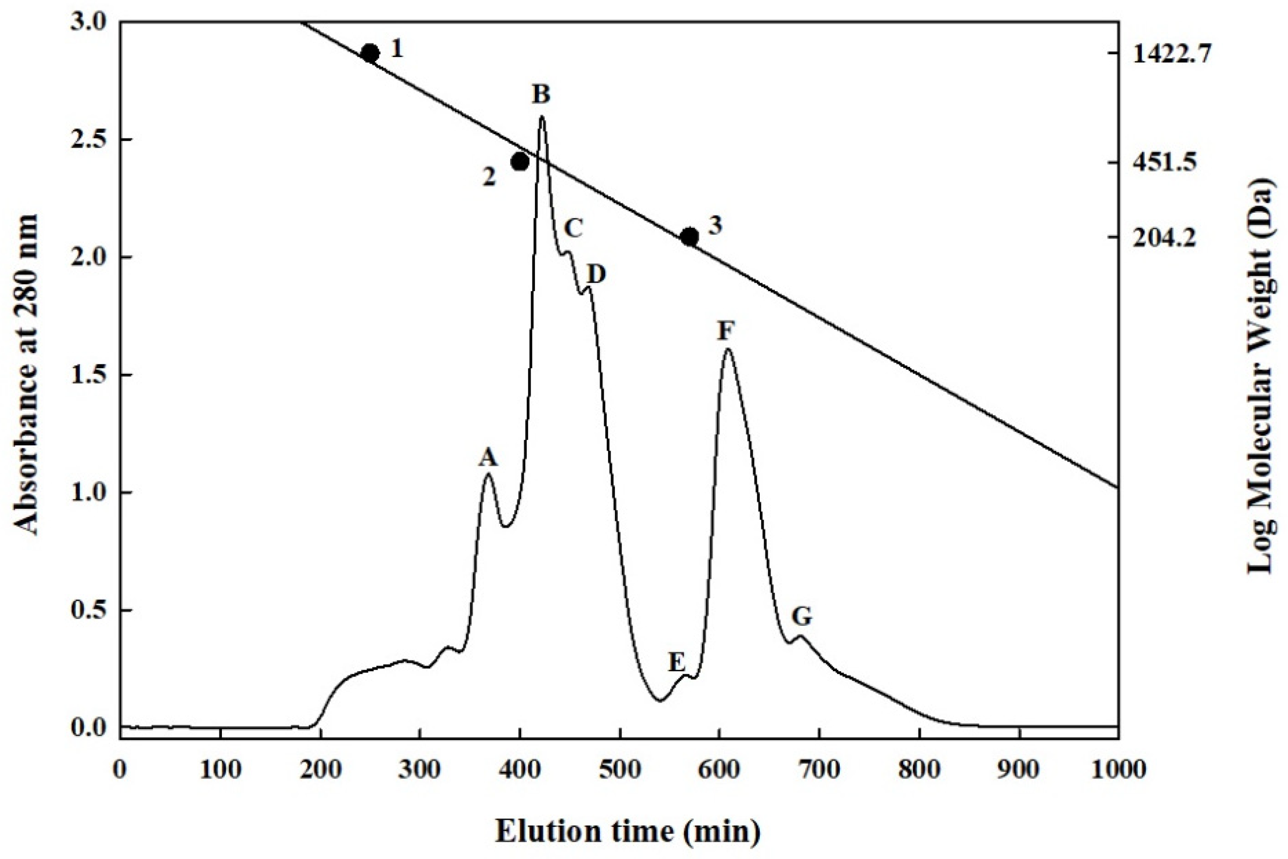
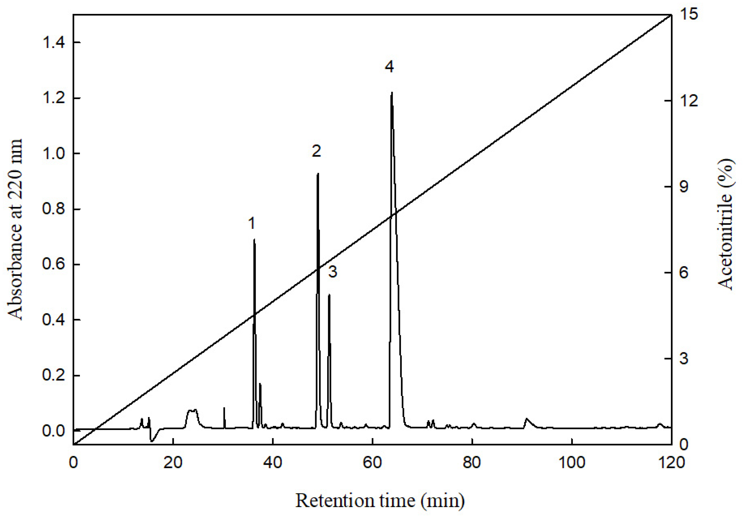
2.4. Separation and Purification of the Primary HMGR-Inhibiting Peptides in the HHP-FH-PR6G Group

HHP-FH-PR6G was fractionated through gel filtration chromatography on a Sephadex G-25 column to examine its molecular weight distribution and HMGR-inhibiting effects. As shown in the spectra in Figure 1, mainly seven peaks were separated. The seven fractions (A–G) were collected and their HMGR-inhibiting capacities were analysed. Their molecular weights were 810–600, 560–420, 420–370, 370–240, 230–190, 180–100, and 100–80 Da, respectively. The peptide content of fraction A was the highest. At a peptide concentration of 4 μg/mL, the HMGR-inhibiting rates of the fractions were 74.4%, 24.4%, 25.6%, 93.3%, 47.8%, 73.3%, and 65.6%, respectively. Fraction D exhibited the greatest inhibition (Table 4); according to its molecular weight distribution (370–240 Da), this is likely ascribable to the peptide composition of 2–4 amino acids. Pak et al. [38] noted that peptides comprising 3–8 amino acids can interact with the active site of HMGR and induce configuration changes that deactivate HMGR.

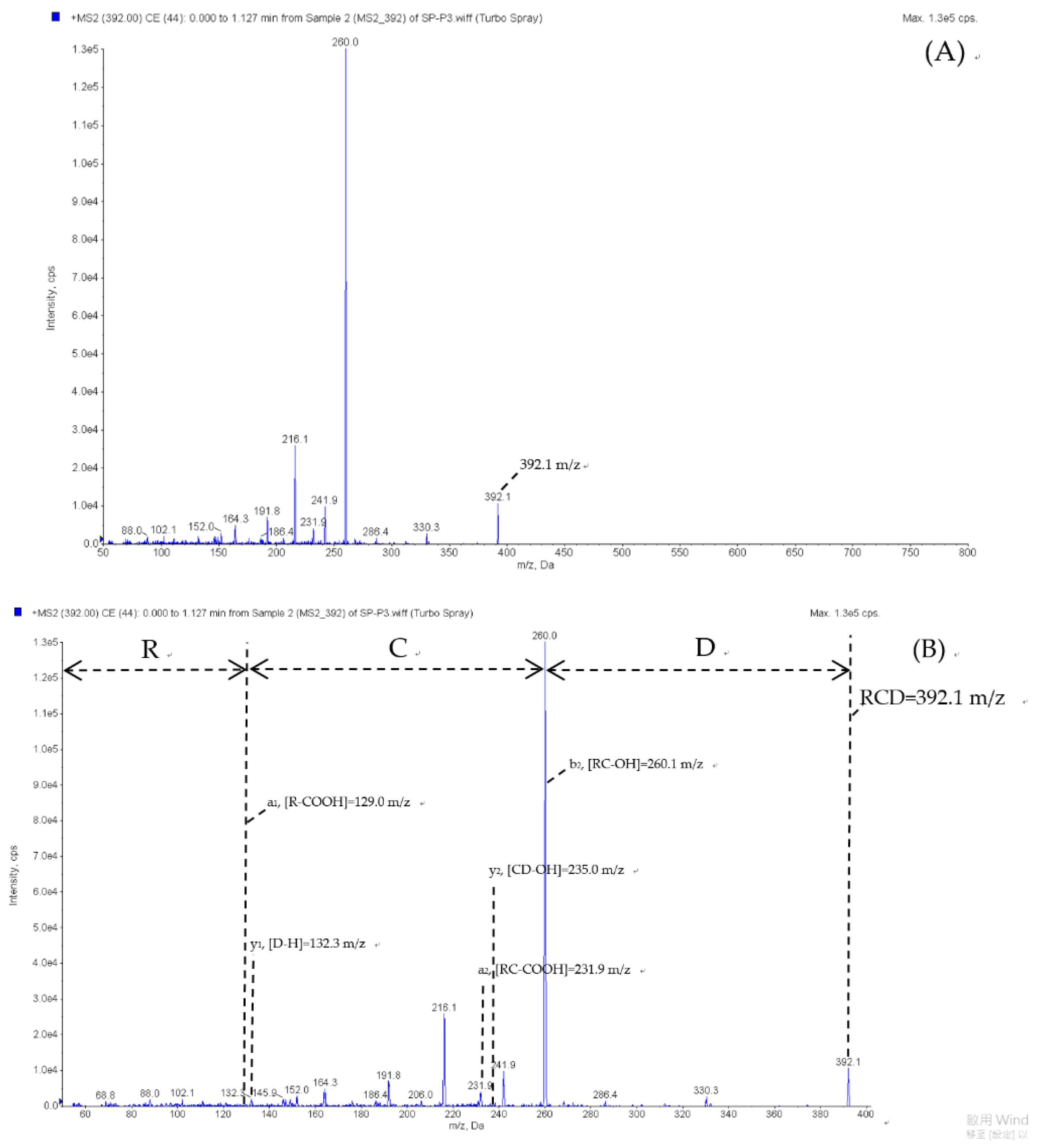
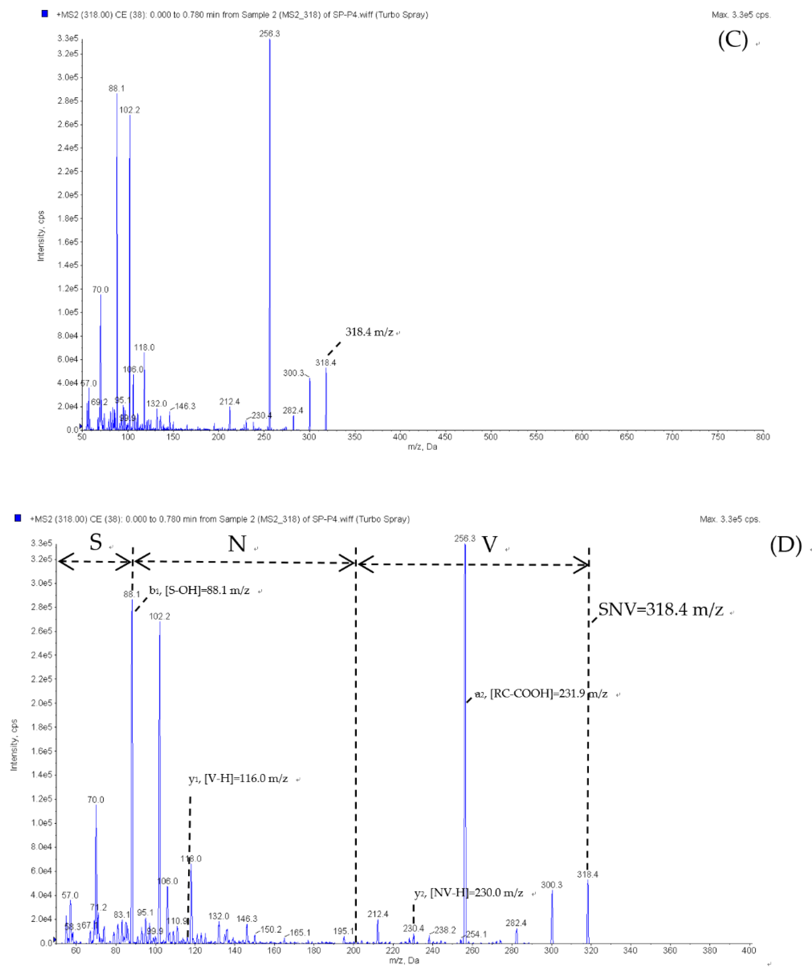
2.5. Peptide Sequence Identification

Fraction D was the primary HMGR-inhibiting peptide fraction in the HHP-FH-PR6P group. Thus, RP-HPLC was performed (on an ODS C18 column) for separation and purification. As shown in Figure 2, four main peaks (Peak 1-Peak 4) were obtained from gradient elution with 0–15% acetonitrile solution containing 0.1% trifluoroacetic acid over 120 min. After elution, their concentrations were 4.6%, 6.3%, 6.5%, and 8.1%, respectively. Analysis of the peptide concentrations and HMGR-inhibiting capacities of the four main peaks revealed that the peptide concentrations of peaks 1 and 2 were below the method detection limit (optical density = 0.01), and thus were presumably not peptides. The peptide concentrations of peaks 3 and 4 were 0.04 and 0.48 mg/mL, respectively (data not shown), and their HMGR inhibition rates were 74.5% and 29.8%, respectively (Table 5).
In a study on cowpea protein hydrolysates obtained through hydrolysis with gastrointestinal enzymes, three main peaks were separated after RP-HPLC. Their HMGR-inhibiting rates were 57.1%, 50%, and 47.8%, respectively [8]. In the present study, the collection of peaks 3 and 4 was followed by the analysis and identification of their peptide sequences through ESI/MS/MS. Two rounds of mass spectrometry, scanning, and detection were then conducted using various mass-to-charge ratio signals (300–800 and 50–800 m/z) to obtain the secondary mass spectrum (Figure A1). Using Analyst software version 1.51 (AB Sciex), the peptide sequences and mass-to-charge ratios of peaks 3 and 4 were determined to be Arg-Cys-Asp (RCD) and 392.1 m/z and Ser-Asn-Val (SNV) and 318.4 m/z, respectively. Search and alignment of RCD performed using the Uniprot Database [39] revealed that it was mainly derived from mod (f212–214) in S. platensis protein, and that SNV was derived from three proteins in S. platensis, namely adenine deaminase (f245–247), methyltransferase (f282–284), and PIN (f73–75). Finally, the IC50 of the HMGR inhibition of RCD and SNV were determined to be 6.9 and 20.1 μΜ, respectively (Table 5).
In a study by e Silva et al. [40], the Gln-Asp-Phe (QDF) peptide was separated from cowpea β-vignin protein. Its HMGR-inhibiting activity in vitro was dose-dependent and its IC50 was 12.8 μM. In silico simulation indicated that inhibitory effects were achieved through the binding of aspartate (D) in the QDF peptide sequence to the Ser589 residue in the HMGR structure. Therefore, the stronger HMGR inhibition of RCD in the present study is likely associated with the aspartate (D) in the sequence. Pak et al. [41] used statin structures as a reference for designing and synthesizing a peptide sequence to serve as an HMGR inhibitor. Compared with the FGYVAE peptide sequence (IC50 = 0.4 μM), the SFGYVAE peptide sequence with serine (S) added to its N-terminus (a structure similar to that of atorvastatin, a statin) exhibited higher HMGR inhibition. Its IC50 of 0.033 μM was 12 times that of the aforementioned sequence. According to the researchers, the oxygen atom of the hydroxyl group of the Ser side-chain presumably resulted in greater inhibitory capacity. This may explain the HMGR-inhibiting effects of the SNV peptide sequence in the present study. In addition, statins derivatives also form a complex with HMGR by interacting with the residues, including Arg-590, Asn-755, Glu-559, Ser565, Arg568, etc., which are found in the catalytic site of HMGR and are important for catalysis, thereby inhibiting the activity of the enzyme [42]. The present study results revealed that SNV and RCD bound to HMGR in a similar manner as reference compounds (Simvastatin) and formed hydrogen bonds with critical residues like Glu559, Ser565, and Arg568 (Figure A2 and Figure A3). The fact that HMGR-inhibiting activity was higher in RCD and SNV is probably associated with their peptide sequence structures and the functional groups of the side chains. The present study has not elucidated the mechanism by which peptides inhibit HMGR activity. To further understand peptides’ effects in cholesterol reduction, their chemical properties and hydrophobic–hydrophilic balance should be considered, as should their amino acid structures and molecular weights.

3. Materials and Methods

3.1. Materials

Spirulina platensis powder was purchased from Da Yi Biotech & Health Food Co., Ltd. (Chiayi, Taiwan).
Umamizyme G, Protease A, Peptidase R, Protin SD-AY10, Protease N, Protin NY100, and with a nominal activity level of 70, 50,000, 420, 80,000, 150,000, 700,000 U/g, respectively, were supplied by Amano Enzyme, Inc. (Yokohama, Nagoya, Japan). Alcalase 2.4 L FG and Neutrase with a nominal activity level of 200,000 and 60,000 U/g, respectively, were supplied by Novozymes A/S Enzyme Inc. (Bagsvaerd, Denmark). Freeze-dried powders containing a mixture of lactic acid bacteria (Lactobacillus casei, Lactobacillus acidophilus, Lactococcus latis subspecies lactise, Lactococcus latis subspecies cremoris, Lactococcus latis subsp lavtis bv. diacetylactis, Saccharomyces cerevisiae, Saccharomyces lactis) were purchased from Lyo-San, Inc. (Lachute, QC, Canada). Digestive enzymes (Pepsin and Pancreatin) and other chemicals of analytical grade were obtained from Sigma Chemical Co. (St. Louis, MO, USA).

3.2. Preparation of S. platensis Solution by HHP-Assisted Enzymatic Hydrolysis

Spirulina powder was mixed with deionized water at a 1:9 (w/w) ratio and homogenised in a high-speed blender (Vita-Mix TNC5200, Vitamix Co., Cleveland, OH, USA) for 2 min. The resulting solution was sealed in a vacuum bag (Nylon/PE) and then sterilised with boiling water in a water bath for 30 min. Next, the solution was rapidly cooled down to room temperature, and commercial enzymes (Umamizyme G, Protease A, or Peptidase R) were added at a 1% concentration (the ratio of enzyme to Spirulina weight was 1:100, w/w). The respective solutions were mixed with 5 mL of deionised water and then sterilised and passed through a 0.2-µm filter membrane. This was followed by hydrolysis under HHP (100 MPa) (TFS-100, Toyo-Koatsu Innoway Co. Ltd., Nishiku, Hiroshima, Japan) and atmospheric pressure (0.1 MPa) at 50 °C for 24 h. The enzymatic reaction was terminated at 90 °C for 10 min. The hydrolysate was then centrifuged at 2710× g (CR21, Hitachi Co. Ltd., Minato-ku, Tokyo, Japan) for 30 min, after which the supernatant was extracted and passed through No. 2 filter paper. Finally, this filtrate was freeze dried into powder for use in the experimental analysis.

3.3. Preparation of Fermented S. platensis through HHP-Assisted Enzymatic Hydrolysis

The sterilised S. platensis solution was prepared according to the method mentioned in Section 3.2. On an aseptic operating table, 0.1% mixed lactic acid bacteria powder (w/v) was added to the S. platensis solution and fermented at 42 °C for 5 h to allow the bacterial concentration to reach 2.1 × 10⁷ CFU/mL. Next, 1% Peptidase R (w/w) was added. The protease was first prepared with an appropriate amount of deionised water and sterilised using a 0.2-μm filter membrane. This was followed by HHP hydrolysis (100 MPa) at 50 °C for 0, 3, and 6 h (high hydrostatic pressure-assisted Peptidase R hydrolysis after fermentation, HHP-FH-PR0-6). The lactic fermented S. platensis solution served as the control group (HHP treatment without protease after fermentation, HHP-FH). After HHP hydrolysis, heating was conducted at 90 °C for 10 min to terminate the enzymatic reaction, and the solution was centrifuged at 2710× g (Hitachi high refrigerated centrifuge CR21) for 30 min. The filtrate (obtained using No. 2 filter paper) was freeze-dried into powder for use in experimental analysis.

3.4. Determination of Free Amino Acid Content

The free amino acid content was determined as described previously [43,44], with minor modifications. First, 0.5 mL of diluted S. platensis hydrolysate solution (50 mg/mL) was added to an 84 °C water bath with 1 mL of Cd-ninhydrin reagent for 5 min of heating. Next, the solution was rapidly cooled to room temperature in the water bath, and its absorbance was measured at 507 nm using a spectrophotometer (EVOLUTION 60S, Thermo Fisher Scientific, Inc., Waltham, MA, USA). Finally, the obtained leucine standard calibration curve was converted to free amino acid content.

3.5. Determination of Soluble Protein Content

The measurement of soluble protein content was slightly modified from the Folin–Lowry method [44,45,46]. First, 0.1 mL of solution was extracted from the S. platensis hydrolysate (50 mg/mL); to this, 0.5 mL of Reagent A and 4 mL of Reagent B (Bio-Rad Dc Protein Assay Kit, Bio-Rad Labortories, Inc., Berkeley, CA, USA) was added. Subsequently, the solution was mixed thoroughly by shaking and let stand at room temperature for 15 min. Finally, its absorbance was measured at 540 nm (EVOLUTION 60S, Thermo Fisher Scientific, Inc.). The standard calibration curve was obtained with bovine serum albumin as the analytical standard and was converted into soluble protein content.

3.6. Peptide Content Determination

Peptide content was determined through the methods used by Church et al. [47] and Lin et al. [44], with slight modifications. The reaction was conducted using orthophthaldialdehyde reagent, and a standard calibration curve (obtained from Leu-Gly reference material) was used for conversion into peptide content. Before the measurement, the sample solution (50 mg/mL) was passed through a 0.22-µm filter membrane, and 50 µL of the filtrate was added to a solution containing 2 mL of o-phthaldialdehyde. After shaking, it was let stand at room temperature for 2 min, and then a spectrophotometer (EVOLUTION 60S, Thermo Fisher Scientific, Inc.) was used to measure the absorbance at 340 nm.

3.7. Determination of HMGR-Inhibiting Capacity

The analysis of HMGR-inhibiting capacity was performed using a modified version of the approach developed by e Silva et al. [40]. First, 0.3 g of sample powder was made up with 10 mL of deionised water and passed through a 0.22-μm polyvinylidene fluoride filter membrane. After the filtrate was obtained using a 5-kDa ultrafiltration membrane, it was diluted to determine the peptide content. The sample peptide concentration was then diluted to 4 μg/mL with deionised water. Finally, an HMGR assay kit (CS-1090, Sigma Chemical., Co.) was used to determine the inhibitory capacity of the sample. Pravastatin, an HMGR-inhibiting drug, was used as the control group.
The assay kit was used as follows. Into a 96-well plate was added 181 μL of buffer solution (pH 7.4), 1 μL of the sample or pravastatin, 4 μL of NADPH, 12 μL of 3-hydroxy-3-methylglutaryl-CoA substrate, and 2 μL of HMGR. The mixture was shaken at 200 rpm for 20 s using an enzyme-linked immunosorbent assay (ELISA) reader (Bio-Tek Synergy HT & Take 3 Plate, BioTek Instruments, Inc., Winooski, VT, USA), followed by a 10-min reaction at 37 °C. During the reaction, the ELISA reader was used to detect the absorbance at 340 nm every 20 s until measurement termination at 600 s. Finally, the absorbance decrement at 340 nm within each unit of time was calculated [10,48,49]. NADPH consumption was calculated according to the following formula (unit: μmol/min/mgP):
( Δ 340 / m i n s a m p l e Δ 340 / m i n b l a n k ) × T V 12.44 × V × 0.6 × L P
where 12.44 is twice the absorption extinction coefficient εmM (6.22 mM−1cm−1) used to calculate the amount of NADPH consumed at 340 nm, meaning that two NADPHs were consumed in the reaction. The negative slope of absorbance in the sample and blank within each 1-min interval is denoted by ∆340/min sample and ∆340/min blank, respectively. TV is the total volume of the reaction (mL), V is the amount of protease used in the reaction (mL), 0.6 mgP/mL is the protease concentration, and LP is the length of the light path (cm; 1 cm for cuvettes and 0.55 cm for plates).
Inhibition   rate   ( % ) = ( Units / mgP control Units / mgP sample   Unit / mgP control ) × 100 %
Regarding the definition of the IC50, a graph was generated using the natural logarithm of the concentration of peptides in the sample and the HMGR inhibition rate to obtain a regression curve and its regression equation. The regression equation was used to calculate the peptide content required to inhibit HMGR activity by 50%. Pravastatin, used as a positive control, had significantly high HMG-CoA reductase inhibitory activity (Inhibition (%) = 89.6 ± 2.8%/4 µg/mL). The data used in this study were all averages of three readings or standard deviations (SDs).

3.8. In Vitro Gastrointestinal Digestion

Digestion was simulated in vitro with slight modifications to previously published methods [50,51]. A 45-g sample of the HHP-FH-PR6 powder was dissolved in 1500 mL of 0.1 M KCl–HCl (pH 2) buffer solution (w/v). Pepsin (EC 3.423.1; 1:10,000) was added to make the protease-protein ratio reach 1/25 (w/w) and the mixture was allowed to react at 37 °C for 4 h. The pH was then adjusted to 7 with 2N NaOH to terminate the reaction. Next, 250 mL of hydrolysate was heated in a boiling water bath for 10 min to deactivate the protease. Pancreatin at a protease-protein ratio of 1:25 (w/w) was added to the remaining hydrolysate, and the mixture was allowed to react at 37 °C for 4 h, heated in a boiling water bath, and deactivated over 10 min to terminate the reaction. Subsequently, the hydrolysate was centrifuged at 6930× g (Hitachi high refrigerated centrifuge CR21) for 30 min, after which the supernatant was freeze-dried into powder for later use (F-HHP-PR6G).

3.9. Gel Filtration Chromatography Analysis of S. platensis Protein Hydrolysate

Gel filtration chromatography of S. platensis protein hydrolysate was performed using the methods of Chen et al. [14] and Lin et al. [51], with slight modifications. The freeze-dried hydrolysate (HHP-FH-PR6G) with the highest HMGR-inhibiting activity was purified and separated using gel filtration chromatography (Sephadex G-25 column, 1.6 × 90 cm; Amersham Pharmacia Biotech AB, Uppsala, Sweden) and equilibrated with 0.02% deionised aqueous solution containing NaN3. The HHP-FH-PR6G powder was prepared at a concentration of 25 mg/mL. After passage through a filter membrane (molecular weight 5000 Da), 5 mL of the filtrate obtained was used as the injection volume; the 0.02% deionised aqueous solution was used as the eluent, and the flow rate of the peristaltic pump (Gilson MP1/LF, Gillson Medical Electronics, Villiers-le-Bel, France) was maintained at 0.5 mL/min. The eluate (5 mL/tube) was collected using a fraction collector (FC203B, Gillson Medical Electronics), and its absorbance was measured at 280 nm. Finally, the elution time and absorbance value were plotted as the horizontal and vertical axes, respectively, and standard calibration curve derived from the standard materials was converted into molecular weight of the sample. The standard materials and their molecular weights were as follows: bacitracin: 1422.0 Da; glycine-glycine-tyrosine-arginine: 451.5 Da; and tryptophan: 204.2 Da.

3.10. Purification of the Peptides for HMG-CoA Reductase Inhibitory Activity

Purification of the peptides of HMG-CoA reductase inhibitory activity from protein hydrolysate was performed by following the method described by Chen et al. [25] and Lin et al. [51] with minor modifications.

3.10.1. Sample Preparation

After the in vitro gastrointestinal digestion, the freeze-dried HHP-FH-PR6G sample was prepared to a concentration of 25 mg/mL. Following gel filtration chromatography on a Sephadex G-25 column, freeze-dried powders of fraction D were dissolved in 1 mL of deionised water; the solution was injected into the RP-HPLC system to obtain the liquid chromatogram.

3.10.2. Chromatographic Conditions Involving Acetonitrile (Concentration: 0–15%)

Regarding the chromatographic conditions, the mobile phases were eluent A (a solution containing water and 0.1% trifluoroacetic acid) and eluent B (acetonitrile solution containing 0.1% trifluoroacetic acid). The concentration of eluent B was increased from 0% to 15% within 120 min for gradient elution. The flow rate was 1 mL/min, and the sample injection volume was 500 μL. A reversed-phase C18 column (Synergi 4 μ Hydro-RP 80 Å, 10 mm × 250 mm; particle size 4 μm; Phenomenex, Torrance, CA, USA) was used as the chromatographic column and a Synergi 4 μ Hydro-RP 80A column (50 mm × 10 mm; particle size 4 μm) was used as the guard column. Detection was conducted at a wavelength of 220 nm. The main peaks with HMGR-inhibiting activity were collected, and each peak was set at a flow rate of 1 mL/min. The sample injection volume was 20 µL, and the C12 column (Joupiter 4 µm Proteo 90 A, 250 mm × 4.6 mm, Phenomenex, Torrance, CA, USA) with lower hydrophobicity was placed under the same gradient elution conditions. The guard column was 4 μ Proteo 90 A (4 mm × 3 mm; particle size 4 μm). Detection was conducted at a wavelength of 220 nm. After column chromatography analysis confirmed that the collected material corresponded to a single peak, the confirmed peaks were collected and freeze dried for peptide sequence analysis.

3.11. ESI/MS/MS and Peptide Data Analyses

The peptide sequence analysis was performed using RP-HPLC peptide purification and collection methods as well as electrospray ionization tandem mass spectrometry (ESI/MS/MS) peptide sequence identification methods previously described by Chen et al. [25] and Lin et al. [51], with minor modifications. Before the analysis, the concentration of the prepared sample must first be increased. The concentration of HHP-FH-PR6G was increased starting from 20 mg/mL, and fraction D, which had the highest inhibitory rate, was purified and collected through gel filtration chromatography on the Sephadex G-25 column. Material from three columns to the left and the right of the peaks of fraction D was collected in one column. Material from this column was freeze dried into powder and dissolved in 1 mL of deionised water. RP-HPLC was performed using the previously described procedure to purify and separate the peaks of the peptides on a semi-preparative C18 column. Next, the main peaks separated from RP-HPLC were collected five times, after which the C12 column was used to confirm that each peak was a single component through the aforementioned method. The collected mixtures were then freeze-dried into powders and their HMGR-inhibiting capacities were determined. The peptide sequence was analysed through ESI-MS/MS. Finally, deionised water was added to the freeze-dried powder of the aforementioned peaks to form a sample solution with a concentration of 700–1000 pmol/μL. A 100-μL sample was injected into the tandem mass spectrometer. Through electrospray ionization, ions were formed from the ion source. Next, a quadrupole ion trap was used as a mass analyser to select the precursor ions and record the mass-to-charge ratio (m/z ratio) of the precursor ions in the positive charged mode (5500 V; mass spectrometry range: 250–350 m/z) to obtain the primary mass spectrum. Subsequently, the precursor ion with the highest signal peak in the mass spectrum was selected and introduced into the collision chamber, and helium was used as the collision gas at a flow rate of 7 µL/min. Collision-induced dissociation was used to induce molecular fragmentation to generate product ion fragments. Subsequently, the m/z ratio and the number of product ions were analysed through tandem mass spectrometry (MS/MS; range: 50–350 m/z) to obtain the secondary mass spectrum, which was then analysed and compared to obtain the amino acid sequence of the peptides by using Analyst software version 1.5.1 (AB Sciex). The identified peptide sequences were aligned using the UniProt database [39] to verify the amino acid sequences of the S. platensis protein, and pairwise sequence alignment tools were used to determine the same sequences [52]. After the identified peptide sequences were aligned, the corresponding proteins were identified as mod (accession number: Q307B8), adenine deaminase (accession number: A0A3P3ZKR6), methyltransferase (accession number: Q306Z9), and PIN (accession number: Q307D1). Subsequently, the identified active peptides underwent solid-phase peptide synthesis, and their HMGR-inhibiting activity was assessed. An RP-HPLC column (ODS C12) was used for the qualitative analysis of these peptides through the previously described method, with the synthetic peptides as standards.

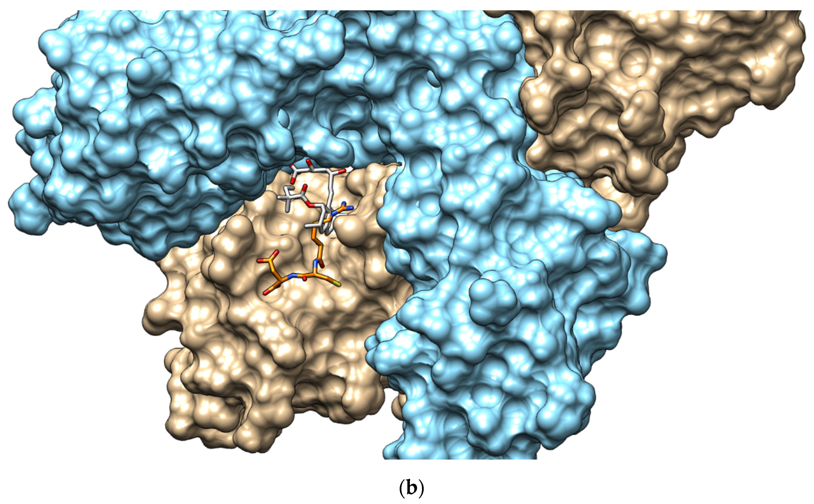
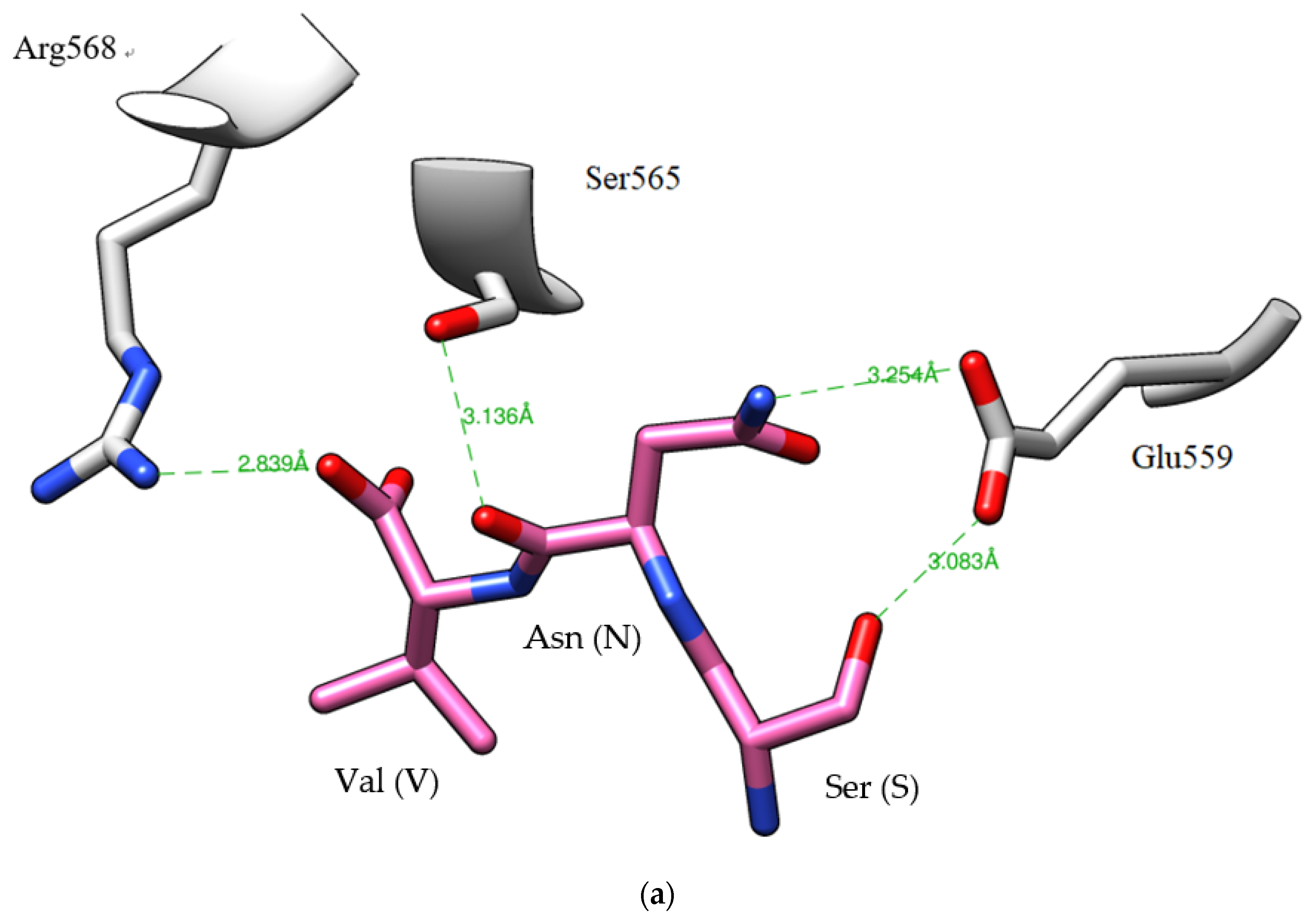
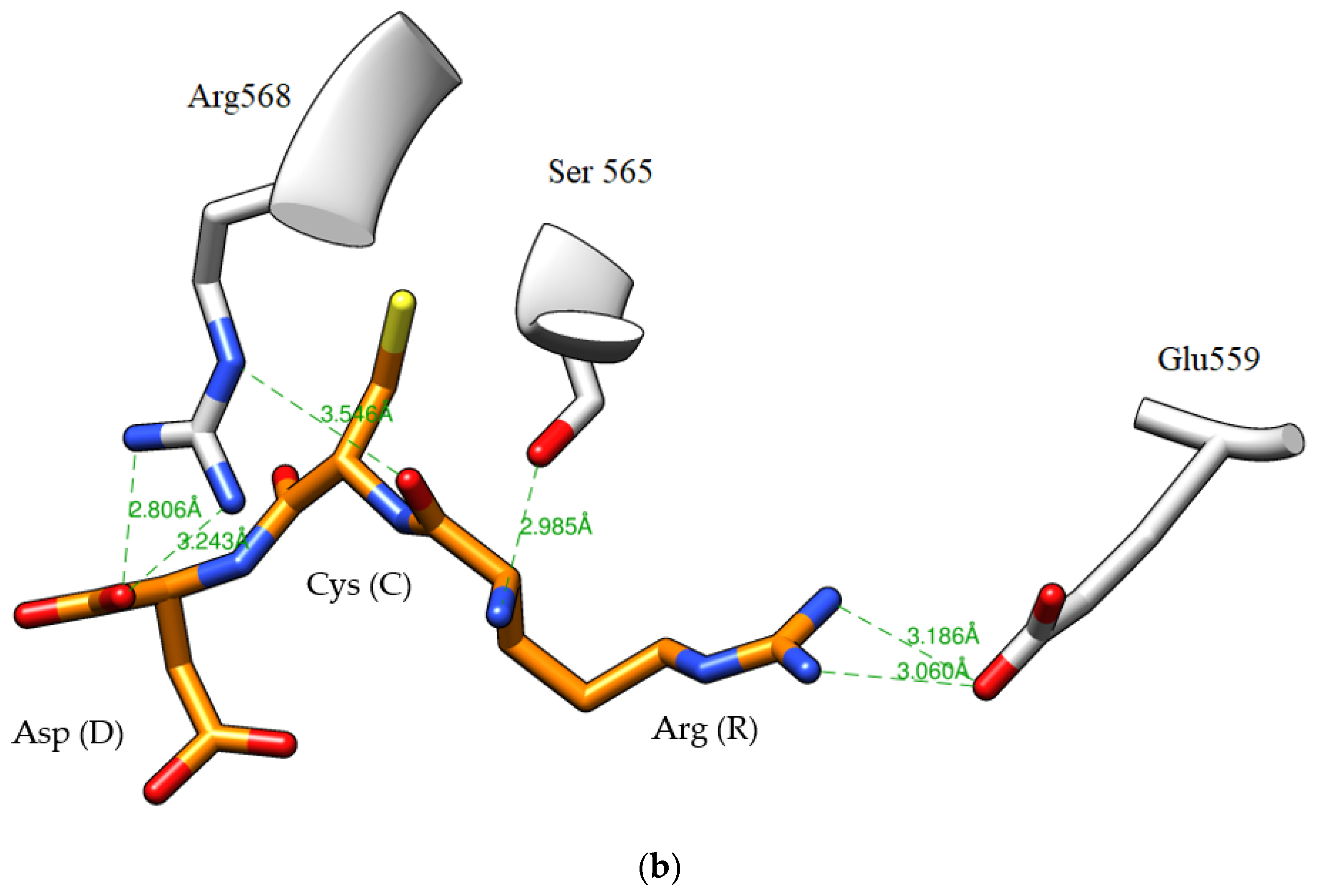
3.12. Analysis of HMGR–Peptide Molecular Docking

Docking simulation was conducted to investigate molecular interactions between human HMGR and active peptides because predicting such interactions can reveal useful information regarding the existence of active peptides. HMG-peptide molecular docking was analysed following the method of e Silva et al. [40], with slight modifications. The Protein Data Bank (PDB) was used as the reference for the HMGR inhibitor (structure of simvastatin), and HMGR (PDB ID: 1HW9) was obtained from http://www.rcsb.org/pdb/ (accessed on 1 January 2021) [53]. First, DockPrep Chimera 1.14 software was used to remove unnecessary water molecules and ligands from the HMGR structure (PDB ID: 1HW9) and to add hydrogen atoms in random orientations [54]. AutoDock-VINA (version 4.2.6) was then used to convert the PDB document into PDBQT format, thereby confirming its coordinates and charge signal. ChemDraw Ultra 12.0 software was used to visualise the peptide structure, and the lowest activation energy of the peptides was determined for stabilisation. An analysis was conducted, centred on the coordinates of the crystal structure of simvastatin (X = 3.93, Y = −9.20, Z = −11.33; PDB ID: 1HW9) set in AutoDock VINA software. The docking dimension (10 × 10 × 10 Å, cubic box) served as the simvastatin-HMGR docking area. In addition, the root mean square deviation (RMSD) of the lowest docked ligand was selected for reshaping the simvastatin docking box dimension, which was used to analyse the molecular coupling of active peptides. Finally, the HMGR-peptide molecular docking was performed, two-dimensional and three-dimensional graphs were drawn, and the HMGR-peptide bond energy was calculated [40].

3.13. Statistical Analysis

Except for the analytical data on gel filtration chromatography, and RP-HPLC chromatography, which are presented as the means of triplicate experiments, the experimental data are presented as means ± standard deviations. SAS software (version 9.4 TS1M5, SAS Institute Inc., Cary, NC, USA) [55] was employed to analyse the differences in the experimental results through a general linear model procedure. Duncan’s multiple range test was conducted to compare the means of multiple groups.

4. Conclusions

After S. platensis was subjected to lactic acid fermentation and hydrolysis with Peptidase R under an HHP of 100 MPa for 6 h (HHP-FH-PR6), bioactive peptides were produced. The soluble protein, peptide, and free amino acid content were respectively 1.3, 3.3, and 4.3 times of those in the HHP-F6 group. The HMGR-inhibiting capacity was favourable, and the IC50 was 2.9 μg/mL. After in vitro simulated gastrointestinal digestion, the inhibitory capacity remained high, and the IC50 was 3.5 μg/mL. Seven peaks (A–G) in the HHP-FH-PR6P group were identified using gel filtration chromatography. The inhibition rate of fraction D was the highest at 93.3%. After further purification and sequence identification, the peptide sequences were identified as Arg-Cys-Asp and Ser-Asn-Val, and their IC50 were 6.9 and 20.1 μM, respectively. This study revealed that the hypocholesterolemic peptides could be derived from a protein hydrolysate of S. platensis and used to serve as a basis for developing functional food that prevent hypercholesterolemia.

Author Contributions

G.-W.C. proposed and designed the experiment. G.-W.C. and M.-H.Y. participated in the experiment. G.-W.C. analyzed the data and composed the manuscript. G.-W.C. revised the manuscript and was responsible for supervising the research. All authors have read and agreed to the published version of the manuscript.

Funding

This research was funded by the National Science Council, Taiwan, Republic of China, grant numbers MOST 107-2218-E-019-002 and MOST 108-2221-E-019-040-MY3.

Data Availability Statement

Not applicable.

Acknowledgments

The authors would like to thank Hong-Ting Victor Lin (Department of Food Science, National Taiwan Ocean University, Taiwan) for providing molecular docking software and equipment.

Conflicts of Interest

The authors declare no conflict of interest.

Appendix A

Figure A1. Identification of molecular mass and amino acid sequence of the peak D3 and D4 from HHP-FH-PR6 by ESI/MS/MS. (A,B) the MS/MS spectra of peptide RCD (m/z 392.1). (C,D) the MS/MS spectra of peptide SNV (m/z 318.4).
Catalysts 11 00873 g0a1aCatalysts 11 00873 g0a1b
Figure A2. Docking analysis of simvastatin and the (a) SNV peptide and the (b) RCD peptide with the HMG-CoA reductase (PDB 1HW9). Both the simvastatin and the peptides are shown in sticks, whereas the surface of the HMG-CoA reductase was shown. The simvastatin (C, light grey; N, blue; O, red; S, yellow), SNV (C, pink; N, blue; O, red; S, yellow), and RCD (C, orange; N, blue; O, red; S, yellow) are colored by elements.
Catalysts 11 00873 g0a2aCatalysts 11 00873 g0a2b
Figure A3. The interactions between tripeptides (a) SNV and (b) RCD and the HMG-CoA reductase (PDB 1HW9). The tripeptides (C, orange; N, blue; O, red; S, yellow) and the HMG-CoA reductase (C; light grey; N, blue; O, red) are colored by elements.
Catalysts 11 00873 g0a3aCatalysts 11 00873 g0a3b

References

  1. Laslett, L.; Alagona, P.; Clark, B.A.; Drozda, J.P.; Saldivar, F.; Wilson, S.R.; Poe, C.; Hart, M. The worldwide environment of cardiovascular disease: Prevalence, diagnosis, therapy, and policy issues: A report from the American College of Cardiology. J. Am. Coll. Cardiol. 2012, 60, S1–S49. [Google Scholar] [CrossRef] [Green Version]
  2. Roth, G.A.; Johnson, C.; Abajobir, A.; Abd-Allah, F.; Abera, S.F.; Abyu, G.; Alla, F. Global, regional, and national burden of cardiovascular diseases for 10 causes, 1990 to 2015. J. Am. Coll. Cardiol. 2017, 70, 1–25. [Google Scholar] [CrossRef]
  3. Opie, L.H.; Frishman, W.H.; Thadani, U. Angiotensin-converting enzyme inhibitors and conventional vasodilators. In Drugs for the Heart, 4th ed.; Opie, L.H., Ed.; Saunders: Philadelphia, PA, USA, 1994; pp. 105–143. [Google Scholar]
  4. Wenniger, L.M.; Beuers, U. Bile salts and cholestasis. Dig. Liver Dis. 2010, 42, 409–418. [Google Scholar] [CrossRef]
  5. DeBose-Boyd, R.A. Feedback regulation of cholesterol synthesis: Stero-accelerated ubiquitination and degradation of HMG-CoA reductase. Cell Res. 2008, 18, 609–621. [Google Scholar] [CrossRef] [Green Version]
  6. Thompson, P.D.; Clarkson, P.; Karas, R.H. Statin-associated myopathy. JAMA 2003, 289, 1681–1690. [Google Scholar] [CrossRef]
  7. Pak, V.V.; Koo, M.S.; Kasymova, T.D.; Kwon, D.Y. Isolation and identification of peptides from soy 11S-globulin with hypocholesterolemic activity. Chem. Nat. Compd. 2005, 41, 710–714. [Google Scholar] [CrossRef]
  8. Marques, M.R.; Fontanari, G.G.; Pimenta, D.C.; Soares-Freitas, R.M.; Arêas, J.A.G. Proteolytic hydrolysis of cowpea proteins is able to release peptides with hypocholesterolemic activity. Food Res. Int. 2015, 77, 43–48. [Google Scholar] [CrossRef]
  9. Ashraf, J.; Liu, L.; Awais, M.; Xiao, T.; Wang, L.; Zhou, X.; Tong, L.T.; Zhou, S. Effect of thermosonication pre-treatment on mung bean (Vigna radiate) and white kidney bean (Phaseolus vulgaris) proteins: Enzymatic hydrolysis, cholesterol lowering activity and structural characterization. Ultrason. Sonochem. 2020, 66, 105121. [Google Scholar] [CrossRef] [PubMed]
  10. Soares, R.A.M.; Mendonça, S.; De Castro, L.Í.A.; Menezes, A.C.C.C.C.; Arêas, J.A.G. Major peptides from amaranth (Amaranthus cruentus) protein inhibit HMG-CoA reductase activity. Int. J. Mol. Sci. 2015, 16, 4150–4160. [Google Scholar] [CrossRef] [Green Version]
  11. Nieto, I.M.P.; del Moral, M.P.; Garcia, M.C. Evaluation of the relationship between the peptide profile and the lipid-lowering properties of olive seeds hydrolysates as a tool for tuning hypocholesterolemic functionality. Food Funct. 2020, 11, 4973–4981. [Google Scholar]
  12. Nopianti, R.; Oktaviani, S. Fractionation of anticholesterol bioactive compounds from bekasam (Indonesian fermented fish product). Pertanika J. Trop. Agric. Sci. 2017, 40, 195–204. [Google Scholar]
  13. Kwon, D.Y.; Oh, S.W.; Lee, J.S.; Yang, H.J.; Lee, S.H.; Lee, J.H.; Lee, Y.B.; Sohn, H.S. Amino acid substitution of hypocholesterolemic peptide originated from glycinin hydrolyzate. Food Sci. Biotechnol. 2002, 11, 55–61. [Google Scholar]
  14. Chen, G.W.; Tsai, J.S.; Sun, P.B. Purification of angiotensin I-converting enzyme inhibitory peptides and antihypertensive effect of milk produced by protease-facilitated lactic fermentation. Int. Dairy J. 2007, 17, 641–647. [Google Scholar] [CrossRef]
  15. Chen, G.W.; Tsai, J.S.; Sun, P.B. Cardiovascular effects of whey from prozyme 6-facilitated lactic acid bacteria fermentation of milk. J. Food Biochem. 2007, 31, 639–655. [Google Scholar] [CrossRef]
  16. Tsai, J.S.; Chen, T.J.; Pan, B.S.; Gong, S.D.; Chung, M.Y. Antihypertensive effect of bioactive peptides produced by protease-facilitated lactic acid fermentation of milk. Food Chem. 2008, 106, 552–558. [Google Scholar] [CrossRef]
  17. Chen, G.W.; Liao, K.J. Method of Combining Pressure and Enzyme to Extract Hydrolysate of Animal-Derived Composition. Taiwan Patent No. TWI600379B, 1 October 2017. [Google Scholar]
  18. Masson, P.; Tonello, C.; Balny, C. High-pressure biotechnology in medicine and pharmaceutical science. J. Biomed. Sci. 2001, 1, 85–88. [Google Scholar] [CrossRef] [Green Version]
  19. Eisenmenger, M.J.; Reyes-De-Corcuera, J.I. High pressure enhancement of enzymes: A Review. Enzyme Microb. Technol. 2009, 45, 331–347. [Google Scholar] [CrossRef]
  20. Marciniak, A.; Suwal, S.; Naderi, N.; Pouliot, Y.; Doyen, A. Enhancing enzymatic hydrolysis of food proteins and production of bioactive peptides using high hydrostatic pressure technology. Trends Food Sci. Technol. 2018, 80, 187–198. [Google Scholar] [CrossRef]
  21. Knudsen, J.C.; Otte, J.; Olsen, K.; Skibsted, L.H. Effect of high hydrostatic pressure on the conformation of β-lactoglobulin A as assessed by proteolytic peptide profiling. Int. Dairy J. 2002, 12, 791–803. [Google Scholar] [CrossRef]
  22. Quirós, A.; Chichón, R.; Recio, I.; López-Fandiño, R. The use of high hydrostatic pressure to promote the proteolysis and release of bioactive peptides from ovalbumin. Food Chem. 2007, 104, 1734–1739. [Google Scholar] [CrossRef]
  23. Zhang, T.; Jiang, B.; Miao, M.; Mu, W.M.; Li, Y.H. Combined effects of high-pressure and enzymatic treatments on the hydrolysis of chickpea protein isolates and antioxidant activity of the hydrolysates. Food Chem. 2012, 135, 904–912. [Google Scholar] [CrossRef]
  24. Garcia-Mora, P.; Peñas, E.; Frias, J.; Zieli’nski, H.; Wiczkowski, W.; Zieli’nska, D.; Martínez-Villaluenga, C. High-pressure-assisted enzymatic release of peptides and phenolics increases angiotensin converting enzyme I inhibitory and antioxidant activities of Pinto bean hydrolysates. J. Agric. Food Chem. 2016, 64, 1730–1740. [Google Scholar] [CrossRef] [PubMed] [Green Version]
  25. Chen, G.W.; Lin, H.T.V.; Huang, L.W.; Lin, C.H.; Lin, Y.H. Purification and identification of cholesterol micelle formation inhibitory peptides of hydrolysate from high hydrostatic pressure-assisted protease hydrolysis of fermented seabass byproduct. Int. J. Mol. Sci. 2021, 22, 5295. [Google Scholar] [CrossRef] [PubMed]
  26. Lin, Y.H.; Li, C.H.; Lin, C.H.; Yan, Y.Z.; Chuang, Y.Y.; Chen, G.W. Application and development status of an ultra-high pressure processing technique in aquatic food. Fish. Ext. Rep. 2017, 47, 17–32. [Google Scholar]
  27. Chen, G.W.; Liao, K.J. Method of Combining Pressure and Enzyme to Extract Hydrolysate of Cubilose. Taiwan Patent No. TWI516212B, 11 January 2016. [Google Scholar]
  28. Seo, Y.C.; Choi, W.S.; Park, J.H.; Park, J.O.; Jung, K.H.; Lee, H.Y. Stable isolation of phycocyanin from Spirulina platensis associated with high-pressure extraction process. Int. J. Mol. Sci. 2013, 14, 1778–1787. [Google Scholar] [CrossRef] [PubMed]
  29. Food and Agriculture Organization of the United Nations. The State of World Fisheries and Aquaculture. 2018. Available online: https://www.un.org/youthenvoy/2013/09/fao-food-and-agriculture-organization-of-the-united-nations/ (accessed on 1 June 2021).
  30. Kuddus, M.; Singh, P.; Thomas, G.; Al-Hazimi, A. Recent developments in production and biotechnological applications of C-phycocyanin. BioMed Res. Int. 2013, 2013, 742859. [Google Scholar] [CrossRef] [PubMed] [Green Version]
  31. Ou, Y.; Lin, L.; Pan, Q.; Yang, X.; Cheng, X. Preventive effect of phycocyanin from Spirulina platensis on alloxan-injured mice. Environ. Toxicol. Pharmacol. 2012, 34, 721–726. [Google Scholar] [CrossRef] [PubMed]
  32. Joventino, I.P.; Alves, H.G.; Neves, L.C.; Pinheiro-Joventino, F.; Leal, L.K.A.; Neves, S.A.; Ferreira, F.V.; Brito, G.A.C.; Viana, G.B. The microalga Spirulina platensis presents anti-inflammatory action as well as hypoglycemic and hypolipidemic properties in diabetic rats. J. Complementary Integr. Med. 2012, 9, 7. [Google Scholar] [CrossRef] [PubMed]
  33. Virginia, A.; Rachmawati, H.; Riani, C.; Retnoningrum, D.S. Study of HMG-CoA reductase inhibition activity of the hydrolyzed product of snakehead fish (Channa striata) skin collagen with 50 kDa collagenase from Bacillus licheniformis F11. 4. Sci. Pharm. 2016, 84, 81–88. [Google Scholar] [CrossRef] [PubMed] [Green Version]
  34. Heinrich, M.; Kulozik, U. Study of chymosin hydrolysis of casein micelles under ultra high pressure: Effect on re-association upon pressure release. Int. Dairy J. 2011, 21, 664–669. [Google Scholar] [CrossRef]
  35. Ludikhuyze, L.; Van Loey, A.; Indrawati; Smout, C.; Hendrickx, M. Effects of combined pressure and temperature on enzymes related to quality of fruits and vegetables: From kinetic information to process engineering aspects. Crit. Rev. Food Sci. Nutr. 2003, 43, 527–586. [Google Scholar] [CrossRef] [PubMed]
  36. Rinto, D.R.; Yasni, S.; Suhartono, M.T. Novel HMG-CoA reductase inhibitor peptide from Lactobacillus acidophilus isolated from Indonesian fermented food bekasam. IJPCBS 2017, 5, 195–204. [Google Scholar]
  37. Lin, Y.H.; Chen, G.W.; Yeh, C.H.; Song, H.; Tsai, J.S. Purification and identification of angiotensin I-converting enzyme inhibitory peptides and the antihypertensive effect of Chlorella sorokiniana protein hydrolysates. Nutrients 2018, 10, 1397. [Google Scholar] [CrossRef] [PubMed] [Green Version]
  38. Pak, V.V.; Koo, M.; Kim, M.J.; Yang, H.J.; Yun, L.; Kwon, D.Y. Modeling an active conformation for linear peptides and design of a competitive inhibitor for HMG-CoA reductase. J. Mol. Recognit. 2008, 21, 224–232. [Google Scholar] [CrossRef] [PubMed]
  39. UniProt Database. Available online: http://www.uniprot.org (accessed on 1 January 2021).
  40. E Silva, M.B.D.C.; da Cruz Souza, C.A.; Philadelpho, B.O.; da Cunha, M.M.N.; Batista, F.P.R.; da Silva, J.R.; Ferreira, E.S. In vitro and in silico studies of 3-hydroxy-3-methyl-glutaryl coenzyme a reductase inhibitory activity of the cowpea Gln-Asp-Phe peptide. Food Chem. 2018, 259, 270–277. [Google Scholar] [CrossRef]
  41. Pak, V.V.; Koo, M.; Kwon, D.Y.; Yun, L. Design of a highly potent inhibitory peptide acting as a competitive inhibitor of HMG-CoA reductase. Amino Acids 2012, 43, 2015–2025. [Google Scholar] [CrossRef]
  42. Da Costa, R.F.; Freire, V.N.; Bezerra, E.M.; Cavada, B.S.; Caetano, E.W.S.; Filho, J.L.L.; Albuquerque, E.L. Explaining statin inhibition effectiveness of HMG-CoA reductase by quantum biochemistry computations. Phys. Chem. Chem. Phys. 2012, 14, 1389–1398. [Google Scholar] [CrossRef] [PubMed]
  43. Doi, E.; Shibata, D.; Matoba, T. Modified colorimetric ninhydrin methods for peptidase assay. Anal. Biochem. 1981, 118, 173–184. [Google Scholar] [CrossRef]
  44. Lin, Y.H.; Chen, C.A.; Tsai, J.S.; Chen, G.W. Preparation and identification of novel antihypertensive peptides from the in vitro gastrointestinal digestion of marine cobia skin hydrolysates. Nutrients 2019, 11, 1351. [Google Scholar] [CrossRef] [PubMed] [Green Version]
  45. Cooper, T.G. Spectrophotometry. In The Tools of Biochemistry, 1st ed.; Wiley-Interscience: Hoboken, NJ, USA, 1977; pp. 53–55. [Google Scholar]
  46. Lowry, O.H.; Resebrough, N.J.; Farr, A.L.; Randall, R.J. Protein measurement with the folin phenol reagent. J. Biol. Chem. 1951, 193, 265–275. [Google Scholar] [CrossRef]
  47. Church, F.C.; Swaisgood, H.E.; Porter, H.D.; Catignani, G.L. Spectrophotometric assay using o-phthaldialdehyde for determination of proteolysis in milk and isolated milk proteins. J. Dairy Sci. 1983, 66, 1219–1227. [Google Scholar] [CrossRef]
  48. Ram, H.; Jaipal, N.; Charan, J.; Kashyap, P.; Kumar, S.; Tripathi, R.; Singh, B.P.; Siddaiah, C.N.; Hashem, A.; Allah, E.F.A. Phytoconstituents of an ethanolic pod extract of Prosopis cineraria triggers the inhibition of HMG-CoA reductase and the regression of atherosclerotic plaque in hypercholesterolemic rabbits. Lipids Health Dis. 2020, 19, 1–11. [Google Scholar] [CrossRef] [PubMed] [Green Version]
  49. Koning, A.J.; Roberts, C.J.; Wright, R.L. Different subcellular localization of Saccharomyces cerevisiae HMG-CoA reductase isozymes at elevated levels corresponds to distinct endoplasmic reticulum membrane proliferations. Mol. Biol. Cell. 1996, 7, 769–789. [Google Scholar] [CrossRef] [PubMed] [Green Version]
  50. Wu, J.; Ding, X. Characterization of inhibition and stability of soy-protein-derived angiotensin I-converting enzyme inhibitory peptides. Food Res. Int. 2002, 35, 367–375. [Google Scholar] [CrossRef]
  51. Lin, Y.H.; Tsai, J.S.; Chen, G.W. Purification and identification of hypocholesterolemic peptides from freshwater clam hydrolysate with in vitro gastrointestinal digestion. J. Food Biochem. 2017, 41, e12385. [Google Scholar] [CrossRef]
  52. Pairwise Sequence Alignment Software. Available online: https://www.ebi.ac.uk/Tools/psa/ (accessed on 1 January 2021).
  53. Hafidz, K.A.; Puspitasari, N.; Azminah, A.Y.; Artha, Y.; Mun’im, A. HMG-CoA Reductase inhibitory activity of Gnetum gnemon seed extract and identification of potential inhibitors for lowering cholesterol level. J. Young Pharm. 2017, 9, 559. [Google Scholar] [CrossRef] [Green Version]
  54. Trott, O.; Olson, A.J. AutoDock Vina: Improving the speed and accuracy of docking with a new scoring function, efficient optimization, and multithreading. J. Comput. Chem. 2010, 31, 455–461. [Google Scholar] [CrossRef] [PubMed] [Green Version]
  55. SAS Institute Inc. SAS®9.4 TS1M5 User’s Guide; SAS Institute Press: Cary, NC, USA, 2019; p. 35. [Google Scholar]
Figure 1. Sephadex G-25 column chromatography of peptides separated from HPP-FH-PR6G. ●—(Standard materials) 1—Bacitracin (1422.7 Da); 2—Gly-Gly-Tyr-Arg (451.5 Da); 3—Tryptophan (204.2 Da). HPP-FH-PR6G: lactic fermented S. platensis hydrolysates produced by Peptidase R under HHP for 6 h and then rehydrolysis with gastrointestinal proteases.
Catalysts 11 00873 g001
Figure 2. An elution profile of the fraction D from Figure 1 by reversed-phase HPLC. C18 Column: Synergi 4 μm Hydro-RP 80A (10 × 250 mm; particle size, 4 μm; Phenomenex, Torrance, CA, USA); elution A (deionized water containing 0.1% trifluoroacetic acid (TFA) and B (100% acetonitrile containing 0.1% TFA); mobile phase: linear gradient from 0 to 15% of B within 120 min; and flow rate of 1.0 mL/min at room temperature, and detection at 220 nm.
Catalysts 11 00873 g002
Table 1. Effect of proteases and HHP conditions on soluble protein, peptide, and free amino acid content and HMG-CoA reductase inhibitory activities of spirulina hydrolysates *.
Pressure
(MPa)
Enzymes Soluble Protein
(mg/g)
Peptide Content
(mg/g)
Free Amino Acid
(mg/g)
Inhibition
(%)
-Pravastatin (positive control)- §--89.6 ± 2.8 a
0.1Umamizyme G483.9 ± 1.4 b487.1 ± 0.7 d158.9 ± 0.2 e20.8 ± 7.6 d
Protease A283.5 ± 2.9 e338.7 ± 0.9 f140.5 ± 0.8 f41.5 ± 1.9 c
Peptidase R416.7 ± 3.3 c585.8 ± 1.8 b237.0 ± 0.3 b40.6 ± 2.8 c
100Umamizyme G514.9 ± 3.2 a519.9 ± 0.8 c165.3 ± 1.0 d24.5 ± 5.7 d
Protease A337.5 ± 1.9 d459.1± 1.9 e198.8 ± 0.1 c51.9 ± 1.0 c
Peptidase R414.4 ± 0.7 c699.1 ± 3.8 a269.2 ± 1.8 a67.0 ± 2.8 b
* Means ± standard deviation (n = 3). Different superscripts (a–f) in the same column indicate significant difference (p < 0.05) between samples. Proteolysis at 50 °C for 24 h. The inhibition (%) of HMG-CoA reductase was measured using the concentration 4 μg/mL peptide of each sample. § Undetected.
Table 2. Effect of proteases and HHP conditions on soluble protein, peptide, and free amino acid content and HMG-CoA reductase inhibitory activities of lactic fermented spirulina hydrolysates *.
SampleHydrolysis Time
(h)
Soluble Protein
(mg/g)
Peptide Content
(mg/g)
Free Amino Acid
(mg/g)
Inhibition
(%)
-Pravastatin (positive control)- §--89.5 ± 2.3 a
HHP-F 0242.3 ± 4.3 c96.8 ± 0.6 c24.0 ± 0.1 c57.9 ± 3.5 d
3242.2 ± 5.9 c102.9 ± 2.9 c25.9 ± 0.2 c62.0 ± 0.6 d
6244.8 ± 4.2 c103.6 ± 1.7 d26.7 ± 0.3 c63.2 ± 1.8 d
HHP-FH-PR 3291.1 ± 3.4 b261.5 ± 5.0 b78.5 ± 0.2 b70.8 ± 2.3 c
-6328.1 ± 1.4 a339.8 ± 7.6 a115.2 ± 7.4 a78.4 ± 0.6 b
* Means ± standard deviation (n = 3). Different superscripts (a–e) in the same column indicate significant difference (p < 0.05) between samples. HHP-F: hydrolysate from spirulina hydrolysed without commercial enzyme during HHP after fermentation for 5 h. HHP-FH-PR: hydrolysate from spirulina hydrolysed with Peptidase R during HHP after fermentation for 5 h. § Undetected.
Table 3. Effect of digestion by gastrointestinal proteases on peptide content and HMG-CoA reductase inhibitory activity of HHP-FH-PR6 *.
SamplePeptide Content
(mg/g)
Inhibition
(%)
IC50 Value
(μg Peptide/mL)
HHP-FH-PR6318.7 ± 0.4 a78.4 ± 0.6 a2.9 ± 0.1 a
Pepsin §225.5 ± 3.8 c65.6 ± 1.1 a,b3.0 ± 0.1 a
Pepsin + Pancreatin §270.7 ± 0.1 b58.9 ± 5.5 b3.5 ± 0.2 a
* Means ± standard deviation (n = 3). Different superscripts (a–c) in the same column indicate significant difference (p < 0.05) between samples. The inhibition (%) of HMG-CoA reductase was measured using 4 μg/mL peptide concentration of each sample. The peptide concentration required to inhibit 50% of HMG-CoA reductase activity. § Pepsin hydrolysed for 4 h followed by pancreatin hydrolysed for 4 h.
Table 4. Peptide content and HMG-CoA reductase inhibitory activities of the size exclusion chromatographic fractions obtained from HPP-FH-PR6G *.
FractionMolecular Weight
(Da)
Peptide Concentration
(mg/mL)
Inhibition
(%)
A810-60032.0 ± 0.374.4 ± 1.1 b
B560-4209.1 ± 0.124.4 ± 0.0 d
C420-3704.0 ± 0.025.6 ± 5.6 d
D370-2403.1 ± 0.093.3 ± 0.0 a
E230-1900.2 ± 0.047.8 ± 1.1 c
F180-1000.2 ± 0.073.3 ± 2.2 b
G100-800.3 ± 0.065.6 ± 3.3 b
* Mean ± standard deviation (n = 3). Different superscripts in the same column indicate significant different (p < 0.05) between samples. The inhibition (%) of HMG-CoA reductase was measured using 4 μg/mL peptide concentration of each samples.
Table 5. Peptide sequences, HMG-CoA reductase inhibitory activities and IC50 of various peaks (D1–4) from HHP-FH-PR6G *.
PeaksSequencesInhibition
(%)
IC50
(μM)
Origin
D3Arg-Cys-Asp74.5 ± 2.16.9Mod (f212–214)
D4Ser-Asn-Val29.8 ± 0.020.1Adenine deaminase (f245–247)
Methyltransferase (f282–284)
PIN (f73–75)
* Means ± standard deviation (n = 3). The peptide concentration required to inhibit 50% of HMG-CoA reductase activity. The peptide sequence alignment of Spirulina platensis proteins (Mod, Adenine deaminase, Methyltransferase and PIN).
Publisher’s Note: MDPI stays neutral with regard to jurisdictional claims in published maps and institutional affiliations.

