Article

Simultaneous Photocatalytic Sugar Conversion and Hydrogen Production Using Pd Nanoparticles Decorated on Iron-Doped Hydroxyapatite

1
Department of Earth Resources Engineering, Kyushu University, Fukuoka 819-0395, Japan
2
Department of Chemistry, Faculty of Engineering and Technology, SRM Institute of Science and Technology, Kattankulathur 603203, Tamil Nadu, India
*
Authors to whom correspondence should be addressed.
Catalysts 2023, 13(4), 675; https://doi.org/10.3390/catal13040675
Submission received: 8 February 2023 / Revised: 26 March 2023 / Accepted: 28 March 2023 / Published: 30 March 2023

Abstract

:
Pd nanoparticles (PdNPs) were successfully deposited on the surface of Fe(III)-modified hydroxyapatite (HAp), which was subsequently used as a photocatalyst for simultaneous photocatalytic H2 evolution and xylose conversion. The structural phase and morphology of the pristine HAp, FeHAp, and Pd@FeHAp were examined using XRD, SEM, and TEM instruments. At 20 °C, Pd@FeHAp provided a greater xylose conversion than pristine HAp and FeHAp, about 2.15 times and 1.41 times, respectively. In addition, lactic acid and formic acid production was increased by using Pd@FeHAp. The optimal condition was further investigated using Pd@FeHAp, which demonstrated around 70% xylose conversion within 60 min at 30 °C. Moreover, only Pd@FeHAp produced H2 under light irradiation. To clarify the impact of Fe(III) doping in FeHAp and heterojunction between PdNPs and FeHAp in the composite relative to pure Hap, the optical and physicochemical properties of Pd@FeHAp samples were analyzed, which revealed the extraordinary ability of the material to separate and transport photogenerated electron-hole pairs, as demonstrated by a substantial reduction in photoluminescence intensity when compared to Hp and FeHAp. In addition, a decrease in electron trap density in the Pd@FeHAp composite using reversed double-beam photoacoustic spectroscopy was attributed to the higher photocatalytic activity rate. Furthermore, the development of new electronic levels by the addition of Fe(III) to the structure of HAp in FeHAp may improve the ability to absorb light by lessening the energy band gap. The photocatalytic performance of the Pd@FeHAp composite was improved by lowering charge recombination and narrowing the energy band gap. As a result, a newly developed Pd@FeHAp composite might be employed as a photocatalyst to generate both alternative H2 energy and high-value chemicals.

Graphical Abstract

1. Introduction

Hydrogen is regarded a clean fuel and a sustainable energy carrier, as it can be derived from natural sources and its combustion yields only water. However, photocatalytic water splitting is constrained by endergonic thermodynamics and the slow kinetics of the O2 evolution half-reaction [1,2]. Consequently, non-renewable and overpriced sacrificial agents such as triethanolamine and methanol are widely used to assist the H2 production [3,4,5,6,7,8,9,10]. Scalable and environmentally-friendly H2 production may be possible via photo reformation of xylose, the second most abundant sugar in lignocellulosic biomass [11,12,13,14]. Photo reforming necessitates the utilization of a photocatalyst that can simultaneously undergo oxidation of xylose into value-added biochemicals and reduction of water to hydrogen in the presence of photogenerated charge carriers [15,16,17,18,19]. Among the various biochemicals produced, lactic acid is a key component of the biorefinery process used in numerous industries, including the formulation of cosmetics, the manufacture of pharmaceuticals, and biodegradable polylactide [20,21,22]. Additionally, formic acid can be produced and used in several applications such as medicine, food, and agriculture [23,24,25,26,27].
Calcium hydroxyapatite (HAp) usually has a high adsorption capacity and amphoteric characteristics. These properties set it apart as a potential adsorbent or catalytic support with tunable selectivity, making it appealing for both fundamental and practical research. The primary drawback of HAp is its wide bandgap energy larger than 4 eV and therefore, the photocatalytic reaction is less active when exposed to solar light. In addition, the photocatalytic efficiency of single phase of HAp is limited by the high rate of photogenerated charge carrier recombination. Hence, there has been extensive research into metal-doped HAp as a solution to overcome these problems of solar light harvesting and fast charge carrier recombination [28]. For instance, Fe(III) ions stand out among various metal ions due to their exceptional ability to absorb solar light, both UV and visible light [29,30,31]. Therefore, the optical and electrical properties of HAp can be altered by inserting Fe ions into its crystalline structure to produced iron-doped HAp (FeHAp), thus lowering the bandgap energy [32]. Due to these alterations, solar light absorption can be improved. On the other hand, incorporating palladium nanoparticles into the HAp structure has shown promising performance towards methane oxidation reaction [33], for green oxidation of alcohols [34] and glucose electrooxidation [35] as reported in previous literatures. The current conventional available system for sugar conversion and hydrogen production utilized HAp-based materials is summarized in Table 1. The recent research works using HAp-based materials were mainly applied for H2 production, glucose oxidation, and isomerization of glucose to fructose. However, the application of HAp-based materials for simultaneous photocatalytic H2 evolution and sugar conversion to value-added chemicals is still limited.
Commonly, Pt nanoparticles (PtNPs) have been used as a co-catalyst for H2 evolution reaction. However, due to the high cost of PtNPs, Pd nanoparticle (PdNPs) could be the alternative co-catalyst for H2 evolution [45]. To reduce the experimental cost, sugar could be used as a sacrificial reagent instead of expensive chemicals such as triethanolamine [3,4]. To the best of our knowledge, no study has been reported on the decoration of Pd nanoparticle (PdNPs) on Fe-doped HAp (Pd@FeHAp) for application in photocatalysis. Herein, we have synthesized HAp derived from calcium oxide through a hydrothermal method, following an ion exchange reaction to produce FeHAp. After that, the deposition of Pd NPs on the surface of FeHAp was conducted using chemical reduction. Its photocatalytic performance was evaluated towards the simultaneous conversion of xylose into value-added biochemicals such as lactic acid and formic acid and evolution of hydrogen from water. The materials were analyzed using the following methods: X-ray powder diffraction (XRD), UV–Vis diffuse reflectance spectroscopy (UV–Vis/DRS), photoluminescence spectroscopy (PL), X-ray photoelectron spectroscopy (XPS), and transmission electron microscopy energy-dispersive X-ray spectroscopy (TEM-EDX). Using reversed double-beam photoacoustic spectroscopy (RDB-PAS), the surface electronic structure of the produced sample and electron transport in the composite were investigated.

2. Results and Discussion

2.1. Phase Structure and Chemical Composition

Figure 1a presents the results of the PXRD analysis of the crystal phase structures of HAp, FeHAp, and Pd@FeHAp composites. All the synthesized samples showed distinct diffraction peaks at 26.0°, 31.9°, 32.3°, and 33.1°, which correspond to the hydroxyapatite crystal phase (JCPDS No. 09-0432). Additionally, no peaks of new Fe species, such as iron oxide and metallic iron, were observed in the FeHAp sample during the Fe(III) modification. This result indicated that the Fe(III) might be able to replace the structural Ca in HAp. Furthermore, the loading of PdNPs using NaBH4 as reducing reagent has no influence to the main structure of FeHAp because Pd(II) ions can adsorb on the surface of HAp via the electrostatic interaction between the positive charge of Pd(II) ions and negative hydroxyl group on the surface of HAp. Thus, the deposition of PdNPs on the surface of HAp will not distort the main structure of FeHAp. However, the characteristic peak of PdNPs was not observed in the diffraction pattern of Pd@FeHAp due to low concentration of PdNPs loading. Thus, to confirm the loading amount of PdNPs in the composite and Fe in the sample, XRF analysis was used. In Table 2, the XRF result showed that the FeHAp and Pd@FeHAp showed smaller Ca content compared with pure HAp, while the appearance of Fe atom was found, confirming the substitution of Fe(III) ions with Ca(II) ions in the HAp structure during the modification step. Additionally, the ratio of Ca/Fe is about 9.9%, which is close to the added value of the Ca(II)/Fe(III) ratio, while the observed Pd content is about 0.23%. In addition, the chemical vibrational compositions of HAp, FeHAp and Pd@FeHAp were conducted by Raman spectroscopy as shown in Figure 1b. The strongest peak corresponding to the symmetric stretching of P−O bonds at 950 cm−1 and the weak band positions at 1050 cm−1 and 1080 cm−1 of asymmetric ν3(P−O) stretching were observed in all samples. In addition, the other band vibrational modes originated from phosphate group at 576 cm−1, 590 cm−1, and 616 cm−1 including the O-P-O bending modes at 430 cm−1 and 450 cm−1 were shown in all samples [46].

2.2. Optical and Surface Electronic Properties

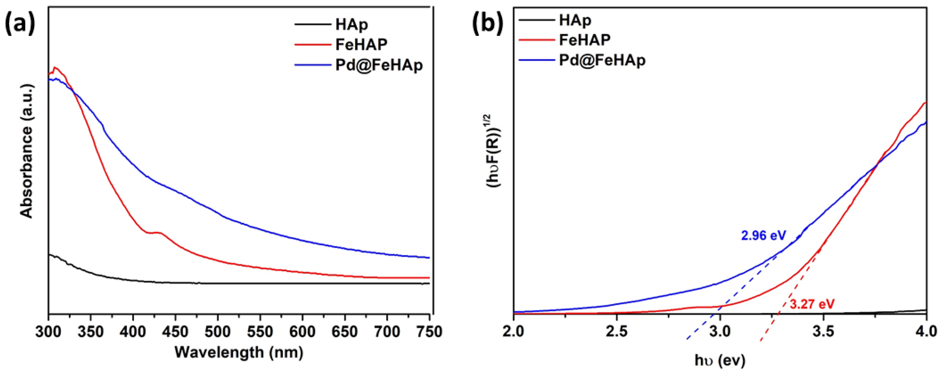
The light absorption and energy band gaps of HAp, FeHAp, and Pd@FeHAp composites were determined using UV-DRS investigations. Figure 2a indicates that HAp has lowest absorbance, suggesting that the pure HAp has poor light absorption ability in both UV and visible light regions. However, after Fe(III) introduction in the HAp structure, FeHAp composite showed strong light absorption in the 300–500 nm wavelength range, making it an efficient photocatalyst in the UV and near-visible light region. Contrarily, compared to HAp and FeHAp, the Pd@FeHAp composites absorbed much more UV and visible light (approximate wavelength range of 300–600 nm). This result indicated that after being decorated with PdNPs, the optical properties of Pd@FeHAp had improved. Moreover, using Tauc’s equation [47], the calculated Eg values of HAp, FeHAp, and Pd@FeHAp were >4.0 eV, 3.27 eV, and 2.96 eV, respectively, as shown in Figure 2b. Pd@FeHAp composite had a lower Eg than pure HAp and FeHAp, indicating that the existence of the PdNPs on the surface of Pd@FeHAp could harvest more visible light region. Thus, the lowered Eg values of the composites indicate that the photocatalytic activity of the composites for the simultaneous reactions may be enhanced in consequence of the increased production of electron-hole pairs during light irradiation.

2.3. Morphology Results

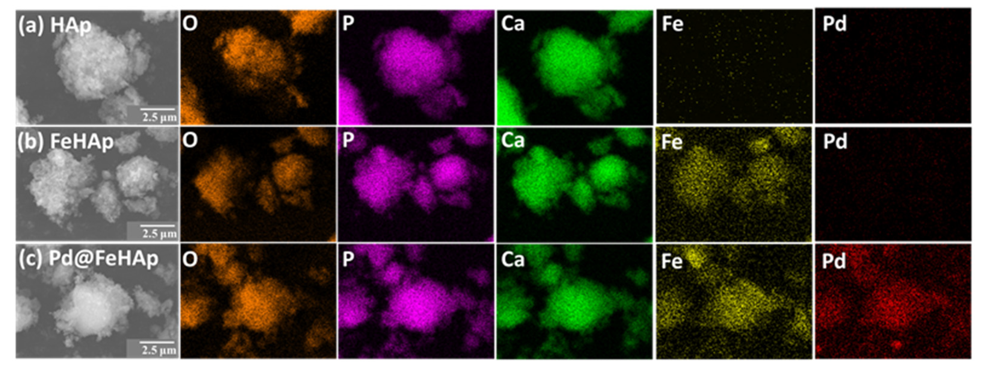
SEM-EDX images of the surface morphologies of HAp, FeHAp, and Pd@FeHAp are shown in Figure 3a–c. Pure HAp surface morphology in Figure 3a showed aggregates of tiny HAp particles arranged in a spherical form with a diameter about 7.5 µm. Surprisingly, the addition of Fe(III) in the HAp structure caused the smaller particle size of FeHAp, indicating some distortion by substitution of Ca(II) by Fe(III) as shown in Figure 3b. Moreover, the difference of particle size of Pd@FeHAp and FeHAp could not be observe (Figure 3c). SEM-EDX mappings were used to clarify the distribution of each element in HAp, FeHAp, and Pd@FeHAp. It can be seen that HAp, FeHAp, and Pd@FeHAp showed homogenous distribution of the main three elements of O, P, and Ca which come from hydroxy appetite structure. Moreover, the Fe signal, which was well overlapping with O, P, and Ca, was detected in the FeHAp and Pd@FeHAp, suggesting that the Fe(III) might dope into the HAp structure to form Fe(III)-doped HAp. Additionally, Pd signal was detected in a similar location with the Ca, O, and P, suggesting good distribution of PdNPs on the surface of HAp in the composites.
The presence of PdNPs in the Pd@FeHAp composite was investigated using TEM and STEM analysis. The high angle annular dark field (HAADF-STEM) images of Pd@FeHAp in Figure 4a,b reveals the dark spot on the surface of FeHAp in the bright field image and the bright spot on the surface of FeHAp in the dark field image, both of which point to the presence of heavy metal nanoparticles on the surface of FeHAp. Additionally, the particle size of the PdNPs in the Pd@FeHAp was investigated by TEM observation in different magnifications (Figure 4c–e). The particle size of PdNPs could be observed around 2.2–3.5 nm. The larger particle size in the Pd@FeHAp might be due to the aggregation of the smaller PdNPs. STEM-EDX mapping results showed Ca, P, O, Fe, and Pd signals in the Pd@FeHAp composites. However, the signals of Pd were observed at the same location as the dark spot in the bright field STEM image, confirming the deposition of PdNPs in the composite (Figure 4f). Thus, the formation process of the PdNPs on the surface of Pd@FeHAp might be due to the adsorption of Pd2+ on the hydroxide functional group on the surface of HAp via an electrostatic interaction force, following by reduction of the Pd2+ on the surface of HAp by chemical reduction.

2.4. Chemical State and Band Positions

XPS was used to determine the chemical states of Ca, P, O, Fe, and Pd as well as the valence band top (VBT) locations in Hap, FeHAp, and Pd@FeHAp. In order to identify the main elements present, the survey XPS spectra of Hap, FeHAp, and Pd@FeHAp were examined as shown in Figure 5a. The signal of Ca 2p, P 2p, and O 1s were observed as major components of HAp and both modified samples. Moreover, two peaks of Fe 2p3/2 and Fe 2p1/2 signals were detected FeHAp and Pd@FeHAp, confirming the existence of Fe ions in the samples. In contrast, the Pd signal in Figure 5a was difficult to observe in survey scans because of low Pd concentration. A narrow scan of the Ca 2p spectra of HAp, FeHAp, and Pd@FeHAp are shown in Figure 5b. The strong and weak signals at 346.8 eV and 349.8 eV corresponded to Ca 2p3/2 and Ca 2p1/2, respectively [48]. In Figure 5c, the signals of the P 2p3/2 and P 2p1/2 in HAp, FeHAp, and Pd@FeHAp can be attributed to PO43− in the structure of hydroxyapatite [49,50]. Additionally, the signals of Fe 2p in the FeHAp and Pd@FeHAp were observed at 712.3 eV, indicating the Fe (III) ions species in the structure [51]. The surface mole ratio of Fe/Ca of the FeHAp and Pd@FeHAp were investigated to compare with the XRF results to elucidate the location of the Fe in the composites. The XPS results showed the Fe/Ca of the FeHAp and Pd@FeHAp at about 0.203 and 0.201, respectively, while the XRF results suggests a lower Fe/Ca ratio of FeHAp (0.077) and Pd@FeHAp (0.072). These results indicated that the Fe ions might exist on the surface of HAp by substitution with the outer Ca surface of HAp. Additionally, the two peaks of Pd 3d5/2 = 336.4 eV and Pd 3d3/2 = 341.7 eV were found in the Pd@FeHAp sample as shown in Figure 5d, confirming the presence of decorated Pd(0) on the surface of FeHAp [35,52]. The valence band (VB) location of HAp, FeHAp, and Pd@FeHAp composite were also determined using XPS [53]. The VB position was estimated by the interception of a linear line from the slope of the XPS signal in the VB region at y = 0. In Figure 5f, the energy around 2.2 eV, 1.9 eV, and 0.46 eV, respectively, were VB positions for HAp, FeHAp, and Pd@FeHAp. The conduction band bottom (CBB) positions of FeHAp and Pd@FeHAp could be calculated based on the combination of VB and Eg results to be −1.37 and −2.5 eV, respectively.

2.5. Simultaneous Photocatalytic Xylose Conversion and Hydrogen Evolution

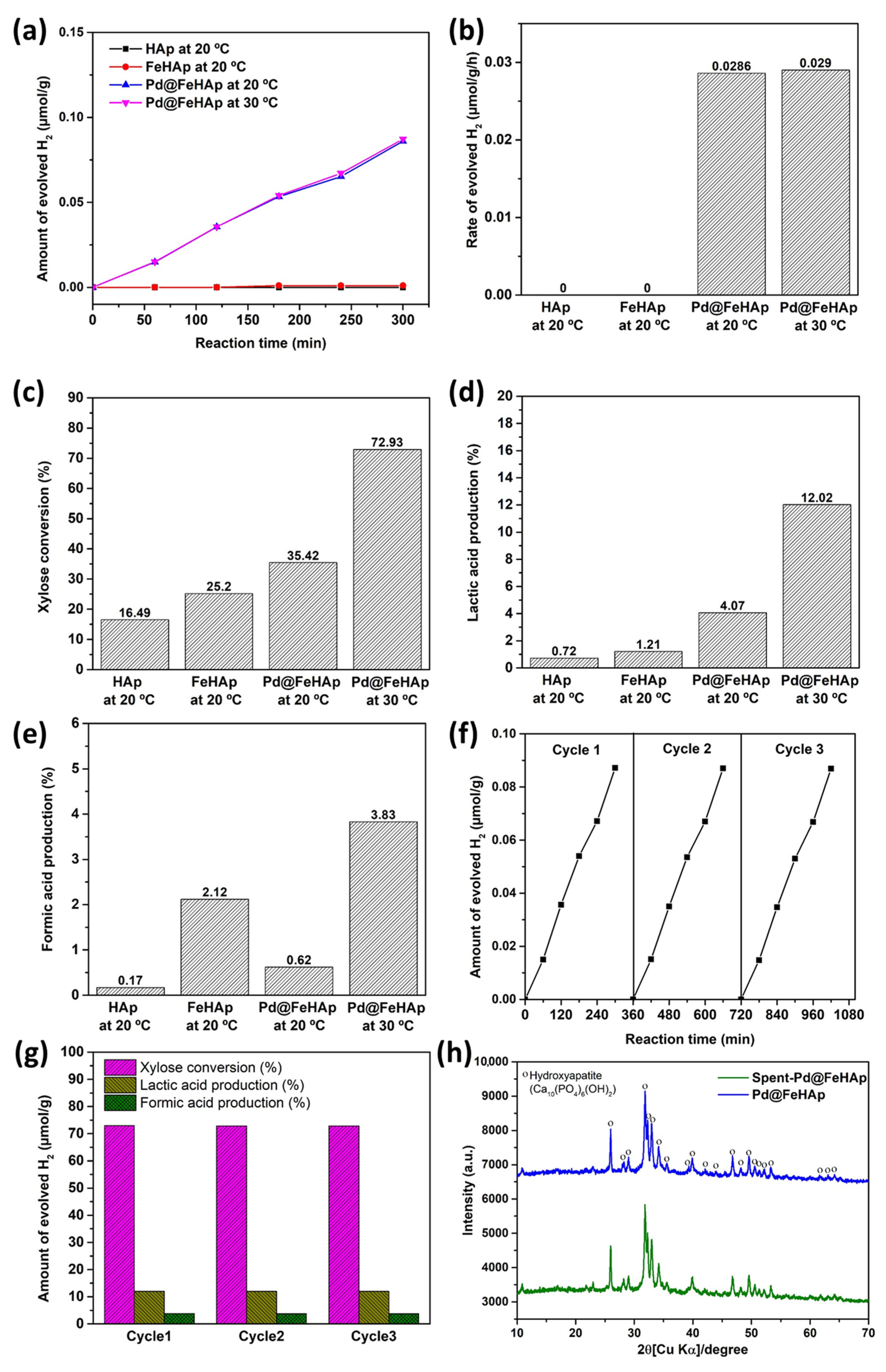
The simultaneous photocatalytic hydrogen evolution and xylose conversion using HAp, FeHAp, and Pd@FeHAp was conducted as shown in Figure 6a. HAp and FeHAp displayed less production of H2 evolution than Pd@FeHAp at 20 °C and the rates of evolved H2 of HAp, FeHAp, and Pd@FeHAp were 0.00, 0.00, and 0.0286 µmol/g/h, respectively. This result suggested that PdNPs can act as a co-catalyst for H2 production to promote the reduction of H+ in the water to H2. In addition, when the reaction temperature was increased to 30 °C, the rate of evolved H2 for Pd@FeHAp was 0.0290 µmol/g/h. The results from the effect of reaction temperature indicated that the reaction temperature has no effect for the H2 evolution. In addition, the photocatalytic H2 production using Pd@HAp, which was produced in the same manner as the Pd@FeHAp, was performed to emphasize the role of Fe in the HAp. As shown in Figure S1, it can be seen that the Pd@HAp could not be generated under light irradiation, while the Pd@FeHAp could produce H2 at about 0.0862 µmol/g at 30 °C. The poor production of H2 over Pd@HAp might be due to the large Eg and lower light absorption ability of pristine HAp. This result indicated that the introduction of Fe into the structure of HAp could reduce the Eg of HAp from >4.0 eV to 3.27 eV, resulting in the change of its properties from insulator to semiconductor, which can work as photocatalysts. Simultaneously, xylose was converted to small molecules during the H2 evolution reaction. The xylose conversions at 20 °C were around 16.5%, 25.2%, and 35.42% for HAp, FeHAp, and Pd@FeHAp, respectively, as shown in Figure 6c. After Fe(III) introduction into the structure of HAp, the conversion efficiency was improved due to reduction of Eg by new fermi level of Fe(III). When PdNPs were decorated on the surface of FeHAp in Pd@FeHAp sample, the photocatalytic xylose conversion efficiency increased due to less recombination of photogenerated charge carriers by the formation heterojunction between PdNPs and FeHAp. Interestingly, the photocatalytic activity of the Pd@FeHAp dramatically enhanced when the reaction temperature was raised to 30 °C because the conversion of xylose is an endothermic reaction, which is preferable at a high reaction temperature. Basically, xylose could be oxidized to several small organic compounds such as xylitol, erythritol, lactic acid, formic acid, and xylonic acid [5,15,54]. However, in this work, the product yields from xylose conversion were investigated based on two important high-value converted products such as lactic acid and formic acid. For this experiment, due to the high basic condition reaction, the final products are basically in their basic forms, such as lactate and formate. However, after the reaction, the solution was acidified using 1M H2SO4 (pH below 7). Thus, the final product forms are acids such as lactic acid and formic acid. At 20 °C, Pd@FeHAp showed higher production of lactic acid than the other samples, as shown in Figure 6d, while the higher formic acid yield was obtained by FeHAp (Figure 6e). In addition, based on the HPLC spectrum of the solution after the simultaneous photocatalytic sugar conversion and hydrogen production over Pd@HAp for 4 h at 30 °C (Figure S2), some other by-products except formic and lactic acid could be formed, such as succinic acid, citric acid, and some unidentified peaks that may be related to some small organic compounds, indicating the conversion of glucose xylose to small organic molecules. Additionally, the pH of the sugar solution after 4 h of reaction was measured and compared with the original pH of the sugar solution before reaction. The results show that the pH of the solution slightly changes after 4 h of the reaction from 12.96 to 12.83 due to the formation of lactate and formate. Catalyst stability and recyclability are significant aspects in their practical use. The photocatalytic H2 evolution and xylose conversion to lactic acid and formic acid over Pd@FeHAp was retained over three cycles, as shown in Figure 6f,g, respectively. In addition, the structural stability of Pd@FeHAp after the photocatalytic reaction was investigated through XRD. In Figure 6h, the Pd@FeHAp had great stability after the simultaneous photocatalytic H2 evolution and xylose conversion. Additionally, in this work, we transformed the non-active HAp which could not generate H2 to possible photocatalyst for the production of H2 by surface Fe modification. Thus, the production of hydrogen is quite lower than normal photocatalyst such as TiO2. However, the results of this work can suggest the possible use of Fe-doped HAp as a photocatalyst for hydrogen production. In addition, Pd NPs was used as co-catalyst instead of Pt NPs, which is highly active and common as a co-catalyst for hydrogen production due to its lower price. Additionally, the xylose was used as a sacrificial agent instead of a triethanolamine, which is a common reagent in H2 production, but has a higher cost compared with xylose. Thus, the combination of Fe-doped HAp and PdNPs can be considered as an alternative photocatalyst due to being cost effective.

2.6. Examination of the Transfer and Separation of Charges

The formation of heterojunction between FeHAp and PdNPs were investigated using PL measurements to examine the separation and transfer of e and h+ pairs. A low PL intensity often indicates a low rate of e- and h+ pair recombination. As seen in Figure 7, reduced PL emission intensity of FeHAp compared to HAp at around 400 nm for a 350 nm excitation wavelength suggested that the new fermi level of Fe substitution in HAp structure could trap the excited electron from valence band and avoid e--h+ pair recombination. Moreover, the PdNPs decoration on the surface of FeHAp improved the charge carrier separation.
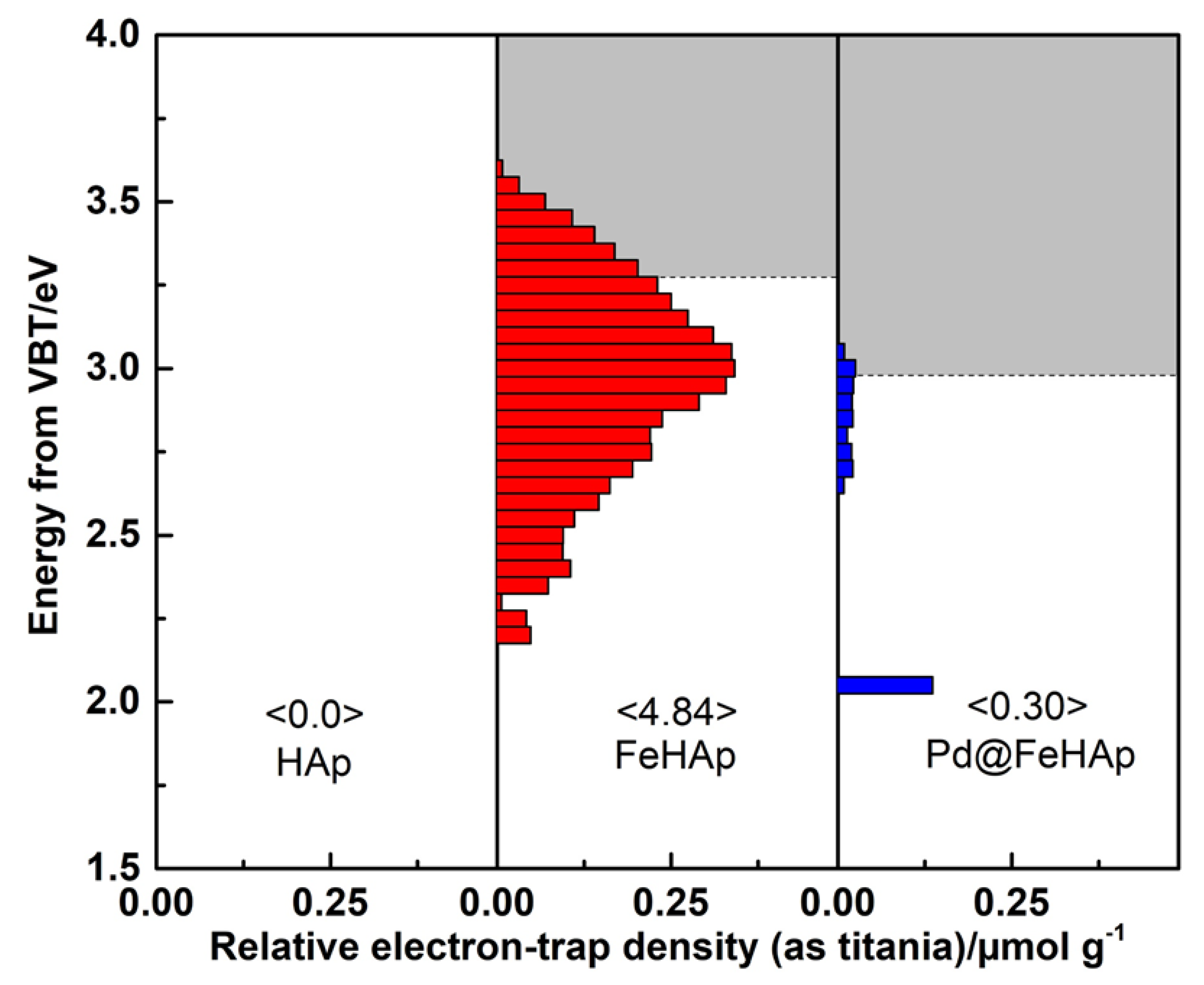
The surface electronic characteristics of HAp, FeHAp, and Pd@FeHAp were also examined using reversed double-beam photoacoustic spectroscopy (RDB-PAS), which could have an influence on photocatalytic activity and electron transfer of the composite [55,56,57]. The energy-resolved distribution of electron trap (ERDT) pattern as a function of the energy from the valence band top (VBT) vs. the electron trap density was conducted for the HAp, FeHAp, and Pd@FeHAp samples as shown in Figure 8. It is evidence that HAp does not have an electron trap because of its large energy band gap, but after Fe modification for FeHAp, the modified sample displayed an electron accumulation density peak at about 3.1 eV, which was close to Eg of the FeHAp sample. This suggests that the Fe atom in the HAp structure may be able to generate a new electron trapped (ETs) state close to CBB. It is interesting that the ERDT patterns of the Pd@FeHAp composites could not be seen, suggesting that the PdNPs introduction on the surface of FeHAp promoted the migration of electrons from FeHAp to the surface of PdNPs. The decrease in electrons in the ETs of FeHAp might be due to the electron transfer from FeHAp to PdNPs through a heterojunction, which prevented charge carrier recombination and enhanced photocatalytic activity.

2.7. Photocatalytic Mechanism

A potential photocatalytic simultaneous photocatalytic H2 evolution and xylose conversion process using Pd@FeHAp composite is shown in Scheme 1. The VB and CB of Pd@FeHAp composite were 0.46 and −2.5 eV, respectively. After light irradiation, the excited e- moved from the VB of FeHAp to a new electronic state created by the Fe(III) Fermi level in the structure. Concurrently, h+ were created at the VB. The decoration of PdNPs on FeHAp prevented the recombination of e and h+ pairs owing to the migration of e to the surface of PdNPs through heterojunction. Through a photocatalytic process, the accumulated electrons on the surface of PdNPs can react with H+ and produce the H2. Simultaneously, xylose can react with h+ in the VB of FeHAp and generate hydroxyl radicals to form formic acid and lactic acid. Thus, by forming a heterojunction using Pd@FeHAp, more active electrons and hydroxyl radicals were available for the simultaneous photocatalytic H2 evolution and xylose conversion.

3. Materials and Methods

3.1. Chemicals

All pure analytical-grade chemicals used in this experiment, namely calcium oxide (CaO), iron (III) nitrate nonahydrate (Fe(NO3)3•9H2O), diammonium hydrogen phosphate ((NH4)2HPO4), xylose (C5H10O5), sodium hydroxide (NaOH), and palladium(II) nitrate (Pd(NO3)2) were supplied by Fujifilm Wako Pure Chemical Co. (Osaka, Japan). None of the chemicals used underwent any further purification.

3.2. Preparation of HAp and FeHAp

A mixture of CaO (s) and (NH4)2HPO4 (s) with a molar ratio of Ca/P = 1.67 in 50 mL of DI water was utilized to prepare hydroxyapatite under hydrothermal treatment. The initial pH was measured at 14.5. The Teflon autoclave was heated at 200 °C for 48 h. The obtained solid was then filtered and washed several times with ethanol and DI water. Additionally, the solid products were dried for 24 h at 75 °C. This product was denoted as HAp.
For Fe(III) modification on HAp, an ion exchange method was applied to introduce Fe(III) to as-prepared HAp by suspending HAp in Fe(NO3)3•9H2O (aq) with a molar ratio of Ca/Fe = 10 under stirring at room temperature for 30 min. After that, the final solid product was filtered and washed with ethanol and DI water many times. The solid product was dried at 75 °C for 24 h. The resulting product was identified using FeHAp.

3.3. Preparation of Pd@HAp and Pd@FeHAp

Pd nanoparticles (PdNPs) were decorated on the surface of FeHAp by chemical reduction using NaBH4. The synthetic method began with dissolving 3.2 mg of Pd(NO3)2 in 20 mL of DI water. Then, 32.25 mg of FeHAp was suspended in Pd solution under stirring at room temperature to achieve 0.5% Pd loading on the composite. After 30 min, 1 mL of 1 M NaBH4 (aq) was then added to the mixture solution to form Pd nanoparticle under stirring for 30 min at room temperature. After that, the synthetic product was filtered and washed with ethanol and DI water several times before drying at 75 °C for 24 h. Pd@FeHAp was the designation given to the final product. In addition, the Pd@HAp was prepared in the same manner as the Pd@FeHAp for comparison of the photocatalytic H2 production.

3.4. Characterization

X-ray diffraction (XRD) using the Ultima IV was used to identify the crystal phases of all materials (RIGAKU, Akishima, Japan). The chemical composition was figured out by Raman spectroscopy (Thermo, DXR Smart Raman, Waltham, MA, USA) using laser wavelength 532 nm. With high magnification, scanning electron microscopy (SEM, VE-9800, Keyence, Osaka, Japan) and STEM-EDX (JEM-2100HCKM, JEOL, Tokyo, Japan) were used to examine the morphology, particle sizes, and surface properties of all samples. The ability of the pure and composite materials to absorb light was measured using UV–Vis diffuse reflectance spectroscopy (UV-DRS, UV-2450 Shimadzu, Kyoto, Japan). All samples’ VB sites were identified by X-ray photoelectron spectroscopy (XPS, ESCA 5800; ULVAC-PHI, Inc., Kanagawa, Japan). In this work, the C-C of C 1s from contaminated carbon from the machine at 284.6 was used as a reference for calibration of the XPS spectra. The elemental compositions of all samples were determined by XRF spectroscopy using Rigaku ZSX Primus II in the wavelength dispersive mode (Akishima, Japan). Photoluminescence (PL) spectroscopic studies were carried out using an FP-6600 spectrofluorometer (FP-6600 spectrofluorometer, JASCO Corporation, Tokyo, Japan). An energy-resolved distribution of electron trap (ERDT) pattern of the generated materials was discovered using reversed double-beam photoacoustic spectroscopy (RDB-PAS) from 600 to 300 nm using an amplifier to double the photoacoustic signals.

3.5. Simultaneous Photocatalytic Sugar Conversion and Hydrogen Production

To elucidate the efficacy of photocatalytic activity of the obtained samples, the simultaneous photocatalytic sugar conversion and hydrogen production were conducted. Typically, 60 mg of the produced samples was added in 120 mL of 1000 ppm xylose in 1 M NaOH under N2 purging for 30 min. Then, a 100 W Hg lamp was used to irradiate the suspensions. While the light was on, the produced H2 gas was sampled to identify the concentration of H2 using gas chromatography (GC, Nexis GC-2030, Shimadzu, Kyoto, Japan), with the Shincarbon ST column, TCD detector, and 50 mL/min flow rate of Ar gas as a mobile phase. In addition, the 0.5 mL from the photocatalytic solution was acidified using 0.5 mL of 1M H2SO4 for high-pressure liquid chromatography (HPLC) analysis. Thus, xylose, lactic acid, and formic acid concentration were determined by HPLC (JASCO, CO-2065 Plus, Tokyo, Japan) using cation-exchange column (Aminex HPX-87H, 300 × 7.8 mm, Bio-Rad) and eluted with the flow rate 0.6 mL/min of 5 mM H2SO4 at 50 °C. The detection of Xylose was calculated from RI detector, whereas the organic acid was determined by a UV detector at wavelength 210 nm. The xylose conversion and production yield were calculated following these equations as shown below [58];
Xylose   conversion   % = initial   concentration remained   concentration initial   concentration × 100
Product   yield   % = mole   of   product mole   of   initial   xylose × carbon   numbers   in   1   mol   product   carbon   numbers   in   1   mol   xylose × 100

4. Conclusions

Pd@FeHAp composites were successfully prepared using a decoration of PdNPs on the surface of Fe(III) modification HAp. Pd@FeHAp provided a greater rate production of H2 and xylose conversion than HAp and FeHAp, resulting from the lower recombination of charge carriers. According to PL studies, the formation of heterojunction between PdNPs and FeHAp in Pd@FeHAp composite reduced the recombination of electron-hole pairs. The surface electronic investigation using RDB-PAS analysis supported the PL result as the electron from VB of HAp was accumulated in the Fermi level of Fe(III). The heterojunction formation between PdNPs and FeHAp caused the lessened electron density in ERDT patterns due to electron migration from FeHAp to PdNPs. Thus, the heterojunction formation in Pd@FeHAp gave more active electrons and hydroxyl radicals for the simultaneous photocatalytic H2 evolution and xylose conversion. Additionally, it is well known that hydroxyapatite can be prepared by natural wastes such as eggshells, animal bones, and cockle shells, and Fe is the fourth most abundant element in Earth’s crust. Moreover, the price of Pd is cheaper than that of Pt (a common cocatalyst), which is widely used in photocatalytic H2 evolution. Thus, the concept of production of Pd@FeHAp as a low-cost photocatalyst for simultaneous H2 evolution and lactic production can promote natural waste recycling in advanced purposes with a cost-effectiveness route. Thus, this work offers an alternative Pd@FeHAp photocatalyst for effective simultaneous photocatalytic H2 evolution and sugar conversion to produce biochemicals and alternative energy.

Supplementary Materials

The following supporting information can be downloaded at: https://www.mdpi.com/article/10.3390/catal13040675/s1, Figure S1:Time course of photocatalytic H2 production over Pd@HAp and Pd@FeHAp; Figure S2: HPLC spectrum of the solution after the simultaneous photocatalytic sugar conversion and hydrogen production over Pd@HAp for 4 h at 30 °C.

Author Contributions

C.C.: Conceptualization, Investigation, Formal analysis, Writing—original draft, Writing—review and editing. Y.N.: Formal analysis. S.S.: Formal analysis. K.S. (Kaiqian Shu): Formal analysis. J.T.: Formal analysis. A.S.: Formal analysis. K.S. (Karthikeyan Sekar): Formal analysis. K.S. (Keiko Sasaki): Conception, interpretation, editing the manuscript. All authors have read and agreed to the published version of the manuscript.

Funding

The work was supported by the Japan Society for the Promotion of Science (JSPS) KAKENHI (A) [No. JSPS JP22H00266]. This work was partly supported by Advanced Research Infrastructure for Materials and Nanotechnology Grant Number JPMXP1222KU1009 in Japan sponsored by the Ministry of Education, Culture, Sports, Science and Technology (MEXT), Japan. This work was partly supported by 2022 Research Start Program 202208 to Chitiphon Chuaicham.

Data Availability Statement

Not applicable.

Conflicts of Interest

The authors declare no conflict of interest.

References

  1. Zhang, Y.; Heo, Y.-J.; Lee, J.-W.; Lee, J.-H.; BajGai, J.; Lee, K.-J.; Park, S.-J. Photocatalytic Hydrogen Evolution via Water Splitting: A Short Review. Catalysts 2018, 8, 655. [Google Scholar] [CrossRef] [Green Version]
  2. Takanabe, K. Photocatalytic Water Splitting: Quantitative Approaches toward Photocatalyst by Design. ACS Catal. 2017, 7, 8006–8022. [Google Scholar] [CrossRef]
  3. Ramis, G.; Bahadori, E.; Rossetti, I. Design of efficient photocatalytic processes for the production of hydrogen from biomass derived substrates. Int. J. Hydrogen Energy 2021, 46, 12105–12116. [Google Scholar] [CrossRef]
  4. Cai, M.; Cao, S.; Zhuo, Z.; Wang, X.; Shi, K.; Cheng, Q.; Xue, Z.; Du, X.; Shen, C.; Liu, X.; et al. Fabrication of Ni2P Cocatalyzed CdS Nanorods with a Well-Defined Heterointerface for Enhanced Photocatalytic H2 Evolution. Catalysts 2022, 12, 417. [Google Scholar] [CrossRef]
  5. Puga, A.V. Photocatalytic production of hydrogen from biomass-derived feedstocks. Coord. Chem. Rev. 2016, 315, 1–66. [Google Scholar] [CrossRef]
  6. Jeon, T.H.; Koo, M.S.; Kim, H.; Choi, W. Dual-Functional Photocatalytic and Photoelectrocatalytic Systems for Energy- and Resource-Recovering Water Treatment. ACS Catal. 2018, 8, 11542–11563. [Google Scholar] [CrossRef]
  7. Granone, L.I.; Sieland, F.; Zheng, N.; Dillert, R.; Bahnemann, D.W. Photocatalytic conversion of biomass into valuable products: A meaningful approach? Green Chem. 2018, 20, 1169–1192. [Google Scholar] [CrossRef]
  8. Schneider, J.T.; Firak, D.S.; Ribeiro, R.R.; Peralta-Zamora, P. Use of scavenger agents in heterogeneous photocatalysis: Truths, half-truths, and misinterpretations. Phys. Chem. Chem. Phys. 2020, 22, 15723–15733. [Google Scholar] [CrossRef]
  9. Xia, B.; Zhang, Y.; Shi, B.; Ran, J.; Davey, K.; Qiao, S.Z. Photocatalysts for Hydrogen Evolution Coupled with Production of Value-Added Chemicals. Small Methods 2020, 4, 1499–1506. [Google Scholar] [CrossRef]
  10. Antonietti, M.; Savateev, A. Splitting Water by Electrochemistry and Artificial Photosynthesis: Excellent Science but a Nightmare of Translation? Chem. Rec. 2018, 18, 969–972. [Google Scholar] [CrossRef]
  11. Zhao, H.; Li, C.-F.; Yong, X.; Kumar, P.; Palma, B.; Hu, Z.-Y.; Van Tendeloo, G.; Siahrostami, S.; Larter, S.; Zheng, D.; et al. Coproduction of hydrogen and lactic acid from glucose photocatalysis on band-engineered Zn1-xCdxS homojunction. iScience 2021, 24, 102109. [Google Scholar] [CrossRef] [PubMed]
  12. Rohini, B.; Hebbar, H.U. Photocatalytic Conversion of Xylose to Xylitol over Copper Doped Zinc Oxide Catalyst. Catal. Lett. 2021, 151, 2583–2594. [Google Scholar] [CrossRef]
  13. Chen, L.; Huang, Y.; Zou, R.; Ma, J.; Yang, Y.; Li, T.; Li, M.; Hao, Q.; Xie, H.; Peng, X. Regulating TiO2/MXenes catalysts to promote photocatalytic performance of highly selective oxidation of d-xylose. Green Chem. 2021, 23, 1382–1388. [Google Scholar] [CrossRef]
  14. Liu, K.; Ma, J.; Yang, X.; Jin, D.; Li, Y.; Jiao, G.; Yao, S.; Sun, S.; Sun, R. Boosting electron kinetics of anatase TiO2 with carbon nanosheet for efficient photo-reforming of xylose into biomass-derived organic acids. J. Alloys Compd. 2022, 906, 164276. [Google Scholar] [CrossRef]
  15. Sanwald, K.E.; Berto, T.F.; Eisenreich, W.; Jentys, A.; Gutiérrez, O.Y.; Lercher, J.A. Overcoming the Rate-Limiting Reaction during Photoreforming of Sugar Aldoses for H2-Generation. ACS Catal. 2017, 7, 3236–3244. [Google Scholar] [CrossRef]
  16. Imizcoz, M.; Puga, A.V. Assessment of Photocatalytic Hydrogen Production from Biomass or Wastewaters Depending on the Metal Co-Catalyst and Its Deposition Method on TiO2. Catalysts 2019, 9, 584. [Google Scholar] [CrossRef] [Green Version]
  17. Puga, A.V.; Barka, N.; Imizcoz, M. Simultaneous H2 Production and Bleaching via Solar Photoreforming of Model Dye-polluted Wastewaters on Metal/Titania. ChemCatChem 2020, 13, 1513–1529. [Google Scholar] [CrossRef]
  18. Uekert, T.; Bajada, M.A.; Schubert, T.; Pichler, C.M.; Reisner, E. Scalable Photocatalyst Panels for Photoreforming of Plastic, Biomass and Mixed Waste in Flow. ChemSusChem 2021, 14, 4190–4197. [Google Scholar] [CrossRef]
  19. Kasap, H.; Achilleos, D.S.; Huang, A.; Reisner, E. Photoreforming of Lignocellulose into H(2) Using Nanoengineered Carbon Nitride under Benign Conditions. J. Am. Chem. Soc. 2018, 140, 11604–11607. [Google Scholar] [CrossRef] [Green Version]
  20. Ma, J.; Yang, X.; Yao, S.; Guo, Y.; Sun, R. Photocatalytic Biorefinery to Lactic Acid: A Carbon Nitride Framework with O Atoms Replacing the Graphitic N Linkers Shows Fast Migration/Separation of Charge. ChemCatChem 2022, 14, e202200097. [Google Scholar] [CrossRef]
  21. Qin, H.-J.; Zhang, Y.-H.; Wang, Z.; Yang, G.-H. Photocatalytic Conversion of Fructose to Lactic Acid by BiOBr/Zn@SnO2 Material. Catalysts 2022, 12, 719. [Google Scholar] [CrossRef]
  22. Ma, J.; Jin, D.; Li, Y.; Xiao, D.; Jiao, G.; Liu, Q.; Guo, Y.; Xiao, L.; Chen, X.; Li, X.; et al. Photocatalytic conversion of biomass-based monosaccharides to lactic acid by ultrathin porous oxygen doped carbon nitride. Appl. Catal. B Environ. 2021, 283, 119520. [Google Scholar] [CrossRef]
  23. Suhag, M.H.; Tateishi, I.; Furukawa, M.; Katsumata, H.; Khatun, A.; Kaneco, S. Photocatalytic Hydrogen Production from Formic Acid Solution with Titanium Dioxide with the Aid of Simultaneous Rh Deposition. ChemEngineering 2022, 6, 43. [Google Scholar] [CrossRef]
  24. Jin, B.; Yao, G.; Wang, X.; Ding, K.; Jin, F. Photocatalytic Oxidation of Glucose into Formate on Nano TiO2 Catalyst. ACS Sustain. Chem. Eng. 2017, 5, 6377–6381. [Google Scholar] [CrossRef]
  25. Roongraung, K.; Chuangchote, S.; Laosiripojana, N. Enhancement of Photocatalytic Oxidation of Glucose to Value-Added Chemicals on TiO2 Photocatalysts by A Zeolite (Type Y) Support and Metal Loading. Catalysts 2020, 10, 423. [Google Scholar] [CrossRef]
  26. Ricke, S.C.; Dittoe, D.K.; Richardson, K.E. Formic Acid as an Antimicrobial for Poultry Production: A Review. Front. Vet. Sci. 2020, 7, 563. [Google Scholar] [CrossRef]
  27. García, V.; Catalá-Gregori, P.; Hernández, F.; Megías, M.D.; Madrid, J. Effect of Formic Acid and Plant Extracts on Growth, Nutrient Digestibility, Intestine Mucosa Morphology, and Meat Yield of Broilers. J. Appl. Poult. Res. 2007, 16, 555–562. [Google Scholar] [CrossRef]
  28. Piccirillo, C.; Castro, P.M.L. Calcium hydroxyapatite-based photocatalysts for environment remediation: Characteristics, performances and future perspectives. J. Environ. Manag. 2017, 193, 79–91. [Google Scholar] [CrossRef]
  29. Zhang, L.; Chuaicham, C.; Balakumar, V.; Sekar, K.; Ohtani, B.; Sasaki, K. Determination of the roles of FeIII in the interface between titanium dioxide and montmorillonite in FeIII-doped montmorillonite/titanium dioxide composites as photocatalysts. Appl. Clay Sci. 2022, 227, 106577. [Google Scholar] [CrossRef]
  30. Saxena, V.; Sharma, S.; Pandey, L.M. Fe(III) doped ZnO nano-assembly as a potential heterogeneous nano-catalyst for the production of biodiesel. Mater. Lett. 2019, 237, 232–235. [Google Scholar] [CrossRef]
  31. Khan, H.; Swati, I.K. Fe3+-doped Anatase TiO2 with d–d Transition, Oxygen Vacancies and Ti3+ Centers: Synthesis, Characterization, UV–vis Photocatalytic and Mechanistic Studies. Ind. Eng. Chem. Res. 2016, 55, 6619–6633. [Google Scholar] [CrossRef]
  32. Dos Santos Silva, D.; Villegas, A.E.C.; Bonfim, R.d.P.F.; Salim, V.M.M.; De Resende, N.S. Iron-substituted hydroxyapatite as a potential photocatalyst for selective reduction of CO2 with H2. J. CO2 Util. 2022, 63, 102102. [Google Scholar] [CrossRef]
  33. Boukha, Z.; Choya, A.; Cortés-Reyes, M.; de Rivas, B.; Alemany, L.J.; González-Velasco, J.R.; Gutiérrez-Ortiz, J.I.; López-Fonseca, R. Influence of the calcination temperature on the activity of hydroxyapatite-supported palladium catalyst in the methane oxidation reaction. Appl. Catal. B Environ. 2020, 277, 119280. [Google Scholar] [CrossRef]
  34. Shokouhimehr, M.; Yek, S.M.-G.; Nasrollahzadeh, M.; Kim, A.; Varma, R.S. Palladium Nanocatalysts on Hydroxyapatite: Green Oxidation of Alcohols and Reduction of Nitroarenes in Water. Appl. Sci. 2019, 9, 4183. [Google Scholar] [CrossRef] [Green Version]
  35. Ulas, B.; Yilmaz, Y.; Koc, S.; Kivrak, H. Hydroxyapatite supported PdxIn100-x as a novel electrocatalyst for high-efficiency glucose electrooxidation. Int. J. Hydrogen Energy 2023, 48, 6798–6810. [Google Scholar] [CrossRef]
  36. Patria, R.D.; Islam, M.K.; Luo, L.; Leu, S.-Y.; Varjani, S.; Xu, Y.; Wong, J.W.-C.; Zhao, J. Hydroxyapatite-based catalysts derived from food waste digestate for efficient glucose isomerization to fructose. Green Synth. Catal. 2021, 2, 356–361. [Google Scholar] [CrossRef]
  37. Jung, K.; Kim, Y.; Chung, W.-J.; Kwon, K.-Y. Hydroxyapatite Supported Ruthenium Catalysts for Hydrogen Generation from Borane Dimethyl Amine. Bull. Korean Chem. Soc. 2015, 36, 2797–2798. [Google Scholar] [CrossRef]
  38. Liu, X.; Yang, Y.; Su, S.; Yin, D. Efficient Oxidation of Glucose into Sodium Gluconate Catalyzed by Hydroxyapatite Supported Au Catalyst. Catal. Lett. 2016, 147, 383–390. [Google Scholar] [CrossRef]
  39. Nur, A.; Nazriati, N.; Fajaroh, F.; Arthaningrum, A.; Nurcahyani, I.; Cipto, D.L.R.P.; Kurniawan, F. Electrosynthesis of Cu/hydroxyapatite as the catalyst for hydrogen production via NaBH4 hydrolysis. IOP Conf. Ser. Mater. Sci. Eng. 2021, 1070, 012023. [Google Scholar] [CrossRef]
  40. Hou, Q.; Laiq Ur Rehman, M.; Bai, X.; Qian, H.; Lai, R.; Xia, T.; Yu, G.; Tang, Y.; Xie, H.; Ju, M. Enhancing the reusability of hydroxyapatite by barium modification for efficient isomerization of glucose to fructose in ethanol. Fuel 2023, 338, 127308. [Google Scholar] [CrossRef]
  41. Malpica-Maldonado, J.J.; Melo-Banda, J.A.; Martínez-Salazar, A.L.; Garcia-Hernández, M. Synthesis and characterization of Ni-Mo2C particles supported over hydroxyapatite for potential application as a catalyst for hydrogen production. Int. J. Hydrogen Energy 2019, 44, 12446–12454. [Google Scholar] [CrossRef]
  42. Mitsudome, T.; Urayama, T.; Kiyohiro, T.; Maeno, Z.; Mizugaki, T.; Jitsukawa, K.; Kaneda, K. On-demand Hydrogen Production from Organosilanes at Ambient Temperature Using Heterogeneous Gold Catalysts. Sci. Rep. 2016, 6, 37682. [Google Scholar] [CrossRef] [PubMed] [Green Version]
  43. Yaemsunthorn, K.; Randorn, C. Hydrogen production using economical and environmental friendly nanoparticulate hydroxyapatite and its ion doping. Int. J. Hydrogen Energy 2017, 42, 5056–5062. [Google Scholar] [CrossRef]
  44. Yan, T.; Li, N.; Jiang, Z.; Guan, W.; Qiao, Z.; Huang, B. Self-sacrificing template synthesis of CdS quantum dots/Cd-Hap composite photocatalysts for excellent H2 production under visible light. Int. J. Hydrogen Energy 2018, 43, 20616–20626. [Google Scholar] [CrossRef]
  45. Liang, Z.; Ouyang, B.; Wang, T.; Liu, X.; Huo, H.; Liu, D.; Feng, H.; Ma, J.; Deng, K.; Li, A.; et al. Pt modified TiO2/NiO p-n junction with enhanced surface reaction and charge separation for efficient photocatalytic hydrogen evolution. Int. J. Hydrogen Energy 2022, 47, 10868–10876. [Google Scholar] [CrossRef]
  46. Prekajski, M.; Jokic, B.; Kalijadis, A.; Maletaskic, J.; Stankovic, N.; Lukovic, J.; Matovic, B. Synthesis of silver doped hydroxyapatite nanospheres using Ouzo effect. Process. Appl. Ceram. 2016, 10, 169–174. [Google Scholar] [CrossRef] [Green Version]
  47. Mehta, S.K.; Kumar, S.; Chaudhary, S.; Bhasin, K.K. Nucleation and growth of surfactant-passivated CdS and HgS nanoparticles: Time-dependent absorption and luminescence profiles. Nanoscale 2010, 2, 145–152. [Google Scholar] [CrossRef]
  48. Chang, M.C.; Tanaka, J. XPS study for the microstructure development of hydroxyapatite–collagen nanocomposites cross-linked using glutaraldehyde. Biomaterials 2002, 23, 3879–3885. [Google Scholar] [CrossRef]
  49. Gomes, G.C.; Borghi, F.F.; Ospina, R.O.; López, E.O.; Borges, F.O.; Mello, A. Nd:YAG (532 nm) pulsed laser deposition produces crystalline hydroxyapatite thin coatings at room temperature. Surf. Coat. Technol. 2017, 329, 174–183. [Google Scholar] [CrossRef]
  50. Govindasamy, P.; Kandasamy, B.; Thangavelu, P.; Barathi, S.; Thandavarayan, M.; Shkir, M.; Lee, J. Biowaste derived hydroxyapatite embedded on two-dimensional g-C3N4 nanosheets for degradation of hazardous dye and pharmacological drug via Z-scheme charge transfer. Sci. Rep. 2022, 12, 11572. [Google Scholar] [CrossRef]
  51. Li, L.; Ma, P.; Hussain, S.; Jia, L.; Lin, D.; Yin, X.; Lin, Y.; Cheng, Z.; Wang, L. FeS2/carbon hybrids on carbon cloth: A highly efficient and stable counter electrode for dye-sensitized solar cells. Sustain. Energy Fuels 2019, 3, 1749–1756. [Google Scholar] [CrossRef]
  52. Li, D.; Zhao, X.; Zhou, Q.; Ding, B.; Zheng, A.; Peng, Q.; Hou, Z. Vicinal hydroxyl group-inspired selective oxidation of glycerol to glyceric acid on hydroxyapatite supported Pd catalyst. Green Energy Environ. 2022, 7, 691–703. [Google Scholar] [CrossRef]
  53. Chuaicham, C.; Sekar, K.; Balakumar, V.; Mittraphab, Y.; Shimizu, K.; Ohtani, B.; Sasaki, K. Fabrication of graphitic carbon nitride/ZnTi-mixed metal oxide heterostructure: Robust photocatalytic decomposition of ciprofloxacin. J. Alloys Compd. 2022, 906, 164294. [Google Scholar] [CrossRef]
  54. Chong, R.; Li, J.; Ma, Y.; Zhang, B.; Han, H.; Li, C. Selective conversion of aqueous glucose to value-added sugar aldose on TiO2-based photocatalysts. J. Catal. 2014, 314, 101–108. [Google Scholar] [CrossRef]
  55. Unwiset, P.; Chen, G.; Ohtani, B.; Chanapattharapol, K.C. Correlation of the Photocatalytic Activities of Cu, Ce and/or Pt-Modified Titania Particles with their Bulk and Surface Structures Studied by Reversed Double-Beam Photoacoustic Spectroscopy. Catalysts 2019, 9, 1010. [Google Scholar] [CrossRef] [Green Version]
  56. Ratova, M.; Tosheva, L.; Kelly, P.J.; Ohtani, B. Characterisation and properties of visible light-active bismuth oxide-titania composite photocatalysts. Sustain. Mater. Technol. 2019, 22, e00112. [Google Scholar] [CrossRef]
  57. Chuaicham, C.; Inoue, T.; Balakumar, V.; Tian, Q.; Ohtani, B.; Sasaki, K. Visible light-driven ZnCr double layer oxide photocatalyst composites with fly ashes for the degradation of ciprofloxacin. J. Environ. Chem. Eng. 2022, 10, 106970. [Google Scholar] [CrossRef]
  58. Li, L.; Shen, F.; Smith, R.L.; Qi, X. Quantitative chemocatalytic production of lactic acid from glucose under anaerobic conditions at room temperature. Green Chem. 2017, 19, 76–81. [Google Scholar] [CrossRef]
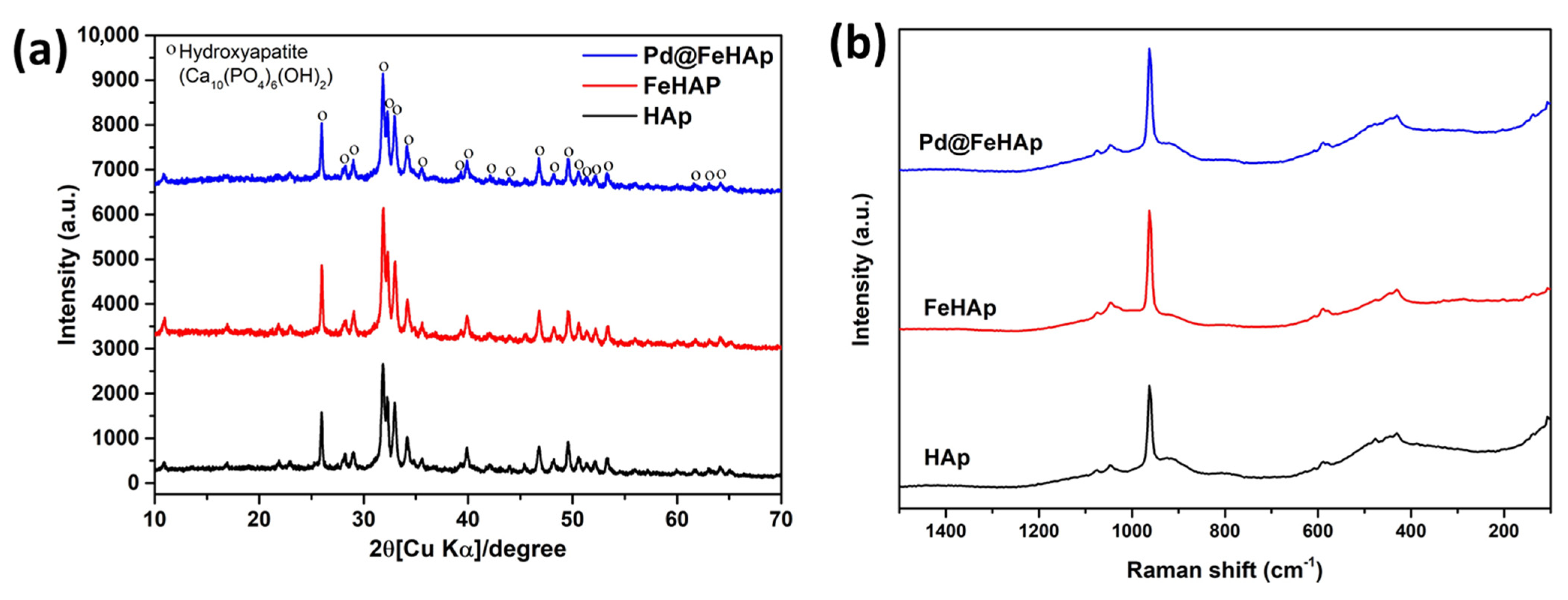
Figure 1. (a) XRD patterns and (b) Raman spectra of HAp, FeHAp, and Pd@FeHAp.
Catalysts 13 00675 g001
Figure 2. (a) UV–Vis DRS spectra and (b) energy band gap plots of HAp, FeHAp, and Pd@FeHAp.
Catalysts 13 00675 g002
Figure 3. SEM-EDX images of (a) HAp, (b) FeHAp, and (c) Pd@FeHAp.
Catalysts 13 00675 g003
Figure 4. STEM images (right = dark filed, left = bright filed) of Pd@FeHAp with magnification at (a) 200 nm, (b) 100 nm, and high-resolution TEM images of Pd@FeHAp at (c) 200 nm, (d) 100 nm, (e) 50 nm, and (f) STEM-EDX images of Pd@FeHAp.
Catalysts 13 00675 g004
Figure 5. XPS spectra of HAp, FeHAp, and Pd@FeHAp: (a) survey spectra, (b) Ca 2p, (c) P 2p, (d) Fe 2p, (e) Pd 3d, and (f) valence band energy region.
Catalysts 13 00675 g005
Figure 6. (a) Time course, (b) kinetics plots of photocatalytic H2 production (c) xylose conversion, (d) lactic acid production, (e) formic acid production over HAp, FeHAp, and Pd@FeHAp, (f) reusability for H2 over the Pd@FeHAp for 4 h, (g) xylose conversion and acid productions over the Pd@FeHAp for 4 h and (h) XRD patterns of fresh Pd@FeHAp and spent Pd@FeHAp after 4 h of the reaction.
Catalysts 13 00675 g006
Figure 7. PL spectra of HAp, FeHAp, and Pd@FeHAp.
Catalysts 13 00675 g007
Figure 8. ERDT/CBB patterns including relative total electron trap density in brackets of HAp, FeHAp, and Pd@FeHAp.
Catalysts 13 00675 g008
Scheme 1. Photocatalytic mechanism of simultaneous photocatalytic xylose conversion and hydrogen evolution.
Catalysts 13 00675 sch001
Table 1. Recent reports of using hydroxyapatite (HAp)-based materials for H2 production and sugar conversion.
MaterialsApplicationsProcessesRef.
HApSugar conversionIsomerization of glucose to fructose[36]
Ru/HApH2 productionHydrolysis of borane dimethyl amine[37]
Au/HApSugar conversionGlucose oxidation to sodium gluconate[38]
Cu/HApH2 productionHydrolysis of NaBH4[39]
Ba/HApSugar conversionIsomerization of glucose to fructose[40]
Ni-Mo2C/HApH2 productionBiomass gasification[41]
Au/HApH2 productionHydrolytic oxidation of organosilanes[42]
PaIn/HApSugar conversionGlucose electrooxidation for direct glucose fuel cells[35]
Ti-HApH2 productionPhotocatalytic water splitting[43]
CdS quantum dots/Cd-HApH2 productionPhotocatalytic H2 evolution[44]
Table 2. Elemental composition (%wt) of HAp, FeHAp, and Pd@FeHAp.
OPCaFePd
HAp47.4917.6034.91--
FeHAp48.2516.3531.953.45-
Pd@FeHAp48.2716.3931.893.210.23
Disclaimer/Publisher’s Note: The statements, opinions and data contained in all publications are solely those of the individual author(s) and contributor(s) and not of MDPI and/or the editor(s). MDPI and/or the editor(s) disclaim responsibility for any injury to people or property resulting from any ideas, methods, instructions or products referred to in the content.