Review

Application of Perovskite Nanocrystals as Fluorescent Probes in the Detection of Agriculture- and Food-Related Hazardous Substances

1
Maize Research Institute, Heilongjiang Academy of Agricultural Sciences, Harbin 150086, China
2
State Key Laboratory of Luminescent Materials and Devices, South China University of Technology, Guangzhou 510640, China
3
Harbin Technician College, Harbin 150500, China
4
School of Chemistry and Chemical Engineering, Harbin Institute of Technology, Harbin 150001, China
*
Authors to whom correspondence should be addressed.
Polymers 2023, 15(13), 2873; https://doi.org/10.3390/polym15132873
Submission received: 19 May 2023 / Revised: 19 June 2023 / Accepted: 21 June 2023 / Published: 29 June 2023
(This article belongs to the Special Issue Polymers/Their Hybrid Materials for Optoelectronic Applications)

Abstract

:
Halide perovskite nanocrystals (PNCs) are a new kind of luminescent material for fluorescent probes. Compared with traditional nanosized luminescent materials, PNCs have better optical properties, such as high fluorescence quantum yield, tunable band gap, low size dependence, narrow emission bandwidth, and so on. Therefore, they have broad application prospects as fluorescent probes in the detection of agriculture- and food-related hazardous substances. In this paper, the structure and basic properties of PNCs are briefly described. The water stabilization methods, such as polymer surface coating, ion doping, surface passivation, etc.; are summarized. The recent advances of PNCs such as fluorescent probes for detecting hazardous substances in the field of agricultural and food are reviewed, and the detection effect and mechanism are discussed and analyzed. Finally, the problems and solutions faced by PNCs as fluorescent probes in agriculture and food were summarized and prospected. It is expected to provide a reference for further application of PNCs as fluorescent probes in agriculture and food.

1. Introduction

With the rapid development of Chinese agriculture and food in recent decades, the excessive and indiscriminate use of some hazardous substances (such as pesticides, antibiotics, industrial pollutants, harmful ion pollutants, etc.) is also increasing. As a result, these hazardous substances may cause harm to human beings through agricultural products and food. In order to ensure that the residues of these related hazardous substances do not exceed the standard, fluorescent probes are used as indicators under the excitation of a certain wavelength of light. Through the detection of the generated fluorescence, qualitative or quantitative analysis of the detected substances can be realized. Fluorescent probes play an important role in agriculture and food because of their advantages of simple operation, high sensitivity, and non-destructive visualization [1,2,3].
In the past few decades, fluorescent probes based on semiconductor quantum dots (QDs) have gained wide attention due to their convenience, speed, low cost, and low limit of detection (LOD). To date, a large number of QD-based fluorescent probes have been developed to detect a variety of agricultural- and food-related contaminants, such as pesticides, harmful ion contaminants, chemical dyes, etc. Currently, QDs are generally composed of IV, II-VI, IV-VI, or II-V elements, such as CdTe, ZnO, and ZnS QDs [4]. However, due to the low quantum yield of these QDs, the sensitivity of fluorescent probes prepared by surface modification is low. Therefore, it is necessary to develop new semiconductor fluorescent probes with excellent photoluminescence (PL) performance and high fluorescence quantum yield. Halide PNCs have become a new promising fluorescent probe material in recent years due to their excellent properties, such as high fluorescence quantum yield, tunable band gap, low size dependence, narrow emission bandwidth, and so on [5,6].
In this paper, as shown in Figure 1, the structure and basic properties of halide PNC luminescent materials and the methods to improve the water stability of PNCs are briefly introduced. On this basis, the research progress of PNCs as fluorescent probes for detecting hazardous substances in the field of agricultural and food is reviewed, and the detection effect and mechanism are discussed and analyzed. Finally, the problems and solutions faced by PNCs as fluorescent probes in agriculture and food were summarized and prospected. It is expected to provide a reference for further application of PNCs as fluorescent probes in agricultural and food.

1.1. Structure and Basic Properties of PNCs

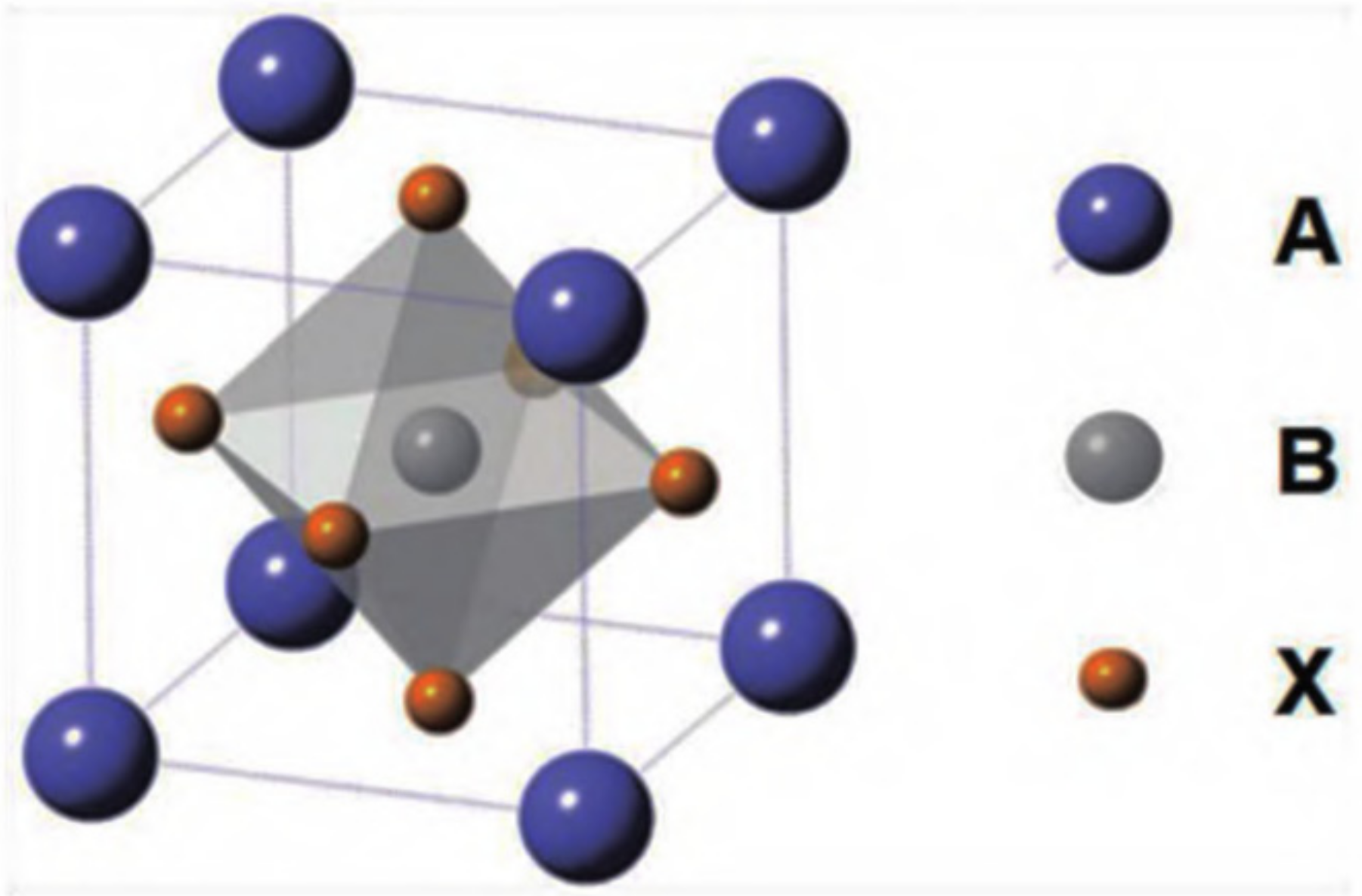
Halide perovskite is a new type of semiconductor material with the molecular formula ABX3, where A mainly represents alkali metal cation cesium (Cs+), methylamine ion (MA, CH3NH3+), or formamidine ion (FA, CH(NH2)2+); B represents metal cations such as Pb2+, Sn2+, etc.; and X stands for the halide anion (Cl, Br, I, or their combination). As shown in Figure 2, the lattice structure of perovskite, such as CaTiO3, has a three-dimensional (3D) angular shared [BX6] octahedron, which is formed by the coordination of the B site in the center and six X site anions. Each octahedron is connected through the shared angle to form an extended 3D network structure, while A is embedded in the gap of the network structure [7]. Halide perovskite can have three different crystalline structures: rhombic, tetragonal, and cubic phases. The cubic phase can exist stably at high temperatures [8]. When A is an organic group such as MA or FA, it is called an organic–inorganic hybrid perovskite, whereas when A is Cs+, it is called an all-inorganic perovskite.
However, the low exciton binding energy of bulk phase perovskite materials makes carriers easy to be trapped by defects in the lattice, leading to slow radiation recombination of carriers in bulk phase perovskite. To address this problem, efforts have been made to reduce the size of perovskite crystals or reduce its structural dimension to enhance carrier restriction within the perovskite lattice [9,10]. Nano-structured perovskites, such as PNCs, perovskite nanowires, and perovskite nanosheets, can improve the photoluminescent quantum yield (PLQY) of materials due to the quantum domain effect. Among them, PNCs have three dimensions below 100 nm, and perovskite quantum dots (PQDs) have three dimensions below 20 nm, exhibiting a clear quantum limiting effect and high PLQY, which is the most widely studied. The band gap and color of halide perovskite can be continuously adjusted from near-infrared to the violet region by modifying the halide elements and their respective composition, covering the entire visible spectral region. Moreover, the narrow spectral distribution of PNCs is crucial for easy detection in fluorescent probes. Numerous simple and effective synthetic methods are available for obtaining PNCs, such as hot injection [11], room-temperature reprecipitation [12], solvothermal synthesis [13], microwave method [14], and ultrasonication [15], among others.

1.2. Methods to Improve the Stability of PNCs

Although PNCs possess excellent properties, as ionic crystals they have a large surface energy and are susceptible to external influences, leading to instability and degradation of their structures. For instance, water, light radiation, and temperature changes can all cause fluorescence quenching and structural degradation [16]. In applications such as agriculture and food, where hazardous substance detection requires a water environment, stability in water is crucial for PNCs to function effectively as fluorescent probes. Fortunately, recent studies have developed a range of strategies to address the limitations of PNCs, including surface coating, metal ion doping, and surface passivation.

1.2.1. Surface Coating by Polymer, Inorganic Oxide, and Porous Materials

The surface of the PNCs is coated and isolated from the environment, which greatly improves the stability of the PNCs, especially the water resistance. At present, PNCs are coated with polymer, inorganic oxide, and porous materials. The polymer forms a dense network on the NC surface, which can effectively prevent environmental erosion. Researchers usually dissolve or expand the polymer in toluene, N, N-dimethylformamide, and other solvents, realize the encapsulation protection of PNCs through in situ growth or adsorption, and then remove the solvent under vacuum and high temperature to obtain polymer–PNCs composite materials [17,18]. As shown in Figure 3, Xuan et al. used divinylbenzene, ethyl acetate, and azodiisobutyronitrile (AIBN) as reaction raw materials to prepare superhydrophobic organic polymers through solvothermal methods. High-quality PQDs were prepared by hot injection, and the cyclohexane solution of PQDs was mixed with the organic polymer to fully absorb the QDs into the pores on the surface of the polymer, forming a complex (CPB@SHFW). Fluorescence quantum efficiency remained at 91% after immersion in water for one month, and the contact angle of the complex was maintained at 150°. The pores of the polymer provided a superhydrophobic environment to improve the water stability of the QDs, while also acting as a framework to maintain the excellent luminous performance of PQDs. This study successfully achieved the preparation of “water-resistant” perovskite QDs [19].
Although the hydrophobic strategy used to coat PNCs has improved their stability, the application of fluorescent probes often requires PNCs to be dispersed in a water environment. To address this challenge, the introduction of amphiphilic polymers that are both hydrophilic and hydrophobic has been shown to be an effective solution. Avugadda et al. reported the creation of CsPbBr3 NCs in capsules that maintain a PLQY of approximately 60% for over two years in water, using a few scalable fabrication steps that involve an automated routine. For this purpose, an encapsulating amphiphilic polymer, Polystyrene-block-poly(acrylic acid) (PS-b-PAA), with low molecular weight (34 kDa) was selected. As illustrated in Figure 4, this polymer, in the presence of NCs in toluene and with the addition of methanol (MeOH) in a single-phase system, facilitates the formation of polymer capsules containing the NCs. This method does not require prior surface modification of the as-synthesized NCs and is insensitive to their surface coating. CsPbBr3@PS-b-PAA is soluble in water, and its emission remains stable in a saline solution [20].
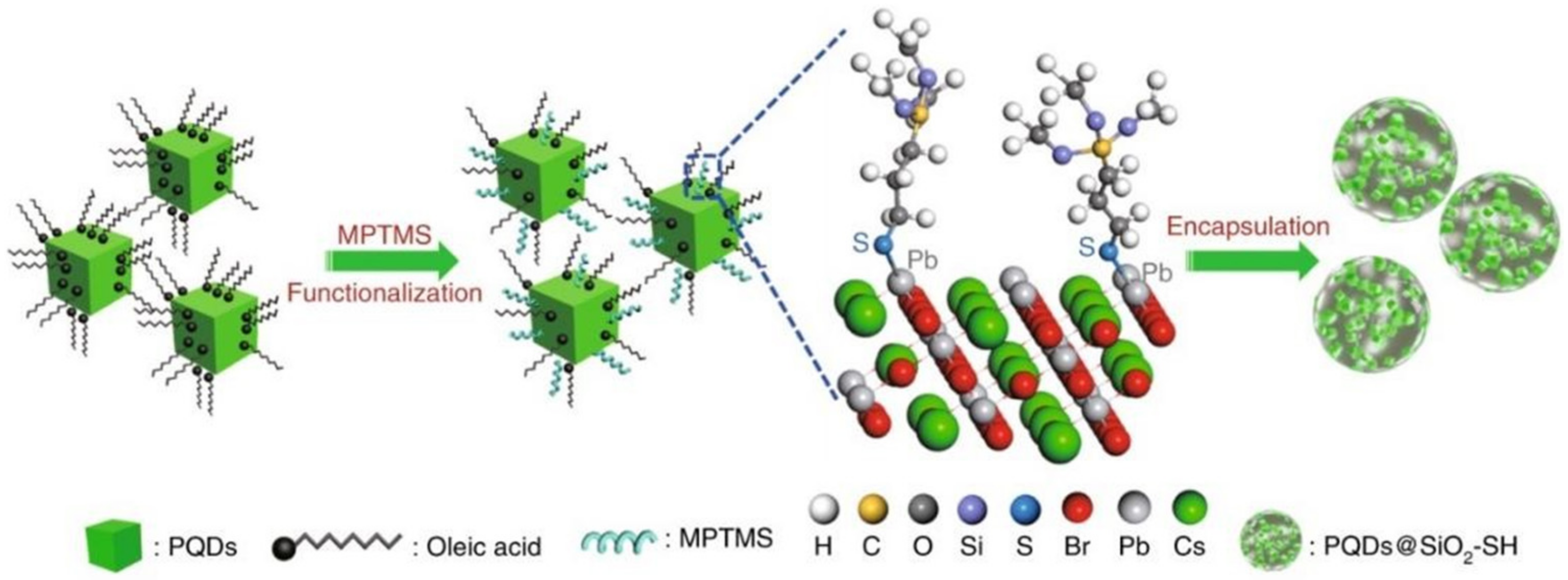
Compared to polymers, inorganic nano oxide coatings can also play a role in stabilizing PNCs, with the advantage of better water dispersion. Nano SiO2 has become a typical representative of inorganic materials used for coating PNCs due to its high heat resistance, stability, and transparency. Li et al. have developed a simple method for producing water-resistant PQDs@SiO2 nanodots (wr-PNDs) that can be easily dispersed in water and maintain their emission for up to six weeks, demonstrating an unprecedented water-resistance capability. The synthetic process for creating wr-PNDs is illustrated in Figure 5. Initially, pristine PQDs are dispersed in a mixed solvent of toluene and (3-mercaptopropyl)trimethoxysilane (C6H15O3Si-SH, MPTMS), followed by the addition of de-ionized water to initiate the hydrolysis of MPTMS. MPTMS molecules can be tightly adsorbed onto the PQDs due to the formation of Pb-S bonding. These bound molecules reduce the interfacial energy between the PQDs and silica by providing additional steric stabilization, thereby preventing phase separation between the PQDs and silica. Silica-encapsulated PQDs-Pb-S-SiO2-SH nanodots are produced as hydrolysis and condensation of MPTMS are triggered by the added water [21].
Porous materials provide a high degree of porosity that can be used to prevent self-aggregation of PNCs and reduce environmental erosion, making it another viable method for enhancing the stability of PNCs coated with inorganic materials. A universal synthesis method of PNCs with pore domain limiting-shell isolation dual protection was developed by You et al.; as depicted in Figure 6. In this approach, perovskite precursor was injected into mesoporous silicon, and PNCs were grown in situ in the mesoporous silicon pores through heating and annealing. Next, dense Al2O3 shells were deposited on the mesoporous silicon surface through atomic deposition technology, which effectively isolates the impact of the environment on PNCs, owing to the abundant -OH on the mesoporous silicon surface. The resulting mSiO2-CsPbBr3@AlOxNCs can remain stably dispersed in water for more than 90 days and can effectively shield the ion exchange effect, thereby significantly enhancing the stability of PNCs and expanding their practical application range [22]. Likewise, Zhang et al. synthesized ceramic-like stable and highly luminescent CsPbBr3 NCs by embedding CsPbBr3 in silica derived from molecular sieve templates at high temperature (600–900 °C). The resulting CsPbBr3-SiO2 powder exhibits high PLQY (~71%) and excellent stability, which can be compared with the ceramic Sr2SiO4:Eu2+ green phosphor [23].
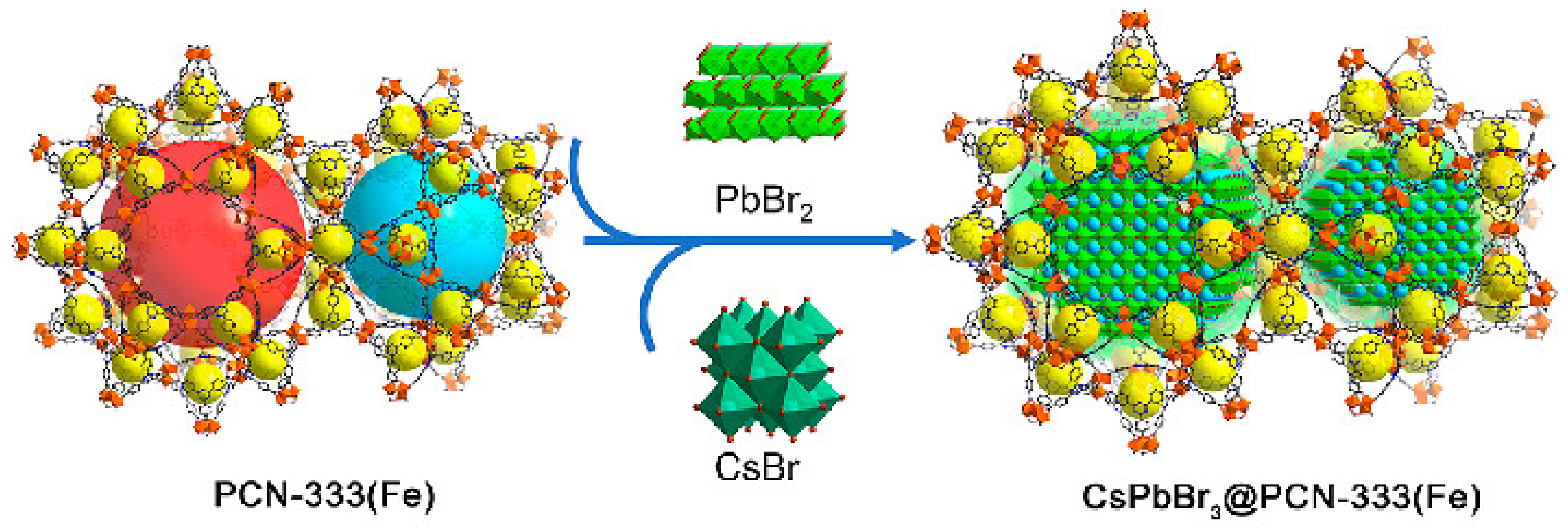
Metal–organic framework compounds (MOFs) are a novel class of porous materials that are assembled by inorganic metals and organic ligands. Due to their high porosity and large specific surface area, MOFs are often utilized as carriers for nanoparticles, drugs, and other materials. Incorporating PNCs into the pores of MOFs can significantly enhance their stability. However, in order to function as a carrier for PNCs, the MOF design must address several challenges. Firstly, the MOF matrix must remain stable during PNCs incorporation and fluorescence detection. As many bivalent metal-based MOFs are sensitive to water or ambient moisture, high-valent metal-based MOFs are ideal hosts that provide stable pore structures for encapsulation. Secondly, the aperture of MOFs must be sufficiently large to accommodate PNCs. While micropore MOFs with apertures smaller than 2 nm are frequently used as encapsulation materials for perovskite nanoparticles, the limited space can hinder QD growth and damage the micropore structure. Therefore, mesoporous MOFs with pore sizes greater than 4 nm are more suitable for PNC fixation. In the case of CsPbBr3 nanocrystals, QDs with complete crystal structures are more suitable for fluorescence detection applications to ensure efficient light absorption and prevent dense surface recombination. Simultaneously, the mesoporous cage can effectively separate the QDs and realize the quantum limited domain of perovskite. As illustrated in Figure 7, Qiao et al. encapsulated CsPbBr3 nanocrystals into a stable ferric organic skeleton (MOF) with mesoporous cages (5.5 and 4.2 nm) via a sequential deposition pathway to obtain a perovskite–MOF composite CSPbBr3@PN-333 (Fe). The CsPbBr3 nanocrystals are stabilized by the restriction of the MOF cage without aggregation or leaching [24]. Wu et al. employed a sequential deposition method to encapsulate MAPbI3 in cheap iron-based MOFs (PSN-221) to obtain the composite MAPbI3@PCN-221(Fex) (x = 0–1), which they used for photocatalytic CO2 reduction. Due to the protection offered by PSN-221 (Fex), this composite catalytic system exhibits ultra-high stability and can operate continuously in water for more than 80 h. Additionally, the maximum efficiency of photocatalytic CO2 is 38 times higher than that of pure PCN-221(Fex) [25].

1.2.2. Ion Doping

Ion doping is a widely used and effective method for introducing heteroatoms into the lattice, making it one of the most important approaches for modifying semiconductors. This method is valuable because it can maintain the structure and properties of the original main crystal to a large extent. Perovskite structures have a high tolerance for impurity doping, enabling various impurity atoms to be doped into the lattice of PQDs. This doping strategy can significantly improve the stability of perovskite and regulate its photoelectric characteristics [26]. The typical perovskite structure (ABX3) consists of corner-sharing [BX6]4− octahedra, with A-site cations confined within the cuboctahedra cages. In the ABX3 structure, the ionic radii of A-site, B-site, and X-site ions must correspond to the Goldschmidt tolerance factor t [27]:
t = r A + r X 2 r B + r X
The Goldschmidt tolerance factor is defined by the formula t = (rA + rX)/√2(rB + rX), where rA, rB, and rX are the ionic radii of the A-site, B-site, and X-site elements, respectively [26]. To maintain a stable perovskite structure, t should fall between 0.813 and 1.107, with a range of 0.9 ≤ t ≤ 1 generally considered optimal for the cubic phase [7]. The [BX6]4− framework confines the A-site cations, and those that are either too large or too small can cause the cubic-like crystal structure to distort, warp, and eventually break down. MA (2.70 Å) is generally the most suitable size for the cubic perovskite structure, whereas FA (2.79 Å) and Cs (1.88 Å) are slightly too large or small to serve as ideal A-sites [28], leading to deviations from the equilibrium interplanar distances. A mixed A-site cation system that combines larger and smaller ions can help to stabilize the 3D perovskite framework and overcome these issues. For instance, Protesescu et al. replaced some Cs+ ions with FA+ ions to improve the structural stability of CsPbI3 PQDs, resulting in a higher t value relative to pure CsPbI3 PQDs. The resulting FA0.1Cs0.9PbI3 PQDs display a 3D orthogonal structure, stable red-light emission (685 nm), a PLQY over 70%, and can maintain their PLQY in solution (with less than 5% relative decrease) after several months of storage at ambient conditions, owing to the restricted transformation from 3D cubic perovskite structure to 1D polycrystals [29]. Triple cations such as (Cs, FA)PbI3 or (Cs, MA, FA)PbI3 have also demonstrated improved stability by promoting phase structure stability, and multi-cationic doping of A sites has emerged as a key method for obtaining high-quality perovskite films [30]. However, it is rarely reported in the field of PNCs [31]. In addition, Wang et al. synthesized ultra-stable quasi-2D PQDs by introducing butadiamine cation (BA+) into methylamine lead bromide perovskite (MAPbBr3), as shown in Figure 8. The quasi-2D perovskite (BA)2(MA)x−1PbxBr3x+1, which has reduced dimensionality, exhibits higher luminous efficiency and better environmental stability than traditional 3D perovskite due to the higher exciton binding energy and formation energy of the reduced dimension perovskite [32].
The incorporation of dopants to replace B-site Pb2+ in perovskite structures has been shown to significantly enhance their stability or modify their optical properties, examples of which include Zn2+, Ni2+, and RE ions [33,34]. The smaller ionic radius of Zn2+ (0.74 Å) compared to Pb2+ (1.19 Å) causes the perovskite lattice to contract, leading to an increase in exciton binding energy and promoting radiative recombination within CsPbI3 PQDs [35]. In addition, Mn2+ doping also increases the Goldschmidt tolerance factor (t), enhancing structural stability [36]. Similarly, the introduction of Ni2+ ions into CsPbI3 PQDs results in lattice contraction due to the smaller ionic radius of Ni2+ (≈0.69 Å) relative to Pb2+ (≈1.19 Å), resulting in a blue shift in the PL and a higher quantum efficiency, as shown in Figure 9a,b. Moreover, Ni2+ doping improves the stability of PNCs by increasing t relative to CsPbI3 PQDs, leading to a more stable perovskite structure. The stability of Ni:CsPbBr3 PNCs with distinct Ni/Pb ratios against moisture and UV light was tested and shown in Figure 9c,d, providing compelling evidence for the beneficial effects of Ni2+ dopants on emission performance [37].

1.2.3. Surface Passivation

The large specific surface area of PNCs results in a high concentration of surface atoms, which in turn leads to an abundance of dangling bonds and defects. These defects can trap electrons in the conduction band and cause non-radiative energy loss, thereby reducing the stability of nanocrystals. Surface passivation using appropriate ligands is an effective strategy to improve the stability of PNCs. However, commonly used ligands like oleic acid and oleylamine are dynamic and can easily desorb in the purification process, leading to PNC aggregation and loss of optical properties. Thus, ligands that bind more firmly to the surface of PNCs are required for better passivation, reducing the impact of water and oxygen and improving stability. Polydentate ligands, containing multiple functional groups, are shown to bind more strongly to nanocrystals and provide improved stability. For instance, Figure 10 illustrates a strategy for introducing polydentate ligand of AHDA to synthesis stable PNCs. The polyamine chelating ligand N′-(2-aminoethyl)-N′-hexadecylethane-1, 2-diamine (AHDA) can anchor the PNC surface lattice with a higher binding energy (2.36 eV) than that of the commonly used oleylammonium ligands (1.47 eV). The strong chelation effect achieves ultra-stable CsPbI3 PNCs with a high binding energy, exhibiting excellent stability even under harsh conditions such as repeated purification, polar solvents, heat, and light [38]. The use of mercaptan materials such as 2-amino-ethylmercaptan (AET) can also lead to tightly bound ligand layers on the surface of PNCs, resulting in high stability, even in water. The AET-CsPbI3 QDs (in both solutions and films) retained more than 95% of the initial PL intensity in water after 1 h [39]. These strong chelating ligand strategies offer promising avenues for achieving stable and high-performance PNCs for a range of applications.
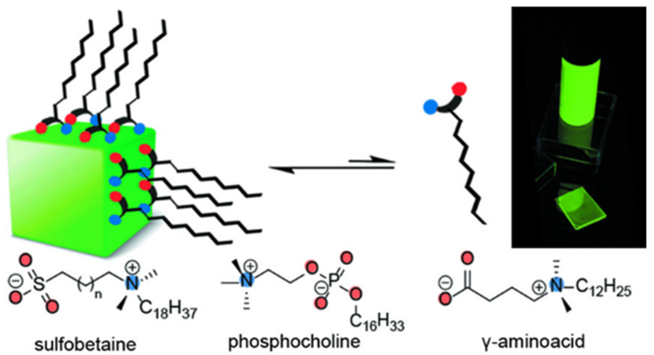
In recent years, zwitterionic organic ligands have emerged as a promising approach for surface engineering of PNCs and defect elimination, owing to their multifunctional zwitterionic structure. As shown in Figure 11, Krieg et al. proposed a surface passivation strategy for PQDs using long-chain zwitterionic ligands such as sulfobetaine and phosphocholine, which comprise both cationic and anionic groups. These zwitterionic ligands can coordinate with both surface cations and anions and are firmly attached to the surface of PQDs through the chelation effect, thereby obtaining PQDs with high stability. For example, 3-(N,Ndimethyloctadecylammonio) propanesulfonate can result in much improved chemical durability. The sulfobetaine-capped CsPbBr3 NCs retained PLQYs in the range of 70–90% for 28–50 days under ambient conditions [40]. Moreover, they demonstrated that the strong binding between the amphoteric lecithin and PQDs provides CsPbBr3 PQDs solutions with superior colloidal stability over a wide range of concentrations [41]. In another study, Wang et al. used polymers containing multiple sulfobetaine amphoteric groups to achieve desirable surface coatings and CsPbBr3 PQDs that maintain bright PL emission even after 1.5 years of storage in polar solvents such as acetone and ethanol [42].
In addition, the presence of long alkyl chain organic ligands in PQD films severely impedes charge transport and interlayer coupling, calling for the development of more conductive organic semiconductor ligands to prepare PNCs with favorable optoelectrical properties. Pan et al. demonstrated that the incorporation of organic semiconductor molecules (rhodamine B derivative, COM) into CsPbBr3 NCs resulted in COM-CsPbBr3 NCs with a high PLQY of 82% and exceptional stability under harsh commercial accelerated operating stability tests such as high temperature (85 °C) and high humidity (85%). The product exhibits remarkable endurance against high temperature and high humidity, retaining 84% of the initial PL intensity value for 300 h. The stability of CsPbBr3 NCs can be significantly improved by the strong interaction between organic semiconductor molecules and the quasi-type II heterostructure formed by CsPbBr3 and moderate photocarrier transfer. Furthermore, the diversity and versatility of organic semiconductors present a broad prospect for their combined application in the field of luminescence and display [43].
Table 1 summarizes methods to improve the stability of PNCs and their stability performance. Surface coating is a way from outside to inside, playing the main role of encapsulation. Metal ion doping and surface passivation are ways to improve the intrinsic structural stability from inside to outside. They are not absolute by classification and may work together to improve the stability of PNCs sometimes.

2. Application of PNCs as Fluorescent Probes in the Detection of Agriculture- and Food-Related Hazardous Substances

2.1. The Detection of Agriculture-Related Hazardous Substances

2.1.1. Pesticide Residue Detection

Pesticides are crucial in minimizing crop losses, ensuring food security, preventing the emergence and spread of plant diseases and insect pests, and improving health conditions. However, their use also leads to environmental pollution, pesticide residues, and other issues. Pesticide residues refer to the presence of trace amounts of pesticide precursor, toxic metabolites, degradants, and impurities that remain in grains, vegetables, fruits, livestock products, aquatic products, soil, and water bodies even after the application of pesticides in agricultural production. Currently, the annual production of chemical pesticides worldwide is nearly 4.1 million tons, and these synthetic compounds were utilized as pesticides, fungicides, algaecides, deworming agents, defoliants, and other pesticides [44]. The substantial use of pesticides, particularly organic pesticides, has resulted in severe pesticide pollution. Ingesting food containing high levels of highly toxic pesticide residues can lead to acute poisoning in both humans and animals. While long-term consumption of agricultural products with excessive pesticide residues may not cause acute poisoning, it can result in chronic poisoning in humans, leading to the development of diseases, inducing cancer, and even affecting future generations [45]. Therefore, the real-time and rapid detection of pesticide residues in agricultural products is of great significance for global food safety and environmental safety. Researchers have conducted significant research on rapid detection technologies for pesticide residues, especially for organophosphorus, organochlorine, triazine herbicides, and other pesticides, yielding some remarkable achievements.

Organophosphorus Pesticide

Organophosphorus pesticides are a type of pesticide that contains organic compounds with phosphorus. They are widely used in agricultural production to control plant diseases and insect pests but can leave residues in crops. Ensuring food safety requires rapid and accurate detection of organophosphorus pesticides.
Dimethoate oxide (OMT) is an organophosphorus insecticide with a strong imbibition insecticidal effect. It can be absorbed into the plant stem and leaves and transmitted to other parts of the plant to kill harmful pests. Huang et al. successfully synthesized a new MIPs@CsPbBr3 QDs composite using imprinting technology with a sol–gel reaction, as shown in Figure 12. APTES-capped CsPbBr3 QDs were used as a fluorescent carrier, and TMOS was used as the cross-linker to gradually form a silica matrix, improving the stability of perovskites, and synthesized water-soluble PQDs. OMT was selected as the template molecule due to its hydrogen bonding interaction with APTES. Loading the MIPs with OMT significantly quenched the fluorescence of MIPs@CsPbBr3 QDs, with a linear range of OMT from 50 to 400 ng/mL and a LOD of 18.8 ng/mL. After removing the OMT template via solvent extraction, the resulting MIP@CsPbBr3 QD composites showed selective recognition ability towards specific template-shaped molecules such as dimethoate, with an imprinting factor of 3.2, indicating excellent specificity of the MIPs for inorganic metal halide (IMH) perovskites [4].
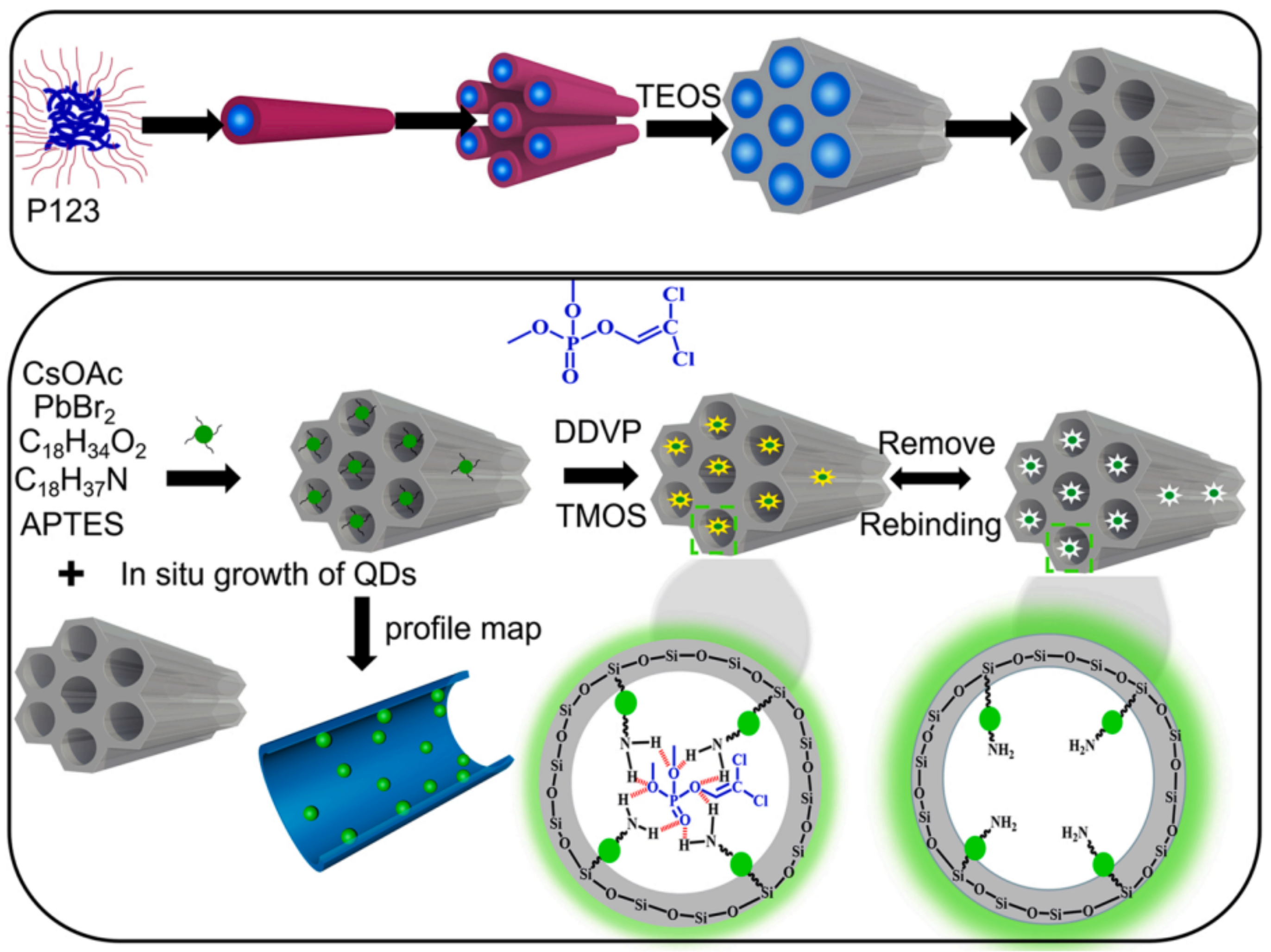
2,2-dichlorovinyl dimethyl phosphate (DDVP) is a highly effective and wide-spectrum organophosphorus insecticide commonly used to control various pests on crops such as cotton, fruit trees, vegetables, sugarcane, tobacco, tea, and mulberry. However, on 27 October 2017, the World Health Organization’s International Agency for Research on Cancer classified DDVP as a group 2B carcinogen. In a study by Huang et al.; molecularly imprinted mesoporous silica (MIMS) with in situ perovskite CsPbBr3 quantum dots (QDs) was prepared and applied for the fluorescence detection of DDVP, as shown in Figure 13. The QDs stability was greatly improved by encapsulating them in mesoporous silica, resulting in higher analytical performance. The design of PQDs-encapsulated MIMS is based on PQDs being encapsulated into mesoporous silica and acting as a transducer for recognition signal fluorescent readout, while molecular imprinting on the surface of mesoporous silica provides selectivity. CsPbBr3 QDs with uniform size and high fluorescence intensity were first grown directly in the channels of mesoporous silica SBA-15. Then, molecular imprinting was applied to the surface of QDs-encapsulated mesoporous silica using a sol–gel method. The mesoporous silica provided a solid support for molecular imprinting and acted as a medium for the in situ synthesis of QDs. The QDs’ fluorescent properties were expected to be affected once the template molecules were captured in the binding sites, reducing the distance between the template and the QDs. Under optimized conditions, the QDs-encapsulated MIMS showed a linear relationship between DDVP concentration of 5–25 μg/L, and the LOD was 1.27 μg/L. The recovery of DDVP increased from 87.4% to 101% [46].
Phoxim is a highly effective broad-spectrum organophosphorus insecticide, exhibiting properties of stomach toxicity, contact pesticide, and long-lasting effect under dark conditions. To detect phoxim specifically and sensitively in samples, Tan et al. synthesized perovskite CsPbBr3 QDs embedded in molecularly imprinted polymers (MIP) by thermal injection. As depicted in Figure 14, CsPbBr3 QDs with high-brightness cores were encapsulated by MIPs using a sol–gel approach initiated by trace moisture in the air. To overcome the disadvantage of using siloxane monomers with simple organic groups, a multifunctional monomer, BUPTEOS, was synthesized with phenyl and hydrogen bond donors. During pre-polymerization, BUPTEOS and phoxim formed a complex through hydrogen bond interactions and π-π accumulation, leading to the formation of specific imprinted cavities in the MIP/QDs composites upon slow sol-gel molecular imprinting process and removal of templates. The resulting MIP/QDs complex demonstrated excellent selectivity to phoxim, with a blotting factor of 3.27. Compared to previous studies on detection of organophosphorus pesticides, the MIP/QDs fluorescent probe exhibited high sensitivity and specificity. Under optimized conditions, the fluorescence quenching of MIP/QDs showed a good linear correlation with the concentration range of 5–100 ng/mL, and the LOD was 1.45 ng/mL. By encapsulating the QDs of the novel MIP/perovskite CsPbBr3 QDs fluorescent probe in the imprinted silica matrix, the stability of the QDs in phoxim detection was significantly improved. In this study, siloxane functional monomers with two functional groups were synthesized by in situ slow hydrolysis of organosilicon monomers [47].

Triazine Herbicides

Triazine herbicides can be classified into symmetrical triazines (1,3,5-triazine) and asymmetric triazines (1,2,4-triazine). The main commercial herbicide is symmetrical triazine, which can be further divided into thiomethyl-s-triazine (e.g., prometryn, ametryn, and terbutryn), simazine, terbuthylazine, fluoroalkyl-s-triazine (e.g., indaziflam and triaziflam), and methoxytriazine (e.g., prometon and terbumeton) [48].
Prometryn is commonly used for pre- and post-bud weeding in cotton and bean fields as well as for algae removal in aquatic products. Zhang et al. developed a novel molecularly imprinted electrochemical luminescence (MIECL) sensor by using perovskite quantum dots (PQDs) coated with a molecularly imprinted silica layer (MIP/CsPbBr3-QDs) as recognition and response elements. As shown in Figure 15, the MIP/CsPbBr3-QDs layer was immobilized onto the surface of a glassy carbon electrode (GCE) via one-pot electropolymerization to enhance water resistance, improve the stability of CsPbBr3-QDs, and reduce film shedding. The highly selective and ultrasensitive MIECL sensor was successfully constructed for prometryn analysis in fish and seawater samples. The LODs for fish and seawater samples were 0.010 μg/kg and 0.050 μg/L, respectively. The recoveries of fish and seawater samples were 88.0% to 106.0%, with a relative standard deviation lower than 4.2%. The MIECL sensor based on MIP/CsPbBr3-QDs shows good stability, accuracy, and precision and can sensitively detect prometryn in aquatic products and environmental samples [49].
Simazine is a highly effective selective herbicide that has been listed as a category 3 carcinogen by the International Agency for Research on Cancer (IARC) on 27 October 2017. To detect simazine in aquatic products, Pan et al. developed a novel MIECL sensor based on the molecular-imprinted polymer perovskite (MIP-CsPbBr3) and Ru(bpy)32+ luminescent molecules. Under optimal experimental conditions, MIP-CsPbBr3-GCEs were exposed to different concentrations of simazine solution (0.1–500 μg/L) and analyzed using the MIECL sensor. The ECL strength showed a linear relationship with the logarithm of simazine concentration within the range of 0.1–500.0 μg/L, and the limit of detection (LOD) was 0.06 μg/L. The MIECL sensor method was validated by analyzing fish and shrimp samples with recovery rates of 86.5–103.9% and relative standard deviations lower than 1.6%. The developed MIECL sensor demonstrated excellent selectivity, sensitivity, reproducibility, accuracy, and precision for the determination of simazine in actual aquatic samples [50].

Organochlorine Pesticides

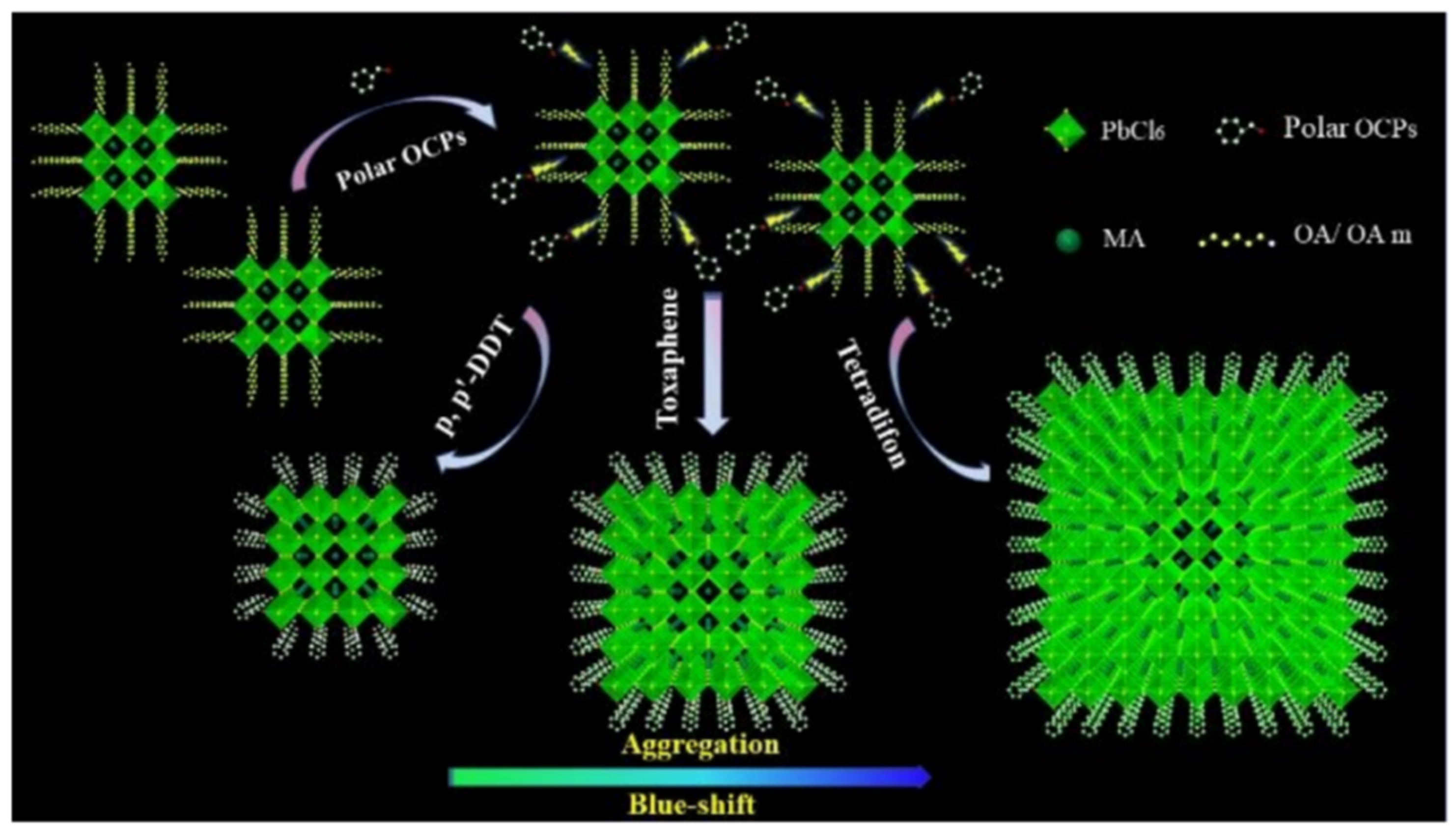
Organochlorine pesticides are a group of organic compounds containing organochlorine elements, commonly used to prevent and control plant diseases and insect pests. They can be categorized into two main types: benzene-based and cyclopentadiene-based. Yang et al. demonstrated that CH3NH3PbBr3 quantum dots (MAPB-QDs) undergo a blue shift in fluorescence spectra upon exposure to polar organochlorine pesticides (OCPs), providing a method for the detection of these compounds. The presence of polar OCPs alters the structure and emission properties of MAPB-QDs by desorbing the capping ligands OA and OAm from their surface, resulting in defect sites that can be occupied by polar OCPs. This allows the chlorine element in OCPs to fully dope into the QDs, increasing the band gap of the MAPB-QDs and causing a blue shift in their wavelength. To address the insufficient stability of MAPB-QDs in the presence of moisture, the researchers mixed MAPB-QDs with PDMS to create a colorimetric card that can be used for the rapid detection of OCPs in actual samples. This study represents the first use of MAPB-QDs for the detection of polar OCPs (Figure 16) [51].

Other Pesticides

Clodinafop is a herbicide belonging to the aryloxy phenoxy propionate class, soluble in various organic solvents but decomposes under strong acidic or basic conditions. Vajubhai et al. synthesized CsPbI3 PQDs using a microwave radiation method and utilized them for detecting clodinafop in samples via fluorescence spectrometry. Figure 17 illustrates the simple synthesis method of CsPbI3 PQDs utilizing PbI2, Cs2CO3, OAm, OA, and ODE as reagents. The resulting PQDs showed a strong red emission at 686 nm and were well-dispersed in hexane. They were utilized as a probe for fluorescence detection of clodinafop in an organic phase (hexane), where the strong red emission at 686 nm was quenched by clodinafop. The study found a good linear relationship between the quenching intensity and the concentration of clodinafop in the range of 0.1–100 μM, with a LOD of 34.70 nM. Furthermore, the fluorescent probes were integrated into liquid–liquid microextraction for the fluorescence analysis of clodinafop in fruits, vegetables, and grains. The developed probes showed a good linearity to the concentration of clodinafop in the range of 0.1−5.0 μM, with a LOD of 34.70 nM, and exhibited a good recovery rate (97−100%) and low relative standard deviation. The results indicate that CsPbI3 PQDs can be used as a sensor for the quantitative detection of clodinafop in samples [1].
Propanil is a highly selective contact herbicide that contains amides, primarily used in rice fields or seedling fields to control barnyard grass and other gramineous and dicotyledonous weeds. The MIP-QDs, under optimized parameters, showed good linearity in the concentration range of paspalum from 1.0 μg/L to 2.0 × 10⁴ μg/L. The developed MIP-QD-based fluorescent probe exhibited a good recovery rate ranging from 87.2% to 112.2%, with relative standard deviations of less than 6.0% for fish and seawater samples. The LOD of paspalum in fish and seawater was 0.42 μg/kg and 0.38 μg/L, respectively. Fluorescence test strips based on MIP-QD also showed satisfactory recovery rate ranging from 90.1% to 111.1%, and the LOD of paspalum in seawater samples was 0.6 μg/L. The developed fluorescent probe and test strip were successfully used for the detection of paspalum in environmental and aquatic products [52].
As shown in Table 2, PNCs demonstrate good stability, high sensitivity, low LOD, and high recovery rate for conventional pesticides. Furthermore, they can be developed into test strips for rapid detection with broad and convenient application prospects.

2.1.2. Environmental Pollutant Detection

Uranium is a radioactive element found in various chemical forms in the environment, with uranyl ion (UO22+) being the most common valence state under normal conditions. Inhalation or ingestion of U can lead to irreversible kidney damage, destruction of biomolecules, and DNA damage. According to World Health Organization guidelines, the concentration of UO22+ in drinking water should be less than 30 ppb. As most uranium complexes are highly soluble in water and mobile in aquatic environments, detecting its presence is essential for monitoring agroecological systems.
Halali et al. synthesized all-inorganic CsPbBr3 PQDs with bright PL through thermal injection and used them for UO22+ sensing. As shown in Figure 18, the first step involved the adsorption of UO22+ on CsPbBr3 PQDs, and zeta potential measurements confirmed the process of electrostatic attraction and adsorption, facilitating quenching. Dynamic light scattering (DLS) revealed that CsPbBr3 PQDs treated with uranyl ions had significantly larger hydrodynamic radii (1244 nm) than the untreated probe (47.9 nm), indicating adsorption of UO22+ on CsPbBr3 PQDs. Fluorescence quenching resulted from the adsorbed UO22+ ions on the surface of CsPbBr3 PQDs. The ultra-low LOD of the probe was 83.33 nM (19.83 ppb), and it demonstrated rapid detection of UO22+ even in non-polar solvents such as toluene. This study suggests new avenues for designing probes (such as PQDs) and analytes (such as metal ions) for trace level sensing of metal ions [53].
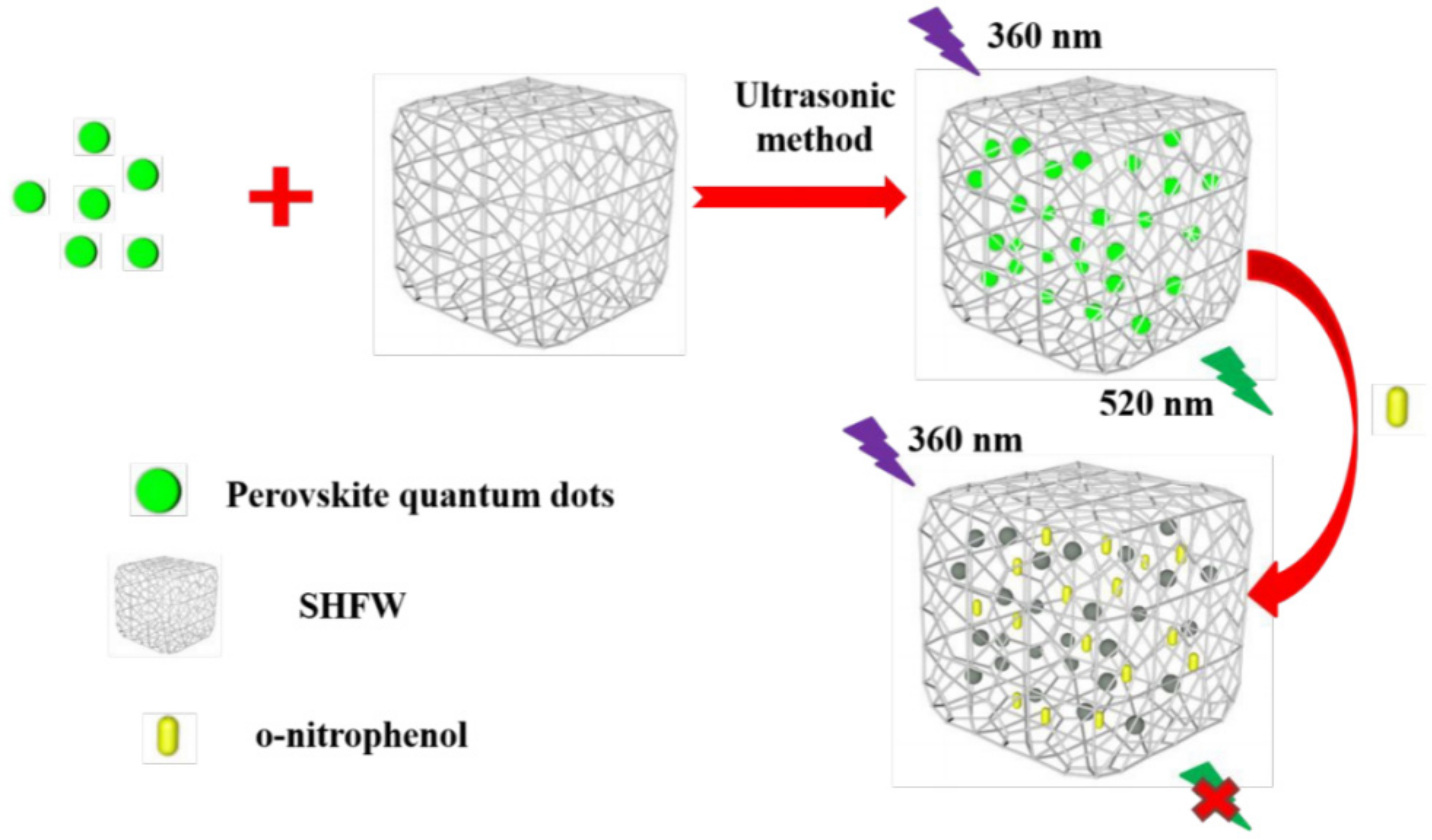
O-nitrophenol (ONP) is a member of the phenolic compound family and is widely used as an intermediate in organic synthesis, dyes, medicines, and other products. ONP is also employed as a photographic developer, pH indicator, and photosensitive material, and as a rubber antioxidant. However, ONP is highly toxic and is considered a priority pollutant for environmental analysis and monitoring. Inhaling ONP can cause adverse effects such as headache, dizziness, and nausea, and serious exposure can lead to damage to the central nervous system or the liver/kidney function. Deng et al. developed a molecularly imprinted fluorescent probe using a combination of highly luminescent PQDs and molecular imprinting technology to rapidly detect ONP (see Figure 19). To enhance the hydrophobic properties of the PQDs, a superhydrophobic porous organic polymer framework (SHFW) was created by combining a superhydrophobic material with a pore-forming agent. This SHFW protected the PQDs from contact with water vapor in the air and increased their stability. The CsPbBr3@SHFW fluorescent probe was designed using SHFW-modified CsPbBr3 QDs to detect ONP in river silt. The probe exhibited high selectivity for ONP, and under optimal detection conditions, the linear range of CsPbBr3@SHFW for ONP detection was 0–280 μM, with a limit of detection of 7.69 × 10⁻³ μM [54].
Table 3 summarizes the PNC-based fluorescent probes for environmental pollutant detection. High fluorescence performance of CsPbBr3 PQDs proved them to be efficient fluorescent probes for the detection of pollutants U and ONP.

2.1.3. Ion Detection

Cation

As industrialization and urbanization continue to progress, harmful ion pollutants have become a major source of land and water pollution, with highly toxic ions posing a global concern. Conventional detection methods are beset with issues such as high cost, long detection cycle, low accuracy, and low efficiency. Thus, the development of simple and selective sensors is crucial for detecting harmful ion pollutants. Compared to other methods, the fluorescence-based method has advantages such as low cost, high sensitivity, fast detection, and ease of use. Using PNCs to develop trace detection methods represents a breakthrough in new methods.
Liu et al. synthesized all-inorganic CsPbBr3 PQDs via the thermal injection method as a fluorescent probe for the detection of Cu2+ in organic phase. The cubic-phase CsPbBr3-NC exhibits a quantum yield as high as 90%. The PL intensity of CsPbBr3 PQD was quenched significantly within seconds of adding Cu2+ owing to effective electron transfer from PQD to Cu2+, as revealed by absorption spectra and transient PL lifetime experiments. The sensor detected Cu2+ in the range of 0 to 100 nM, with a low LOD of 0.1 nM, demonstrating excellent sensitivity and selectivity for Cu2+ in hexane [55].
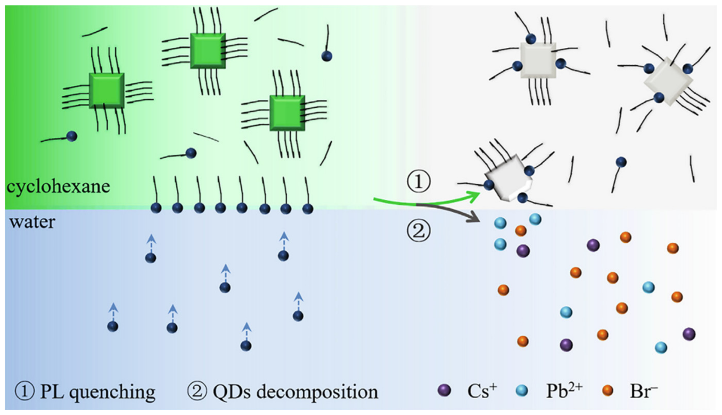
Li et al. established a method for detecting Cu2+ in aqueous solution based on CsPbBr3 (CPB) QDs. A strong organic ligand (oleylamine, OAm) was added, and due to the strong interaction between the amine ligand and metal ions, OAm in cyclohexane captured Cu2+ from water, forming an OAm-Cu2+ complex at the cyclohexane/water interface (Figure 20). These Cu2+ complexes rapidly diffused into cyclohexane, ultimately quenching the PL of CPB QDs. The fluorescence probe based on CPB QDs exhibited a wide linear range (106 M–102 M), a short response time (1 min), and good selectivity for Cu2+. The metal ion phase transfer strategy avoided direct application of unstable PQDs in a water environment [56].
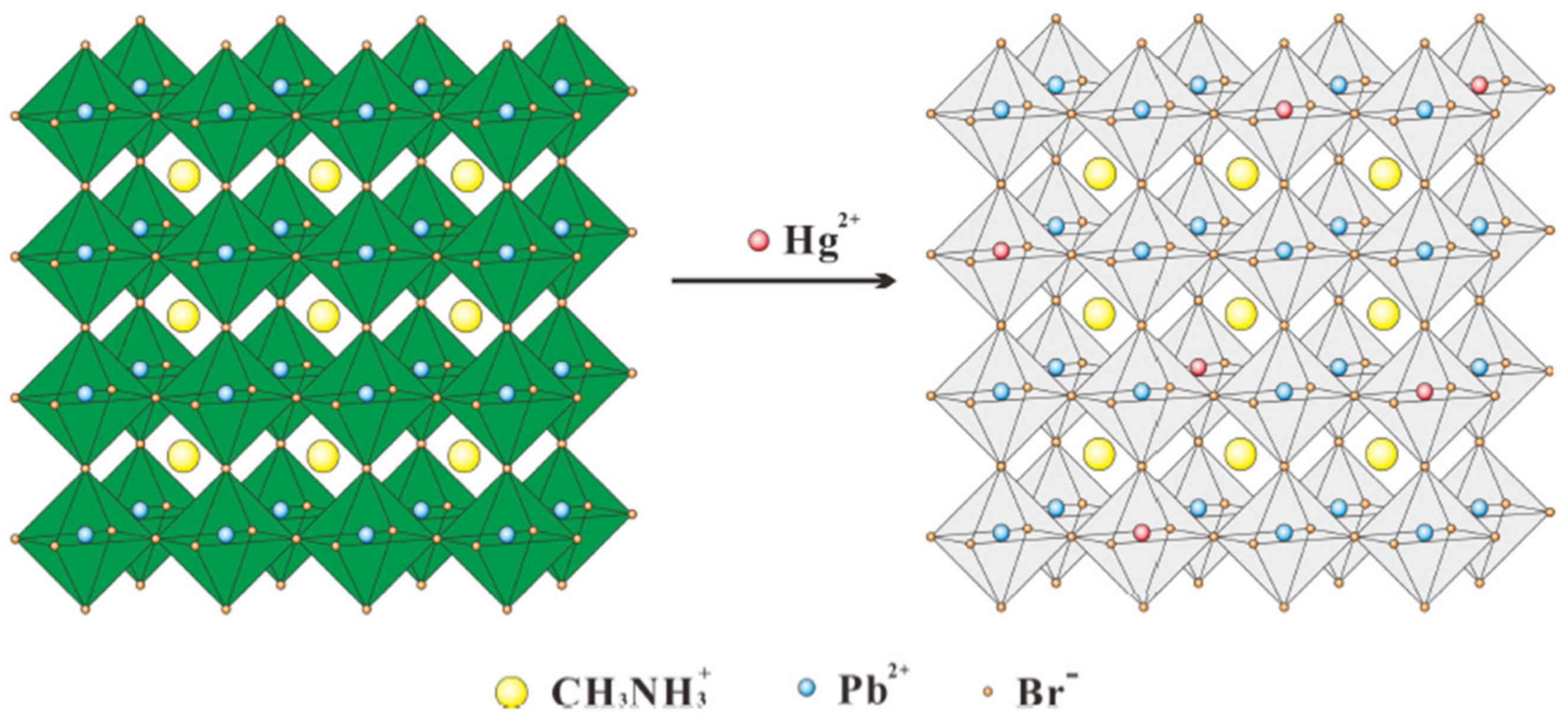
Mercury ion pollution is a significant issue that impacts human health and environmental safety. To address this problem, Lu et al. developed a novel fluorescence nanosensor based on the surface ion exchange mechanism for rapid, highly sensitive, and selective visual detection of mercury ions (Hg2+), using CH3NH3PbBr3 PQDs. CH3NH3PbBr3 PQDs were synthesized using ligand-assisted reprecipitation (LARP) technology, without adding any modifier. These PQDs exhibit strong green fluorescence with a high quantum yield of 50.28%. The main principle of the sensor is based on the fact that the strong green fluorescence of CH3NH3PbBr3 QDs at 520 nm is significantly quenched by Hg2+, and the blue shift occurs with the increase of Hg2+ fluorescence due to surface ion exchange, which differs from most other detection mechanisms shown in Figure 21. The LOD of the sensor is 0.124 nM (24.87 ppt) within the range of 0 nM to 100 nM. The fluorescence intensity of PQDs remains unaffected by interfering ions such as Cd2+, Pb2+, Cu2+, etc.; indicating high selectivity of the fluorescent probe towards Hg2+. The detection of Hg2+ using CH3NH3PbBr3 PQDs has several advantages, including simple operation, small sample size, high efficiency, and visual detection [57].
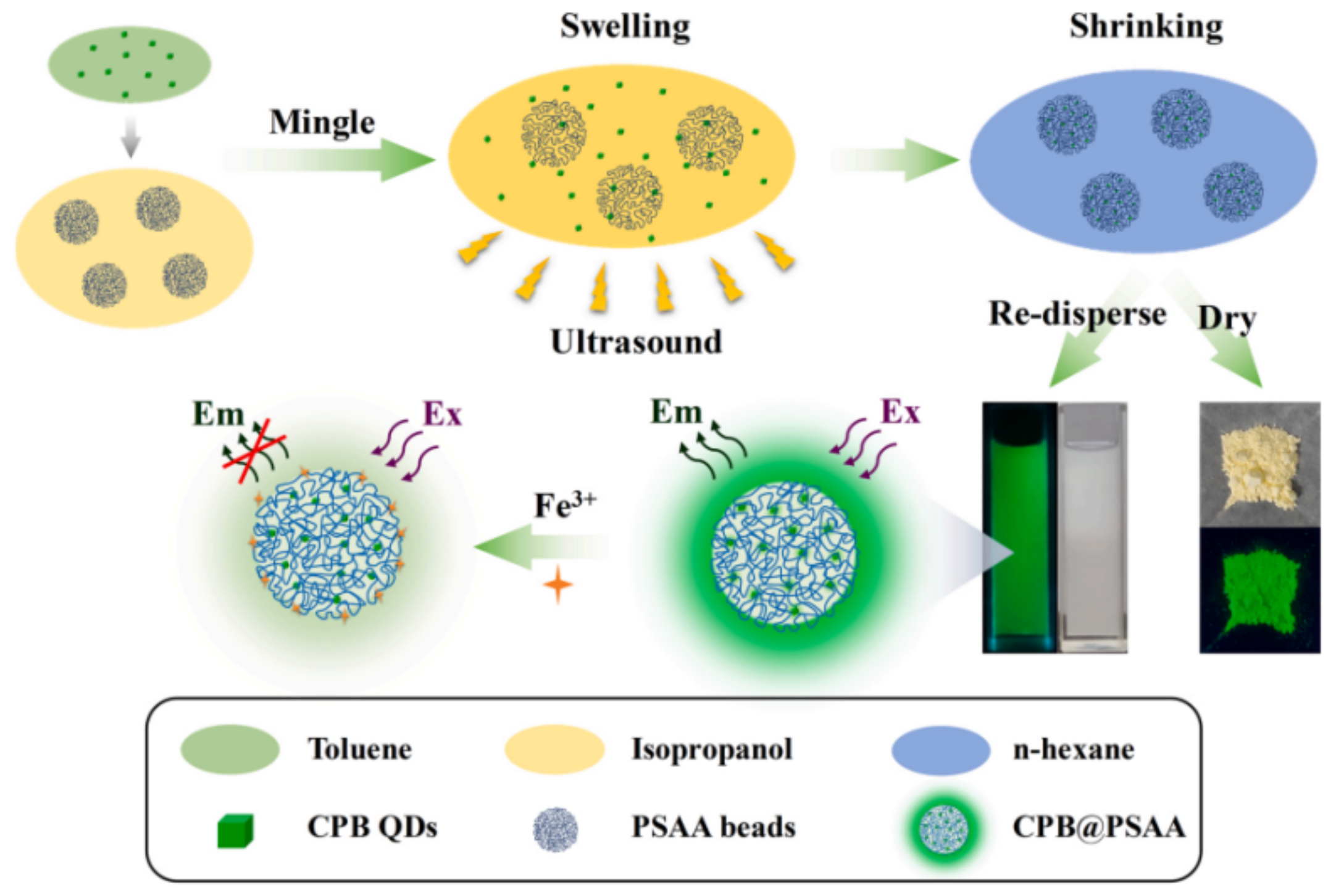
Chen et al. developed a novel method for detecting Fe(III) by encapsulating CsPbBr3 PQDs into poly(styrene/acrylamide) nanospheres using an improved expansion and contraction strategy, as illustrated in Figure 22. The CPB PQDs were dissolved in toluene, which also served as a swelling agent for PSAA polymers, while isopropanol was used as a dispersant for PSAA spheres. After ultrasonic swelling treatment, the composites were rapidly extracted from n-hexane. The resulting CPB@PSAA composites were well-dispersed in an aqueous solution and maintained their bright fluorescence for over 12 months. The fluorescence of the solution could be selectively quenched by the presence of iron ions, enabling the selective sensing of Fe(III). The results demonstrated a good linear relationship between 5 and 150 μM under optimal conditions, with an LOD of approximately 2.2 μM. Moreover, Fe(III) sensing was successfully applied to Yangtze River water, human serum, and tea with satisfactory results [58].
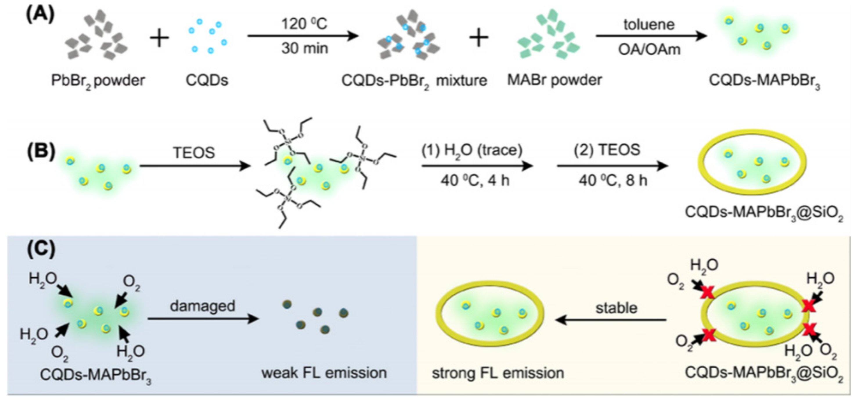
In 2019, Wang et al. developed a novel composite material, carbon quantum dots (CQDs) doped MAPbBr3 PQDs encapsulated with ultra-stable SiO2, for potential applications in fluorescence analysis. The composite material was prepared by using carboxy-rich CQDs and MAPbBr3 to form hydrogen-bond interactions, as depicted in Figure 23, which significantly improved the thermal stability of CQDs-MAPbBr3. Furthermore, the composite material was encapsulated with highly dense SiO2 via in situ growth, which greatly enhanced its water resistance and stability. The composite material exhibited excellent water and thermal stability for over 9 months in aqueous solution and maintained strong fluorescence emission even after annealing at 150 °C. The composite material was then employed for fluorescence analysis to detect Zn2+ and Ag+ [59].
Yan et al. developed a novel fluorescence detection platform for selective detection of Pb2+ ions based on the excellent luminescence properties of lead halide perovskite CH3NH3PbBr3 (MAPbBr3) and CsPbBr3. As shown in Figure 24, a high concentration of CH3NH3Br (MABr) solution was employed as a fluorescent probe. Upon adding PbBr2, a rapid chemical reaction occurred to form MAPbBr3, which exhibited a significant luminous response under ultraviolet light. The fluorescence probe mechanism for Pb2+ is attributed to the excellent PL properties of MAPbBr3 in MAPbBr3@MABr solution. Hence, the non-fluorescent MABr demonstrated a sensitive and selective luminescence response to Pb2+ under UV irradiation. Moreover, the reaction of MABr and PbBr2 facilitated the conversion of Pb2+ to MAPbBr3, enabling the extraction of Pb2+ from waste products. Figure 24 shows the detailed experimental process and results of this study [60].
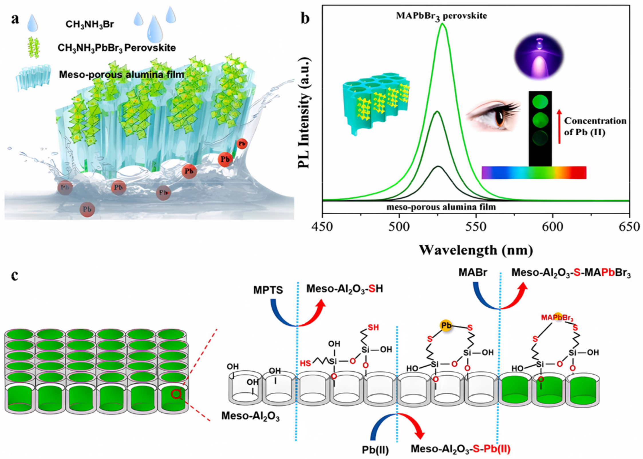
When MABr encounters Pb (II) bound to sulfhydryl groups, it leads to the growth of high-fluorescence (MAPbBr3) perovskite in situ in sulfhydryl-functionalized mesoporous alumina films. This sulfhydryl modification significantly enhances the extraction ability of mesoporous alumina membranes for Pb (II), as reported by Wang et al. Figure 25 illustrates the selection of a commercial meso-Al2O3 film with a pore size of 20 nm as a prototype for 3-mercaptopropyltriethoxysilane (MPTS) modification, due to the strong Pb-S bonding. The hydroxyl groups on the surface of the meso-Al2O3 film facilitate the sulfydryl modification through silane hydrolysis, as shown in Figure 25c. The meso-Al2O3-SH film efficiently enriches Pb(II) in aqueous solutions, which is subsequently dried in an oven. Excess MABr solution is added to the film, and the solvent is evaporated in an oven at 60 °C. Upon drying, MAPbBr3 nanocrystals form on the skeleton or in the holes of the meso-Al2O3-SH film, exhibiting green fluorescence emission under the excitation of 365 nm ultraviolet light (Figure 25a). The PL intensity of the MAPbBr3 nanocrystals increases linearly with the increase of Pb(II) concentration in the sample solution, enabling the visual determination of Pb(II) (Figure 25b). Under optimal conditions, the adsorption capacity of Pb (II) by sulfhydryl-functionalized membranes reaches 94.9 μg/g. Due to the strong extraction ability of Pb (II), the fluorescence opening determination of Pb (II) is realized by forming MAPbBr3 perovskite at the site of sulfhydryl-functional alumina film. The LOD is as low as 5 × 10⁻³ μg/mL. This method integrates the extraction and determination of Pb (II) without background, effectively addressing the problem of low concentration detection and promoting the entire analysis process [61].

Anion

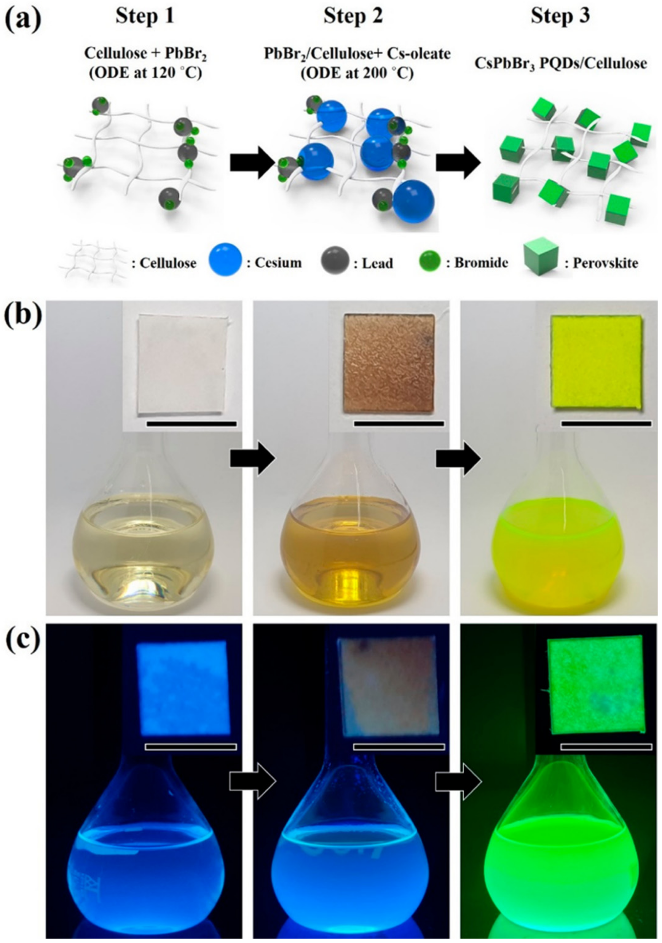
Chlorine and iodine residual trace elements during tap water disinfection can pose significant potential harm to human health. Park et al. synthesized CsPbBr3 perovskite quantum dots (PQDs) cellulose composites using a thermal injection method, as depicted in Figure 26. The composites showed excellent stability, durability, and photoluminescence (PL) properties under various humidity conditions. Initially, cellulose fibers were stabilized with PbBr2 precursors to adsorb ions between fibers (Figure 26a). Subsequently, Cs-oleate was added to the PbBr2/cellulose composite at a higher temperature of 200 °C, leading to the nucleation of CsPbBr3 PQDs and their immediate physical aggregation with cellulose fibers. The successful synthesis of CsPbBr3 PQDs and their integration with cellulose nanofibers were confirmed through green PL under UV field (Figure 26c). A portable sensor for early diagnosis was developed based on this composite, which used the rapid anion exchange strategy between halide anions in cellulose fiber PQDs with a high porous structure to detect iodide and chloride ions in real water samples, with detection values of 2.56 mM and 4.11 mM, respectively. CsPbBr3 PQDs/cellulose composites have the advantages of using low-cost materials for one-pot continuous synthesis, high sensitivity, reasonable detection time, simple and portable visual detection, flexibility of practical application, and a hydrophilic sensing platform. The response detection time can be shortened to change the color, indicating the possibility of real-time and on-site detection [62].
Table 4 summarizes the PNC-based fluorescent probes developed for ion detection. The probes have demonstrated high sensitivity, rapid response times, and ease of use in detecting Cu2+, Hg2+, Fe3+, Zn2+, Ag+, Pb2+, I, and Cl anions in environmental samples. These features make PNC-based fluorescent probes promising candidates for visual detection of harmful ions with great potential for practical applications.

2.2. The Detection of Food-Related Hazardous Substances

2.2.1. Antibiotic Detection

The overuse of antibiotics can lead to the proliferation of resistant bacteria, which can have harmful effects on human health and cause allergic reactions. Furthermore, the presence of antibiotic residues in food, due to their usage in livestock, is a significant concern. Hence, a simple and highly sensitive technique is required for detecting antibiotics in food samples. Roxithromycin (ROX) is a new generation of macrolide antibiotics that mainly targets Gram-positive bacteria, anaerobic bacteria, chlamydia, and mycoplasma. Han et al. have developed a novel CsPbBr3-loaded PHEMA MIP nanogel with high stability against water and oxidation, using multifunctional MIP nanogel synthesized from four 2-(hydroxyethyl)methacrylate (HEMA) derivatives with different functions. First, MIP antioxidant-nanogels were prepared with ROX as a template, and then perovskite nanoparticles were loaded into nanogels via in situ synthesis through the hot-injection method. The developed CsPbBr3-loaded MIP antioxidant-nanogels exhibited excellent stability to air/water and enhanced stability to water-based solvents. Finally, these nanogels were successfully applied for the selective and sensitive detection of ROX antibiotics in animal-derived food products, as shown in Figure 27. The limit of selectivity and sensitivity to ROX was 1.7 × 10⁻⁵ μg/mL (20.6 pM), and the detection results demonstrated good recovery, indicating the excellent performance of the developed MIP antioxidant-nanogel loaded with CsPbBr3 [63].
Tetracycline (TC) is a class of broad-spectrum antibiotics with phenanthrene nuclei discovered in the 1940s. This class of antibiotics is widely used to treat infections caused by Gram-positive and negative bacteria, intracellular mycoplasma, chlamydia, and Rickettsia. In addition, tetracycline is often used as a growth promoter for animals in some countries, including the United States.
In Figure 28, Jia et al. developed a perovskite-composited ratiometric fluorescent nanosensor with good water stability for detecting tetracycline. The sensor was inspired by the respective advantages of perovskite and rare earth elements. The team prepared rod-like MAPbBr3@PbBr(OH) perovskite using the hydration method, with the PbBr(OH) surface layer providing dual advantages of preventing water from entering PNCs and dispersing PNCs in the matrix, resulting in high stability of MAPbBr3@PbBr(OH) in water. Under UV irradiation, the perovskite nanorods emitted stable green fluorescence, which served as the fluorescence internal standard for ratiometric fluorescent nanosensors. The silica shell was used to cover the surface of PQDs, which facilitated the subsequent surface modification of europium compounds. Upon the addition of TC, the green fluorescence intensity of MAPbBr3@PbBr(OH)@SiO2-Cit-Eu decreased slowly, while the energy transfer from TC to Eu3+ led to the gradual enhancement of red fluorescence. The detection system exhibited a multi-color visual transformation from green to red fluorescence, which could be applied to the visual rapid detection of TC in actual liquid foods and have great potential application value. The sensor has a good linear response in the range of 0–25 μM, and the detection limit is 11.15 nM. Qualitative and semi-quantitative visual sensing of tetracycline on the surface of a variety of foods (e.g., eggs, apples, bananas, pears, and oranges) can be achieved with the help of color analysis software on a smartphone [64].
Thuy et al. developed a new fluorescent probe for detecting trace amounts of tetracycline (TC) in food samples by preparing highly stable Cs4PbBr6/CsPbBr3 perovskite nanocrystals (PNCs) protected by perfluorooctane triethoxy-silane fluorocarbons on their surface. The high hydrophobic groups formed a waterproof layer, which rendered the PNCs highly stable in water. The fluorescent probes demonstrated high sensitivity and selectivity for TC detection in food samples under optimized conditions, with a limit of detection (LOD) of 76 nM and a linear range of 0.4–10 μM [65].
In Figure 29, Wang et al. developed a novel CsPbBr3@BN fluorescent probe for the highly sensitive detection of TC with an LOD as low as 6.5 μg/L. The green-emitting CsPbBr3 quantum dots (QDs) were successfully adhered to BN nanosheets, and upon the addition of TC, the CsPbBr3@BN fluorescence was sensitively quenched due to electron transfer between them. The fluorescent probe was successfully applied in the determination of TC in honey and milk samples [66]. Furthermore, Wang et al. synthesized a novel fluorescent probe for TC in highly polar ethanol at room temperature. The silicon layer was easily modified by in situ hydrolysis of 3-amino-propyl triethoxysilane (APTES) on the surface of IPQDs at room temperature, producing a new fluorescent probe without water and initiator that could be stably stored in ethanol. The method had high selectivity and sensitivity to TC in ethanol, and the LOD could even reach 76 nM. The fluorescence quenching mechanism was mainly due to electron transfer between TC and IPQDs. The sensor was successfully applied to the detection of trace TC in practical samples. These studies laid a foundation for improving the stability of PNCs and their development in the field of TC detection [67].
Salari et al. developed a chemiluminescent (CL) probe for the highly selective and sensitive determination of cefazolin (CFZ) antibiotic in food, water, and biological samples. The CL probe consists of CsPbBr3 quantum dots (QDs) in an organic phase and Fe(II) and K2S2O8 in an aqueous medium. The synthesis of CsPbBr3 QDs from CsBr and PbBr2 by a solution-based method at room temperature is illustrated in Figure 30. The designed probe exhibited a linear range of 25–300 nM with a low LOD of 9.6 nM and a recovery rate of 94% to 106% for CFZ, enhancing the CL signal of the probe. This research has the potential to advance the development of CL probes for the detection of antibiotics in various fields [68].
By analyzing the chemical formula of ciprofloxacin hydrochloride (C17H18FN3O3·HCl·H2O), the concentration of its solution can be determined by measuring the Cl ionic concentration. CsPbBr3 NCs have superior optical properties and high anion exchange capacity, enabling precise and sensitive detection of Cl ionic concentrations. Shi et al. developed colorimetric test strips based on CsPbBr(3−x)Clx NCs for rapid and convenient detection of ciprofloxacin hydrochloride in food. As depicted in Figure 31, at room temperature, CsPbBr3 NCs and C17H18FN3O3·HCl·H2O undergo an anion exchange reaction where some of the Br ions are replaced by Cl ions. Upon exposure to different concentrations of ciprofloxacin hydrochloride, the emitted light of CsPbBr3NC changes from 513 nm to 442 nm, and the color of the test strip changes immediately after exposure to different ciprofloxacin solutions [69].
Table 5 summarizes the PNC-based fluorescent probes developed for detecting antibiotics in food samples. Fluorescent probes using CsPbBr3 and MAPbBr3 NCs were successfully applied to detect antibiotics such as ROX, TC, CFZ, and ciprofloxacin hydrochloride. The results show excellent stability, high selectivity and sensitivity, low LOD, and good recovery rate. Furthermore, the use of color analysis software in smartphones enables qualitative and semi-quantitative visual sensing of antibiotics on various food surfaces, which provides a basis for improving the stability of PQDs and their potential for use in detection applications.

2.2.2. Detection of Microbial Toxins, Pathogens, and Carcinogens

Aflatoxin B1 (AFB1) contamination is one of the most serious problems in food safety. Su et al. have developed a photoelectrochemical (PEC) immunosensing platform for sensitive detection of AFB1 in peanut and corn samples using CsPbBr3 NCs and amorphous TiO2 [CsPbBr3/a-TiO2]. AFB1-BSA conjugates labeled with alkaline phosphatases (ALP) were used as competitors in the competitive immunoreaction triggered on anti-AFB1 antibody-coated microplates. In Figure 32, photoelectrochemically active CsPbBr3/a-TiO2 nanocomposites were dropwise added onto a fluorine-doped tin oxide (FTO) electrode. The ALP collected on the microplate hydrolyzed ascorbic acid-2-phosphate (AAP) to produce ascorbic acid (AA), which enhanced the photocurrent of CsPbBr3/a-TiO2 nanocomposites. The concentration of AFB1 in food can be monitored by detecting the photocurrent of the CsPbBr3/a-TiO2-modified electrode. The PEC platform exhibited a working range of 0.01~15 ng/mL and a low detection limit of 2.8 pg/mL. The accuracy of this method was found to be acceptable compared with the reference aflatoxin enzyme-linked immunosorbent assay (ELISA). The developed PEC immunosensing platform shows promise in the sensitive and accurate detection of AFB1 in foodstuffs [70]. In addition, Li et al. synthesized CH3NH3PbBr3 (MAPB) QDs@SiO2 nanospheres via the encapsulation of MAPB QDs with a network structure of APTES hydrolyzed and condensed with SiO2. They constructed an electrochemical luminescence (ECL) platform using MAPB QDs@SiO2 for the highly selective and ultra-sensitive detection of AFB1, with a LOD of 8.5 fg/mL. The sensor exhibited good recovery rates for AFB1 in real corn oil samples [71].
Staphylococcus aureus contamination of food at 20–37 °C can result in the production of enterotoxin, causing acute gastroenteritis symptoms such as nausea, vomiting, and diarrhea within 1–5 h of consumption. Food poisoning is mainly caused by enterotoxins rather than bacteria. Staphylococcal enterotoxin (SEs) consists mainly of S. aureus enterotoxins A, B, C, D, and E (SEA, SEB, SEC, SED, and SEE), and the minimum dose of enterotoxin required to cause food poisoning is 1–7.2 g/kg body weight. Xu et al. developed CsPbBr3@mesoporous silica nanomaterials (MSN) and converted it into CsPb2Br5@MSN in an aqueous phase for immunoassay of Staphylococcal enterotoxin (SEs). This additive-free surface-enhanced Raman scattering (SERS) immunoassay can be used for the simple, sensitive, and reproducible detection of SEC. The research can be extended to the development of various perovskite composites, providing the possibility of exploring more SERS detection probes for food safety monitoring [72].
In food processing, waxing can maintain the fresh flavor of fruit. However, artificial wax applied to the surface of fruit often contains morpholine, which is easily converted into nitroso-morpholine in the human body and can cause liver or kidney cancer. In addition, artificial wax also contains protein substances that can cause allergic reactions. Ye et al. [73] prepared a lead-free Cs2PdBr6 perovskite humidity sensor, which was used for the first time to detect artificial wax on fruit. The manufactured humidity sensor has a response time of 0.7 s and a recovery time of 1.7 s. The impedance varies by 105 Ω when the relative humidity increases from 11% to 95%. The oleic acid-modified Cs2PdBr6 sensor has the advantages of high sensitivity and fast response/recovery. It can be used to distinguish the waxy/wax-free apples and oranges from the surface moisture.
Table 6 summarizes PNC-based fluorescent probes for the detection of microbial toxins, pathogens, and carcinogens in food. PNCs of sPbBr3, CH3NH3PbBr3, and lead-free Cs2PdBr6 were used to detect B1 (AFB1), SEs, nitrite, and artificial wax on fruit and showed high sensitivity and selectivity and has a wide range of application potential.

2.2.3. Detection of Food Spoilage Gas

The odor of ammonia is a result of protein breakdown by microorganisms in spoiled food. To detect ammonia, Huang et al. developed a highly sensitive and selective CsPbBr3 QDs film sensor that is fully reversible. Real-time dynamic passivation of PQDs was studied by monitoring changes in PL intensity, as shown in Figure 33. The sensing device consists of a silicon detector and a PQDs film, excited at 365 nm under the flow of ammonia gas. Exposure to ammonia gas led to a remarkable increase in fluorescence of the QDs film, which could even be observed by the naked eye, and returned to its original state upon removal of the gas. The response–recovery cycles of this process are shown in the schematic diagram in Figure 33. The sensor can detect ammonia in a wide range of 25–350 ppm, with an LOD as low as 8.85 ppm, and a fast response time of ≈10 s and a recovery time of ≈30 s are achieved, respectively. This novel sensor has potential applications in food safety and environmental monitoring [74].
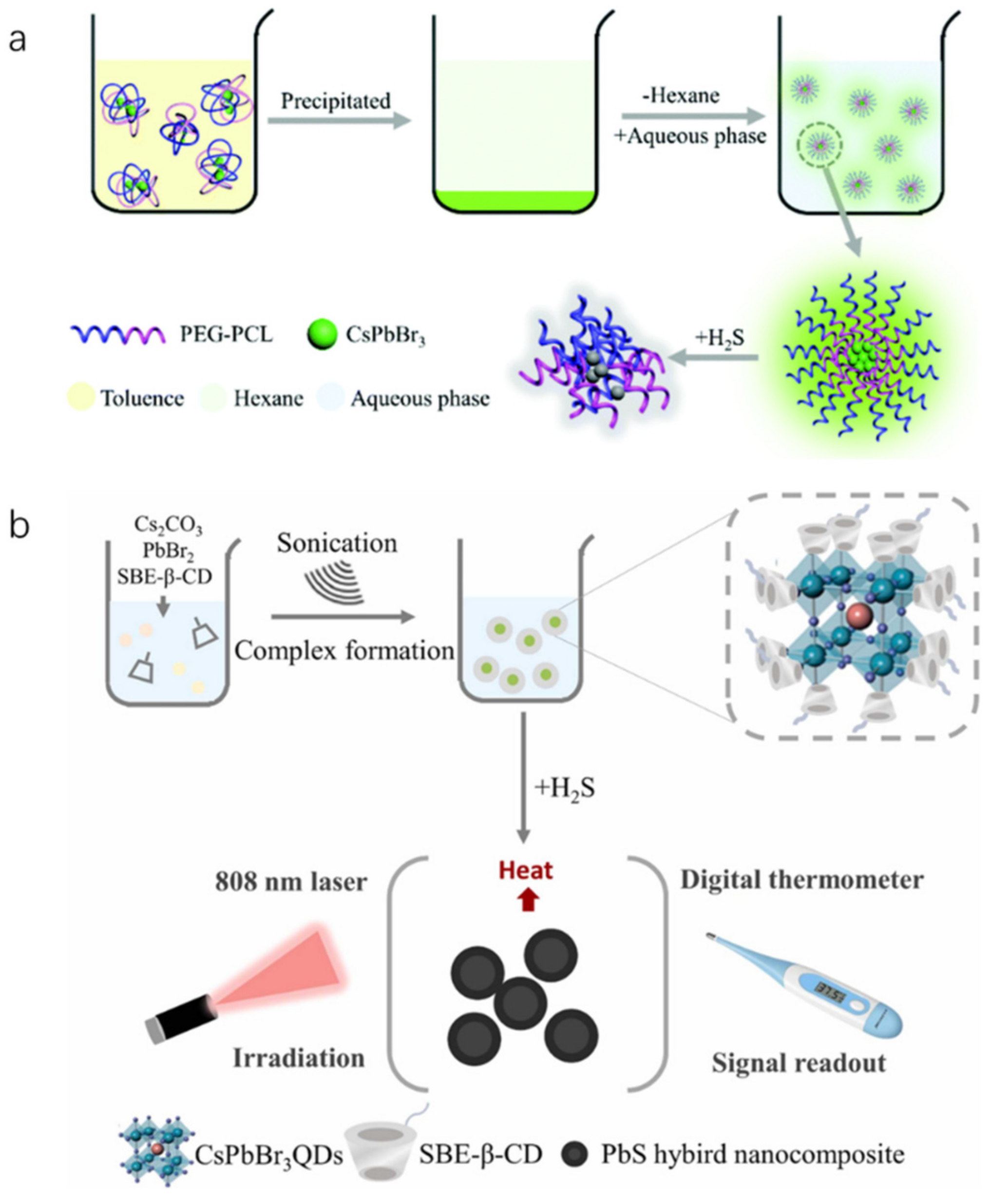
Hydrogen sulfide (H2S) is a toxic gas that emits a rotten egg smell, and exposure to high levels of H2S in food can be detrimental to human health. Li et al. developed water-soluble inorganic PQDs (CsPbBr3@PEG-PCL nanoparticles) by encapsulating CsPbBr3 QDs with a block copolymer of polyethylene glycol polycaprolactone (PEG-PCL), as illustrated in Figure 34a. The fluorescence of CsPbBr3@PEG-PCL nanoparticles remains stable in aqueous solution for at least 15 days under light and room temperature conditions, making it ideal for H2S detection. The fluorescence intensity of CsPbBr3@PEG-PCL nanoparticles is quenched by H2S, and thus, a highly sensitive H2S fluorescent probe can be constructed based on fluorescence intensity quenching. The fluorescence signal has a linear relationship with H2S concentration in the range of 0–32.00 μM, with a limit of detection (LOD) of 37.65 nM. CsPbBr3-based sensors are also capable of accurately detecting H2S in wine samples, cells, and zebrafish [75]. Another type of water-soluble inorganic PQDs (CsPbBr3@SBE-β-CD nanoparticles) was obtained by passivating the surface ligands with sulfobutyl ether-β-cyclodextrin (SBE-β-CD). This nanoparticle can be used as a photothermal probe to react with H2S and produce a substance with high photothermal effect. Under 808 nm laser irradiation, the temperature change of the system was measured using a thermometer as a signal reading device to quantitatively analyze H2S, as shown in Figure 34b. There is a linear relationship between temperature and H2S concentration in the range of 0.5–6000.0 μM, with a LOD of 0.19 μM. This sensor can achieve the quantitative detection of H2S in water medium [76].
Zu et al. proposed a synthesis method for all-inorganic CsPbBr3 QDs that can be directly stored in aqueous solution, maintaining excellent fluorescence stability and dispersion, as illustrated in Figure 35. By adding CTAB aqueous solution, CsPbBr3 QDs and mineral oil can be rapidly processed by ultrasonic to form CsPbBr3@CO complex. In aqueous solution, when the concentration of CTAB as a surfactant exceeds the critical micellar concentration (CMC), spherical or rodlike micelles may be formed, with hydrophobic nuclei surrounded by hydration shells. These micelles can encapsulate hydrophobic substances, enhancing their solubility and dispersion in water. As fluorescent probes, these micelles exhibit good selectivity for the detection of hydrogen sulfide (H2S) in food and have a short detection time. The fluorescent probe demonstrates a linear relationship between the H2S concentration and the fluorescence intensity in the range of 0.15–105.0 μM, with an LOD of 53.0 nM, satisfying the H2S detection requirements for most food or biological samples [77].
Table 7 summarizes fluorescent probes based on PNCs for detecting food spoilage. Modified CsPbBr3 QDs exhibit high sensitivity and selectivity in detecting gases such as ammonia and H2S in wine, beer, and biological samples (zebrafish), with rapid response and recovery times.

2.2.4. Detection of Harmful Additives in Food

Sudan dyes, classified as carcinogens, are commonly used as phenylazo derivatives in industry but are strictly prohibited from use in food. He et al. utilized CsPbX3 PQDs of varying colors within molecularly imprinted polymers as fluorescent probes for the sensitive and selective detection of Sudan Red I in food. The fluorescence intensity of MIPs-CsPbX3 microspheres was significantly quenched after loading Sudan Red I. The linear response was good within the range of 0.5–150 μg/L, with an LOD of 0.3 μg/L and recoveries between 95.27–105.96% [2]. Wu et al. developed a simple and effective fluorescence detection platform for Sudan I-IV utilizing CsPbBr3 PQDs, as shown in Figure 36. Under UV excitation, the CsPbBr3 QDs exhibited bright green fluorescence, which was efficiently quenched by Sudan I-IV due to significant overlap between the absorption band of Sudan I-IV and the fluorescence spectrum of CsPbBr3 QDs. After optimization, the quantification of fluorescence quenching efficiency of CsPbBr3 QDs was found to be correlated with the logarithmic concentrations of Sudan I-IV (100–10000, 0.1–1000, 0.1–2000, and 0.4–1000 ng). The LODs were 3.33, 0.03, 0.03, and 0.04 ng/mL, respectively [78].
Rhodamine 6G (Rh6G) is an anthraquinone dye that has found wide application in the textile and food industries. However, due to its teratogenic, carcinogenic, and other biological toxicities to both humans and animals, it is crucial to detect it effectively in low concentration ranges. Chan et al. synthesized monodispersed PEGylated CsPbBr3/SiO2 QDs using the LARP synthesis method at an ambient temperature of 23 °C. In Figure 37, the CsPbBr3/SiO2 QDs were encapsulated in mPEG-DSPE phospholipid micelles to provide excellent aqueous solubility and stability. This nanosensor exhibits good sensitivity and selectivity for Rh6G and can also detect it in biological samples containing complex interference backgrounds. The operation range is 0–10 mg/mL, and the LOD is 0.01 mg/mL [79]. Similarly, Wang et al. prepared a CsPbBr3 PQDs/polystyrene fiber film (CPB QDs/PS FM) using a one-step electrospinning method. Due to the excellent optical properties of CPB QDs, the ultra-low LOD for Rh6G detection is 0.01 ppm, and FRET efficiency is 18.80% in 1 ppm Rh6G aqueous solution [80].
Furthermore, RhoB is a harmful substance that often appears in printing and dye wastewater or as an additive in food. Traditional detection systems are not sensitive enough and often require a large number of sample solutions (>1 mL) to concentrate to a higher concentration. Ni et al. fabricated a reusable perovskite nanocomposite fiber paper using microwave and electrospinning methods, which consisted of CsPbBr3 QDs grown in situ in a high concentration solid polymer fiber. This perovskite fiber paper can be easily recycled and has the advantages of ultra-sensitive detection (0.01 ppm), rapid detection (<3 min), and minimal dose (<25 μL), which are obviously superior to traditional detection systems [81].
Shi et al. synthesized oil-soluble all-inorganic CsPbBrI2 QDs using the thermal injection method for the detection of basic yellow dyes that are illegally added to food and beverages. Their results showed a linear range of 1–500 μg/mL, a LOD of 0.78 μg/mL, and recoveries of 95.27–98.84% at levels of 8, 40, 80, and 400 μg/mL, with relative standard deviations of less than 3.52% [82].
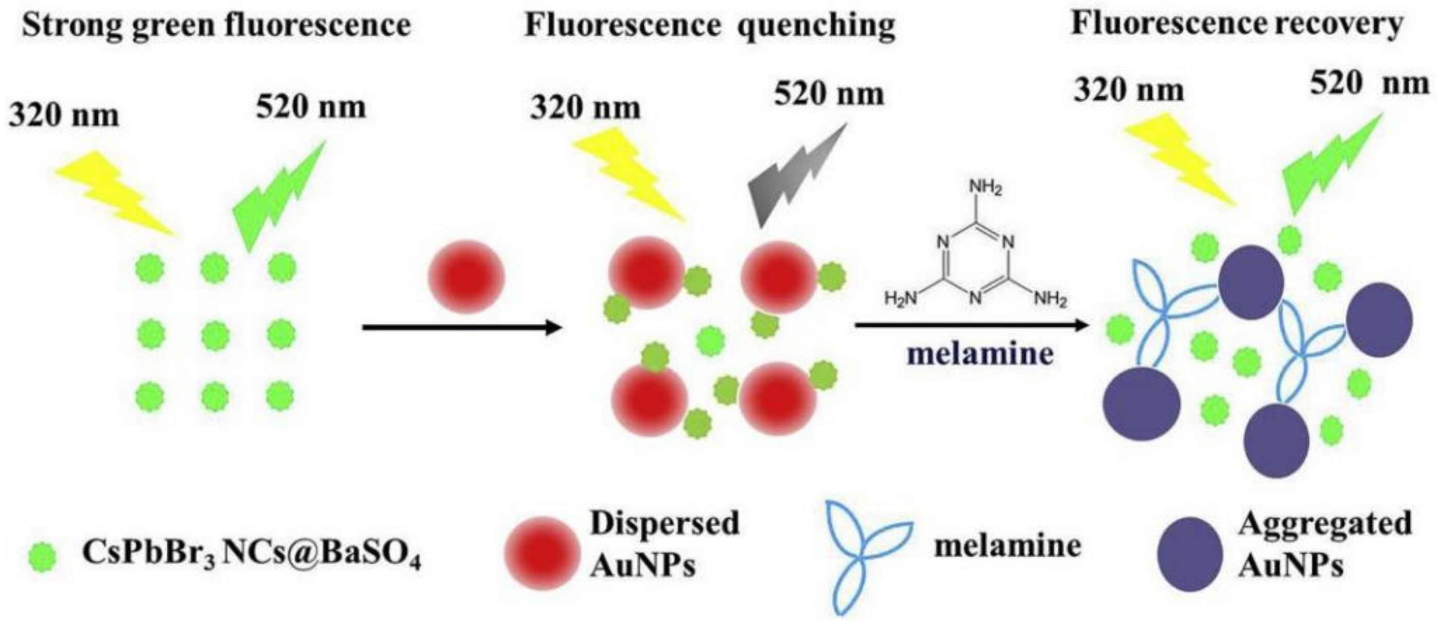
Li et al. developed an ultra-sensitive fluorescent nanosensor for the detection of melamine using PNCs (CsPbBr3NCs@BaSO4). Figure 38 illustrates the nanosensor’s principle. The negatively charged citrate-stabilized AuNPs were mixed with positively charged CsPbBr3 NCs@BaSO4, causing them to assemble together through electrostatic interaction. This aggregation led to strong fluorescence quenching of the CsPbBr3 NCs@BaSO4 due to the inner filter effect of the AuNPs. However, upon the addition of melamine, the color of the solution changed from wine red to blue–grey, indicating the agglomeration of the AuNPs. This aggregation decreased the inner filter effect of the AuNPs, resulting in a recovery of the fluorescence of the CsPbBr3 NCs@BaSO4. The nanosensor was used for the determination of trace melamine in dairy products, achieving a LOD of 0.42 nmol/L and a relative standard deviation of 4.0% by repeated determination of 500.0 nmol/L (n = 11) [83].
To achieve efficient electrochemical luminescence (ECL) in PQDs, Sun et al. proposed the synthesis of Cs4PbBr6@CsPbBr3 PQD nanoacanthospheres (PNAs) with optimized conditions resulting in ECL activity four times higher than 3D (CsPbBr3) PQDs, which was then applied to Bisphenol A (BPA) ECL sensing [84].
Table 8 summarizes the use of modified CsPbBr3 and CsPbBrI2 QDs as PNC-based fluorescent probes for detecting harmful additives in food. These probes showed good selectivity and sensitivity in detecting Sudan I–IV, Rh6G, RhoB, basic yellow dye, melamine, and bisphenol A, with the advantages of rapid determination and minimal dose. This demonstrates their potential practical applications and superiority over traditional detection systems.

2.2.5. Edible oil Quality Inspection

Edible oil undergoes complex changes during storage, with the most important being the decomposition of oil into free fatty acids and other products under the influence of light, heat, air, and other factors, commonly referred to as rancidity. Peroxide is an intermediate product formed during the process of oil oxidation and rancidity, which is unstable and can decompose into aldehydes, ketones, and other oxides. This not only affects the flavor and nutritional value of food but also poses risks to human health. China’s National Testing Standard GB/T 2716-2018 specifies that the peroxide value of edible oil should be <0.25 g/100 g. Currently, the peroxide value of cooking oil is usually determined by titration or potential methods, which are not reproducible, sensitive, and environmentally friendly, and the methods applied are cumbersome to operate. Although spectrophotometry and high-performance liquid chromatography have been developed to overcome these shortcomings, there is still a need for real-time and visual detection methods that are simple and rapid.
The peroxide number of edible oil is related to its quality, and classical determination methods for the peroxide number are unsatisfactory due to their complexity and poor reproducibility in the analytical process and their incapability of field rapid detection. To overcome this, a colorimetric sensing method based on the wavelength shift of CsPbBr3 NCs with the addition of oleylammonium iodide (OLAM-I) was developed for the determination of peroxide number in edible oil. As shown in Figure 39, by performing halide exchange with OLAM-I, the fluorescence emission wavelength of CsPbBr3 NCs gradually red-shifts, with the degree of red-shift proportional to the amount of OLAM-I added. This results in a color change from green to yellow and finally to red, reflecting the halogen-exchange characteristics of CsPbBr3 NCs and the redox reaction between OLAM-I and peroxides in edible oil. This method enables the visual detection of the peroxidation value of edible oil samples, with a detection process that only takes about 15 min and has been proven to be convenient and accurate [85].
The quality of edible oil has a significant impact on human health, with excessive acid number (AN), 3-chloro-1,2-propylene glycol (3-MCPD), and moisture content (MC) being the key factors to monitor. To address this issue, Zhao et al. developed orange luminescent oil-soluble CsPbBr1.5I1.5 QDs and applied them to detect AN, 3-MCPD, and MC in edible oil by employing fluorescence quenching and wavelength shift. The mechanism diagram of the detection method for hazardous substances in edible oil is shown in Figure 40. By adding varying concentrations of benzoic acid into the oil, the orange-emitting fluorescence of oil-soluble CsPbBr1.5I1.5 QDs was quenched by edible oil with different ANs, and a good linear relationship was observed between the fluorescence intensity of CsPbBr1.5I1.5 QDs and AN concentration, enabling quantitative detection of AN in edible oil. Additionally, the unique halogen exchange reaction between CsPbBr1.5I1.5 QDs and 3-MCPD allowed for the sensitive and specific detection of 3-MCPD, as the emission peak of CsPbBr1.5I1.5 QDs blue-shifted with increasing 3-MCPD concentration. To monitor the MC of edible oil, water-sensitive PQDs were prepared to establish ratiometric fluorescent probes. The orange-emitting CsPbBr1.5I1.5@MSNs were used as detection probes, which were generally quenched when exposed to water, while green-emitting quasi-two-dimensional (2D) CsPbBr3 NSs served as reference probes due to their stable fluorescence properties in aqueous solution. The ratiometric fluorescent probes based on these two materials were established for MC detection in edible oil, with limits of detection (LODs) of 0.71 mg KOH/g, 39.8 μg/mL 3-MCPD, and 0.45% MC. By leveraging this detection principle, the researchers achieved quantitative detection of AN, 3-MCPD, and MC in edible oil, demonstrating great potential for monitoring the quality of edible oil [86].
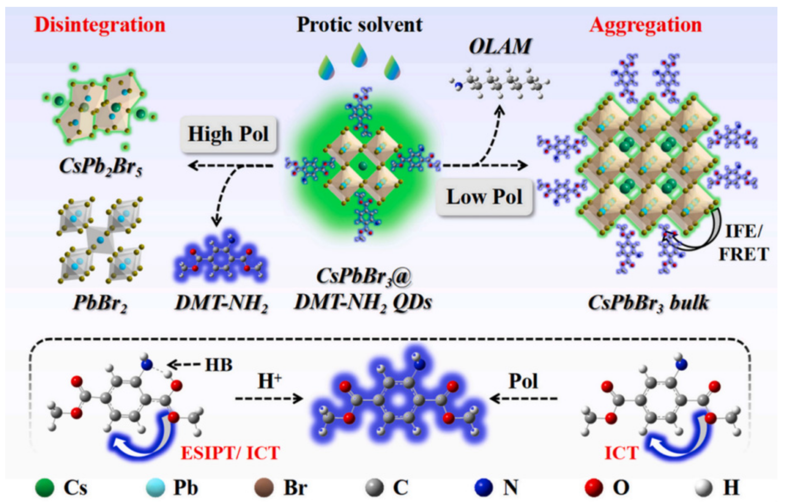
Accurately detecting trace moisture in edible oil is crucial to ensure its quality and safety. Although all inorganic PQDs are potential candidates for optoelectronic applications, their stability is often restricted by environmental factors, especially high polarity materials. In contrast, perovskite-structured QDs are highly suitable as polarity sensors due to their instability to polar materials. To address this, CsPbBr3 PQDs were synthesized at low temperature, which were modified with a functional ligand dimethyl aminoterephthalate (CsPbBr3@DMT-NH2 QDs). The resulting dual-emission QD displayed highly sensitive fluorescence turn-on/off dual response and a distinct fluorescence color change from green to blue in the presence of water. This enabled the establishment of ratiometric fluorescence sensing and visual ratiometric chromaticity detection methods for water assay, with ultralow LODs of 0.006% (v/v) and 0.01% (v/v), respectively. These methods accurately detected trace water in edible oils (with a recovery rate of 93.0% to 108.0%, and a relative error of ≤5.33%). The study also revealed that the high polarity levels of protic solvents can disintegrate the QDs, while the relatively low polarity levels promote their aggregation, ultimately leading to green fluorescence quenching. Furthermore, the deprotonation and polarity of water enhance the blue fluorescence of DMT-NH2 by promoting excited-state intramolecular proton transfer and intramolecular charge transfer, as shown in Figure 41. This is the first report on dual-response ratiometric fluorescence PQDs for ultrasensitive water detection in edible oils [87].
To ensure the quality and safety of edible oil, the analysis of total polar substance (TPM) is crucial. However, the development of appropriate TPM analysis methods has been challenging in the food safety field. A paper-based fluorescent probe using CsPbBr3 QDs was synthesized and utilized for rapid detection of TPM in different types of edible oils. The results showed a linear relationship for olive oil (17–31.5%), soybean oil (25–31.5%), and sunflower oil (21.5–33%). Real-time monitoring of TPM content was achieved using the probe, and the potential for a more rapid detection method was explored. These findings indicate that CsPbBr3 QDs can serve as a new fluorescent probe for the rapid detection of TPM in edible oil, with practical application value. Furthermore, Table 9 summarizes the use of PNC-based fluorescent probes for edible oil quality inspection, including modified CsPbBr3 NCs and CsPbBr1.5I1.5 QDs, which demonstrate the convenience and accuracy of detecting peroxide value, AN, 3-MCPD, MC, and TPM. These perovskite nanomaterials not only expand the application of bioanalysis but also provide a new material and method for monitoring the safety of oil-phase foods in multiple ways [88].

3. Summary and Prospects

In summary, the fluorescent probe based on PNCs is expected to replace the traditional inorganic QDs because of its high fluorescence quantum yield and sensitivity based on PNCs to detect hazardous substances in agriculture and food. Nowadays, PNCs have been used in agro-ecological environment to detect pesticide residue, environmental pollutant, and harmful ions. At the same time, PNCs have also been applied in food to detect antibiotics, microorganisms, food spoilage gases, additives, and edible oil quality. Compared with the fluorescence analysis method constructed by traditional QDs, PNCs as fluorescent probes are just in their infancy. Therefore, there are still many problems in the practical application of PNCs, and more research is needed.
(1)
The external environment (light, humidity, temperature, etc.) is easy to affect photoelectric properties of PNCs. Although, many modification methods have been proved to be effective to achieve PNCs in aqueous solutions, some of these methods are complex or cannot be used as fluorescent probes in the field of agriculture and food. Simple and novel water-stabilized method that is suitable for detection in agriculture and food should be further developed in the future. For example, the exploration of organic semiconductor ligands to prepare PNCs materials with good optoelectrical properties and heterostructures.
(2)
Lead is an important component of PNCs structure, and it is a huge obstacle, which hinders PNCs in practice in agriculture and food. Non-contact detection (gas or separated liquid samples) may be more suitable for the lead-based PNCs fluorescent probe. The development of novel and efficient lead-free PNCs fluorescent probe is more preferred and should be the future trend in this area.
(3)
The specificity of the fluorescent probe based on PNCs can be further developed to selectively detect specific analyte. Combined with molecular imprinting techniques, it has been proved to be an effective way to improve its selectivity. Especially, the novel characterizing methods should be paid more attention, such as MIECL platform, PEC immunosensing platform, etc.
(4)
The integration of fluorescent probe based on PNCs, excitation light source, and fluorescence detector into a portable instrument is still a big challenge. The future practice of fluorescent probe based on PNCs in the field of agriculture and food should tend to portable, real-time, and visualized detection. Fluorescent probe based on PNCs prefer to be developed into the form of test strip. Ratio fluorescence is a good method to get visual sensing with high accuracy. In addition, with the help of the color analysis software in smart phones, qualitative and semi-quantitative visual sensing can be easily realized, which has a good application prospect.
In short, with the deepening of research, fluorescent probe based on PNCs should be constantly improved and innovated. The development of highly sensitive, highly selective, portable, and smart fluorescent probe based on PNCs in the field of agriculture and food is expected.

Author Contributions

Funding acquisition, W.Z. and T.Y.; writing-original draft preparation, W.Z., J.Z. and F.K.; writing—review and editing, W.Z. and T.Y.; validation and supervision, T.Y.; formal analysis and investigation, W.Z. All authors have read and agreed to the published version of the manuscript.

Funding

This work was supported by the Open Fund of the State Key Laboratory of Luminescent Materials and Devices, South China University of Technology (Grant No. 2022-skllmd-08), the National Key Research and Development Program of China (No. 2022YFD1200704--2), the National Key Research and Development Program of China (No. 2021YFD1201000), and the Heilongjiang Provincial Postdoctoral Science Foundation (Grant No. LBH-TZ0604).

Institutional Review Board Statement

Not applicable.

Data Availability Statement

Not applicable.

Conflicts of Interest

The authors declare no conflict of interest.

References

  1. Vajubhai, G.N.; Chetti, P.; Kailasa, S.K. Perovskite quantum dots for fluorescence turn-off detection of the clodinafop pesticide in food samples via liquid–liquid microextraction. ACS Appl. Nano Mater. 2022, 5, 18220–18228. [Google Scholar] [CrossRef]
  2. He, J.; Yu, L.; Jiang, Y.; Lü, L.; Han, Z.; Zhao, X.; Xu, Z. Encoding CsPbX3 perovskite quantum dots with different colors in molecularly imprinted polymers as fluorescent probes for the quantitative detection of Sudan I in food matrices. Food Chem. 2023, 402, 134499. [Google Scholar] [CrossRef] [PubMed]
  3. Gupta, A.; Mehta, S.K.; Kunal, K.; Mukhopadhyay, K.; Singh, S. Quantum dots as promising nanomaterials in agriculture. In Agricultural Nanobiotechnology; Woodhead Publishing: Southton, UK, 2022; pp. 243–296. [Google Scholar]
  4. Huang, S.; Guo, M.; Tan, J.; Geng, Y.; Wu, J.; Tang, Y.; Liang, Y. Novel fluorescent probe based on all-inorganic perovskite quantum dots coated with molecularly imprinted polymers for highly selective and sensitive detection of omethoate. ACS Appl. Mater. Interfaces 2018, 10, 39056–39063. [Google Scholar] [CrossRef] [PubMed]
  5. Shangguan, Z.; Zheng, X.; Zhang, J.; Lin, W.; Guo, W.; Li, C.; Wu, T.; Lin, Y.; Chen, Z. The stability of metal halide perovskite nanocrystals—A key issue for the application on quantum-dot-based micro light-emitting diodes display. Nanomaterials 2020, 10, 1375. [Google Scholar] [CrossRef]
  6. Jia, D.; Xu, M.; Mu, S.; Ren, W.; Liu, C. Recent progress of perovskite nanocrystals in chem/bio sensing. Biosensors 2022, 12, 754. [Google Scholar] [CrossRef] [PubMed]
  7. Wei, Y.; Cheng, Z.; Lin, J. An overview on enhancing the stability of lead halide perovskite quantum dots and their applications in phosphor-converted LEDs. Chem. Soc. Rev. 2019, 48, 310–350. [Google Scholar] [CrossRef] [PubMed]
  8. Stoumpos, C.C.; Malliakas, C.D.; Kanatzidis, M.G. Semiconducting tin and lead iodide perovskites with organic cations: Phase transitions, high mobilities, and near-infrared photoluminescent properties. Inorg. Chem. 2013, 52, 9019–9038. [Google Scholar] [CrossRef]
  9. Liu, Y.; Pan, G.; Wang, R.; Shao, H.; Wang, H.; Xu, W.; Song, H. Considerably enhanced exciton emission of CsPbCl3 perovskite quantum dots by the introduction of potassium and lanthanide ions. Nanoscale 2018, 10, 14067–14072. [Google Scholar] [CrossRef]
  10. Huang, S.; Wang, B.; Zhang, Q.; Li, Z.; Shan, A.; Li, L. Postsynthesis potassium modification method to improve stability of CsPbBr3 perovskite nanocrystals. Adv. Opt. Mater. 2018, 6, 1701106. [Google Scholar] [CrossRef]
  11. Protesescu, L.; Yakunin, S.; Bodnarchuk, M.I.; Krieg, F.; Caputo, R.; Hendon, C.H.; Kovalenko, M.V. Nanocrystals of cesium lead halide perovskites (CsPbX(3), X = Cl, Br, and I): Novel optoelectronic materials showing bright emission with wide color gamut. Nano Lett. 2015, 15, 3692–3696. [Google Scholar] [CrossRef] [Green Version]
  12. Di Stasio, F.; Christodoulou, S.; Huo, N.; Konstantatos, G. Near-unity photoluminescence quantum yield in cspbbr3 nanocrystal solid-state films via postsynthesis treatment with lead bromide. Chem. Mater. 2017, 29, 7663–7667. [Google Scholar] [CrossRef]
  13. Chen, M.; Zou, Y.; Wu, L.; Pan, Q.; Yang, D.; Hu, H.; Zhang, Q. Solvothermal synthesis of high-quality all-inorganic cesium lead halide perovskite nanocrystals: From nanocube to ultrathin nanowire. Adv. Funct. Mater. 2017, 27, 1701121. [Google Scholar] [CrossRef]
  14. Pan, Q.; Hu, H.; Zou, Y.; Chen, M.; Wu, L.; Yang, D.; Zhang, Q. Microwave-assisted synthesis of high-quality “all-inorganic” CsPbX3 (X = Cl, Br, I) perovskite nanocrystals and their application in light emitting diodes. J. Mater. Chem. C 2017, 5, 10947–10954. [Google Scholar] [CrossRef]
  15. Tong, Y.; Bladt, E.; Aygüler, M.F.; Manzi, A.; Milowska, K.Z.; Hintermayr, V.A.; Feldmann, J. Highly luminescent cesium lead halide perovskite nanocrystals with tunable composition and thickness by ultrasonication. Angew. Chem. Int. Ed. 2016, 55, 13887–13892. [Google Scholar] [CrossRef] [PubMed]
  16. Zhu, M.; Bai, J.; Chen, R.; Li, H. Synthesis and properties of B-site doped all-inorganic perovskite quantum dots. Chin. J. Appl. Chem. 2021, 38, 1541. [Google Scholar]
  17. Wei, Y.; Deng, X.; Xie, Z. Enhancing the stability of perovskite quantum dots by encapsulation in crosslinked polystyrene beads via a swelling–shrinking strategy toward superior water resistance. Adv. Funct. Mater. 2017, 27, 1703535. [Google Scholar] [CrossRef]
  18. An, H.; Kim, W.K.; Wu, C.; Kim, T.W. Highly-stable memristive devices based on poly (methylmethacrylate): CsPbCl3 perovskite quantum dot hybrid nanocomposites. Org. Electron. 2018, 56, 41–45. [Google Scholar] [CrossRef]
  19. Xuan, T.; Huang, J.; Liu, H.; Lou, S.; Cao, L.; Gan, W.; Wang, J. Super-hydrophobic cesium lead halide perovskite quantum dot-polymer composites with high stability and luminescent efficiency for wide color gamut white light-emitting diodes. Chem. Mater. 2019, 31, 1042–1047. [Google Scholar] [CrossRef]
  20. Avugadda, S.K.; Castelli, A.; Dhanabalan, B.; Fernandez, T.; Silvestri, N.; Collantes, C.; Arciniegas, M.P. Highly emitting perovskite nanocrystals with 2-year stability in water through an automated polymer encapsulation for bioimaging. ACS Nano 2022, 16, 13657–13666. [Google Scholar] [CrossRef]
  21. Li, S.; Lei, D.; Ren, W.; Guo, X.; Wu, S.; Zhu, Y.; Jen, A.K.Y. Water-resistant perovskite nanodots enable robust two-photon lasing in aqueous environment. Nat. Commun. 2020, 11, 1192. [Google Scholar] [CrossRef] [Green Version]
  22. You, C.Y.; Li, F.M.; Lin, L.H.; Lin, J.S.; Chen, Q.Q.; Radjenovic, P.M.; Li, J.F. Ultrastable monodispersed lead halide perovskite nanocrystals derived from interfacial compatibility. Nano Energy 2020, 71, 104554. [Google Scholar] [CrossRef]
  23. Zhang, Q.; Wang, B.; Zheng, W.; Kong, L.; Wan, Q.; Zhang, C.; Li, L. Ceramic-like stable CsPbBr3 nanocrystals encapsulated in silica derived from molecular sieve templates. Nat. Commun. 2020, 11, 31. [Google Scholar] [CrossRef] [PubMed] [Green Version]
  24. Qiao, G.Y.; Guan, D.; Yuan, S.; Rao, H.; Chen, X.; Wang, J.A.; Yu, J. Perovskite quantum dots encapsulated in a mesoporous metal–organic framework as synergistic photocathode materials. J. Am. Chem. Soc. 2021, 143, 14253–14260. [Google Scholar] [CrossRef] [PubMed]
  25. Wu, L.Y.; Mu, Y.F.; Guo, X.X.; Zhang, W.; Zhang, Z.M.; Zhang, M.; Lu, T.B. Encapsulating perovskite quantum dots in iron-based metal-organic frameworks (MOFs) for efficient photocatalytic CO2 reduction. Angew. Chem. Int. Ed. 2019, 58, 9491–9495. [Google Scholar] [CrossRef]
  26. Liu, B.; Guo, X.; Gao, D. Research progress on the improvement of the stability of perovskite quantum dots. Chem. Ind. Eng. Prog. 2021, 40, 247–258. [Google Scholar] [CrossRef]
  27. Pering, S.R.; Deng, W.; Troughton, J.R.; Kubiak, P.S.; Ghosh, D.; Niemann, R.G.; Cameron, P.J. Azetidinium lead iodide for perovskite solar cells. J. Mater. Chem. A 2017, 5, 20658–20665. [Google Scholar] [CrossRef] [Green Version]
  28. Filip, M.R.; Eperon, G.E.; Snaith, H.J.; Giustino, F. Steric engineering of metal-halide perovskites with tunable optical band gaps. Nat. Commun. 2014, 5, 5757. [Google Scholar] [CrossRef] [Green Version]
  29. Protesescu, L.; Yakunin, S.; Kumar, S.; Bär, J.; Bertolotti, F.; Masciocchi, N.; Kovalenko, M.V. Dismantling the “red wall” of colloidal perovskites: Highly luminescent formamidinium and formamidinium–cesium lead iodide nanocrystals. ACS Nano 2017, 11, 3119–3134. [Google Scholar] [CrossRef]
  30. Wu, X.; Jiang, Y.; Chen, C.; Guo, J.; Kong, X.; Feng, Y.; Gao, J. Stable triple cation perovskite precursor for highly efficient perovskite solar cells enabled by interaction with 18C6 stabilizer. Adv. Funct. Mater. 2020, 30, 1908613. [Google Scholar] [CrossRef]
  31. Xu, L.; Yuan, S.; Zeng, H.; Song, J.J. A comprehensive review of doping in perovskite nanocrystals/quantum dots: Evolution of structure, electronics, optics, and light-emitting diodes. Mater. Today Nano 2019, 6, 100036. [Google Scholar] [CrossRef]
  32. Wang, J.; Liu, X.; Zhou, L.; Shen, W.; Li, M.; He, R. Highly luminescent and stable quasi-2D perovskite quantum dots by introducing large organic cations. Nanoscale Adv. 2021, 3, 5393–5398. [Google Scholar] [CrossRef]
  33. Zhou, Y.; Chen, J.; Bakr, O.M.; Sun, H.T. Metal-doped lead halide perovskites: Synthesis, properties, and optoelectronic applications. Chem. Mater. 2018, 30, 6589–6613. [Google Scholar] [CrossRef] [Green Version]
  34. Zhuang, X.; Sun, R.; Zhou, D.; Liu, S.; Wu, Y.; Shi, Z.; Song, H. Synergistic effects of multifunctional lanthanides doped CsPbBrCl2 quantum dots for efficient and stable MAPbI3 perovskite solar cells. Adv. Funct. Mater. 2022, 32, 2110346. [Google Scholar] [CrossRef]
  35. Shen, X.; Zhang, Y.; Kershaw, S.V.; Li, T.; Wang, C.; Zhang, X.; Rogach, A.L. Zn-alloyed CsPbI3 nanocrystals for highly efficient perovskite light-emitting devices. Nano Lett. 2019, 19, 1552–1559. [Google Scholar] [CrossRef]
  36. Song, P.; Qiao, B.; Song, D.; Cao, J.; Shen, Z.; Xu, Z.; Al-Ghamdi, A. Modifying the crystal field of CsPbCl3: Mn2+ nanocrystals by co-doping to enhance its red emission by a hundredfold. ACS Appl. Mater. Interfaces 2020, 12, 30711–30719. [Google Scholar] [CrossRef]
  37. Kim, H.; Bae, S.R.; Lee, T.H.; Lee, H.; Kang, H.; Park, S.; Kim, S.Y. Enhanced optical properties and stability of CsPbBr3 nanocrystals through nickel doping. Adv. Funct. Mater. 2021, 31, 2102770. [Google Scholar] [CrossRef]
  38. Zeng, Q.; Zhang, X.; Bing, Q.; Xiong, Y.; Yang, F.; Liu, H.; Yang, B. Surface Stabilization of Colloidal Perovskite Nanocrystals via Multi-amine Chelating Ligands. ACS Energy Lett. 2022, 7, 1963–1970. [Google Scholar] [CrossRef]
  39. Bi, C.; Kershaw, S.V.; Rogach, A.L.; Tian, J. Improved stability and photodetector performance of CsPbI3 perovskite quantum dots by ligand exchange with aminoethanethiol. Adv. Funct. Mater. 2019, 29, 1902446. [Google Scholar] [CrossRef]
  40. Krieg, F.; Ochsenbein, S.T.; Yakunin, S.; Ten Brinck, S.; Aellen, P.; Süess, A.; Kovalenko, M.V. Colloidal CsPbX3 (X = Cl, Br, I) nanocrystals 2.0: Zwitterionic capping ligands for improved durability and stability. ACS Energy Lett. 2018, 3, 641–646. [Google Scholar] [CrossRef] [Green Version]
  41. Krieg, F.; Ong, Q.K.; Burian, M.; Rainò, G.; Naumenko, D.; Amenitsch, H.; Kovalenko, M.V. Stable ultraconcentrated and ultradilute colloids of CsPbX3 (X= Cl, Br) nanocrystals using natural lecithin as a capping ligand. J. Am. Chem. Soc. 2019, 141, 19839–19849. [Google Scholar] [CrossRef] [Green Version]
  42. Wang, S.; Du, L.; Jin, Z.; Xin, Y.; Mattoussi, H. Enhanced stabilization and easy phase transfer of CsPbBr3 perovskite quantum dots promoted by high-affinity polyzwitterionic ligands. J. Am. Chem. Soc. 2020, 142, 12669–12680. [Google Scholar] [CrossRef] [PubMed]
  43. Pan, Q.; Hu, J.; Fu, J.; Lin, Y.; Zou, C.; Di, D.; Cao, M. Ultrahigh stability of perovskite nanocrystals by using semiconducting molecular species for displays. ACS Nano 2022, 16, 12253–12261. [Google Scholar] [CrossRef] [PubMed]
  44. Rajput, S.; Sharma, R.; Kumari, A.; Kaur, R.; Sharma, G.; Arora, S.; Kaur, R. Pesticide residues in various environmental and biological matrices: Distribution, extraction, and analytical procedures. Environ. Dev. Sustain. 2022, 24, 6032–6052. [Google Scholar] [CrossRef]
  45. Damalas, C.A.; Eleftherohorinos, I.G. Pesticide exposure, safety issues, and risk assessment indicators. Int. J. Environ. Res. Public Health 2011, 8, 1402–1419. [Google Scholar] [CrossRef]
  46. Huang, S.; Tan, L.; Zhang, L.; Wu, J.; Zhang, L.; Tang, Y.; Liang, Y. Molecularly imprinted mesoporous silica embedded with perovskite CsPbBr3 quantum dots for the fluorescence sensing of 2, 2-dichlorovinyl dimethyl phosphate. Sens. Actuators B Chem. 2020, 325, 128751. [Google Scholar] [CrossRef]
  47. Tan, L.; Guo, M.; Tan, J.; Geng, Y.; Huang, S.; Tang, Y.; Liang, Y. Development of high-luminescence perovskite quantum dots coated with molecularly imprinted polymers for pesticide detection by slowly hydrolysing the organosilicon monomers in situ. Sens. Actuators B Chem. 2019, 291, 226–234. [Google Scholar] [CrossRef]
  48. Chen, D.; Guan, J.; Zhang, G. Research status of microbial degradation of pesticides. J. Henan Inst. Sci. Technol. (Nat. Sci. Ed.) 2020, 48, 39–46. [Google Scholar]
  49. Zhang, R.R.; Gan, X.T.; Xu, J.J.; Pan, Q.F.; Liu, H.; Sun, A.L.; Zhang, Z.M. Ultrasensitive electrochemiluminescence sensor based on perovskite quantum dots coated with molecularly imprinted polymer for prometryn determination. Food Chem. 2022, 370, 131353. [Google Scholar] [CrossRef]
  50. Pan, Q.F.; Jiao, H.F.; Liu, H.; You, J.J.; Sun, A.L.; Zhang, Z.M.; Shi, X.Z. Highly selective molecularly imprinted-electrochemiluminescence sensor based on perovskite/Ru (bpy)32+ for simazine detection in aquatic products. Sci. Total Environ. 2022, 843, 156925. [Google Scholar] [CrossRef]
  51. Yang, Y.; Han, A.; Hao, S.; Li, X.; Luo, X.; Fang, G.; Liu, J.; Wang, S. Fluorescent methylammonium lead halide perovskite quantum dots as a sensing material for the detection of polar organochlorine pesticide residues. Analyst 2020, 145, 6683–6690. [Google Scholar] [CrossRef]
  52. Liu, C.X.; Zhao, J.; Zhang, R.R.; Zhang, Z.M.; Xu, J.J.; Sun, A.L.; Shi, X.Z. Development and application of fluorescent probe and test strip based on molecularly imprinted quantum dots for the selective and sensitive detection of propanil in fish and seawater samples. J. Hazard. Mater. 2020, 389, 121884. [Google Scholar] [CrossRef] [PubMed]
  53. Halali, V.V.; Rani, R.S.; Balakrishna, R.G.; Budagumpi, S. Ultra-trace level chemosensing of uranyl ions; scuffle between electron and energy transfer from perovskite quantum dots to adsorbed uranyl ions. Microchem. J. 2020, 156, 104808. [Google Scholar] [CrossRef]
  54. Deng, P.; Wang, W.; Liu, X.; Wang, L.; Yan, Y. A hydrophobic polymer stabilized CsPbBr3 sensor for environmental pollutant detection. New J. Chem. 2021, 45, 930–938. [Google Scholar] [CrossRef]
  55. Liu, Y.; Tang, X.; Zhu, T.; Deng, M.; Ikechukwu, I.P.; Huang, W.; Qiu, F. All-inorganic CsPbBr3 perovskite quantum dots as a photoluminescent probe for ultrasensitive Cu 2+ detection. J. Mater. Chem. C 2018, 6, 4793–4799. [Google Scholar] [CrossRef]
  56. Li, Q.; Zhou, W.; Yu, L.; Lian, S.; Xie, Q. Perovskite quantum dots as a fluorescent probe for metal ion detection in aqueous solution via phase transfer. Mater. Lett. 2021, 282, 128654. [Google Scholar] [CrossRef]
  57. Lu, L.Q.; Tan, T.; Tian, X.K.; Li, Y.; Deng, P. Visual and sensitive fluorescent sensing for ultratrace mercury ions by perovskite quantum dots. Anal. Chim. Acta 2017, 986, 109–114. [Google Scholar] [CrossRef]
  58. Chen, M.; An, J.; Hu, Y.; Chen, R.; Lyu, Y.; Hu, N.; Liu, Y. Swelling-shrinking modified hyperstatic hydrophilic perovskite polymer fluorescent beads for Fe (III) detection. Sens. Actuators B Chem. 2020, 325, 128809. [Google Scholar] [CrossRef]
  59. Wang, J.; Li, M.; Shen, W.; Su, W.; He, R. Ultrastable carbon quantum dots-doped MAPbBr3 perovskite with silica encapsulation. ACS Appl. Mater. Interfaces 2019, 11, 34348–34354. [Google Scholar] [CrossRef]
  60. Yan, J.; He, Y.; Chen, Y.; Zhang, Y.; Yan, H. CH3NH3Br solution as a novel platform for the selective fluorescence detection of Pb2+ ions. Sci. Rep. 2019, 9, 15840. [Google Scholar] [CrossRef] [Green Version]
  61. Wang, S.; Huang, Y.; Zhang, L.; Li, F.; Lin, F.; Wang, Y.; Chen, X. Highly selective fluorescence turn-on determination of Pb (II) in Water by in-situ enrichment of Pb (II) and MAPbBr3 perovskite growth in sulfydryl functionalized mesoporous alumina film. Sens. Actuators B Chem. 2021, 326, 128975. [Google Scholar] [CrossRef]
  62. Park, B.; Kang, S.M.; Lee, G.W.; Kwak, C.H.; Rethinasabapathy, M.; Huh, Y.S. Fabrication of CsPbBr3 perovskite quantum dots/cellulose-based colorimetric sensor: Dual-responsive on-site detection of chloride and iodide ions. Ind. Eng. Chem. Res. 2019, 59, 793–801. [Google Scholar] [CrossRef]
  63. Han, J.; Sharipov, M.; Hwang, S.; Lee, Y.; Huy, B.T.; Lee, Y.I. Water-stable perovskite-loaded nanogels containing antioxidant property for highly sensitive and selective detection of roxithromycin in animal-derived food products. Sci. Rep. 2022, 12, 3147. [Google Scholar] [CrossRef]
  64. Jia, L.; Xu, Z.; Zhang, L.; Li, Y.; Zhao, T.; Xu, J. The fabrication of water-stable perovskite-europium hybrid polychromatic fluorescence nanosensor for fast visual sensing of tetracycline. Appl. Surf. Sci. 2022, 592, 153170. [Google Scholar] [CrossRef]
  65. Thuy, T.T.; Huy, B.T.; Kumar, A.P.; Lee, Y.I. Highly stable Cs4PbBr6/CsPbBr3 perovskite nanoparticles as a new fluorescence nanosensor for selective detection of trace tetracycline in food samples. J. Ind. Eng. Chem. 2021, 104, 437–444. [Google Scholar] [CrossRef]
  66. Wang, W.; Deng, P.; Liu, X.; Ma, Y.; Yan, Y. A CsPbBr3 quantum dots/ultra-thin BN fluorescent probe for stability and highly sensitive detection of tetracycline. Microchem. J. 2021, 162, 105876. [Google Scholar] [CrossRef]
  67. Wang, T.; Wei, X.; Zong, Y.; Zhang, S.; Guan, W. An efficient and stable fluorescent sensor based on APTES-functionalized CsPbBr3 perovskite quantum dots for ultrasensitive tetracycline detection in ethanol. J. Mater. Chem. C 2020, 8, 12196–12203. [Google Scholar] [CrossRef]
  68. Salari, R.; Amjadi, M.; Hallaj, T. Perovskite quantum dots as a chemiluminescence platform for highly sensitive assay of cefazolin. Spectrochim. Acta Part A Mol. Biomol. Spectrosc. 2023, 285, 121845. [Google Scholar] [CrossRef] [PubMed]
  69. Shi, X.; Kralj, M.; Zhang, Y. Colorimetric test strips based on cesium lead bromide perovskite nanocrystals for rapid detection of ciprofloxacin hydrochloride. J. Phys. Condens. Matter 2022, 34, 304002. [Google Scholar] [CrossRef]
  70. Su, L.; Tong, P.; Zhang, L.; Luo, Z.; Fu, C.; Tang, D.; Zhang, Y. Photoelectrochemical immunoassay of aflatoxin B1 in foodstuff based on amorphous TiO2 and CsPbBr3 perovskite nanocrystals. Analyst 2019, 144, 4880–4886. [Google Scholar] [CrossRef] [PubMed]
  71. Li, J.; Wang, Q.; Xiong, C.; Deng, Q.; Zhang, X.; Wang, S.; Chen, M.M. An ultrasensitive CH3NH3PbBr3 quantum dots@ SiO2-based electrochemiluminescence sensing platform using an organic electrolyte for aflatoxin B1 detection in corn oil. Food Chem. 2022, 390, 133200. [Google Scholar] [CrossRef] [PubMed]
  72. Xu, Y.; Shi, L.; Jing, X.; Miao, H.; Zhao, Y. SERS-active composites with Au–Ag Janus nanoparticles/perovskite in immunoassays for Staphylococcus aureus enterotoxins. ACS Appl. Mater. Interfaces 2022, 14, 3293–3301. [Google Scholar] [CrossRef]
  73. Ye, W.; Cao, Q.; Cheng, X.F.; Yu, C.; He, J.H.; Lu, J.M. Lead-free Cs2PdBr6 perovskite-based humidity sensor for artificial fruit waxing detection. J. Mater. Chem. A 2020, 8, 17675–17682. [Google Scholar] [CrossRef]
  74. Huang, H.; Hao, M.; Song, Y.; Dang, S.; Liu, X.; Dong, Q. Dynamic passivation in perovskite quantum dots for specific ammonia detection at room temperature. Small 2020, 16, 1904462. [Google Scholar] [CrossRef] [PubMed]
  75. Luo, F.; Li, S.; Cui, L.; Zu, Y.; Chen, Y.; Huang, D.; Lin, Z. Biocompatible perovskite quantum dots with superior water resistance enable long-term monitoring of the H2S level in vivo. Nanoscale 2021, 13, 14297–14303. [Google Scholar] [CrossRef]
  76. Li, S.; Zhang, Y.; Zu, Y.; Chen, Y.; Luo, F.; Huang, D.; Lin, Z. Photothermal sensor based on water-soluble CsPbBr3@ sulfobutylether-β-cyclodextrins nanocomposite using a thermometer as readout. Sens. Actuators B Chem. 2022, 355, 131301. [Google Scholar] [CrossRef]
  77. Luo, F.; Zhang, Y.; Zu, Y.; Li, S.; Chen, Y.; Chen, Z.; Lin, Z. Quick preparation of water-soluble perovskite nanocomposite via cetyltrimethylammonium bromide and its application. Microchim. Acta 2022, 189, 68. [Google Scholar] [CrossRef] [PubMed]
  78. Wu, C.; Lu, Q.; Miu, X.; Fang, A.; Li, H.; Zhang, Y. A simple assay platform for sensitive detection of Sudan I–IV in chilli powder based on CsPbBr3 quantum dots. J. Food Sci. Technol. 2018, 55, 2497–2503. [Google Scholar] [CrossRef]
  79. Chan, K.K.; Yap, S.H.K.; Giovanni, D.; Sum, T.C.; Yong, K.T. Water-stable perovskite quantum dots-based FRET nanosensor for the detection of rhodamine 6G in water, food, and biological samples. Microchem. J. 2022, 180, 107624. [Google Scholar] [CrossRef]
  80. Wang, Y.; Zhu, Y.; Huang, J.; Cai, J.; Zhu, J.; Yang, X.; Li, C. CsPbBr3 perovskite quantum dots-based monolithic electrospun fiber membrane as an ultrastable and ultrasensitive fluorescent sensor in aqueous medium. J. Phys. Chem. Lett. 2016, 7, 4253–4258. [Google Scholar] [CrossRef]
  81. Ni, D.J.; Zhang, J.; Cao, Z.K.; Li, R.; Xu, T.F.; Sang, H.W.; Long, Y.Z. Supersensitive and reusable perovskite nanocomposite fiber paper for time-resolved single-droplet detection. J. Hazard. Mater. 2021, 403, 123959. [Google Scholar] [CrossRef]
  82. Shi, L.; Xu, C.; Fan, Y. Determination of basic yellow in food by fluorescence colorimetry based on perovskite nanomaterials. J. Food Saf. Qual. 2021, 12, 3658–3664. [Google Scholar]
  83. Li, Q.; Wang, H.; Yue, X.; Du, J. Perovskite nanocrystals fluorescence nanosensor for ultrasensitive detection of trace melamine in dairy products by the manipulation of inner filter effect of gold nanoparticles. Talanta 2020, 211, 120705. [Google Scholar] [CrossRef] [PubMed]
  84. Sun, R.; Yu, X.; Chen, J.; Zhang, W.; Huang, Y.; Zheng, J.; Chi, Y. Highly Electrochemiluminescent Cs4PbBr6@ CsPbBr3 perovskite nanoacanthospheres and their application for sensing bisphenol A. Anal. Chem. 2022, 94, 17142–17150. [Google Scholar] [CrossRef] [PubMed]
  85. Zhu, Y.; Li, F.; Huang, Y.; Lin, F.; Chen, X. Wavelength-shift-based colorimetric sensing for peroxide number of edible oil using CsPbBr3 perovskite nanocrystals. Anal. Chem. 2019, 91, 14183–14187. [Google Scholar] [CrossRef] [PubMed] [Green Version]
  86. Zhao, Y.; Xu, Y.; Shi, L.; Fan, Y. Perovskite nanomaterial-engineered multiplex-mode fluorescence sensing of edible oil quality. Anal. Chem. 2021, 93, 11033–11042. [Google Scholar] [CrossRef]
  87. Wang, S.; Yu, L.; Wei, Z.; Xu, Q.; Zhou, W.; Xiao, Y. Dual-response ratiometric fluorescence based ligand-functionalized CsPbBr3 perovskite quantum dots for sensitive detection of trace water in edible oils. Sens. Actuators B Chem. 2022, 366, 132010. [Google Scholar] [CrossRef]
  88. Huangfu, C.; Feng, L. High-performance fluorescent sensor based on CsPbBr3 quantum dots for rapid analysis of total polar materials in edible oils. Sens. Actuators B Chem. 2021, 344, 130193. [Google Scholar] [CrossRef]

Figure 1. Schematic illustration of the outline.
Polymers 15 02873 g001
Figure 2. Typical ABX3 perovskite crystal structure. Reproduced from ref. [7] Copyright (2019), with permission from Royal Society of Chemistry.
Polymers 15 02873 g002
Figure 3. Schematic illustration for preparation of CPB@SHFW composites. Reproduced from ref. [19] Copyright (2019), with permission from American Chemical Society.
Polymers 15 02873 g003
Figure 4. (a) Schematic illustration of the capsules embedding the CsPbBr3 NCs. (b,c) Photographs of aged samples of (b) Cs-oleate and (c) DDAB-coated CsPbBr3 NCs dispersed in water under normal and UV light. Reproduced from ref. [20] Copyright (2022), with permission from American Chemical Society.
Polymers 15 02873 g004
Figure 5. Synthesis of wr-PNDs by encapsulating into SiO2. Reproduced from ref. [21] Copyright (2020), with permission from Springer Nature.
Polymers 15 02873 g005
Figure 6. Synthetic procedure of the mSiO2-ABX3@AlOx (A = MA, FA, Cs, B = Pb, Mn, X = Cl, Br, I) NCs. Reproduced from ref. [22] Copyright (2020), with permission from Elsevier.
Polymers 15 02873 g006
Figure 7. Schematic illustration of the preparation of CsPbBr3@PCN-333(Fe). Reproduced from ref. [24] Copyright (2021), with permission from American Chemical Society.
Polymers 15 02873 g007
Figure 8. Schematic representation of metal halide perovskites with 3D and quasi-2D structures. Reproduced from ref. [32] Copyright (2021), with permission from Royal Society of Chemistry.
Polymers 15 02873 g008
Figure 9. (a) Schematic of CsPbBr3 PNCs with and without Ni2+ substitution. (b) PLQY with Ni/Pb feed molar ratios of 0, 1.25, 2.5, 3.75, and 5; insets show photographs of Ni:CsPbBr3 PNCs in hexane under 365 nm UV irradiation. Normalized PL intensity of CsPbBr3 and Ni:CsPbBr3 PNCs (Ni/Pb = 2.5), (c) under constant UV radiation (365 nm, 4 W), and (d) in deionized water. Reproduced from ref. [37] Copyright (2021), with permission from Wiley-VCH.
Polymers 15 02873 g009
Figure 10. Multi-amine AHDA ligands stabilize the CsPbI3 surface. Reproduced from ref. [38] Copyright (2022), with permission from American Chemical Society.
Polymers 15 02873 g010
Figure 11. Surface engineering of PNCs with zwitterionic organic ligands, including 3-(N,N-dimethyloctadecylammonio) propanesulfonate (n = 1), N-hexadecylphosphocholine, and N,N-dimethyldodecylammoniumbutyrate. Reproduced from ref. [39] Copyright (2019), with permission from Wiley-VCH.
Polymers 15 02873 g011
Figure 12. Schematic illustration of the preparation of the MIPs@CsPbBr3 QD sensor. Reproduced from ref. [4] Copyright (2018), with permission from American Chemical Society.
Polymers 15 02873 g012
Figure 13. Schematic illustration of the preparation of QDs-encapsulated MIMS. Reproduced from ref. [46] Copyright (2020), with permission from Elsevier.
Polymers 15 02873 g013
Figure 14. Schematic illustration of the preparation of MIP/CsPbBr3 QDs composites. Reproduced from ref. [47] Copyright (2019), with permission from Elsevier.
Polymers 15 02873 g014
Figure 15. Schematic illustration for the fabrication process and prometryn determination principle of ultrasensitive MIECL sensor. Reproduced from ref. [49] Copyright (2022), with permission from Elsevier.
Polymers 15 02873 g015
Figure 16. Schematic illustration of the reaction mechanism for MAPB-QDs and polar OCPs. Reproduced from ref. [51] Copyright (2020), with permission from Royal Society of Chemistry.
Polymers 15 02873 g016
Figure 17. Schematic illustration of microwave synthesis of CsPbI3 PQDs for sensing of clodinafop. Reproduced from ref. [1] Copyright (2022), with permission from American Chemical Society.
Polymers 15 02873 g017
Figure 18. Adsorption mechanism UO22+ ions on CsPbBr3 PQDs due to the zeta potential process. Reproduced from ref. [53] Copyright (2020), with permission from Elsevier.
Polymers 15 02873 g018
Figure 19. The schematic diagram of the synthetic CsPbBr3@SHFW for fluorescence detection of ONP. Reproduced from ref. [54] Copyright (2021), with permission from Royal Society of Chemistry.
Polymers 15 02873 g019
Figure 20. Schematic diagram for phase transfer of Cu2+ from water to cyclohexane induced PL quenching (①) and decomposition (②) of CPB QDs. Reproduced from ref. [56] Copyright (2021), with permission from Elsevier.
Polymers 15 02873 g020
Figure 21. An illustration of the ion exchange process on the surface of CH3NH3PbBr3 QDs. Reproduced from ref. [57] Copyright (2017), with permission from Elsevier.
Polymers 15 02873 g021
Figure 22. Synthetic diagram of the preparation of CPB@PSAA composites for the detection of iron ion. Reproduced from ref. [58] Copyright (2020), with permission from Elsevier.
Polymers 15 02873 g022
Figure 23. Schematic illustration of the synthesis process of (A) CQDs-MAPbBr3, (B) CQDs-MAPbBr3@SiO2, and (C) Comparison of CQDs-MAPbBr3 and CQDs-MAPbBr3@SiO2 under air and moisture conditions. Reproduced from ref. [59] Copyright (2019), with permission from American Chemical Society.
Polymers 15 02873 g023
Figure 24. Schematic illustration for the luminescent response of the MABr solution to Pb2+. Reproduced from ref. [60] Copyright (2019), with permission from Springer Nature.
Polymers 15 02873 g024
Figure 25. (a) Schematic of Pb(II) extraction and MAPbBr3 perovskite formation. (b) Schematic of fluorescence turn-on and Pb(II) determination. (c) Schematic of sulfydryl functionalization, Pb(II) enrichment, and on-site conversion to MAPbBr3 perovskite. Reproduced from ref. [61] Copyright (2021), with permission from Elsevier.
Polymers 15 02873 g025
Figure 26. (a) Scheme showing the stepwise process for generating CsPbBr3 PQDs/cellulose composites. (b) Color change under visible light and (c) photoluminescence under UV light. Reproduced from ref. [62] Copyright (2020), with permission from American Chemical Society.
Polymers 15 02873 g026
Figure 27. Schematic illustration of ROX sensing in animal-derived food using CsPbBr3-loaded MIP antioxidant-nanogels. Reproduced from ref. [63] Copyright (2022), with permission from Springer Nature.
Polymers 15 02873 g027
Figure 28. Preparation strategy of MAPbBr3@PbBr(OH)@SiO2-Cit-Eu ratio fluorescent probe and visual sensing schematic of TC. Reproduced from ref. [64] Copyright (2022), with permission from Elsevier.
Polymers 15 02873 g028
Figure 29. Schematic illustration of the synthesis of CsPbBr3@BN for sensitive detection of TC. Reproduced from ref. [66] Copyright (2021), with permission from Elsevier.
Polymers 15 02873 g029
Figure 30. Synthesis process of CsPbBr3 QDs via LARP. Reproduced from ref. [68] Copyright (2023), with permission from Elsevier.
Polymers 15 02873 g030
Figure 31. Schematic diagram of the anion exchange reaction of CsPbBr3 to CsPbBr(3−x)Clx. Reproduced from ref. [69] Copyright (2022), with permission from IoP Publishing.
Polymers 15 02873 g031
Figure 32. Schematic illustration of the PEC immunosensing platform for detecting AFB1 using CsPbBr3/a-TiO2 nanocomposites and enzyme immunoassay. Reproduced from ref. [70] Copyright (2019), with permission from Royal Society of Chemistry.
Polymers 15 02873 g032
Figure 33. Schematic diagram of the response–recovery cycles of the dynamic passivation of PQDs. Reproduced from ref. [74] Copyright (2020), with permission from Wiley-VCH.
Polymers 15 02873 g033
Figure 34. (a) Preparation process of CsPbBr3@PEG-PCL nanoparticle and the detection of H2S. (b) Preparation process of CsPbBr3@SBE-β-CD nanoparticles and their H2S photothermal sensor. Reproduced from ref. [75] Copyright (2021), with permission from Royal Society of Chemistry. Reproduced from ref. [76] Copyright (2022), with permission from Elsevier.
Polymers 15 02873 g034
Figure 35. The encapsulation process of the CsPbBr3@CO complex.
Polymers 15 02873 g035
Figure 36. Schematic illustration of the Sudan dye detection mechanism using the CsPbBr3 QDs. Reproduced from ref. [78] Copyright (2018), with permission from Springer Nature.
Polymers 15 02873 g036
Figure 37. Synthesis and encapsulation of PEGylated CsPbBr3/SiO2 QDs, and FRET-based sensing mechanism of R6G using the proposed nanosensor. Reproduced from ref. [79] Copyright (2022), with permission from Elsevier.
Polymers 15 02873 g037
Figure 38. Schematic illustration of turn-on fluorescent melamine nanosensor based on the inner filter effect of the AuNPs on CsPbBr3 NCs@BaSO4. Reproduced from ref. [80] Copyright (2016), with permission from American Chemical Society.
Polymers 15 02873 g038
Figure 39. Mechanism for determination of peroxide in edible oils by using CsPbBr3 NCs. Reproduced from ref. [85] Copyright (2019), with permission from American Chemical Society.
Polymers 15 02873 g039
Figure 40. Schematic illustration of CsPbBr1.5I1.5 QDs -engineered multiplex-mode fluorescence sensing of AN, 3-MCPD, and MC in edible oil. Reproduced from ref. [86] Copyright (2021), with permission from American Chemical Society.
Polymers 15 02873 g040
Figure 41. Response mechanism of CsPbBr3@DMT-NH2 QDs to water and polar protic solvents. Reproduced from ref. [87] Copyright (2022), with permission from Elsevier.
Polymers 15 02873 g041
Table 1. Summary of methods to improve the stability of PNCs and their stability performance.
MethodsModification MaterialWater-Stabilized PNCsStability of PNCsRef.
Surface coatingPolymerDivinylbenzene, ethyl acetate, and AIBNCPB@SHFWPLQY remained at 91%, 1 month in water[19]
PS-b-PAACsPbBr3@PS-b-PAAPLQY remained at 60%, 2 years in water[20]
Inorganic oxideC6H15O3Si-SHPQDs-Pb-S-SiO2-SHPLQY remained at 80%, 13 h in water[21]
Porous materialsMesoporous siliconmSiO2-CsPbBr3@AlOxOver 20% PL emission intensity, 90 days in water[22]
Ferric organic skeleton MOFsCsPbBr3@PN-333 (Fe)The Li–O2 battery can be cycled stably for more than 200 h at 0.01 mA cm−2 under illumination[24]
MAPbI3@PCN-221(Fex)The catalytic system can operate continuously in water for more than 80 h[25]
Metal ion dopingA siteCs+ ions with FA+FA0.1Cs0.9PbI3 PQDsPLQY remained at 95%, several months in solution[29]
B siteNi2+Ni:CsPbBr3 PNCsThe stability of Ni:CsPbBr3 PNCs are higher than CsPbBr3 PNCs [37]
Surface passivationStrong chelating ligandsThe polyamine chelating ligand: AHDA AHDA-CsPbI3 PNCsPLQY remained at 60%, after 15 purification cycles[38]
SH ligands: 2-amino-ethylmercaptan (AET)AET-CsPbI3-QDsPLQY remained at 95%, 1 h in water[39]
Zwitterionic organic ligandsSulfobetaine or phosphocholineSulfobetaine-capped CsPbBr3 NCsPLQY remained at 70–90% for 28–50 days under ambient conditions[40]
Organic semiconductor ligandsRhodamine B derivative (COM)COM-CsPbBr3 NCs 84% of the initial PL intensity, 300 h under HT 85 °C and HH 85%[43]
Table 2. Summary of PNC-based fluorescent probes for pesticide residue detection.
Pesticide TypeTarget AnalyteMolecular StructureFluorescent ProbeLinearity
Range
LODRecovery RateRef.
OrganophosphorusOMTPolymers 15 02873 i001MIPs@CsPbBr350–400 ng/mL18.8 ng/mL [4]
DDVPPolymers 15 02873 i002CsPbBr3 QDs5–25 μg/L1.27 μg/L87.4–101%[46]
PhoximPolymers 15 02873 i003CsPbBr3 QDs5–100 ng/mL1.45 ng/mL [47]
Triazine herbicidesPrometrynPolymers 15 02873 i004MIP/CsPbBr3-
QDs
0.010 μg/kg, 0.050 μg/L88.0–106.0%[49]
SimazinePolymers 15 02873 i005MIP-CsPbBr30.1–500.0 μg/L0.06 μg/L86.5–103.9%[50]
OrganochlorineOCPsMAPB-QDs [51]
Other pesticidesClodinafopPolymers 15 02873 i006CsPbI3 PQDs0.1–5.0 μM34.70 nM97–100%[1]
PropanilPolymers 15 02873 i007MIP-QDs1.0 μg/L–
2 × 10⁴ μg/L
0.42 μg/kg,
0.38 μg/L
87.2–112.2%[52]
Table 3. Summary of PNC-based fluorescent probes for environmental pollutant detection.
Target AnalyteMolecular Structure Fluorescent ProbeLinearity
Range
LODRef.
UUCsPbBr3 PQD0–3300 nM (3.3 μM)0–3300 nM (3.3 μM)[53]
O-nitrophenol (ONP)Polymers 15 02873 i008CsPbBr3@SHFW0–280 μM7.69 × 10−3 μM[54]
Table 4. Summary of PNC-based fluorescent probes for ion detection.
Ion TypeTarget AnalyteFluorescent ProbeLinearity
Range
LODRef.
CationCu2+CsPbBr3 (PQD)0–100 nM0.1 nM[55]
Cu2+CsPbBr3 (CPB)106 M–102 M [56]
Hg2+CH3NH3PbBr3 (QDs)0–100 nM0.124 nM (24.87 ppt)[57]
Fe(III)CPB@PSAA5–150 μM2.2 μM[58]
Zn2+, Ag+CQD-MAPbBr3@SiO2 [59]
Pb2+CH3NH3PbBr3 (MAPbBr3) [60]
Pb2+MAPbBr3 5 × 10⁻³ μg/mL[61]
AnionI, ClCsPbBr3 PQDs 2.56 mM, 4.11 mM[62]
Table 5. Summary of PNC-based fluorescent probes for antibiotic detection in food.
Target AnalyteMolecular Structure of the Target AnalyteFluorescent ProbeLinearity
Range
LODRecovery RateRef.
RoxithromycinPolymers 15 02873 i009MIP-CsPbBr3 1.7 × 10⁵μg/mL
(20.6 pM)
[63]
TetracyclinePolymers 15 02873 i010MAPbBr3 @PbBr(OH)@SiO2-Cit-Eu0–25 μM11.15 nM [64]
Cs4PbBr6/CsPbBr30.4–10 μM76 nM [65]
CsPbBr3@BN0–0.44 mg/L6.5 μg/L [66]
APTES@IPQDs [67]
CefazolinPolymers 15 02873 i011CsPbBr3 QDs25–300 nM9.6 nM94–106%[68]
Ciprofloxacin hydrochloridePolymers 15 02873 i012CsPbBr(3−x)Clx NCs [69]
Table 6. Summary of PNC-based fluorescent probes for the detection of microbial toxins, pathogens, and carcinogens in food.
Target AnalyteMolecular StructureFluorescent ProbeLinearity RangeLODRef.
B1 (AFB1)Polymers 15 02873 i013CsPbBr3/a-TiO20.01~15 ng/mL2.8 pg/mL[70]
MAPB QDs@SiO2 and MAPB 8.5 fg/mL[71]
SEsStaphylococcal enterotoxinCsPb2Br5 @MSN [72]
Artificial wax on fruitPolymers 15 02873 i014Cs2PdBr6 [73]
Table 7. Summary of PNC-based fluorescent probes for the detection of food spoilage gas in food.
Target AnalyteFluorescent ProbeLinearity RangeLODRef.
NH3CsPbBr3 QDs25–350 ppm8.85 ppm[74]
H2SCsPbBr3@PEG-PCL [75]
H2SCsPbBr3@SBE-β-CD0.5–6000.0 μM0.19 μM[76]
H2SCsPbBr3@CO0.15–105.0 μM53.0 nM[77]
Table 8. Summary of PNC-based fluorescent probes for the detection of harmful additives in food.
Target AnalyteMolecular StructureFluorescent ProbeLinearity
Range
LODRecovery RateRef.
Sudan red IPolymers 15 02873 i015CsPbX30.5–150 μg/L0.3 μg/L95.27–105.96%[2]
Sudan red I–IVPolymers 15 02873 i016CsPbBr3 3.33, 0.03, 0.03, 0.04 ng/mL [78]
Rhodamine 6GPolymers 15 02873 i017CsPbBr3/SiO2 QDs0–10 mg/mL0.01 mg/mL [79]
CPBQDs/PSFM 0.01 ppm [80]
RhoBPolymers 15 02873 i018CsPbBr3-PVDF 0.01 ppm [81]
Basic yellow dyePolymers 15 02873 i019CsPbBrI2 QDs1–500 μg/mL0.78 μg/mL95.27–98.84%[82]
MelaminePolymers 15 02873 i020CsPbBr3NCs@BaSO4 0.42 nmol/L [83]
Bisphenol APolymers 15 02873 i021Cs4PbBr6@CsPbBr3 PQD [84]
Table 9. Summary of PNC-based fluorescent probes for edible oil quality inspection.
Target AnalyteMolecular StructureFluorescent ProbeLinearity
Range
LODRecovery RateRef.
Peroxide valueCH3(CH2)7CH=CH(CH2)7CH2NH3ICsPbBr3 NCs [85]
AN,
3-MCPD,
MC
Excessive acid number
Polymers 15 02873 i022, H2O
CsPbBr1.5I1.5@MSNs 0.71 mg KOH/g, 39.8 μg/mL 3-MCPD, 0.45% MC [86]
H2OH2OCsPbBr3@DMT-NH2
QDs
0.006% (v/v), 0.01% (v/v)93.0~108.0%[87]
TPMPolymers 15 02873 i023CsPbBr3 QD17–31.5%, 25–31.5%, 21.5–33% [88]
Disclaimer/Publisher’s Note: The statements, opinions and data contained in all publications are solely those of the individual author(s) and contributor(s) and not of MDPI and/or the editor(s). MDPI and/or the editor(s) disclaim responsibility for any injury to people or property resulting from any ideas, methods, instructions or products referred to in the content.