

Chitosan Hemostatic Dressings: Properties and Surgical Applications

 ,
, 


Abstract

1. Introduction
2. Chitosan-Activated Hemostasis
- (i).
- The presence of opposite charges between chitosan and erythrocytes causes positively charged glucosamine to attract negatively charged red blood cells, causing agglutination and promoting coagulation [24]. In particular, the binding that chitosan establishes with red blood cells may be related to the increase in its molecular weight and the degree of entanglement due to the particular intermolecular hydrogen bonding force or the electrostatic repulsion between the polyelectrolyte molecules [25].
- (ii).
- Stimulating platelet adhesion and aggregation is not straight forward, as it depends on a number of properties, including mobility of the surface chains, surface chemical composition, hydrogen bonding properties, charge density and hydrophobicity/hydrophilicity [26].
- (iii).
- By exploiting covalent/hydrogen bonds and reversible hydrophobic interactions between chitosan and plasma proteins (interpolymer complexation), it is possible to obtain an effective physical barrier, defined as a blood protein–membrane barrier, directly at the bleeding site. This process goes beyond normal blood coagulation, as it activates independent coagulation, which is particularly suitable for patients with coagulopathy [27,28].
- (iv).
3. Chitosan Hemostatic Dressings
3.1. HemCon Patches
3.1.1. HemCon Patch® Pro and HemCon® Bandage
- Remove the sheath following hospital protocol.
- Allow a small amount of blood to leak out to surround the puncture site.
- Do not clean the puncture site or moisten it with saline solution. Blood isneeded to facilitate the adhesionprocess.
- With the printed side facing up, place HemCon Patch® PRO directly over the puncture site. The patch can be cut to size. Do not remove the backing.
- Maintain digital pressure along the entire vascular access tract until bleeding is controlled.
- The patch will adhere to the site where bleeding is present [44].
3.1.2. HemCon ChitoFlex
3.2. Celox
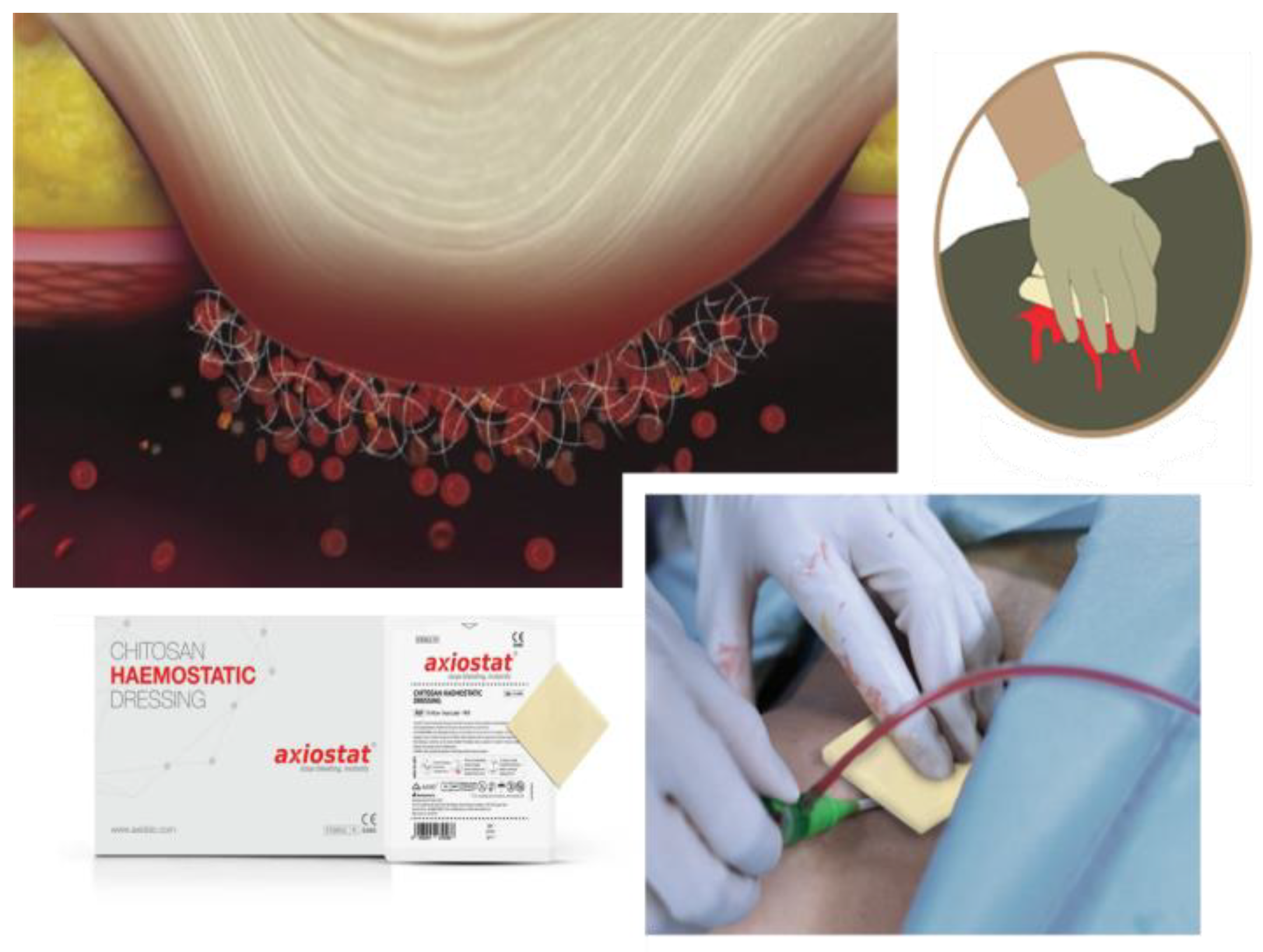
3.3. Axiostat
3.4. TraumaStat
3.5. ChitoSeal
4. Surgical Applications of Chitosan Dressings
5. Conclusions and Future Perspectives
Author Contributions
Funding
Institutional Review Board Statement
Data Availability Statement
Conflicts of Interest
References
- Mohamed, E.; Fitzgerald, A.; Tsuzuki, T. The Role of Nanoscale Structures in the Development of Topical Hemostatic Agents. Mater. Today Nano 2021, 16, 100137. [Google Scholar] [CrossRef]
- Moore, E.E.; Moore, H.B.; Kornblith, L.Z.; Neal, M.D.; Hoffman, M.; Mutch, N.J.; Schöchl, H.; Hunt, B.J.; Sauaia, A. Trauma-Induced Coagulopathy. Nat. Rev. Dis. Primers 2021, 7, 30. [Google Scholar] [CrossRef]
- Malik, A.; Rehman, F.U.; Shah, K.U.; Naz, S.S.; Qaisar, S. Hemostatic Strategies for Uncontrolled Bleeding: A Comprehensive Update. J. Biomed. Mater. Res. B Appl. Biomater. 2021, 109, 1465–1477. [Google Scholar] [CrossRef] [PubMed]
- Chiara, O.; Cimbanassi, S.; Bellanova, G.; Chiarugi, M.; Mingoli, A.; Olivero, G.; Ribaldi, S.; Tugnoli, G.; Basilicò, S.; Bindi, F.; et al. A systematic review on the use of topical hemostats in trauma and emergency surgery. BMC Surg. 2018, 18, 68. [Google Scholar] [CrossRef] [PubMed]
- Shah, A.; Palmer, A.J.R.; Klein, A.A. Strategies to Minimize Intraoperative Blood Loss during Major Surgery. Br. J. Surg. 2020, 107, e26–e38. [Google Scholar] [CrossRef] [PubMed]
- Zhong, Y.; Hu, H.; Min, N.; Wei, Y.; Li, X.; Li, X. Application and outlook of topical hemostatic materials: A narrative review. Ann. Transl. Med. 2021, 9, 577. [Google Scholar] [CrossRef] [PubMed]
- Zou, C.Y.; Li, Q.J.; Hu, J.J.; Song, Y.T.; Zhang, Q.Y.; Nie, R.; Li-Ling, J.; Xie, H.Q. Design of biopolymer-based hemostatic material: Starting from molecular structures and forms. Mater. Today Bio 2022, 17, 100468. [Google Scholar] [CrossRef] [PubMed]
- Yu, P.; Zhong, W. Hemostatic materials in wound care. Burn. Trauma 2021, 9, tkab019. [Google Scholar] [CrossRef] [PubMed]
- Jiang, S.; Liu, S.; Lau, S.; Li, J. Hemostatic biomaterials to halt non-compressible hemorrhage. J. Mater. Chem. B 2022, 10, 7239–7259. [Google Scholar] [CrossRef]
- Li, X.F.; Lu, P.; Jia, H.R.; Li, G.; Zhu, B.; Wang, X.; Wu, F.G. Emerging materials for hemostasis. Coord. Chem. Rev. 2023, 475, 214823. [Google Scholar] [CrossRef]
- Gheorghiță, D.; Moldovan, H.; Robu, A.; Bița, A.I.; Grosu, E.; Antoniac, A.; Corneschi, I.; Antoniac, I.; Bodog, A.D.; Băcilă, C.I. Chitosan-Based Biomaterials for Hemostatic Applications: A Review of Recent Advances. Int. J. Mol. Sci. 2023, 24, 10540. [Google Scholar] [CrossRef] [PubMed]
- Saini, S.; Dhiman, A.; Nanda, S. Immuno modulatory properties of chitosan: Impact on wound healing and tissue repair. Endocr. Metab. Immune Disord. Drug Targets 2020, 20, 1611–1623. [Google Scholar] [CrossRef] [PubMed]
- Curcio, F.; Perri, P.; Piro, P.; Galassi, S.; Sole, R.; Trombino, S.; Cassano, R. Synthetic Haemostatic Sealants: Effectiveness, Safety, and In Vivo Applications. Pharmaceuticals 2024, 17, 288. [Google Scholar] [CrossRef]
- Feng, P.; Luo, Y.; Ke, C.; Qiu, H.; Wang, W.; Zhu, Y.; Hou, R.; Xu, L.; Wu, S. Chitosan-based functional materials for skin wound repair: Mechanisms and applications. Front. Bioeng. Biotechnol. 2021, 9, 650598. [Google Scholar] [CrossRef] [PubMed]
- Wang, Y.-W.; Liu, C.-C.; Cherng, J.-H.; Lin, C.-S.; Chang, S.-J.; Hong, Z.-J.; Liu, C.-C.; Chiu, Y.-K.; Hsu, S.-D.; Chang, H. Biological effects of chitosan-based dressing on hemostasis mechanism. Polymers 2019, 11, 1906. [Google Scholar] [CrossRef] [PubMed]
- Bhoyar, S.D.; Malhotra, K.; Madke, B. Dressing materials: A comprehensive review. J. Cutan. Aesthetic Surg. 2023, 16, 81–89. [Google Scholar] [CrossRef] [PubMed]
- Notario-Pérez, F.; Martín-Illana, A.; Cazorla-Luna, R.; Ruiz-Caro, R.; Veiga, M.D. Applications of Chitosanin Surgical and Post-Surgical Materials. Mar. Drugs 2022, 20, 396. [Google Scholar] [CrossRef]
- Qi, L.; Zhang, C.; Wang, B.; Yin, J.; Yan, S. Progress in hydrogels for skin wound repair. Macromol. Bioscience. 2022, 22, 2100475. [Google Scholar] [CrossRef]
- Trombino, S.; Curcio, F.; Di Gioia, M.L.; Armentano, B.; Poerio, T.; Cassano, R. Multifunctional Membranes Based on β-Glucans and Chitosan Useful in Wound Treatment. Membranes 2022, 12, 121. [Google Scholar] [CrossRef]
- Jones, M.; Kujundzic, M.; John, S.; Bismarck, A. Crab vs. Mushroom: A Review of Crustacean and Fungal Chitin in Wound Treatment. Mar. Drugs 2020, 18, 64. [Google Scholar] [CrossRef]
- Lim, C.; Hwang, D.S.; Lee, D.W. Intermolecular interactions of chitosan: Degree of acetylation and molecular weight. Carbohydr. Polym. 2021, 259, 117782. [Google Scholar] [CrossRef] [PubMed]
- Gatto, M.; Ochi, D.; Yoshida, C.M.P.; da Silva, C.F. Study of chitosan with different degrees of acetylation as cardboard paper coating. Carbohydr. Polym. 2019, 210, 56–63. [Google Scholar] [CrossRef] [PubMed]
- Hu, Z.; Zhang, D.Y.; Lu, S.T.; Li, P.W.; Li, S.D. Chitosan-based composite materials for prospective hemostatic applications. Mar. Drugs 2018, 16, 273. [Google Scholar] [CrossRef] [PubMed]
- Cao, S.; Xu, G.; Li, Q.; Zhang, S.; Yang, Y.; Chen, J. Double cross linking chitosan sponge with antibacterial and hemostatic properties for accelerating wound repair. Compos. Part B Eng. 2022, 234, 109746. [Google Scholar] [CrossRef]
- Jin, H.; Wang, Z. Advances in alkylated chitosan and its applications for hemostasis. Macromol 2022, 2, 346–360. [Google Scholar] [CrossRef]
- Shao, H.; Wu, X.; Xiao, Y.; Yang, Y.; Ma, J.; Zhou, Y.; Chen, W.; Qin, S.; Yang, J.; Wang, R.; et al. Recent research advances on polysaccharide-, peptide-, and protein-based hemostatic materials: A review. Int. J. Biol. Macromol. 2024, 261, 129752. [Google Scholar] [CrossRef] [PubMed]
- Kim, K.; Ryu, J.H.; Koh, M.-Y.; Yun, S.P.; Kim, S.; Park, J.P.; Jung, C.-W.; Lee, M.S.; Seo, H.-I.; Kim, J.H.; et al. Coagulopathy-independent, bioinspired hemostatic materials: A full research story from preclinical models to a human clinical trial. Sci. Adv. 2021, 7, eabc9992. [Google Scholar] [CrossRef] [PubMed]
- Lee, V.K.; Lee, T.; Ghosh, A.; Saha, T.; Bais, M.V.; Bharani, K.K.; Chag, M.; Parikh, K.; Bhatt, P.; Namgung, B.; et al. An architecturally rational hemostat for rapid stopping of massive bleeding on anticoagulation therapy. Proc. Natl. Acad. Sci. USA 2024, 121, e2316170121. [Google Scholar] [CrossRef] [PubMed]
- Guo, Y.; Wang, M.; Liu, Q.; Liu, G.; Wang, S.; Li, J. Recent Advances in the Medical Applications of Hemostatic Materials. Theranostics 2023, 13, 161–196. [Google Scholar] [CrossRef]
- Sultankulov, B.; Berillo, D.; Sultankulova, K.; Tokay, T.; Saparov, A. Progress in the Development of Chitosan-Based, Biomaterials for Tissue Engineering and Regenerative Medicine. Biomolecules 2019, 9, 470. [Google Scholar] [CrossRef]
- Shakiba-Marani, R.; Ehtesabi, H. A Flexible and Hemostatic Chitosan, Polyvinyl Alcohol, Carbon Dot Nanocomposite Sponge for Wound Dressing Application. Int. J. Biol. Macromol. 2023, 224, 831–839. [Google Scholar] [CrossRef] [PubMed]
- Peng, H.T. Hemostatic agents for prehospital hemorrhage control: A narrative review. Mil. Med. Res. 2020, 7, 13. [Google Scholar] [CrossRef]
- Mu, L.; Wu, L.; Wu, S.; Ye, Q.; Zhong, Z. Progress in chitin/chitosan and their derivatives for biomedical applications: Where we stand. Carbohydr. Polym. 2024, in press. [Google Scholar] [CrossRef]
- Chen, K.Y.; Chen, Y.C.; Lin, T.H.; Yang, C.Y.; Kuo, Y.W.; Lei, U. Hemostatic enhancement via chitosan is independent of classical clotting pathways—A quantitative study. Polymers 2020, 12, 2391. [Google Scholar] [CrossRef] [PubMed]
- Zhou, X.; Zhang, X.; Zhou, J.; Li, L. An investigation of chitosan and its derivatives on red blood cell agglutination. RSC Adv. 2017, 7, 12247–12254. [Google Scholar] [CrossRef]
- Chen, Z.; Yao, X.; Liu, L.; Guan, J.; Liu, M.; Li, Z.; Yang, J.; Huang, S.; Wu, J.; Tian, F.; et al. Blood coagulation evaluation of N-alkylated chitosan. Carbohydr. Polym. 2017, 173, 259–268. [Google Scholar] [CrossRef] [PubMed]
- Gordy, S.D.; Rhee, P.; Schreiber, M.A. Military applications of novel hemostatic devices. Expert Rev. Med. Devices 2011, 8, 41–47. [Google Scholar] [CrossRef] [PubMed]
- Morin-Crini, N.; Lichtfouse, E.; Torri, G.; Crini, G. Applications of chitosan in food, pharmaceuticals, medicine, cosmetics, agriculture, textiles, pulp and paper, biotechnology, and environmental chemistry. Environ. Chem. Lett. 2019, 17, 1667–1692. [Google Scholar] [CrossRef]
- Zou, Y.; Jin, X.; Zhang, X.; Kong, X.; Zhang, Q.; Xie, X.; Liu, C.; Ke, L.; Liu, W.; Wang, W. A multifunctional biomedical patch based on hyperbranched epoxy polymer and MXene. Sci. China Technol. Sci. 2021, 64, 2744–2754. [Google Scholar] [CrossRef]
- Ribeiro, D.M.L.; Júnior, A.R.C.; de Macedo, G.H.R.V.; Chagas, V.L.; Silva, L.d.S.; Cutrim, B.d.S.; Santos, D.M.; Soares, B.L.L.; Zagmignan, A.; Miranda, R.d.C.M.d.; et al. Polysaccharide-based formulations for healing of skin-related wound infections: Lessons from animal models and clinical trials. Biomolecules 2019, 10, 63. [Google Scholar] [CrossRef]
- Jangid, N.K.; Hada, D.; Rathore, K. Chitosan as an emerging object for biological and biomedical applications. J. Polym. Eng. 2019, 39, 689–703. [Google Scholar] [CrossRef]
- Salis, A.; Rassu, G.; Budai-Szűcs, M.; Benzoni, I.; Csányi, E.; Berkó, S.; Maestri, M.; Dionigi, P.; Porcu, E.P.; Gavini, E.; et al. Development of thermosensitive chitosan/glicerophospate injectable in situ gelling solutions for potential application in intraoperative fluorescence imaging and local therapy of hepatocellular carcinoma: A preliminary study. Expert Opin. DrugDeliv. 2015, 12, 1583–1596. [Google Scholar] [CrossRef] [PubMed]
- Fusteș-Dămoc, I.; Măluțan, T.; Mija, A. High content chitosan-based materials with high performance properties. Int. J. Biol. Macromol. 2022, 223, 263–272. [Google Scholar] [CrossRef] [PubMed]
- Valachová, K.; Šoltés, L. Versatile Use of Chitosan and Hyaluronan in Medicine. Molecules 2021, 26, 1195. [Google Scholar] [CrossRef] [PubMed]
- Littlejohn, L.F.; Devlin, J.J.; Kircher, S.S.; Lueken, R.; Melia, M.R.; Johnson, A.S. Comparison of Celox-A, ChitoFlex, WoundStat, and combat gauze hemostatic agents versus standard gauze dressing in control of hemorrhage in a swine model of penetrating trauma. Acad. Emerg. Med. 2011, 18, 340–350. [Google Scholar] [CrossRef] [PubMed]
- Pourshahrestani, S.; Zeimaran, E.; Kadri, N.A.; Mutlu, N.; Boccaccini, A.R. Polymeric hydrogel systems as emerging biomaterial platforms to enable hemostasis and wound healing. Adv. Healthc. Mater. 2020, 9, 2000905. [Google Scholar] [CrossRef]
- Gustafson, S.B.; Fulkerson, P.; Bildfell, R.; Aguilera, L.; Hazzard, T.M. Chitosan dressing provides hemostasis in swine femoral arterial injury model. Prehospital Emerg. Care 2007, 11, 172–178. [Google Scholar] [CrossRef] [PubMed]
- Clay, J.G.; Grayson, J.K.; Zierold, D. Comparative testing of new hemostatic agents in a swine model of extremity arterial and venous hemorrhage. Mil. Med. 2010, 175, 280–284. [Google Scholar] [CrossRef]
- Welch, M.; Barratt, J.; Peters, A.; Wright, C. Systematic review of prehospital haemostatic dressings. BMJ Mil. Health 2020, 166, 194–200. [Google Scholar] [CrossRef]
- Kong, C.; Chen, S.; Wang, X.; Hu, C.; Li, B.; Fu, R.; Zhang, J. Hemoadhican, a Tissue Adhesion Hemostatic Material Independent of Blood Coagulation. Adv. Healthc. Mater. 2023, 12, 2300705. [Google Scholar] [CrossRef]
- Hu, B.; Bao, G.; Xu, X.; Yang, K. Topical hemostatic materials for coagulopathy. J. Mater. Chem. B 2022, 10, 1946–1959. [Google Scholar] [CrossRef] [PubMed]
- Singh, G.; Nayal, A.; Malhotra, S.; Koul, V. Dual functionalized chitosan based composite hydrogel for haemostatic efficacy and adhesive property. Carbohydr. Polym. 2020, 247, 116757. [Google Scholar] [CrossRef] [PubMed]
- Sánchez-Machado, D.I.; López-Cervantes, J.; Martínez-Ibarra, D.M.; Escárcega-Galaz, A.A.; Vega-Cázarez, C.A. The use of chitosan as a skin-regeneration agent in burns injuries: A review. e-Polymers 2022, 22, 75–86. [Google Scholar] [CrossRef]
- Gupta, R.; Mohanty, S.; Verma, D. Current status of hemostatic agents, their mechanism of action, and future directions. J. Bioact. Compat. Polym. 2023, 38, 77–105. [Google Scholar] [CrossRef]
- Ghosh, S.; Sarkhel, S.; Ghosh, K.; Dhar, S.; Karar, S.; Roychowdhury, V. Plant-Derived Hemostats. Rev. Bras. Farmacogn. 2023, 33, 259–271. [Google Scholar] [CrossRef]
- Sinha, N.; Mazumdar, A.; Mitra, J.; Sinha, G.; Baunthiyal, S.; Baunthiyal, S. Chitosan based Axiostat dental dressing following extraction in cardiac patients under antiplatelet therapy. Int. J. Oral. Health Med. Res. 2017, 3, 65–67. [Google Scholar]
- Zadeh Mehrizi, T.; Shafiee Ardestani, M.; Rezayat, S.M.; Javanmard, A. A review study of the use of modified chitosan as a new approach to increase the preservation of blood products (erythrocytes, platelets, and plasma products): 2010–2022. Nanomed. J. 2023, 10, 16–32. [Google Scholar]
- Gao, Y.; Sarode, A.; Kokoroskos, N.; Ukidve, A.; Zhao, Z.; Guo, S.; Flaumenhaft, R.; Gupta, A.S.; Saillant, N.; Mitragotri, S. A polymer-based systemic hemostatic agent. Sci. Adv. 2020, 6, eaba0588. [Google Scholar] [CrossRef]
- Wang, L.; You, X.; Dai, C.; Tong, T.; Wu, J. Hemostatic nanotechnologies for external and internal hemorrhage management. Biomater. Sci. 2020, 8, 4396–4412. [Google Scholar] [CrossRef]
- Xiao, X.; Wu, Z. A Narrative Review of Different Hemostatic Materials in Emergency Treatment of Trauma. Emerg. Med. Int. 2022, 2022, 6023261. [Google Scholar] [CrossRef]
- Meucci, F.; Stolcova, M.; Caniato, F.; Sarraf, M.; Mattesini, A.; Di Mario, C. The Essentials of Femoral Vascular Access and Closure. Interv. Cardiol. Princ. Pract. 2022, 21–33. [Google Scholar] [CrossRef]
- Mistry, P.A.; Konar, M.N.; Latha, S.; Chadha, U.; Bhardwaj, P.; Eticha, T.K. Chitosan superabsorbent biopolymers in sanitary and hygiene applications. Int. J. Polym. Sci. 2023, 4717905. [Google Scholar] [CrossRef]
- Ousey, K.; Rippon, M.G.; Rogers, A.A.; Totty, J.P. Considerations for an ideal post-surgical wound dressing aligned with antimicrobial stewardship objectives: A scoping review. J. Wound Care 2023, 32, 334–347. [Google Scholar] [CrossRef]
- Matica, M.A.; Aachmann, F.L.; Tøndervik, A.; Sletta, H.; Ostafe, V. Chitosan as a Wound Dressing Starting Material: Antimicrobial Properties and Mode of Action. Int. J. Mol. Sci. 2019, 20, 5889. [Google Scholar] [CrossRef]
- Tang, W.; Wang, J.; Hou, H.; Li, Y.; Wang, J.; Fu, J.; Lu, L.; Gao, D.; Liu, Z.; Zhao, F.; et al. Application of chitosan and its derivatives in medical materials. Int. J. Biol. Macromol. 2023, 240, 124398. [Google Scholar] [CrossRef] [PubMed]
- Xia, Y.; Yang, R.; Wang, H.; Li, Y.; Fu, C. Application of chitosan-based materials in surgical or postoperative hemostasis. Front. Mater. 2022, 9, 994265. [Google Scholar] [CrossRef]
- Kantak, M.N.; Bharate, S.S. Analysis of clinical trials on biomaterial and therapeutic applications of chitosan: A review. Carbohydr. Polym. 2022, 278, 118999. [Google Scholar] [CrossRef]
- Vallabh, K. Acute upper limb ischemia. In Vascular Emergencies: Expert Management for the Emergency Physician; Cambridge University Press: Cambridge, UK, 2013; p. 109. [Google Scholar]
- Bizari, D.; Khoshmohabat, H.; SalahshourKordestani, S.; Zarepur, R. Comparison of HemoFoam® and Conventional Gauze Dressing on Hemostasis of Vascular Access Site in Hemodialysis Patients. Galen Med. J. 2019, 8, 1395. [Google Scholar] [CrossRef]
- Binnetoglu, K.; Kumandas, A.; Ekici, H.; Ozbaykus, A.C.; Tiryaki, M. Comparison of the algan hemostatic agent with celox in rat femoral artery bleeding model. Medicine 2021, 10, 1469–1473. [Google Scholar] [CrossRef]
- Minici, R.; Serra, R.; Maglia, C.; Guzzardi, G.; Spinetta, M.; Fontana, F.; Venturini, M.; Laganà, D. Efficacy and Safety of Axiostat® Hemostatic Dressing in Aiding Manual Compression Closure of the Femoral Arterial Access Site in Patients Undergoing Endovascular Treatments: A preliminary Clinical Experience in Two Centers. J. Pers. Med. 2023, 13, 812. [Google Scholar] [CrossRef]
- Radhakrishna, S.; Shukla, V.; Shetty, S.K. Is chitosan dental dressing better than cotton gauze in achieving hemostasis in patients on antithrombotics? J. Oral Maxillofac. Surg. 2023, 81, 224–231. [Google Scholar] [CrossRef] [PubMed]
- Lestari, W.; Yusry, W.N.A.W.; Haris, M.S.; Jaswir, I.; Idrus, E. A glimpse on the function of chitosan as a dental hemostatic agent. Jpn Dent. Sci. Rev. 2020, 56, 147–154. [Google Scholar] [CrossRef] [PubMed]
- Rajendra, K.; Vempalli, S.; Kadiyala, M.; Sharma, V.; Karipineni, S.; Gunturu, S.; Patil, D.B. Effect of platelet-rich fibrin versus chitosan-based Axiostat hemostatic agent following dental extraction in cardiac patients on antiplatelet therapy: A comparative study. Natl. J. Maxillofac. Surg. 2021, 12, 361–366. [Google Scholar] [PubMed]


| Commercial Name | Form | Composition | Mode of Action |
|---|---|---|---|
| HemCon | Rigid and hard sponge bandage | Non-woven chitosan fibers | Amine groups on chitosan interact with RBCs and induce clotting |
| HemCon Patch® Pro | Flexible and contouring dressing | Chitosan acetate | With anionic erythrocytes, the chitosan salts of the device ‘cross-link’ rapidly, adhering strongly to the wound surface |
| HemCon® Bandage | Impregnated dressing | Chitosan acetate | Freeze-dried chitosan acetate salt, mainly used for emergencies to stop blood loss, enhance platelet function |
| HemCon ChitoFlex | Rolled chitosan gauze | Chitosan | Chitosan binds to red blood cells and platelets and forms a mucoadhesive physical barrier around the bleeding site |
| Celox | Dressing based on chitosan granules and flake form | Chitosan | Swelling of the chitosan granules |
| Axiostat | Highly porous chitosan matrix | Chitosan | Electrostatic interaction between positively charged biopolymer of Axiostat and the negatively charged blood cells |
| TraumaStat | Chitosan gauze | Polyethylene fibers, coated with chitosan and filled with precipitated silica | Mobilization of Ca2+ to increase platelet activation and coagulation factor activation for clot formation |
| ChitoSeal | Chitosan dressing | Supported with a cellulose coating | Amine groups of chitosan interact with RBCs and induce clotting |
Disclaimer/Publisher’s Note: The statements, opinions and data contained in all publications are solely those of the individual author(s) and contributor(s) and not of MDPI and/or the editor(s). MDPI and/or the editor(s) disclaim responsibility for any injury to people or property resulting from any ideas, methods, instructions or products referred to in the content. |
© 2024 by the authors. Licensee MDPI, Basel, Switzerland. This article is an open access article distributed under the terms and conditions of the Creative Commons Attribution (CC BY) license (https://creativecommons.org/licenses/by/4.0/).
Share and Cite
Cassano, R.; Perri, P.; Scarcello, E.; Piro, P.; Sole, R.; Curcio, F.; Trombino, S. Chitosan Hemostatic Dressings: Properties and Surgical Applications. Polymers 2024, 16, 1770. https://doi.org/10.3390/polym16131770
Cassano R, Perri P, Scarcello E, Piro P, Sole R, Curcio F, Trombino S. Chitosan Hemostatic Dressings: Properties and Surgical Applications. Polymers. 2024; 16(13):1770. https://doi.org/10.3390/polym16131770
Chicago/Turabian StyleCassano, Roberta, Paolo Perri, Edoardo Scarcello, Paolo Piro, Roberta Sole, Federica Curcio, and Sonia Trombino. 2024. "Chitosan Hemostatic Dressings: Properties and Surgical Applications" Polymers 16, no. 13: 1770. https://doi.org/10.3390/polym16131770
APA StyleCassano, R., Perri, P., Scarcello, E., Piro, P., Sole, R., Curcio, F., & Trombino, S. (2024). Chitosan Hemostatic Dressings: Properties and Surgical Applications. Polymers, 16(13), 1770. https://doi.org/10.3390/polym16131770