1. Introduction
Wound healing is a complex and dynamic physiological process triggered by a cellular response to injury and composed of overlapping and orchestrated phases, such as hemostasis, inflammation, proliferation, and remodeling. Each of these phases is tightly regulated by the interplay between cells such as keratinocytes, fibroblasts, leukocytes, macrophages, endothelial cells, and platelets. These cells release an array of growth factors and cytokines, such as transforming growth factor-beta (TGF-β), vascular endothelial growth factor (VEGF), interleukin-6 (IL-6), and tumor necrosis factor-alpha (TNF-α), which coordinate the progression of the wound healing process and ensure successful tissue repair [
1,
2,
3].
Diabetes mellitus (DM) is a chronic metabolic disorder characterized by persistently high blood glucose levels, which lead to various systemic and local complications. Diabetic microangiopathy is one of the most common complications of hyperglycemia. This vascular damage compromises the skin’s barrier function and delays wound healing. The poor healing of diabetic wounds is mainly characterized by prolonged inflammation, which disrupts normal progression through the healing phases and leads to stagnation of the proliferative phase. Furthermore, the extracellular matrix in diabetic wounds tends to exhibit reduced collagen deposition, a factor critical for wound strength and tissue remodeling [
4,
5,
6]. These changes in the healing process of diabetic patients are compounded by imbalances in the plasmatic concentrations of pro-inflammatory cytokines such as IL-6, TNF-α, and VEGF, which further exacerbate the impaired healing response in diabetic individuals [
7,
8].
Diabetic wounds bring great pain to patients and impose an economic burden on individuals and society, thus requiring treatment strategies that are more effective and less expensive [
9,
10]. Bacterial cellulose (BC)-based dressings are an affordable and effective strategy for the treatment of skin lesions [
11,
12,
13,
14], as are melatonin-based products that have proven effects on healing [
15,
16,
17,
18,
19].
Therefore, this study aims to evaluate the effects of an innovative, accessible, and effective dressing produced based on bacterial cellulose and used in conjunction with melatonin and to describe its effects on the regulation and expression of inflammatory cytokines, VEGF, PCNA, and collagen in the healing of cutaneous wounds in an experimental model of rats with diabetes.
2. Method
2.1. Experimental Design
Albino rats (Rattus norvegicus albinus) of the Wistar lineage, 60 days old, weighing approximately 250 ± 30 g, from the vivarium of the Department of Animal Morphology and Physiology (DMFA) of the Federal Rural University of Pernambuco (UFRPE) were used. The animals were maintained in an environment with a controlled temperature (22 ± 1 °C) and photoperiod (12 h light and 12 h dark), and food and water were provided ad libitum.
The animals were evaluated daily, during an experimental period of 14 days and randomly divided into 4 groups, with 4 animals in each, as described below:
GC—control group with non-diabetic rats; GDCC—diabetic rats treated with a commercial healing agent; GDCB—diabetic rats treated with bacterial cellulose; and GDMCB—diabetic rats treated with melatonin and bacterial cellulose.
This study followed the principles established by the Brazilian laws on the use and creation of animals (n° 11.794/2008), which regulate research with animals in Brazil. The study was approved by the Ethics Committee of the Use of Animals of the Federal Rural University of Pernambuco (CEUA—UFRPE) under registration no. 052/2019.
2.2. Diabetes Induction
Induction was performed by intraperitoneal administration of a streptozotocin solution (Sigma Chemical Co., St. Louis, MO, USA) after a 14 h fasting period. Streptozotocin was diluted in 10 mM sodium citrate buffer with pH 4.5, and a single dose consisted of 60 mg/kg of animal weight. The non-diabetic animals received equivalent doses of saline solution. After 30 min, all animals were fed normally. Diabetes was confirmed 5 days after induction. Only animals that presented glycemia above 200 mg/dL (Accu-Chek Activ Kit glucometer, Accu-Chek, Singapore) were included in the study, except for the control group [
20].
2.3. Melatonin Treatment
Melatonin, N-acetyl-5-methoxytryptamine (Sigma Chemical Co., St. Louis, MO, USA), was administered in daily injections intraperitoneally from 6:00 p.m. to 7:00 p.m. at 10 mg/kg; it was dissolved in 0.2 mL of ethanol and diluted in 0.9 mL of 0.9% NaCl.
2.4. Treatment with Bacterial Cellulose and Commercial Healing
A bacterial cellulose (BC) hydrogel and a commercial healing agent (CC) were applied directly to each animal’s wound once a day with a dose of approximately 0.4 mL. The commercial healing agent used was an amorphous hydrogel with alginate, chosen because of its wide use and similarities to the BC hydrogel regarding physicochemical characteristics. The BC hydrogel was produced from sugarcane molasses at the Sugarcane Experimental Station (EECC) of the Federal Rural University of Pernambuco (UFRPE) and supplied by the company POLISA®, specializing in biopolymers for health. Bacterial cellulose is produced by propagating Gluconacetobacter hansenii in a sterile culture obtained from sugarcane molasses. The polymeric mass is mechanically processed to obtain the gel and, subsequently, the hydrogel. All stages of production are carried out in accordance with the standard operating procedure, a process developed and patented by Polisa Biopolímeros para Saúde Ltda. Samples of BC hydrogel dressings tested in this study were prepared from a 0.7% concentration.
2.5. Immunohistochemistry
For immunohistochemical analysis, silanized slides were deparaffinized and rehydrated in xylene and alcohols. Antigenic retrieval was performed using a citrate buffer solution (pH 8.0) at a high temperature in the microwave for 5 min. Endogenous peroxidase was inhibited using a solution of hydrogen peroxide (3%) in methanol. The nonspecific antigen–antibody reaction was blocked by incubating the slides in PBS and 5% bovine serum albumin (BSA) for one hour. IL-6 (sc-32296, Santa Cruz Biotechnology, Santa Cruz, CA, USA), TNFα (sc-33639, Santa Cruz Biotechnology, Santa Cruz, CA, USA), VEGF (MBS2540134, MyBioSource, San Diego, CA, USA), and PCNA (Santa Cruz Biotechnology, Santa Cruz, CA, USA) antibodies were diluted 1:50 in 1% PBS/BSA and incubated for 1 h. Subsequently, the slides were treated with the secondary antibody for 30 min. The antigen–antibody reaction was observed through a brown precipitate after application of 3,3′-diaminobenzidine for four minutes and counterstained with hematoxylin. Images of the IL6, TNFα, and VEGF slides were captured using a Sony
® Video camera, coupled to the Olympus
® Bx50 microscope, and the images were submitted to the Gimp 2.0 application for quantification through the use of an RGB Histogram (red–green–blue) [
21,
22].
For PCNA, cell counts were performed using a Weibel graticule with 25 observation points in a 10× eyepiece. Three slides per group were used, and four fields/slide were analyzed. In each field, 300 cells were counted and transformed into percentage of marked cells [
23].
2.6. Evaluation and Quantification of Collagen I and III
For the evaluation and quantification of type I and III collagen, the sections were stained using the Picrosirius Red histochemical technique. After mounting with Entellan
®, the slides were photomicrographed under polarized light in an Axio microscope (Imager.M2m/Zeiss, Oberkochen, Germany) with an AxioCam camera (HRc/Zeiss) attached. Five images per section were obtained under 200× magnification and processed using the ImageJ
® software version 1.52, where color markings were made by selecting the corresponding pixels in all images. In this way, the percentage of area occupied in the image by each of the two markings was obtained. For collagen type I, pixels were marked in red, and for type III, pixels were marked in green [
24].
3. Results
During the experimental period of this study, there were no animal losses, and no complications, including procedure-related infection, were observed.
3.1. Immunohistochemistry (IL-6, TNF-α, VEGF, and PCNA)
Immunohistochemistry for IL-6 in the wounds showed weak staining after 14 days in the GDCB and GDMCB groups, whereas the GC and GDCC groups showed a strong staining pattern. Regarding the quantification of IL-6 expression in pixels, we observed a significant decrease in pixels in the GDMCB group compared to the other groups (
Figure 1).
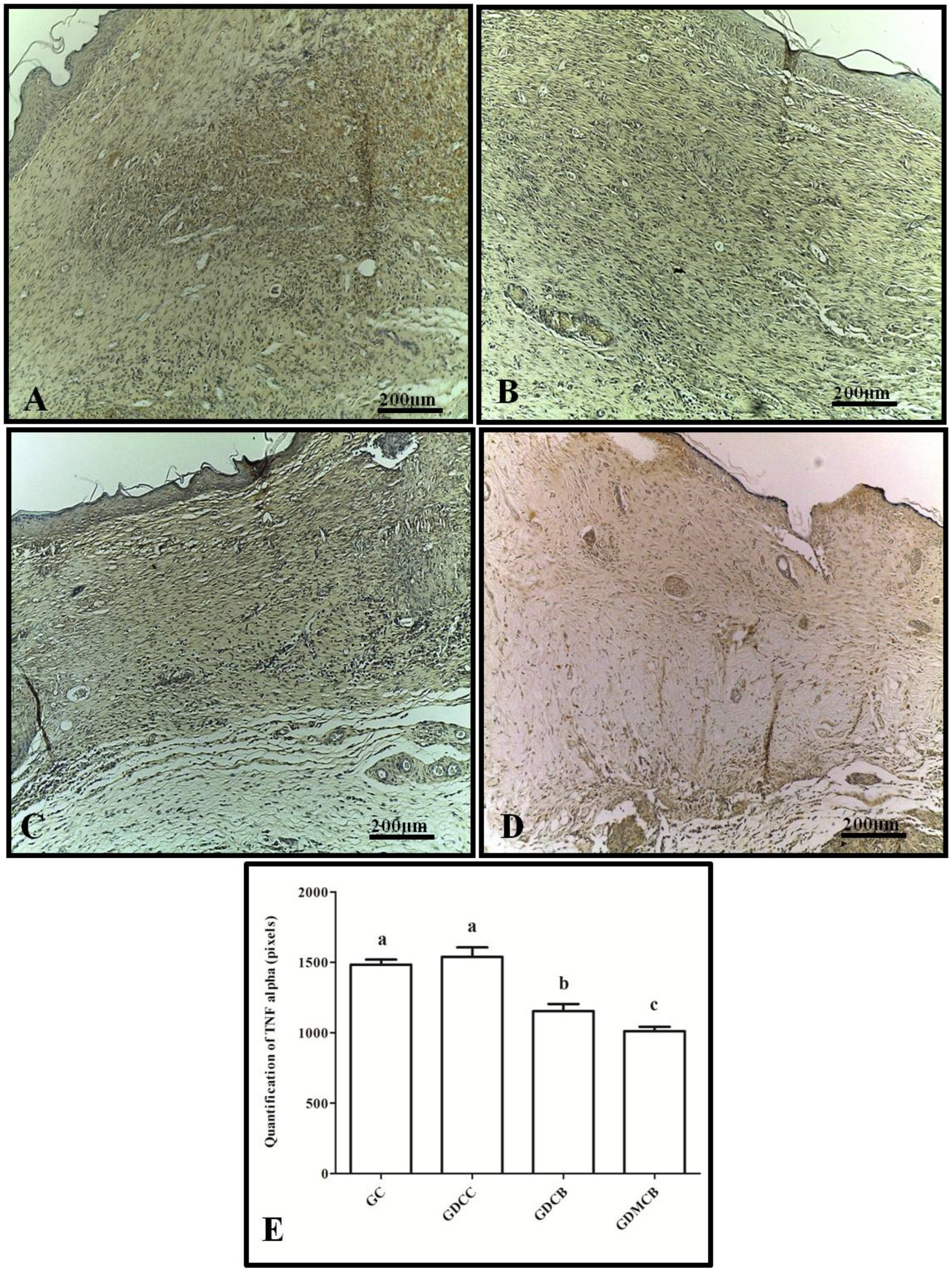
The analysis of TNF-α in the wounds revealed weak staining after 14 days in the GDCB and GDMCB groups, whereas the GC and GDCC groups showed a strong staining pattern, but no statistical difference was found. Regarding quantification of TNF-α expression in pixels, we observed a significant decrease in pixels in the GDMCB group compared to the other groups (
Figure 2).
For VEGF, the wounds showed weak staining after 14 days in the GDCB and GDMCB groups, whereas the GC and GDCC groups showed a strong staining pattern, but no statistical difference was found. Regarding the quantification of VEGF expression in pixels, we observed a significant decrease in pixels in the GDCB and GDMCB groups compared to the other groups (
Figure 3).
Regarding cell proliferation, PCNA was marked in all groups in the epithelial layer, and marking was weak after 14 days in the GC and GCC groups, without statistical difference, whereas the GDCB and GDMCB groups showed a strong staining pattern but no statistical difference. Regarding the quantification of positive PCNA cells, we observed an increase in the proliferation indices, along with a significant increase in pixels in the GDCB and GDMCB groups compared to the other experimental groups (
Figure 4).
3.2. Evaluation and Quantification of Collagen I and III
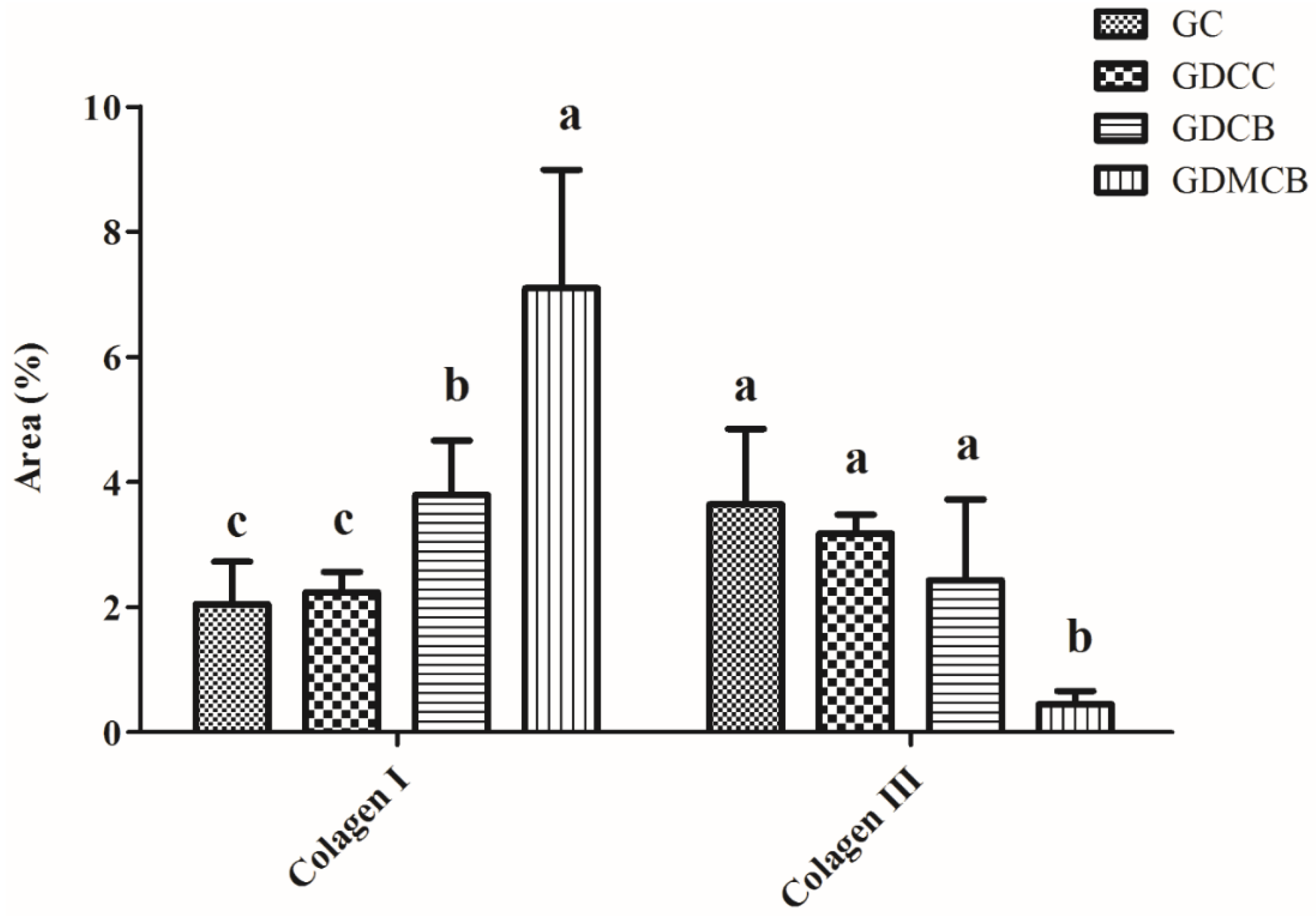
Quantification of type I collagen (mature collagen) revealed a larger concentration (by area quantification) in the GDCB and GDMCB groups, and quantification of type III collagen (immature) revealed a smaller concentration in the GDMCB group at 14 days after operation (
Figure 5).
The evaluation of the collagen fibers showed that the GDMCB group presented organized thick type I collagen fibers parallel to the surface, whereas the GC and GDCC groups, after 14 days, still presented a predominance of type III collagen fibers in fragmented and randomly arranged forms and few type I fibers (
Figure 6).
4. Discussion
Here, we demonstrate that the application of melatonin in conjunction with the use of a bacterial cellulose dressing significantly accelerates skin repair in an experimental model of diabetes. The combined treatment appears to mediate the production of cytokines and collagen, both critical elements in wound healing, especially in diabetic conditions where repair processes are often compromised. By analyzing the effects of this combination therapy, we provide new insights into the mechanisms underlying tissue repair in a hyperglycemic environment.
4.1. Bacterial Cellulose in Wound Healing
The bacterial cellulose hydrogel presented in this study was produced microbiologically using the bacterium
Gluconacetobacter hansenii, with sugarcane molasses (
Saccharum officinarum) being used as the raw material. From this bacterial cellulose hydrogel, many biomedical byproducts can be generated, such as perforated membranes, intact membranes, tubes, films, and sponges. These byproducts can be used for various purposes, such as urethral reconstruction, tympanic membrane perforation, and other applications [
14].
Bacterial cellulose films were used on animals with skin wounds of various sizes, caused by different traumas or tumor excisions requiring healing by secondary intention. In this study, it was observed that granulation tissue demonstrated accelerated growth in the initial phase of re-epithelialization, occurring, on average, by the fifth day after the start of treatment, and the granulation tissue filled the entire wound space. Regarding the healing time, all wounds treated with the sugarcane-derived biopolymer progressed without complications, showing a shorter healing time compared to wounds of similar size [
11].
In more recent studies involving humans, bacterial cellulose was used as a dressing in the form of membranes for the treatment of varicose ulcers. The size of the dressing varied from 2 × 2 cm to 6 × 60 cm, and the thickness varied from 0.01 to 0.02 mm. This study compared the use of bacterial cellulose with a control group that received conventional treatment with triglyceride oil. The efficacy of the treatment was evaluated based on the degree of wound healing, the size of the wound area, tissue characteristics during the healing process, and the number of fully healed wounds. This study demonstrated that the bacterial cellulose membrane has ideal properties as a dressing, as it maintains moisture in the wound bed, absorbs excess exudate, limits infectious processes, and protects the wound from mechanical trauma [
12].
In a new study, perforated membranes coupled with a sponge, both made of bacterial cellulose biopolymer, were used in the form of sterile films measuring 10 × 8 cm and 0.01 to 0.02 mm in thickness for the treatment of adult patients with pressure ulcers [
13]. The primary outcome assessed was the healing process (tissue characteristics), and the secondary outcome was the healing time. It is important to highlight that no signs of toxic reactions associated with the bacterial cellulose biopolymer were observed, demonstrating that it is a non-toxic and biocompatible dressing. The membrane displayed the essential characteristics of an ideal dressing, making it a potential option for wound coverage. The authors concluded that the bacterial cellulose membrane was effective in treating pressure ulcers, as it acted as a granulation tissue inducer, reducing the depth of the wound. Being an innovative, low-cost dressing, it represents an important therapeutic alternative for this challenging condition.
4.2. Melatonin in Wound Healing
Melatonin (N-acetyl-5-methoxytryptamine) is the primary product of the pineal gland, although it can also be secreted by other organs, such as the retina and skin. It has been shown to exert anti-inflammatory effects and is beneficial for the treatment of cardiovascular diseases, neurological disorders, and obesity-related diabetes. One study prepared injectable hydrogels loaded with melatonin and investigated their effects in a full-thickness wound model featuring rats [
15]. When compared to the control group and a hydrogel without melatonin, the melatonin-loaded hydrogel significantly increased the wound closure percentage, promoted granulation tissue proliferation and re-epithelialization, accelerated collagen deposition, and enhanced angiogenesis and collagen III formation, among other healing factors. These results suggest that the melatonin-loaded hydrogel promoted granulation tissue formation and accelerated wound healing.
An in vitro study demonstrated that melatonin promotes the healing of diabetic wounds by regulating the activity of primary keratinocytes derived from rats [
17]. The authors suggested that melatonin reduced high glucose-induced mRNA expression and the release of pro-inflammatory cytokines, including tumor necrosis factor-α, interleukin (IL)-1β, IL-6, and IL-8, in keratinocytes. They also stated that melatonin inhibited oxidative stress and that a high glucose-induced reduction in keratinocyte migration and proliferation, along with increased apoptosis, were counteracted by melatonin treatment. Collectively, these results suggest that melatonin is a potential therapeutic strategy for improving impaired diabetic wound healing by regulating keratinocyte activity.
Finally, a recent study aimed to evaluate the wound healing potential of melatonin when applied to diabetic rat wounds. In this study, a melatonin-containing formulation was developed and topically applied to skin wounds in diabetic rats [
19]. The stability of melatonin in the formulation, macroscopic analysis results and the wound contraction index, histological analysis results, and collagen content were evaluated. The study concluded that topical application of melatonin optimized skin wound healing in diabetic rats by attenuating the inflammatory process and promoting an early increase in mononuclear cells at the wound site. The treatment accelerated cutaneous wound contraction, facilitated early collagen fiber maturation, increased collagen deposition, and promoted faster scar connective tissue formation.
4.3. The Role of Cytokines in Wound Healing
Cytokines play a pivotal role in the regulation of the wound healing process. IL-6, TNF-α, and VEGF are some of many cytokines released during tissue repair and inflammation. These pro-inflammatory cytokines are essential for orchestrating the complex interplay of immune cells, growth factors, and structural proteins that drive tissue regeneration. IL-6, for instance, exhibits dual functions depending on the stage of healing. During the early phases of repair, IL-6 exerts chemoattractive effects on neutrophils, promoting their migration to the wound site and facilitating the clearance of pathogens and debris. In the later stages, IL-6 promotes keratinocyte proliferation, contributing to the re-establishment of the epidermal layer [
25]. Similarly, TNF-α, another critical cytokine, stimulates endothelial cells to produce nitric oxide (NO) and express adhesion molecules, which are essential for leukocyte recruitment and activation at the injury site [
25].
Our findings indicated that the expression of IL-6 and TNF-α was significantly reduced in the group treated with both melatonin and bacterial cellulose at the end of the experiment. This decrease can be attributed to the anti-inflammatory and immunomodulatory effects of melatonin, as shown by previous studies [
18,
20]. Melatonin negatively regulates pro-inflammatory cytokines such as IL-6 and tumor necrosis factor-α, preventing the translocation of nuclear factor kappa B (NF-κB), a transcription factor that drives the production of these pro-inflammatory cytokines. By preventing NF-κB translocation to the nucleus and its binding to DNA, melatonin effectively downregulates the expression of these cytokines, thus attenuating inflammation [
18,
26]. These findings align with previous studies that have highlighted melatonin’s ability to suppress the inflammatory response, particularly in hyperglycemic conditions.
VEGF, a key angiogenic factor, is also crucial for wound healing, particularly in diabetic patients, where impaired angiogenesis is a major obstacle to effective repair. VEGF can be activated by hyperglycemia. Interestingly, our study results revealed a lower quantification of VEGF in the GDCB and GDMCB groups at 14 days after operation. This reduction in VEGF expression likely reflects the advanced stage of wound healing in these groups, where angiogenesis is no longer required because the wound has nearly healed. This supports the hypothesis that a defect in endogenous VEGF production may contribute to the impaired reparative angiogenesis observed in diabetic wounds. Previous studies have shown that the administration of VEGF can improve healing outcomes in diabetic models, further underscoring its role in restoring proper vascularization in impaired wounds [
7,
27,
28].
4.4. Cell Proliferation
Beyond cytokine modulation, our study highlights the influence of bacterial cellulose and melatonin on cell proliferation, particularly in the context of keratinocyte activation and re-epithelialization. Bacterial cellulose hydrogel has been shown to promote cell proliferation, enhance in vitro cell viability, support the differentiation of mesenchymal stem cells, and help in reprogramming the essential macrophage population to promote cell proliferation and tissue repair, facilitating the transition from the inflammatory phase of healing to the proliferative phase of healing [
29,
30,
31,
32,
33]. Studies have also shown that both bacterial cellulose hydrogel and melatonin accelerate re-epithelialization by activating keratinocytes, a critical step in the wound healing process [
17,
33,
34,
35,
36,
37,
38]. In our study, we observed enhanced epithelium labeling, suggesting that both the bacterial cellulose hydrogel and melatonin, whether applied individually or in combination, may act synergistically to improve keratinocyte function, thereby facilitating closure of diabetic wounds. These findings provide promising evidence that this combination therapy could be particularly beneficial in the treatment of chronic wounds in diabetic patients, where re-epithelialization is often delayed.
4.5. The Role of Collagen in Wound Healing
Collagen is essential in wound healing because it provides structural integrity to the newly formed tissue. During the healing process, the initial granulation tissue rich in collagen type III and blood vessels is gradually replaced by collagen type I and a few blood vessels, resulting in tissue that more closely resembles the normal dermal structure of healthy individuals, where collagen type I constitutes approximately 80% of the collagen content in the extracellular matrix of the dermis, while type III collagen accounts for about only 10% [
39]. The present study showed an increase in the amount and thickness of type I collagen at the end of the experiments in the GDMCB group compared to the other groups. This observation is consistent with previous reports that bacterial cellulose can enhance collagen deposition, supporting the maturation of granulation tissue into a more stable and organized dermal matrix [
11,
40,
41]. Furthermore, melatonin has been shown to enhance collagen synthesis, particularly in diabetic models, by mitigating oxidative stress and promoting fibroblast activity [
15,
19,
42]. The combined effects of bacterial cellulose and melatonin in our study suggest that this therapeutic strategy not only accelerates wound closure but also improves the quality of the regenerated tissue, ensuring stronger and more resilient skin.
5. Conclusions
It can be concluded that treating skin lesions of diabetic rats supplemented with melatonin using a bacterial cellulose-based dressing has positive effects in regulating the expression of inflammatory cytokines, vascular endothelial growth factor, and collagen, showing that this association may be a possible therapy approach in wound healing.
By modulating cytokine expression, promoting cell proliferation, and improving collagen deposition, this combination therapy addresses several of the key factors that contribute to impaired healing in diabetes. These findings pave the way for further research into the clinical potential of melatonin and bacterial cellulose in the treatment of chronic wounds, particularly in patients with diabetes.
Author Contributions
J.G.M.d.S.: conceptualization, method, writing, editing, and formal analysis. I.M.F.d.M.: method and writing. É.R.A.: method and writing. G.M.d.O.: method and writing. A.A.d.S.: method and writing. D.N.A.: method, discussion, and reviewing the manuscript. I.M.F.C.: method and writing. F.C.M.P.: supervision, project management, and review. J.L.d.A.A.: supervision, project management, and review. V.W.T.: supervision, project management, funding acquisition, and method. Á.A.C.T.: supervision, project management, funding acquisition, and method. All authors have read and agreed to the published version of the manuscript.
Funding
This study was carried out with the support of Coordenação de Aperfeiçoamento de Pessoal de Nível Superior, Brazil (CAPES), by providing a scholarship, protocol code 88887.611499/2021-00.
Institutional Review Board Statement
The animal study protocol was approved by the Institutional Ethics Committee of Federal Rural University of Pernambuco (protocol code 052/2019, approved on 19 June 2019).
Data Availability Statement
The original contributions presented in the study are included in the article, further inquiries can be directed to the corresponding authors.
Acknowledgments
We are grateful for the support of the Department of Animal Morphology and Physiology and the Graduate Program of Animal Bioscience at the Federal Rural University of Pernambuco (UFRPE). We are also grateful for the support we received from The Coordination for the Improvement of Higher Education Personnel (CAPES), foundation of the Ministry of Education (MEC). The authors also extend their thanks to the Keizo Asami Immunopathology Laboratory (LIKA) and to the Federal University of Pernambuco (UFPE).
Conflicts of Interest
The authors declare no conflict of interest.
References
- Brem, H.; Tomic-Canic, M. Cellular and molecular basis of wound healing in diabetes. J. Clin. Investig. 2007, 117, 1219–1222. [Google Scholar] [CrossRef] [PubMed]
- Chen, C.; Lin, Z.; Liu, W.; Hu, Q.; Wang, J.; Zhuang, X.; Guan, S.; Wu, X.; Hu, T.; Quan, S.; et al. Emodin accelerates diabetic wound healing by promoting anti-inflammatory macrophage polarization. Eur. J. Pharmacol. 2022, 936, 175329. [Google Scholar] [CrossRef] [PubMed]
- Silva, J.G.M.; de Melo, I.M.F.; de Alves, É.R.; de Araújo, A.C.C.; Teixeira, V.W.; Teixeira, Á.A.C. Uso de Melatonina na Cicatrização de Feridas: Uma Revisão. Temas Saúde 2022, 22, 317–329. [Google Scholar] [CrossRef]
- Shamiya, Y.; Ravi, S.P.; Coyle, A.; Chakrabarti, S.; Paul, A. Engineering nanoparticle therapeutics for impaired wound healing in diabetes. Drug Discov. Today 2022, 27, 1156–1166. [Google Scholar] [CrossRef] [PubMed]
- Yang, Y.; Zhao, X.; Yu, J.; Chen, X.; Wang, R.; Zhang, M.; Zhang, Q.; Zhang, Y.; Wang, S.; Cheng, Y.; et al. Bioactive skin-mimicking hydrogel band-aids for diabetic wound healing and infectious skin incision treatment. Bioact. Mater. 2021, 6, 3962–3975. [Google Scholar] [CrossRef]
- Adeleke, O.; Oboh, G.; Adefegha, S.; Osesusi, A. Effect of aqueous extract from root and leaf of Sphenocentrum jollyanum pierre on wounds of diabetic rats: Influence on wound tissue cytokines, vascular endothelial growth factor and microbes. J. Ethnopharmacol. 2022, 293, 115266. [Google Scholar] [CrossRef]
- Bitto, A.; Minutoli, L.; Altavilla, D.; Polito, F.; Fiumara, T.; Marini, H.; Galeano, M.; Calò, M.; Cascio, P.L.; Bonaiuto, M.; et al. Simvastatin enhances VEGF production and ameliorates impaired wound healing in experimental diabetes. Pharmacol. Res. 2008, 57, 159–169. [Google Scholar] [CrossRef]
- Sorkhabi, R.; hossein Ahoor, M.; Haghjo, A.G.; Tabei, E.; Taheri, N. Assessment of tear inflammatory cytokines concentration in patients with diabetes with varying severity of involvement. Exp. Eye Res. 2022, 224, 109233. [Google Scholar] [CrossRef]
- Sinclair, A.J.; Abdelhafiz, A.H.; Forbes, A.; Munshi, M. Evidence-based diabetes care for older people with Type 2 diabetes: A critical review. Diabet. Med. 2019, 36, 399–413. [Google Scholar] [CrossRef]
- Zha, W.; Wang, J.; Guo, Z.; Zhang, Y.; Wang, Y.; Dong, S.; Liu, C.; Xing, H.; Li, X. Efficient delivery of VEGF-A mRNA for promoting diabetic wound healing via ionizable lipid nanoparticles. Int. J. Pharm. 2023, 632, 122565. [Google Scholar] [CrossRef]
- de Oliveira Cardoso Coelho, O.C.; Carrazoni, P.G.; da Cunha Monteiro, V.L.; de Assis Dutra, M.F.; Mota, R.A.; Tenório Filho, F. Biopolímero produzido a partir da cana-de-áçucar para cicatrização cutânea. Acta Cir. Bras. 2002, 17, 11–13. [Google Scholar] [CrossRef]
- Cavalcanti, L.M.; Pinto, F.C.; Oliveira, G.M.; Lima, S.V.; Aguiar, J.L.; Lins, E.M. Efficacy of bacterial cellulose membrane for the treatment of lower limbs chronic varicose ulcers: A randomized and controlled trial. Rev. Col. Bras. Cir. 2017, 44, 72–80. [Google Scholar] [CrossRef] [PubMed]
- de Oliveira, G.M.; Vieira, J.M.S.; da Silva, J.G.M.; de Albuquerque, É.L.M.S.; de Albuquerque, A.V.; de Aguiar, J.L.A.; Pinto, F.C. Curativo de celulose bacteriana para o tratamento de lesões por pressão em pacientes hospitalizados. Rev. Enferm. Atual Derme 2019, 87, 1–9. [Google Scholar] [CrossRef]
- da Silva, J.G.M.; Pinto, F.C.M.; de Oliveira, G.M.; da Silva, A.A.; Campos Júnior, O.; da Silva, R.O.; Teixeira, V.W.; de Melo, I.M.; Paumgartten, F.J.; de Souza, T.P.; et al. Non-clinical safety study of a sugarcane bacterial cellulose hydrogel. Res. Soc. Dev. 2020, 9, e960997932. [Google Scholar] [CrossRef]
- Chen, K.; Tong, C.; Yang, J.; Cong, P.; Liu, Y.; Shi, X.; Liu, X.; Zhang, J.; Zou, R.; Xiao, K.; et al. Injectable melatonin-loaded carboxymethyl chitosan (CMCS)-based hydrogel accelerates wound healing by reducing inflammation and promoting angiogenesis and collagen deposition. J. Mater. Sci. Technol. 2021, 63, 236–245. [Google Scholar] [CrossRef]
- Cho, J.H.; Bhutani, S.; Kim, C.H.; Irwin, M.R. Anti-inflammatory effects of melatonin: A systematic review and meta-analysis of clinical trials. Brain Behav. Immun. 2021, 93, 245–253. [Google Scholar] [CrossRef]
- Song, R.; Ren, L.; Ma, H.; Hu, R.; Gao, H.; Wang, L.; Chen, X.; Zhao, Z.; Liu, J. Melatonin promotes diabetic wound healing in vitro by regulating keratinocyte activity. Am. J. Transl. Res. 2016, 8, 4682–4693. [Google Scholar]
- Pugazhenthi, K.; Kapoor, M.; Clarkson, A.N.; Hall, I.; Appleton, I. Melatonin accelerates the process of wound repair in full-thickness incisional wounds. J. Pineal Res. 2008, 44, 387–396. [Google Scholar] [CrossRef]
- de Souza, T.R.; Rocha, V.L.; de Rincon, G.C.N.; de Oliveira Junior, E.R.; Celes, M.R.N.; Lima, E.M.; Amaral, A.C.; Miguel, M.P. Topical application of melatonin accelerates the maturation of skin wounds and increases collagen deposition in a rat model of diabetes. J. Tissue Viability 2022, 31, 606–613. [Google Scholar] [CrossRef]
- Ferreira de Melo, I.M.; Martins Ferreira, C.G.; Lima da Silva Souza, E.H.; Almeida, L.L.; Bezerra de Sá, F.; Cavalcanti Lapa Neto, C.J.; de Castro, M.V.; Teixeira, V.W.; Teixeira, Á.A. Melatonin regulates the expression of inflammatory cytokines, VEGF and apoptosis in diabetic retinopathy in rats. Chem. Biol. Interact. 2020, 327, 109183. [Google Scholar] [CrossRef]
- Oberholzer, M.; Östreicher, M.; Christen, H.; Brühlmann, M. Methods in quantitative image analysis. Histochem. Cell Biol. 1996, 105, 333–355. [Google Scholar] [CrossRef] [PubMed]
- Lee, E.-S.; Kim, J.-H.; Im, S.; Lee, K.B.; Sohn, S.; Kang, W.H. Application of computerized image analysis in pigmentary skin diseases. Int. J. Dermatol. 2001, 40, 45–49. [Google Scholar] [CrossRef] [PubMed]
- Weibel, E.R. Principles and methods for the morphometric study of the lung and other organs. Lab. Investig. 1963, 12, 131–155. [Google Scholar] [PubMed]
- Dias, F.C.R.; de Gomes, M.L.M.; de Melo, F.C.S.A.; Menezes, T.P.; Martins, A.L.; do Cupertino, M.C.; Otoni, W.C.; da Matta, S.L. Pfaffia glomerata hydroalcoholic extract stimulates penile tissue in adult Swiss mice. J. Ethnopharmacol. 2020, 261, 113182. [Google Scholar] [CrossRef]
- Balbino, C.A.; Pereira, L.M.; Curi, R. Mecanismos envolvidos na cicatrização: Uma revisão. Rev. Bras. Ciências Farm. 2005, 41, 27–51. [Google Scholar] [CrossRef]
- Reiter, R.J.; Tan, D.; Osuna, C.; Gitto, E. Actions of Melatonin in the Reduction of Oxidative Stress. J. Biomed. Sci. 2000, 7, 444–458. [Google Scholar] [CrossRef]
- Galeano, M.; Deodato, B.; Altavilla, D.; Cucinotta, D.; Arsic, N.; Marini, H.; Torre, V.; Giacca, M.A.; Squadrito, F. Adeno-associated viral vector-mediated human vascular endothelial growth factor gene transfer stimulates angiogenesis and wound healing in the genetically diabetic mouse. Diabetologia 2003, 46, 546–555. [Google Scholar] [CrossRef]
- Galiano, R.D.; Tepper, O.M.; Pelo, C.R.; Bhatt, K.A.; Callaghan, M.; Bastidas, N.; Bunting, S.; Steinmetz, H.G.; Gurtner, G.C. Topical Vascular Endothelial Growth Factor Accelerates Diabetic Wound Healing through Increased Angiogenesis and by Mobilizing and Recruiting Bone Marrow-Derived Cells. Am. J. Pathol. 2004, 164, 1935–1947. [Google Scholar] [CrossRef]
- Favi, P.M.; Benson, R.S.; Neilsen, N.R.; Hammonds, R.L.; Bates, C.C.; Stephens, C.P.; Dhar, M.S. Cell proliferation, viability, and in vitro differentiation of equine mesenchymal stem cells seeded on bacterial cellulose hydrogel scaffolds. Mater. Sci. Eng. C 2013, 33, 1935–1944. [Google Scholar] [CrossRef]
- Lima, F.D.; Pinto, F.C.; Andrade-da-Costa, B.L.; Silva, J.G.; Campos Júnior, O.; Aguiar, J.L. Biocompatible bacterial cellulose membrane in dural defect repair of rat. J. Mater. Sci. Mater. Med. 2017, 28, 37. [Google Scholar] [CrossRef]
- Cherng, J.-H.; Chou, S.-C.; Chen, C.-L.; Wang, Y.-W.; Chang, S.-J.; Fan, G.-Y.; Leung, F.S.; Meng, E. Bacterial Cellulose as a Potential Bio-Scaffold for Effective Re-Epithelialization Therapy. Pharmaceutics 2021, 13, 1592. [Google Scholar] [CrossRef] [PubMed]
- Hsu, C.-Y.; Lin, S.-C.; Wu, Y.-H.; Hu, C.-Y.; Chen, Y.-T.; Chen, Y.-C. The Antimicrobial Effects of Bacterial Cellulose Produced by Komagataeibacter intermedius in Promoting Wound Healing in Diabetic Mice. Int. J. Mol. Sci. 2022, 23, 5456. [Google Scholar] [CrossRef] [PubMed]
- Oliveira, G.M.O.; Gomes Filho, A.O.; Silva, J.G.M.; Silva, A.G.; Lins, E.M.; Oliveira, M.D.L.D.; Andrade, C.A.S. Bacterial cellulose biomaterials for the treatment of lower limb ulcers. Rev. Colégio Bras. Cir. 2023, 50, e20233536. [Google Scholar] [CrossRef] [PubMed]
- Drobnik, J. Wound healing and the effect of pineal gland and melatonin. J. Exp. Integr. Med. 2012, 2, 12. [Google Scholar] [CrossRef]
- Khan, S.; Ul-Islam, M.; Ikram, M.; Islam, S.U.; Ullah, M.W.; Israr, M.; Jang, J.H.; Yoon, S.; Park, J.K. Preparation and structural characterization of surface modified microporous bacterial cellulose scaffolds: A potential material for skin regeneration applications in vitro and in vivo. Int. J. Biol. Macromol. 2018, 117, 1200–1210. [Google Scholar] [CrossRef]
- Mohamad, N.; Loh, E.Y.X.; Fauzi, M.B.; Ng, M.H.; Mohd Amin, M.C.I. In vivo evaluation of bacterial cellulose/acrylic acid wound dressing hydrogel containing keratinocytes and fibroblasts for burn wounds. Drug Deliv. Transl. Res. 2019, 9, 444–452. [Google Scholar] [CrossRef]
- Vorotelyak, E.A.; Malchenko, L.A.; Rogovaya, O.S.; Lazarev, D.S.; Butorina, N.N.; Brodsky, V.Y. Melatonin Stimulates Epithelium Migration in Wound Models In Vitro and In Vivo. Bull. Exp. Biol. Med. 2019, 168, 242–246. [Google Scholar] [CrossRef]
- Radu, C.D.; Verestiuc, L.; Ulea, E.; Lipsa, F.D.; Vulpe, V.; Munteanu, C.; Bulgariu, L.; Pașca, S.; Tamas, C.; Ciuntu, B.M.; et al. Evaluation of Keratin/Bacterial Cellulose Based Scaffolds as Potential Burned Wound Dressing. Appl. Sci. 2021, 11, 1995. [Google Scholar] [CrossRef]
- Li, J.; Chen, J.; Kirsner, R. Pathophysiology of acute wound healing. Clin. Dermatol. 2007, 25, 9–18. [Google Scholar] [CrossRef]
- Lima, S.V.C.; Machado, M.R.; Pinto, F.C.M.; de Lira, M.M.M.; de Albuquerque, A.V.; Lustosa, E.S.; Silva, J.G.; Campos, O. A new material to prevent urethral damage after implantation of artificial devices: An experimental study. Int. Braz. J. Urol. 2017, 43, 335–344. [Google Scholar] [CrossRef]
- Cavalcante, A.R.T.; de Lima, R.P.; de Souza, V.S.B.; Pinto, F.C.M.; Campos Júnior, O.; da Silva, J.G.M.; de Albuquerque, A.V.; de Andrade Aguiar, J.L. Effects of bacterial cellulose gel on the anorectal resting pressures in rats submitted to anal sphincter injury. Heliyon 2018, 4, e01058. [Google Scholar] [CrossRef]
- Ragothaman, M.; Kannan Villalan, A.; Dhanasekaran, A.; Palanisamy, T. Bio-hybrid hydrogel comprising collagen-capped silver nanoparticles and melatonin for accelerated tissue regeneration in skin defects. Mater. Sci. Eng. C 2021, 128, 112328. [Google Scholar] [CrossRef]
| Disclaimer/Publisher’s Note: The statements, opinions and data contained in all publications are solely those of the individual author(s) and contributor(s) and not of MDPI and/or the editor(s). MDPI and/or the editor(s) disclaim responsibility for any injury to people or property resulting from any ideas, methods, instructions or products referred to in the content. |
© 2024 by the authors. Licensee MDPI, Basel, Switzerland. This article is an open access article distributed under the terms and conditions of the Creative Commons Attribution (CC BY) license (https://creativecommons.org/licenses/by/4.0/).


 ,
, 
