Article

Ecofriendly Bioagents, Parthenocissus quinquefolia, and Plectranthus neochilus Extracts to Control the Early Blight Pathogen (Alternaria solani) in Tomato

1
Plant Pathology Institute, Agriculture Research Center (ARC), Alexandria 21616, Egypt
2
Center Agricultural Pesticides Laboratory, Agriculture Research Center (ARC), Alexandria 21616, Egypt
3
Department of Floriculture, Ornamental Horticulture and Garden Design, Faculty of Agriculture (El-Shatby), Alexandria University, Alexandria 21545, Egypt
4
Botany and Microbiology Department, College of Science, King Saud University, P.O. Box 2455, Riyadh 11451, Saudi Arabia
5
Forestry and Wood Technology Department, Faculty of Agriculture (El-Shatby), Alexandria University, Alexandria 21545, Egypt
6
Department of Plant Pathology, Faculty of Agriculture (El-Shatby), Alexandria University, Alexandria 21545, Egypt
*
Author to whom correspondence should be addressed.
Agronomy 2021, 11(5), 911; https://doi.org/10.3390/agronomy11050911
Submission received: 6 April 2021 / Revised: 25 April 2021 / Accepted: 1 May 2021 / Published: 6 May 2021
(This article belongs to the Section Horticultural and Floricultural Crops)

Abstract

:
Background: early blight disease caused by Alternaria solani is one of the most destructive diseases of the tomato, reducing tomato production globally. Methods: four fungal isolates were collected from four tomato cultivars and identified through morphological characterization and polymerase chain reaction (PCR) amplification of the internal transcript spacer (ITS) region. Plectranthus neochilus and Parthenocissus quinquefolia methanol extracts and the bioagents Trichoderma viride and Pseudomonas fluorescens were used as antifungal agents in vitro and in vivo and compared with chlorothalonil, a reference chemical fungicide. HPLC analysis of the plant extracts was used to identify the main flavonoid compounds, namely, rutin and myricetin. Results: molecular characterization showed that the fungal isolates belonged to A. solani. The results of in vitro antifungal activity studies revealed that chlorothalonil, at a concentration of 2500 mg/L, showed the highest inhibition percentage of fungal growth (IPFG) against A. solani (84.4%), followed by the bioagents T. viride and P. fluorescens, with IPFG values of 72.9% and 67.9%, respectively. Moderate to weak activity was found against A. solani when P. neochilus and P. quinquefolia extracts were applied at a concentration of 2500 mg/L, with an IPFG value of 54% for both extracts. The results of in vivo spray application showed that T. viride and chlorothalonil, as well as P. fluorescens, significantly reduced the disease index of early blight, and followed by the P. neochilus and P. quinquefolia extracts. By HPLC, the flavonoid compounds rutin and myricetin were identified in P. neochilus (leaf) with amounts of 2429.60 and 75.92 mg/100 g of extract, and in P. quinquefolia (fruit), with amounts of 1891.60 and 241.06 mg/100 g of extract, respectively. Conclusions: the results of the bioactivity of plant extracts and the bioagents indicate a vital role as antifungal activity against A. solani.

1. Introduction

The tomato (Solanum lycopersicum L.) is one of the most important vegetable crops [1]. It is susceptible to various diseases caused by different pathogens, such as bacteria, viruses, nematodes, and fungi [2]. Early blight disease, one of the most common tomato diseases, is caused by the fungal pathogen Alternaria solani, which usually infects solanaceous crops, including tomato, potato, pepper, and eggplant [3,4]. The common symptoms of Alternaria diseases are the creation of necrotic spots in concentric rings with a yellow chlorotic halo, which affects plants by reducing the photosynthetic area [5,6]. This pathogen causes significant damage at all growth stages and in all aerial parts of tomato, leading to a 35–78% loss in fruit yield [7,8].
Morphological and pathological variations among A. solani isolates have been widely studied by many researchers [9,10,11]. Most of the assays used in the disease diagnosis of early blight depend on visual assessment of the symptoms, spore load counting, and lesion diameter measurement [12]. Recently, PCR was used for the detection of Alternaria spp. in tomato samples based on ribosomal internal transcribed spacer (ITS) DNA sequence analysis [13].
There are various methods to control A. solani, such as cultivation of disease-free transplants (resistant varieties), crop rotation, and application of biological control agents, such as Trichoderma viride and Pseudomonas fluorescens [14]. In nature, these are harmless bacterial and fungal species that protect the roots of plants from diseases [15,16]. Moreover, protective fungicides and plant extracts have been used [14,17,18,19,20]. Plant extracts such as Parthenocissus quinquefolia and Plectranthus neochilus extracts are used as antifungal agents [21,22,23]. Long-term effective management strategies usually use a combination of two or more measures for disease control [24].
Plectranthus neochilus is a perennial, aromatic, succulent herb [25], and its essential oil is used for antifungal activity against Rhizopus stolonifer [26], and its antimicrobial [23], antischistosomal [27], and insecticidal activities [28]. The major essential oil constituents are β-caryophyllene, α-thujene, α-pinene, β-pinene, germacrene D, and caryophyllene oxide [27], as well as the fatty acid esters α-amyrin, sitosterol, and stigmasterol. In addition, flavone cirsimaritin was isolated from the ethanol extract [29]. Extracts of P. neochilus were found to be rich in polyphenols and flavonoid glycosides (rutin and naringin) [30].
Parthenocissus quinquefolia (L.) Planch. (Virginia creeper) is a deciduous climber plant that belongs to the Vitaceae family and is native to North America, and can be found in Southern Africa, and Australia [31,32]. This plant has been used medically to treat scrofula and chronic cutaneous affections due to its antibacterial, antifungal, and antioxidant properties [22,33]. The chemical constituents include 3,4,5-trihydroxy-benzoic acid, pallidol, piceatannol, resveratrol, resveratrol trans-dehydrodimer, cyphostemmin A and B, quercetin-3-O-α-L-rhamnoside, and myricetin-3-O-α-L-rhamnoside [34]. Reducing sugars, anthraquinones, alkaloids, flavonoids, saponins, tannins, terpenoids, and some glycosides were identified in the plant extracts [35]. Moreover, P. quinquefolia is considered a dye resource because it is rich in pigments such as anthocyanins [36]. Anthocyanins are particularly abundant in the fruits and flowers, as well as in stems, roots, and leaves [37,38]. The flavonoid content of P. quinquefolia leaves (4.07%) and seeds (2.3%) is important for further development and utilization of the biologically active components of P. quinquefolia [39,40].
This study is designed and carried out for the documentation and evaluation the activity of two bioagents Trichoderma viride and Pseudomonas fluorescens as well as natural extracts from P. neochilus and P. quinquefolia against the growth of molecularly identified Alternaria solani isolates, the causal pathogen of tomato early blight in vitro and in vivo. The obtained resulted were compared to those for chemical fungicide (chlorothalonil). Furthermore, and for the characterization of two main flavonoid compounds, rutin and myricetin, were identified by chromatographic analysis, HPLC.

2. Materials and Methods

2.1. Isolation of the Fungal Pathogen

A standard tissue isolation technique was used to obtain fungal pathogen cultures as described by Naik et al. [41]. The leaves were microscopically examined to confirm the presence of the early blight fungi. Isolation trials were performed on field-infected tomato plant cultivars Dosera 023, Ajyad 7, and Marina HajinF2, and small-infected samples were washed with sterile distilled water (SDW). These pieces were placed on potato dextrose agar (PDA) medium, and incubated for 5 days at 25 ± 2 °C. The culture was purified by a single-spore isolation technique [42].

2.2. Pathogenicity Test
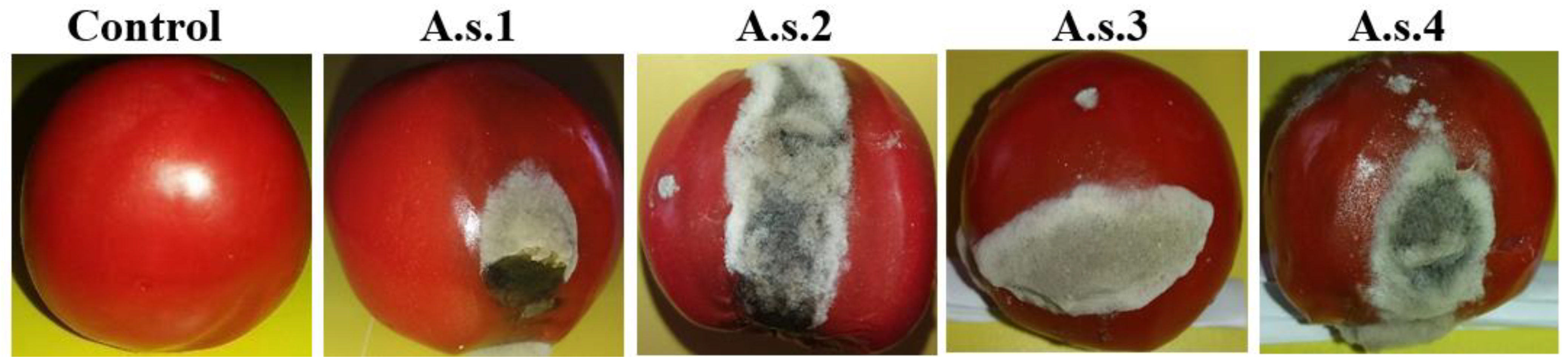
2.2.1. Tomato Fruit

Pure cultures of the fungal pathogen were obtained by the single-spore isolation method, and these cultures were used for pathogenicity tests by following Koch’s postulates [43]. Healthy tomato fruit were taken, and their surfaces were sterilized with ethanol (70%). Artificial infection was carried out using 5 μL of the fungal spore suspension; a spore suspension with a concentration of 3 × 10⁶ spore/mL was used to inoculate each tomato, where the suspension was placed on each fruit, and the fruit was placed under humidified conditions in an incubator at 27 °C for one week [44].

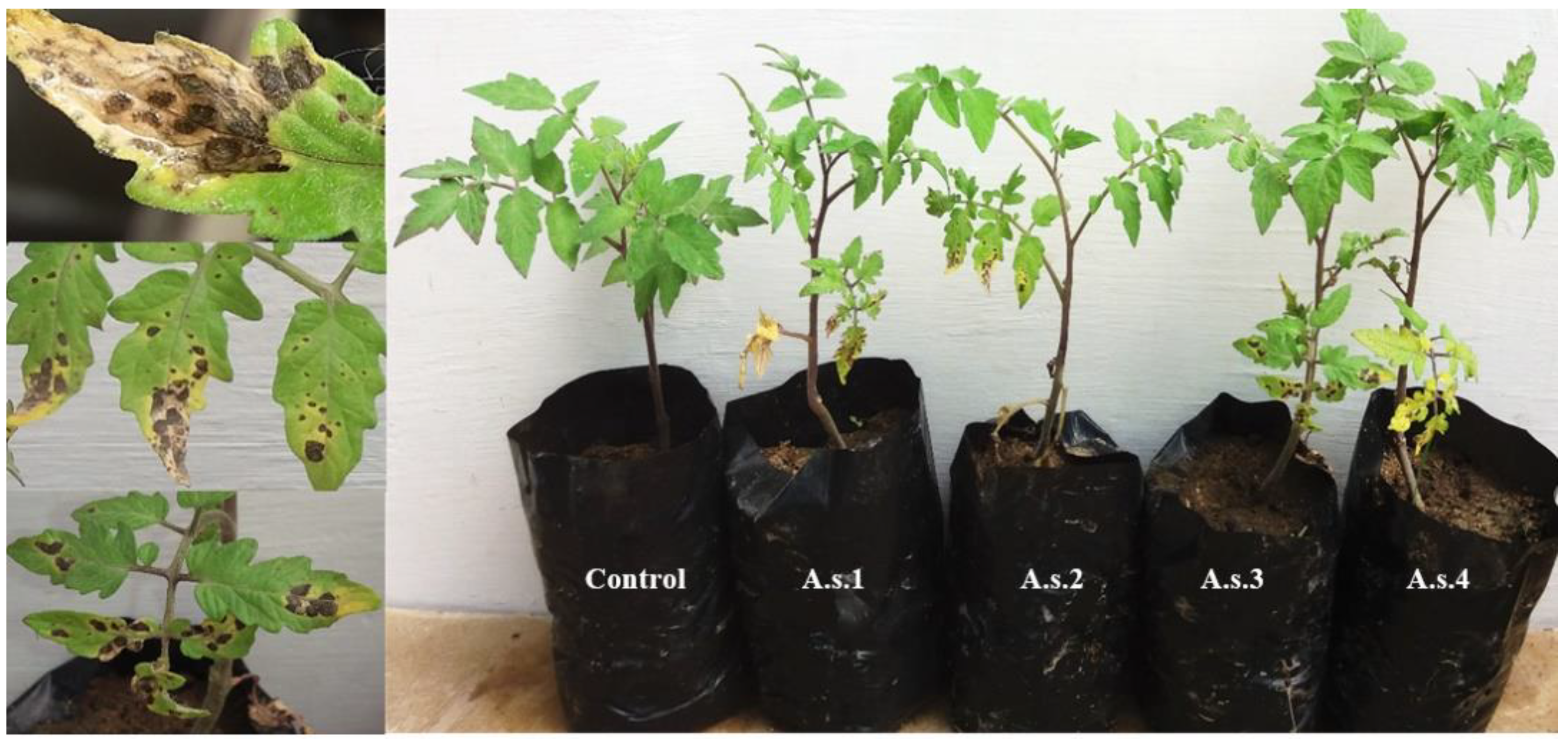
2.2.2. Tomato Seedlings

Seeds of the tomato cultivar Dosera were grown in a greenhouse, and the soil used for cultivation was sterilized by an autoclave. The temperature for plant growth was maintained at 28 °C to 32 °C, and the relative humidity was maintained at 40 to 60%; the plants were allowed to attain a height of 150–200 mm. The collection of tomato plants have been done under the permission at Agriculture Research Center (ARC), Alexandria, Egypt.
Four replications were used in the pathogenicity test. A spore suspension with a concentration of 3 × 10⁶ spore/mL (containing 0.01% Tween 20) was sprayed on leaves, and the degree of leaf infection was studied by visual observation of the extent of lesion development on the leaves, which was assessed for 10 days after inoculation [45]. Observations of the severity of the disease on the foliage were recorded using a 0–5 scale, as shown in Table 1 [46], and the percentage disease index (PDI) was determined by using the following formula and the description of the disease scale [47]: PDI = (A/B × C) × 100, where A is the sum of all ratings, B is the number of plants, and C is the maximum rating.

2.3. Identification of the Fungal Pathogen

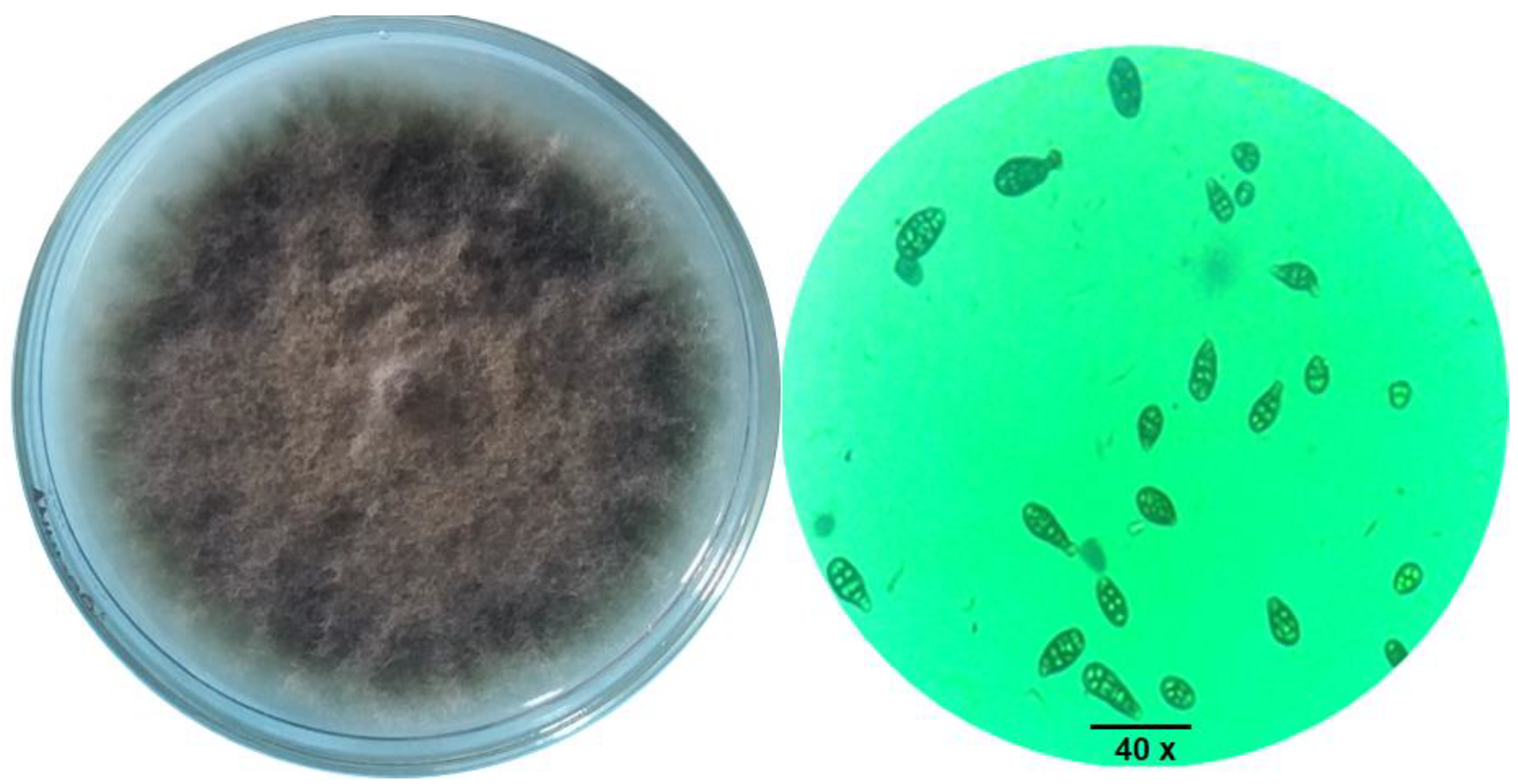
2.3.1. Cultural and Morphological Characteristics

The fungal isolates were identified by microscopic examination, including examination of the structure, size, and shape of the conidia. The isolates were identified according to the criterial for cultural and morphological characteristics described by Naik et al. [41].

2.3.2. Molecular Characterization via Polymerase Chain Reaction (PCR) Amplification of the Internal Transcript Spacer (ITS) Region

After obtaining pure cultures of the fungal isolates, DNA was extracted from these isolates using a rapid mini-preparation procedure [48,49]. The ITS DNA region of these isolates was amplified via PCR using the universal primers ITS1 (5′-TCCGTAGGTGAACCTGCGG-3′) and ITS4 (5′-TCCTCCGCTTATTGA TATGC-3′), which amplified the ITS regions and 5.8S genes encoded by fungal species. Amplification of the ITS rDNA was performed in a total volume of 25 μL, containing 12.5 μL of PCR Green Master Mix (Thermo Scientific™, Gloucester, United Kingdom), 3 μL of template DNA, 8.5 μL of molecular-grade water, and 0.5 μL each of the universal forward primer (ITS1) and reverse primer (ITS4). The optimized thermal profile for PCR was as follows: initial denaturation at 95 °C for 3 min; 35 cycles of denaturation at 94 °C for 30 s, and annealing at 55 °C for 2 min; and a final extension at 72 °C for 10 min. The PCR products were separated on a 1.5% agarose gel in 0.5X Tris-borate-EDTA (TBE) buffer at 65 volts for 15 min, run parallel to a standard DNA molecular marker, and visualized under a UV transilluminator.

2.4. Sequencing of the ITS Region and Phylogenetic Analysis

The obtained ITS rDNA (500–700 bp) regions of selected isolates were sent for sequencing (Macrogen, Scientific Services Company, Seoul, Korea). The sequences were compared to those in GenBank (http://www.ncbi.nlm.nih.gov, accessed on 4 April 2020) using NCBI BLAST. The sequences obtained were submitted to GenBank. The ITS sequences of fungal strains were downloaded from the GenBank database and used in the phylogenetic analyses as reference sequences. All the DNA sequences were aligned with the program CLUSTALW [50]. The resulting multiple-alignment file was used for phylogenetic analyses. The evolutionary history was inferred using the Maximum Parsimony method. The evolutionary history was inferred using the Maximum Parsimony method. The consensus tree inferred from 10 most parsimonious trees is shown. Branches corresponding to partitions reproduced in less than 50% trees are collapsed. The consistency index is 1.000000 (1.000000), the retention index is 1.000000 (1.000000), and the composite index is 1.000000 (1.000000) for all sites and parsimony-informative sites (in parentheses). The percentage of parsimonious trees in which the associated taxa clustered together are shown next to the branches. The MP tree was obtained using the subtree–pruning–regrafting (SPR) algorithm [51] with search level 0, in which the initial trees were obtained by the random addition of sequences (10 replicates). The analysis involved 10 nucleotide sequences. Sequence gaps were treated as missing data. There were a total of 288 positions in the final dataset.

2.5. Evaluation of Bioagents and Plant Extracts against the Early Blight Pathogen Compared to a Chemical Fungicide In Vitro and In Vivo

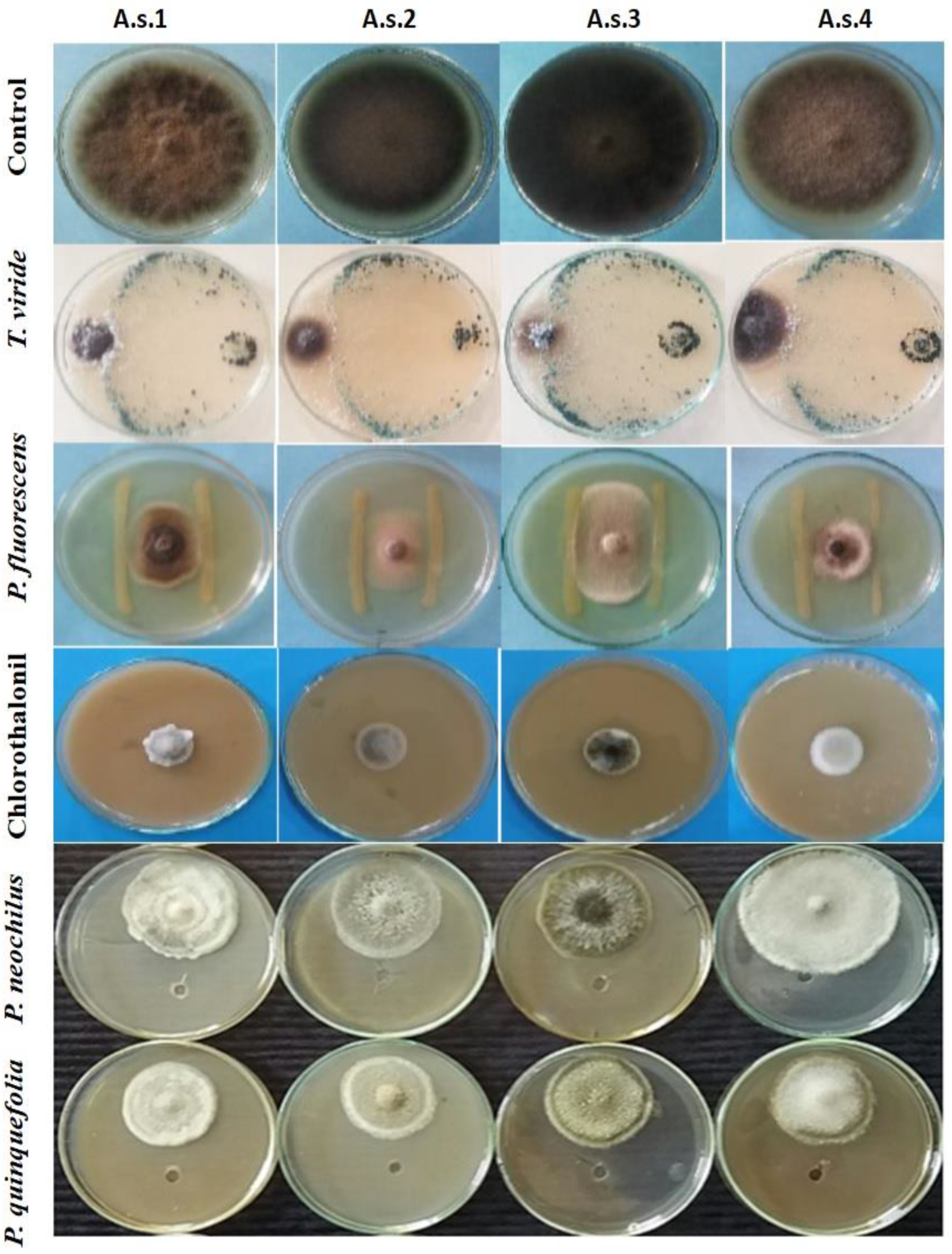
2.5.1. Efficacy of Biological Control Agents In Vitro

Two biological control agents, namely, Trichoderma viride (accession no. MW647090) and Pseudomonas fluorescens (accession no. MW647093), were evaluated for their efficacy against the fungal pathogen using a dual-culture technique [19,49]. Fifteen milliliters of PDA was poured into 9-cm-diameter Petri dishes and allowed to solidify. A 0.5-cm disc of the pathogen was taken from growing margins of a 7-day-old culture and placed at one end of the Petri dish. The T. viride strain isolated in this study from an infected tomato field (0.5 cm disc) was inoculated on the opposite side of the same Petri dish. In the case of the bacterial antagonist, the fungus was centered between two P. fluorescens lines in Petri dishes and incubated for 7 days at 27 °C. The activity of the antagonistic organisms was recorded by measuring the colony diameter in each treatment and comparing it to the control value [52].

2.5.2. Plant Extracts and Their Bioactivity In Vitro

P. quinquefolia fruit and P. neochilus leaves were collected from Alexandria, Egypt. The samples were air-dried under laboratory conditions and ground using a small laboratory Wiley mill. Approximately 50 g each of the P. quinquefolia fruit and P. neochilus leaf samples were extracted with 200 mL of methanol for three days at room temperature and then filtered using Whatman No. 1 filter paper [53]. Subsequently, the solvent was evaporated, and the extracts were concentrated under vacuum using a rotary evaporator at 45 °C. Furthermore, the crude extracts were stored in sealed vials at 4 °C until further use for in vitro screening of antimicrobial activity [54].
The extracts were prepared at concentrations of 2500, 1250, and 625 mg/L by dissolving the extract in dimethyl sulfoxide (DMSO 99.99%) and tested against the growth of the isolated fungus. Wells with a 6-mm diameter were cut out from the PDA medium and filled with 80 µL of each extract. Fungal isolates were grown on PDA and placed at one end of the Petri dish. Each antimicrobial assay was performed in triplicate. The plates were incubated at an appropriate growth temperature (27 °C) for 7 days. The assessment of antimicrobial activity was based on the measurement of linear growth of fungi on the agar surface around the well [55].

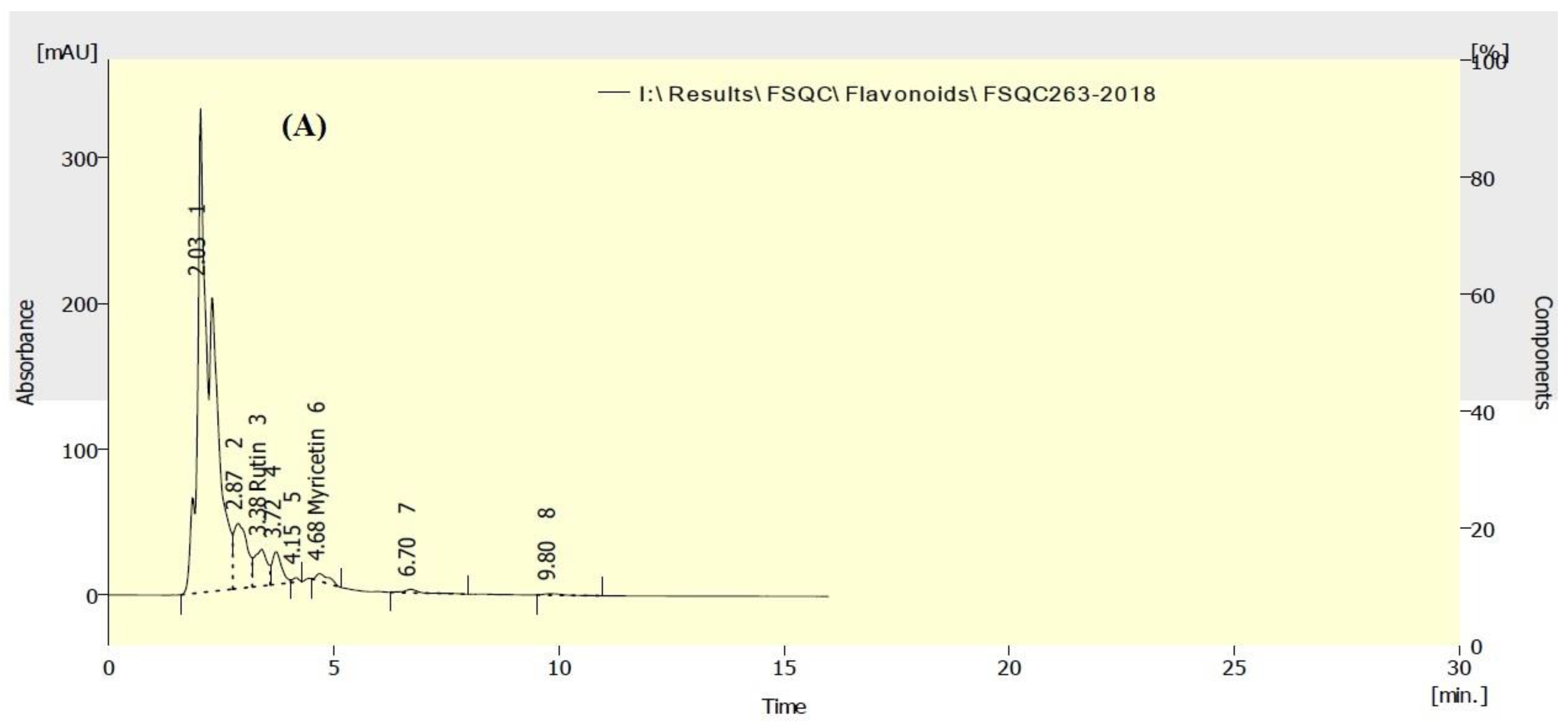
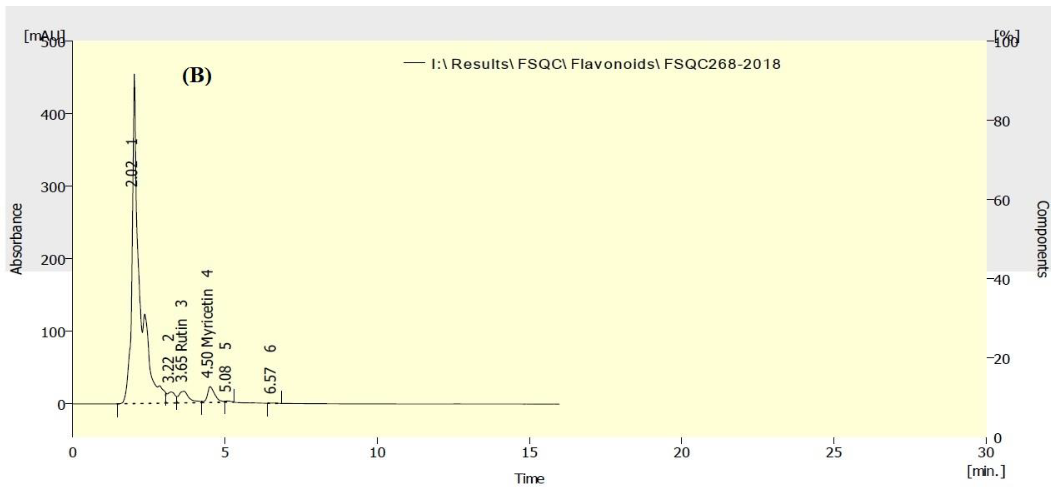
2.6. HPLC Analysis of Flavonoids

An HPLC instrument (Smartline, Knauer, Germany), equipped with a binary pump and a Zorbax Eclipse plus C18 column (150 mm × 4.6 mm i.d.) (Agilent Technologies, Santa Clara, CA, USA) and operated at 35 °C, was used to identify the flavonoid compounds in the methanol extracts of P. quinquefolia fruit and P. neochilus leaves. The conditions used were as follows: eluent methanol:H2O with 0.5% H3PO4, 50:50; flow rate, 0.7 mL/min; and injected volume, 20 µL. The UV detector was set at 273 nm, and data integration was performed using ClarityChrom@ chromatography software, version 7.2.0 (KnauerWissenschaftlicheGeräte GmbH, HegauerWeg 38, 14163 Berlin, Germany) [20,56,57]. Standard flavonoids rutin, myricetin, quercetin, naringenin, kaempferol, and apigenin were used.

2.7. In Vitro Evaluation of Fungicide

The efficacy of the fungicide chlorothalonil (Brado 72% SC®) as a chemical positive control was tested against the isolated fungus at three concentrations: 2500 (1×) Ministry of Agriculture Recommendation), 1250, and 625 mg/L. The fungicide was added to PDA medium after sterilization. A 0.5-cm disc of the fungal isolate was removed and placed at the center of a Petri dish and incubated for 7 days at 27 °C, and the activity of the fungicide was recorded by measuring the colony diameter of the tested fungus in each treatment and comparing it to the control value [58,59].
The bioagents, plant extracts, and chemical positive control (chlorothalonil) were tested with a completely randomized design in triplicate. Then, the plates were incubated until fungal growth covered the surface of PDA medium in the control treatment [60]. The efficacy of each treatment was determined by measuring linear growth (cm), and the data are expressed as the percentage of mycelial growth inhibition compared with the control using the following formula [19,61]: mycelial growth inhibition (%) = (T0Ta/T0) × 100; where T0 and Ta are the average diameters (mm) of fungal colonies under the control and experimental treatments, respectively.

2.8. Control of Early Blight Disease in Tomato In Vivo

The biocontrol agents and plant extracts as well as the chemical fungicide were screened on tomato seedlings. After the plants attained a height of 20 cm, a spore suspension of the fungal isolate (A.s.1) containing 3 × 10⁶ spore/mL was sprayed on the tomato seedlings. T. viride (106 spore/mL), and P. fluorescens (108 CFU/mL), as well as the plant extracts and fungicide (2500 mg/L), were sprayed onto the tomato seedlings one day after inoculation. The experiment was conducted in a randomized complete block design with four replications. Data on disease severity were obtained after three weeks of all treatments [62]. The percent disease index (PDI) was calculated [63], as was the percent reduction in PDI (%) [2].

2.9. Statistical Analysis

The reduction in linear growth of the pathogen as an effect of treatment with biotic and biocontrol agents was analyzed using analysis of variance in a completely randomized design using a computer program, Statistical Analysis System (SAS), and compared with the values for of the control. Means among the treatments were compared using minimum significant difference measured by Tukey’s Studentized Range (HSD) Test at Alpha 0.05 [64].

3. Results

3.1. Isolation of the Fungal Pathogen

Four fungal isolates of the early blight disease pathogen (Alternaria spp.) were obtained from field-infected plants of the tomato cultivars Dosera, 023, Marina Hajin F2, and Ajyad7.

3.2. Pathogenicity Test

3.2.1. Tomato Fruits

The pathogenicity test of the four isolates showed their ability to infect artificially inoculated tomato fruits after one week, in which black spots appeared around the infected area, and fungal growth increased as the incubation period increased (Figure 1).

3.2.2. Tomato Seedlings

Artificial infection of tomato seedlings was carried out under greenhouse conditions on 15-day-old seedlings of the tomato cultivar Dosera. The disease symptoms were observed one week after inoculation as browning of the tissue followed by necrosis. The spots produced were oval in shape, and in the later stage, these spots expanded, and concentric circles were formed and were surrounded by a yellow halo. Finally, the spots changed from brown to dark brown (Figure 2), and the non-inoculated plants did not exhibit disease symptoms. Table 2 shows the percent disease index (PDI), and the degree of leaf infection was investigated by visual observation.

3.3. Identification of the Fungal Pathogen

3.3.1. Cultural and Morphological Characteristics

Four purified fungal isolates, namely, A.s.1, A.s.2, A.s.3, and A.s.4, were identified based on morphological characteristics. The conidia were brown to olivaceous brown, were solitary and straight or exhibited ellipsoidal tapering, and had transverse and longitudinal septate. According to the microscopic images of the four pathogenic fungi and preliminary evaluation, all the isolates belonged to A. solani. The color of the observed colonies was dark brown or olivaceous brown, and the colonies were smooth on PDA medium (Figure 3).

3.3.2. Molecular Characterization through Sequence Analysis of the ITS Region and Phylogenetic Tree of Alternaria Solani Isolates

The fungal isolates were identified via amplification and sequencing of the ITS region. The four isolates produced a PCR product of approximately 500–700 bp, and the ITS sequences were submitted to GenBank. The accession numbers are listed in Table 2. The accession numbers of the ITS sequences were MT279570, MT279571, MT279572, and MT279573. The DNA sequence obtained for each fungal isolate showed 99.5% homology with the A. solani sequences available in GenBank, as determined by utilizing the BLAST tool. The level of similarity reported here confirms the morphological identification of the isolates. In the phylogenetic tree of the ITS region, four A. solani isolates obtained in this study were compared with six isolates collected from GenBank (GU395512, MT135014, KX452728, KF999007, HQ270456, and MT199327), and high genetic similarity to the reported isolates was found (Figure 4).

3.4. Evaluation of Bioagents, Plant Extracts and a Chemical Fungicide against A. solani Isolates In Vitro

The data presented in Table 3 show the highly significant effects of the tested biocontrol agents T. viride (Accession no. MW647090) and P. fluorescens (accession no. MW647093) and plant extracts (P. neochilus and P. quinquefolia) against the growth of A. solani isolates compared with the effect of chlorothalonil 72% as a chemical positive control.
It is evident that chlorothalonil 72% was the most suppressive agent against all the A. solani isolates, with inhibition percentage of fungal growth (IPFG) values that ranged from 84.44% against the isolate A.s.3 at a concentration 2500 mg/L to 77.78% against the isolate A.s.1 at the same concentration. In addition to the positive chemical control (chlorothalonil), the biocontrol agents T. viride and P. fluorescens were found to have high IPFG values of 72.99% and 67.93%, respectively. Moderate to weak activity was found against the A. solani isolate when the P. neochilus and P. quinquefolia extracts were applied at a concentration of 2500 mg/L, and the highest IPFG value (54.01%) of both extracts was observed against the A. solani isolate (A.s.1). The lowest IPFG value (31%) of both extracts was observed against the A.s.3 isolate at the same concentration (2500 mg/L) (Figure 5). Moreover, as shown in Table 3, as the concentrations of chlorothalonil and the P. neochilus and P. quinquefolia extracts increased, the IPFG values against the growth of A. solani isolates increased.

3.5. HPLC Analysis of Flavonoids in P. neochilus and P. quinquefolia Extracts

The main flavonoid compounds identified in the P. neochilus (leaf) and P. quinquefolia (fruit) methanolic extracts by HPLC analysis were rutin (2429.60 and 1891.60 mg/100 g of plant extract, respectively) and myricetin (75.92 and 241.06 mg/100 g of plant extract, respectively) (Table 4). The HPLC chromatograms of the flavonoids identified in the P. neochilus and P. quinquefolia extracts are summarized in Figure 6a,b.

3.6. Evaluation of Bioagents, Plant Extracts and a Chemical Fungicide against A. solani Isolates In Vivo

Table 5 shows the efficacy of the bioagents T. viride and P. fluorescens, as well as the P. neochilus and P. quinquefolia plant extracts and the fungicide chlorothalonil, in reducing the severity of early blight disease in vivo. The data were recorded after 21 days of application. Table 5 shows that all the bioagents and plant extracts tested, in addition to the fungicide chlorothalonil, reduced, though to different extents, the disease index of A. solani compared to that of the inoculated control. It was evident that T. viride and chlorothalonil were superior to all the other treatments in reducing the disease severity (80%) of A. solani, followed by P. fluorescens (70%). The plant extracts of P. neochilus and P. quinquefolia showed moderate effects on the reduction in the disease index (70 and 65%), respectively, compared to the control (Table 5).

4. Discussion

Early blight disease in tomato caused by Alternaria species is known as a severe and destructive fungal disease in Egypt [65]. Four isolates of the fungus A. solani, which is associated with early blight in tomato plants, were investigated. These isolates were obtained from different tomato fields in Egypt. Morphological identification of the fungal isolates was performed according to the morphological characteristics reported by Simmons, such as colony morphology, size, and shape of conidia and pattern of conidial septation among the tested isolates [66]. The characteristics of this pathogen were consistent with the characteristics described by the Commonwealth Mycological Institute, Kew, Surrey, England [67]. Thus, the pathogen was identified as A. solani [68], according to morphological characterization and amplification and sequencing of the ITS region. Based on previous studies, A. solani is the main cause of early blight disease in the family Solanaceae [69]. Furthermore, molecular techniques are suitable methods for analysis, particularly for researchers who are not familiar with the conventional characterization of fungi [70]. One of these methods is sequencing of the ITS region of ribosomal DNA, which distinguishes Alternaria spp. from other pathogens very well [71,72]. The data obtained from molecular studies confirmed the morphological characterization of the tested fungal isolates obtained in this research [7,73,74].
Recently, there has been increasing concern regarding the use of ecofriendly bioagents to control plant pathogens [19,75,76]. Moreover, biological control of early blight pathogens is an attractive alternative to conventional chemical control through the selection and exploitation of fungal and bacterial strains antagonistic to the pathogens that cause early blight in tomato [77,78,79]. The genus Pseudomonas contains a number of strains that are useful for plant protection [80], for example, strains with the ability to produce antibiotics and siderophores [81]. The results obtained from in vitro and in vivo studies revealed that T. viride and P. fluorescens isolates were suppressive to A. solani isolates, and disease severity was investigated [82,83,84]. Additionally, these results are consistent with those of Casida and Lukezie, who reported that Pseudomonas strain 679-2 was able to reduce the severity of early blight disease caused by A. solani [85].
Furthermore, Trichoderma viride, due to its antagonistic activity, is considered a potential biological control agent against many plant pathogenic fungi [49,86]. Trichoderma sp. controls pathogen growth via the production of extracellular enzymes, antibiotics, and antifungal metabolites [16,84,87,88]. The results of this study are consistent with the observed effectiveness of four fungicides, chlorothalonil, copper chloride oxide, and azoxystrobin, at different concentrations against A. solani; the results showed that the fungicides significantly reduced the radial growth of the tested isolates of A. solani [89,90].
Plant defense responses involve the activation of multiple coordinated and apparently complementary defense reactions involving the production of phytoalexins or other antimicrobial compounds, the formation of physical barriers through increased cross-linking, and elicitation of the hypersensitive response [91,92,93]. The induction of plant defense responses also seems to involve several signal transduction cascades [94].
Many attempts have been made to biologically control plant diseases through induction of resistance in the host against the corresponding pathogen by saprophytic bacteria [95], including the use of Pseudomonas fluorescens to control of tobacco mosaic virus [96] and in rice against sheath blight disease [97].
A number of biochemical alterations observed following treatment with bacterial inducers of systemic resistance, such as induction of phytoalexins; induction and/or stimulation of key enzymes, including peroxidase, phenylalanine ammonia lyase, chalcone synthase, chitinase, and β-1,3-glucanase [98,99,100]; and stress-related proteins, have been implicated in the mechanism of resistance [100]. The accumulation of phenolics, callose deposition, and lignification have also been reported and linked to the phenomenon of acquired resistance [101,102,103].
The P. neochilus and P. quinquefolia extracts showed moderate effects against the early blight pathogen A. solani in vitro and in vivo. These results are in line with those of El-Hefny et al. [56], who used an acetone extract of Withania somnifera fruit, which contains flavonoids (rutin and myricetin) at 3%, to inhibit the growth of fungal mycelia of Fusarium culmorum by 84.07% and Rhizoctonia solani by 67.03%.
Flavonoids have been proposed to control fungal pathogens via their inhibitory effect on fungal spore germination [104,105]. Moreover, they cause disruption of the fungal plasma membrane, induction of mitochondrial dysfunction and inhibition of cell wall formation, cell division, RNA, and protein synthesis and the efflux-mediated pumping system [106]. Furthermore, flavonoid compounds, such as catechin and rutin extracted from pomegranate peel, have potent inhibitory effects against Colletotrichum gloeosporioides, a fungus that infects Persea americana [107]. Flavonoids inhibit many varieties of eukaryotic enzymes, and this inhibition of enzymes may be due to the interaction of enzymes with different parts of the flavonoid molecule, such as carbohydrates, phenyl rings, phenols, and benzopyrone rings [108]. Moreover, the antimicrobial activity of P. neochilus extracts has been associated with the lipophilicity of their chemical constituents, mainly monoterpenes and sesquiterpenes, which are often the main chemicals therein [23,109]. The antimicrobial activity of P. quinquefolia may be due to some phenolic compounds in these extracts [110].

5. Conclusions

In agriculture, there is an important need for alternate ecofriendly materials to control plant diseases. This study provides insights for the development of new phytosanitary products based on plant extracts of Plectranthus neochilus and Parthenocissus quinquefolia and on the bioagents Trichoderma viride and Pseudomonas fluorescens for the control of A. solani in tomato plants. The tested bioagents, plant extracts, and the fungicide chlorothalonil were significantly reduced disease index of A. solani. T. viride and chlorothalonil were suggested to be superior in the reduction of disease severity of A. solani followed by P. fluorescens extract. In vivo, and with spray application of the tested agents, T. viride, chlorothalonil followed by P. fluorescens extract were observed the most reduction in the disease index of early blight. This study suggested and recommended alternatives to chemical pesticides to achieve organic production.

Author Contributions

Conceptualization, A.A.M.; data curation, A.A.M. and M.M.Z.E.-D.; formal analysis, M.M.S., M.M.Z.E.-D., M.E.-H., and N.A.A.; funding acquisition, D.A.A.F., A.A.H., and H.M.A.; investigation, N.A.A. and M.Z.M.S.; methodology, A.A.M., M.M.S., M.M.Z.E.-D., M.E.-H., and N.A.A.; resources, N.A.A., M.Z.M.S., H.M.A.; software, H.M.A.; supervision, N.A.A.; validation, M.E.-H. and N.A.A.; Writing—original draft, A.A.M., M.M.S., M.M.Z.E.-D., and N.A.A.; Writing—review and editing, A.A.M., D.A.A.F., A.A.H., M.Z.M.S., and M.E.-H. All co-authors contributed to writing and revising the article. All authors have read and agreed to the published version of the manuscript.

Funding

This research was supported by the Deanship of Scientific Research at King Saud University for funding this work through Research group no. RG 1435-011.

Institutional Review Board Statement

Not applicable.

Informed Consent Statement

Not applicable.

Data Availability Statement

Not applicable.

Acknowledgments

The authors would like to extend their sincere appreciation to the Deanship of Scientific Research at King Saud University for funding this work through Research group no. RG 1435-011.

Conflicts of Interest

The authors declare no conflict of interest.

References

  1. Abada, K.A.; Mostafa, S.H.; Hillal, M.R. Effect of some chemical salts on suppressing the infection by early blight disease of tomato. Egypt. J. Appl. Sci. 2008, 23, 47–58. [Google Scholar]
  2. Sahu, D.K.; Khare, C.P.; Singh, H.K.; Thakur, M.P. Evaluation of newer fungicide for management of early blight of tomato in Chhattisgarh. Bioscan 2013, 8, 1255–1259. [Google Scholar]
  3. Song, W.; Ma, X.; Tan, H.; Zhou, J. Abscisic acid enhances resistance to Alternaria solani in tomato seedlings. Plant. Physiol. Biochem. 2011, 49, 693–700. [Google Scholar] [CrossRef]
  4. Carneiro, S.M.; Romano, E.D.; Pignoni, E.; Teixeira, M.Z.; da Costa Vasconcelos, M.E.; Gomes, J.C. Effect of biotherapic of Alternaria solani on the early blight of tomato plant and the in vitro development of the fungus. Int. J. High. Dilution Res. 2010, 9, 147–155. [Google Scholar]
  5. Chaerani, R.; Groenworld, R.; Stam, P.; Voorrips, R.E. Assessment of early blight (Alternaria solani) resistance in tomato using a droplet inoculation method. J. Gen. Plant. Pathol. 2007, 73, 96–103. [Google Scholar] [CrossRef] [Green Version]
  6. Kokaeva, L.Y.; Belosokhov, A.F.; Doeva, L.Y.; Skolotneva, E.S.; Elansky, S.N. Distribution of Alternaria species on blighted potato and tomato leaves in Russia. J. Plant. Dis. Prot. 2015, 125, 205–212. [Google Scholar] [CrossRef]
  7. Blancard, D.; Laterrot, H.; Marchoux, G.; Candresse, T. A Colour Handbook—Tomato Diseases: Identification, Biology and Control; Manson Publishing Manson Publishing Limited: London, UK, 2012; p. 688. [Google Scholar]
  8. Mallik, I.; Arabiat, S.; Pasche, J.S.; Bolton, M.D.; Patel, J.S.; Gudmestad, N.C. Molecular characterization and detection of mutations associated with resistance to succinate dehydrogenase inhibiting fungicides in Alternaria solani. Phytopathology 2014, 104, 40–49. [Google Scholar] [CrossRef] [Green Version]
  9. Khan, A.A. Resistance of two tomato species to five isolates of A. solani. Asian J. Plant. Sci. 2002, 1, 703–704. [Google Scholar] [CrossRef] [Green Version]
  10. Verma, K.P.; Singh, S.; Gandhi, S.K. Variability among Alternaria solaniisolates associated with early blight of tomato. Indian Phytopathol. 2007, 60, 180–186. [Google Scholar]
  11. Gannibal, P.B.; Orina, A.S.; Mironenko, N.V.; Levitin, M.M. Differentiation of the closely related species, Alternaria solani and A. tomatophila, by molecular and morphological features and aggressiveness. Eur. J. Plant. Pathol. 2014, 139, 609–623. [Google Scholar] [CrossRef]
  12. Bock, C.H.; Poole, G.H.; Parker, P.E.; Gottwald, T.R. Plant disease severity estimated visually, by digital photography and image analysis, and by hyperspectral imaging. Crit. Rev. Plant. Sci. 2010, 29, 59–107. [Google Scholar] [CrossRef]
  13. Pavóna, M.A.; Lunab, A.; de la Cruza, S.; Gonzáleza, I.; Martína, R.; Garcíaa, T. PCR based assay for the detection of Alternaria species and correlation with HPLC determination of altenuene, alternariol and alternariol monomethyl ether production in tomato products. Food Control 2012, 25, 45–52. [Google Scholar] [CrossRef]
  14. Rani, S.; Singh, R.; Gupta, S. Development of integrated disease management module for early blight of tomato in Jammu. J. Pharmacogn. Phytochem. 2017, 6, 268–273. [Google Scholar]
  15. Babu, S.; Seetharaman, K.; Nandakumar, R.; Johnson, I. Efficacy of fungal antagonists against leaf blight of tomato caused by Alternaria solani (Ell. and Mart.). J. Biol. Control 2000, 14, 79–81. [Google Scholar]
  16. Koley, S.; Mahapatra, S.S.; Kole, P.C. In vitro efficacy of bio-control agents and botanicals on the growth inhibition of Alternaria Solanicausing early leaf blight of tomato. Int. J. Bio-Resour. Environ. Agric. Sci. 2015, 1, 114–118. [Google Scholar]
  17. Sharma, R.K.; Patel, D.R.; Chaudhari, D.R.; Kumar, V.; Patel, M.M. Effect of Some Fungicides against Early Blight of Tomato (Lycopersicon esculentum Mill.) Caused by Alternaria solani (Ell. and Mart.) Jones and Grout and their Impact on Yield. Int. J. Curr. Microbiol. Appl. Sci. 2018, 7, 1395–1401. [Google Scholar] [CrossRef]
  18. Hernández-Ochoa, J.S.; Levin, L.N.; Hernández-Luna, C.E.; Contreras-Cordero, J.F.; Niño-Medina, G.; Chávez-Montes, A.; López-Sandin, I.; Gutiérrez-Soto, G. Antagonistic Potential of Macrolepiota sp. Against Alternaria solani as Causal Agent of Early Blight Disease in Tomato Plants. Gesunde Pflanz. 2020, 72, 69–76. [Google Scholar] [CrossRef]
  19. Mohamed, A.A.; Behiry, S.I.; Ali, H.M.; EL-Hefny, M.; Salem, M.Z.M.; Ashmawy, N.A. Phytochemical Compounds of Branches from P. halepensis Oily Liquid Extract and S. terebinthifolius Essential Oil and Their Potential Antifungal Activity. Processes 2020, 8, 330. [Google Scholar] [CrossRef] [Green Version]
  20. Ashmawy, N.A.; Salem, M.Z.M.; El Shanhorey, N.; Al-Huqail, A.A.; Ali, H.M.; Behiry, S.I. Eco-friendly wood-biofungicidal and antibacterial activities of various Coccoloba uvifera L. leaf extracts: HPLC analysis of phenolic and flavonoid compounds. BioResources 2020, 15, 4165–4187. [Google Scholar] [CrossRef]
  21. Fazli, R.; Ishtiaq, S. Integrated control of Alternaria solani with Trichoderma spp. And fungicides under in vitro conditions. Sairhad J. Agric. 2010, 26, 613–619. [Google Scholar]
  22. Simões, M.F.; Rijo, P.; Duarte, A.; Barbosa, D.; Matias, D.; Delgado, J.; Cirilo, N.; Rodriguez, B. Two new diterpenoids from Plectranthuss pecies. Phytochem. Lett. 2010, 3, 221–225. [Google Scholar] [CrossRef]
  23. Crevelin, E.J.; Caixeta, S.C.; Dias, H.J.; Groppo, M.; Cunha, W.R.; Martins, C.H.G.; Crotti, A.D.M. Antimicrobial activity of the essential oil of Plectranthus neochilus against cariogenic bacteria. Evid. Based Complement. Altern. Med. 2015, 2015, 102317. [Google Scholar] [CrossRef] [Green Version]
  24. Ticha, M.B.; Meksi, N.; Attia, H.E.; Haddar, W.; Guesmi, A.; Jannet, H.B.; Mhenni, M.F. Ultrasonic extraction of Parthenocissus quinquefolia colorants: Extract identification by HPLC-MS analysis and cleaner application on the phytodyeing of natural fibres. Dyes Pigment. 2017, 141, 103–111. [Google Scholar] [CrossRef]
  25. Paton, A.; Mwanyambo, M.; Culham, A. Phylogenetic study of Plectranthus, Coleus and allies (Lamiaceae): Taxonomy, distribution and medicinal use. Bot. J. Linn. Soc. 2018, 188, 355–376. [Google Scholar] [CrossRef]
  26. Aguiar, G.P.; Lima, K.A.; Severiano, M.E.; Groppo, M.; Ambrósio, S.R. Antifungal Activity of the essential oils of Plectranthus neochilus (Lamiaceae) and Tagetes erecta (Asteraceae) cultivated in Brazil. Int. J. Complement. Altern. Med. 2018, 11, 343. [Google Scholar]
  27. Caixeta, S.C.; Magalhães, L.G.; de Melo, N.I.; Wakabayashi, K.A.; de P. Aguiar, G.; de P. Aguiar, D.; Mantovani, A.L.; Alves, J.M.; Oliveira, P.F.; Tavares, D.C.; et al. Chemical composition and in vitro schistosomicidal activity of the essential oil of Plectranthus neochilus grown in Southeast Brazil. Chem. Biodiver. 2011, 8, 2149–2157. [Google Scholar] [CrossRef]
  28. Baldin, E.L.; Crotti, A.E.; Wakabayashi, K.A.; Silva, J.P.; Aguiar, G.P.; Souza, E.S.; Veneziani, R.C.; Groppo, M. Plant-derived essential oils affecting settlement and oviposition of Bemisia tabaci (Genn.) biotype B on tomato. J. Pest. Sci. 2013, 86, 301–308. [Google Scholar] [CrossRef]
  29. Viana, A.J.S. Estudo Químico e de Atividade Aiológica de Plectranthus Neochilus Schltr. (Lamiaceae). Master’s Thesis, Federal University of Vales do Jequitinhonha e Mucuri, Teófilo Otoni, Brasil, 2011. (In Spanish). [Google Scholar]
  30. Matias, D.; Nicolai, M.; Fernandes, A.S.; Saraiva, N.; Almeida, J.; Saraiva, L.; Faustino, C.; Díaz-Lanza, A.M.; Reis, C.P.; Rijo, P. Comparison Study of Different Extracts of Plectranthus madagascariensis, P. neochilus and the Rare P. porcatus (Lamiaceae): Chemical Characterization, Antioxidant, Antimicrobial and Cytotoxic Activities. Biomolecules 2019, 9, 179. [Google Scholar] [CrossRef] [PubMed] [Green Version]
  31. Invasive Species Compendium, Parthenocissus quinquefolia (Virginia creeper). Available online: https://www.cabi.org/isc/datasheet/44676 (accessed on 21 November 2019).
  32. Gledhill, D. The Names of Plants, 3rd ed.; Cambridge University Press: Cambridge, UK, 2010; pp. 292, 324, ISBN 9780521866453 (hardback), ISBN 9780521685535 (paperback). [Google Scholar] [CrossRef]
  33. Shaheen, S.; Manzoor, A. Phytochemical screening and antioxidant potential of Parthenocissus quinquefolia (L.) planch extracts of bark and stem. Pak. J. Pharm. Sci. 2018, 31, 1813–1816. [Google Scholar]
  34. Yang, J.; Wang, A.; Ji, T.; Su, Y. Chemical constituents from Parthenocissus quinquefolia. Zhongguo Zhong yao za zhi Zhongguo zhongyao zazhi China J. Chin. Mater. Medica 2010, 35, 1573–1576. [Google Scholar]
  35. Khan, Z.U.D.; Faisal, S.; Perveen, A.; Sardar, A.A.; Siddiqui, S.Z. Phytochemical properties and antioxidant activities of leaves and fruits extracts of Parthenocissus quinquefolia (L.) Planch. Bang J. Bot. 2018, 47, 33–38. [Google Scholar]
  36. Cardon, D. Natural Dyes, Sources Tradition. Technology and Science; Archetype: London, UK, 2007; p. 778. ISBN 190498200X, 9781904982005. [Google Scholar]
  37. Francis, F.J. Colorants; Egan Press: St. Paul, MN, USA, 1999; ISBN 1-891127-00-4. [Google Scholar]
  38. Stafford, H.A. Anthocyanins and betalains: Evolution of the mutually exclusive pathways. Plant. Sci. 1994, 101, 91–98. [Google Scholar] [CrossRef]
  39. Gai, C.Y.; Liu, H.M.; Li, J.; Li, J. Extraction and Content Determination of Total Flavonoid in Parthenocissus Quinquefolia. Spec. Wild Econ. Anim. Plant. Res. 2010, 2, 20. [Google Scholar]
  40. Shi, J.; Han, X.; Zhang, Y.; Sun, T.; Diao, H.; Cao, X. Extraction techniques and identification of flavonoids in parthenocissus seeds. China Med. Her. 2010, 18, 33. [Google Scholar]
  41. Naik, M.K.; Prasad, Y.; Bhat, K.V.; Rani, D. Morphological, physiological, pathogenic and molecular variability among isolates of Alternaria solani from tomato. Indian Phytopathol. 2010, 63, 168–173. [Google Scholar]
  42. Johnston, A.; Booth, C. Plant Pathologist’s Pocket Book; Oxford and IBH Pub. Co.: Calcutta, India, 1983; p. 136. [Google Scholar]
  43. Agrios, G.N. Plant Pathology, 5th ed.; Elsevier Academic Press: Burlington, MA, USA, 2005; pp. 79–103. [Google Scholar]
  44. Hussein, G.S. The use of PCR to detect Alternaria fungi which isolated from tomato fruit. Biochem. Cell. Arch. 2019, 19, 4203–4206. [Google Scholar]
  45. Chohan, S.; Perveen, R.; Abid, M.; Naz, M.S.; Akram, N. Morpho-physiological Studies Management and Screening of Tomato Germplasm against Alternaria solani the Causal Agent of Tomato Early Blight. Int. J. Agric. Biol. 2015, 17, 111–118. [Google Scholar]
  46. Barratt, R.W.; Horsfall, J.G. An improved grading system for measuring plant disease. Phytopathology 1945, 35, 655. [Google Scholar]
  47. Pandey, K.K.; Pandey, P.K.; Kalloo, G.; Banerjee, M.K. Resistance to early blight of tomato with respect to various parameters of disease epidemics. J. Gen. Plant. Pathol. 2003, 69, 364–371. [Google Scholar] [CrossRef]
  48. Edel, V.; Christian, S.; Gautheron, N.; Recorbet, G.; Alabouvette, C. Genetic diversity of Fusarium oxysporum populations isolated from different soils in France. FEMS Microbiol. Ecol. 2001, 36, 61–71. [Google Scholar] [CrossRef]
  49. Mohamed, A.A.; Gomaa, F.H. Molecular Characterization and Biological Control of Some Rice Seed-borne Fungal Pathogens. J. Phytopathol. Pest. Manag. 2019, 6, 40–53. [Google Scholar]
  50. Kumar, S.; Stecher, G.; Tamura, K. MEGA7: Molecular Evolutionary Genetics Analysis version 7.0 for bigger datasets. Mol. Biol. Evol. 2016, 33, 1870–1874. [Google Scholar] [CrossRef] [Green Version]
  51. Mount, D.W. Maximum parsimony method for phylogenetic prediction. Cold Spring Harb. Protoc. 2008, 2008. [Google Scholar] [CrossRef]
  52. Molloy, D.P. Biological control of Zebra mussels. In Proceedings of the 3rd California Conference on Biological Control, University of California, Davis, CA, USA, 15–16 August 2002; pp. 86–94. [Google Scholar]
  53. Salem, M.Z.M.; Mansour, M.M.; Elansary, H.O. Evaluation of the effect of inner and outer bark extracts of Sugar Maple (Acer saccharum var. saccharum) in combination with citric acid against the growth of three common molds. J. Wood Chem. Technol. 2019, 39, 136–147. [Google Scholar] [CrossRef]
  54. Salem, M.Z.M.; Ali, H.M.; El-Shanhorey, N.A.; Abdel-Megeed, A. Evaluation of extracts and essential oil from Callistemon viminalis leaves: Antibacterial and antioxidant activities, total phenolic and flavonoid contents. Asian Pac. J. Trop. Med. 2013, 6, 785–791. [Google Scholar] [CrossRef] [Green Version]
  55. Nene, Y.L.; Thapaya, P.N. Evaluation of fungicides. In Fungicide in Plant Disease Control; Oxford and IBH Publishing Company: New Delhi, India, 1993; p. 531. [Google Scholar]
  56. EL-Hefny, M.; Salem, M.Z.M.; Behiry, S.I.; Ali, H.M. The Potential Antibacterial and Antifungal Activities of Wood Treated with Withania somnifera Fruit Extract, and the Phenolic, Caffeine, and Flavonoid Composition of the Extract According to HPLC. Processes 2020, 8, 113. [Google Scholar] [CrossRef] [Green Version]
  57. Al-Huqail, A.A.; Behiry, S.I.; Salem, M.Z.M.; Ali, H.M.; Siddiqui, M.H.; Salem, A.Z.M. Antifungal, antibacterial, and antioxidant activities of Acacia saligna (Labill.) HL Wendl. flower extract: HPLC analysis of phenolic and flavonoid compounds. Molecules 2019, 24, 700. [Google Scholar] [CrossRef] [Green Version]
  58. Derbalah, A.S.; El-Mahrouk, M.S.; El-Sayed, A.B. Efficacy and safety of some plant extracts against tomato early blight disease caused by Alternaria solani. Plant. Pathol. J. 2011, 10, 115–121. [Google Scholar] [CrossRef] [Green Version]
  59. NAShwA, S.M.; Abo-ElyouSr, K.A. Evaluation of various plant extracts against the early blight disease of tomato plants under greenhouse and field conditions. Plant. Prot. Sci. 2013, 4, 74–79. [Google Scholar] [CrossRef] [Green Version]
  60. Qasem, J.R.; Abu-Blan, H.A. Fungicidal activity of some common weed extracts against different plant pathogenic fungi. J. Phytopathol. 1996, 144, 157–161. [Google Scholar] [CrossRef]
  61. Sundar, A.R.; Das, N.D.; Krishnaveni, D. In-vitro Antagonism of Trichoderma spp. against two Fungal Pathogens of Castor. Ind. J. Plant. Prot. 1995, 23, 152–155. [Google Scholar]
  62. El-Katatny, M.H.; Emam, A.S. Control of postharvest tomato rot by spore suspension and antifungal metabolites of Trichoderma harzianum. J. Microbiol. Biotech. Food Sci. 2012, 1, 1505–1528. [Google Scholar]
  63. Wheeler, B.E.J. An Introduction to Plant Diseases; J. Wiley and Sons Limited: London, UK, 1969; p. 301. [Google Scholar]
  64. SAS. User Guide: Statistics (Release 8.02); SAS Institute: Cary, NC, USA, 2001. [Google Scholar]
  65. Ashour, A.M.A. A protocol suggested for managing tomato early blight. Egypt J. Phytopathol. 2009, 37, 9–20. [Google Scholar]
  66. Simmons, E. Alternaria: An Identification Manual; CBS Fungal Biodiversity Centre: Utrecht, The Netherlands, 2007. [Google Scholar]
  67. Ellis, M.B. Dematiaceous Hyphomycetes; Commonwelth Mycological Institute: Kew, UK, 1971; p. 608. [Google Scholar]
  68. Ramjegathesh, R.; Ebenezar, E.G. Morphological and physiological characters of Alternaria Alternata causing leaf blight diseases of onion. Int. J. Plant. Pathol. 2012, 3, 34–44. [Google Scholar] [CrossRef] [Green Version]
  69. Dang, H.X.; Pryor, B.; Peever, T.; Lawrence, C.B. The Alternaria genomes database: A comprehensive resource for a fungal genus comprised of saprophytes, plant pathogens, and allergenic species. BMC Genom. 2015, 16, 239. [Google Scholar] [CrossRef] [Green Version]
  70. Pryor, B.M.; Michailides, T.J. Morphological, Pathogenic, and Molecular Characterization of Alternaria Isolates Associated with Alternaria Late Blight of Pistachio. Phytopathology 2002, 92, 406–416. [Google Scholar] [CrossRef] [Green Version]
  71. Kusaba, M.; Tsuge, T. Phologeny of Alternaria fungi known to produce host-specific toxins on the basis of variation in internal transcribed spacers of ribosomal DNA. Curr. Genet. 1995, 28, 491–498. [Google Scholar] [CrossRef] [PubMed]
  72. Xie, G.; Tan, S.; Yu, L. Morphological and molecular identification of pathogenic fungal of post-harvest tomato fruit during storage. Afr. J. Microbiol. Res. 2012, 6, 4805–4809. [Google Scholar]
  73. Peralta, I.E.; Knapp, S.; Spooner, D.M. New species of wild tomatoes (Solanum section Lycopersicon: Solanaceae) from Northern Peru. Syst. Bot. 2005, 30, 424–434. [Google Scholar] [CrossRef]
  74. Chaerani, R.; Voorrips, R.E.; Roeland, E. Tomato early blight (Alternaria solani): The pathogen, genetics and breeding for resistance. J. Gen. Plant. Pathol. 2006, 13, 335–347. [Google Scholar] [CrossRef]
  75. Behiry, S.I.; EL-Hefny, M.; Salem, M.Z.M. Toxicity effects of Eriocephalus africanus L. leaf essential oil against some molecularly identified phytopathogenic bacterial strains. Nat. Prod. Res. 2020, 34, 3394–3398. [Google Scholar] [CrossRef]
  76. Ashmawy, N.A.; Behiry, S.I.; Al-Huqail, A.A.; Ali, H.M.; Salem, M.Z.M. Bioactivity of Selected Phenolic Acids and Hexane Extracts from Bougainvilla spectabilis and Citharexylum spinosum on the Growth of Pectobacterium carotovorum and Dickeya solani Bacteria: An Opportunity to Save the Environment. Processes 2020, 8, 482. [Google Scholar] [CrossRef]
  77. Adhikari, P.; Oh, Y.; Panthee, D.R. Current status of early blight resistance in tomato: An update. Int. J. Mol. Sci. 2017, 18, 2019. [Google Scholar] [CrossRef] [Green Version]
  78. Aldiba, A.; Escov, I. Biological Control of Early Blight on Potato Caused by Alternaria Solani by Some Bioagents. In Proceedings of the 1st International Symposium Innovations in Life Sciences (ISILS 2019), Belgorod, Russia, 10–11 October 2019; Atlantis Press: Paris, France, 2019; pp. 103–107. [Google Scholar] [CrossRef]
  79. Evidente, A.; Andolfi, A. Fungal phytotoxins for control of cirsium arvense and Sonchus arvensis. Pest. Technol. 2011, 5, 1–17. [Google Scholar]
  80. Mercado-Blanco, J.; Bakker, P.A. Interactions between plants and beneficial Pseudomonas spp.: Exploiting bacterial traits for crop protection. Antonie van Leeuwenhoek 2007, 92, 367–389. [Google Scholar] [CrossRef]
  81. Drehe, I.; Simonetti, E.; Ruiz, J.A. Contribution of the siderophores pyoverdine and enantio-pyochelin to fitness in soil of Pseudomonas protegens Pf-5. Curr. Microbiol. 2018, 75, 1560–1565. [Google Scholar] [CrossRef]
  82. Latha, P.; Anand, T.; Prakasam, V.; Jonathan, E.I.; Paramathma, M.; Samiyappan, R. Combining Pseudomonas, Bacillus an Trichoderma strains with organic amendments and micronutrient to enhance suppression of collar and root rot disease in physic nut. Appl. Soil Ecol. 2011, 49, 215–223. [Google Scholar] [CrossRef]
  83. Zape, A.S.; Gade, R.M.; Singh, R.A.V.I.N.D.R.A.; Deshmukh, V.A. Efficacy of different antagonist against the Sclerotium rolfsii, Rhizoctonia solani and Fusarium solani. Bioscan 2014, 9, 1431–1434. [Google Scholar]
  84. Vallabhaneni, S.D. Biocontrol of Rhizoctonia solani in tobacco (Nicotiana tabacum) seed beds using Pseudomonas fluorescens. Agric. Res. 2016, 5, 137–144. [Google Scholar] [CrossRef]
  85. Casida, L.E.; Lukezic, F.L. Control of leaf spot diseases of alfalfa and tomato with applications of the bacterial predator Pseudomonas strain 679-2. Plant Dis. 1992, 76, 1217. [Google Scholar] [CrossRef]
  86. Mohamed, H.A.; Haggag, W.M. Biocontrol potential of salinity tolerant mutants of Trichoderma harzianum against Fusarium oxysporum. Braz. J. Microbiol. 2006, 37, 181–191. [Google Scholar] [CrossRef] [Green Version]
  87. Montealegre, J.; Valderrama, L.; Sanchez, S.; Herrera, R.; Besoainand, X.; Perez, L.M. Biological control of Rhizoctonia solani intomatoes with Trichoderma harzianum mutants. Electron. J. Biotechnol. 2010, 13, 1–11. [Google Scholar] [CrossRef] [Green Version]
  88. Mandal, D.; Pal, R.; Mohanty, A.K. Management of bacterial leaf blight of rice in an integrated way. J. Mycopathol. Res. 2017, 54, 539–541. [Google Scholar]
  89. Singh, P.C.; Singh, D. In vitro evaluation of fungicides against Alternaria alternata. Ann. Plant Prot. Sci. 2006, 14, 500–502. [Google Scholar]
  90. Mesta, R.K.; Benagi, V.I.; Shankergroud, I.; Megeri, S.N. Effect of dates of sowing and correlation of weather parameters on the incidence of Alternaria blight of sunflower. Karnataka J. Agric. Sci. 2009, 22, 441–443. [Google Scholar]
  91. Lattanzio, V.; Lattanzio, V.M.; Cardinali, A. Role of phenolics in the resistance mechanisms of plants against fungal pathogens and insects. Phytochem. Adv. Res. 2006, 661, 23–67. [Google Scholar]
  92. Heyman, J.; Canher, B.; Bisht, A.; Christiaens, F.; De Veylder, L. Emerging role of the plant ERF transcription factors in coordinating wound defense responses and repair. J. Cell Sci. 2018, 131, jcs.208215. [Google Scholar] [CrossRef] [PubMed] [Green Version]
  93. Brotman, Y.; Landau, U.; Cuadros-Inostroza, Á.; Takayuki, T.; Fernie, A.R.; Chet, I.; Viterbo, A.; Willmitzer, L. Trichoderma-plant root colonization: Escaping early plant defense responses and activation of the antioxidant machinery for saline stress tolerance. PLoS Pathog. 2013, 9, e1003221. [Google Scholar] [CrossRef]
  94. Heil, M.; Bostock, R.M. Induced systemic resistance (ISR) against pathogens in the context of induced plant defences. Ann. Bot. 2002, 89, 503–512. [Google Scholar] [CrossRef] [PubMed] [Green Version]
  95. Kokoskova, B.; Pavela, R.; Pouvova, D. Effectiveness of plant essential oils against Erwinia amylovora, Pseudomonas syringae pv. syringae and associated saprophytic bacteria on/in host plants. J. Plant. Pathol. 2011, 93, 133–139. [Google Scholar]
  96. Shen, L.; Wang, F.; Yang, J.; Qian, Y.; Dong, X.; Zhan, H. Control of tobacco mosaic virus by Pseudomonas fluorescens CZ powder in greenhouses and the field. Crop. Prot. 2014, 56, 87–90. [Google Scholar] [CrossRef]
  97. Nandakumar, R.; Babu, S.; Viswanathan, R.; Raguchander, T.; Samiyappan, R. Induction of systemic resistance in rice against sheath blight disease by Pseudomonas fluorescens. Soil Biol. Biochem. 2001, 33, 603–612. [Google Scholar] [CrossRef]
  98. Deice Raasch-Fernandes, L.; Bonaldo, S.M.; de Jesus Rodrigues, D.; Magela Vieira-Junior, G.; Regina Freitas Schwan-Estrada, K.; Rocco da Silva, C.; Gabriela Araújo Verçosa, A.; Lopes de Oliveira, D.; Wender Debiasi, B. Induction of phytoalexins and proteins related to pathogenesis in plants treated with extracts of cutaneous secretions of southern Amazonian Bufonidae amphibians. PLoS ONE 2019, 14, e0211020. [Google Scholar] [CrossRef]
  99. Meena, B.; Radhajeyalakshmi, R.; Marimuthu, T.; Vidhyasekaran, P.; Doraiswamy, S.; Velazhahan, R. Induction of pathogenesis-related proteins, phenolics and phenylalanine ammonia-lyase in groundnut by Pseudomonas fluorescens. J. Plant. Dis. Prot. 2000, 107, 514–527. [Google Scholar]
  100. Sivakumar, G.; Sharma, R.C. Induced biochemical changes due to seed bacterization by Pseudomonas fluorescens in maize plants. Indian Phytopathol. 2003, 56, 34–137. [Google Scholar]
  101. Akram, A.; Ongena, M.; Duby, F.; Dommes, J.; Thonart, P. Systemic resistance and lipoxygenase-related defence response induced in tomato by Pseudomonas putida strain BTP1. BMC Plant Biol. 2008, 8, 1–12. [Google Scholar] [CrossRef] [Green Version]
  102. Trouvelot, S.; Varnier, A.L.; Allegre, M.; Mercier, L.; Baillieul, F.; Arnould, C.; Gianinazzi-Pearson, V.; Klarzynski, O.; Joubert, J.M.; Pugin, A.; et al. A β-1, 3 glucan sulfate induces resistance in grapevine against Plasmopara viticola through priming of defense responses, including HR-like cell death. Mol. Plant. Microbe Interact. 2008, 21, 232–243. [Google Scholar] [CrossRef] [Green Version]
  103. Cohen, Y.; Rubin, A.E.; Kilfin, G. Mechanisms of induced resistance in lettuce against Bremia lactucae by DL-β-amino-butyric acid (BABA). Eur. J. Plant. Pathol. 2010, 126, 553–573. [Google Scholar] [CrossRef]
  104. Cushnie, T.T.; Lamb, A.J. Antimicrobial activity of flavonoids. Inter. J. Antimicrob. Agent. 2005, 26, 343–356. [Google Scholar] [CrossRef]
  105. Peralta, M.A.; da Silva, M.A.; Ortega, M.G.; Cabrera, J.L.; Paraje, M.G. Antifungal activity of a prenylated flavonoid from Dalea elegans against Candida albicans biofilms. Phytomedicine 2015, 22, 975–980. [Google Scholar] [CrossRef]
  106. Al Aboody, M.S.; Mickymaray, S. Anti-fungal efficacy and mechanisms of flavonoids. Antibiotics 2020, 9, 45. [Google Scholar] [CrossRef] [PubMed] [Green Version]
  107. Nair, M.S.; Saxena, A.; Kaur, C. Characterization and antifungal activity of pomegranate peel extract and its use in polysaccharide-based edible coatings to extend the shelf-life of capsicum (Capsicum annuum L.). Food Bioproc. Technol. 2018, 11, 1317–1327. [Google Scholar] [CrossRef]
  108. Havsteen, B.H. The biochemistry and medical significance of the flavonoids. Pharmacol. Therap. 2002, 96, 67–202. [Google Scholar] [CrossRef]
  109. Edris, A.E. Pharmaceutical and therapeutic potentials of essential oils and their individual volatile constituents, a review. Phytother. Res. 2007, 21, 308–323. [Google Scholar] [CrossRef]
  110. Rattanata, N.; Daduang, S.; Phaetchanla, S.; Bunyatratchata, W.; Promraksa, B.; Tavichakorntrakool, R.; Uthaiwat, P.; Boonsiri, P.; Daduang, J. Antioxidant and antibacterial properties of selected Thai weed extracts. Asian Pac. J. Trop. Biomed. 2014, 4, 890–895. [Google Scholar] [CrossRef] [Green Version]
Figure 1. Artificially inoculated tomato fruits with Alternaria spp. isolates (A.s.1, A.s.2, A.s.3, and A.s.4) showing black spots and gray mycelial fungal growth compared to healthy control after one week from the inoculation.
Agronomy 11 00911 g001
Figure 2. Artificially infection of Dosera tomato cultivar seedlings with four Alternaria spp. isolates (a spore suspension with a concentration of 3 × 10⁶ spore/mL) showing early blight symptoms for 10 days after inoculation.
Agronomy 11 00911 g002
Figure 3. Colonial morphology and growth pattern of Alternaria solani on PDA medium (left), and microscopic examination of conidial spores at 40× magnification (right).
Agronomy 11 00911 g003
Figure 4. Phylogenetic tree of Alternaria solani isolates (Acc. numbers, MT279570, MT279571, 325 MT279572, and MT279573) obtained in this study compared with ITS sequences by maximum parsimony. A. solani isolates 326 collected from GenBank (Acc. numbers, GU395512, MT135014, KX452728, KF999007, HQ270456 327, and MT199327).
Agronomy 11 00911 g004
Figure 5. Antagonistic activity of bioagents (T. viride, P. fluorescens), Chlorothalonil fungicide at concentration 625 mg/L, P. neochilus and P. quinquefolia extracts at concentration of 2500 mg/L against A. solani compared to the control in vitro.
Agronomy 11 00911 g005
Figure 6. HPLC chromatograms for quantification of rutin and myricetin in (A) Plectranthus neochilus and (B) Parthenocissus quinquefolia extracts.
Agronomy 11 00911 g006aAgronomy 11 00911 g006b
Table 1. Description of disease scale.
NumberSymptoms
0No symptom spot on the leaf
11–20% leaf area infected and covered by spot
221–40% leaf area infected and covered by spot
341–60% leaf area infected and covered by spot
461–80% leaf area infected and covered by spot
580% leaf area infected and covered by spot
Table 2. Origin, percent disease index and accession numbers of four isolates of Alternaria solani used in this study.
Isolates CodesCultivarsPlant Parts Accession NumberPercent Disease Index (PDI) *
A.s.1DoseraTomato fruitMT27957045%
A.s.2023Tomato fruitMT27957130%
A.s.3Marina Hajin F2Tomato leavesMT27957225%
A.s.4Ajyad 7Tomato leavesMT27957320%
* Average of four replicates.
Table 3. Antifungal activity of T. viride and P. fluorescens bioagents, P. neochilus and P. quinquefolia plant extracts and Chlorothalonil fungicide (Brado72%SC®) on A. solani isolates under in vitro condition.
TreatmentConcentration Inhibition Percentage of Fungal Growth (IPFG) %
A.s.1A.s.2A.s.3A.s.4
T. viride106 spore/mL72.99 ± 0.42 b *71.43 ± 0.75 c65.26 ± 0.47 c63.98 ± 1.42 c
P. fluorescens108 CFU/mL67.93 ± 0.42 c64.93 ± 0.74 d49.76 ± 0.93 d53.76 ± 0.53 d
P. neochilus extract625 mg/L48.11 ± 0.73 e28.57 ± 0.75 h23.94 ± 1.62 g10.21 ± 0.53 g
1250 mg/L51.89 ± 0.73 de41.56 ± 0.75 fg28.17 ± 0.81 ef31.18 ± 1.42 f
2500 mg/L54.01 ± 1.69 d50.21 ± 1.56 e31.45 ± 0.46 e37.09 ± 0.93 e
P. quinquefolia extract625 mg/L48.52 ± 0.84 e39.39 ± 1.56 g26.76 ± 0.81 fg7.52± 1.42 g
1250 mg/L50.63 ± 0.73 de45.45 ± 1.98 ef28.17 ± 0.81 ef30.64 ± 0.93 f
2500 mg/L54.01 ± 1.11 d48.05 ± 0.74 e30.98 ± 0.81 e35.48 ± 0.93 ef
Chlorothalonil fungicide (Chemical positive control)625 mg/L73.70 ± 0.37 b 73.71 ± 0.98 bc75.93 ± 0.37 b74.44 ± 1.28 b
1250 mg/L76.67 ± 0.00 ab77.78 ± 0.64 ab80.74 ± 0.37 a78.89 ± 0.64 ab
2500 mg/L77.78 ± 0.64 a82.59 ± 0.97 a84.44 ± 0.64 a81.85 ± 0.74 a
Control00.0 f0.0 i0.0 h0.0 h
Minimum Significant Difference *3.985.493.975.07
p-value <0.00010.0005<0.0001<0.0001
*: Means with the same letter/s within the same column are not significant difference according to minimum significant difference measured by Tukey’s studentized range (HSD) test at Alpha 0.05.
Table 4. Flavonoid compounds identified of the methanol extracts from P. neochilus leaves and P. quinquefolia fruits by HPLC.
Flavonoid CompoundFlavonoids (mg/100 g of Plant Extract)
P. neochilus (Leaves)P. quinquefolia (Fruits)
Rutin2429.601891.60
Myricetin75.92241.06
QuercetinND *ND
NaringeninNDND
KaempferolNDND
ApigeninNDND
* ND: Not detected.
Table 5. Efficacy of bio-agents, plant extracts, and chemical fungicide tested as foliar spray on severity of early blight disease.
TreatmentsConcentrationPDI * %Reduction in PDI %
T. viride106 spore/mL2080
P. fluorescens108 CFU/mL2575
P. neochilus extract2500 mg/L3070
P. quinquefolia extract2500 mg/L3565
Chlorothalonil fungicide2500 mg/L2080
Control Alternaria3 × 106 spore/mL100--
PDI *, percent disease index.
Publisher’s Note: MDPI stays neutral with regard to jurisdictional claims in published maps and institutional affiliations.

