Communication

Biological and Molecular Characterization of Clover Yellow Vein Virus Infecting Trifolium repens in China

1
College of Horticulture and Plant Protection, Inner Mongolia Agricultural University, Hohhot 010018, China
2
College of Life Sciences, Henan Normal University, Xinxiang 453007, China
3
College of Life Sciences and Technology, Inner Mongolia Normal University, Hohhot 010022, China
4
College of Plant Protection, Northeast Agricultural University, Harbin 150030, China
*
Authors to whom correspondence should be addressed.
Agronomy 2023, 13(5), 1193; https://doi.org/10.3390/agronomy13051193
Submission received: 8 March 2023 / Revised: 5 April 2023 / Accepted: 20 April 2023 / Published: 24 April 2023
(This article belongs to the Special Issue Molecular Evolution of Plant RNA Viruses)

Abstract

:
White clover (Trifolium repens L.) is an important perennial legume forage and ornamental plant, and is widely distributed and cultivated in the world. Recently, white clover plants showing symptoms of leaf mosaic and redding were observed in Hohhot, Inner Mongolia of China. In this work, flexuous filamentous viral particles of about 700 × 13 nm in size were observed in the symptomatic leaf samples. The infection of clover yellow vein virus (ClYVV) was confirmed by small RNA sequencing and RT-PCR validation. Mechanical inoculation assays showed that this ClYVV isolate (ClYVV-IM) can infect a range of herbaceous species, including Nicotiana benthamiana, N. occidentalis, Chenopodium quinoa, C. amaranticolor, Vicia faba, Vigna unguiculata, and Solanum lycopersicum, causing various symptoms. The complete genome sequence of ClYVV-IM consists of 9565 nt and shared sequence identities, ranging from 83.05% to 96.30%, with those of the other ClYVV isolates published in GenBank. Phylogenetic analyses based on the polyprotein nucleotide and amino acid sequences clustered 15 ClYVV isolates into two groups and ClYVV-IM located in Group I. Two potential recombination events located at 914–2970 nt and 5153–5694 nt were detected in the genome of ClYVV-IM. To our knowledge, this is the first report of occurrence and complete genome of ClYVV infecting white clover in China.

1. Introduction

White clover (Trifolium repens L.), belonging to the family Leguminosae, is a perennial forage species with high nutritional quality [1,2]. White clover has a strong nitrogen fixation ability because it is the host of Rhizobia bacteria that could transform atmospheric nitrogen into ammonia, contributing to soil quality [3,4]. Therefore, it is a significantly important species in pasture ecosystems. Moreover, it is also widely cultivated as ornamental plants at gardens and roadsides [5]. Viral infections, however, cause severe yield and quality losses in white clover, and affect its nitrogen-fixing capacity [6].
Clover yellow vein virus (ClYVV) is a member of the genus Potyvirus in the family Potyviridae [7]. The virion of ClYVV is filamentous and has a single-stranded positive-sense genome attached to a genome-linked protein at 5’-end and a poly (A) at 3’-end [8]. The genome RNA contains a single large open reading frame (ORF) which is translated and cleaved into ten mature proteins of P1, HC-Pro, P3, 6K1, CI, 6K2, VPg, NIa-Pro, Nib, and CP, and a small ORF which is produced by a frameshift in the P3 cistron and expressed as a fusion protein (P3N-PIPO) [8,9]. ClYVV was first reported from white clover in England [10], and it was later reported to cause significant damage to many leguminous crops and forage plants around the world [11]. In China, the occurrence of ClYVV has only been reported in hyacinth bean (Dolichos lablab) and common bean (Phaseolus vulgaris) from Shandong Province, and in broad bean (Vicia faba) from Anhui Province [12,13,14]. However, few reports about the occurrence of ClYVV in clover in China are available. In this work, we presented the occurrence of the ClYVV isolate infecting white clover in China and the biological and molecular characteristics.

2. Materials and Methods

2.1. Viral Source

Viral-like disease symptoms of leaf mosaic and redding were observed in white clover on the campus of Inner Mongolia Agricultural University in Hohhot, Inner Mongolia, China (Figure 1a). Fresh leaf samples were collected for virus identification by electron microscopy, and some were frozen in liquid nitrogen for 2 min and stored at −86 ℃ for mechanical sap transmission, small RNA sequencing, and determination of genomic sequence.

2.2. Electron Microscopy

Leaf samples were ground in 0.01 M PBS buffer (1:10 w/v) and centrifuged at 9000 rpm for 3 min. The supernatant was mounted on carbon-coated copper grids with an aperture of 37 μm for 5 min, stained with 2% (w/v) sodium phosphotungstic acid solution for 20 min, dried for 15 min, and observed under a transmission electron microscope (Hitachi H-7650) [15].

2.3. Sap Transmission Experiments

A range of herbaceous species, including Nicotiana benthamiana, N. occidentalis, Chenopodium quinoa, C. amaranticolor, Vicia faba, Vigna unguiculata, and Solanum lycopersicum, were mechanically inoculated with the sap of the leaf samples [16]. The inoculum was prepared by grinding leaf samples in 0.01 M PBS buffer (1:10 w/v) and mixed with a small amount of 600-mesh carborundum. The indicator plants were inoculated by gently rubbing the inoculum on leaves using a paintbrush and rinsing with tap water after 5 min. Meanwhile, five plants for each indicator species were inoculated with PBS buffer as negative controls. All the inoculated plants were maintained in a greenhouse at 25 ± 3 ℃ with a 16 h light period and monitored for symptom development. The systemic leaves of noninoculated were collected from each plant at 21 days post inoculation (dpi) and stored at −86 ℃ for further RT-PCR detection.

2.4. RNA Sequencing and RT-PCR Validation

Total RNA was extracted from leaf samples using TRIzol reagent (Invitrogen, Carlsbad, CA, USA) according to the manufacturer’s instructions. A small RNA library was constructed using a Small RNA Sample Prep Kit (Illumina, San Diego, CA, USA) and sequenced by Biomarker Technologies using the Illumina HiSeq2000 platform. The original image data from sequencing were transformed to raw data by base-calling. The raw data were cleaned by trimming adapter sequences and removing the reads longer than 35 nt and shorter than 18 nt using an in-house Perl script from Biomarker Technologies. Then, the repeated sequences and noncoding RNA, including ribosomal RNA (rRNA), transfer RNA (tRNA), small cytoplasmic RNA (scRNA), small nuclear RNA (snRNA), and small nucleolar RNA (snoRNA), were removed using Bowtie [17]. The remaining clean reads were assembled into contigs using Velvet [18]. The contigs were used for BLAST analysis against the GenBank Virus RefSeq databases with e-values of 10−5 to identify viral sequences [19].
In order to validate the result of the small RNA sequencing, specific detection primers were designed based on the obtained contigs (Supplementary Table S1). Single-stranded cDNA was synthesized from the total RNA using M-MLV Reverse Transcriptase (Promega, Madison, WI, USA) with random hexamer primer. PCR was carried out in a 20 μL reaction mixture containing 10.0 μL KOD OneTM PCR Master Mix (TOYOBO, Osaka, Japan), 1.0 μL cDNA, and 0.6 μL, 10 μM upstream and downstream primers, with 35 cycles of 98 ℃ for 10 s, 60 ℃ for 5 s, and 68 ℃ for 20 s.

2.5. Determination of ClYVV-IM Genome Sequence

Four pairs of primers, ClY-F1/ClY-R1, ClY-F2/ClY-R2, ClY-F3/ClY-R3, and ClY-F4/ClY-R4 (Supplementary Table S1), were designed based on the contigs from ClYVV and used for RT-PCR amplification, as described above, to determine the complete genome of ClYVV in this study (ClYVV-IM). The 5’ and 3’ terminal sequences were amplified using SMARTerTM RACE cDNA Amplification Kit (Clontech) according to the manufacturer’s instructions. All the amplicons were cloned into pMD18-T Simple vector (TaKaRa, Shiga, Japan) and sequenced. The complete genome of ClYVV-IM was assembled using Vector NTI (Invitrogen, Waltham, MA, USA) based on sequences of overlapping regions. The open reading frames (ORFs) were identified by ORFfinder (https://www.ncbi.nlm.nih.gov/orffinder/ accessed on 1 October 2022).

2.6. Genetic Variation, Phylogenetic and Recombination Analysis

All ClYVV genome sequences available in GenBank database were retrieved and compared with those of ClYVV isolates obtained in this work. Pairwise identities of nucleotide and amino acid sequences were calculated using SDT 1.0 [20]. Sequence alignment was performed using the Muscle algorithm in MEGA 11 [21]. Phylogenetic trees were constructed by maximum likelihood (ML) method with 1000 bootstrap replicates using MEGA 11. Recombination analysis was performed using seven methods in the RDP4 software package [22], including RDP, Geneconv, Chimera, BootScan, MaxChi, SiScan, and 3Seq, and only recombination events predicted by at least five methods with p-value < 0.01 were accepted.

3. Results

3.1. Electron Microscopy

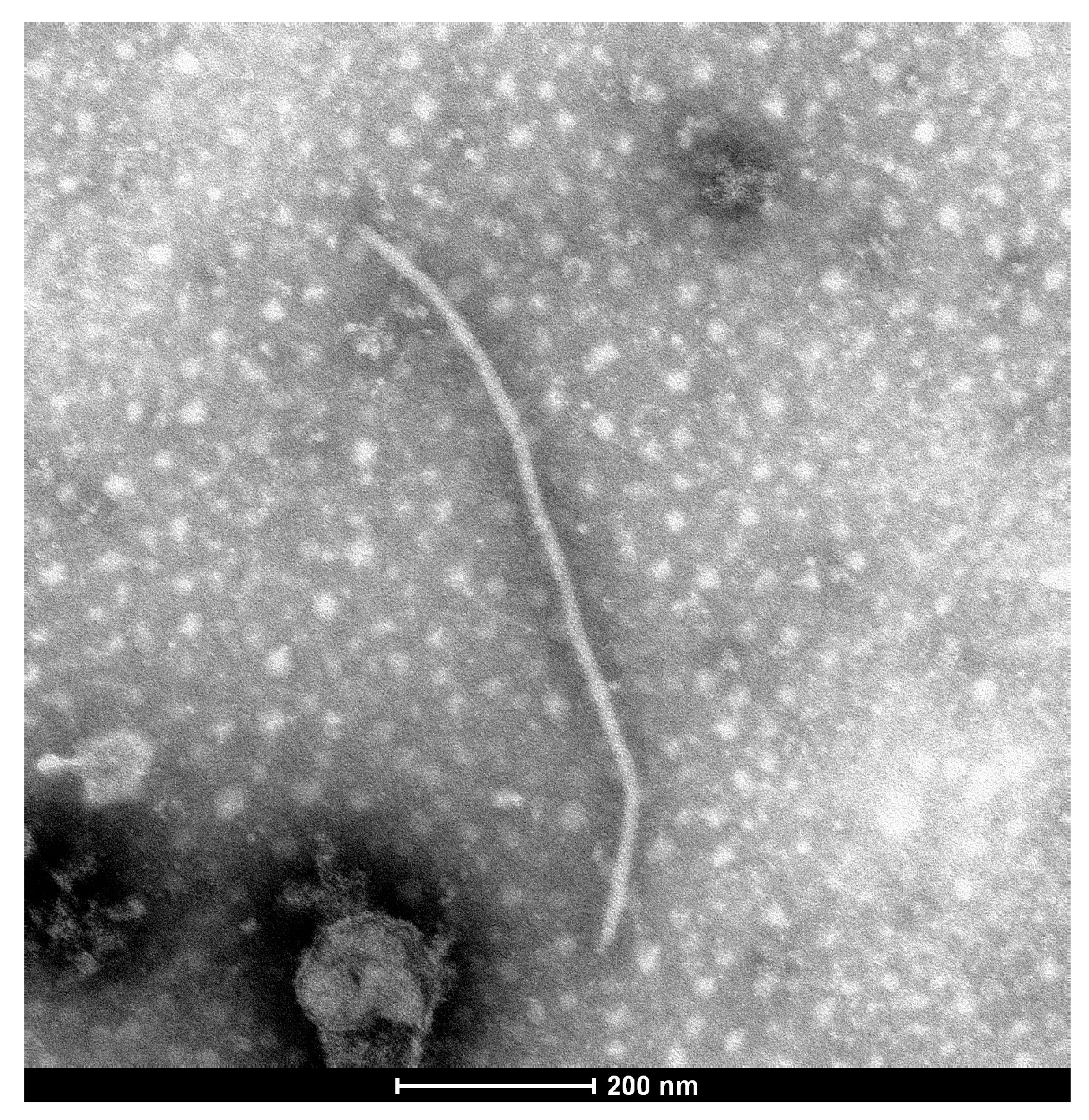
In September 2021, white clover plants with typical viral disease symptoms of leaf mosaic and redding were observed on the campus of Inner Mongolia Agricultural University in Hohhot, Inner Mongolia, China (Figure 1a). In the crude sap of the original symptomatic white clover leaves, flexuous filamentous viral particles of about 700 nm in length and 13 nm in width were observed using transmission electron microscopy (Figure 2). The characteristics of these particles were consistent with the morphology and dimensions of potyviruses [23]. No particles were observed in white clover plants of healthy controls.

3.2. Sap Transmission Experiments

The crude sap of the symptomatic leaf samples was mechanically inoculated to a range of herbaceous species. No symptoms developed on the inoculated leaves of all tested herbaceous species, while the noninoculated systemic leaves developed different viral disease symptoms at 14 to 21 dpi. Both N. occidentalis and C. amaranticolor showed the symptoms of leaf crinkle and chlorotic spots (Figure 1b,c). N. benthamiana had the symptoms of leaf crinkle and mosaic (Figure 1d), while V. faba exhibited the symptoms of leaf crinkle and vein clearing (Figure 1e). The symptoms of leaf distortion and chlorotic spots were observed on C. quinoa (Figure 1f). V. unguiculata and S. lycopersicum developed similar symptoms of leaf distortion (Figure 1g,h).

3.3. Small RNA Sequencing and RT-PCR Validation

In order to further identify the virus species in the infected samples, the small RNAs of three pooled symptomatic leaf samples were sequenced by high-throughput sequencing. A total of 18,026,757 raw reads with Q30 of 98.90% were obtained from the samples. After trimming adapter sequences, a total of 15,809,674 clean reads with length of 18–35 nt remained for further analyses. Using Velvet software, the clean reads were assembled into 1564 contigs with N50 of 69. BLAST analysis against the GenBank Virus RefSeq databases showed that 62 contigs mapped to viral genomes, including 60 contigs mapped to ClYVV, one mapped to apple stem grooving virus (ASGV), and one mapped to ixeridium yellow mottle-associated virus 2 (IxYMaV-2).
The pooled leaf sample of white clover used for small RNA sequencing was further detected by RT-PCR with specific primers of ClYVV, ASGV, and IxYMaV-2 (Supplementary Table S1). RT-PCR detection with specific primers of ASGV or IxYMaV-2, however, showed negative results, indicating false positive of ASGV and IxYMaV-2. Only ClYVV was RT-PCR-positive using the specific primers ClYVV-F/ClYVV-R. Moreover, the systemic leaves of the indicator plants were detected by RT-PCR for ClYVV as well. Three plants for each species were detected and all of them were positive for ClYVV infection. It was indicated that the viral disease of white clover in this study was associated with the infection of ClYVV.

3.4. Characterization of ClYVV-IM Genome

Four segments, covering the near-full-length genome of ClYVV-IM, with lengths of 2490 bp, 2531 bp, 2555 bp, and 2112 bp, were amplified using the primer pairs ClY-F1/ClY-R1, ClY-F2/ClY-R2, ClY-F3/ClY-R3, and ClY-F4/ClY-R4, respectively. The complete genome sequence of ClYVV-IM was assembled from these four segments and two terminal 5‣ and 3‣ sequences obtained by RACE. It was submitted to the GenBank database with accession number of OP617198 (accessed on 1 October 2022). The full genome of ClYVV-IM is 9565 nt excluding 3‣ poly (A) tail, and has a 171 nt 5‣-UTR and a 175 nt 3‣-UTR. It encodes a large polyprotein of 3072 aa from 172 to 9390 nt, which is cleaved into ten mature proteins of P1 (302 aa), HC-Pro (457 aa), P3 (348 aa), 6K1 (53 aa), CI (635 aa), 6K2 (53 aa), VPg (191 aa), Nia-Pro (243 aa), Nib (519 aa), and CP (271 aa) (Figure 3). The PIPO is embedded within the P3 cistron from 2910 to 3140 nt, which starts at the conserved motif G2A6 and is expressed as a P3N-PIPO fusion product (Figure 3).
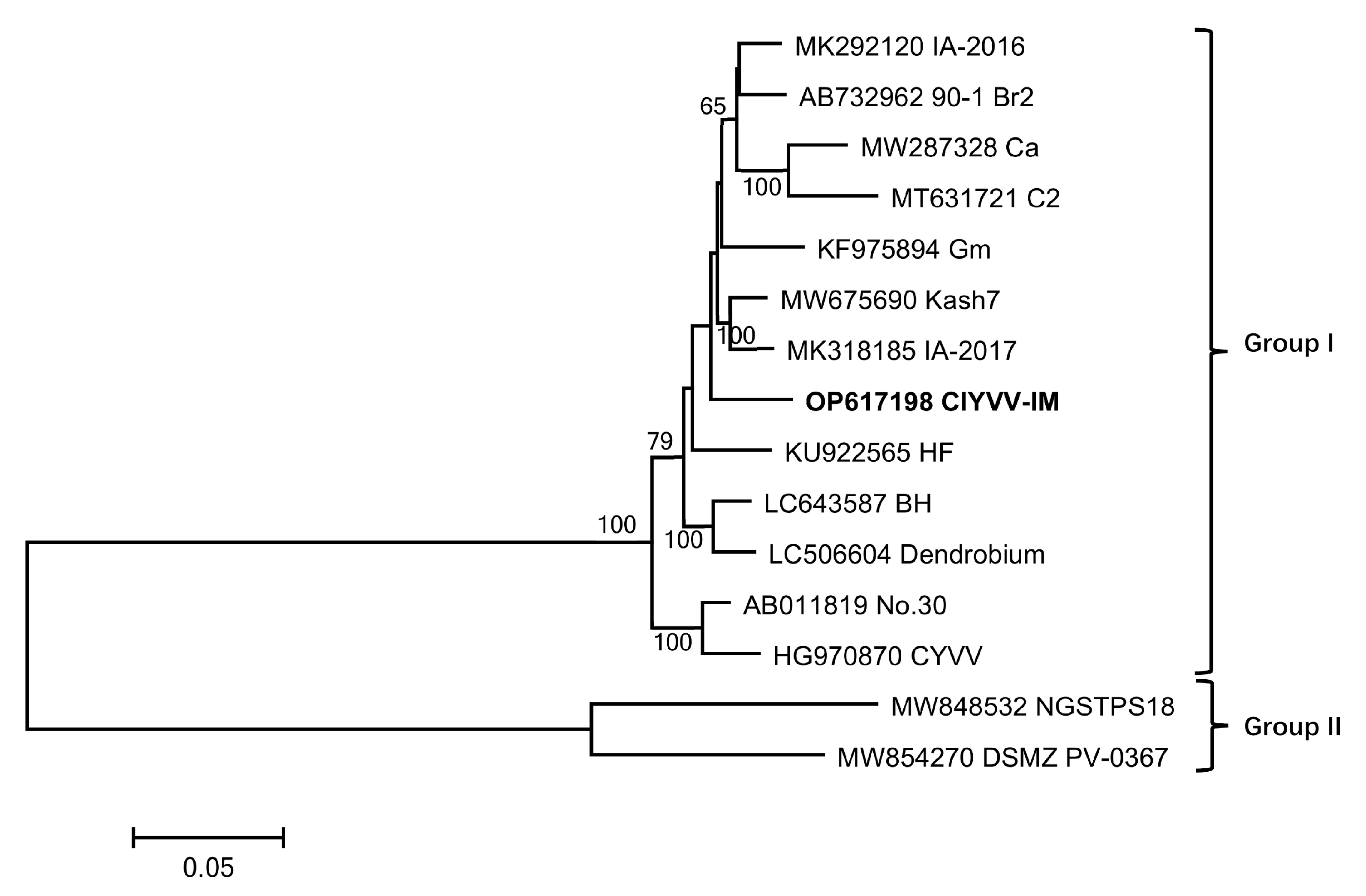
BLAST analysis retrieved the complete genomic sequences of ten ClYVV isolates around the world and four near-full-length genomes which covered the whole large ORF (Table 1). Pairwise alignments revealed that ClYVV-IM shared genome sequence identities ranging from 83.05% with ClYVV-NGSTPS18 (a broad bean isolate from Germany) to 96.30% with ClYVV-Kash7 (a common bean isolate from India) (Table 1). The polyprotein of ClYVV-IM shared 82.91–96.32% and 92.80–98.80% with that of other ClYVV isolates available in GenBank at nucleotide and amino acid levels, respectively (Table 1). Phylogenetic analyses by ML method based on the polyprotein nucleotide (Figure 4) and amino acid (Supplementary Figure S1) sequences showed a similar result that the 15 ClYVV isolates were clustered into two groups. The two isolates from Germany (ClYVV-NGSTPS18 and ClYVV- DSMZ-PV-0367) were clustered into Group II, and the other 13 isolates, including ClYVV-IM in this study, were clustered into Group I. No host-species-specific correlation to the phylogenetic distribution was observed. Using seven methods in the RDP4 software, a total of 15 potential recombination events were detected among the 11 ClYVV full-length genomes (Table 2). Two potential recombination events located at 914–2970 nt and 5153–5694 nt were detected in genome of ClYVV-IM by six and seven methods in RDP4, respectively.

4. Discussion

Infection of viruses causes serious losses in the yield and quality of white clover. Several viruses, such as alfalfa mosaic virus (AMV), white clover mosaic virus (WCMV, Traverse, MI, USA), red clover vein mosaic virus (RCVMV), soybean dwarf virus (SbDV), beet western yellow virus (BWYV), and ClYVV, have been reported to infect white clover around the world [5,24,25,26]. Although ClYVV was first reported in white clover, most of the research about its infection has focused on Leguminosae vegetable crops such as common bean, hyacinth bean, broad bean, snap bean, etc. [12,13,14,27]. Graziery is an indispensable part of the agricultural production in northern China, and white clover is one of the most important forage species in pasture ecosystems. However, the information about ClYVV infecting white clover in China is limited. In this work, we proved the occurrence of ClYVV in white clover and described the characteristics of the ClYVV-IM occurring in northern China. Our findings will contribute to the development of strategies to control ClYVV in white clover.
Mixed infections of two or more virus species in a single plant are common in nature, and this phenomenon may affect symptom phenotypes of viral disease. Coinfection of AMV and WCMV in white clover, for example, results in severer symptoms than infection by individual virus [5]. However, it is difficult to identify all of the virus in a single sample using conventional methods based on serological reaction or specific primers. Next-generation sequencing (NGS) of small RNAs is increasingly used for virus identification in plant tissues [28]. Through sequencing the whole small RNAs, including the ones from viruses by the plant-RNA-silencing machinery, this method can identify all of the viruses in the tested plant tissues theoretically. Based on the results of small RNA sequencing and RT-PCR validation, it was proved that the tested white clover was infected with ClYVV only, without any other virus.
Recombination is one of the most important driving forces in the evolution of plant viruses and it explains a considerable amount of genetic diversity in natural populations [29,30]. Many intra- and interspecies recombination events have been detected from potyvirus [31]. The present study predicted a total of 15 recombination events from nine of the eleven ClYVV isolates with complete genome sequences, accounting for 81.8%. The results showed that recombination plays an important role in process of evolution for ClYVV.
In summary, this is the first report of occurrence and complete genome sequence of ClYVV infecting white clover in China.

Supplementary Materials

The following supporting information can be downloaded at: https://www.mdpi.com/article/10.3390/agronomy13051193/s1, Figure S1: Phylogenetic analysis of clover yellow vein virus based on polyprotein amino acid sequences by maximum likelihood method with 1000 bootstrap replicates using MEGA 11; Table S1: Primers used in this study.

Author Contributions

Conceptualization, Z.L., L.Z. and S.S.; methodology, L.X. and P.S.; software, M.Z.; validation, P.S., B.Z. and L.Z.; formal analysis, Z.L. and L.X.; investigation, L.X. and B.Z.; resources, M.Z.; data curation, L.Z.; writing—original draft preparation, Z.L. and L.X.; writing—review and editing, S.S.; visualization, M.Z.; supervision, L.Z.; project administration, Z.L. and S.S.; funding acquisition, Z.L. and S.S. All authors have read and agreed to the published version of the manuscript.

Funding

This research was funded by the Natural Science Foundation of Inner Mongolia, China (grant numbers 2022QN03018, 2019MS03021, 2020MS03014), the Research Program of science and technology at Universities of Inner Mongolia Autonomous Region (grant numbers NJZY21458, NJZY22521), the Research Start-up Funds for High-level Researchers in Inner Mongolia Agricultural University (grant numbers NDYB2018–3, NDYB2019–1, NDYB2018–14), and the Construction Program of ‘Double First-Class’ academic discipline collaborative innovation achievement (grant number LJGXCG2022).

Data Availability Statement

The sequence data have been submitted to GenBank databases under accession number OP617198. Addresses are as follows: GenBank http://www.ncbi.nlm.nih.gov (accessed on 1 October 2022).

Conflicts of Interest

The authors declare no conflict of interest.

References

  1. Isobe, S.N.; Hisano, H.; Sato, S.; Hirakawa, H.; Okumura, K.; Shirasawa, K.; Sasamoto, S.; Watanabe, A.; Wada, T.; Kishida, Y. Comparative genetic mapping and discovery of linkage disequilibrium across linkage groups in white clover (Trifolium repens L.). G3-Genes Genom. Genet. 2012, 2, 607–617. [Google Scholar] [CrossRef]
  2. Wu, F.; Ma, S.; Zhou, J.; Han, C.; Zhang, X. Genetic diversity and population structure analysis in a large collection of white clover (Trifolium repens L.) germplasm worldwide. PeerJ 2021, 9, e11325. [Google Scholar] [CrossRef] [PubMed]
  3. Abberton, M.T.; Fothergill, M.; Collins, R.P.; Marshall, A.H. Breeding forage legume for sustainable and profitable farming systems. Asp. Appl. Biol. 2006, 80, 81–87. [Google Scholar]
  4. Griffiths, A.G.; Barrett, B.A.; Simon, D.; Khan, A.K.; Bickerstaff, P.; Anderson, C.B.; Franzmayr, B.K.; Hancock, K.R.; Jones, C.S. An integrated genetic linkage map for white clove (Trifolium repens L.) with alignment to Medicago. BMC Genom. 2013, 14, 388. [Google Scholar] [CrossRef] [PubMed]
  5. Liang, Q.; Wei, L.; Xu, B.; Calderón-Urrea, A.; Xiang, D. Study of viruses co-infecting white clover (Trifolium repens) in China. J. Integr. Agric. 2017, 16, 1990–1998. [Google Scholar] [CrossRef]
  6. Panter, S.; Chu, P.G.; Ludlow, E.; Garrett, R.; Kalla, R.; Jahufer, M.Z.Z.; de Lucas Arbiza, A.; Rochfort, S.; Mouradow, A.; Smith, K.F.; et al. Molecular breeding of transgenic white clover (Trifolium repens L.) with field resistance to Alfalfa mosaic virus through the expression of its coat protein gene. Transgenic Res. 2012, 21, 619–632. [Google Scholar] [CrossRef] [PubMed]
  7. Hart, J.P.; Griffiths, P.D. Resistance to Clover yellow vein virus in common bean germplasm. Crop Sci. 2014, 54, 2609–2617. [Google Scholar] [CrossRef]
  8. Hisa, Y.; Suzuki, H.; Atsumi, G.; Choi, S.H.; Nakahara, K.S.; Uyeda, I. P3N-PIPO of Clover yellow vein virus exacerbates symptoms in pea infected with White clover mosaic virus and is implicated in viral synergism. Virology 2014, 449, 200–206. [Google Scholar] [CrossRef]
  9. Chuang, B.Y.; Miller, W.A.; Atkins, J.F.; Firth, A.E. An overlapping essential gene in the Potyviridae. Proc. Natl. Acad. Sci. USA 2008, 105, 5897–5902. [Google Scholar] [CrossRef]
  10. Hollings, M.; Nariani, T.K. Some properties of clover yellow vein, a virus from Trifolium repens L. Ann. Appl. Biol. 1965, 56, 99–109. [Google Scholar] [CrossRef]
  11. Hart, J.P.; Griffiths, P.D. A serious of eIF4E alleles at the Bc-3 locus are associated with recessive resistance to Clover yellow vein virus in common bean. Theor. Appl. Genet. 2013, 126, 2849–28663. [Google Scholar] [CrossRef] [PubMed]
  12. Li, C.S.; Zhu, H.C. Identification of Clover yellow vein virus infecting Phaseolus vulgaris. J. Shandong Agric. Univ. 1989, 4, 223–226. [Google Scholar]
  13. Li, C.S. Identification of Clover yellow vein virus infecting Dolichos lablab. Virol. Sin. 1991, 6, 223–226. [Google Scholar]
  14. Zhang, C.; Zheng, H.; Yan, D.; Han, K.; Peng, J.; Lu, Y. Identification of Clover yellow vein virus infecting broad bean in China by deep sequencing and characterization of virus-derived small interfering RNAs. Acta Agric. Univ. Zhejiangensis 2018, 30, 406–412. [Google Scholar]
  15. Song, S.; Cui, J.; Lei, G.; Chen, Y.; Yang, M.; Li, Z.; Zhang, J. Occurrence, infectivity and molecular characterization of hosta virus X in North-east China. Can. J. Plant Pathol. 2020, 42, 595–603. [Google Scholar] [CrossRef]
  16. Li, Z.; Sun, P.; Zhang, L.; Song, S. First report of basella rugose mosaic virus infecting Anredera cordifolia in mainland of China. Crop Prot. 2021, 139, 105350. [Google Scholar] [CrossRef]
  17. Langmead, B.; Trapnell, C.; Pop, M.; Salzberg, S.L. Ultrafast and memory-efficient alignment of short DNA sequences to the human genome. Genome Biol. 2009, 10, R25. [Google Scholar] [CrossRef]
  18. Zerbino, D.R.; Birney, E. Velvet: Algorithms for de novo short read assembly using de Bruijn graphs. Genome Res. 2008, 18, 821–829. [Google Scholar] [CrossRef]
  19. Wu, Q.; Luo, Y.; Lu, R.; Lau, N.; Lai, E.C.; Palese, P. Virus discovery by deep sequencing and assembly of virus-derived small silencing RNAs. Proc. Natl. Acad. Sci. USA 2010, 107, 1606–1611. [Google Scholar] [CrossRef]
  20. Muhire, B.M.; Varsani, A.; Martin, D.P. SDT: A virus classification tool based on pairwise sequence alignment and identity calculation. PLoS ONE 2014, 9, e108277. [Google Scholar] [CrossRef]
  21. Tamura, K.; Stecher, G.; Kumar, S. MEGA11: Molecular evolutionary genetics analysis version 11. Mol. Biol. Evol. 2021, 38, 3022–3027. [Google Scholar] [CrossRef] [PubMed]
  22. Martin, D.P.; Murrell, B.; Golden, M.; Khoosal, A.; Muhire, B. RDP4: Detection and analysis of recombination patters in virus genomes. Virus Evol. 2015, 1, vev003. [Google Scholar] [CrossRef] [PubMed]
  23. Nury, S.; Hosseini, A.; Gibbs, A.J.; Mohammadi, M. Poisono hemlock virus (PHVY), a novel potyvirus from Iranian Conium maculatum (Apiaceae). Australas Plant Pathol. 2020, 49, 119–216. [Google Scholar] [CrossRef]
  24. Denny, B.L.; Guy, P.L. Incidence and spread of viruses in white-clover pastures of the South Island, New Zealand. Australas Plant Path. 2009, 38, 270–276. [Google Scholar] [CrossRef]
  25. Pratt, M.J. Studies on clover yellow mosaic and white clover mosaic viruses. Can. J. Bot. 2011, 39, 655–665. [Google Scholar] [CrossRef]
  26. Fletcher, J.; Tang, J.; Blouin, A.; Ward, L.; MacDiarmid, R.; Ziebell, H. Red clover mosaic virus- A novel virus to New Zealand that is widespread in Legumes. Plant Dis. 2016, 100, 890–895. [Google Scholar] [CrossRef]
  27. Larsen, R.C.; Miklas, P.N.; Eastwell, K.C.; Grau, C.R. A strain of Clover yellow vein virus that causes severe pod necrosis disease in snap bean. Plant Dis. 2008, 92, 1026–1032. [Google Scholar] [CrossRef]
  28. Liang, P.; Navarro, B.; Zhang, Z.; Wang, H.; Lu, M. Identification and characterization of a novel geminivirus with a monopartite genome infecting apple tree. J. Gen. Virol. 2015, 96, 2411–2420. [Google Scholar] [CrossRef]
  29. Roossinck, M.J. Mechanisms of plant virus evolution. Annu. Rev. Phytopathol. 1997, 35, 191–209. [Google Scholar] [CrossRef]
  30. Boulila, M. Molecular evidence for recombination in Prunus necrotic ringspot virus. Plant Mol. Biol. Rep. 2009, 27, 189–198. [Google Scholar] [CrossRef]
  31. Li, X.; Zhu, T.; Yin, X.; Zhang, C.; Chen, J.; Tian, Y.; Liu, J. The genetic structure of Turnip mosaic virus population reveals the rapid expansion of a new emergent lineage in China. Virol. J. 2017, 14, 165. [Google Scholar] [CrossRef] [PubMed]
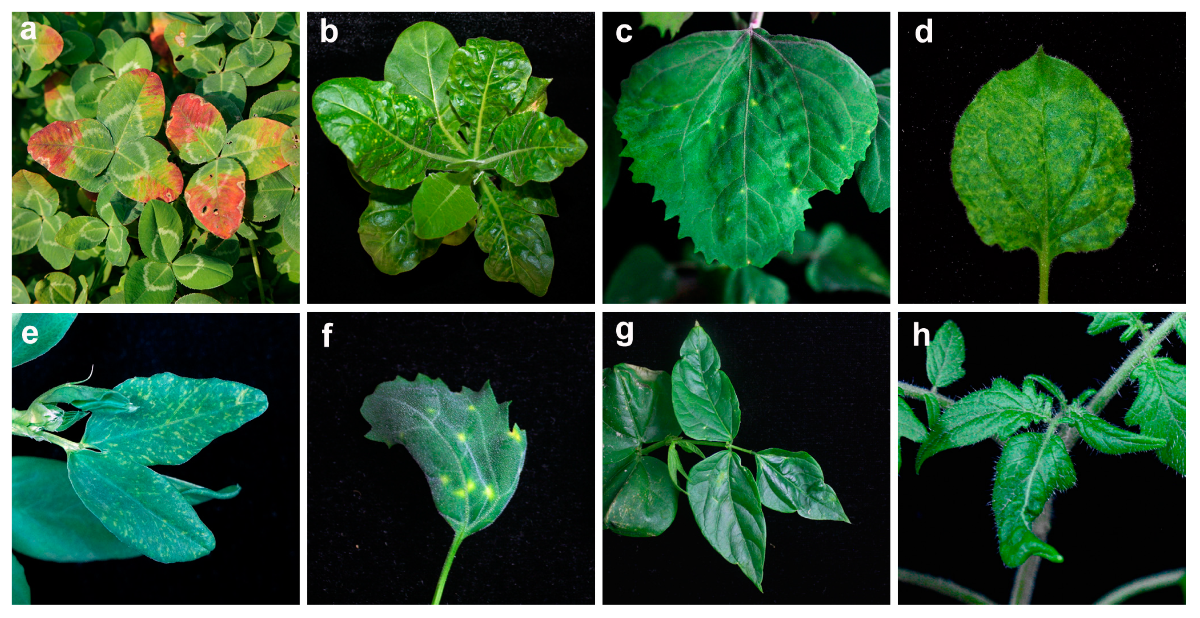
Figure 1. Clover yellow vein virus-induced symptoms of leaf mosaic and redding in naturally infected Trifolium repens (a), leaf crinkle and chlorotic spots in inoculated Nicotiana occidentalis (b) and Chenopodium amaranticolor (c), leaf crinkle and mosaic in inoculated N. benthamiana (d), leaf crinkle and vein clearing in inoculated Vicia faba (e), leaf distortion and chlorotic spots in inoculated C. quinoa (f), leaf distortion in inoculated Vigna unguiculata (g) and Solanum lycopersicum (h).
Agronomy 13 01193 g001
Figure 2. Transmission electron micrographs of clover yellow vein virus particle from crude extracts of ClYVV-IM-infected white clover leaves.
Agronomy 13 01193 g002
Figure 3. Genomic structure of clover yellow vein virus ClYVV-IM isolate. The nucleotide positions of the cleavage sites are indicated above, and the amino acid sequences of the cleavage sites are shown below. P1, the first protein; HC-Pro, helper component-proteinase; P3, the third protein; 6K1, 6 kDa protein 1; CI, cytoplasmic inclusion protein; 6K2, 6 kDa protein 2; VPg, viral genome-linked protein; NIa-Pro, nuclear inclusion a protease; NIb, nuclear inclusion b protease; CP, coat protein; PIPO, pretty interesting potyvirus ORF.
Agronomy 13 01193 g003
Figure 4. Phylogenetic analysis of clover yellow vein virus based on polyprotein nucleotide sequences by maximum likelihood method with 1000 bootstrap replicates using MEGA 11. The accession numbers and isolate names are indicated, and ClYVV-IM determined in this study is marked in bold.
Agronomy 13 01193 g004
Table 1. Pairwise sequence identities between ClYVV-IM in this study and other published complete and near-full-length genome of ClYVV isolates.
IsolateGenBank No.HostCountryGenome Identities (%)Polyprotein Identities (%)
NucleotideNucleotideAmino Acid
Kash7 aMW675690Phaseolus vulgarisIndia96.3096.3298.60
IA-2017 aMK318185Glycine maxUSA96.1096.1098.50
IA-2016 aMK292120Glycine maxUSA96.0296.0298.80
HF aKU922565Vicia fabaChina95.6295.6398.40
BH aLC643587Aquilegia buergerianaSouth Korea95.6195.5098.50
Gm aKF975894Glycine maxSouth Korea95.3895.3798.60
Dendrobium aLC506604Dendrobium spp.South Korea95.3395.3198.60
No. 30 aAB011819--95.1695.0298.70
Ca aMW287328Centella asiaticaUSA94.5794.5297.90
NGSTPS18 aMW848532Vicia fabaGermany83.0582.9192.80
CYVV bHG970870Trifolium repensAustralia 94.3498.50
C2 bMT631721Crotalaria micansUSA 93.9497.70
DSMZ-PV-0367 bMW854270Phaseolus vulgarisGermany 83.1593.10
90–1 Br2 bAB732962Pisum sativumJapan 95.8598.70
a ClYVV isolates with complete genome sequence. b ClYVV isolates with near-full-length genome which covered the complete large ORF.
Table 2. Recombination events among ClYVV isolates predicted by RDP4.
Event No.RecombinantParental IsolateRegion (nt)p-Value a
MajorMinorRGBMCS3S
1IMGmNo. 305153–56941.0 × 10−113.33 × 10−111.62 × 10−123.80 × 10−89.77 × 10−98.62 × 10−133.59 × 10⁻¹⁰
Kash7
IA-2017
IA-2016
HF
BH
Dendrobium
2IMDendrobiumHF914–29706.6 × 10−5-2.97 × 10−37.53 × 10−83.30 × 10−79.70 × 10−101.77 × 10⁻²³
3IA-2016Kash7Ca1999–45395.0 × 10−7-1.62 × 10−42.42 × 10−95.20 × 10−103.55 × 10−53.58 × 10−4
4HFIA-2017No. 303069–45798.8 × 10−113.47 × 10−119.25 × 10−122.15 × 10−121.54 × 10−134.17 × 10−149.64 × 10⁻¹⁷
5BHIA-2016No. 303676–41811.41 × 10−44.29 × 10−41.67 × 10−45.43 × 10−103.31 × 10−92.74 × 10−107.76 × 10⁻⁹
6BHNo. 30IA-20173676–41908.86 × 10−113.47 × 10−119.25 × 10−122.15 × 10−121.54 × 10−134.17 × 10−149.64 × 10⁻¹⁷
7BHIA-2017IA-20162971–41902.41 ×10−43.40 × 10−41.15 × 10−44.11 × 10−42.13 × 10−49.96 × 10−71.12 × 10−5
8BHDendrobiumIM69–4063.55 × 10−59.55 × 10−43.59 × 10−56.21 × 10−51.17 × 10−63.80 × 10−76.90 × 10−9
9BHDendrobiumNo. 30835–27022.39 × 10−214.95 × 10−104.16 × 10−129.15 × 10−213.79 × 10−184.15 × 10−154.49 × 10⁻³³
10DendrobiumGmCa867–16623.43 × 10−114.18 × 10−42.57 × 10−101.59 × 10−71.26 × 10−66.02 × 10−103.13 × 10⁻⁴
11DendrobiumIA-2017IA-20162842–36752.41 × 10−43.40 × 10−41.15 × 10−44.11 × 10−42.13 × 10−49.96 × 10−71.12 × 10−5
12DendrobiumIA-2016No. 303676–41811.41 × 10−44.29 × 10−41.67 × 10−45.43 × 10−103.31 × 10−92.74 × 10−107.76 × 10⁻⁹
13No. 30DendrobiumHF948–15996.69 × 10−5-2.97 × 10−37.53 × 10−83.30 × 10−79.70 × 10−101.77 × 10⁻²³
14No. 30IMBH2703–56945.53 × 10−113.66 × 10−72.20 × 10−123.78 × 10−63.93 × 10−31.14 × 10−161.44 × 10−50
15CaIA-2016No. 304540–56943.00 × 10−14-2.97 × 10−122.72 × 10−151.44 × 10⁻¹⁶-4.76 × 10⁻²⁶
a p-values for each recombination event detected by methods: R, RDP; G, GENECOVN; B, BOOTSCAN; M, MAXCHI; C, CHIMAERA; S, SISCAN; 3S, 3SEQ.
Disclaimer/Publisher’s Note: The statements, opinions and data contained in all publications are solely those of the individual author(s) and contributor(s) and not of MDPI and/or the editor(s). MDPI and/or the editor(s) disclaim responsibility for any injury to people or property resulting from any ideas, methods, instructions or products referred to in the content.