Article

PsFT, PsTFL1, and PsFD Are Involved in Regulating the Continuous Flowering of Tree Peony (Paeonia × lemoinei ‘High Noon’)

1
State Key Laboratory of Efficient Production of Forest Resources, Beijing Key Laboratory of Ornamental Plants Germplasm Innovation & Molecular Breeding, National Engineering Research Center for Floriculture, Key Laboratory of Genetics and Breeding in Forest Trees and Ornamental Plants of Ministry of Education, Peony International Institute, School of Landscape Architecture, Beijing Forestry University, Beijing 100083, China
2
State Key Laboratory of Systematic and Evolutionary Botany, Institute of Botany, Chinese Academy of Sciences, Beijing 100093, China
*
Author to whom correspondence should be addressed.
Agronomy 2023, 13(8), 2071; https://doi.org/10.3390/agronomy13082071
Submission received: 2 July 2023 / Revised: 26 July 2023 / Accepted: 2 August 2023 / Published: 6 August 2023
(This article belongs to the Special Issue Flowering and Flower Development in Plants)

Abstract

:
Tree peonies are an economically important crop with flowers of high ornamental value. Most tree peony cultivars in gardens are once-flowering, and the continuous flowering (CF) trait has been revealed only in a few tree peony cultivars, such as ‘High Noon’ (‘HN’). However, the molecular mechanism underlying its CF remains unclear. Here, we demonstrated that PsTFL1 functions as a floral inhibitor via the ectopic expression of PsTFL1 in transgenic Arabidopsis thaliana plants. Our findings suggest that PsFT and PsTFL1 interact with PsFD, and the detected interactions may occur in the nucleus. Compared with the non-CF variety, the gene expression patterns of PsFT, PsTFL1, and PsFD during the flower development indicate that these three genes may be related to the CF habit in tree peony ‘HN’. These findings will aid future investigations of CF behavior and promote the breeding of tree peonies and other perennial woody plants.

1. Introduction

The transition from vegetative to reproductive development (flowering) requires environmental signals, such as the photoperiod and vernalization, which trigger the expression and regulation of genes and their integration in the shoot apical meristem (SAM). Continuous flowering (CF), a unique reproductive process, has been reported to be regulated by flowering integrator genes [1,2,3]. Some flowering integrator genes include FLOWERING LOCUS T (FT), TERMINAL FLOWER1 (TFL1), SUPPRESSOR OF OVEREXPRESSION OF CONSTANS1 (SOC1), and LEAFY (LFY) [4,5,6,7], and the roles of FT and TFL1 have been widely studied in Arabidopsis [8], Antirrhinum [9], Rosa [1,10,11], Jatropha [12], and many other species. The FT and TFL1 belong to the phosphatidyl ethanolamine-binding protein (PEBP) family; however, FT promotes flowering, and TFL1 represses it [1,4,8,9,10,11,12,13]. Structural studies of FT and TFL1 have revealed that they both exist in an external loop along with an adjacent peptide, serving as a potential ligand-binding site. The structures formed by the rest of the proteins play a key role in determining the antagonistic functions of FT and TFL1 [14]. The molecular antagonistic mechanism between these two genes has also been investigated in some plants. In contrast to the role of FT as a florigen that promotes plant flowering, the overexpression or RNAi silencing of TFL1 genes in rape [15], ragweed [16], apple [17], and pear [18] results in delayed or early flowering.
FD, a member of the basic region leucine zipper (bZIP) family, interacts with FT and can have a major effect on early flowering [14]. FT has been reported to be a florigen in previous studies [10,14,19,20], and an FT/FD heterodimer formed from the combination of FT with FD binds to the promoter of APETALA1 (AP1) to activate flowering initiation [19,20,21,22]. In rice, 14-3-3 (the FT-like protein in rice) and FD form a hexametric structure, which has been referred to as the ‘florigen activation complex’ [13]. However, in Arabidopsis, TFL1 represses the floral transition and maintains the indeterminacy of the shoot apex [17,23,24]. In contrast to loss-of-function phenotypes, overexpression of TFL1 delays flowering and inhibits the transition of the inflorescence meristem (IM) to the floral meristem (FM), resulting in an extended IM stage [25,26]. Therefore, TFL1 as a negative regulator regulates the phase transition from vegetative growth to reproductive growth and from IM to FM in the shoot apical meristem (SAM). TFL1 is involved in transcriptional repression mechanisms that control FM identity genes, which are also FT-targeted [27]. Studies of the fusion of TFL1 with activator or repressor domains have revealed that TFL1 does not possess the functional domains commonly found in transcription factors or regulators. Therefore, TFL1 and FT act as adhesive proteins rather than transcriptional regulators and interact with FD-like proteins [14].
In Chinese rose and woodland strawberry, the recessive mutation of KSN (homolog of TFL1) results in continuous flowering and determinate/indeterminate growth, making the TFL1 homologs provide promising candidate genes for examining the remontancy of flowering [10]. The TFL1 protein sequence is highly similar to FT, and TFL1 shares 71% of amino acid residues, including 55% identical residues, with FT [27]. Because of a retrotransposon insertion, the transcription of RoKSN is blocked in CF roses, and the absence of the floral repressor induces continuous blooming [11]. In Fragaria vesca, a 2-bp deletion in the coding region of the TFL1 homolog introduces a frame shift that leads to CF [11]. In Rosa, fluorescence resonance energy transfer (FRET) assays have been used to clarify competition between RoFT and RoKSN for interaction with RoFD. The FRET results suggest that RoFT and RoKSN are able to interact strongly with RoFD, with a FRET value of >10% [10]. Advances in our understanding of CF in model plants can provide insights into the remontancy and CF of other species such as tree peonies.
Tree peony species, or Mudan, is a woody species in the genus Paeonia. Known as the ‘king of flowers’, it has been cultivated in China since the Tang Dynasty (618 A.D.). More than 1000 cultivars of tree peonies have been released worldwide [28], and they can be divided into three flowering classes: once-flowering (OF), autumn-flowering (AF), and continuous-flowering (CF) classes [5,28,29,30]. Particularly, the CF trait has drawn more attention, but the molecular regulatory mechanisms underlying CF in tree peonies remain elusive. We previously cloned PsFT and showed that its overexpression can promote flowering in transgenic Arabidopsis thaliana plants, suggesting that PsFT may be associated with flowering [29,30]. In CF roses, some studies have revealed that the interaction between FT, TFL1, and FD in flower bud differentiation is related to the regulatory mechanism of CF [1,10]. Does such a regulatory mechanism exist in CF tree peonies? In this study, we isolated PsTFL1 and PsFD in tree peonies and analyzed the sequences of FT/TFL1/FD and the structures of the proteins that they encode. We also studied the expression patterns of PsFT, PsTFL1, and PsFD during bud differentiation in CF and OF tree peonies and explored their protein–protein interactions. These findings provide insights into the understanding of the molecular mechanism of CF in tree peonies and will aid future studies concerning the CF habits of plants, especially in ornamental species such as roses and tree peonies.

2. Materials and Methods

2.1. Plant Materials

Two classes of tree peonies with different flowering traits were used in this study: CF and OF. The CF cultivar P. × lemoinei ‘HN’ was obtained from the Jiufeng Experiment Station of Beijing Forestry University (Haidian District, Beijing, China, 40°03′ N, 116°05′ E). P. delavayi var. lutea, a potential CF resource, was obtained from GuoSe Peony Nursery (Yanqing district, Beijing, China, 40°45′ N, 115°97′ E). The OF cultivars P. suffruticosa ‘LYH’ were also obtained from GuoSe Peony Nursery. Changes in gene expression during the floral differentiation process in the common buds of CF ‘HN’, the potential CF potential P. delavayi var. lutea, and the OF ‘LYH’ were analyzed. The flower buds were collected from 25 May to 5 September every 10 days in CF ‘HN’, OF ‘LYH’, and the potential CF potential P. delavayi var. lutea was analyzed also. Common flower bud initiation coincides with the enlargement of the meristem, and bract primordia become visible in early July, according to Zhou et al. [29]. The apical buds used for expression analysis were immediately frozen in liquid nitrogen. Changes in gene expression in the CF bud of ‘HN’ were also characterized during the differentiation process. CF buds in ‘HN’ were divided into four periods (bud sprouting, S1; bud development and elongation, S2; flower establishment, S3; and flower expansion, S4) according to their morphological characteristics. To compare the expression patterns in common buds in the dormant stage, we collected common buds from ‘HN’.

2.2. Sequence Isolation

Total RNA was extracted using an Aidlab RNA Isolation Kit (Aidlab Biotechnologies Co., Ltd., Beijing, China), and cDNA was synthesized using the FastQuant RT Kit with DNase (Tiangen Biotech, Beijing, China). The raw gene fragments screened from the transcriptome sequencing database were sequenced in a previous study [29]. Polymerase chain reaction (PCR) and the rapid amplification of cDNA ends (SMARTer® RACE 5′/3′Kit) were used to obtain TFL1 and FD from buds. The primers used are presented in Table S1. The FT gene was sequenced in a previous study [30].

2.3. Protein Structure and Phylogenetic Analysis

The Online SWISS-MODEL (https://swissmodel.expasy.org/interactive, (accessed on 18 August 2022)) software was used to predict the secondary structures of PsFT, PsTFL1, and PsFD. The sequences from other species were analyzed along with sequences from ‘HN’. The complete nucleotide sequences, protein sequences, and corresponding annotation information for all these species were downloaded from the JGI and NCBI databases. The BLAST method was used to identify homologous genes of FT in 10 species (or cultivars), TFL1 in 11 species (or cultivars), and FD in 14 species (or cultivars) from the protein database (Table S2). We used RA × ML software v1.0 to construct the maximum-likelihood (ML) phylogenetic tree. The tree topology was visualized using online software (https://itol.embl.de/, (accessed on 9 May 2023)). Support (BS) values for each node were calculated based on 10,000 bootstrap replicates.

2.4. Expression Analysis

To investigate the expression levels of PsFT, PsTFL1, and PsFD at different times during flower bud differentiation, we conducted real-time fluorescent quantitative PCR using a CFX96 Real-Time PCR System (Bio-rad, Hercules, CA, USA) in a 20-µL reaction volume comprising 10 µL of FastStart Universal SYBR Green Ⅱ Master Mix (TaKaRa, Tokyo, Japan), 6 µL of ddH2O, 1 µL of 10 mM forward and reverse primers, and 2 µL of diluted cDNA as the template for target genes. Real-time PCR was performed with the following thermal cycling conditions 95 °C for 10 min, followed by 40 cycles of 2 min at 95 °C, 5 s at 95 °C, and 30 s at annealing temperature (PsFT: 55 °C; PsTFL1: 50 °C; PsFD: 60 °C). Melting curve analysis was conducted using the default settings of the CFX96 Real-Time PCR System (Bio-rad, Hercules, CA, USA). The annealing temperature ranged from 50–60 °C (PsFT: 55 °C, PsTFL1: 50 °C, PsFD: 60 °C, and reference gene: 60 °C). There were three replicates for each sample. The qRT-PCR results were analyzed using the 2−ΔΔCt method [31], and all primers used in this study are listed in Table S1.

2.5. Subcellular Localization

The full-length open reading frame (ORF) of three genes (PsFT, PsTFL1, and PsFD) were inserted into the 35S-GFP vector. Three vectors (35S::FT-GFP, 35S::TFL1-GFP, and 35S::FD-GFP) were generated and transformed into tobacco epidermal cells for two days under low-light density and room-temperature conditions (Invitrogen, Carlsbad, CA, USA). Autofluorescence showed the chlorophyll fluorescence signal was excited at a wavelength of 640 nm and emitted at a wavelength of 675 nm. The protoplasts were observed via a laser-scanning confocal microscope (Leica, Wetzlar, Germany).

2.6. Plant Transformation

Full-length PsTFL1 ORF was cloned onto the vector pEarleyGate 100. The resulting construct, PsTFL1-PEG100, was transformed into A. thaliana (Col-0) using the floral dip method [30]. After vernalization for 2 days at 2 °C, the transformants (T0 lines) from the dipped plants that survived exposure to the Basta solution (1/500) were transplanted into a substrate consisting of vermiculite and nutrient soil (v:v = 1:1) in a growth chamber at 21 °C under long-day (LD) conditions (16 h photoperiod, light intensity: 50 mmol·m–2·s–1). Morphological observations of 15 plants from the second generation (T2) were recorded. The number of rosette leaves and the day when the first flower bloomed were determined.

2.7. Yeast 2-Hybrid (Y2H) Analysis

Y2H analysis was used to clarify the interactions among PsFT, PsTFL1, and PsFD using the Y2H gold system (Clontech, Mountain View, CA, USA). The ORFs of PsFT and PsTFL1 (without signal peptide) were introduced into the pGBKT7 vectors as bait. The ORFs of PsFD and PsTFL1 (without signal peptide) were introduced into pGADT7 as prey. Transformed yeast cells were assayed for growth on synthetic dropout SD/-Leu with agar, SD/-Leu/-Trp with agar, SD/-Leu/-Trp/-His with agar, and SD/-Ade/-His/-Leu/-Trp with agar containing X-α-galactosidase (X-α-gal) and aureobasidin A (AbA).

2.8. Bimolecular Fluorescence Complementation (BiFC)

The coding regions of PsTFL1 and PsFT were introduced into the pCAMBIA1300-35S-YC155 vector and PsFD into the pCAMBIA1300-35S-NY173 vector using a Gateway cloning system (Invitrogen, Carlsbad, CA, USA). The loaded vectors were transferred into Agrobacterium tumefaciens (GV3101) and then infiltrated into the abaxial air spaces of 4-week-old Nicotiana benthamiana plants. Fluorescent signals and bright-field pictures were taken using a confocal laser scanning microscope (LeicaSP8, Wetzlar, Germany).

2.9. Co-IP Assay

To confirm the heterodimization of PsFT-PsFD and PsTFL1-PsFD, we performed protein co-immunoprecipitation assays. The full-length coding sequences of PsFT and PsTFL1 were recombined into the pBinGFP2 vector to be expressed as PsFT-GFP and PsTFL1-GFP, respectively, and PsFD was recombined into the pCAMBIA vector to be expressed as PsFD-Myc. Three-week-old N. benthamiana plants were transiently co-expressed with anti-flag antibodies (Abmart) using a Pierce Crosslink IP kit (Thermo, Shanghai, China) following the manufacturer’s instructions. Finally, a 10% SDS-polyacrylamide gel immunoblot analysis was performed with anti-GFP (Abmart) and anti-Myc (Abmart) antibodies.

2.10. Sequencing Analysis and Primer Synthesis

All resultant constructs were commercially sequenced, and all primers (Table S1) were synthesized by RuiBiotech (Beijing, China).

3. Results

3.1. Comparison of Tree Peony Flowering Traits

Most tree peony cultivars in gardens are OF; they generally complete floral differentiation before winter and then flower in the following spring with dormancy release through winter. Common buds continue to flower in the following spring after winter dormancy. The CF trait has been revealed only in a few tree peony cultivars, such as ‘High Noon’ (‘HN’), which can continuously bloom throughout the annual growth cycle from spring through summer to autumn as the buds differentiate continuously [29,30] (Figure 1). We described the buds in the two classes of cultivars blooming in spring (late April to early May) as common buds and those blooming in other specific times as CF buds. Our previous studies indicated that the bract primordia of common buds become visible in early July both in CF and in OF cultivars, which corresponds to the initiation of flower bud differentiation, and the CF buds of CF cultivars; additionally, the common buds in all cultivars differentiate simultaneously [29]. These varieties differ significantly in their flowering times, which makes them an excellent system for exploring the molecular regulatory mechanisms underlying CF in tree peonies.

3.2. Sequencing Analyses of FD and TFL1 in Tree Peonies

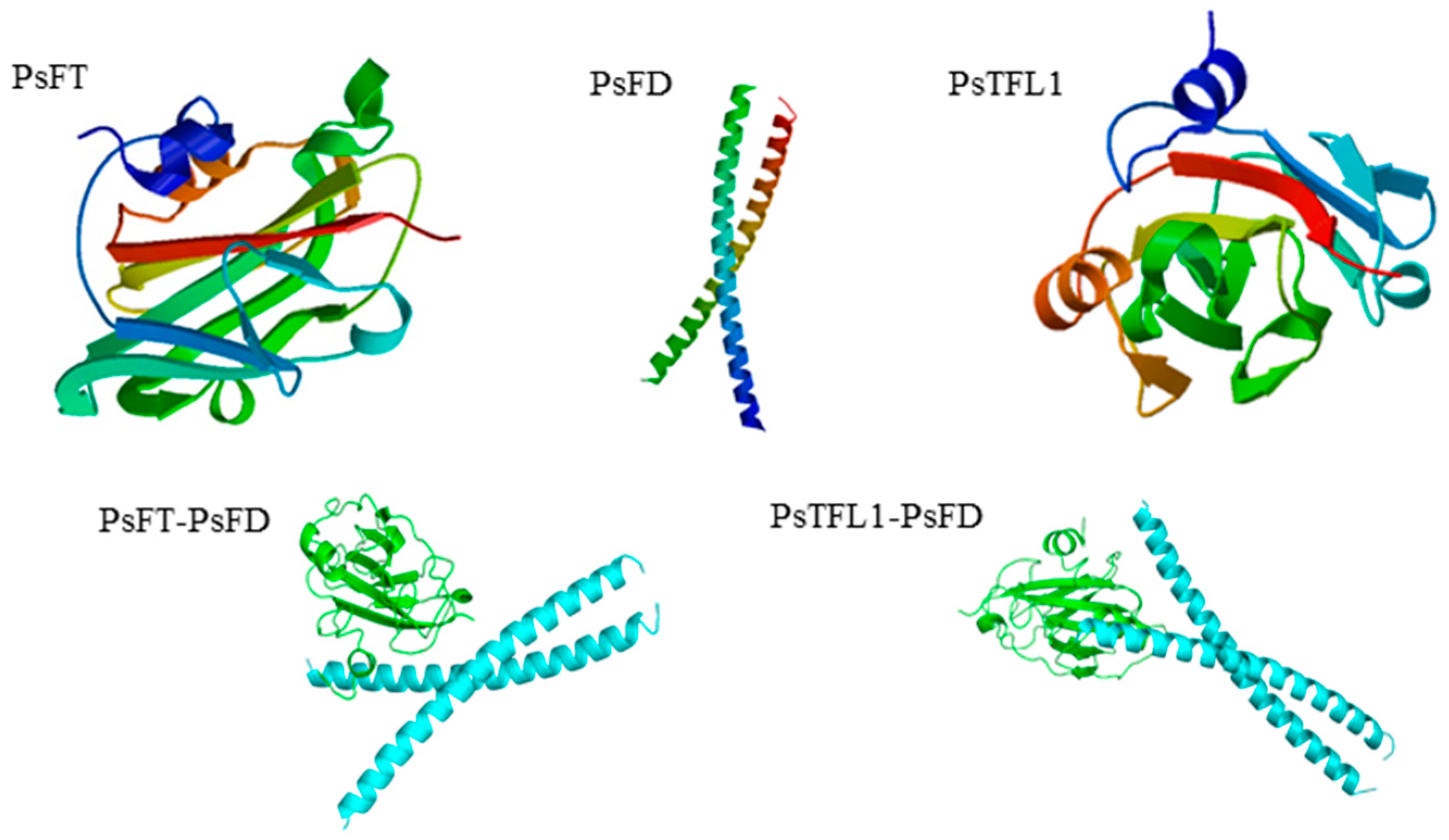
The complete full-length sequences of PsFD and PsTFL1 were amplified using the RACE method. Multiple sequence alignment showed detailed information on the coding sequences and corresponding amino acid (aa) sequences of these two genes (Figure S1). PsTFL1 and PsFD contained 171 aa and 249 aa, respectively. Phylogenetic analysis revealed homologous genes in each clade—FT-like, TFL1-like, and FD-like (Figure S2)—and these homologous genes belonged to their own clade. The PsTFL1 protein showed high sequence similarity to the PsFT protein, and both proteins, belonging to the PEBP family, had greater than 70% sequence conservation (Figure S1a,b). They were not only very similar in the overall protein fold but also in the putative ligand binding and neighboring effector sites (Figure 2). However, there were substantial differences in the FD-like clade among species (Figure S1c). PsFD, a member of the basic region leucine zipper (bZIP) protein family, contained a chained mode structure (Figure 2). Moreover, the heterodimers could be formed as PsFD-PsFT and PsFD-PsTFL1 with high confidence (Figure 2).

3.3. Expression of PsTFL1, PsFD, and PsFT in Different Flowering Cultivars

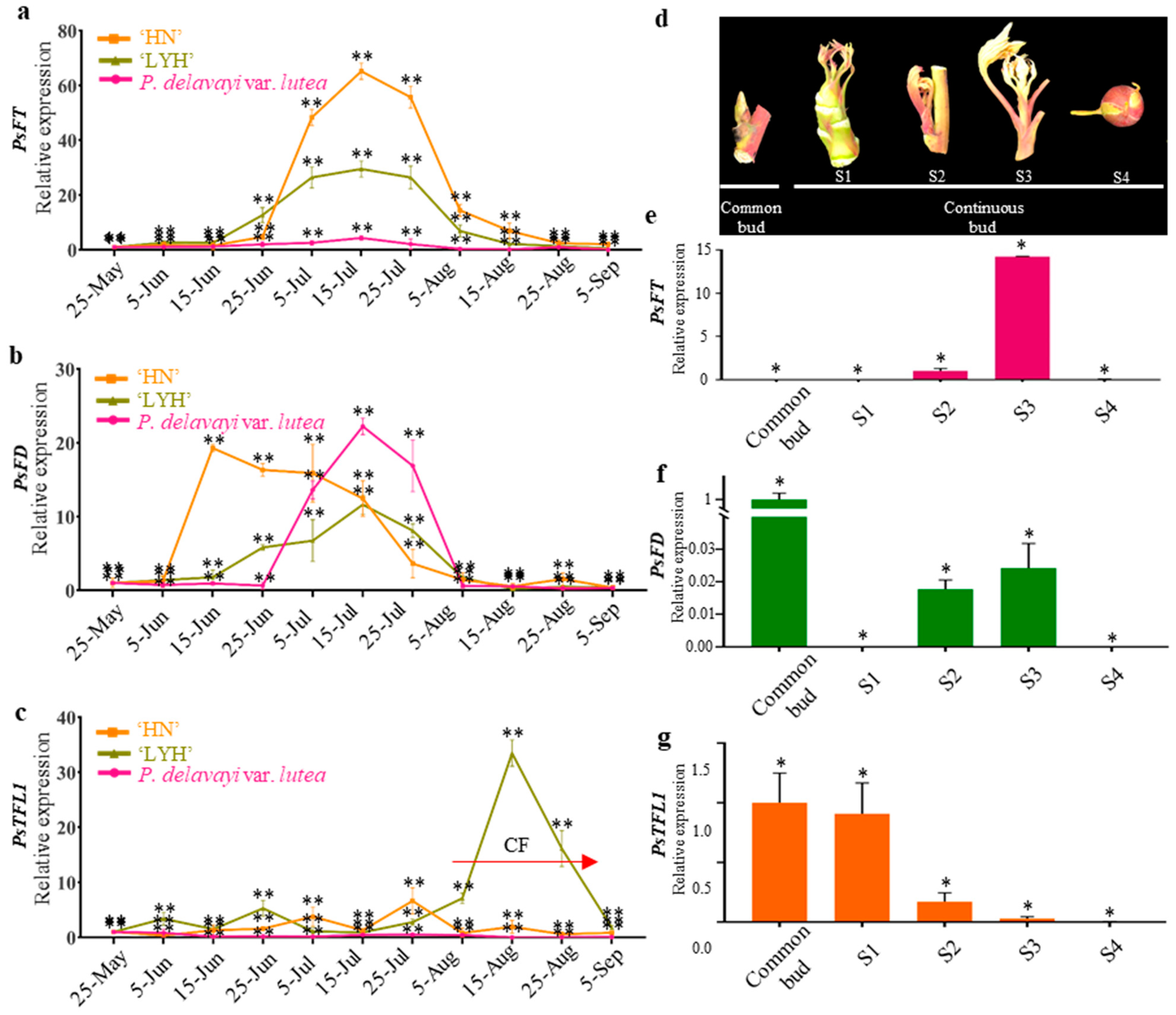
The qRT-PCR results of common buds in ‘HN’, ‘LYH’, and P. delavayi var. lutea revealed that PsFT expression increased continuously from late June to mid-July and then decreased from mid-July to early September (Figure 3a). The expression of PsFD from early June to mid-August was consistent with that of PsFT, although its variation was observed in the three peony cultivars (or species) (Figure 3b). These findings indicate that the expression of PsFT and PsFD increased as floral bud differentiation was initiated with bract primordia formation, as in a previous study [29]. The expression level of PsFT and PsFD was increased in the process of flower bud differentiation, despite differences in degrees among the three cultivars (species). However, the expression of PsTFL1 was only detected in the OF cultivar ‘LYH’, and the expression started to be elevated around August 5, reached the peak around August 15, and decreased until it became silent around September 5, which was exactly the CF stage (Figure 3c). However, this phenomenon was not observed in CF ‘HN’ and potential CF P. delavayi var. lutea, thus implying the essential role of PsTFL1 in CF.
We further analyzed the expression patterns of PsTFL1, PsFT, and PsFD in different floral buds in HN and found that during the flower bud differentiation of CF ‘HN’, the expression of PsTFL1 gradually decreased as the expression of PsFT increased (Figure 3d–g). In ‘HN’ common buds in dormancy, the expression of PsFT was hardly detected, but the expression of PsFD and PsTFL1 was high (Figure 3e–g). During CF bud differentiation, the increased expression of PsFT and PsFD was accompanied by a decrease in PsTFL1 expression. The results indicate that PsFT and PsFD were at peak expression during the S3 stage of the CF buds in CF ‘HN’, while the expression of PsTFL1 gradually decreased from the S1 to S4 stages for the CF buds.

3.4. Subcellular Localization of PsTFL1, PsFD, and PsFT

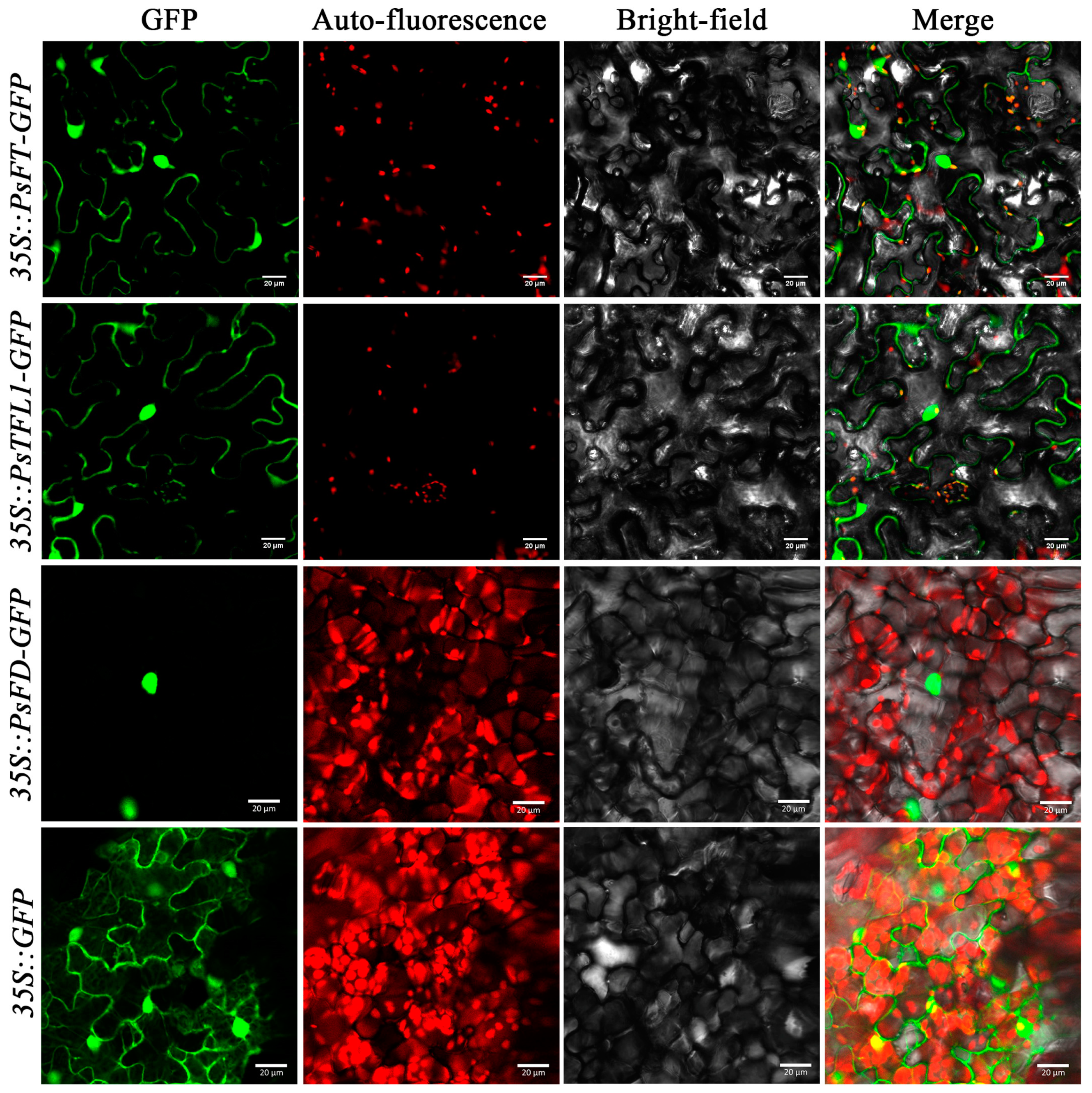
To reveal the subcellular localization of PsTFL1, PsFD, and PsFT, we made the indicated constructs to express fusion proteins with GFP driven by a 35S promoter (Figure 4). In this experiment, the fused GFP fluorescence signal could indicate the subcellular localization of the proteins of interest. In this study, laser confocal microscopy revealed that the PsFT-GFP and PsTFTL1-GFP proteins were primarily expressed in the nucleus and cytoplasm, while the PsFD-GFP protein was expressed in the nucleus (Figure 4). The GFP control (35S::GFP) demonstrated the subcellular localization of the GFP proteins alone, which was observed in the nucleus and cytoplasm. Considering the autofluorescence signal and the merged image (Figure 4), we concluded that PsFT and PsTFL1 are localized in the cytoplasm and nucleus, whereas PsFD is localized in the nucleus.

3.5. Ectopic Expression of PsTFL1 in A. thaliana

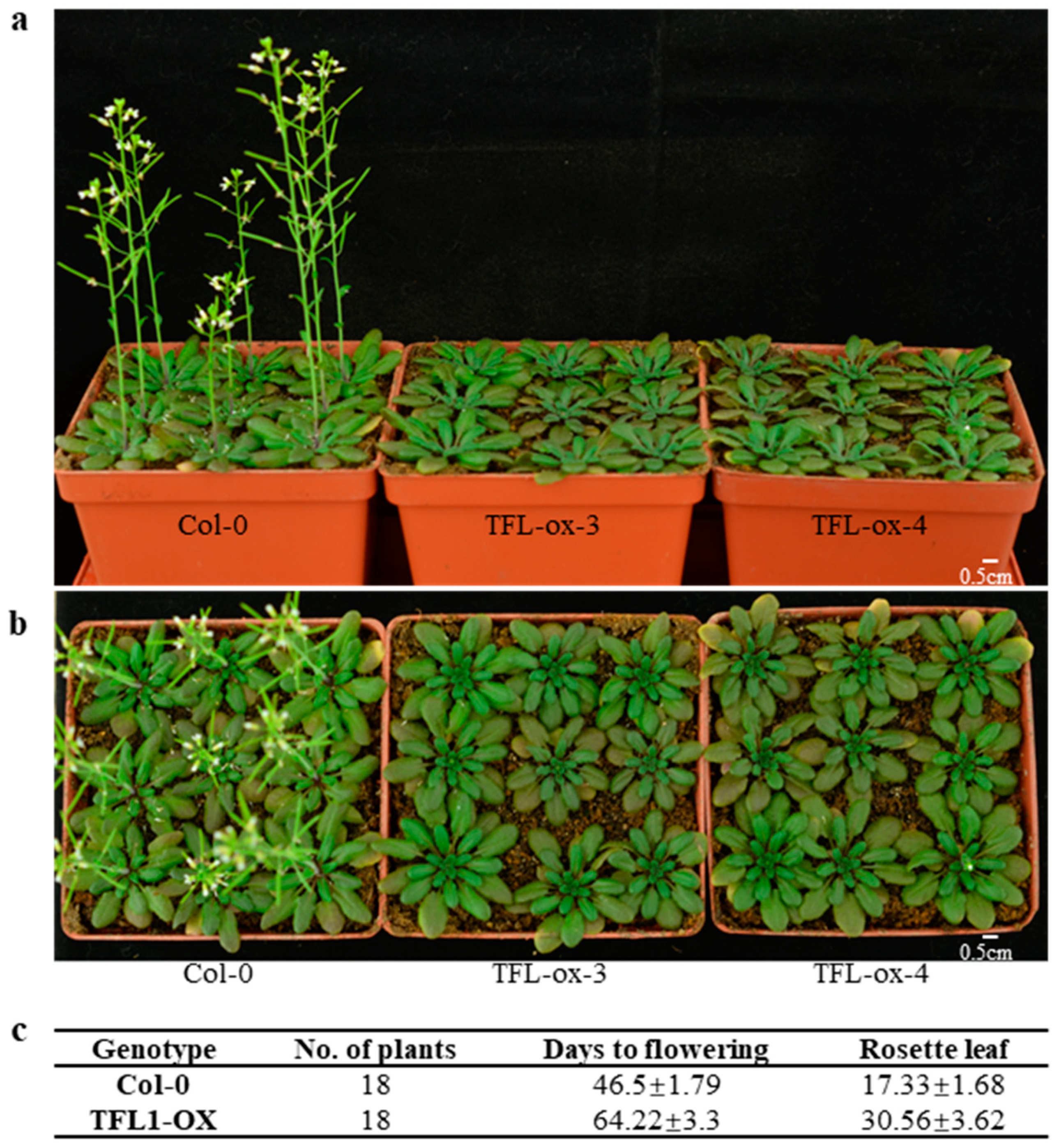
To examine the role of PsTFL1 in the bud differentiation regulations of CF ‘HN’, we generated PsTFL1-overexpressing transgenic A. thaliana plants, and 15 independent kan-resistant T1 plants were obtained. The T2 lines were again filtrated with a kan solution, and morphological observations were taken from 18 plants from the second generation (T2). Under LD conditions, transgenic T2 lines exhibited later flowering characters and more rosette leaves than WT plants (Figure 5a,b). As quantified, the average number of rosette leaves in transgenic Arabidopsis lines was around 30, and the time from germination to flowering was around 64 days. In contrast, the number of rosette leaves in wild-type (WT) Arabidopsis plants was around 17, and the time from germination to flowering was around 46 days (Figure 5c). Therefore, the ectopic expression of PsTFL1 in A. thaliana revealed the function of PsTFL1 in delaying blooming.

3.6. PsFT and PsTFL1 Physically Interact with PsFD

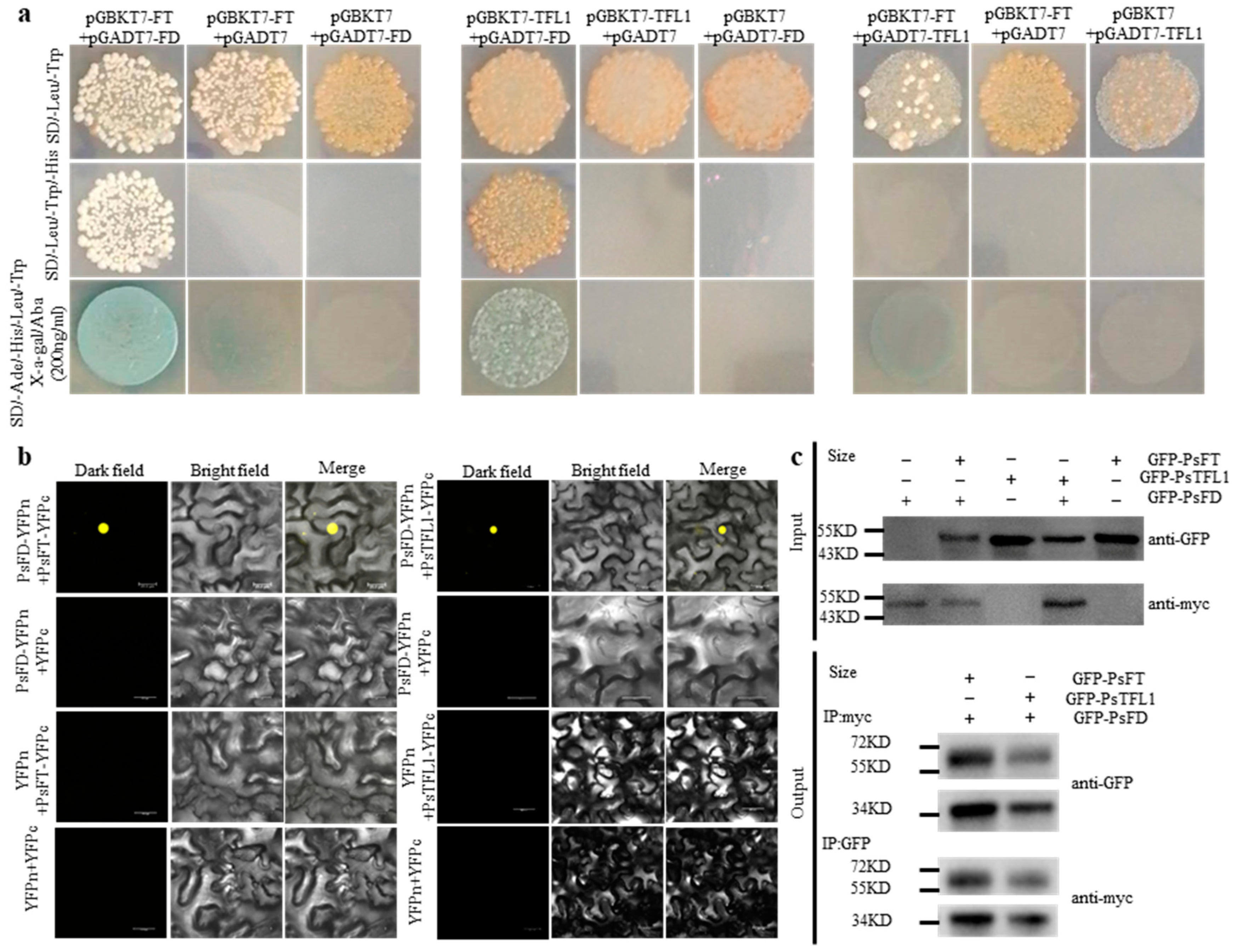
To investigate the direct interaction between PsFT and PsTFL1 with PsFD, as hypothesized (Figure 2), we performed yeast two-hybrid (Y2H) assays. The results showed that the yeast cells co-transformed with PsFT-PsFD or PsTFL1-PsFD exhibited survival and growth, indicating that PsFT and PsTFL1 can interact with PsFD in vitro, while PsFT did not interact with PsTFL1 (Figure 6a). We further conducted BiFC and Co-IP assays to further clarify the observed interactions between the above pairs of proteins. In BiFC assays, a yellow fluorescent protein fluorescence signal was observed in the epidermal cells of tobacco leaves when PsFD-YFPn and PsFT-YFPc, as well as PsFD-YFPn and PsTFL1-YFPc, were co-expressed compared with the set negative controls (Figure 6b), indicating that PsFD physically interacts with PsFT and PsTFL1 in plant cells. In Co-IP assays, we found that PsFT and PsTFL1 were recombined into pBinGFP2 vectors to yield GFP-PsFT and GFP-PsTFL1, respectively, and PsFD was recombined into pCAMBIA to yield the CAM-PsFD vector (Figure 6c). GFP-PsFT and GFP-PsTFL1 co-immunoprecipitated with CAM-PsFD at a protein size from 55 to 72 kD in both anti-GFP and anti-myc media (Figure 6c). These results confirmed the existence of protein–protein interactions between PsFD-PsFT and PsFD-PsTFL1.

4. Discussion

Clarifying the regulatory mechanisms of plant flowering has implications for crop production and breeding [32]. Many studies have examined the molecular mechanism underlying the CF phenotype to promote improvement in the ornamental and economic value of plants. FT, TFL1, and FD have been shown to be involved in the differentiation of CF buds in some perennial plants such as R. rugosa [1], R. hybrid RI [10,11], and Fragaria vesca [11], but whether these genes play an important role in CF tree peonies as well remains unclear. The results of this study proposed the regulatory roles of PsFT, PsTFL1, and PsFD in CF ‘HN’ tree peonies.

4.1. The Different Roles of PsFT-PsFD and PsTFL1-PsFD in Flowering Control

The FT and TFL1 genes are members of the PEBP family, and PEBP proteins are generally well conserved [8]. FT is a mobile protein that triggers flowering, and its sequence is highly conserved across species [12,20,33]. In this study, we found that the key amino acid residues of PsFT and PsTFL1 are conserved, but they are different in PsFD. Moreover, they are not only very similar in the overall protein fold, but they are also similar in the putative ligand binding site. This structure can act as a switch between two downstream possibilities in Arabidopsis, FT converts FD into a strong activator, while TFL1 converts FD into a strong repressor [14]. This similar site suggested that the biological functions of these molecules are similar [8,14,34]. PsFT and PsTFL1 may possess a buckle structure that binds with PsFD. The similarity in their binding site and interaction confidence analysis, with PsFD–PsFT = 0.8671 and PsFD–PsTFL1 = 0.8551, indicates that PsFT and PsTFL1 can both interact with PsFD.
In A. thaliana, both FT and TFL1 were localized in the nucleus and cytoplasm and interacted with FD in the nucleus [27,35]. In this study, subcellular localization assays showed that PsFD was localized in the nucleus, and PsFT and PsTFL1 were localized both in the nucleus and cytoplasm. These results indicated that there is overlap in the localization of PsFT, PsTFL1, and PsFD in the cell, which is necessary for their interactions, and that PsFT and PsTFL1 can interact with PsFD in the nucleus in tree peonies, similar to their function in A. thaliana [8,35]. In this study, our Y2H, BiFC, and Co-IP assays revealed interactions between PsFT, PsTFL1, and PsFD and confirmed protein–protein interactions between PsFT and PsFD and between PsTFL1 and PsFD. Furthermore, the differential structure of the functional groups of PsFT and PsTFL1 might have different functions in tree peonies. We reported previously that the overexpression of the PsFT gene promoted flowering in transgenic A. thaliana [30]. In this study, we confirmed that PsTFL1 delayed flowering and resulted in more rosette leaves in transgenic Arabidopsis plants. In line with the findings in Arabidopsis [14], we suggested that PsFT-PsFD or PsTFL1-PsFD may promote or delay flowering in tree peonies.

4.2. The Role of PsFT, PsTFL1, and PsFD in Regulating CF

As a florigen, FT promotes flower induction or regulates blooming [12], and its homologs have often been reported in apple [36,37], Arabidopsis [8,20], poplar [38], citrus [39], loquat [40], and other species. In loquat, there are two copies of FT; the expression of EjFT1 is involved in bud sprouting and leaf development in mid-May, and EjFT2 is involved in floral bud induction in mid-June [40]. However, in ‘HN’, we only detected a single copy of PsFT, an FT-like gene. The expression of PsFT in common bud differentiation increased continuously from late June to mid-July and then decreased from mid-July to early September. PsFT is also expressed during CF in CF cultivars. Our previous study showed that transferring PsFT from ‘HN’ into A. thaliana significantly advanced flowering compared with WT plants [30]. The PsFT sequenced from tree peony ‘HN’ was detected to be expressed during common bud differentiation, CF flowering. PsFT may act as a florigen in tree peonies.
In this work, a single-copy TFL1 homolog sequence was identified in ‘HN’. We found that the expression of PsTFL1 remained low in the CF ‘HN’ and CF potential P. delavayi during common bud differentiation from early June to late August but increased as bud differentiation finished in the OF ‘LYH’ when a peak appeared from 5 August to 15 August. PsTFL1 was always expressed both in the common buds in dormant stages of all cultivar classes, but its expression gradually decreased from S1 to S4 in CF buds of ‘HN’. This might suggest that the low expression of PsTFL1 in CF tree peonies could allow them to flower continuously under suitable conditions. The overall expression of KSN (a homologous gene of TFL1 in rose) has been shown to inhibit the floral transition in a CF rose, and the expression of KSN was significantly repressed in the CF cultivar compared with OF varieties [1,10,11]. Therefore, we deduced that PsTFL1 might inhibit bud differentiation, and the decrease in its expression during floral differentiation might be related to the flowering of CF ‘HN’ in mid-August.
We found that the expression of PsFD was first upregulated in CF ‘HN’ on 5 June, followed by ‘LYH’ and P. delavayi on 15 June and 25 June. After entering the floral differentiation stage, the expression of PsFD first increased, and this was followed by an increase in the expression of PsFT. Therefore, PsFD might mediate the expression of PsFT, thereby regulating the process of floral differentiation. In the development and flowering of ‘HN’, the expression of both PsFD and PsFT increased, and the expression of PsTFL1 decreased. PsFT, PsTFL1, and PsFD may play a role in inducing CF in ‘HN’. In dormant buds of ‘HN’, we found that PsFT was weak expressed; however, PsTFL1 and PsFD were highly expressed. These findings suggest that PsFD may be involved in the expression of PsFT and PsTFL1 in CF cultivars. The function of FD is not limited to the regulation of flowering time, but it also involves several aspects of plant development [36]. FD may function as a molecular connector that can form an FD–FT or FD–TFL1 florigen activation or inhibition complex by binding with FT and TFL1 [14].
According to the expression curves during the bud differentiation of CF ‘HN’, the expression of PsTFL1 was gradually reduced, while the expression of PsFT was increased, which coincides with the increased expression of PsFD. In common buds from ‘HN’, a high expression of PsTFL1 coincides with the absence of PsFT expression, indicating that PsTFL1 and PsFD could be related to the inhibition of flowering in the common buds of ‘HN’. Therefore, PsFT likely plays a key role in flower bud differentiation and flowering in tree peony, and it is inhibited by PsTFL1. The results of our study showed that PsFT, PsTFL1, and PsFD may be related to the CF trait in the tree peony ‘HN’.
In our study, the results of the qRT-PCR assays suggested that PsFD and PsFT could be expressed during common bud differentiation, common flowering, and CF flowering in ‘HN’. The expression level of PsTFL1 remained low during the common floral differentiation of CF ‘HN’ and CF potential P. delavayi compared with OF ‘LYH’. Furthermore, PsTFL1 expression gradually decreased from S1 to S4 in the CF buds of ‘HN’. We also found that the expression of PsFT and PsFD increased during CF in CF ‘HN’. The regulation of PsFD, PsFT, and PsTFL1 could be correlated with CF flowering in ‘HN’. Ectopic expression in A. thaliana revealed that PsFT acts as a florigen and PsTFL1 acts as a repressor protein. Protein–protein interaction assays revealed that PsFT and PsTFL1 could interact with PsFD. Our findings suggest that the regulation of PsFT and PsTFL1 may be involved in the flowering or lack thereof in tree peonies during common and CF flowering.

5. Conclusions

In this study, we demonstrated that PsTFL1 functions as a floral inhibitor in tree peonies. Our findings suggest that PsFT and PsTFL1 can interact with PsFD to form a complex, and the gene regulation patterns of PsFD, PsFT, and PsTFL1 may be involved in the CF flowering of ‘HN’. However, the mechanisms underlying the differences in expression require further clarification.

Supplementary Materials

The following supporting information can be downloaded at: https://www.mdpi.com/article/10.3390/agronomy13082071/s1, Figure S1. Alignments of the deduced protein sequences. a. PsFT and its homologous proteins from other plants. b. PsTFL1 and its homologous proteins from other plants. c. PsFD and its homologous proteins from other plants. Figure S2. Maximum likelihood phylogenetic tree of the indicated putative proteins. a. PsFT and PsTFL1. b. PsFD with 10,000 bootstrap replicates (G-box binding factor 3 of Zea mays as an outgroup of PsFD). Table S1. Sequences of oligonucleotide primers used in this study. Table S2. Information of genes used in multiple sequence alignment.

Author Contributions

Conceived and designed the experiments: F.C. and L.Z. Performed the experiments: L.Z. Analyzed data: L.Z., F.C. and C.H. Drafted the paper: L.Z. and Z.G. Modified the paper: F.C., H.H. and C.H. Approved the paper: F.C. All authors have read and agreed to the published version of the manuscript.

Funding

The National Natural Science Foundation of China (Grant No.31972446). The funders had no role in study design, data collection, analysis, decision to publish, or manuscript preparation.

Acknowledgments

Thank you to Ruiyuan Biotech Company (Nanjing, China) for the assistance in protein model construction and interaction confidence analysis.

Conflicts of Interest

The authors declare that they have no known competing financial interest or personal relationship that could have appeared to influence the work reported in this paper.

References

  1. Bai, M.; Liu, J.Y.; Fan, C.G.; Chen, Y.Q.; Chen, H.; Lu, J.; Sun, J.J.; Ning, G.G. KSN heterozygosity is associated with continuous flowering of Rosa rugosa Purple branch. Hortic. Res. 2021, 8, 26. [Google Scholar] [CrossRef] [PubMed]
  2. Simpson, G.G.; Gendall, A.R.; Dean, C. When to switch to flowering. Annu. Rev. Cell Dev. Biol. 1999, 15, 19–25. [Google Scholar] [CrossRef]
  3. Srikanth, A.; Schmid, M. Regulation of flowering time: All roads lead to Rome. Cell Mol. Life Sci. 2011, 68, 2013–2037. [Google Scholar] [CrossRef] [PubMed]
  4. Yi, X.; Gao, H.; Yang, Y.; Yang, S.; Luo, L.; Yu, C.; Wang, J.; Cheng, T.; Zhang, Q.; Pan, H. Differentially expressed genes related to flowering transition between once- and continuous-flowering Roses. Biomolecules 2022, 12, 58. [Google Scholar] [CrossRef] [PubMed]
  5. Wang, S.L.; Beruto, M.; Xue, J.Q.; Zhu, F.Y.; Liu, C.J.; Yan, Y.M.; Zhang, X.X. Molecular cloning and potential function prediction of homologous SOC1 genes in tree peony. Plant Cell Rep. 2015, 34, 1459–1471. [Google Scholar] [CrossRef]
  6. Blumel, M.; Dally, N.; Jung, C. Flowering time regulation in crops-what did we learn from Arabidopsis? Curr. Opin. Biotechnol. 2015, 32, 121–129. [Google Scholar] [CrossRef]
  7. Andrés, F.; Coupland, G. The genetic basis of flowering responses to seasonal cues. Nat. Rev. Genet. 2012, 13, 627–639. [Google Scholar] [CrossRef]
  8. Coelho, C.P.; Minow, M.A.; Chalfun-Júnior, A.; Colasanti, J. Putative sugarcane FT/TFL1 genes delay flowering time and alter reproductive architecture in Arabidopsis. Front. Plant Sci. 2014, 5, 221. [Google Scholar] [CrossRef] [Green Version]
  9. Banfield, M.J.; Brady, R.L. The structure of Antirrhinum centroradialis protein (CEN) suggests a role as a kinase regulator. J. Mol. Biol. 2000, 297, 1159–1170. [Google Scholar] [CrossRef]
  10. Randoux, M.; Davière, J.M.; Jeauffre, J.; Thouroude, T.; Pierre, S.; Toualbia, Y.; Perrotte, J.; Reynoird, J.P.; Jammes, M.J.; Oyant, L.H.; et al. RoKSN, a floral repressor, forms protein complexes with RoFD and RoFT to regulate vegetative and reproductive development in rose. New Phytol. 2014, 202, 161–173. [Google Scholar] [CrossRef] [Green Version]
  11. Iwata, H.; Gaston, A.; Remay, A.; Thouroude, T.; Jeauffre, J.; Kawamura, K.; Oyant, L.H.; Araki, T.; Denoyes, B.; Foucher, F. The TFL1 homologue KSN is a regulator of continuous flowering in rose and strawberry. Plant J. 2012, 69, 116–125. [Google Scholar] [CrossRef] [PubMed]
  12. Li, C.Q.; Luo, L.; Fu, Q.T.; Niu, L.; Xu, Z.F. Isolation and functional characterization of JcFT, a FLOWERING LOCUS T (FT) homologous gene from the biofuel plant Jatropha curcas. BMC Plant Biol. 2014, 14, 125. [Google Scholar] [CrossRef] [Green Version]
  13. Taoka, K.; Ohki, I.; Tsuji, H.; Furuita, K.; Hayashi, K.; Yanase, T.; Yamaguchi, M.; Nakashima, C.; Purwestri, Y.A.; Tamaki, S.; et al. 14-3-3 proteins act as intracellular receptors for rice Hd3a florigen. Nature 2011, 476, 332–335. [Google Scholar] [CrossRef]
  14. Ahn, J.H.; Miller, D.; Winter, V.J.; Banfield, M.J.; Lee, J.H.; Yoo, S.Y.; Henz, S.R.; Brady, R.L.; Weigel, D. Divergent external loop confers antagonistic activity on floral regulators FT and TFL1. EMBO J. 2006, 25, 605–614. [Google Scholar] [CrossRef] [PubMed]
  15. Guo, Y.; Hans, H.; Christian, J.; Molina, C. Mutations in single FT- and TFL1-paralogs of rapeseed (Brassica napus L.) and their impact on flowering time and yield components. Front. Plant Sci. 2014, 5, 282. [Google Scholar] [CrossRef] [PubMed] [Green Version]
  16. Kardailsky, I.; Shukla, V.K.; Ahn, J.H.; Dagenais, N.; Christensen, S.K.; Nguyen, J.T.; Chory, J.; Harrison, M.; Weigel, D. North European invasion by common ragweed is associated with early flowering and dominant changes in FT/TFL1 expression. J. Exp. Bot. 2018, 69, 2647–2658. [Google Scholar] [CrossRef] [Green Version]
  17. Flachowsky, H.; Szankowski, I.; Waidmann, S.; Peil, A.; Tränkner, C.; Hanke, M.V. The MdTFL1 gene of apple (Malus × domestica Borkh.) reduces vegetative growth and generation time. Tree Physiol. 2012, 32, 1288–1301. [Google Scholar] [CrossRef]
  18. Freiman, A.; Shlizerman, L.; Golobovitch, S.; Yablovitz, Z.; Korchinsky, R.; Cohen, Y.; Samach, A.; Chevreau, E.; Roux, P.L.; Patocchi, A.; et al. Development of a transgenic early flowering pear (Pyrus communis L.) genotype by RNAi silencing of PcTFL1-1 and PcTFL1-2. Planta 2012, 235, 1239–1251. [Google Scholar] [CrossRef]
  19. Yamaguchi, A.; Kobayashi, Y.; Goto, K.; Abe, M.; Araki, T. TWIN SISTER OF FT (TSF) acts as a floral pathway integrator redundantly with FT. Plant Cell Physiol. 2005, 46, 1175–1189. [Google Scholar] [CrossRef] [Green Version]
  20. Wigge, P.A.; Kim, M.C.; Jaeger, K.E.; Busch, W.; Schmid, M.; Lohmann, J.U.; Weigel, D. Integration of spatial and temporal information during floral induction in Arabidopsis. Science 2005, 309, 1056–1059. [Google Scholar] [CrossRef]
  21. Khan, M.R.; Ai, X.Y.; Zhang, J.Z. Genetic regulation of flowering time in annual and perennial plants. Wiley Interdiscip. Rev. RNA 2014, 5, 347–359. [Google Scholar] [CrossRef]
  22. Harig, L.; Beinecke, F.A.; Oltmanns, J.; Muth, J.; Müller, O.; Rüping, B.; Twyman, R.M.; Fischer, R.; Prüfer, D.; Noll, G.A. Proteins from the FLOWERING LOCUS T-like subclade of the PEBP family act antagonistically to regulate floral initiation in tobacco. Plant J. 2012, 72, 908–921. [Google Scholar] [CrossRef]
  23. Alvarez, J.; Guli, C.L.; Yu, X.H.; Smyth, D.R. Terminal flower: A gene affecting inflorescence development in Arabidopsis thaliana. Plant J. 1992, 2, 103–116. [Google Scholar] [CrossRef]
  24. Shannon, S.; Meeks-Wagner, D.R.A. mutation in the Arabidopsis TFL1 gene affects inflorescence meristem development. Plant Cell 1991, 3, 877–892. [Google Scholar] [CrossRef] [PubMed]
  25. Kardailsky, I.; Shukla, V.K.; Ahn, J.H.; Dagenais, N.; Christensen, S.K.; Nguyen, J.T.; Chory, J.; Harrison, M.; Weigel, D. Activation tagging of the floral inducer FT. Science 1999, 286, 1962–1965. [Google Scholar] [CrossRef] [PubMed] [Green Version]
  26. Hanzawa, Y.; Money, T.; Bradley, D. A single amino acid converts a repressor to an activator of flowering. Proc. Natl. Acad. Sci. USA 2005, 21, 7748–7753. [Google Scholar] [CrossRef]
  27. Hanano, S.; Goto, K. Arabidopsis TERMINAL FLOWER1 is involved in the regulation of flowering time and inflorescence development through transcriptional repression. Plant Cell 2011, 23, 3172–3184. [Google Scholar] [CrossRef] [Green Version]
  28. Cheng, F.Y. Advances in the breeding of tree peonies and a cultivar system for the cultivar group. Int. J. Plant Breed. 2007, 1, 89–104. [Google Scholar]
  29. Zhou, H.; Cheng, F.Y.; Wang, R.; Zhong, Y.; He, C.Y. Transcriptome comparison reveals key candidate genes responsible for the unusual reblooming trait in tree peonies. PLoS ONE 2013, 8, e79996. [Google Scholar] [CrossRef] [Green Version]
  30. Zhou, H.; Cheng, F.Y.; Wu, J.; He, C.Y. Isolation and functional analysis of Flowering Locus T in tree peonies (PsFT). J. Am. Soc. Hortic. Sci. 2015, 140, 265–271. [Google Scholar] [CrossRef] [Green Version]
  31. Livak, K.J.; Schmittgen, T.D. Analysis of relative gene expression data using real-time quantitative PCR and the 2(-Delta Delta C(T)) method. Methods 2001, 25, 402–408. [Google Scholar] [CrossRef]
  32. Eshed, Y.; Lippman, Z.B. Revolutions in agriculture chart a course for targeted breeding of old and new crops. Science 2019, 366, eaax0025. [Google Scholar] [CrossRef]
  33. Laurie, R.E.; Diwadkar, P.; Jaudal, M.; Zhang, L.; Hecht, V.; Wen, J.; Tadege, M.; Mysore, K.S.; Putterill, J.; Weller, J.L.; et al. The Medicago FLOWERING LOCUS homolog, MtFTa11, is a key regulator of flowering time. Plant Physiol. 2011, 156, 2207–2224. [Google Scholar] [CrossRef] [Green Version]
  34. Yang, L.; Wang, H.N.; Hou, X.H.; Zou, Y.P.; Han, T.S.; Niu, X.M.; Zhang, J.; Zhao, Z.; Todesco, M.; Balasubramanian, S.; et al. Parallel evolution of common allelic variants confers flowering diversity in Capsella rubella. Plant Cell 2018, 30, 1322–1336. [Google Scholar] [CrossRef] [PubMed] [Green Version]
  35. Abe, M.; Kobayashi, Y.; Yamamoto, S.; Daimon, Y.; Yamaguchi, A.; Ikeda, Y.; Ichinoki, H.; Notaguchi, M.; Goto, K.; Araki, T. FD, a bZIP Protein mediating signals from the floral pathway integrator FT at the shoot apex. Science 2005, 309, 1052–1056. [Google Scholar] [CrossRef] [PubMed]
  36. Cheng, X.F.; Li, G.F.; Krom, N.; Tang, Y.H.; Wen, J.Q. Genetic regulation of flowering time and inflorescence architecture by MtFDa and MtFTa1 in Medicago truncatula. Plant Physiol. 2021, 185, 161–178. [Google Scholar] [CrossRef] [PubMed]
  37. Kotoda, N.; Hayashi, H.; Suzuki, M.; Igarashi, M.; Hatsuyama, Y.; Kidou, S.I.; Igasaki, T.; Nishiguchi, M.; Yano, K.; Shimizu, T.; et al. Molecular characterization of FLOWERING LOCUT T-like genes of apple (Malus × domestica Borkh.). Plant Cell Physiol. 2010, 5, 561–575. [Google Scholar] [CrossRef] [Green Version]
  38. Hsu, C.Y.; Adams, J.P.; Kim, H.; No, K.; Ma, C.; Strauss, S.H.; Drnevichc, J.; Vanderveldea, L.; Ellisa, J.D.; Ricea, B.M.; et al. FLOWERING LOCUS T duplication coordinates reproductive and vegetative growth in perennial poplar. Proc. Natl. Acad. Sci. USA 2011, 108, 10756–10761. [Google Scholar] [CrossRef]
  39. Endo, T.; Shimada, T.; Fujii, H.; Kobayashi, Y.; Araki, T.; Omura, M. Ectopic expression of an FT homolog from Citrus confers an early flowering phenotype on trifoliate orange (Poncirus trifoliata L. Raf.). Transgenic Res. 2005, 14, 703–712. [Google Scholar] [CrossRef]
  40. Reig, C.; Gil-Muñoz, F.; Vera-Sirera, F.; García-Lorca, A.; Martínez-Fuentes, A.; Mesejo, C.; Pérez-Amador, M.A.; Agustí, M. Bud sprouting and floral induction and expression of FT in loquat [Eriobotrya japonica (Thunb.) Lindl.]. Planta 2017, 246, 915–925. [Google Scholar] [CrossRef]
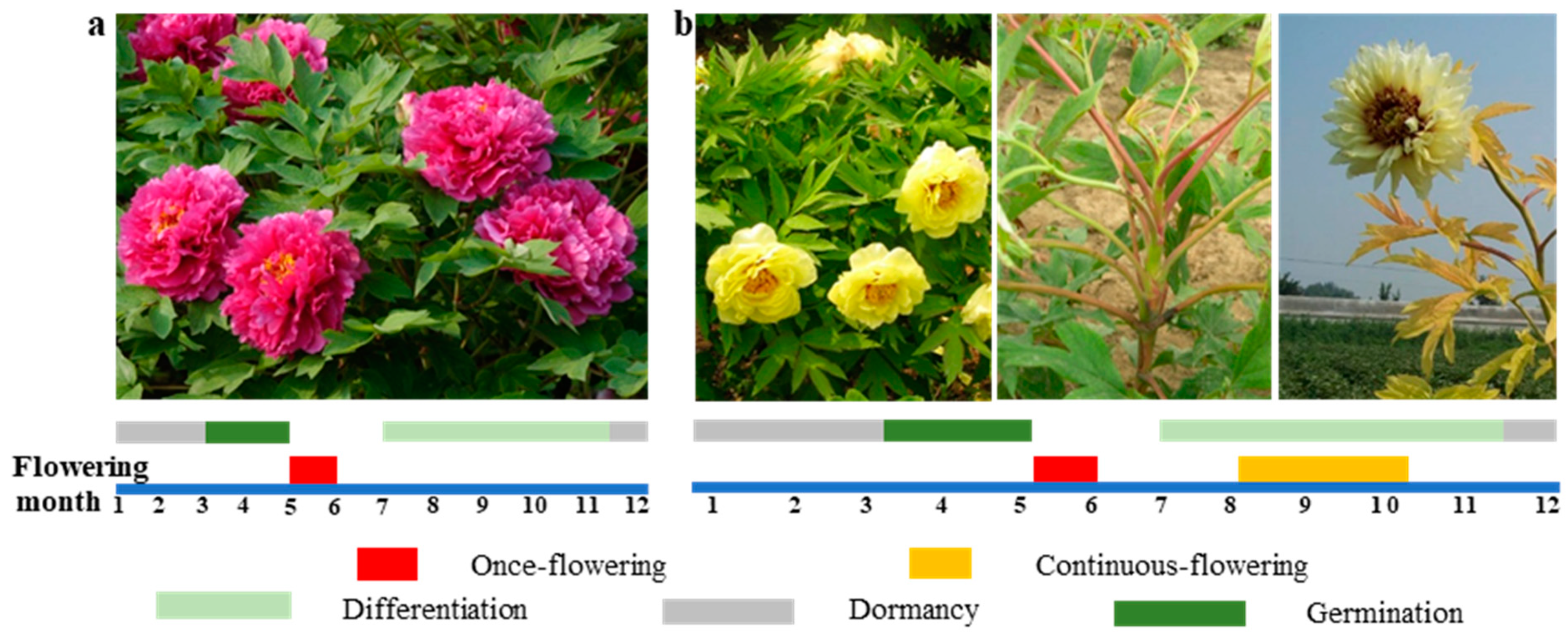
Figure 1. Different blooming modes of tree peonies. (a) OF genotype ‘Luo Yang Hong’ (‘LYH’) that only flowers in spring. After the first flower appears in early May, scale buds enter the bud primordium differentiation stage in late May. The bud primordium differentiation stage is from late May to November, and floral induction occurs in early July. The dormancy stage is in mid-November to the following March. Budding growth is initiated in March of the following year, and flowering is initiated in early May. (b) CF genotype (‘HN’). After the first flower appears in early May, a portion of the buds continue germinating, and floral induction occurs in early July. CF bud differentiation is completed shortly after, and flowering occurs between early August and early October. The overwinter buds enter the floral induction stage in early July, the dormancy stage is from mid-November to the following March, and the germination stage begins in early March of the following year. Anthesis is initiated in early May. Common flowering is indicated by a red box; CF is indicated by yellow box.
Agronomy 13 02071 g001
Figure 2. Secondary protein structures of PsFT, PsTFL1, and PsFD; interaction model construction of PsFD-PsFT and PsFD-PsTFL1. (Interaction confidence analysis: PsFD-PsFT: 0.8671; PsFD-PsTFL1: 0.8551).
Agronomy 13 02071 g002
Figure 3. Expression patterns of PsFT, PsFD, and PsTFL1 in buds during flower differentiation. (a) PsFT. (b) PsFD. (c) PsTFL1. Red arrow indicates the period of CF flowering. (d) The morphological structure of common and CF bud in ‘HN’. (eg) Expression pattern of PsFT (e), PsFD (f), and PsTFL1 (g) in common and CF buds of ‘HN’. The common buds of OF ‘HN’ and the S1, S2, S3, and S4 stages of CF ‘HN’ were collected in mid-September during continuous flowering. The curves show the mean values of three biological replicates for each sample. Data are the means of three biological replicates, and three technical replicates were performed for each biological replicate. Error bars show the standard deviation. Horizontal axis: plant data; vertical axis: gene expression levels relative to PsUBIQUITIN. Significance levels are calculated via analysis of variance (ANOVA) and indicated by asterisks: * (p < 0.05); ** (p < 0.01).
Agronomy 13 02071 g003
Figure 4. Subcellular localization of PsFT, PsTFL1, and PsFD in tobacco epidermal cells. GFP, Green fluorescence protein; Auto-fluorescence, chlorophyll fluorescence signal; Merge, merged image of GFP, auto-fluorescence, and bright-field images. Bars = 20 μm.
Agronomy 13 02071 g004
Figure 5. PsTFL1 from tree peonies induced late flowering in transgenic A. thaliana. (a) The transgenic line that constitutively expressed PsTFL1 exhibited the late-flowering phenotype in contrast to the wild-type (WT) and vector controls under long-day conditions. (b) Vertical view. Bars = 0.5 cm. (c) The statistics of days to flowering and rosette leaves in WT and over expression of PsTFL1 in A. thaliana.
Agronomy 13 02071 g005
Figure 6. Protein–protein interactions among PsFT, PsTFL1, and PsFD. (a) Yeast two-hybrid assay. Yeast transformed with combinations of PsFT-PsFD and PsTFL1-PsFD with PsFT and PsTFL1, as pGBKT7-bait and PsFD as pGADT7-bait were cultured on both nonselective media (SD/leu/-trp) and selective media (X-a-gal/Aba). (b) BiFC assays were conducted to confirm the interaction between PsFT-PsFD and PsTFL1-PsFD in tobacco leaves. Dark-field, bright-field, and merged images are shown, Bars = 25 μm. (c) Co-immunoprecipitation assays. Input: PsFD-pCAMBIA and PsFT-pBinGFP2, as well as PsFD-pCAMBIA and PsTFL1-pBinGFP2, were co-expressed in N. benthamiana. Immunoprecipitated proteins (IPs) were analyzed via immunoblotting and probing with either anti-GFP (α-GFP) or anti-myc (α-myc). Proteins were extracted and subjected to Western blotting. Output: The bands observed between 55 kDa and 72 kDa in the output are the result of the binding between PsFT-PsFD and PsTFL1-PsFD in the Co-IP experiment.
Agronomy 13 02071 g006
Disclaimer/Publisher’s Note: The statements, opinions and data contained in all publications are solely those of the individual author(s) and contributor(s) and not of MDPI and/or the editor(s). MDPI and/or the editor(s) disclaim responsibility for any injury to people or property resulting from any ideas, methods, instructions or products referred to in the content.