Article

Molecular Mechanism of Exogenous Magnesium in Regulating Cation Homeostasis in Roots of Peanut Seedlings under Salt Stress

College of Land and Environment, Shenyang Agricultural University, No. 120 Dongling Road, Shenhe District, Shenyang 110866, China
*
Author to whom correspondence should be addressed.
Agronomy 2024, 14(4), 724; https://doi.org/10.3390/agronomy14040724
Submission received: 22 February 2024 / Revised: 22 March 2024 / Accepted: 28 March 2024 / Published: 1 April 2024
(This article belongs to the Special Issue Legume-Rhizobial Symbiosis under Stress Conditions)

Abstract

:
Salt stress seriously hinders the normal growth of plant seedling roots. Magnesium, as one of the essential medium elements for plant growth, can effectively alleviate the damage of salt stress to plant roots, but the key genes involved and their mechanism are still unclear. The purpose of this study was to explore the related molecular mechanism of exogenous magnesium regulating cation homeostasis in peanut seedlings under salt stress. Firstly, according to plant physiology experiments, it was found that exogenous magnesium treatment significantly improved the tolerance of peanut seedlings to salt stress. After that, the transcriptome data were integrated, and further gene expression analysis showed that the expression of genes such as CNGC1, NCLs, and NHX7 was regulated under exogenous magnesium treatment, which effectively reduced the accumulation of sodium ions in cells. At the same time, exogenous magnesium also regulates the expression of genes such as ACAs and POTs and maintains the homeostasis of calcium and potassium ions in cells. These results reveal the molecular mechanism of exogenous magnesium regulating the cation homeostasis of peanut seedlings under salt stress, which provides an important reference for further revealing the key genes of salt tolerance in plants.

1. Introduction

Globally, soil salinization poses a major threat to plant growth and productivity [1]. Affecting more than 1 billion hectares of land [2], the lack of freshwater resources and unreasonable irrigation methods have aggravated salinization [3] and brought huge economic losses to irrigated areas [4].
Ion stress caused by salt stress is a direct challenge for plants. When the content of Na+ in the environment exceeds the coordination ability of plant cells, high concentrations of Na+ can seriously damage the ion homeostasis in plant cells [5,6], causing physiological disorders and even the death of the cells [7]. At the same time, high concentrations of sodium ions in the rhizosphere will reduce the content of K+ and Ca2+ in plant cells [8], resulting in water loss and root system withering [9]. Ionic stress caused by salt stress can also lead to secondary oxidative stress and secondary osmotic stress.
Magnesium is one of the medium-amount elements necessary for plants and is rich in plant cells [10]. It is involved in many key physiological processes such as photosynthesis, signaling, ATP formation, and cell ion homeostasis [11,12]. It has been found that a sufficient magnesium content can improve the salt tolerance of plants [13]. Adding magnesium oxide under salt stress can improve the antioxidant enzyme activity of sweet potatoes, improve nutrient acquisition [14], and increase the chlorophyll content of sunflowers [15]. It has also been reported that magnesium improves plant tolerance to salt stress and maintains the intracellular ion balance by regulating enzyme activity, ion channels, and transporter proteins, thereby improving plant resilience [16]. Magnesium ions can enhance the activity of K+ transporter protein OsHKT1 to improve rice K+ homeostasis [17], and can also regulate H-ATPase activity [18]. However, as a common divalent cation, whether exogenous magnesium can mediate the cation exchange in plant roots and enable the excretion of Na+ from plant cells is rarely reported.
Peanut (Arachis hypogaea L.) is a major oilseed crop across the world and is often considered salt-sensitive [19]. In the initial stage of peanut growth, salt stress mainly causes harm to roots and inhibits seed germination and seedling growth [20,21], and has additionally been shown to seriously affect peanut growth and pod yield [22]. Many studies have found that exogenous magnesium can alleviate the damage caused by salt stress to the roots of peanut seedlings, but little is known about the molecular mechanism of exogenous magnesium alleviating salt stress, particularly from studies using omics methods.
Therefore, the purpose of this study was to explore the effects of exogenous magnesium on the phenotype, ion dynamic changes, and related gene expression of peanut roots under salt stress through physiological and transcriptome analysis, so as to further study the molecular mechanism of exogenous magnesium alleviating salt stress in peanut roots.

2. Materials and Methods

2.1. Test Material Culture

In this study, the salt-intolerant variety “Haihua No. 1” peanut was used as the test material [23], and the peanut seeds were purchased from the Qilu Seed Industry. Peanut seeds with full particles and well-proportioned size were selected and rinsed three times with sterile water, and then soaked for 10 h at a constant temperature of 41 °C. After the seeds had fully absorbed the water, they were placed in a disinfection tray and covered with a wet towel to moisturize. The seeds were protected from light for 4 days at 29 °C [24]. Selected individuals whose root system length had grown to more than twice the length of the long axis of the seed were randomly placed in black plastic containers and continued to be protected from light at 25 °C for 4 days. Afterward, individuals with uniform growth were selected for the experimental treatment.

2.2. Experiment Design

The experimental treatments included (1) blank control (CK): distilled water treatment; (2) salt stress treatment (Na): NaCl, 75 mM; (3) exogenous magnesium treatment (Mg): MgCl2, 4 mM; and (4) exogenous magnesium treatment under salt stress (Na_Mg): NaCl, 75 mM; MgCl2, 4 mM.
The plant roots’ positions at 12, 24, 36, 48, 60, and 72 h after treatment (a total of six time points) were selected for the determination of physiological phenotypic indices. Peanut root systems at 48 h after treatment were selected for transcriptome sequencing and RT-qPCR assay.

2.3. Test Method

2.3.1. Determination of Physiological Phenotypic Indicators

In this experiment, 0.1 g of dried plant material was accurately weighed, passed through a 0.5 mm mesh screen, put into a ceramic crucible, and heated in an electric furnace at 300 °C for 30 min. Following this, the sample was ashed at 500 °C for 2 h. The hot sample was dissolved in 1:1 (v/v) nitric acid, and distilled water was added to bring the total volume to 50 mL. After filtration, the liquid was placed in a centrifuge tube.
The Na+ and K+ content was determined using flame photometry [25]. A total of 10 mL of the solution was taken for testing; then, 0.2 mL of 0.1 mol/L Al2 (SO4) 3 was added to the solution (to reduce the interference of Ca2+ in the Na measurement), and 10 mL of distilled water was added before measurement on the machine. The contents of Na+ and K+ in the sample were transformed using a NaCl and KCl standard solution (mg/g D.M), respectively. EDTA titration was used to measure the Ca2+ content and magnesium ion content [26]. For each sample, 15 mL of the solution was taken for testing and the pH was adjusted to 7 with NaOH solution. Then, 2 mL of triethanolamine solution (1: 3, v/v) was added as a masking agent (to remove the influence of chelating metal ions), and a K-B indicator (chrome blue potassium: phenanthroline green b = m:m = 1:2.5) and 0.01 mol/L EDTA standard solution (EDTA-2Na) were used. When the solution changed from pink to blue, the reaction was considered complete. According to the reaction ratio (mg/g D.M) between the Ca and EDTA, the Ca2+ content in the plant samples was determined. In addition, 10 mL of the above-mentioned liquid was taken for testing (containing 1~5 mg of calcium). The sample was diluted to 50 mL with water in a triangular flask, and then 2 mL of 1:1 triethanolamine and 5 mL of ammonia buffer were added. The sample was shaken well, and then 0.1 g of K-B indicator was added before titration with 0.01 mol/L of EDTA standard solution until the color changed from purple to blue–green. The total content of Ca2+ and Mg2+ in the plant samples was then determined, and the Mg2+ content was finally obtained according to the difference.

2.3.2. Transcriptome (RNA-Seq) Sequencing

The total RNA was extracted from roots quick-frozen at −80 °C using TRIzol reagent (Invitrogen, Carlsbad, CA, USA). The total RNA quantity and purity were determined using a Bioanalyzer 2100 and RNA Nano LabChip kit 6000 (Agilent, Santa Clara, CA, USA). The mRNA was purified from the total RNA using the Oligo-T oligonucleotide-attached magnetic bead system (Invitrogen). The mRNA fragments were cleaved via reverse transcription using the RNA-Seq Sample Preparation Kit (Illumina, San Diego, CA, USA) to create the final cDNA library. cDNA sequencing was performed using the LC Sciences Double-Ended (150 bp) Illumina Hi-Seq 4000 System (Illumina, San Diego, CA, USA).

2.3.3. RT-qPCR Detection

To confirm the reliability of the transcriptome data, we isolated RNA from the samples using the RNAiso Plus kit (TAKARA, Kyoto, Japan), enriched mRNA using magnetic beads, and purified the mRNA using probe hybridization. The mRNA was reverse-transcribed to cDNA using the GoScript TM Reverse Transcription System (Promega, Beijing, China). mRNA relative expression analysis was performed using the QuantStudio TM 5 Real-Time Fluorescent Quantitative PCR System (Thermo Fisher Scientific, Waltham, MA, USA). A GoTaq qPCR premix kit (Promega, Beijing, China) was used to provide cDNA reverse transcriptase and fluorescent dyes. In this study, three biological replicates were performed in RNA-Seq and RT-qPCR analysis. ACTIN2 (LOC112715878) was selected as the reference gene. Information about these selected genes (gene ID, primer sequences) is provided in Table S1.

2.4. Data Processing and Data Visualization Methods

2.4.1. Screening of Differentially Expressed Genes (DEGs)

The differentially expressed genes (DEGs) in each paired comparison group were calculated using edgeR (R-bag) [27]. The screening criteria for each pair of comparison groups were based on two criteria: ① For each gene, the average count under at least one treatment is greater than 50, and the absolute value of the difference multiple of the two groups of counts (the absolute value of the average ratio, |log2FC| is greater than 1). ② After the p-values of all of the genes that met the previous criterion had been calculated using the batch t test, the FDR value was calculated via the BH method, and all genes with an FDR less than 0.05 were kept as a list of differentially expressed genes in the paired comparison group.

2.4.2. GO Enrichment Analysis and KEGG Enrichment Analysis

The GO and KEGG enrichment analysis calculation methods were based on the method described by Subramanian [28]; the specific cloud platform link is (https://www.omicshare.com/tools/(accessed on 14 October 2023)), and the website link for calculation using cluster profiler 4.0 (R package) is (https://bioconductor.org/packages/release/bioc/html/clusterProfiler.html (accessed on 12 September 2023)) [29].

3. Results

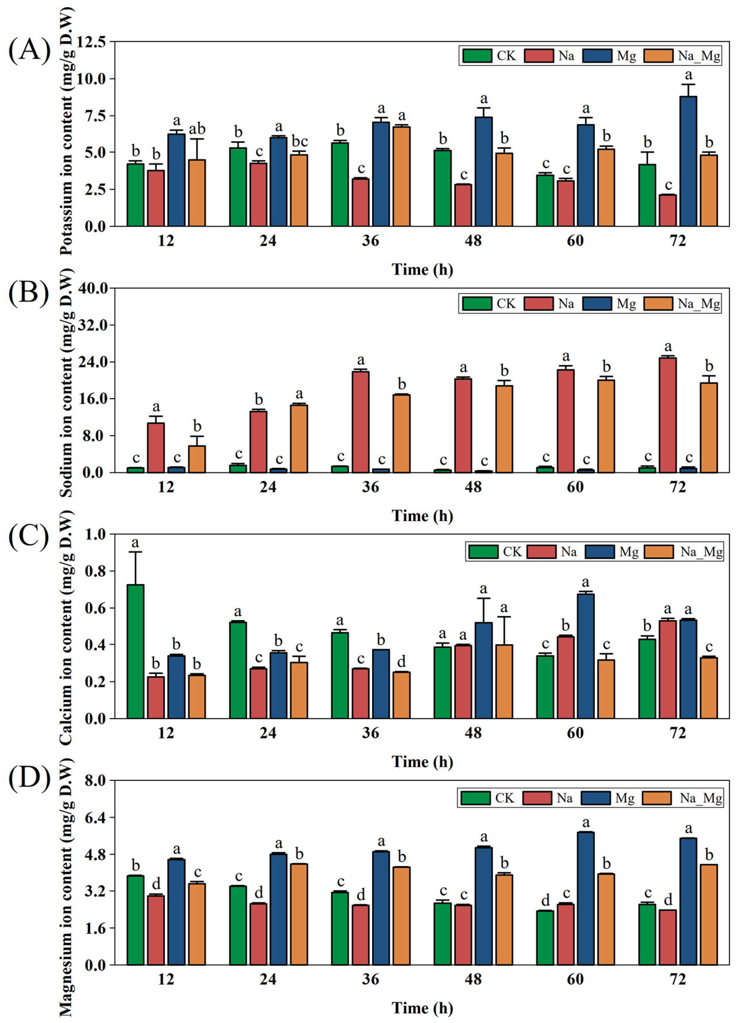
3.1. Exogenous Magnesium Alleviates Root Growth and Ion Homeostasis of Peanut Seedlings under Salt Stress

In this experiment, we investigated the effect of exogenous magnesium on the salt tolerance of the main roots of seedlings under salt stress. As shown in Figure S1, under salt stress, the root system turned brown, showed symptoms of beard root necrosis, and gradually wilted. Compared with plants treated with Na alone, the root withering in plants treated with the Na_Mg combination slowed down. In order to visualize the dynamic effects of exogenous Mg on peanut seedlings under salt stress at the physiological level, the physiological indices of peanut roots at six consecutive time points (12 h, 24 h, 36 h, 48 h, 60 h, and 72 h) under four different treatments were determined as K+ content, Na+ content, Ca2+ content, and Mg2+ content (Figure 1).
In this study, the contents of K+, Na+, Ca2+, and Mg2+ were detected. With the increase in time, the content of K+ in the Na_Mg treatment group was in a dynamic equilibrium, and the content of K+ in the Na treatment group gradually decreased, which was significantly lower than that in the other three treatment groups after 36 h (p < 0.05) (Figure 1A). As expected, the Na+ content in the peanut roots under Na treatment and Na_Mg treatment showed an upward trend, which was significantly higher than for the other treatments (p < 0.05); however, after 48 h, the content in the Na_Mg treatment group was significantly lower than that in the Na treatment group (p < 0.05) (Figure 1B). Interestingly, with the increase in time, the content of Ca2+ in the Mg treatment group increased gradually, while the content of Ca2+ in the Na treatment group and Na_Mg treatment group fluctuated (Figure 1C). Importantly, the Mg2+ content in the Na+ treatment group decreased continuously at all time points, while the Mg2+ content in the Na_Mg treatment group was in a dynamic balance; the Mg2+ content in the Na_Mg treatment group was significantly higher than that in the Na+ treatment group (p < 0.05) (Figure 1D).
The results showed that exogenous magnesium can effectively alleviate the adverse effects of salt stress on peanut root system growth, significantly inhibit the absorption of Na+ and the loss of K+ and Ca2+, and increase the absorption of Mg2+ under salt stress.

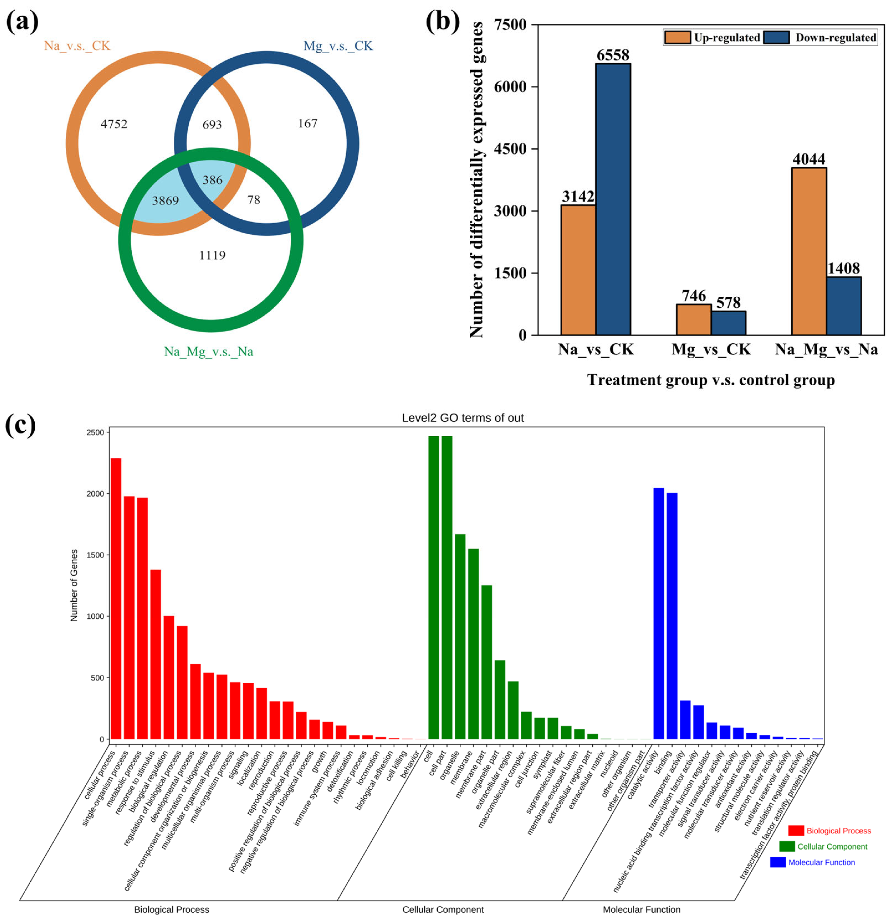
3.2. DEG Screening and KEGG Enrichment Analysis of DEGs

In order to comprehensively study the molecular mechanism of exogenous magnesium in alleviating salt stress in the peanut seedling root system, 12 mRNA sequencing libraries were constructed using the Illumina RNA sequencing platform. These included three biological repeats of the root system treatments using distilled water (CK), 75 mM of NaCl (Na), 4 mM of MgCl2 (Mg), and 75 mM of NaCl and 4 mM of MgCl2 (Na_Mg), respectively, resulting in 12 sub-transcription groups. As shown in Table S2, among the 12 sequencing libraries, the lowest number of Clean Reads is 41,591,826, and the highest number is 51,694,788, with Q30 greater than 92.75% and a CG content ranging from 44.64% to 44.94% (Table S2). Pearson’s correlation coefficient R analysis was performed on the total count quantity, and the average value of R was above 0.98 (Figure S2), and principal component analysis (PCA) was performed to project each sample point onto the first and second principal components of the matrix of all sample counts (Figure S3). The differences between the two treatment groups could be identified, and the biological repeatability of the samples in each treatment group met the requirements of downstream data analysis, indicating that the transcriptome sequencing data could be used for further analysis.
Three paired comparison groups, Na vs. CK, Mg vs. CK, and Na_Mg vs. Na, were established (treatment group vs. control group). Compared with CK, 9700 DEGs were identified in the Na treatment group (upregulated by 3142, downregulated by 6558); compared with the Mg treatment group, 1324 DEGs were identified in the CK treatment group (upregulated by 746, downregulated by 578); and compared with Na treatment group, 5452 DEGs were identified in the Na_Mg treatment group (upregulated by 4044, downregulated by 1408) (Figure 2b).
The DEGs of three paired comparison groups were classified using a Venn diagram, as shown in Figure 2a, and 4255 genes were screened out, which were effectively expressed under salt stress and regulated by exogenous magnesium, that is, DEGs at the intersection of two paired comparison groups (Na vs. CK and Na_Mg vs. Na) (Table S3).
KEGG and MapMan enrichment analyses were carried out on these 4255 differentially expressed genes. The results showed that KEGG enriched the first 25 DEG pathways according to the ranking of the p-values (Figure S4). It can be seen that the first five pathways are phenylpropanoid biosynthesis, flavonoid biosynthesis, secondary metabolite biosynthesis, isoflavone biosynthesis, and plant MAPK signaling pathway. MapMan enriched the related metabolism overview pathways (Figures S9 and S10), and the first three metabolism overview pathways are photorespiration, tetrapyrrole, and metabolism.
Meanwhile, GO enrichment was performed for these 4255 differentially expressed genes (Figure 2c), showing that the two pairwise comparison groups, Na vs. CK and Na_Mg vs. Na, differed significantly in terms of biological processes, cytoskeleton, and molecular functions. In the biological process category, the top three enriched GO terms were cellular processes (GO:0009987), single-organism processes (GO:0044699), and metabolic processes (GO:0008152). In the molecular functions category, the top three enriched GO terms are cell (GO:0005623), cell part (GO:0044464), and organelle (GO:0043226). In the bioprocess category, the top three enriched GO terms were catalytic activity (GO:0003824), bound (GO:0005488), and transporter protein activity (GO:0005215) (Table S4).
It can be seen that the addition of exogenous magnesium under salt stress may have induced the differential expression of genes related to catalytic activity mediated by enzymes and genes related to ion transporter proteins in cell membranes as well as organelle membranes. This study mainly involves the analysis of ion transport channels related to the addition of exogenous magnesium under salt stress, including the Mg2+ channel, Na+ channel, Ca2+ channel, H+ channel, and K+ channel (Table S5).

3.3. Mining Differentially Expressed Genes of Magnesium Ion in Response to Cation Homeostasis of Peanut Seedlings under Salt Stress

Under salt stress, a high concentration of sodium ions accumulated in the plant root system, which broke the ion homeostasis in cells and caused toxicity [30,31]. Cation balance in plant root systems is very important for reducing the content of sodium ions in cells. In previous studies, the effect of magnesium ions on the ion balance under salt stress was ignored [28,32,33]. In this study, DEGs related to Na+, K+, Ca2+, Mg2+, and H+ ion transport and exchange were mined based on the comparison between Swissprot annotation and gene expression count, and a schematic diagram and related heat map were drawn.

3.3.1. Regulation of Exogenous Mg2+ on Key Genes of Mg2+ Transport Channel and Non-Selective Ion Channel

Figure 3 shows the excavated expression quantities of 14 DEGs related to Mg2+ balance. There are five magnesium transporter MRS2−s genes, seven possible magnesium transporter NIPA genes, and two magnesium/proton exchanger MHX genes. Eleven of these genes had consistent expression patterns, with most MRS2−s, most NIPAs, and two MHX being downregulated after Na treatment, while these genes were upregulated after Na_Mg treatment. There were three genes with opposite expression patterns: one MRS2−1 (LOC112779112) and two NIPAs (LOC112789048 and LOC112766238) were upregulated after Na treatment, and downregulated after Na_Mg treatment. After adding exogenous magnesium under salt stress, MRS2−4 and most NIPAs were upregulated, so that Mg2+ outside the cell entered the cytoplasm, and MHX was upregulated, so that Mg2+ in vacuoles entered the cytoplasm, which promoted the absorption of Mg2+ in the cytoplasm.
The schematic diagram shows the subcellular localization, function, and regulation of these ion channels. The abbreviations in the diagram are as follows: NIPAs, probable magnesium transporter NIPAs; MRS2−1/3/5, magnesium transporter MRS2−1/3/5; MRS2−4, magnesium transporter MRS2−4; MHX, magnesium/proton exchanger; CNGC1, cyclic nucleotide-gated ion channel 1; CNGC9, putative cyclic nucleotide-gated ion channel 9; CNGC20, probable cyclic nucleotide-gated ion channel 20; and GLRs, glutamate receptors. Treatment: CK, treatment by adding distilled water; Na, treatment by adding 75 mM of NaCl solution; Mg, treatment by adding 4 mM of MgCl2 solution; and Na_Mg, treatment by adding 75 mM of NaCl solution and 4 mM of MgCl2 solution. The direction of the arrow represents the transport direction of the relevant ions. The direction of ion transport on the same side is represented by the double arrow. For example, the downward arrow of MHX in the figure indicates that Mg2+ on the same side as the arrow is transported downward from the cytoplasm to the vacuole through the magnesium/proton exchanger MHX.
The DEG expression quantities of 19 non-selective ion channels related to Na+ balance were also excavated (Figure 3) and were found to comprise 15 glutamate receptor GLR genes and four cyclic nucleotide-gated ion channel CNGCs. Among them, eight GLRs were upregulated and the other seven were downregulated after Na_Mg treatment. In addition, three CNGCs (LOC112712077, LOC112777506, and LOC112750839) were upregulated after Na treatment, but these genes were downregulated after Na_Mg treatment, while CNGC1 expressed the opposite pattern. Most of the CNGC genes were upregulated after salt stress but downregulated after adding exogenous magnesium under salt stress, which restricted some of the Na+ from entering the cytoplasm.

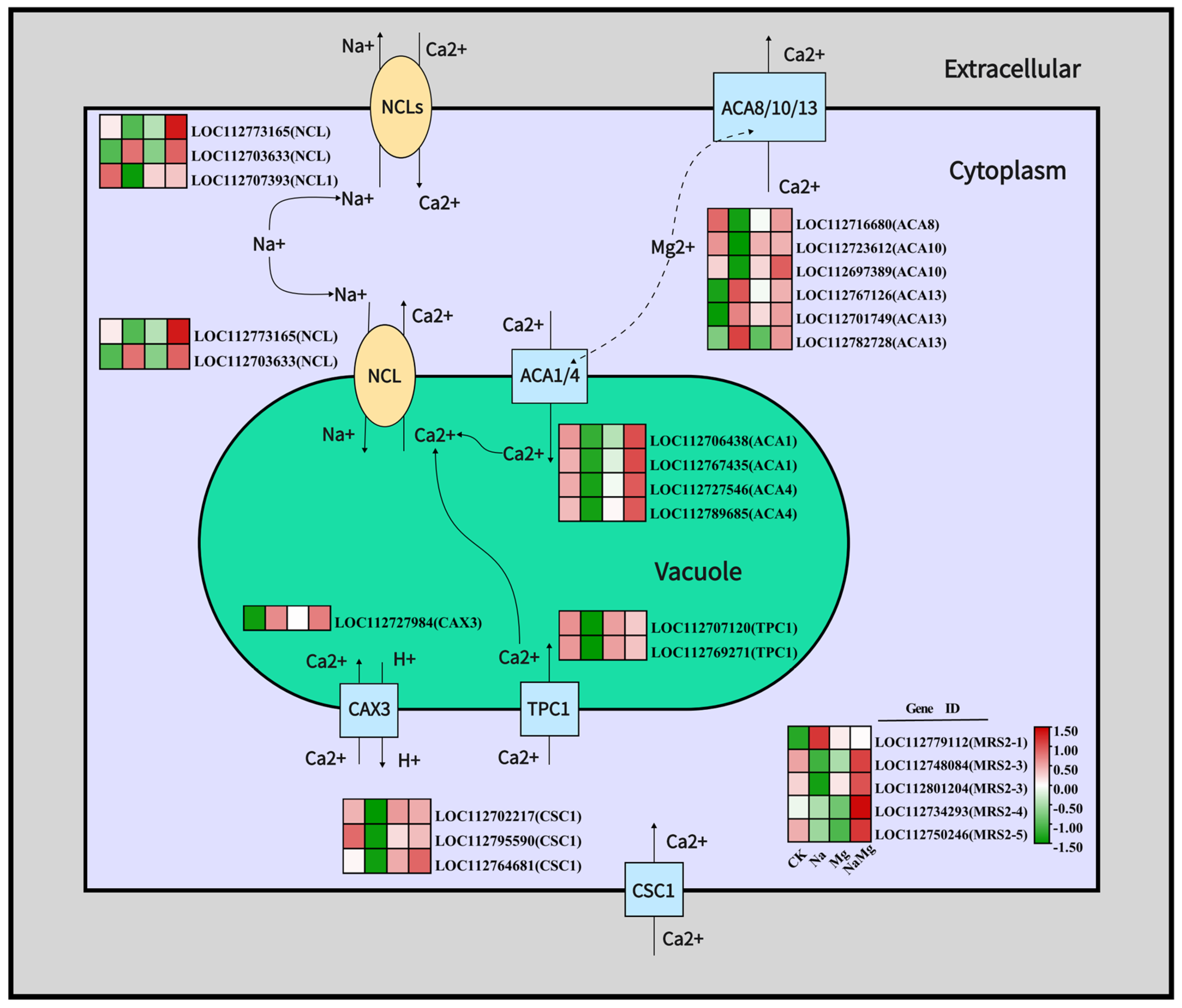
3.3.2. Regulation of Exogenous Mg2+ on Key Genes of Ca2+ Transport Channel and Na+/Ca2+ Ion Exchange Channel

As shown in Figure 4, the expression quantities of the 16 excavated DEGs related to Ca2+ balance were demonstrated, comprising 10 calcium transport ATPase ACA genes, two double-pore calcium channel protein TPC1 genes, one vacuole cation/proton exchanger CAX3 gene, and three calcium permeable stress-gated cation channel one CSC1 genes. Among them, 12 genes had the same expression patterns, including three CSC1s, two TPC1s, and seven ACAs. These were downregulated after Na treatment, but upregulated after Na_Mg treatment. Four genes exhibited the opposite expression patterns: three ACA13s and one CAX3 were upregulated after Na treatment, while three ACA13s were downregulated after Na_Mg treatment; differently, one CAX3 was downregulated after Na_Mg treatment.
The expression quantities of three DEGs related to Na+/Ca2+ ion exchange were also discovered (Figure 4) and comprised two sodium–calcium ion exchangers NCL and one sodium–calcium ion exchanger NCL1. Among them, one NCL (LOC112773165) and one NCL1 (LOC112707393) were downregulated after Na treatment, but upregulated after Na_Mg treatment. However, one NCL (LOC112703633) expressed the opposite pattern and was upregulated after the Na treatment and downregulated after the Na_Mg treatment. After adding exogenous magnesium under salt stress, the overall upregulation of Ca2+-transport-related differentially expressed genes can be seen. It can also be seen that part of the Ca2+ is transferred from the cytoplasm to the extracellular region, and another part is transferred from the cytoplasm to the vacuole. On the whole, NCL and NCL1 were upregulated after the addition of exogenous magnesium under salt stress, which promoted the exchange of sodium/calcium ions outside the cell and in the vacuoles, enabled Na+ exchange outside the cell or sealed in vacuoles, and reduced the content of Na+ in the cytoplasm.

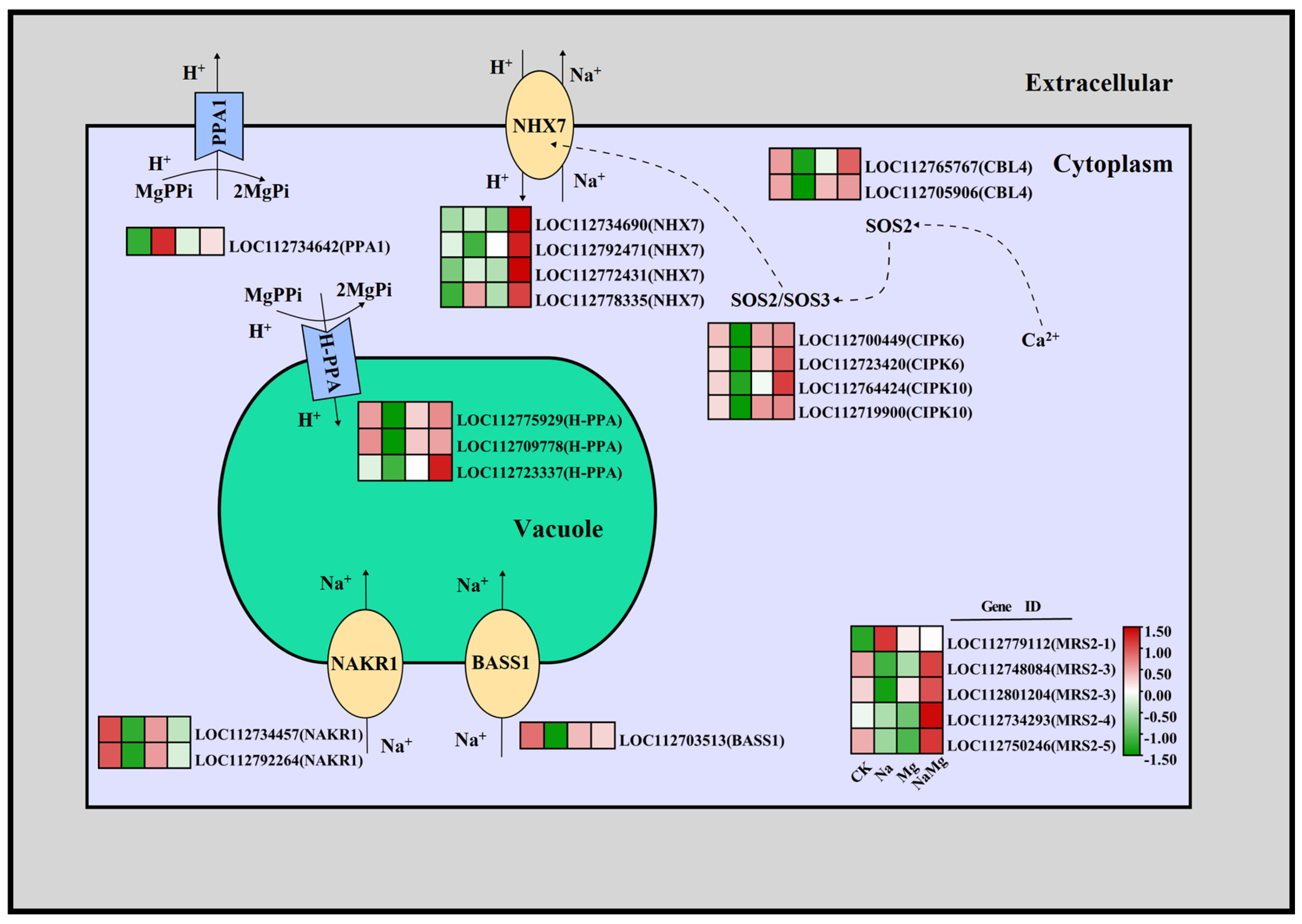
3.3.3. Regulation of Exogenous Mg2+ on Key Genes of H+ Transport Channel, Na+ Transport Channel, and Na+/H+ Ion Exchange Channel

Plants maintain ion homeostasis by reducing the concentration of Na+ in the cytoplasm. This is mainly achieved by expelling sodium ions from the cytoplasm. One of the pathways involved in this is the SOS (salt overly sensitive) salt discharge pathway, which transports Na+ to exchange H+ for salt discharge, and the other is Na+ ion exchange with other substances. Na+ exchange channels located in the plasma membrane transport Na+ to the outside of cells, and Na+ exchange channels located in the vacuoles are responsible for maintaining the separation of Na+ [34].
Ten DEGs are expressed in the SOS salt discharge pathway (sodium/hydrogen ion exchange channel) (Figure 5), including four NHX7 (SOS1) genes, two CIPK6 (SOS2) genes, two CIPK10 (SOS2) genes, and two CBL4 (SOS3) genes. Among them, seven gene expression patterns are consistent, including one NHX7 (LOC112792471) gene, two CIPK6 genes, two CIPK10 genes, and two CBL4 genes. These genes were downregulated after Na treatment, but they were all significantly upregulated after Na_Mg treatment. Three NHX7 genes exhibited the opposite expression (LOC112734690, LOC112772431, and LOC112778335) and were upregulated after Na treatment and continued to be upregulated after Na_Mg treatment. Overall, NHX7 was upregulated and CIPKs and CBL4 were downregulated under salt stress, whereas the genes of the SOS salt discharge pathway were upregulated overall after the addition of exogenous magnesium under salt stress; this activated the SOS salt discharge pathway to enable Na+ excretion from the cytoplasm into the extracellular space, reducing the Na+ content in the cytoplasm.
Three DEGs were expressed in other sodium ion exchange channels related to Na+ balance (Figure 5): two protein sodium and potassium deficiency NAKR1 genes and one possible sodium/metabolite co-transporter BASS1 gene. The expression patterns of these three DEGs were consistent, with two NAKR1 genes and one BASS1 gene downregulated after Na treatment and upregulated after Na_Mg treatment. This demonstrates that these genes are downregulated during salt stress and upregulated after adding exogenous magnesium under salt stress, which seals Na+ from outside the cytoplasm into the vacuole and reduces the content of Na+ in the cytoplasm.
At the same time, four DEGs related to H+ balance were also identified (Figure 5): three pyrophosphate tonoplast proton pump H−PPA genes and one soluble inorganic pyrophosphate 1 PPA1 gene. Among them, the three H-PPAs were downregulated after Na treatment, but upregulated after Na_Mg treatment, and the expression pattern of the PPA1 was opposite. This shows that the addition of exogenous magnesium under salt stress inhibits the efflux of H+ to the outside of cells and promotes the transport of H+ from the cytoplasm to the vacuoles.

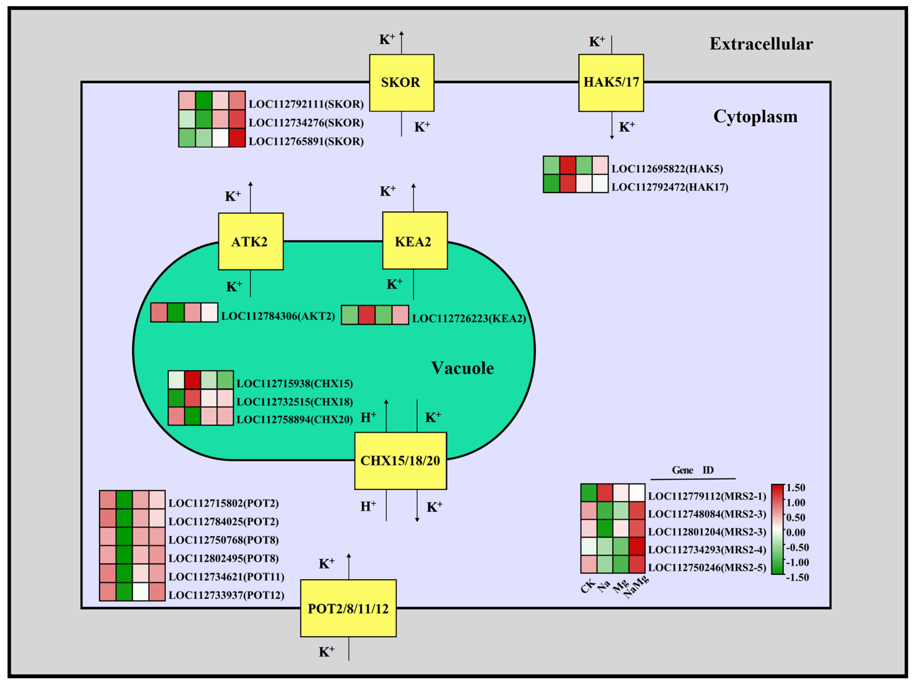
3.3.4. Regulation of Exogenous Mg2+ on Key Genes of K+ Transport Channel

Under salt stress, a high concentration of Na+ usually leads to a lack of K+, and plants need to maintain a high K/Na ratio to regulate the Na/K steady state [30,35]. As shown in Figure 6, 16 DEGs related to K+ balance were excavated: six specific potassium ion absorption channel genes POTs, three highly selective outward rectifier potassium channel SKOR genes, two high-affinity potassium transporter HAKs genes, three cation/H+ antiporter CHXs genes (mainly for K+ and H+ exchange), a highly selective and weak inward rectifier potassium channel AKT2 gene, and a K+ efflux transporter KEA2 gene. Among them, the expression patterns of 12 genes were consistent; six POTs, three SKORs, one CAX20, and one AKT2 were downregulated after Na treatment, but the gene expression was upregulated after Na_Mg treatment. Five genes showed the opposite expression patterns: two HAKs, one KEA2, and two CAXs were upregulated after Na treatment, but downregulated after Na_Mg treatment. However, the expression levels of two HAKs were still significantly higher than for CK. The different expression patterns of differentially expressed genes located on the cell membrane allow the K+ in the cytoplasm and vacuoles to be dynamically balanced. Adding exogenous magnesium under salt stress will make POTs upregulated, and result in compensatory absorption of K+ lost from cells under salt stress, thus inhibiting the loss of total K+.

3.4. RT−qPCR Verification

In order to verify the accuracy of RNA−seq, six key differentially expressed genes regulating cation balance were selected for RT-QCPR detection, as shown in Figure S7, and a Melt Curve Plot and Amplification Curve Plot of these six genes were also created (Figure S8). Then, these two quantitative methods were used to compare whether the expression trends in two paired comparison groups (Na vs. CK and Na_Mg vs. Na) were consistent. The genes were LOC112789685(ACA4), LOC112712077(CNGC1), LOC112775929(H-PPA), LOC112707393(NCL1), LOC112795740(NIPA4), and LOC112733937(POT12), respectively.
Among these six genes, the expression trends of ACA4, CNGC1, H−PPA, NCL1, and POT12 were the same, and the results of the RT-QCPR verification of the NIPA4 gene showed that the expression after Mg treatment was higher than after Na_Mg treatment; however, because the NIPA4 gene is a specific magnesium transport gene, the expression in the Na_Mg treatment group was still significantly higher than in the Na treatment group (p > 0.05). To sum up, the results of these two quantitative methods are basically the same.

4. Discussion

Under salt stress conditions, maintaining the ion homeostasis of plants is essential for normal growth. The analysis of phenotypes and physiological indicators revealed that adding exogenous magnesium under salt stress significantly inhibited Na+ absorption, reduced K+ loss, maintained Ca2+ homeostasis, and increased Mg2+ absorption.
Salt stress first triggers ionic stress, and then subsequently triggers secondary antioxidant stress and secondary osmotic stress [36]. This paper mainly discusses how exogenous magnesium can alleviate the ion stress under salt stress, and the findings of this study show that after exogenous magnesium ions enter the cytoplasm through magnesium ion channels, some cations in the cells are exchanged with Na+ exchange channels, which can reduce the concentration of Na+ in the cytoplasm and reduce the toxicity of Na+. At the same time, this study explored K+-, Ca2+-, and H+-related cation channels, and found that the process of exogenous magnesium in alleviating salt stress in the roots of peanut seedlings was mainly divided into four aspects, including one direct pathway (Na+/Mg2+ direct competition pathway), two indirect pathways (Mg-activated Na+/Ca2+ exchange pathway and Na+/H+ exchange pathway), and the K+ steady-state pathway. Therefore, these pathways are discussed separately in the following paragraphs.
The cyclic nucleotide-gated channel (CNGC) family is a non-selective cation channel that is subjected to Ca2+ signaling; it is involved in plant immune responses and is a very important ion channel for plants to cope with abiotic stresses [37]. Studies have found that Mg2+ can compete with Na+ for cationic binding sites on the root surface [38]. There are also articles reporting that CNGC10 in the meristem and remote elongation regions of plant roots can transport Mg2+ into the cytoplasm, and Na+ can also enter the cytoplasm through CNGC channels [39]. According to Figure 5b, after adding exogenous magnesium under salt stress, the DEGs encoding CNGC are upregulated as a whole, which provides a certain basis for the direct ion exchange of Mg2+ and Na+.
The magnesium transporter (MGT/MRS2) family is the most documented Mg2+ transporter family and has been found and reported in many plants [40,41,42,43], but the magnesium transporter NIPA family has not been deeply studied. It has also been found that members of the OsMGT gene family may be involved in the response to salt stress, and it is further assumed that MGT is involved in abiotic stress [44]. In this study, it was found that after adding exogenous magnesium under salt stress, MRS2-4 located on the cell membrane and NIPA1, NIPA3, and NIPA4 in the NIPA family were significantly upregulated, so that extracellular Mg2+ entered the cytoplasm through the ion channels of the NIPA and MRS2-4 families and the content of magnesium ions in the cytoplasm increased.
Mg2+ not only directly competes with Na+ for CNGC channels on the cell membrane, but also stimulates the exchange of Na+/Ca2+ and Na+/H+ indirectly so that Na+ is transferred out of the cytoplasm.
The first indirect way is that Mg2+ can indirectly activate Na+/Ca2+ exchange. ACA calcium transport ATPase is a magnesium-dependent ion channel. It has been found that plasma membrane ACAs, vacuolar Ca2+ transporters (OsACAs and OsCAXs), and endoplasmic reticulum Ca2+-ATPase (ECA) play an important role in rice salt tolerance [45]. It has also been found that the increase in AtNCL expression in plants makes plants more sensitive to CaCl2 and increases their tolerance to NaCl [46], and the sodium/calcium exchanger NCLX plays an important role in the Ca2+ balance of plants under stress [47]. As shown in Figure 5, the experimental results indicated that the Na_Mg group upregulated ACA1, ACA4, and TPC1 in the vacuoles and upregulated ACAs and NCLs located on the cell membrane and NCLs and CSC1 located on the cytosol compared with the Na-treated group. After Mg2+ enters the cytoplasm, the ACA family ion channels are activated. Part of the Ca2+ in the cytoplasm is transported to the environment through ACA8/10/13, and the Ca2+ in the environment flows back to the cytoplasm through the sodium/calcium exchanger NCL, where Na+ is discharged into the environment. Due to the upregulation of CSC1, the Ca2+ outside the cell may flow back to the cytoplasm, and the Ca2+ flowing back to the cytoplasm and the environment can be used as a signal to activate the salt discharge channels of CNGCs and SOS. However, more Ca2+ participates in ion exchange between the cytoplasm and vacuoles. ACA1/4 and TPC1 are located in the vacuoles and part of the Ca2+ in the cytoplasm enters the vacuoles through ACA1/4 and TPC1; the rest of the Ca2+ is exchanged with H+ through vacuole cation/proton exchanger CAX3 and enters the vacuoles through sodium/calcium exchanger NCL to exchange Na+ and Ca2+. After adding exogenous magnesium under salt stress, the CSC1 located on the cell membrane was upregulated, Mg2+ upregulated ACA1, ACA4, and TPC1 located on the vacuole membrane, and upregulated the NCLs, which allowed the Ca2+ to return to the cytoplasm, maintained the expression of the Ca2+ signal in the cytoplasm, and promoted the Na+/Ca2+ ion exchange on the vacuole membrane to seal Na+ in the vacuole.
Interestingly, in the first indirect pathway, NCLs played an important role in Na+/Ca2+ ion exchange; however, among these three differentially expressed NCL genes, one had the opposite expression pattern to the other two under CK and salt stress. According to the peanut reference protein sequence, the amino acid sequences related to these three NCLs (LOC112773165, LOC112703633, and LOC112707393) were found, respectively, and the amino acid sequences of these three genes were compared. As shown in Figure S5, the amino acid sequences of these three genes were similar, but among them, the sequences of NCL and NCL1 were shorter. Following this, domain analysis was carried out on these genes and the results combined with the calculation results of the phylogenetic tree of multi-sequence alignment are shown in Figure S6. The domains of these three sequences are similar, and they are all composed of domains related to Na+/Ca2+ (membrane Na+/Ca2+ exchange domain, EF-hand domain) and other domains (cytoplasmic domain, transmembrane domain, and non-cytoplasmic domain). Compared with the amino acid sequence of NCL1, the amino acid sequences of two NCLs have one more membrane Na+/Ca2+ exchange domain, while the amino acid sequence of one NCL1 has one more EF-hand domain and the cytoplasm domain is longer. This indicates that the three NCL genes are very similar in structure, and the NCL (LOC112703633) gene should have the same expression pattern as the other two NCL genes.
The second indirect way is that Mg2+ can indirectly activate Na+/H+ exchange. Ca2+, as the second messenger, generates calcium signals to activate SOS3(CBL4) in plants. SOS3 combines with SOS2(CIPK6,10) to activate SOS2, and then the phosphorylation of SOS2 stimulates the activity of SOS1(NHX7), thus triggering the salt discharge pathway of SOS, which is a conservative mechanism to maintain ion homeostasis under salt stress [48,49,50]. After adding exogenous magnesium under salt stress, combined with transcriptome data, it was found that SOS1(NHX7) was significantly upregulated, and most of SOS2(CIPK6,10) was upregulated, but the SOS3(CBL1) gene was downregulated. It was speculated that Mg2+ directly acted on SOS1(NHX7), and Na+/H+ ion exchange occurred on the cell membrane so that the Na+ in the cytoplasm was discharged to the environment. After adding exogenous magnesium under salt stress, it was found that H-PPA was upregulated and PPA1 was downregulated. The high expression of SOS1(NHX7) may be related to the activation of the hydrogen pump by Mg2+. Some studies found that salt stress caused the differential expression of BASS [51]. It has been found that the proton pump in plants can regulate the PH and ion homeostasis of cells under salt stress. Mg2+ can activate H-ATP(PPA1) and H-PPase(H-PPA), catalyze the combination of free Mg2+ and pyrophosphate PPi to form MgPPi, and transport H+ [52]. The overexpression of H-PPase can enhance the salt tolerance of transgenic plants and increase ion retention to enhance the tolerance to NaCl and drought [53,54]. It has been found that H-ATP SCaBP1(SOS3) interacts with PKS5/CIPK11(SOS2) and PKS24/CIPK14(SOS2) and activates them, and the latter phosphorylates and inhibits H-ATPase on the plasma membrane. This inhibition is very important for cell pH regulation [48,55]. This is consistent with the research results in this paper. After adding exogenous magnesium under salt stress, H-PPase(H-PPA) on the vacuole membrane was upregulated and PPA1(H-ATP) on the cell membrane was downregulated. The proton pump H-PPA on the cell membrane was inhibited by the SOS salt discharge pathway and the H+ produced by SOS1(NHX7) was stored in the cytoplasm; however, the Mg2+ in the cytoplasm was activated by the vacuoles. At the same time, part of the Mg2+ in the cytoplasm entered the vacuoles through MRS2-1/3/5 channels on the vacuole membrane and returned to the cytoplasm through the MHX magnesium proton exchanger, which can also enable H+ in the cytoplasm to enter the vacuoles. After adding exogenous magnesium under salt stress, the SOS salt discharge pathway was upregulated, promoting Na+/H+ ion exchange so that Na+ was excreted to the cytoplasm. At the same time, the SOS salt discharge pathway inhibited the expression of PPA1(H-ATP) on the cell membrane and reduced the loss of H+ outside the cell. However, the high expression of H-PPase(H-PPA) on the vacuole membrane enabled the H+ produced by SOS1(NHX7) in the cytoplasm to enter the vacuole. In this study, it was considered that the addition of exogenous magnesium promoted the efflux of Na+ and slowed down the H+ under salt stress.
Exogenous magnesium can also maintain K+ homeostasis in cells. Some studies suggest that the gene expression of POT/HAK family members may be directly regulated by the cytosolic Ca2+ level under salt stress [56]. SKORs participate in the absorption and transportation of potassium from roots, which may regulate the salt tolerance of plants [57]. Some studies have also shown that the transformation of OsHAK21 [58] and OsHAK16 [59] can significantly restore the growth of K+-deficient yeast at low K+ concentrations, participate in K+/Na+ homeostasis and K+ uptake in plants, and enhance the salt sensitivity of plants. The difference is that after the mutant of OsHAK1 is knocked out, the K+ absorption of plants is significantly inhibited and their salt tolerance is improved [60]. The upregulation of POTs on the cell membrane after adding exogenous magnesium can allow potassium ions in the environment to enter the cytoplasm and reduce the loss of potassium ions. As shown in Figure 1a, after the addition of magnesium ions under salt stress, the content of potassium ions fluctuates steadily and the SKOR on the cell membrane is upregulated, but the expression level is low. Thus, the addition of exogenous magnesium maintains the dynamic balance of K+.

5. Conclusions

Combined with physiological and transcriptome methods, the effects of exogenous magnesium on regulating the cation homeostasis of peanut roots under salt stress were analyzed. We found that the cation balance of peanut roots was regulated by four aspects after adding exogenous magnesium: (1) extracellular Mg2+ directly competed with Na+ for the CNGC channel on the cell membrane, which inhibited Na+ from entering the cytoplasm; (2) Mg2+ upregulated the ACA channel on the vacuole membrane, activated the sodium–calcium ion exchanger NCL, and sealed Na+ in the vacuole; (3) Mg2+ upregulated the sodium–hydrogen ion exchange channel NHX7, activated the SOS salt discharge pathway, and activated the proton pump H-PPA on the vacuole membrane so that Na+ was discharged; and (4) Mg2+ also activated K-transport-channel-related POTs and maintained K+ homeostasis in cells. This study clarified the mechanism of magnesium on the cation balance of peanut root systems under salt stress, which provides theoretical support for cultivating new salt-tolerant peanut varieties regulated by magnesium.

Supplementary Materials

The following supporting information can be downloaded at: https://www.mdpi.com/article/10.3390/agronomy14040724/s1, Figure S1: Growth of peanut seedlings under four different treatments. Figure S2: Pearson correlation coefficient analysis of the total count of transcriptome sequencing samples under different treatments. Figure S3: Principal component analysis (PCA) of transcriptome sequencing samples under treatments. Figure S4: KEGG histogram of DEGs enriched in the first 25 pathways between Na vs. CK and Na_Mg vs. Na. Figure S5: Amino acid sequence alignment of three NCL proteins. Figure S6: Domain analysis of amino acid sequences of three NCL proteins. Figure S7: Comparison of expression trend between 2 pairwise comparison groups of 6 differentially expressed genes (DEGs) under two test methods (qPCR and RNA-seq). Figure S8: Melt Curve Plot and Amplification Curve Plot of RT-qPCR analysis results. Figure S9: Metabolism overview—enrichment pathways of MapMan analysis of DEGs between Na vs. CK. Figure S10: Metabolism overview—enrichment pathways of MapMan analysis of DEGs between Na_Mg vs. Na. Table S1: (A) RT-QPCR primer sequence; (B) RNA-seq and RT-QPCR statistics. Table S2: Statistical results of transcriptome sequencing reads. Table S3: Count and Swissprot annotation of differentially expressed genes (DEGs) shared by Na vs. CK and Na_Mg vs. Na in two paired control groups. Table S4: (A) Enrichment data of differentially expressed genes (DEGs) from KEGG shared by Na vs. CK and Na_Mg vs. Na in two paired control groups. (B) Enrichment data of differentially expressed genes (DEGs) from GO shared by Na vs. CK and Na_Mg vs. Na in two paired control groups. Table S5: Two-phase control groups, Na vs. CK and Na_Mg vs. Na, shared the differential expression of Na+, K+, Ca2+, Mg2+, and H+ ion transport channel genes with count and Swissprot annotations.

Author Contributions

Conceptualization, G.L. and R.W.; methodology, R.W.; software, R.W. and X.D.; validation, Y.G., F.H. and R.W.; investigation, R.W.; resources, G.L.; data curation, R.W.; writing—original draft preparation, R.W.; writing—review and editing, R.W. and X.D.; visualization, R.W.; supervision, H.Z.; project administration, R.W. All authors have read and agreed to the published version of the manuscript.

Funding

This research received no external funding.

Data Availability Statement

The data provided in this study are stored in the NCBI SRA database (SRA BioProject PRJNA1066883). Other data included in this study can be found in the manuscript or Supplementary Materials.

Conflicts of Interest

The authors declare no conflicts of interest.

References

  1. Singh, A. Soil salinization management for sustainable development: A review. J. Environ. Manag. 2021, 277, 111383. [Google Scholar] [CrossRef] [PubMed]
  2. Guo, H.; Nie, C.Y.; Li, Z.; Kang, J.; Wang, X.L.; Cui, Y.N. Physiological and transcriptional analyses provide insight into maintaining ion homeostasis of sweet sorghum under salt stress. Int. J. Mol. Sci. 2023, 24, 11045. [Google Scholar] [CrossRef] [PubMed]
  3. Jiang, D.; Lu, B.; Liu, L.; Duan, W.; Meng, Y.; Li, J.; Zhang, K.; Sun, H.; Zhang, Y.; Dong, H.; et al. Exogenous melatonin improves the salt tolerance of cotton by removing active oxygen and protecting photosynthetic organs. BMC Plant Biol. 2021, 21, 331. [Google Scholar] [CrossRef] [PubMed]
  4. Qadir, M.; Quillérou, E.; Nangia, V.; Murtaza, G.; Singh, M.; Thomas, R.J.; Drechsel, P.; Noble, A.D. Economics of salt-induced land degradation and restoration. Nat. Resour. Forum 2014, 38, 282–295. [Google Scholar] [CrossRef]
  5. Yang, Y.; Yao, Y.; Li, J.; Zhang, J.; Zhang, X.; Hu, L.; Ding, D.; Bakpa, E.P.; Xie, J. Trehalose alleviated salt stress in tomato by regulating ROS metabolism, photosynthesis, osmolyte synthesis, and trehalose metabolic pathways. Front. Plant Sci. 2022, 13, 772948. [Google Scholar] [CrossRef] [PubMed]
  6. Li, R.; Fu, R.; Li, M.; Song, Y.; Li, J.; Chen, C.; Gu, Y.; Liang, X.; Nie, W.; Ma, L.; et al. Transcriptome profiling reveals multiple regulatory pathways of tamarix chinensis in response to salt stress. Plant Cell Rep. 2023, 42, 1809–1824. [Google Scholar] [CrossRef] [PubMed]
  7. Boyer, J.S. Plant productivity and environment. Science 1982, 218, 443–448. [Google Scholar] [CrossRef] [PubMed]
  8. Sadak, M.S.; Hanafy, R.S.; Elkady, F.M.A.M.; Mogazy, A.M.; Abdelhamid, M.T. Exogenous calcium reinforces photosynthetic pigment content and osmolyte, enzymatic, and non-enzymatic antioxidants abundance and alleviates salt stress in bread wheat. Plants 2023, 12, 1532. [Google Scholar] [CrossRef]
  9. Hasegawa, P.M.; Bressan, R.A.; Zhu, J.-K.; Bohnert, H.J. Plant cellular and molecular responses to high salinity. Annu. Rev. Plant Physiol. Plant Mol. Biol. 2000, 51, 463–499. [Google Scholar] [CrossRef]
  10. Kobayashi, N.; Tanoi, K. Critical issues in the study of magnesium transport systems and magnesium deficiency symptoms in plants. Int. J. Mol. Sci. 2015, 16, 23076–23093. [Google Scholar] [CrossRef]
  11. Verbruggen, C.E.M.; Hermans, C. Physiological and molecular responses to magnesium nutritional imbalance in plants. Plant Soil 2013, 368, 87–99. [Google Scholar] [CrossRef]
  12. Kleczkowski, L.A.; Igamberdiev, A.U. Magnesium signaling in plants. Int. J. Mol. Sci. 2021, 22, 1159. [Google Scholar] [CrossRef]
  13. Farhangi-Abriz, S.; Ghassemi-Golezani, K. Changes in soil properties and salt tolerance of safflower in response to biochar-based metal oxide nanocomposites of magnesium and manganese. Ecotoxicol. Environ. Saf. 2021, 211, 111904. [Google Scholar] [CrossRef]
  14. Abd El-Mageed, T.A.; Gyushi, M.A.H.; Hemida, K.A.; El-Saadony, M.T.; Abd El-Mageed, S.A.; Abdalla, H.; AbuQamar, S.F.; El-Tarabily, K.A.; Abdelkhalik, A. Coapplication of effective microorganisms and nanomagnesium boosts the agronomic, physio-biochemical, osmolytes, and antioxidants defenses against salt stress in Ipomoea batatas. Front. Plant Sci. 2022, 13, 883274. [Google Scholar] [CrossRef]
  15. Rivelli, A.R.; De Maria, S.; Pizza, S.; Gherbin, P. Growth and physiological response of hydroponically-grown sunflower as affected by salinity and magnesium levels. J. Plant Nutr. 2010, 33, 1307–1323. [Google Scholar] [CrossRef]
  16. White, P.J.; Broadley, M.R. Biofortification of crops with seven mineral elements often lacking in human diets—Iron, zinc, copper, calcium, magnesium, selenium and iodine. New Phytol. 2009, 182, 49–84. [Google Scholar] [CrossRef]
  17. Chen, Z.C.; Yamaji, N.; Motoyama, R.; Nagamura, Y.; Ma, J.F. Up-regulation of a magnesium transporter gene OsMGT1 is required for conferring aluminum tolerance in rice1[W][OA]. Plant Physiol. 2012, 159, 1624–1633. [Google Scholar] [CrossRef]
  18. Tian, G.; Liu, C.; Xu, X.; Xing, Y.; Liu, J.; Lyu, M.; Feng, Z.; Zhang, X.; Qin, H.; Jiang, H.; et al. Effects of magnesium on nitrate uptake and sorbitol synthesis and translocation in apple seedlings. Plant Physiol. Biochem. 2023, 196, 139–151. [Google Scholar] [CrossRef]
  19. Luo, L.; Wan, Q.; Zhang, K.; Zhang, X.; Guo, R.; Wang, C.; Zheng, C.; Liu, F.; Ding, Z.; Wan, Y. AhABI4s negatively regulate salt-stress response in peanut. Front. Plant Sci. 2021, 12, 741641. [Google Scholar] [CrossRef]
  20. Khan, M.A.; Gemenet, D.C.; Villordon, A. Root system architecture and abiotic stress tolerance: Current knowledge in root and tuber crops. Front. Plant Sci. 2016, 7, 1584. [Google Scholar] [CrossRef]
  21. Sharma, S.; Kulkarni, J.; Jha, B. Halotolerant rhizobacteria promote growth and enhance salinity tolerance in peanut. Front. Microbiol. 2016, 7, 1600. [Google Scholar] [CrossRef]
  22. Xu, Y.; Zhang, Z.; Ding, H.; Wen, S.; Zhang, G.; Qin, F.; Dai, L. Comprehensive effects of salt stress and peanut cultivars on the rhizosphere bacterial community diversity of peanut. Arch. Microbiol. 2021, 204, 15. [Google Scholar] [CrossRef]
  23. Wen, S.; Ding, H.; Xu, Y.; Zhang, G.C.; Zhang, Z.M.; Dai, L.X. Photosynthetic and stress-resistant physiological response characteristics of different salt-tolerant peanut varieties to NaCl stress. Acta Bot. Northwest 2021, 41, 1535–1544. (In Chinese) [Google Scholar] [CrossRef]
  24. Miao, Y.; Hong-zhi, L.; Ying, Y.; Ai-min, S.; Li, L.; Hui, H.; Qiang, W.; Hong-wei, Y.; Xiao-he, W. Optimising germinated conditions to enhance yield of resveratrol content in peanut sprout using response surface methodology. Int. J. Food Sci. Technol. 2016, 51, 1754–1761. [Google Scholar] [CrossRef]
  25. Liang, Y. Effects of silicon on enzyme activity and sodium, potassium and calcium concentration in barley under salt stress. Plant Soil 1999, 209, 217–224. [Google Scholar] [CrossRef]
  26. Malmstadt, H.V.; Hadjiioannou, T.P. Plant Analyses, Rapid and Accurate Automatic Titration Method for Determination of Calcium and Magnesium in Plant Material with EDTA Titrant. Available online: https://pubs.acs.org/doi/abs/10.1021/jf60100a005 (accessed on 4 January 2024).
  27. Ziemann, M.; Kaspi, A.; El-Osta, A. Digital expression explorer 2: A repository of uniformly processed RNA sequencing data. GigaScience 2019, 8, giz022. [Google Scholar] [CrossRef]
  28. Subramanian, A.; Tamayo, P.; Mootha, V.K.; Mukherjee, S.; Ebert, B.L.; Gillette, M.A.; Paulovich, A.; Pomeroy, S.L.; Golub, T.R.; Lander, E.S.; et al. Gene set enrichment analysis: A knowledge-based approach for interpreting genome-wide expression profiles. Proc. Natl. Acad. Sci. USA 2005, 102, 15545–15550. [Google Scholar] [CrossRef]
  29. Paysan-Lafosse, T.; Blum, M.; Chuguransky, S.; Grego, T.; Pinto, B.L.; Salazar, G.A.; Bileschi, M.L.; Bork, P.; Bridge, A.; Colwell, L.; et al. InterPro in 2022. Nucleic Acids Res. 2022, 51, D418–D427. [Google Scholar] [CrossRef]
  30. Park, H.J.; Kim, W.-Y.; Yun, D.-J. A new insight of salt stress signaling in plant. Mol. Cells 2016, 39, 447–459. [Google Scholar] [CrossRef]
  31. Horie, T.; Karahara, I.; Katsuhara, M. Salinity tolerance mechanisms in glycophytes: An overview with the central focus on rice plants. Rice 2012, 5, 11. [Google Scholar] [CrossRef]
  32. Ren, W.; Chen, L.; Wang, Q.; Ren, Y. Transcriptome and metabolome analysis of upland cotton (Gossypium hirsutum) seed pretreatment with MgSO4 in response to salinity stress. Life 2022, 12, 921. [Google Scholar] [CrossRef]
  33. Xuan, T.D.; Huong, C.T.; Quan, N.V.; Anh, L.H.; Khanh, T.D.; Rayee, R. Improvement of salinity tolerance in rice seedlings by exogenous magnesium sulfate application. Soil Syst. 2022, 6, 69. [Google Scholar] [CrossRef]
  34. Zhu, J.-K. Salt and drought stress signal transduction in plants. Annu. Rev. Plant Biol. 2002, 53, 247–273. [Google Scholar] [CrossRef] [PubMed]
  35. van Zelm, E.; Zhang, Y.; Testerink, C. Salt tolerance mechanisms of plants. Annu. Rev. Plant Biol. 2020, 71, 403–433. [Google Scholar] [CrossRef] [PubMed]
  36. Tahjib-Ul-Arif, M.; Siddiqui, M.N.; Sohag, A.A.M.; Sakil, M.A.; Rahman, M.M.; Polash, M.A.S.; Mostofa, M.G.; Tran, L.-S.P. Salicylic acid-mediated enhancement of photosynthesis attributes and antioxidant capacity contributes to yield improvement of maize plants under salt stress. J. Plant Growth Regul. 2018, 37, 1318–1330. [Google Scholar] [CrossRef]
  37. Chen, L.; Wang, W.; He, H.; Yang, P.; Sun, X.; Zhang, Z. Genome-wide identification, characterization and experimental expression analysis of CNGC gene family in Gossypium. Int. J. Mol. Sci. 2023, 24, 4617. [Google Scholar] [CrossRef] [PubMed]
  38. Tian, X.-Y.; He, D.-D.; Bai, S.; Zeng, W.-Z.; Wang, Z.; Wang, M.; Wu, L.-Q.; Chen, Z.-C. Physiological and molecular advances in magnesium nutrition of plants. Plant Soil 2021, 468, 1–17. [Google Scholar] [CrossRef]
  39. Chen, Z.C.; Peng, W.T.; Li, J.; Liao, H. Functional dissection and transport mechanism of magnesium in plants. Semin. Cell Dev. Biol. 2018, 74, 142–152. [Google Scholar] [CrossRef] [PubMed]
  40. Xu, X.-F.; Qian, X.-X.; Yang, Z.-N. Slowing development facilitates arabidopsis mgt mutants to accumulate enough magnesium for pollen formation and fertility restoration. Front. Plant Sci. 2021, 11, 621338. [Google Scholar] [CrossRef]
  41. Li, J.; Yokosho, K.; Liu, S.; Cao, H.R.; Yamaji, N.; Zhu, X.G.; Liao, H.; Ma, J.F.; Chen, Z.C. Diel magnesium fluctuations in chloroplasts contribute to photosynthesis in rice. Nat. Plants 2020, 6, 848–859. [Google Scholar] [CrossRef]
  42. Bin, M.; Yi, G.; Zhang, X. Discovery and characterization of magnesium transporter (MGT) gene family in Citrus sinensis and their role in magnesium deficiency stress. Plant Growth Regul. 2023, 100, 733–746. [Google Scholar] [CrossRef]
  43. Tang, Y.; Yang, X.; Li, H.; Shuai, Y.; Chen, W.; Ma, D.; Lü, Z. Uncovering the role of wheat magnesium transporter family genes in abiotic responses. Front. Plant Sci. 2023, 14, 1078299. [Google Scholar] [CrossRef] [PubMed]
  44. Mohamadi, S.F.; Babaeian Jelodar, N.; Bagheri, N.; Nematzadeh, G.; Hashemipetroudi, S.H. New insights into comprehensive analysis of magnesium transporter (MGT) gene family in rice (Oryza Sativa L.). 3 Biotech 2023, 13, 322. [Google Scholar] [CrossRef] [PubMed]
  45. Yamada, N.; Theerawitaya, C.; Cha-um, S.; Kirdmanee, C.; Takabe, T. Expression and functional analysis of putative vacuolar Ca2+-transporters (CAXs and ACAs) in roots of salt tolerant and sensitive rice cultivars. Protoplasma 2014, 251, 1067–1075. [Google Scholar] [CrossRef] [PubMed]
  46. Li, P.; Zhang, G.; Gonzales, N.; Guo, Y.; Hu, H.; Park, S.; Zhao, J. Ca2+-regulated and diurnal rhythm-regulated Na+/Ca2+ exchanger AtNCL affects flowering time and auxin signalling in Arabidopsis. Plant Cell Environ. 2016, 39, 377–392. [Google Scholar] [CrossRef]
  47. Singh, A.K.; Kumar, R.; Tripathi, A.K.; Gupta, B.K.; Pareek, A.; Singla-Pareek, S.L. Genome-wide investigation and expression analysis of sodium/calcium exchanger gene family in rice and Arabidopsis. Rice 2015, 8, 21. [Google Scholar] [CrossRef] [PubMed]
  48. Zhu, J.-K. Abiotic stress signaling and responses in Plants. Cell 2016, 167, 313–324. [Google Scholar] [CrossRef] [PubMed]
  49. Fu, H.; Yu, X.; Jiang, Y.; Wang, Y.; Yang, Y.; Chen, S.; Chen, Q.; Guo, Y. Salt overly sensitive 1 is inhibited by clade D protein phosphatase 2C D6 and D7 in Arabidopsis thaliana. Plant Cell 2023, 35, 279–297. [Google Scholar] [CrossRef] [PubMed]
  50. Ma, L.; Ye, J.; Yang, Y.; Lin, H.; Yue, L.; Luo, J.; Long, Y.; Fu, H.; Liu, X.; Zhang, Y.; et al. The SOS2-SCaBP8 complex generates and fine-tunes an AtANN4-dependent calcium signature under salt stress. Dev. Cell 2019, 48, 697–709.e5. [Google Scholar] [CrossRef] [PubMed]
  51. Huang, L.; Kuang, L.; Li, X.; Wu, L.; Wu, D.; Zhang, G. Metabolomic and transcriptomic analyses reveal the reasons why hordeum marinum has higher salt tolerance than Hordeum vulgare. Environ. Exp. Bot. 2018, 156, 48–61. [Google Scholar] [CrossRef]
  52. Meyer, K.; Stecca, K.L.; Ewell-Hicks, K.; Allen, S.M.; Everard, J.D. Oil and protein accumulation in developing seeds is influenced by the expression of a cytosolic pyrophosphatase in Arabidopsis[C][W][OA]. Plant Physiol. 2012, 159, 1221–1234. [Google Scholar] [CrossRef]
  53. Abdoli, S.; Ghassemi-Golezani, K.; Alizadeh-Salteh, S. Responses of ajowan (Trachyspermum ammi L.) to exogenous salicylic acid and iron oxide nanoparticles under salt stress. Environ. Sci. Pollut. Res. 2020, 27, 36939–36953. [Google Scholar] [CrossRef] [PubMed]
  54. Ghassemi-Golezani, K.; Abdoli, S. Improving ATPase and PPase activities, nutrient uptake and growth of salt stressed ajowan plants by salicylic acid and iron-oxide nanoparticles. Plant Cell Rep. 2021, 40, 559–573. [Google Scholar] [CrossRef] [PubMed]
  55. Himabindu, Y.; Chakradhar, T.; Reddy, M.C.; Kanygin, A.; Redding, K.E.; Chandrasekhar, T. Salt-tolerant genes from halophytes are potential key players of salt tolerance in glycophytes. Environ. Exp. Bot. 2016, 124, 39–63. [Google Scholar] [CrossRef]
  56. Li, Z.; Geng, W.; Tan, M.; Ling, Y.; Zhang, Y.; Zhang, L.; Peng, Y. Differential responses to salt stress in four white clover genotypes associated with root growth, endogenous polyamines metabolism, and sodium/potassium accumulation and transport. Front. Plant Sci. 2022, 13, 896436. [Google Scholar] [CrossRef] [PubMed]
  57. Long-Tang, H.; Li-Na, Z.; Li-Wei, G.; Anne-Aliénor, V.; Hervé, S.; Yi-Dong, Z. Constitutive expression of CmSKOR, an outward K+ channel gene from melon, in Arabidopsis thaliana involved in saline tolerance. Plant Sci. 2018, 274, 492–502. [Google Scholar] [CrossRef] [PubMed]
  58. Shen, Y.; Shen, L.; Shen, Z.; Jing, W.; Ge, H.; Zhao, J.; Zhang, W. The potassium transporter OsHAK21 functions in the maintenance of ion homeostasis and tolerance to salt stress in rice. Plant Cell Environ. 2015, 38, 2766–2779. [Google Scholar] [CrossRef] [PubMed]
  59. Feng, H.; Tang, Q.; Cai, J.; Xu, B.; Xu, G.; Yu, L. Rice OsHAK16 functions in potassium uptake and translocation in shoot, maintaining potassium homeostasis and salt tolerance. Planta 2019, 250, 549–561. [Google Scholar] [CrossRef]
  60. Chen, G.; Hu, Q.; Luo, L.; Yang, T.; Zhang, S.; Hu, Y.; Yu, L.; Xu, G. Rice potassium transporter OsHAK1 is essential for maintaining potassium-mediated growth and functions in salt tolerance over low and high potassium concentration ranges. Plant Cell Environ. 2015, 38, 2747–2765. [Google Scholar] [CrossRef]
Figure 1. Major cation content of peanut seedlings under different treatments. (A) Potassium ion content; (B) sodium ion content; (C) calcium ion content; and (D) magnesium ion content. Treatment: CK, treatment by adding distilled water; Na, treatment by adding 75 mM of NaCl solution; Mg, treatment by adding 4 mM of MgCl2 solution; and Na_Mg, treatment by adding 75 mM of NaCl solution and 4 mM of MgCl2 solution. Single-factor variance analysis and Duncan’s new multi-range method were used to compare the differences between the treatments. The significance level was p < 0.05. There was no significant difference between the two groups containing the same letters.
Agronomy 14 00724 g001
Figure 2. Screening differentially expressed genes (DEGs). (a) Venn diagram of DEGs in three paired comparison groups; (b) number of DEGs in the three paired comparison groups; (c) GO histogram of DEG enrichment between Na vs. CK and Na_Mg vs. Na. Treatment: CK, treatment by adding distilled water; Na, treatment by adding 75 mM of NaCl solution; Mg, treatment by adding 4 mM of MgCl2 solution; and Na_Mg, treatment by adding 75 mM of NaCl solution and 4 mM of MgCl2 solution. Each treatment contains 3 biological repeats. Three paired comparison groups: Na vs. CK, Mg vs. CK, and Na_Mg vs. Na (experimental group and control group).
Agronomy 14 00724 g002
Figure 3. Schematic diagram and gene expression heat map of DEGs encoding proteins involved in Mg2+ transport channels and non-selective ion channels under different treatments.
Agronomy 14 00724 g003
Figure 4. Schematic diagram and gene expression heat map of DEG encoding proteins involved in Ca2+ transport channel and Na+/Ca2+ exchange channel under different treatments. The schematic diagram shows the subcellular localization, function, and regulation of these ion channels. ACA1/4, calcium-transporting ATPase 1/4; ACA8/10/13, calcium-transporting ATPase 8/10/13; CAX3, vacuolar cation/proton exchanger 3; TPC1, two-pore calcium channel protein 1; CSC1, calcium-permeable stress-gated cation channel 1; and NCL/NCL1, sodium/calcium exchanger NCL/NCL1. Treatment: CK, treatment by adding distilled water; Na, treatment by adding 75 mM of NaCl solution; Mg, treatment by adding 4 mM of MgCl2 solution; and Na_Mg, treatment by adding 75 mM of NaCl solution and 4 mM of MgCl2 solution.
Agronomy 14 00724 g004
Figure 5. Schematic diagram and gene expression heat map of DEG encoding proteins involved in the H+ transport channel, Na+/H+ ion exchange channel (SOS pathway), and other Na+-related channels under different treatments. The schematic diagram shows the subcellular localization, function, and regulation of these ion channels. PPA1, soluble inorganic pyrophosphatase 1; H−PPA, pyrophosphate-energized vacuolar membrane proton pump; NHX7(SOS1), sodium/hydrogen exchanger 7; CIPK6/10(SOS2), CBL-interacting serine/threonine-protein kinase 6/10; CBL4(SOS3), calcineurin B-like protein 4; NAKR1, protein sodium potassium root defective 1; and BASS1, probable sodium/metabolite cotransporter BASS1. Treatment: CK, treatment by adding distilled water; Na, treatment by adding 75 mM of NaCl solution; Mg, treatment by adding 4 mM of MgCl2 solution; and Na_Mg, treatment by adding 75 mM of NaCl solution and 4 mM of MgCl2 solution.
Agronomy 14 00724 g005
Figure 6. Schematic diagram and gene expression heat map of DEG encoding protein involved in K+ transport channel under different treatments. The schematic diagram shows the subcellular localization, function, and regulation of these ion channels. POT2/8/11/12, potassium transporter 2/8/11/12; HAK5/17, probable potassium transporter 5/17; CHX15/18/20, cation/H+ antiporter 15/18/20; AKT2, potassium channel AKT2/3; KEA2, K+ efflux antiporter 2; and SKOR, potassium channel SKOR. Treatment: CK, treatment by adding distilled water; Na, treatment by adding 75 mM of NaCl solution; Mg, treatment by adding 4 mM of MgCl2 solution; and Na_Mg, treatment by adding 75 mM of NaCl solution and 4 mM of MgCl2 solution.
Agronomy 14 00724 g006
Disclaimer/Publisher’s Note: The statements, opinions and data contained in all publications are solely those of the individual author(s) and contributor(s) and not of MDPI and/or the editor(s). MDPI and/or the editor(s) disclaim responsibility for any injury to people or property resulting from any ideas, methods, instructions or products referred to in the content.

