Article

Potential of Thermal and RGB Imaging Combined with Artificial Neural Networks for Assessing Salt Tolerance of Wheat Genotypes Grown in Real-Field Conditions

by
Salah El-Hendawy
1,*,
Muhammad Usman Tahir
1,
Nasser Al-Suhaibani
1,
Salah Elsayed
2,
Osama Elsherbiny
3 and
Hany Elsharawy
4
1
Department of Plant Production, College of Food and Agriculture Sciences, King Saud University, P.O. Box 2460, Riyadh 11451, Saudi Arabia
2
Agricultural Engineering, Evaluation of Natural Resources Department, Environmental Studies and Research Institute, University of Sadat City, Sadat City 32897, Egypt
3
Agricultural Engineering Department, Faculty of Agriculture, Mansoura University, Mansoura 35516, Egypt
4
Precision Agriculture Lab, Department of Life Science Engineering, School of Life Sciences, Technical University of Munich, 85354 Freising, Germany
*
Author to whom correspondence should be addressed.
Agronomy 2024, 14(7), 1390; https://doi.org/10.3390/agronomy14071390
Submission received: 16 May 2024 / Revised: 23 June 2024 / Accepted: 24 June 2024 / Published: 27 June 2024

Abstract

:
Developing new bread wheat varieties that can be successfully grown in saline conditions has become a pressing task for plant breeders. High-throughput phenotyping tools are crucial for this task. Proximal remote sensing is gaining popularity in breeding programs as a quick, cost-effective, and non-invasive tool to assess canopy structure and physiological traits in large genetic pools. Limited research has been conducted on the effectiveness of combining RGB and thermal imaging to assess the salt tolerance of different wheat genotypes. This study aimed to evaluate the effectiveness of combining several indices derived from thermal infrared and RGB images with artificial neural networks (ANNs) for assessing relative water content (RWC), chlorophyll a (Chla), chlorophyll b (Chlb), total chlorophyll (Chlt), and plant dry weight (PDW) of 18 recombinant inbred lines (RILs) and their 3 parents irrigated with saline water (150 mM NaCl). The results showed significant differences in various traits and indices among the tested genotypes. The normalized relative canopy temperature (NRCT) index exhibited strong correlations with RWC, Chla, Chlb, Chlt, and PDW, with R2 values ranging from 0.50 to 0.73, 0.53 to 0.76, 0.68 to 0.84, 0.68 to 0.84, and 0.52 to 0.76, respectively. Additionally, there was a strong relationship between several RGB indices and measured traits, with the highest R2 values reaching up to 0.70. The visible atmospherically resistant index (VARI), a popular index derived from RGB imaging, showed significant correlations with NRCT, RWC, Chla, Chlb, Chlt, and PDW, with R2 values ranging from 0.49 to 0.62 across two seasons. The different ANNs models demonstrated high predictive accuracy for NRCT and other measured traits, with R2 values ranging from 0.62 to 0.90 in the training dataset and from 0.46 to 0.68 in the cross-validation dataset. Thus, our study shows that integrating high-throughput digital image tools with ANN models can efficiently and non-invasively assess the salt tolerance of a large number of wheat genotypes in breeding programs.

1. Introduction

Salinity is a major factor affecting crop production and food security, particularly in arid regions. Industrial activity and population growth have led to higher pollution levels and increased salinity in water sources and agricultural soils. By 2050, nearly half of the arable land will be affected by salinization. Furthermore, salinity impacts the productivity of 12 million hectares each year, leading to an annual economic loss of USD 27.3 billion [1]. In Saudi Arabia, agriculture relies on saline groundwater with an average electrical conductivity (EC) of 4.6 dS m−1 to combat water scarcity [2]. Irrigating with saline water, even at a low EC of 0.5 dS m−1, can result in salt buildup in the topsoil through evaporation, ultimately causing salinity stress. This stress can significantly reduce yields of key food crops such as wheat by 30–60% [3,4]. Therefore, arid countries face a significant risk of salinity stress in their agricultural sector.
Wheat (Triticum aestivum L.) is a widely grown cereal grain and a staple food for 35% of the global population. It is a versatile crop that can be used for various purposes, including making bread, pasta, and other food products. Wheat is rich in nutrients such as carbohydrates, protein, and fiber, making it a valuable source of energy and essential nutrients. Wheat production must double by 2050 to meet increasing demand [5]. However, high salinity levels can significantly decrease wheat crop yield by more than 60% due to the moderate tolerance of wheat plants to salt stress. Salinity stress inhibits plant growth and decreases wheat grain yield by inducing osmotic stress and ion toxicity from an excess of Na+ and Cl ions and restricting the uptake of essential nutrients. This leads to a decrease in various agro-morpho-physiological aspects of plants, ultimately resulting in reduced wheat grain yield and its components [2,3,4]. However, enhancing the salt tolerance of wheat genotypes can reduce the negative impact of salinity-stress components and enhance grain yield by up to 25% [6]. Therefore, improving the salt tolerance of wheat genotypes is crucial for promoting wheat self-sufficiency in countries with extensive areas of land impacted by salinity or dependent on irrigating wheat with saline water. Interestingly, this approach is a more practical and cost-effective solution for farmers compared to expensive methods like excessive gypsum application and leaching. There are only a few wheat genotypes globally that are adapted to salt stress despite significant efforts to enhance salinity tolerance through breeding programs. This is mainly due to the lack of efficient screening methods to comprehensively evaluate the phenotypic traits associated with salinity tolerance and assess the salt tolerance of numerous genotypes quickly, cost-effectively, and non-destructively. To enhance salt tolerance in breeding programs, it is essential to create multiple crossing lines and evaluate them based on morphological, physiological, and biochemical traits associated with salt tolerance mechanisms at different plant organ levels. This method improves the accuracy and effectiveness of assessing salt tolerance in genotypes [7,8,9].
Chlorophylls (Chl) play a vital role in plant photosynthesis, but their levels are greatly impacted by salinity stress. The different components of salinity stress, such as osmotic stress, ion toxicity, and nutrient deficiency, work together to elevate reactive oxygen species (ROS) levels, excess Na+ accumulation in leaf cells, and reduced leaf water content. This can result in the breakdown of Chl or a reduction in Chl production [10,11]. Previous studies have shown that maintaining Chl content under saline conditions can help identify salt-tolerant genotypes. Early detection of Chl content is also important for assessing plant senescence, light-use efficiency, photosynthetic CO2 assimilation, and plant biomass accumulation under high salinity stress. Several studies have reported a positive correlation between Chl content and overall plant salinity tolerance in different field crops [2,10,11,12,13,14]. These findings suggest that accurate estimation of Chl content can be a reliable indicator for assessing the salt tolerance of genotypes.
Another typical response to salt stress is a significant reduction in plant biomass. This is initially caused by lower soil water potential and later by ion toxicity. The toxicity can lead to leaf death, decrease the photosynthetic area of the plant canopy, reduce photosynthetically active radiation, and limit the supply of carbohydrates to different plant organs. Water uptake is also restricted, leading to inhibited plant growth by reducing photosynthesis, transpiration, stomatal conductance, and Chl degradation [15,16]. Therefore, overall plant biomass can serve as an indicator of salt tolerance in different genotypes. In wheat, high salt levels can significantly reduce plant biomass, especially in sensitive genotypes. However, salt-tolerant genotypes may show greater plant biomass than salt-sensitive ones because they can maintain a higher photosynthetic rate under salinity stress. Plant biomass has high heritability, making it a valuable screening criterion for differentiating between salt-tolerant and sensitive genotypes [17,18].
Excessive salt in the root zone inhibits plant water uptake, causing stomatal closure, reduced transpiration, lower water content in plants, and increased canopy temperature [15,16]. Orzechowska et al. [19] found that canopy foliage temperature increased significantly under short-term salinity stress. Sirault et al. [20] also found that plants under salinity stress showed stomatal closure, decreased transpiration rates, increasing the canopy temperature. The connection between a plant’s ability to tolerate salinity and its capacity to maintain high leaf water content and low canopy temperature suggests that assessing these traits can serve as useful screening criteria for assessing salt tolerance in various genotypes.
As aforementioned, the leaf Chl content, plant biomass, relative water content (RWC), and canopy temperature (CT) are valuable indicators for distinguishing salt tolerance among genotypes. However, traditional methods of phenotyping these traits through destructive plant sampling and laboratory analysis are time-consuming, costly, impractical, and not conducive to real-time evaluation. Consequently, plant breeders require efficient phenotyping techniques to accurately assess these traits for numerous genotypes in a timely manner, overcoming the drawbacks of conventional phenotyping plant traits. Recent advancements in high-throughput digital imaging platforms and analysis tools have enhanced plant phenotyping in plant-breeding studies. These tools enable rapid and precise assessment of plant traits, helping breeders identify desirable characteristics more efficiently and accelerate the development of new crop varieties. This technology allows for the analysis of various plant traits, including germination, growth dynamics, biomass production, morphology, and stress indicators [21,22,23].
RGB imagery from conventional digital cameras has been found to be an effective proximal remote sensing tool for assessing various plant traits under different biotic and abiotic stresses [21,22,23,24,25]. This portable tool is affordable, easy to use, and cost-effective for capturing images of multiple samples effortlessly. Generally, RGB imaging is a fast and non-invasive technique for detecting color changes in plant leaves caused by stress, such as chlorophyll deficiency. This tool uses sensors to capture images at specific wavelengths: 400–499 nm (peak at 475 nm) for blue (B), 500–549 nm (peak at 520 nm) for green (G), and 550–750 nm (peak at 650 nm) for red (R) spectrum. Researchers have found that different vegetation indices can be derived from these three primary colors (R, G, and B), such as G/R, R − B, 2G − R − B, and (R − B)/(R + B). These indices can be effectively used to rapidly estimate various morpho-physiological properties of plants whether they are in normal conditions or under stress. In a study by Duan et al. [26], four RGB traits were examined in 40 rice accessions to distinguish between drought-tolerant and drought-sensitive genotypes. The traits included green projected area ratio, total projected area/bounding rectangle area ratio, perimeter/projected area ratio, and total projected plant area/convex hull area ratio. Guo et al. [27] analyzed 507 rice accessions and categorized them into different groups based on biomass, greenness, morphology, and histogram texture. A study by Kim et al. [22] analyzed three RGB traits (plant area, color, and compactness) in drought-tolerant and drought-sensitive rice genotypes. The study found that these traits, extracted from RGB images, effectively distinguished between the two genotypes. The researchers proposed that this tool could be valuable for quick phenotyping in breeding programs and studying genetic responses to drought stress. Petrozza et al. [28] utilized RGB imaging to assess the photosynthetic apparatus, biomass, water content, and overall health of tomato plants under water deficit stress. Elsayed et al. [29] found a strong correlation between the green pixel values in RGB images and the fresh and dry aboveground biomasses as well as nitrogen uptake of a wheat cultivar. However, to our knowledge, the use of RGB images for assessing distinct differences in phenotypic traits between genotypes under salinity-stress conditions is not a common practice.
Thermal imaging is another effective proximal remote sensing tool that can be used to detect salt tolerance among genotypes through monitoring the surface temperature of crop canopies. Salinity stress disrupts the balance between leaf water potential and osmotic potential, resulting in significant changes in leaf turgor pressure. This along with potassium concentration plays a crucial role in regulating stomatal opening under salinity stress [3]. Generally, stomatal closure, transpiration rate, and canopy temperature are interconnected under salinity stress. When stomata close, the canopy surface temperature rises as evaporative cooling decreases by transpiration process. Therefore, genotypes that can regulate stomatal conductance in response to salinity stress are able to control their transpiration rate and effectively cool their canopy. However, phenotyping salinity-stress tolerance based on stomatal conductance measurements is challenging due to the inconsistency caused by stomatal sensitivity to slight environmental variations. According to this theory, using a thermal imaging camera to monitor canopy temperature provides a holistic view of crop conditions, making it a valuable tool for detecting plant stress. Thermal imaging cameras are ideal for measuring canopy temperature, as they offer detailed temperature distribution across the canopy, excluding non-leafy areas. Therefore, this technology is particularly valuable for monitoring the canopy temperature of genotypes grown under stress conditions [21,30,31]. The normalized relative canopy temperature (NRCT) is a valuable index calculated from canopy temperature data. It helps evaluate the vulnerability of various genotypes to abiotic stress [32]. By calculating this index, genotypes can be quickly and easily screened for their tolerance to salinity stress. In this study, we applied this method to evaluate wheat genotypes under salinity-stress conditions.
Researchers are now using artificial intelligence (AI), the adaptive neuro-fuzzy inference system (ANFIS), and artificial neural network (ANN) to determine or predict several plant attributes, moving away from traditional scientific methods. ANNs are efficient tools for analyzing plant traits and responses to environmental factors due to their fast and accurate processing of massive datasets. Their capacity to learn from data and predict outcomes based on patterns makes them ideal for tasks like image analysis, trait identification, and yield prediction in plant phenotyping research. ANNs provide a robust and adaptable method for analyzing plant phenotypic data, allowing researchers to enhance their understanding of plant biology and optimize crop breeding and management strategies [33,34,35,36,37]. ANNs are increasingly used in situations where the relationship between variables is complex or unknown. ANNs are adept at handling noisy data, making them valuable for agricultural data modeling [38]. Therefore, recent studies have applied ANNs in a range of research areas using data obtained from remote sensors. These include predicting seed germination, estimating leaf area in field crops, forecasting harvest metrics, predicting heavy metal accumulation in crops, projecting greenhouse gas emissions, and modeling plant growth responses to climate change [33,34,35,36,37]. For example, García-Martínez et al. [37] used ANN with data from multispectral and RGB images to predict maize grain yield. They achieved correlation coefficients (R2) of 0.080 and 0.093 at 47 and 79 days after sowing, respectively. Ribeiro et al. [34] also reported that the ANN model outperformed the regression models in predicting sesame leaf area, with lower errors and higher R2 values in both training and testing phases.
Feature selection methods in machine learning (ML) aid in identifying crucial characteristics for model analysis. This strategy boosts model performance by eliminating redundant features, mitigating overfitting, and maintaining essential feature representation, thereby enhancing interpretability [33]. Feature selection techniques are becoming increasingly important in the field of predictive modeling [33,39,40,41]. Numerous studies have investigated different methods to reduce data dimensionality. In partial least squares regression (PLSR) models, the importance of a feature can be determined by the regression coefficients assigned to each variable in the model [41]. Decision tree (DT) and random forest (RF) techniques rank variables based on their relevance [41]. Glorfeld [42] created an index using a back-propagation neural network to identify critical variables. Selecting the right hyperparameters is crucial for the performance of ML models [43]. It improves algorithm performance, ensures a stable research foundation, and allows for shaping the predictive model by influencing training behaviors [44,45]. The manipulation of hyperparameters provides a means to significantly enhance prediction accuracy for the target variables.
Limited attention has been given to the effectiveness of using a combination of RGB and thermal imaging to assess salt tolerance traits in various wheat genotypes. Therefore, this study aimed to (i) evaluate the salt tolerance of different wheat genotypes using RWC, Chl content, and plant biomass as screening criteria; (ii) investigate the performance of indices derived from RGB and thermal imaging as non-destructive, rapid, and cost-effective tools for indirectly assessing salt tolerance of wheat genotypes; and (iii) assess the accuracy of predicting plant traits using a combination of ANN models and indices derived from RGB and thermal imaging for the same wheat genotypes under salt-stress conditions.

2. Materials and Methods

2.1. Plant Material, Experimental Design, Growth Conditions, and Salinity Treatment

This study included 22 spring wheat genotypes, including 7 F8 recombinant inbred lines (RILs) from a cross between salt-tolerant Sakha 93 and salt-sensitive Sakha 61, 11 F8 RILs from a cross between Sakha 93 and moderately salt-tolerant Sids1, as well as the parents of two crosses and the commercial cultivar Kawz. The salt tolerance of the three parents was previously assessed under controlled and real saline field conditions based on different agronomic and physiological traits [46,47]. All genotypes were evaluated using a randomized complete block design with three replications. All genotypes were evaluated during the 2019–2020 and 2020–2021 growing seasons at the Experimental Research Station (ERS) of the College of Food and Agriculture Sciences, King Saud University in Riyadh, Saudi Arabia (24°25′ N, 46°34′ E; elevation, 400 m). The average monthly climatic data for precipitation, minimum and maximum temperatures, and relative humidity at the ERS during the wheat growing season (December to April) are presented in Figure 1. Soil samples were collected at a depth of 0–60 cm from different locations on the experimental site to analyze the chemical and physical properties. The analysis revealed that the soil at the ERS has a sandy loam texture with 14.9% clay, 28.4% silt, and 56.7% sand and a pH of 7.85, electrical conductivity of 1.12 dS m−1, bulk density of 1.48 g cm−3, organic matter content of 0.46%, CaCO3 content of 29.22%, and water-holding capacity of 18.56%.
Seeds of each genotype were planted at a rate of 150 kg ha−1 in five rows spaced 20 cm apart and 1.5 m long on 25 November 2019 and 17 November 2020. Phosphorus and potassium were applied at a rate of 100 kg ha−1 of P2O5 and 90 kg ha−1 of K2O, respectively, after seeding. Nitrogen fertilizer was applied at a rate of 150 kg ha−1 in three equal doses: at sowing, at middle of tillering, and at booting growth stages. The other recommended agronomic practices were implemented promptly to ensure a healthy crop.
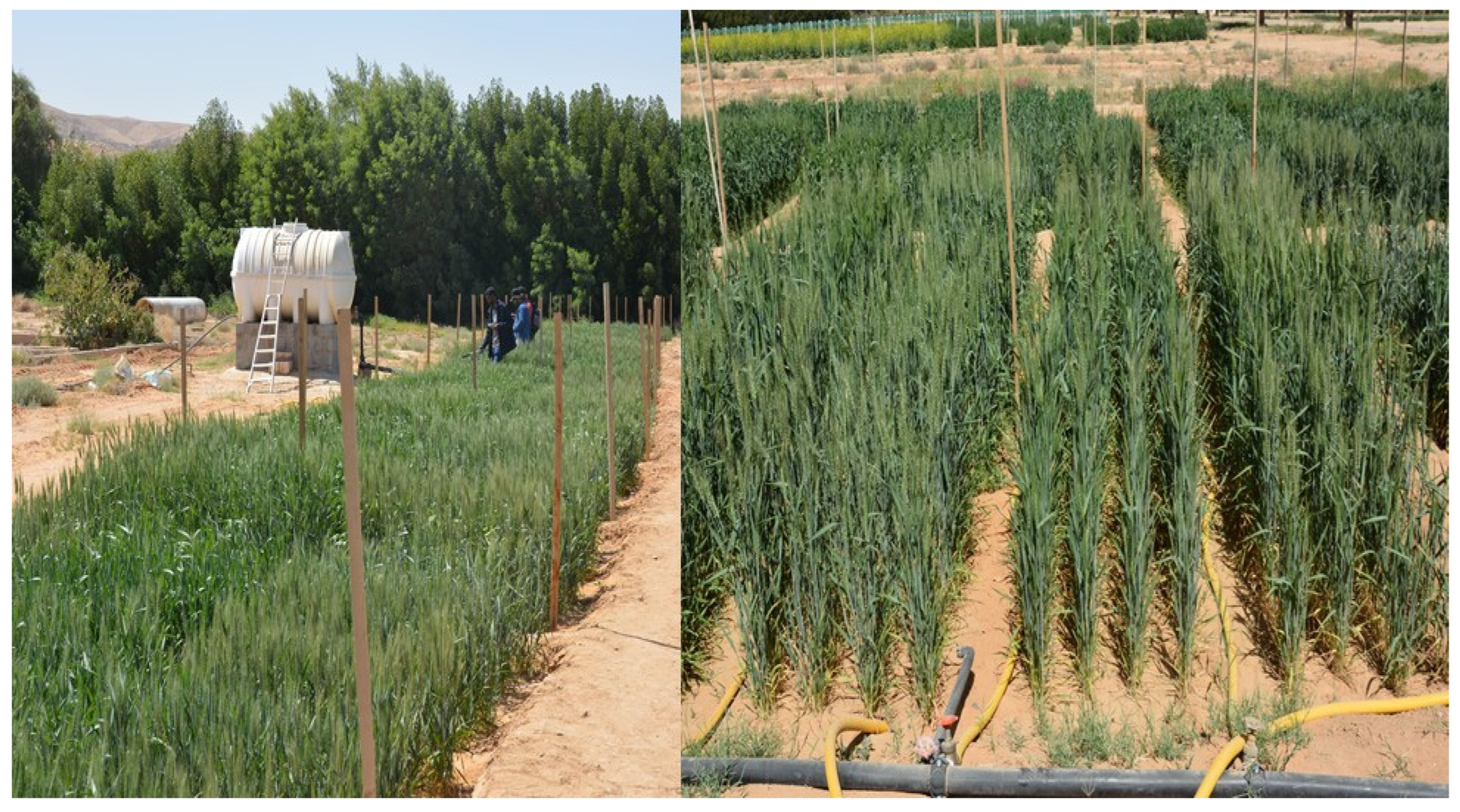
The salt tolerance of the tested genotypes was evaluated under high-salinity conditions (150 mM NaCl). Initially, all genotypes were irrigated with non-saline water for three weeks to avoid salinity stress during germination and seedling establishment. Subsequently, they were irrigated with artificial saline water containing 150 mM NaCl for the remainder of the experiment. The saline water was applied using a low-pressure surface irrigation system, which included a plastic water tank (5.0 m3) and a main line (76 mm diameter) delivering saline water from the tank to each plot. To ensure uniform water distribution to each plot, the main line was divided into sub-main hoses with manual control valves at each plot (see Figure 2).
The irrigation amount was calculated using the crop coefficient (Kc) for wheat from FAO-56 and the reference evapotranspiration rate (ETo) calculated with the modified Penman–Monteith equation. The total irrigation water applied was approximately 5000 m3 per hectare. The two irrigation parameters (ETo and Kc) were adjusted based on weather conditions at the ERS.

2.2. Measurements

2.2.1. Measurements of Relative Water Content, Chlorophyll Contents and Biomass

The different destructive traits were measured at 75 days from sowing when the wheat plants were at the flowering growth stage. The relative water content (RWC) of the leaves was calculated using the following formula:
RWC = (FW − DW)/(TW − DW) × 100
where FW is the fresh weight of approximately 0.20 cm2 of leaf samples taken from the top of four randomly selected plants from each genotype and replicate, TW is the turgid weight of these leaf samples after soaking them in distilled water for 24 h, and DW is the dry weight of these leaf samples after drying them in a hot-air oven at 75 °C until a constant weight was achieved.
Chlorophyll pigments, including chlorophyll a (Chla), chlorophyll b (Chlb), and total chlorophyll (Chlt), were determined using spectrophotometry. Fresh leaf samples (0.4 g) were homogenized in 5 mL of 80% acetone in the dark until the leaves were completely bleached. The extracted sap was then centrifuged at 400 rpm for 5 min and adjusted to a total volume of 15 mL with 80% acetone. The pigment extracts were analyzed for absorption spectra at 645 nm (A645) and 663 nm (A663) using a UV/vis spectrophotometer (UV-2550, Shimadzu, Tokyo, Japan). The concentrations of different Chl pigments (mg g−1 FW) were calculated according to Lichtenthaler and Wellburn [48] as follows:
Chla = (12.72 × A663) − (2.58 × A645) × V/1000 × FW
Chlb = (22.87 × A645) − (4.67 × A663) × V/1000 × FW
Chlt = (8.02 × A663) + (20.21 × A645) × V/1000 × FW
where V and FW are the volume of the extract solution (15 mL) and the weight of the fresh leaf tissue (0.4 g), respectively.
Plant biomass (PDW) was determined by averaging the dry weight of 10 randomly selected plants from each genotype and replicate. The plants were oven-dried at 75 °C for three days until a constant weight was achieved.

2.2.2. Thermal Imaging

Canopy temperature was measured at 75 days after sowing by capturing thermal infrared images between 11:30 and 14:30 under less windy conditions using a handheld thermal infrared camera (Therma CAM SC 3000 infrared camera, FLIR System, Wilsonville, OR, USA) with a 640 × 480 pixels resolution detector and a 45° × 34° wide-angle lens. The camera’s spectral range was 7.5–13 µm with a focal length of 13 mm. It had an accuracy of ±0.2 °C in the temperature range of −20 °C to 600 °C and a thermal sensitivity of ≤0.05 °C at 30 °C. The emissivity settings for measurements were 0.96 for the dry reference and 0.95 for the wet reference [49]. Before using the camera, a temperature-dependent radiometric calibration matrix was applied to correct ambient temperature variations and other non-uniformity noise. The thermal images were taken at a height of 100 cm above the plant canopy in the nadir orientation from five different locations in each plot. The average leaf temperature in each thermal image was calculated using the FLIR Research Pro Software version 5.1. (FLIR System, Wilsonville, OR, USA) by averaging the values of 50 randomly selected leaves within a polygon area fitted around each leaf. These data were then used in the following equation to calculate the normalized relative canopy temperature (NRCT).
NRCT = T T m i n T m a x T m i n
where T is the real infrared temperature measured in the canopy, while Tmin and Tmax represent the lowest and highest temperatures recorded in the entire field trial, respectively.

2.2.3. Digital RGB Imaging

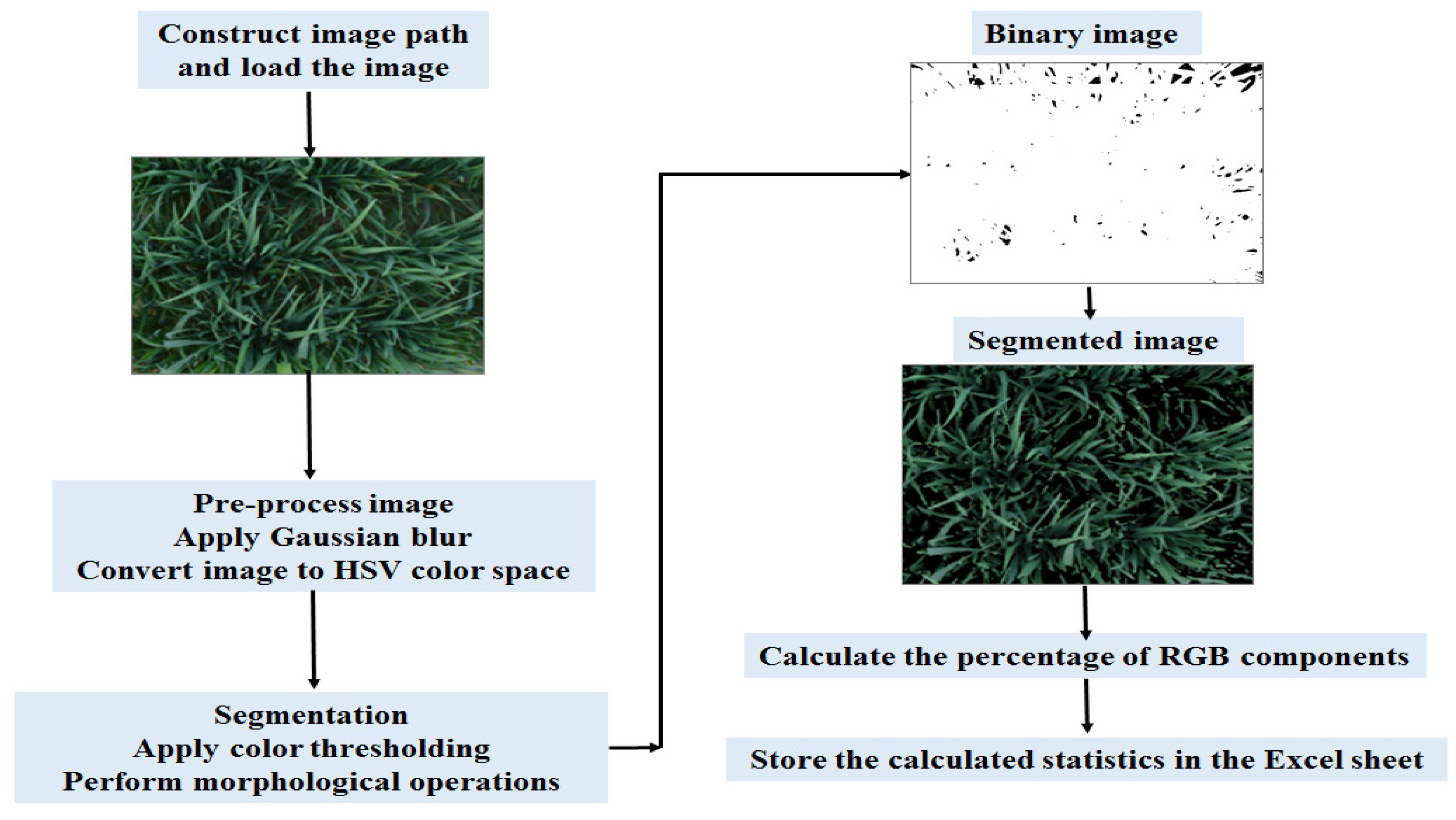
Digital images were taken between 12:00 and 14:00 on the same day as the thermal image acquisition. A 14-megapixel digital camera (Sony Alpha A5000, Sony, Tokyo, Japan) was used to capture photos. The camera was fitted with a Sony 16–50 mm lens set at the minimum focal length, fixed aperture of F3.5, and shutter speed of 1/250. Because the photos were captured under clear-sky conditions, an umbrella was held above the camera to provide shade conditions. This was done to ensure homogeneous lighting and minimize the impact of direct sunlight and leaf reflections on image quality and pixel intensity in stereoscopic images. The camera had a resolution of 3120 × 4160 pixels and captured 8-bit RGB images (0.4–0.7 μm). The camera was handheld and positioned vertically downward at a distance of one meter from the canopy. The flash was always turned off during measurements. To minimize angular distortion, the focal length was adjusted to 18 mm, providing a 76° image angle. A slight increase in exposure time was applied to enhance image segmentation through a controlled overexposure. The images were saved in JPEG format and analyzed using Python software (Version 3.11) and the Open CV library. To remove interference from non-canopy elements like soil, weeds, and straw, image segmentation and extraction were performed. This process ensured that only canopy information was used for feature extraction, as illustrated in Figure 3. The RGB values were normalized to reduce the effects of lighting and color variations [50].

Stages of Image Processing Pipeline for Vegetation Extraction

The pipeline consisted of multiple stages, including RGB channel separation, color index of vegetation extraction (CIVE), Otsu thresholding, masking, segmentation, and RGB color index calculation. Initially, the original image was divided into its red, green, and blue (RGB) color channels. Subsequently, the color index of vegetation extraction (CIVE) was computed, which measures the difference between the green and red color channels to identify vegetation areas in the image [50]. An Otsu threshold was then applied to the CIVE image to segment it into foreground and background regions based on pixel intensity distribution. This step helps in isolating vegetation areas further [51]. Following the Otsu thresholding, a masking operation was performed on the original image using the binary image obtained from the thresholding process as a mask. This operation eliminates non-vegetation areas from the original image, retaining only the vegetation-related parts. The masking operation produced a segmented image that highlights areas with vegetation from the original image. This segmented image is valuable for studying the distribution and properties of vegetation in the scene. Additionally, various RGB color indices were computed for the segmented image.
RGB imagery is composed of three bands, with each pixel’s color represented by three values [52]. The values of these RGB bands in plant canopies are influenced by factors like green biomass area and chlorophyll content. The mean values of RGB bands were extracted as sample features using the equations below:
R = 1 S n u m i = 1 S n u m R i
G = 1 S n u m i = 1 S n u m G i
B = 1 S n u m i = 1 S n u m B i
where R i , G i , and B i are the pixel values for the red, green, and blue bands, respectively, in the digital image; i and S n u m are the first pixel and maximum number of pixels, respectively; R, G, and B are the mean values of the red, green, and blue bands, respectively.
The formula and references of the twenty-one RGB imagery indices that were tested in this study are presented in Table 1.

2.3. Back-Propagation Neural Network (BPNN)

The back-propagation neural network (BPNN) is a popular artificial neural network model [61]. It comprises three layers: the input layer for initial data, the hidden layer as an intermediary, and the output layer for producing results based on the input. This type of neural network is a type of machine learning technique that uses multiple layers to extract more advanced features from raw data. It includes two hidden layers with nodes determined by regression accuracy. These hidden layers contain “activation” nodes also referred to as weights. The output layer delivers the anticipated values of the measured parameters. Artificial neural network (ANN) models are intricate mathematical structures that utilize interconnected neurons or nodes with weighted connections to simulate human cognitive processes for tasks such as pattern recognition and prediction [62,63]. Figure 4 shows the schematic diagram of the methodology of ANNs used in this study to predict the NRCT, RWC, Chla, Chlb, Chlt, and PDW of wheat genotypes. In this research, the input variables consist of 18 color RGB-based VIs. The number of features in each iteration of the loop ranged from 18 down to 1. During each loop, the most important features were selected, while the less important ones were excluded. This process facilitated the comparison of all outputs, enabling the identification of the optimal feature set that could enhance the prediction of salt tolerance in wheat genotypes based on the lowest root mean squared error of cross-validation (RMSECV).
To facilitate the model training process, a transformation was performed to rescale the features. Normalization was applied to individual features (X) to adjust for differences in magnitude between them. Feature normalization (Xnorm) was computed by subtracting the minimum feature value (Xmin) from each feature value and then dividing by the difference between the maximum (Xmax) and minimum feature values, as shown by the following formula:
X n o r m = X X m i n X m a x X m i n
The training process consisted of a minimum of 2000 iterations or continued until the error measurement reached a threshold of 10−4. To determine the optimal number of neurons in the hidden layer, the leave-one-out validation (LOOV) method was used during the validation process on the training dataset. The limited-memory Broyden–Fletcher–Goldfarb–Shanno (LBFGS) parameter, renowned for its efficiency in weight optimization, was utilized [64]. Furthermore, a specific formula was used to pinpoint the most important feature, with the goal of improving the predictive power of the regression model and decreasing the dimensionality of hyperspectral images [43].
M = j = 1 n H I   P j / k = 1 n p   I   P j , k   O j i = 1 n p j = 1 n H I   P i , j / k = 1 n p   I   P i , j , k   O j
where M is the important measure for the input variable, n p is the number of input variables, n H is the number of hidden layer nodes, I P j is the absolute value of the hidden layer weight corresponding to the pth input variable and the jth hidden layer, and O j is the absolute value of the output layer weight corresponding to the jth hidden layer.

2.4. Datasets and Software for Data Analysis

During the training and cross-validation process, we used a total of 44 samples from seasons 1 and 2. We implemented a leave-one-out cross-validation (LOOCV) technique, where the model was trained on all data points except one, which was reserved for validation. This method helps prevent overfitting and improves the model’s predictive accuracy, as discussed in reference [65]. Our data analysis, model development, and data preparation were carried out using Python version 3.7.3. We specifically examined the suitability of the ANN modules from the Scikit-learn package version 0.20.2 for regression tasks. The computations were performed on a system with an Intel Core i7-3630QM CPU running at 2.4 GHz and 8 GB of RAM.

2.5. Model Evaluation

Statistical measures like the coefficient of determination (R2), mean squared error (MSE), and the root mean squared error (RMSE) are commonly used to evaluate the accuracy of regression models. These measures are explained in studies by Malone et al. [66] and Saggi and Jain [67]. In this context, Fact denotes the actual value from laboratory calculations, Fp represents the predicted or simulated value, Fave is the average value, and N stands for the total number of data points.
Root   mean   squared   error   ( R M S E ) = 1 N i = 1 N ( F   a c t F   p ) 2
Mean   squared   error   ( M S E ) = 1 N i = 1 N ( F a c t F p ) 2
Coefficient   of   determination   ( R 2 ) = ( F   a c t F   p ) 2 ( F   a c t F   a v e ) 2

2.6. Statistical Analyses

An analysis of variance (ANOVA) was carried out on a randomized complete block design, followed by a post hoc test (Tukey’s test) at a significance level of 0.05. This was carried out to determine any significant differences in the mean values of the measured destructive traits and RGB indices among the tested genotypes. Mean values with the same letter indicate no significant difference at p ≤ 0.05. Simple linear regression was employed to estimate the relationship between measured destructive traits and NRCT and RGB indices. The significance level of R2 for these regressions was tested at p-values of ≤0.001, 0.01, and 0.05.

3. Results and Discussion

3.1. Genotypic Variations for Measured Destructive Traits

Table 2 shows the effects of year, genotypes, and their interactions on various destructive traits. The analysis of variance indicates that only PDW was influenced by year, while genotypes had a highly significant effect (p < 0.001) on all destructive traits. The interaction between genotype and year had a highly significant effect on chlorophyll pigment contents (Chla, Chlb, and Chlt) but not on RWC and PDW.
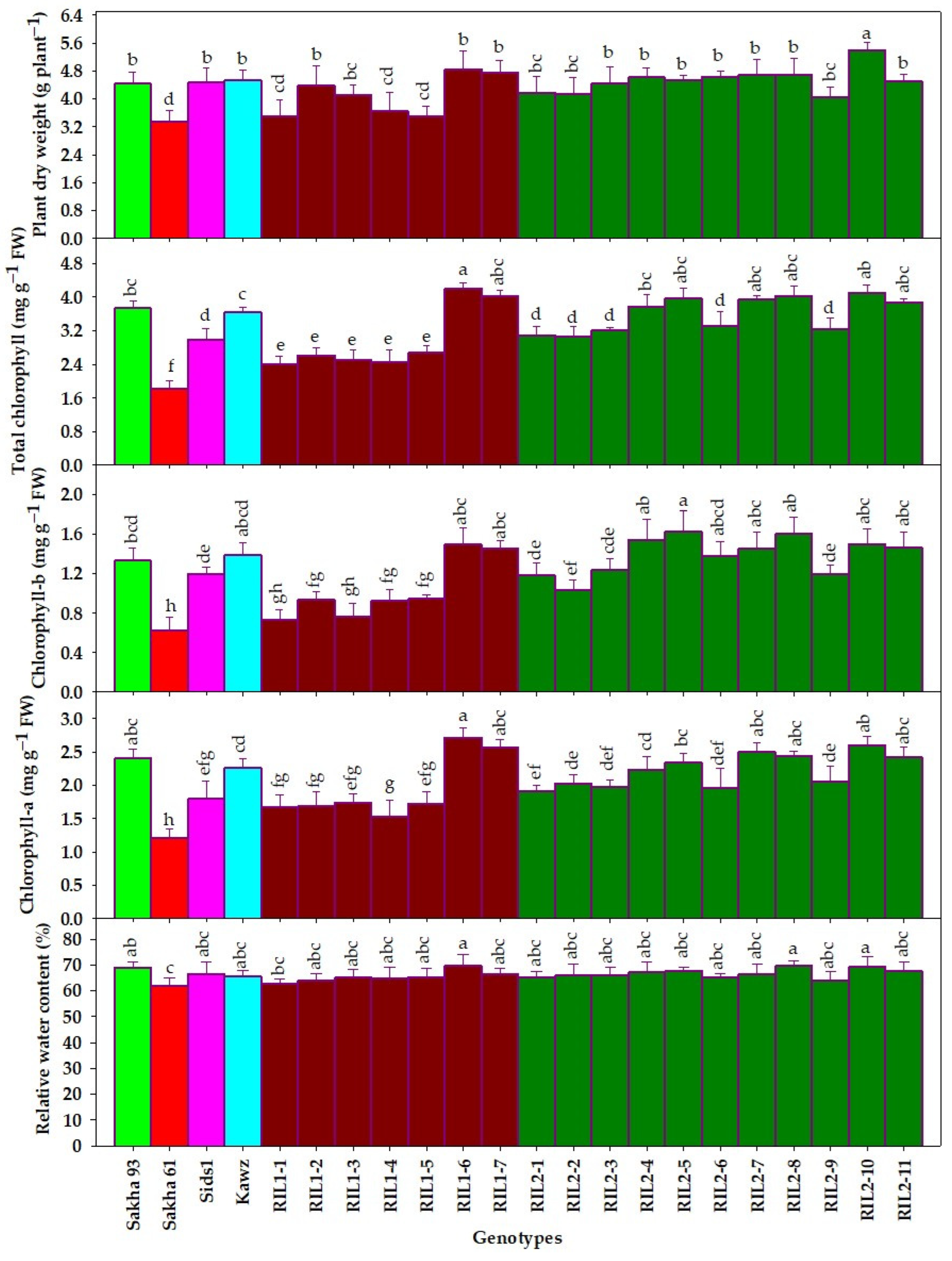
The analysis of variance also showed that genotypes accounted for the majority of the variation in destructive traits (26.85–79.89%), while the year had a smaller influence (0.001–16.32%) (Table 2). Genotypes had the highest contribution to the variation in chlorophyll pigment contents (72.23–79.89%) and a moderate contribution to PDW variation (53.78%), while their contribution to RWC variation was low (26.85%). The year had a greater share in the variability of RWC (16.32%) and PDW (9.03%) compared to chlorophyll pigment contents (0.001–0.53%). The interaction between genotype and year had nearly equal contributions in the variation of various destructive traits (9.71–16.40%) (Table 2). The largest share of genotypes in the variation of the different destructive traits suggests that these traits can serve as reliable screening criteria for assessing the salt tolerance of wheat genotypes. The significant contribution of genotype to the variation in chlorophyll pigment contents (72.23–79.89%) indicates that genotypes with the ability to maintain chlorophyll content under salinity stress are crucial for conferring tolerance to salt stress. In this study, the tested genotypes showed a wide range of chlorophyll pigment contents. Chla contents ranged from 1.21 to 2.72 mg g−1 FW, Chlb from 0.63 to 1.63 mg g−1 FW, and Chlt from 1.83 to 4.21 mg g−1 FW (Figure 5). Additionally, the salt-tolerant genotype Sakha 93 exhibited higher values for chlorophyll pigment contents (Chla, Chlb, and Chlt) compared to the salt-sensitive genotype Sakha 61. Among all tested genotypes, Sakha 61 had the lowest values for these traits. Out of the RILs tested, two (RIL6 and RIL7) from the Sakha 93 and Sakha 61 cross and four (RIL4, RIL5, RIL10, and RIL11) from the Sakha 93 and Sids1 cross showed chlorophyll pigment contents comparable to or higher than those of Sakha 93 (Figure 5). These results confirm the importance of maintaining chlorophyll content under salinity stress to enhance salt-tolerance mechanisms in genotypes. Similarly, previous studies have reported that salt-tolerant genotypes tend to maintain higher chlorophyll content under salinity conditions, while salt-sensitive genotypes experience a significant decrease in chlorophyll levels. This indicates that the variation in Chlorophyll pigments can be a key factor in evaluating the salt tolerance of different genotypes under salt stress [2,11,12,68,69,70]. Due to the inability of salt-sensitive Sakha 61 and other RILs to maintain chlorophyll pigments under salinity stress, they possibly accumulate more Na+ and produce excessive ROS in leaves. This ultimately leads to the degradation of chlorophyll or a reduction in chlorophyll synthesis. Therefore, measuring chlorophyll content accurately can serve as a dependable indicator for assessing the salt tolerance of different genotypes.
Salt stress has various negative effects on plants, leading to changes in their physiology and biochemistry that result in a decrease in plant biomass. When plants are exposed to salinity stress, they must balance their use of photosynthetic energy between adaptation mechanisms and biomass production. This trade-off ultimately leads to a significant reduction in biomass accumulation. Plant biomass is an important indicator of how plants respond to salt stress at the whole-plant level [71,72]. Previous studies have shown that salt-tolerant genotypes tend to have higher plant biomass compared to sensitive genotypes, as they can sustain a higher photosynthetic rate under salinity stress [3,15,17,18,73,74]. Therefore, plant biomass can be a useful screening criterion for distinguishing between salt-tolerant and salt-sensitive genotypes. This study found that PDW measured at flowering stages is a key trait for distinguishing salt tolerance among the tested genotypes. Genotype played a significant role in the variation of PDW, accounting for 53.78% of the total variation (Table 2). Additionally, the tested genotypes displayed a broad range of PDW values, ranging from 3.36 to 5.40 g per plant (Figure 4). Among all tested genotypes, the salt-sensitive genotype Sakha 61 had the lowest values for PDW. In addition, several RILs from both crosses showed PDW comparable to or higher than those of Sakha 93 (Figure 4). These results indicate that plant biomass is a valuable screening criterion for assessing the salt tolerance of genotypes. This result aligns with previous studies that have shown plant biomass to be a valuable trait for screening bread [3,75] and durum wheats [76,77] for salt tolerance.
Relative water content (RWC) is a key physiological parameter used to gauge a plant’s tolerance to salinity stress. It provides valuable insights into the plant’s physiological status when exposed to high salinity levels [1,78,79]. A study on the salt tolerance of alfalfa found that RWC varied among different alfalfa genotypes, and the salt tolerance of these genotypes was positively correlated with their RWC [80]. In this study, the genotype had a lower contribution to the variation of RWC (26.85%) compared to chlorophyll pigment contents and PDW (Table 2). Additionally, there were few differences in RWC among the tested genotypes (Figure 4). This indicated that the RWC was much less affected by salinity stress than other measured traits. This finding contradicts other reports that indicate significant variations in RWC among different genotypes under salinity-stress conditions, and the salt-tolerant genotypes were found to have higher RWC compared to salt-sensitive ones. The slight variations in RWC observed among the genotypes tested in this study may be attributed to osmotic adjustment mechanisms. Salt-sensitive genotypes may accumulate more Na+ ions, while salt-tolerant genotypes may synthesize soluble osmotic adjustment substances to maintain RWC. This finding indicates that although the tested genotypes exhibit varying levels of salt tolerance, their ability to retain their RWC does not vary significantly. Hence, because of the limited genetic influence on RWC variation, it is recommended to use this trait as a screening criterion for assessing salt tolerance only after understanding how various genotypes maintain water content under salinity conditions.
Importantly, the results of this study confirm that the most measured destructive traits in this study could serve as effective screening criteria for assessment of the salt tolerance of wheat genotypes because the genotype effects accounted for the majority of the variation in these traits, as shown in Table 2 and Figure 5. However, traditional methods of phenotyping these traits through destructive plant sampling and laboratory analysis are frequently destructive and labor-intensive, time-consuming, costly, and not conducive to real-time evaluation. Therefore, this creates a bottleneck in plant breeding. Undoubtedly, the introduction of high-throughput phenotyping tools will greatly enhance the indirect assessment of various plant traits related to salt tolerance, facilitating the breeding of salt-tolerant wheat. The data of these tools are presented and discussed in the following sections.

3.2. Performance of Thermal Index to Assess Measured Traits

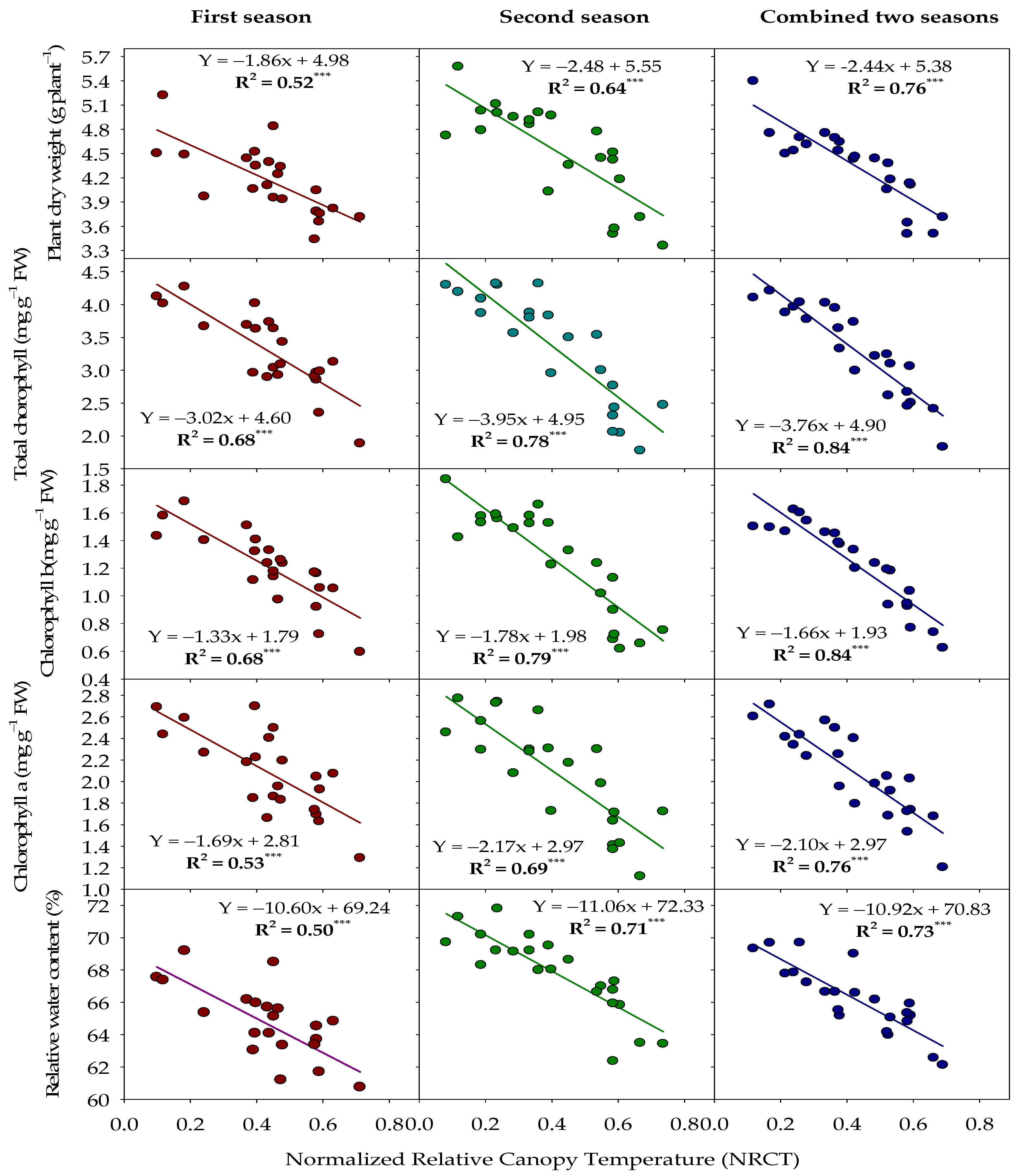
Remote sensing, combined with advanced image analysis, is a valuable tool for comparing different genotypes in field conditions. Thermal imaging, in particular, is effective for detecting plant stress in remote sensing systems. Infrared thermal measurements can evaluate cereal crops’ tolerance to osmotic stress by showing variations in transpiration rates that impact leaf and canopy temperature [20]. High soil salt levels can impede a plant’s ability to absorb water, leading to stomatal closure, reduced water vapor release from leaves, decreased water content, and elevated heat flow in leaves. This results in higher canopy temperatures, which adversely affect plant growth and productivity [20,25,81,82]. Orzechowska et al. [19] observed a significant rise in canopy foliage temperature during short-term salinity stress, while Sirault et al. [20] noted that plants experiencing salinity stress exhibited stomatal closure, reduced transpiration rates, and increased canopy temperature. In general, thermal imaging is preferred over traditional methods like porometry because of its accuracy, non-invasiveness, and speed. Monitoring plant traits under salinity stress can be simplified with a thermal tool that detects infrared radiation (800–1300 nm) to measure canopy temperature changes non-invasively. Therefore, in this study, we aimed to assess the salt tolerance of wheat genotypes using NRCT. We examined the relationship between NRCT and different destructive traits in 22 wheat genotypes across two seasons to investigate this hypothesis. The relationships between NRCT and various traits were found to be strong, with R2 values ranging from 0.50 to 0.73 for RWC, 0.53 to 0.76 for Chla, 0.68 to 0.84 for Chlb, 0.68 to 0.84 for Chlt, and 0.52 to 0.76 for PDW across two seasons (Figure 6). The relationships between NRCT and various measured traits showed the highest R2 values (R2 = 0.68–0.84) when the data were analyzed across two seasons (Figure 6). In a study by Sirault et al. [25], it was found that there is a negative relationship between leaf temperature and salt tolerance. In a study by Vennam et al. [83], it was observed that exposing maize plants to high salinity levels led to a 4 °C increase in canopy temperature. This increase was attributed to a reduction in stomatal conductance and transpiration. Finally, the strong correlation between the thermal index (NRCT) and measured traits confirms the effectiveness of thermal imagery in evaluating the salt tolerance of wheat genotypes.

3.3. The Variation of RGB Indices and the Performance of Indices to Assess Wheat Genotypes Traits under Salinity Conditions

Salinity stress can reduce photosynthesis and cause water deficiencies in plants, leading to changes in plant traits such as water status, pigment content, and dry weight biomass. So, the values of the RGB indices (i.e., the red (R), green (G), and blue (B) band percentages) exhibited significant variation among wheat genotypes under salinity conditions (Table 3). This result indicates that salinity stress had a notable effect on the color characteristics of wheat genotypes. This indicates that using RGB technology could be a cost-effective method to assess the salt tolerance of wheat genotypes. Salinity stress in plants can lead to changes in plant color due to factors like osmotic stress, ion toxicity, and nutrient deficiency. These stressors affect various plant properties, including stomatal conductance, guard cell chloroplasts, leaf pigment concentrations, photosynthesis, and water status. As a result, plants may exhibit significant alterations in their response to visible light. For instance, the activity of guard cell chloroplasts has been shown to be sensitive to both blue and red light [84,85,86].
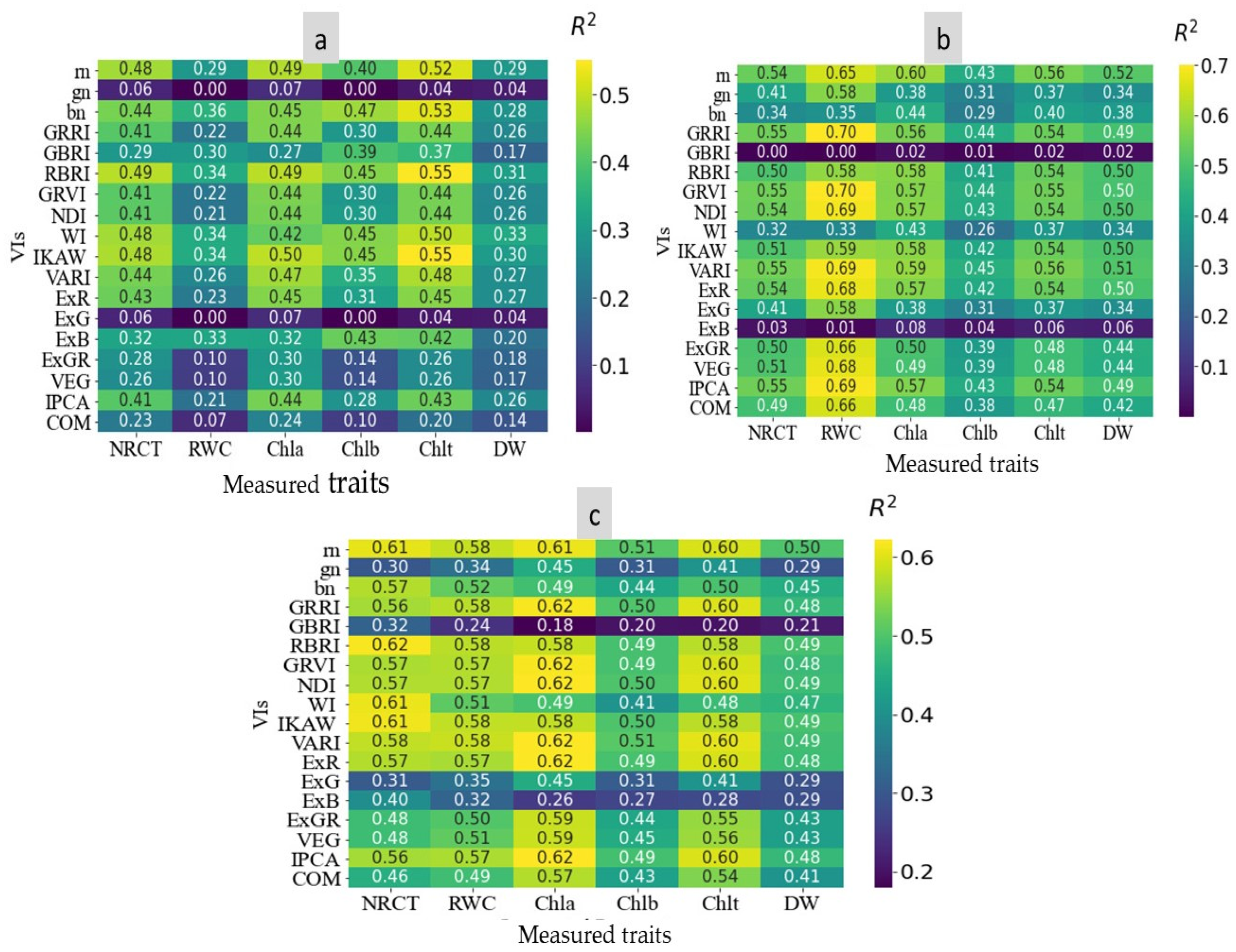
Table 3 shows distinct variations in the RGB indices among the wheat genotypes tested. For instance, rn ranged from 0.280 to 0.313, GRRI ranged from 1.141 to 1.335, RBRI ranged from 0.804 to 0.965, VARI ranged from 0.133 to 0.304, ExG ranged from 0.067 to 0.124, and IPCA ranged from 8.588 to 17.69. Figure 7 shows the relationships between the measured traits and various RGB indices for the first, second, and combined two seasons. The data indicate variations in the relationship between RGB indices and different traits, with the highest R2 values reaching up to 0.70 (Figure 7). There were varying degrees of relationships between the RGB indices and the five measured traits. Across two seasons, the RBRI, GRRI, GRVI, NDI, VARI, EXR, and IPCA showed good relationships with NRCT, RWC, Chla, Chlb, Chlt, and PDW. For example, VARI had strong relationships with NRCT, RWC, Chla, Chlb, Chlt, and PDW, with R2 values of 0.58, 0.58, 0.62, 0.51, 0.60, and 0.49, respectively.
Several studies have demonstrated a strong correlation between plant water content and the visible light spectrum (400–700 nm) [28,86,87]. Schlemmer et al. [87] found a negative linear relationship between RWC and the blue and red spectral regions, while Carter [88] observed that RWC is affected by the blue and red visible range around 480–690 nm. Several studies have demonstrated that vegetation indices, such as the green–red vegetation index (GRVI), derived from RGB images, can accurately predict physiological parameters like Chl content, leaf area index, transpiration rate, stomatal conductance, and RWC. This is attributed to the strong positive correlation between these indices and the physiological indicators [85,89,90]. This indicates that the RGB channels of visible light could be useful for monitoring different plant characteristics related to chlorophyll pigments and water status. These results may be attributed to the varying of these traits among the 22 wheat genotypes tested, leading to the generation of distinct image-derived indices.

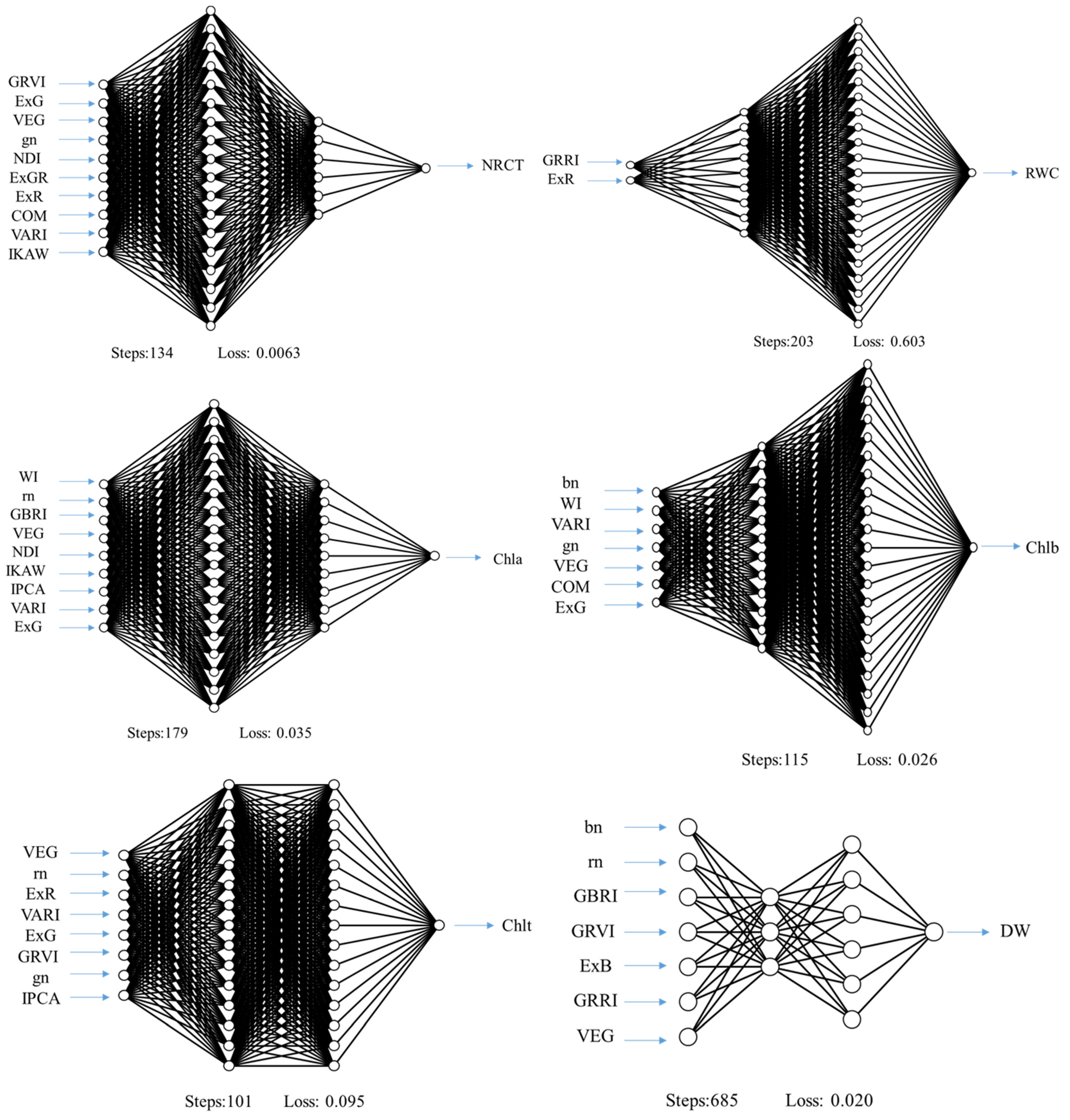
3.4. The Performance of ANN Model to Predict Measured Traits

Robust statistical techniques are essential for accurate predictive power in high-throughput phenotyping of plant traits using multiple indices. Machine learning algorithms have made significant advancements in recent years and are widely used in precision agriculture for precise plant trait predictions [33,34,91,92]. Our study utilized ANN models to accurately predict measured traits by incorporating RGB indices as independent variables (Figure 8). This study used data from season 1 (S1) and season 2 (S2) as well as a combined dataset from both seasons (BS) to train a neural network for predicting the specific variables listed in Table 4. The model’s predictions were evaluated against a separate set of measurements to determine its performance. The study explored various multivariate techniques and showed that these methods significantly enhance predictive accuracy. Independent evaluation was emphasized as the most trustworthy way to gauge the regression model’s effectiveness, as it involves data not used during model training. The ANN-VIs-10-ST model was identified as the best predictive model, showing a strong correlation between its key features and NRCT. This model includes ten essential features for accurately predicting NRCT. It achieved an R2 of 0.895 and an RMSE of 0.055 for the training dataset as well as an R2 of 0.682 and an RMSE of 0.072 for cross-validation. The ANN-VIs-2-S2 model performed exceptionally well in the evaluation by RWC. It achieved high accuracy with R2 scores of 0.806 for the training set and 0.681 for the cross-validation set. The model also demonstrated low RMSE scores of 1.099% for training and 1.082% for cross-validation. The ANN-VIs-9-ST model was found to be the best-performing model for assessing Chla content. It achieved an R2 of 0.866 and an RMSE of 0.136 mg g−1 FW in the training dataset and an R2 of 0.706 with an RMSE of 0.165 mg g−1 FW in the validation dataset. The ANN-VIs-7-ST model showed excellent performance in predicting Chlb content, with R2 values of 0.727 (RMSE = 0.142 mg g−1 FW) for the training set and 0.461 (RMSE = 0.150 mg g−1 FW) for the cross-validation set. For Chlt prediction, the most accurate model was the ANN-VIs-8-ST, with R2 values of 0.899 and 0.737 and RMSE values of 0.190 and 0.240 mg g−1 FW for the training and cross-validation sets, respectively. Additionally, for PDW predictions, the ANN-VIs-7-S1 model outperformed all others, achieving impressive R2 values of 0.947 (RMSE = 0.094 g plant−1) for the training set and 0.645 (RMSE = 0.198 g plant−1) for the cross-validation set. Elsherbiny et al. [91] noted performance improvements in robust prediction through specific training actions, including filtering high-level features and optimizing model hyperparameters. In a study by Ribeiro et al. [34], it was found that ANN modeling outperformed linear regression models in terms of accuracy, reliability, and efficiency. This is because ANNs can better capture complex and nonlinear relationships between input and output values.
This study enhanced wheat genotype trait identification by integrating RGB and thermal images, leading to improved model performance. High-resolution images from ground or UAV-mounted cameras provided accurate data for assessing plant traits in the field. This research facilitates the development of cost-effective phenotyping platforms for large-scale breeding under salt-stress conditions. Sharing these findings could enhance site-specific wheat management through an intelligent system based on the optimal model. The ground-based camera method can be adapted for UAV use, increasing efficiency and enabling crop growth monitoring over extensive areas.
In future research, it would be beneficial to explore the use of thermal and RGB imaging in combination with ANN to evaluate salt tolerance in other important crops such as rice, maize, and barley. This would help demonstrate the wider applicability of these techniques beyond wheat. Additionally, incorporating advanced technologies like hyperspectral or high-resolution 3D imaging could improve the accuracy of stress assessments by providing more detailed and comprehensive data. Further refinement of the neural network models through experimentation with different architectures or the integration of other machine learning methods could enhance prediction accuracy and robustness. Conducting studies under diverse field conditions and extending them over time would help validate and generalize the findings, ensuring the effectiveness of the techniques in various environments and over different seasons. Lastly, investigating the integration of these technologies with current agronomic practices could facilitate their adoption in real-world agricultural settings, leading to more resilient and productive farming systems.

4. Conclusions

Salinization is a rising concern worldwide, impacting agricultural land and freshwater resources. Hence, developing salt-tolerant genotypes is crucial to ensure food and water security in the face of this challenge. The traits measured in this study, including RWC, pigment contents, and plant biomass, indicate that these traits are effective screening criteria for evaluating the salt tolerance of wheat genotypes and predicting their performance in field conditions. However, traditional methods of phenotyping these traits through destructive plant sampling and laboratory analysis are time-consuming, costly, impractical, and not conducive to real-time evaluation. This study demonstrated that thermal and RGB imaging, along with ANN models, can accurately assessing these plant traits in various wheat genotypes under salinity-stress conditions. The results showed a strong correlation between indices derived from thermal and RGB imaging and various plant traits. The correlation values were even higher when data from both seasons were combined. Additionally, combining thermal and RGB imagery indices with an ANN model proved effective in predicting the observed plant attributes. For example, in the evaluation conducted by RWC, the ANN-VIs-2-S2 model demonstrated superior performance. Its accuracy was reflected in R2 scores of 0.806 for the training set and 0.681 for the cross-validation set. Additionally, the model showed RMSE scores of 1.099% for training and 1.082% for cross-validation. This knowledge will aid in developing lightweight, proximal sensors for quick and non-destructive phenotyping of large wheat populations, accelerating the selection of high-quality germplasm for salt-tolerance breeding programs. The study findings were confirmed under a specific level of salinity stress on a single crop. Further research is needed to investigate various salinity levels and crops to verify the reliability and consistency of the results obtained in this study.

Author Contributions

Conceptualization, S.E.-H.; methodology, S.E.-H., N.A.-S., M.U.T., O.E. and S.E.; software, S.E.-H., M.U.T., H.E., O.E. and S.E.; validation, S.E.-H.; formal analysis, S.E.-H., M.U.T., S.E., H.E. and O.E.; investigation, S.E.-H., M.U.T., N.A.-S., O.E. and S.E.; resources, S.E.-H., N.A.-S. and M.U.T.; data curation, S.E.-H., S.E., O.E., M.U.T., H.E. and N.A.-S.; writing—original draft preparation, S.E.-H. and S.E.; writing—review and editing, S.E.-H.; visualization, S.E.-H. and N.A.-S.; supervision, S.E.-H.; project administration, S.E.-H.; funding acquisition, S.E.-H. and N.A.-S. All authors have read and agreed to the published version of the manuscript.

Funding

This research was funded by the Researchers Supporting Project number (RSPD2024R730), King Saud University, Riyadh, Saudi Arabia.

Data Availability Statement

All data are presented within the article.

Acknowledgments

The authors acknowledge the Researchers Supporting Project number (RSPD2024R730), King Saud University, Riyadh, Saudi Arabia.

Conflicts of Interest

The authors declare no conflicts of interest.

References

  1. Chaurasia, S.; Kumar, A.; Singh, A.K. Comprehensive evaluation of morpho-physiological and ionic traits in wheat (Triticum aestivum L.) genotypes under salinity stress. Agriculture 2022, 12, 1765. [Google Scholar] [CrossRef]
  2. El-Hendawy, S.E.; Hassan, W.M.; Al-Suhaibani, N.A.; Refay, Y.; Abdella, K.A. Comparative performance of multivariable agro-physiological parameters for detecting salt tolerance of wheat cultivars under simulated saline field growing conditions. Front. Plant. Sci. 2017, 8, 435. [Google Scholar] [CrossRef]
  3. Oyiga, B.C.; Sharma, R.; Shen, J.; Baum, M.; Ogbonnaya, F.; Léon, J.; Ballvora, A. Identification and characterization of salt tolerance of wheat germplasm using a multivariable screening approach. J. Agron. Crop Sci. 2016, 202, 472–485. [Google Scholar] [CrossRef]
  4. Mansour, E.; Moustafa, E.S.A.; Desoky, E.M.; Ali, M.M.A.; Yasin, M.A.T.; Attia, A.; Alsuhaibani, N.; Tahir, M.U.; El-Hendawy, S.E. Multidimensional evaluation for detecting salt tolerance of bread wheat genotypes under actual saline field growing conditions. Plants 2020, 9, 1324. [Google Scholar] [CrossRef]
  5. Shiferaw, B.; Smale, M.; Braun, H.J.; Duveiller, E.; Reynolds, M.; Muricho, G. Crops that feed the world 10. past successes and future challenges to the role played by wheat in global food security. Food Sec. 2013, 5, 291–317. [Google Scholar] [CrossRef]
  6. Zada, A.; Ali, A.; Binjawhar, D.N.; Abdel-Hameed, U.K.; Shah, A.H.; Gill, S.M.; Hussain, I.; Abbas, Z.; Ullah, Z.; Sher, H.; et al. Molecular and physiological evaluation of bread wheat (Triticum aestivum L.) genotypes for stay green under drought stress. Genes 2022, 13, 2261. [Google Scholar] [CrossRef]
  7. Reynolds, M.; Pask, A.; Mullan, D. Physiological Breeding I: Interdisciplinary Approaches to Improve Crop Adaptation; International Maize and Wheat Improvement Center (CIMMYT): Texcoco, Mexico, 2012. [Google Scholar]
  8. Araus, J.L.; Slafer, G.A.; Royo, C.; Serret, M.D. Breeding for yield potential and stress adaptation in cereals. Crit. Rev. Plant Sci. 2008, 27, 377–412. [Google Scholar] [CrossRef]
  9. El-Hendawy, S.; Al-Suhaibani, N.; Mubushar, M.; Tahir, M.U.; Marey, S.; Refay, Y.; Tola, E. Combining hyperspectral reflectance and multivariate regression models to estimate plant biomass of advanced spring wheat lines in diverse phenological stages under salinity conditions. Appl. Sci. 2022, 12, 1983. [Google Scholar] [CrossRef]
  10. Croft, H.; Chen, J.M.; Luo, X.; Bartlett, P.; Chen, B.; Staebler, R.M. Leaf chlorophyll content as a proxy for leaf photosynthetic capacity. Glob. Chang. Biol. 2017, 23, 3513–3524. [Google Scholar] [CrossRef]
  11. Zhou, X.; Huang, W.; Zhang, J.; Kong, W.; Casa, R.; Huang, Y. A Novel combined Spectral Index for Estimating the Ratio of Carotenoid to Chlorophyll Content to Monitor Crop Physiological and Phenological Status. Int. J. Appl. Earth Obs. 2019, 76, 128–142. [Google Scholar] [CrossRef]
  12. Zhang, L.; Ma, H.; Chen, T.; Pen, J.; Yu, S.; Zhao, X. Morphological and physiological responses of cotton (Gossypium hirsutum L.) plants to salinity. PLoS ONE 2014, 9, e112807. [Google Scholar] [CrossRef]
  13. Pour-Aboughadareh, A.; Sanjani, S.; Nikkhah-Chamanabad, H.; Mehrvar, M.R.; Asadi, A.; Amini, A. Identification of salt-tolerant barley genotypes using multiple-traits index and yield performance at the early growth and maturity stages. Bull. Natl. Res. Cent. 2021, 45, 117. [Google Scholar] [CrossRef]
  14. Tao, R.; Ding, J.; Li, C.; Zhu, X.; Guo, W.; Zhu, M. Evaluating and screening of agro-physiological indices for salinity stress tolerance in wheat at the seedling stage. Front. Plant Sci. 2021, 12, 646175. [Google Scholar] [CrossRef]
  15. Munns, R.; Tester, M. Mechanisms of salt tolerance. Annu. Rev. Plant Biol. 2008, 59, 651–681. [Google Scholar] [CrossRef]
  16. Acosta-Motos, J.R.; Ortuño, M.F.; Bernal-Vicente, A.; Diaz-Vivancos, P.; Sanchez-Blanco, M.J.; Hernandez, J.A. Plant responses to salt stress: Adaptive mechanisms. Agronomy 2017, 7, 18. [Google Scholar] [CrossRef]
  17. Radi, A.A.; Farghaly, F.A.; Hamada, A.M. Physiological and biochemical responses of salt-tolerant and salt-sensitive wheat and bean genotypes to salinity. J. Biol. Earth Sci. 2013, 2, 72–88. [Google Scholar]
  18. Genc, Y.; Taylor, J.; Lyons, G.H.; Li, Y.; Cheong, J.; Appelbee, M.; Oldach, K.; Sutton, T. Bread wheat with high salinity and sodicity tolerance. Front. Plant Sci. 2019, 10, 280. [Google Scholar] [CrossRef]
  19. Orzechowska, A.; Trtílek, M.; Tokarz, K.M.; Szymańska, R.; Niewiadomska, E.; Rozpądek, P.; Wątor, K. Thermal analysis of stomatal response under salinity and high light. Int. J. Mol. Sci. 2021, 22, 4663. [Google Scholar] [CrossRef]
  20. Sirault, X.R.R.; James, R.A.; Furbank, R.T.; Sirault, X.R.R.; James, R.A.; Furbank, R.T. A New screening method for osmotic component of salinity tolerance in cereals using infrared thermography. Funct. Plant Biol. 2009, 36, 970–977. [Google Scholar] [CrossRef]
  21. Banerjee, B.P.; Joshi, S.; Thoday-Kennedy, E.; Pasam, R.K.; Tibbits, J.; Hayden, M.; Spangenberg, G.; Kant, S. High-throughput phenotyping using digital and hyperspectral imaging-derived biomarkers for genotypic nitrogen response. J. Exp. Bot. 2020, 71, 4604–4615. [Google Scholar] [CrossRef]
  22. Kim, S.L.; Lee, N.; Lee, H.; Lee, E.; Cheon, K.-S.; Kim, M.; Baek, J.-H.; Choi, I.; Ji, H.; Yoon, I.S.; et al. High-throughput phenotyping platform for analyzing drought tolerance in rice. Planta 2020, 252, 38. [Google Scholar] [CrossRef]
  23. Rufo, R.; Soriano, J.M.; Villegas, D.; Royo, C.; Bellvert, J. Using unmanned aerial vehicle and ground-based RGB indices to assess agronomic performance of wheat landraces and cultivars in a Mediterranean-type environment. Remote Sens. 2021, 13, 1187. [Google Scholar] [CrossRef]
  24. Hasan, U.; Sawut, M.; Chen, S. Estimating the leaf area index of winter wheat based on unmanned aerial vehicle RGB-image parameters. Sustainability 2019, 11, 6829. [Google Scholar] [CrossRef]
  25. Thoday-Kennedy, E.; Joshi, S.; Daetwyler, H.D.; Hayden, M.; Hudson, D.; Spangenberg, G.; Kant, S. Digital phenotyping to delineate salinity response in safflower genotypes. Front. Plant Sci. 2021, 12, 662498. [Google Scholar] [CrossRef]
  26. Duan, L.; Han, J.; Guo, Z.; Tu, H.; Yang, P.; Zhang, D.; Fan, Y.; Chen, G.; Xiong, L.; Dai, M. Novel digital features discriminate between drought resistant and drought sensitive rice under controlled and field conditions. Front. Plant Sci. 2018, 9, 492. [Google Scholar] [CrossRef]
  27. Guo, Z.; Yang, W.; Chang, Y.; Ma, X.; Tu, H.; Xiong, F.; Jiang, N.; Feng, H.; Huang, C.; Yang, P. Genome-wide association studies of image traits reveal genetic architecture of drought resistance in rice. Mol. Plant 2018, 11, 789–805. [Google Scholar] [CrossRef]
  28. Petrozza, A.; Santaniello, A.; Summerer, S.; Di Tommaso, G.; Di Tommaso, D.; Paparelli, E.; Piaggesic, A.; Peratab, P.; Cellini, F. Physiological responses to Megafol treatments in tomato plants under drought stress: A phenomic and molecular approach. Sci. Hortic. 2014, 174, 185–192. [Google Scholar] [CrossRef]
  29. Elsayed, S.; Barmeier, G.; Schmidhalter, U. Passive reflectance sensing and digital image analysis allows for assessing the biomass and nitrogen status of wheat in early and late tillering stages. Front. Plant Sci. 2018, 9, 1478. [Google Scholar] [CrossRef]
  30. Lapidot, O.; Ignat, T.; Rud, R.; Rog, I.; Alchanati, V.; Klein, T. Use of thermal imaging to detect evaporative cooling in coniferous and broadleaved tree species of the Mediterranean maquis. Agric. For. Meteorol. 2019, 271, 285–294. [Google Scholar]
  31. Stutsel, B.; Johansen, K.; Malbéteau, Y.M.; McCabe, M.F. Detecting plant stress using thermal and optical imagery from an unoccupied aerial vehicle. Front. Plant Sci. 2021, 12, 734944. [Google Scholar]
  32. Elsayed, S.; Rischbeck, P.; Schmidhalter, U. Comparing the performance of active and passive reflectance sensors to assess the normalized relative canopy temperature and grain yield of drought-stressed barley cultivars. Field Crop. Res. 2015, 177, 148–160. [Google Scholar] [CrossRef]
  33. Singh, R.N.; Krishnan, P.; Singh, V.K.; Sah, S.; Das, B. Combining biophysical parameters with thermal and RGB indices using machine learning models for predicting yield in yellow rust affected wheat crop. Sci. Rep. 2023, 13, 18814. [Google Scholar] [CrossRef]
  34. Ribeiro, J.E.; Coêlho, E.S.; de Oliveira, A.K.; da Silva, G.C.; Lopes, W.A.R.; Oliveira, P.H.; da Silva, E.F.; Júnior, A.P.B.; da Silveira, L.M. Artificial neural network approach for predicting the sesame (Sesamum indicum L.) leaf area: A non-destructive and accurate method. Heliyon 2023, 9, e17834. [Google Scholar]
  35. Kim, N.; Ha, K.-J.; Park, N.-W.; Cho, J.; Hong, S.; Lee, Y.-W. A Comparison between major artificial intelligence models for crop yield prediction: Case study of the Midwestern United States, 2006–2015. ISPRS Int. J. Geo Inf. 2019, 8, 240. [Google Scholar] [CrossRef]
  36. Khaki, S.; Wang, L. Crop Yield Prediction Using Deep Neural Networks. In Smart Service Systems, Operations Management, and Analytics; Yang, H., Qiu, R., Chen, W., Eds.; Springer Proceedings in Business and Economics; Springer: Berlin, Germany, 2020; pp. 139–147. [Google Scholar]
  37. García-Martínez, H.; Flores-Magdaleno, H.; Ascencio-Hern’andez, R.; Khalil-Gardezi, A.; Tijerina-Ch’avez, L.; Mancilla-Villa, O.R.; Vázquez-Peña, M.A. Corn grain yield estimation from vegetation indices, canopy cover, plant density, and a neural network using multispectral and RGB images acquired with unmanned aerial vehicles. Agriculture. 2020, 10, 10070277. [Google Scholar]
  38. Salem, S.B.H.; Gaagai, A.; Ben Slimene, I.; Moussa, A.B.; Zouari, K.; Yadav, K.K.; Eid, M.H.; Abukhadra, M.R.; El-Sherbeeny, A.M.; Gad, M.; et al. Applying Multivariate Analysis and Machine Learning Approaches to Evaluating Groundwater Quality on the Kairouan Plain, Tunisia. Water 2023, 15, 3495. [Google Scholar] [CrossRef]
  39. Schuize, F.H.; Wolf, H.; Jansen, H.; Vander, V.P. Applications of artificial neural networks in integrated water management: Fiction or future? Water Sci. Technol. 2005, 52, 21–31. [Google Scholar] [CrossRef]
  40. EIMasry, G.; Sun, D.W.; Allen, P. Near-infrared hyperspectral imaging for predicting colour, pH and tenderness of fresh beef. J. Food Eng. 2012, 110, 127–140. [Google Scholar] [CrossRef]
  41. Strobl, C.; Boulesteix, A.-L.; Kneib, T.; Augustin, T.; Zeileis, A. Conditional variable importance for random forests. BMC Bioinform. 2008, 9, 307. [Google Scholar] [CrossRef]
  42. Glorfeld, L.W. A methodology for simplification and interpretation of backpropagation-based neural network models. Expert Syst. 1996, 10, 37–54. [Google Scholar] [CrossRef]
  43. Melis, G.; Dyer, C.; Blunsom, P. On the state of the art of evaluation in neural language models. arXiv 2017, arXiv:1707.05589. [Google Scholar]
  44. Bergstra, J.; Yamins, D.; Cox, D. Making a science of model search: Hyperparameter optimization in hundreds of dimensions for vision architectures. In Proceedings of the 30th International Conference on Machine Learning (ICML 2013), Atlanta, Gerorgia, 16–21 June 2013; pp. 115–123. [Google Scholar]
  45. Wu, J.; Chen, X.-Y.; Zhang, H.; Xiong, L.-D.; Lei, H.; Deng, S.-H. Hyperparameter optimization for machine learning models based on Bayesian optimization. J. Electron. Sci. Technol. 2019, 17, 26–40. [Google Scholar]
  46. El-Hendawy, S.E.; Hu, Y.C.; Yakout, G.M.; Awad, A.M.; Hafiz, S.E.; Schmidhalter, U. Evaluating salt tolerance of wheat genotypes using multiple parameters. Euro. J. Agron. 2005, 22, 243–253. [Google Scholar] [CrossRef]
  47. El-Hendawy, S.E.; Ruan, Y.; Hu, Y.; Schmidhalter, U. A Comparison of screening criteria for salt tolerance in wheat under field and environment controlled conditions. J. Agron. Crop Sci. 2009, 49, 1–9. [Google Scholar]
  48. Lichtenthaler, H.K.; Wellburn, A.R. Determinations of total carotenoids and chlorophylls A and B of leaf extracts in different solvents. Biochem. Soc. Trans. 1983, 603, 591–592. [Google Scholar] [CrossRef]
  49. Jones, H.G. Application of thermal imaging and infrared sensing in plant physiology and eco-physiology. In Advances in Botanical Research; Academic Press: Cambridge, MA, USA, 2004; pp. 107–163. [Google Scholar]
  50. Mardanisamani, S.; Eramian, M. Segmentation of vegetation and microplots in aerial agriculture images. Plant Phenome J. 2022, 5, e20042. [Google Scholar] [CrossRef]
  51. Upendar, K.; Agrawal, K.N.; Chandel, N.S.; Singh, K. Greenness identification using visible spectral colour indices for site specific weed management. Plant Physiol. Rep. 2021, 26, 179–187. [Google Scholar] [CrossRef]
  52. Kumaseh, M.R.; Luther, L.; Nainggolan, N. Segmentasi citra digital ikan menggunakan metode thresholding. J. Ilm. Sains 2013, 13, 74–79. [Google Scholar] [CrossRef]
  53. Bendig, J.; Yu, K.; Aasen, H.; Bolten, A.; Bennertz, S.; Broscheit, J.; Martin, M.L. Combining UAV-based plant height from crop surface models, visible, and near infrared vegetation indices for biomass monitoring in barley. Int. J. Appl. Earth Obs. Geoinf. 2015, 39, 79–87. [Google Scholar] [CrossRef]
  54. Gitelson, A.A.; Kaufman, Y.J.; Stark, R.; Rundquist, D. Novel algorithms for remote estimation of vegetation fraction. Remote Sens. Environ. 2002, 80, 76–87. [Google Scholar] [CrossRef]
  55. Woebbecke, D.; Meyer, G.; von Bargen, K.; Mortensen, D. Plant species identification, size, and enumeration using machine vision techniques on near-binary images. Int. Soc. Opt. Photonics 1993, 1836, 208–219. [Google Scholar]
  56. Kawashima, S.; Nakatani, M. An algorithm for estimating chlorophyll content in leaves using a video camera. Ann. Bot. 1998, 81, 49–54. [Google Scholar] [CrossRef]
  57. Mao, W.; Wang, Y.; Wang, Y. Real-time detection of between-row weeds using machine vision. In Proceedings of the ASAE Annual Meeting, Las Vegas, NV, USA, 27–30 July 2003; American Society of Agricultural and Biological Engineers: St. Joseph, MI, USA, 2003; p. 1. [Google Scholar]
  58. Hague, T.; Tillett, N.; Wheeler, H. Automated crop and weed monitoring in widely spaced cereals. Precis. Agric. 2006, 7, 21–32. [Google Scholar] [CrossRef]
  59. Saberioon, M.M.; Amin, M.S.M.; Anuar, A.R.; Gholizadeh, A.; Wayayok, A.; Khairunniza- Bejo, S. Assessment of rice leaf chlorophyll content using visible bands at different growth stages at both the leaf and canopy scale. Int. J. Appl. Earth Obs. Geoinf. 2014, 32, 35–45. [Google Scholar] [CrossRef]
  60. Guijarro, M.; Pajares, G.; Riomoros, I.; Herrera, P.J.; Burgos-Artizzu, X.P.; Ribeiro, A. Automatic segmentation of relevant textures in agricultural images. Comput. Electron. Agric. 2011, 75, 75–83. [Google Scholar] [CrossRef]
  61. Schalkoff, R.J. Artificial Neural Networks; McGraw-Hill Higher Education: New York, NY, USA, 1997; ISBN 0-07-057118-X. [Google Scholar]
  62. Haykin, S. Neural Networks: A Comprehensive Foundation, 2nd ed.; Prentice Hall: Upper Saddle River, NJ, USA, 1999. [Google Scholar]
  63. Li, J.; Yoder, R.; Odhiambo, L.O.; Zhang, J. Simulation of nitrate distribution under drip irrigation using artificial neural networks. Irrig. Sci. 2004, 23, 29–37. [Google Scholar] [CrossRef]
  64. Byrd, R.H.; Lu, P.; Nocedal, J.; Zhu, C. A limited memory algorithm for bound constrained optimization. Siam J. Sci. Comput. 1995, 16, 1190–1208. [Google Scholar] [CrossRef]
  65. Zhu, J.; Huang, Z.H.; Sun, H.; Wang, G.X. Mapping forest ecosystem biomass density for Xiangjiang river basin by combining plot and remote sensing data and comparing spatial extrapolation methods. Remote Sens. 2017, 9, 241. [Google Scholar] [CrossRef]
  66. Malone, B.P.; Styc, Q.; Minasny, B.; McBratney, A.B. Digital soil mapping of soil carbon at the farm scale: A spatial downscaling approach in consideration of measured and uncertain data. Geoderma 2017, 290, 91–99. [Google Scholar] [CrossRef]
  67. Saggi, M.K.; Jain, S. Reference evapotranspiration estimation and modeling of the Punjab Northern India using deep learning. Comput. Electron. Agric. 2019, 156, 387–398. [Google Scholar] [CrossRef]
  68. Shah, S.H.; Houborg, M.; McCabe, M.F. Response of chlorophyll, carotenoid and SPAD-502 measurement to salinity and nutrient stress in wheat (Triticum aestivum L.). Agronomy. 2017, 7, 61. [Google Scholar] [CrossRef]
  69. Quan, X.; Liang, X.; Li, H.; Xie, C.; He, W.; Qin, Y. Identification and characterization of wheat germplasm for salt tolerance. Plants 2021, 10, 268. [Google Scholar] [CrossRef] [PubMed]
  70. Irshad, I.; Haq, M.A.; Akhtar, J.; Maqsood, M. Effects of different organic amendments on maize (Zea mays L.) growth in salt affected soil. J. Global Innov. Agric. Sci. 2022, 10, 121–130. [Google Scholar] [CrossRef]
  71. Zhu, X.G.; Long, S.P.; Ort, D.R. What is the maximum efficiency with which photosynthesis can convert solar energy into biomass? Curr. Opin. Biotechnol. 2008, 19, 153–159. [Google Scholar] [CrossRef]
  72. Avolio, M.L.; Hoffman, A.M.; Smith, M.D. Linking gene regulation, physiology, and plant biomass allocation in Andropogon gerardii in response to drought. Plant Ecol. 2018, 219, 1–15. [Google Scholar] [CrossRef]
  73. Xu, Y.; Bu, W.; Xu, Y.; Fei, H.; Zhu, Y.; Ahmad, I.; Nimir, N.E.A.; Zhou, G.; Zhu, G. Effects of Salt Stress on Physiological and Agronomic Traits of Rice Genotypes with Contrasting Salt Tolerance. Plants 2024, 13, 1157. [Google Scholar] [CrossRef] [PubMed]
  74. Khan, M.; Rahman, M.; Hasan, M.; Amin, M.F.; Matin, M.Q.I.; Faruq, G.; Alkeridis, L.A.; Gaber, A.; Hossain, A. Assessment of the salt tolerance of diverse bread wheat (Triticum aestivum L.) genotypes during the early growth stage under hydroponic culture conditions. Heliyon 2024, 10, e29042. [Google Scholar] [CrossRef]
  75. Hamam, K.A.; Negim, O. Evaluation of wheat genotypes and some soil properties under saline water irrigation. Ann. Agric. Sci. 2014, 59, 165–176. [Google Scholar] [CrossRef]
  76. Yousfi, S.; Serret, M.D.; Araus, J.L. Shoot δ15N gives a better indication than ion concentration or D13C of genotypic differences in the response of durum wheat to salinity. Funct. Plant Biol. 2009, 36, 144–155. [Google Scholar] [CrossRef]
  77. Guellim, A.; Catterou, M.; Chabrerie, O.; Tetu, T.; Hirel, B.; Dubois, F.; Ahmed, H.B.; Kichey, T. Identification of Phenotypic and Physiological Markers of Salt Stress Tolerance in Durum Wheat (Triticum durum Desf.) through Integrated Analyses. Agronomy 2019, 9, 844. [Google Scholar] [CrossRef]
  78. Pour-Aboughadareh, A.; Mehrvar, M.R.; Sanjani, S.; Amini, A.; Nikkhah-Chamanabad, H.; Asadi, A. Effects of salinity stress on seedling biomass, physiochemical properties, and grain yield in different breeding wheat genotypes. Acta Physiol. Plant. 2021, 43, 98. [Google Scholar] [CrossRef]
  79. Chen, B.; Bian, X.; Tu, M.; Yu, T.; Jiang, L.; Lu, Y.; Chen, X. moderate salinity stress increases the seedling biomass in oilseed rape (Brassica napus L.). Plants 2023, 12, 1650. [Google Scholar] [CrossRef] [PubMed]
  80. Dos Santos, T.B.; Ribas, A.F.; de Souza, S.G.H.; Budzinski, I.G.F.; Domingues, D.S. Physiological responses to drought, salinity, and heat stress in plants: A review. Stresses 2022, 2, 113–135. [Google Scholar] [CrossRef]
  81. Hu, Y.; Schmidhalter, U. Opportunity and challenges of phenotyping plant salt tolerance. Trends Plant Sci. 2023, 28, 552–566. [Google Scholar] [CrossRef] [PubMed]
  82. Boopal, J.; Sathee, L.; Ramasamy, R.; Pandey, R.; Chinnusamy, V. Influence of incremental short term salt stress at the seedling stage on root plasticity, shoot thermal profile and ion homeostasis in contrasting wheat genotypes. Agriculture 2023, 13, 1946. [Google Scholar] [CrossRef]
  83. Vennam, R.R.; Bheemanahalli, R.; Reddy, K.R.; Dhillon, J. Early-season maize responses to salt stress: Morpho-physiological, leaf reflectance, and mineral composition. J. Agric. Food Res. 2024, 15, 100994. [Google Scholar] [CrossRef]
  84. Olsen, R.L.; Pratt, B.P.; Gump, P.; Kemper, A.; Tallman, G. Red light activates a chloroplast-dependent ion uptake mechanism for stomatal opening under reduced CO2 concentrations in Vicia spp. New Phytol. 2002, 153, 497–508. [Google Scholar] [CrossRef] [PubMed]
  85. Zeiger, E.; Talbott, L.D.; Frechilla, S.; Srivastava, A.; Zhu, J. The guard cell chloroplast: A perspective for the twenty-first century. New Phytol. 2002, 153, 415–424. [Google Scholar] [CrossRef] [PubMed]
  86. Elsayed, S.; El-Hendawy, S.; Khadr, M.; Elsherbiny, O.; Al-Suhaibani, N.; Alotaibi, M.; Tahir, M.U.; Darwish, W. Combining thermal and RGB imaging indices with multivariate and data-driven modeling to estimate the growth, water status, and yield of potato under different drip irrigation regimes. Remote Sens. 2021, 13, 1679. [Google Scholar] [CrossRef]
  87. Schlemmer, M.R.; Francis, D.D.; Shanahan, J.F.; Schepers, J.S. Remotely measuring chlorophyll content in corn leaves with differing nitrogen levels and relative water content. Agron. J. 2005, 97, 106–112. [Google Scholar] [CrossRef]
  88. Carter, G.A. Primary and secondary effects of the water content on the spectral reflectance of leaves. Am. J. Bot. 1991, 78, 916–924. [Google Scholar] [CrossRef]
  89. Taha, M.F.; Mao, H.; Wang, Y.; ElManawy, A.I.; Elmasry, G.; Wu, L.; Memon, M.S.; Niu, Z.; Huang, T.; Qiu, Z. High-Throughput analysis of leaf chlorophyll content in aquaponically grown lettuce using hyperspectral reflectance and RGB images. Plants 2024, 13, 392. [Google Scholar] [CrossRef] [PubMed]
  90. Colovic, M.; Stellacci, A.M.; Mzid, N.; Di Venosa, M.; Todorovic, M.; Cantore, V.; Albrizio, R. Comparative performance of aerial RGB vs. ground hyperspectral indices for evaluating water and nitrogen status in sweet maize. Agronomy 2024, 14, 562. [Google Scholar] [CrossRef]
  91. Elsherbiny, O.; Fan, Y.; Zhou, L.; Qiu, Z. Fusion of feature selection methods and regression algorithms for predicting the canopy water content of rice based on hyperspectral data. Agriculture 2021, 11, 51. [Google Scholar] [CrossRef]
  92. Shen, Y.; Mercatoris, B.; Cao, Z.; Kwan, P.; Guo, L.; Yao, H.; Cheng, Q. Improving wheat yield prediction accuracy using LSTM-RF Framework based on UAV thermal infrared and multispectral imagery. Agriculture 2022, 12, 892. [Google Scholar] [CrossRef]
Figure 1. Average monthly climatic data at the Experimental Research Station during the wheat’s growth stages from December to April in two seasons: 2019–2020 and 2020–2021.
Agronomy 14 01390 g001
Figure 2. Overview of the field experiment showing saline irrigation system and genotypes.
Agronomy 14 01390 g002
Figure 3. Image processing pipeline for vegetation extraction.
Agronomy 14 01390 g003
Figure 4. Flowchart illustrating a general overview of the methods presented for indirectly quantifying normalized relative canopy temperature (NRCT) with relative water content (RWC), chlorophyll a (Chla), chlorophyll b (Chlb), total chlorophyll (Chlt), and dry weight (DW) of wheat genotype.
Agronomy 14 01390 g004
Figure 5. Genotypic variations for measured destructive traits under salinity conditions. The mean value for each genotype with the same letter is not statistically different according to Duncan’s multiple range test at a 0.05 significance level.
Agronomy 14 01390 g005
Figure 6. Relationships between normalized relative canopy temperature (NRCT) with relative water content (RWC), chlorophyll a (Chla), chlorophyll b (Chlb), total chlorophyll (Chlt), and plant dry weight (DW) of wheat genotypes for each season and across two seasons. *** indicates highly significant at p ≤ 0.001 probability level.
Agronomy 14 01390 g006
Figure 7. Coefficient of determinations (R2) for the linear regression of various RGB indices with normalized relative canopy temperature (NRCT), relative water content (RWC), chlorophyll a (Chla), chlorophyll b (Chlb), total chlorophyll (Chlt), and dry weight (DW) of wheat genotypes under salinity conditions for (a) first season, (b) second season, and (c) combined two seasons.
Agronomy 14 01390 g007
Figure 8. The neural network diagrams established for detecting normalized relative canopy temperature (NRCT), relative water content (RWC), chlorophyll a (Chla), chlorophyll b (Chlb), total chlorophyll (Chlt), and dry weight (DW) of wheat genotypes.
Agronomy 14 01390 g008
Table 1. Description of different RGB imagery indices tested in this study.
RGB IndicesFormulaReferences
Red pixel percentage (rn)R/(R + G + B)[52]
Green pixel percentage (rg)G/(R + G + B)[52]
Blue pixel percentage (rb)B/(R + G + B)[53]
Green/red ratio index (GRRI)G/R[53]
Green/blue ratio index (GBRI)G/B[53]
Red/blue ratio index (RBRI)R/B[53]
Green/red vegetation index (GRVI)(G − R)/(G + B)[54]
Normalized difference index (NDI) 128 × [ G R G + R + 1 ] [55]
Woebbecke index (WI) ( G B ) ( R G ) [55]
Kawashima index (IKAW) ( R B ) ( R + B ) [56]
Visible atmospherically resistant index (VARI)(G − R)/(R + G+ B) [54]
Excess red vegetation index (ExR)1.4 × R − G [57]
Excess green vegetation index (ExG)2 × G − R − B [57]
Excess blue vegetation index (ExB)1.4 × B − G [57]
Excess green minus excess red index (ExGR)E × G − E × R [57]
Vegetative index (VEG) G R a × B 1 a , a = 0.667 [58]
Principal component analysis index (IPCA) 0.994   | R B | + 0.961
| G B |   + 0.914   | G R |
[59]
Combination (COM) 0.25 × E x G + 0.3 × E x G R +
0.33 × C I V E + 0.12 × V E G
[60]
Table 2. The sum of squares (%) of the combined analysis of variance for relative water content (RWC, %), chlorophyll a (Chla, mg g−1 FW), chlorophyll b (Chlb, mg g−1 FW), total chlorophyll (Chlt, mg g−1 FW), and plant dry weight (PDW, g plant−1) affected by year, genotype, and their interaction.
Source of VariationDFRWCChlaChlbChltPDW
Sum of Squares (%)
Blocks22.96 ns0.62 ns0.02 ns0.27 ns0.48 ns
Year (Y)116.32 ns0.00 ns0.53 ns0.12 ns9.03 *
Error-122.760.180.150.050.22
Genotypes (G)2126.85 **72.23 ***73.33 ***79.89 ***53.78 ***
G × Y219.71 ns16.40 ***14.43 ***13.74 ***10.50 ns
Error-28441.4010.5711.54 ***5.9325.99
Total131100100100100100
The percentage of the sum of squares for main factors and interactions is expressed relative to the total sum of squares (100%). Significance levels are indicated as ***, **, *, and ns for p-values ≤ 0.001, 0.01, 0.05, or not significant, respectively.
Table 3. The variation values of RGB indices among tested wheat genotypes across two seasons.
GenotypesrngnbnGRRIGBRIRBRIGRVINDIWI
Sakha 930.291 b–e0.367 a–c0.343 a–c1.261 a–d1.071 ab0.850 c–f0.116 a–c−0.115 c–e−0.319 a
Sakha 610.286 de0.366 a–c0.349 a1.282 a–c1.049 b0.820 ef0.123 a–c−0.122 c–e−0.216 a
Sids10.299 a–d0.367 a–c0.335 a–d1.232 b–e1.099 ab0.893 a–e0.104 a–d−0.102 a–e−0.477 ab
Kawz0.312 a0.356 c0.332 b–d1.141 e1.072 ab0.942 ab0.066 e−0.065 a−0.634 ab
RIL1-10.280 e0.373 ab0.348 a1.335 a1.073 ab0.804 f0.143 a−0.141 e−0.241 a
RIL1-20.288 c–e0.375 a0.338 a–d1.304 ab1.111 ab0.853 c–f0.131 ab−0.129 de−0.427 ab
RIL1-30.294 b–e0.369 ab0.337 a–d1.257 a–d1.095 ab0.871 b–f0.114 a–c−0.113 b–e−0.425 ab
RIL1-40.291 b–e0.371 ab0.339 a–d1.27 a–c1.095 ab0.858 b–f0.121 a–c−0.119 c–e−0.396 ab
RIL1-50.292 b–e0.368 a–c0.340 a–c1.261 a–d1.084 ab0.860 b–f0.115 a–c−0.113 c–e−0.374 ab
RIL1-60.315 a0.362 b–c0.325 d1.148 e1.115 ab0.971 a0.069 e−0.069 a−0.800 b
RIL1-70.304 a–c0.361 b–c0.335 a–d1.188 c–e1.076 ab0.907 a–d0.086 cd−0.084 a–c−0.458 ab
RIL2-10.301 a–d0.368 a–c0.331 b–d1.225 b–e1.113 ab0.901 a–d0.101 b–d−0.099 a–d−0.562 ab
RIL2-20.305 a–c0.362 b–c0.334 a–d1.186 c–e1.086 ab0.916 a–d0.085 cd−0.084 a–c−0.504 ab
RIL2-30.291 b–e0.371 ab0.338 a–d1.277 a–c1.097 ab0.860 b–f0.122 a–c−0.119 c–e−0.409 ab
RIL2-40.302 a–d0.367 a–c0.332 b–d1.214 b–e1.105 ab0.910 a–d0.097 b–d−0.095 a–d−0.539 ab
RIL2-50.305 a–c0.366 a–c0.330 b–d1.201 b–e1.111 ab0.926 a–c0.091 b–d−0.091 a–d−0.613 ab
RIL2-60.288 c–e0.368 a–c0.345 ab1.277 a–c1.069 ab0.837 d–f0.122 a–c−0.119 c–e−0.296 a
RIL2-70.305 a–c0.363 a–c0.332 b–d1.190 c–e1.093 ab0.919 a–d0.087 cd−0.086 a–c−0.540 ab
RIL2-80.301 a–d0.364 a–c0.335 a–d1.209 b–e1.086 ab0.899 a–e0.095 b–d−0.094 a–d−0.470 ab
RIL2-90.291 b–e0.368 a–c0.342 a–c1.264 a–c1.078 ab0.853 c–f0.117 a–c−0.115 c–e−0.346 a
RIL2-100.313 a0.363 a–c0.325 d1.159 d–e1.117 a0.965 a0.073 e−0.0725 ab−0.806 b
RIL2-110.306 ab0.367 a–c0.328 cd1.202 b–e1.119 a0.932 a–c0.092 b–d−0.090 a–d−0.637 ab
IKAWVARIExRExGExBExGRVEGIPCACOM
Sakha 93−0.082 c–f0.241 a–c0.040 b–d0.101 a–c0.113 ab0.062 a–c1.194 a–c14.400 a–c6.387 ab
Sakha 61−0.100 ef0.266 a–c0.034 b–d0.098 a–c0.122 a0.064 a–c1.199 a–c15.23 a–c6.388 ab
Sids1−0.057 a–e0.209 b–d0.050 a–d0.102 a–c0.101 ab0.052 a–d1.186 a–d13.062 a–d6.383 a–c
Kawz−0.032 ab0.133 e0.081 a0.067 c0.110 ab−0.014 c1.117 d8.588 d6.346 d
Sakha 93−0.109 f0.304 a0.019 d0.118 ab0.115 ab0.099 a1.241 a17.691 a6.408 a
Sakha 61−0.080 c–f0.268 ab0.029 cd0.124 a0.098 ab0.095 a1.236 a16.43 ab6.408 a
Sids1−0.069 b–f0.233 a–c0.042 b–d0.108 ab0.103 ab0.066 a–c1.201 a–c14.303 a–c6.391 ab
Kawz−0.077 b–f0.248 a–c0.037 b–d0.112 ab0.104 ab0.076 ab1.212 a–c15.15 a–c6.396 ab
RIL1-1−0.076 b–f0.238 a–c0.041 b–d0.104 a–c0.108 ab0.063 a–c1.199 a–c14.42 a–c6.389 ab
RIL1-2−0.015 a0.133 e0.079 a0.084 bc0.093 b0.005 cd1.137 c–d8.764 d6.359 bd
RIL1-3−0.050 a–d0.174 de0.065 ab0.083 bc0.108 ab0.018 b–d1.150 b–d10.804 cd6.364 bd
RIL1-4−0.048 a–d0.200 b–d0.054 a–c0.105 a–c0.095 ab0.051 a–d1.186 a–d12.715 b–d6.384 a–c
RIL1-5−0.044 a–d0.170 de0.066 ab0.085 a–c0.106 ab0.019 b–d1.152 b–d10.716 cd6.365 bd
RIL1-6−0.076 b–f0.250 a–c0.037 b–d0.113 ab0.103 ab0.077 ab1.214 ab15.238 a–c6.396 ab
RIL1-7−0.047 a–d0.193 b–e0.057 a–c0.100 a–c0.098 ab0.043 a–d1.177 a–d12.218 b–d6.379 a–c
RIL2-1−0.0385 a–c0.180 c–e0.060 a–c0.098 a–c0.095 ab0.038 a–d1.170 a–d11.529 b–d6.376 a–c
RIL2-2−0.089 d–f0.257 a–c0.036 b–d0.103 a–c0.115 ab0.067 a–c1.204 a–c15.14 a–c6.390 ab
RIL2-3−0.042 a–d0.172 de0.064 ab0.089 a–c0.102 ab0.025 b–d1.156 b–d10.901 cd6.369 a–c
RIL2-4−0.054 a–e0.191 b–e0.058 a–c0.092 a–c0.105 ab0.035 a–d1.167 a–d11.890 b–d6.373 a–c
RIL2-5−0.080 c–f0.242 a–d0.040 b–d0.104 a–c0.110 ab0.064 a–c1.199 a–c14.53 a–c6.389 ab
RIL2-6−0.018 a0.142 e0.076 a0.088 a–c0.092 b0.012 b–d1.144 b–d9.348 d6.363 bd
RIL2-7−0.036 a–c0.179 de0.061 a–c0.101 a–c0.093 b0.040 a–d1.174 a–d11.609 b–d6.378 a–c
Means followed by the same letter are not significantly different from one another based on Duncan’s multiple range test at a p ≤ 0.05 significance level. The full names of the RGB indices abbreviations are provided in Table 1.
Table 4. The outcomes of the ANN models for season 1 (S1), season 2 (S2), and combined two seasons (CTS).
VariableSeasonSuggested FeaturesParametersTrainingCross-Validation
(h1, h2, f)R2MSERMSER2MSERMSE
NRCTS1GRRI, ExB, IKAW, NDI(3, 18, relu)0.6180.0090.0980.4600.0090.095
S2WI, GBRI, ExB, IPCA, ExG, IKAW(12, 12, logistic)0.9991 × 10−60.0010.5810.0090.093
CTSGRVI, ExG, VEG, gn, NDI, ExGR, ExR COM, VARI, IKAW(18, 6, identity)0.8950.0030.0550.6820.0050.072
RWCS1ExR, NDI, IKAW, RBRI, VEG, ExGR(15, 3, relu)0.5702.4621.5690.2632.0221.422
S2GRRI, ExR(9, 21, relu)0.8061.2081.0990.6811.1711.082
CTSRBRI, GRVI, GBRI, ExGR, VEG, VARI, NDI(15, 3, identity)0.8101.0771.0380.5611.8551.362
ChlaS1VARI(18, 9, logistic)0.9090.0120.1110.5710.0360.190
S2ExB, bn, GRRI, GBRI, ExGR, NDI, VARI, ExR(3, 3, relu)0.7150.0660.2560.5570.0720.268
CTSWI, rn, GBRI, VEG, NDI, IKAW, IPCA, VARI, ExG(18, 9, identity)0.8660.0180.1360.7060.0270.165
ChlbS1NDI, gn(18, 15, relu)0.5340.0300.1740.3420.0240.155
S2IKAW, RBRI, NDI, ExR, GRVI, VARI, VEG, COM, gn, ExB(3, 3, logistic)0.9810.0030.0510.2840.0490.221
CTSbn, WI, VARI, gn, VEG, COM, ExG(12, 21, identity)0.7270.0200.1420.4610.0230.150
ChltS1COM, GRRI, IKAW, RBRI, IPCA, gn, ExG, rn, GRVI, VEG(6, 18, relu)0.8410.0530.2310.6290.0610.246
S2GRVI(3, 9, relu)0.6920.2070.4550.4720.1900.436
CTSVEG, rn, ExR, VARI, ExG, GRVI, gn, IPCA(15, 15, identity)0.8990.0360.1900.7370.0580.240
DWS1bn, rn, GBRI, GRVI, ExB, GRRI, VEG(3, 6, logistic)0.9470.0090.0940.6450.0390.198
S2bn, ExGR, IKAW, WI(21, 21, relu)0.9770.0080.0890.5150.0820.286
CTSRBRI, gn, COM, VARI, rn, GBRI, bn, GRRI, WI(9, 12, identity)0.8270.0340.1840.3380.0730.270
Disclaimer/Publisher’s Note: The statements, opinions and data contained in all publications are solely those of the individual author(s) and contributor(s) and not of MDPI and/or the editor(s). MDPI and/or the editor(s) disclaim responsibility for any injury to people or property resulting from any ideas, methods, instructions or products referred to in the content.