Article

Genome-Wide Analysis of Soybean Mosaic Virus Reveals Diverse Mechanisms in Parasite-Derived Resistance

Jiangsu Key Laboratory for Pathogens and Ecosystems, Jiangsu Engineering and Technology Research Center for Microbiology, College of Life Sciences, Nanjing Normal University, Nanjing 210023, China
*
Author to whom correspondence should be addressed.
Agronomy 2024, 14(7), 1457; https://doi.org/10.3390/agronomy14071457
Submission received: 12 May 2024 / Revised: 18 June 2024 / Accepted: 3 July 2024 / Published: 5 July 2024
(This article belongs to the Special Issue Molecular Advances in Crop Protection and Agrobiotechnology)

Abstract

:
Plant viruses cause severe losses in agricultural production. Parasite-derived resistance (PDR) offers a promising avenue for developing disease-resistant varieties independent of resistance genes. However, for potyviruses with great agricultural importance, such as soybean mosaic virus (SMV), systematic research on viral genes that can be used for PDR has not been conducted. In this study, we transiently expressed the untranslated region (UTR) or each protein-coding cistron of SMV in Nicotiana benthamiana to evaluate their potential role in conferring PDR. A viral suppressor of RNA silencing (VSR) was also applied to investigate the possible mechanisms of the PDR. The results showed that the transient overexpression of UTR and each cistron of SMV could inhibit SMV infection. The expression of VSR in N. benthamiana leaves could compromise UTR and most of the SMV cistron-mediated inhibition of SMV infection, indicating the involvement of RNA silencing in PDR. In comparison, the expression of VSR could not compromise the PDR conferred by coat protein (CP), P3N-PIPO, cylindrical inclusion (CI), and NIa-Pro, suggesting that these viral cistrons may play roles in PDR at the protein level. These results reveal diverse mechanisms in PDR conferred by different viral cistrons and provide potential gene candidates that can be used for transgenic approaches against SMV.

1. Introduction

In modern agricultural production, to achieve high yields, large-scale and high-density cultivation of single crops leads to favorable conditions for the outbreak of plant virus diseases. Additionally, the global agricultural trade accelerates the spread of plant viruses [1,2]. Since tobacco mosaic virus (TMV) was first discovered in the 1890s [3], more than 2000 plant virus species have been identified [4]. Although most plant viruses cause mild or no symptoms [5], some viruses infecting important crops cause annual losses exceeding 30 billion dollars [6,7].
Various plant protection strategies have been considered or applied to reduce the damage of viruses to agricultural production [7]. Due to the obligate parasitism of plant viruses within cells and their synthesis of genetic material using the plant’s transcription and translation systems, it is challenging to develop viricides that do not affect plant growth. Controlling insect vectors that transmit viruses is an effective method; however, the use of large amounts of insecticides can cause environmental problems. A cross-protection strategy based on the superinfection exclusion (SIE) of close-related viruses has also been used to control several plant viruses, such as citrus tristeza virus (CTV, genus Closterovirus, family Closteroviridae) [8] and papaya ringspot virus (PRSV, genus Potyvirus, family Potyviridae) [9]. However, this strategy requires inoculating healthy plants with mild virus isolates, which may cause a reduced level of plant disease and carries the risk of mutating into severe viral strains. Therefore, it is widely recognized that the most economical and efficient method is to breed virus-resistant varieties.
Resistant genes only exist in specific germplasms, including wild relatives of affected crops [10,11,12]. They may be recessive or incompletely dominant or have linkages with unfavorable traits that make them difficult to clone or use. To avoid the problems of using natural resistance, Sanford and Johnston [13] proposed the concept of parasite-derived resistance (PDR) in 1985. This concept is based on parasitic pathogens relying on parasite-encoded functions to interact with hosts. “Such essential functions, which are under the control of the parasite’s genes, might be disrupted by the presence of a corresponding gene product in the host which is (1) dysfunctional; (2) in excess; or (3) appears at the wrong developmental stage in the parasite’s life cycle.” Thus, it is viable to disrupt the infection of viruses by expressing specific or modified viral genes in host cells through transgenic approaches, thereby achieving PDR to viruses.
Abel et al. first reported plant resistance to viruses by PDR in 1986 [14]. This study aimed to achieve effects similar to cross-protection by expressing the coat protein (CP) of TMV instead of inoculating mild TMV isolates. The transgenic tobacco lines expressing CP exhibited low infection rates or delayed symptom appearance when inoculated with TMV particles, but weaker resistance to TMV when inoculated with viral RNA without capsid [15,16]. Moreover, Osbourn et al. concluded that the CP protein rather than the RNA expressed in transgenic plants conferred resistance by inhibiting nucleocapsid disassembly and a later step of virus infection [14,17]. Subsequent research on PDR using CP, replicases, or movement proteins of different viruses has been successfully conducted in various plants, including transgenic papaya against PRSV [18], transgenic plum against plum pox virus (PPV, genus Potyvirus, family Potyviridae) [19], transgenic squash against cucumber mosaic virus (CMV, genus Cucumovirus, family Bromoviridae), watermelon mosaic virus (WMV, genus Potyvirus, family Potyviridae), and zucchini yellow mosaic virus (ZYMV, genus Potyvirus, family Potyviridae) [20], transgenic sweet pepper against CMV [21,22,23], and transgenic tomato against CMV [24]. Some improved cultivars have been authorized for commercial release [25].
Meanwhile, some studies challenged the concept of viral protein-mediated PDR. For example, Lindbo and Dougherty showed that transgenic expression of the untranslatable CP sequence of tobacco etch virus (TEV, genus Potyvirus, family Potyviridae) in tobacco could also obtain PDR when inoculated with either viral particles or viral RNA [26]. It was proposed that RNA-mediated RNA silencing conferred resistance to TEV [27]. Following this concept, transgenic expression of viral sequences-derived hairpin RNA (hpRNA) or artificial microRNAs (amiRNA) was shown as a more efficient and potent strategy to gain virus resistance [28,29]. However, whether the protein or RNA confers PDR alone or simultaneously is still not determined.
Potyvirus, a genus of plant virus with hundreds of species, consists of many agriculturally important viruses, such as potato mosaic virus Y (PVY), soybean mosaic virus (SMV), turnip mosaic virus (TuMV), and those mentioned in previous paragraphs. The genome of potyvirus contains a positive-sense single-stranded RNA with a single open reading frame (ORF) encoding 11 proteins: P1, helper component proteinase (HC-Pro), P3, 6K1, cylindrical inclusion (CI) protein, 6K2, viral genome-linked protein (VPg), nuclear inclusion a protease (NIa-Pro), nuclear inclusion b (NIb), and CP [30]. Among these proteins, HC-Pro functions as a viral suppressor of RNA silencing (VSR), which may attenuate the RNA silencing-mediated PDR. Although the studies [26,31] have revealed that RNA silencing still plays an important role in PDR conferred by CP cistrons against potyviruses, it does not negate the possibility of CP protein in PDR. Furthermore, it is unknown whether other protein-coding cistrons of potyvirus can mediate PDR. If they can, it remains to be explored whether there are different mechanisms of the PDR conferred by different virus cistrons.
In this study, we used an SMV isolate capable of infecting Nicotiana benthamiana as the target virus. We transiently expressed each protein-coding cistron and untranslated region (UTR) of SMV in N. benthamiana. In order to differentiate the possible mechanisms that mediate the PDR, VSRs treatments were carried out. The results of this study revealed the diversity of the mechanisms in conferring PDR by different viral cistrons.

2. Materials and Methods

2.1. Infectious Clones

The infectious clones of SMV-GFP, PVY-GFP, WTMV-GFP, WTMV-RFP, and TMV-GFP used in this study were described in the previous report [32]. The infectious clone of TuMV-GFP and the T7 promoter-based infectious clone pMTC27 [33] containing TNV-AC (tobacco necrosis virus A Chinese isolate from soybean) were kindly provided by Dr. Xiaoming Zhang (Chinese Academy of Science) and Dr. Dawei Li (China Agricultural University), respectively. In order to inoculate N. benthamiana plants with TNV-AC by agroinfiltration, the genomic fragment of TNV-AC was amplified and inserted into pCB301-1B, between the Stu I and Sma I restriction sites. The hepatitis delta virus ribozyme was also attached to the end of TNV-AC.
For the construction of the infectious clones for SMV-RFP and SMV(△P1)-GFP, we followed the established methodology for potyvirus infectious clones [34,35]. This involved assembling the linearized vector pCB301-1B and virus fragments with overlapping regions within yeast. The introduction of RFP and deletion of P1 in the virus fragments were accomplished through overlapping extension PCR. The primer details are provided in Supplementary Table S1.

2.2. DNA Constructs

The plasmid p19-HC-Pro-γb [36], which can simultaneously express the TBSV p19 protein, the TEV HC-Pro protein, and the barley stripe mosaic virus γb protein, was kindly provided by Dr. Zhenghe Li (Zhejiang University).
The viral protein expression plasmids were described previously [35]. The point-mutated P1 was inserted into DNA vector pGD-C-FLAG [37] between the BamH I and Sma I (TaKaRa Biotechnology Co., Ltd., Dalian, China) restriction sites. Point mutations in P1 of SMV were introduced through overlapping extension PCR. The UTR expression plasmids were constructed by cyclizing PCR products amplified using reverse primer pairs on the UTRs.
For the creation of the DNA construct expressing dsRNA of HC-Pro from SMV, a two-step process was followed. Initially, the HC-Pro fragment was inserted into pGD-35S-CAT1 [38], between BamH I and EcoR I (TaKaRa Biotechnology Co., Ltd., Dalian, China), in the forward direction, resulting in pGD-35S-CAT1-HCpro. Subsequently, the same HC-Pro region was inserted into pGD-35S-CAT1-HCpro, between Mun I and Xba I (TaKaRa Biotechnology Co., Ltd., Dalian, China), in the reverse direction, producing pGD-35S-CAT1-dsRNA(HC-Pro).
All plasmids were constructed using the ClonExpress II One Step Cloning Kit (Vazyme Biotech Co., Ltd., Nanjing, China). The primer details are provided in Supplementary Table S1.

2.3. Agroinfiltration

The plasmids containing infectious clones and DNA constructs were transformed into A. tumefaciens EHA105. These transformed agrobacteria were cultured on lysogeny broth plates supplemented with kanamycin and rifampicin. Upon collection, the agrobacteria were suspended in MMA solution (10 mM MES [pH 5.6], 10 mM MgCl2, and 150 mM acetosyringone) at an appropriate concentration. After standing for approximately 2 h, the solution of agrobacteria was used to infiltrate one-month-old N. benthamiana plants using needle-free syringes. All the chemicals used in this experiment were bought from Sangon Biotech Co., Ltd. (Shanghai, China).

2.4. Photograph and Microscopy

For assessment of the viral infection in the leaves, the GFP and RFP signals were excited using a handheld LED lamp LUYOR-3415RG (Shanghai Luyor Instrument Co., Ltd., Shanghai, China). Subsequently, the fluorescence emitted by GFP and RFP was captured using an LP510 filter or a BP590 filter (LUV-590A, Shanghai Luyor Instrument Co., Ltd., Shanghai, China), respectively, with a digital camera. In order to overlay images of the leaves with different fluorescence, Photoshop CC (version 14.0) was used to extract the RGB signals of fluorescent proteins.

2.5. Western Blot

The leaf discs were ground in protein loading buffer containing 50 mM Tris-HCl (pH 6.8), 10% (v/v) glycerol, 2% (w/v) sodium dodecyl sulfate (SDS), 0.25‰ (w/v) bromophenol blue, and 5% (v/v) 2-Mercaptoethanol (Sangon Biotech Co., Ltd., Shanghai, China). Subsequently, the samples were subjected to a water bath at 95 °C for 10 min, followed by centrifugation at 12,000× rpm for 1 min. The proteins present in the supernatant were separated via SDS-polyacrylamide gel electrophoresis and then transferred onto polyvinylidene fluoride (PVDF) membranes (0.22 mm thickness) for Western blot analysis. For primary incubation, rabbit polyclonal antibodies targeting the coat protein (CP) of SMV, WTMV, PVY, TMV, or TNV-AC were utilized to detect the viruses, and a mouse monoclonal antibody (D191041, Sangon Biotech Co., Ltd., Shanghai, China) was used to detect FLAG tagged protein. As a secondary antibody, HRP-conjugated goat anti-rabbit IgG (D110058, Sangon Biotech Co., Ltd., Shanghai, China) or rabbit anti-mouse IgG (D110097, Sangon Biotech Co., Ltd., Shanghai, China) were employed. The Chemistar High-sig ECL Western Blotting Substrate kit (180-501, Tanon Science & Technology Co., Ltd., Shanghai, China) was employed to detect protein blots using an Amersham Imager 680 system (GE Healthcare Bio-Sciences AB, Uppsala, Sweden). Ponceau S was used to stain the Rubisco large subunit to show protein loading.

2.6. RT-PCR

RNA was extracted using the previously described method [38]. The first strand of cDNA was synthesized with a HiScript III 1st Strand cDNA Synthesis Kit (+gDNA wiper) (R312, Vazyme Biotech Co., Ltd., Nanjing, China). PCR was carried out with a 2 × Rapid Taq Master Mix (P222, Vazyme Biotech Co., Ltd., Nanjing, China). The housekeeper gene encoding α-tubulin was used as an internal control. The primers used for PCR are listed in Supplementary Table S2.

2.7. Trypan Blue Staining

The detached leaves were immersed in trypan blue solution containing 50% (v/v) ethyl alcohol, 12.5% (v/v) lactic acid, 12.5% (v/v) water phenol, 12.5% (v/v) glycerol, and 12.5% (v/v) sterile water with 0.025% (w/v) trypan blue (Sangon Biotech Co., Ltd., Shanghai, China) in a plastic box and vacuum for 10 min. Then, the plastic box was placed in boiling water for 10 min and incubated at room temperature overnight. At last, the leaves were destained three times in 250% (w/v) chloral hydrate (Sangon Biotech Co., Ltd., Shanghai, China).

2.8. Data Processing and Statistics

ImageJ (version 1.54J) was employed to quantify both the area and intensity of fluorescence observed in leaf images, to measure the accumulation of CPs detected through Western blot analysis, and to measure the Pearson correlation coefficient of the different fluorescents. The visualization and statistical evaluation of the data were carried out with GraphPad Prism (version 8.0.2). The Student’s t-test was used to compare the means between two groups, and ANOVA (analysis of variance) was used to compare the means among three or more groups.

3. Results

3.1. UTR Inhibits the Infection of the Cognate Potyvirus

Although UTR is a vital component of the virus and is present in the genome of all virus species, no study has explored UTR as a source for PDR. Here, we investigated the UTR of SMV. The 5′ and 3′ UTRs of SMV were tandemly inserted into a transient expression vector and co-agroinfiltrated with the infectious SMV-GFP clone, which harbored a GFP-encoding gene facilitating the observation of infection. The optical densities at 600 nm of the Agrobacterium suspensions harboring viral UTR and each infectious clone were 0.9 and 0.1, respectively. After seven days, the fluorescence intensity of SMV-GFP within the agroinfiltrated region was notably lower when co-agroinfiltrated with UTRs than when co-agroinfiltrated with the empty vector (Figure 1A). However, UTRs from other potyviruses (WTMV, PVY, and TuMV) did not significantly suppress the fluorescence intensity of SMV-GFP. Western blot analysis of CP accumulation of SMV confirmed these findings (Figure 1B).
Further experiments involving co-agroinfiltration of UTRs derived from three other potyviruses, PVY, WTMV, or TuMV, showed that viral infection was significantly suppressed only when the UTR was overexpressed with the cognate virus rather than with other potyviruses (Figure 1). These results indicate that the UTR-mediated repression of potyviruses is species-specific.
Similar experiments were conducted on non-potyviruses, including TMV (genus Tobamovirus, family Virgaviridae) and TNV-AC (genus Alphanecrovirus, family Tombusviridae). After seven days, the UTR of TMV notably repressed the infection of TMV-GFP (Supplementary Figure S1). In contrast, the UTR of TNV-AC did not exhibit apparent impacts on the infection of TNV-AC. This discrepancy indicates that the UTR-mediated PDR might not always hold for other viruses.

3.2. UTR-Mediated Repression of SMV Specifically Relieved by HC-Pro

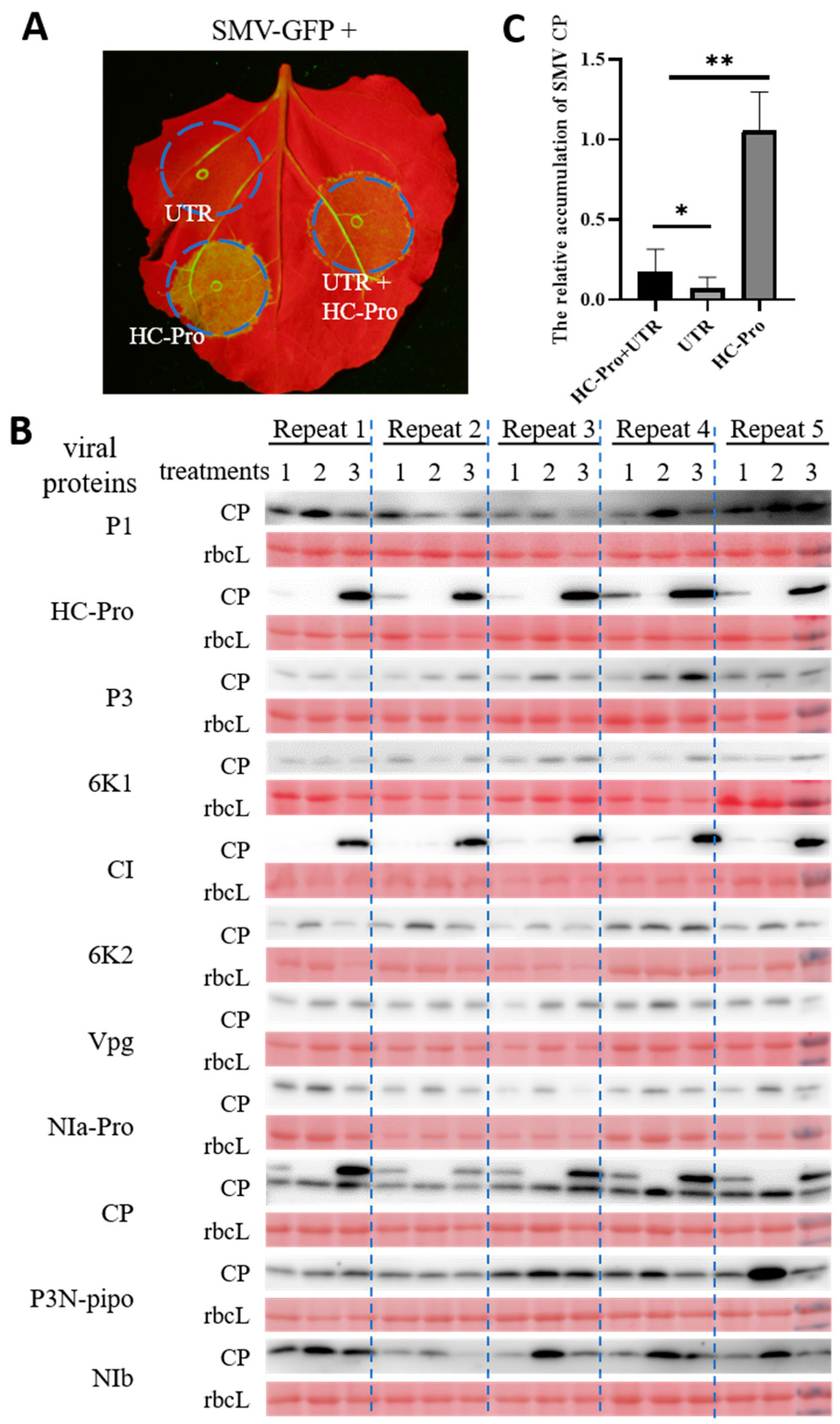
As the UTR-mediated PDR is only effective for the cognate virus, it is easy to speculate that UTR-induced RNA silencing may mediate PDR. In order to check this speculation, the UTR and HC-Pro were co-agroinfiltrated into N. benthamiana leaves along with SMV-GFP (Figure 2A). Although SMV-GFP accumulation was still inhibited by UTR in the presence of HC-Pro (lane 1) when compared with HC-Pro coexpression alone (lane 3), the viral accumulation was significantly higher than the coexpression of UTR alone (lane 2) (Figure 2B,C). These results support the involvement of RNA silencing in UTR-mediated PDR.
In addition to HC-Pro, other potyviral proteins may also encode VSR activities. For example, TuMV Vpg was identified as the VSR, which functions by degrading the gene-silencing components SGS3 and RDR6 [39]. The wheat streak mosaic virus (genus Tritimovirus, family Potyviridae) P1 and the second copy of the P1 of the cucumber vein yellowing virus (genus Ipomovirus, family Potyviridae) also function as VSRs [40,41,42]. We decided to check if more cistrons could compromise the UTR-mediated PDR to SMV. We coexpressed each cistron of SMV in the presence of UTR, similar to those experiments performed with HC-Pro (Figure 2A). However, none of these cistrons compromised the UTR-mediated PDR (Figure 2B and Supplementary Figure S2).

3.3. RNA Silencing Repressor Relieves the UTR-Mediated PDR

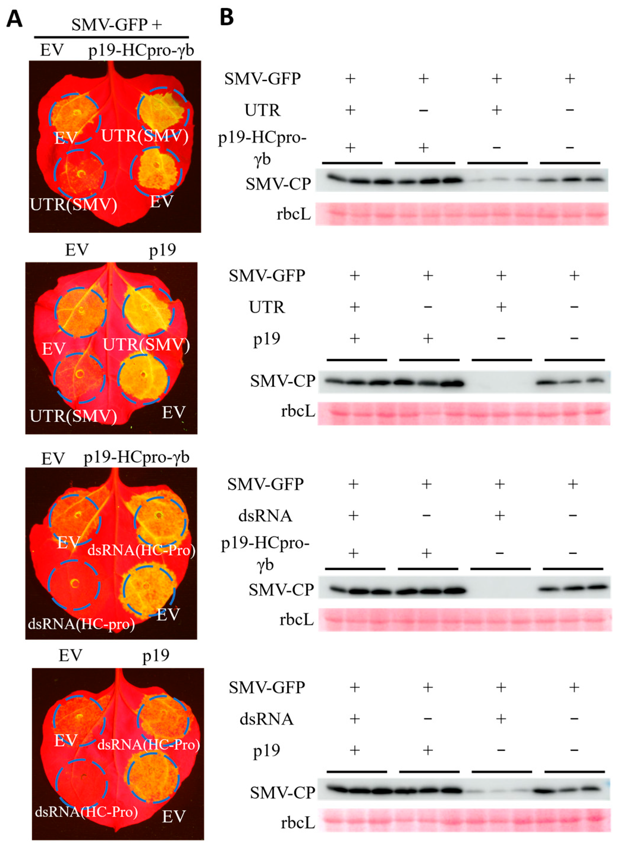
We performed another set of experiments using other VSRs to confirm that RNA silencing is required for UTR-mediated PDR. The Agrobacterium carrying SMV-GFP (OD600 = 0.1) was co-agroinfiltrated with SMV-derived UTR (OD600 = 0.1) together with a plasmid expressing three VSRs (tomato bushy stunt virus p19, tobacco etch virus HC-Pro, and barley stripe mosaic virus γb) or the tomato bushy stunt virus VSR p19 alone (OD600 = 0.8). We found that the coexpression of three VSRs or p19 significantly compromised UTR-mediated PDR as observed by GFP fluorescence (Figure 3A) or detected by Western blotting (Figure 3B).
Further, we replaced the UTR with hairpin RNA-derived dsRNA (OD600 = 0.1), targeting the HC-Pro region of the SMV genome [43], and tested the role of VSRs in dsRNA-mediated SMV suppression. Remarkably, the results obtained with dsRNA were similar to those obtained with UTR (Figure 3). Thus, the above experiments suggest that the suppression of viral infection by UTR is mainly conferred by RNA silencing.

3.4. All the Transiently Expressed Viral Cistrons Repress the Infection of SMV

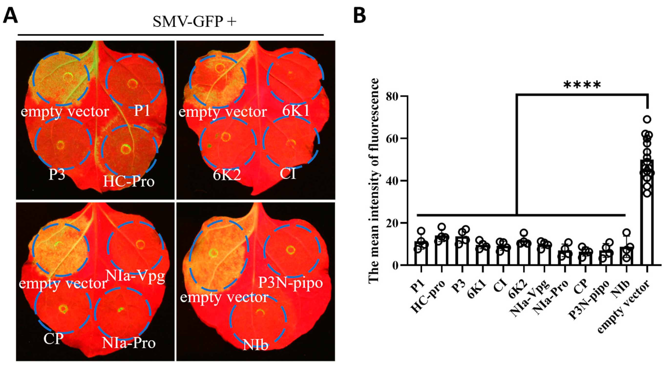
To identify which protein encoded by SMV could potentially be used for PDR, vectors transiently expressing different viral cistrons (OD600 = 0.9) were co-agroinfiltrated with SMV-GFP infectious clones (OD600 = 0.1) into N. benthamiana leaves. Seven days after infiltration, tissues co-agroinfiltrated with an empty vector exhibited bright fluorescence (Figure 4A), indicating efficient infection of SMV-GFP. Conversely, tissues co-agroinfiltrated with any of the viral protein-encoding cistrons displayed a lower GFP signal (Figure 4A,B). These results suggest that all the viral cistrons could confer PDR.

3.5. PDRs Conferred by Different Cistrons Vary in Mechanism

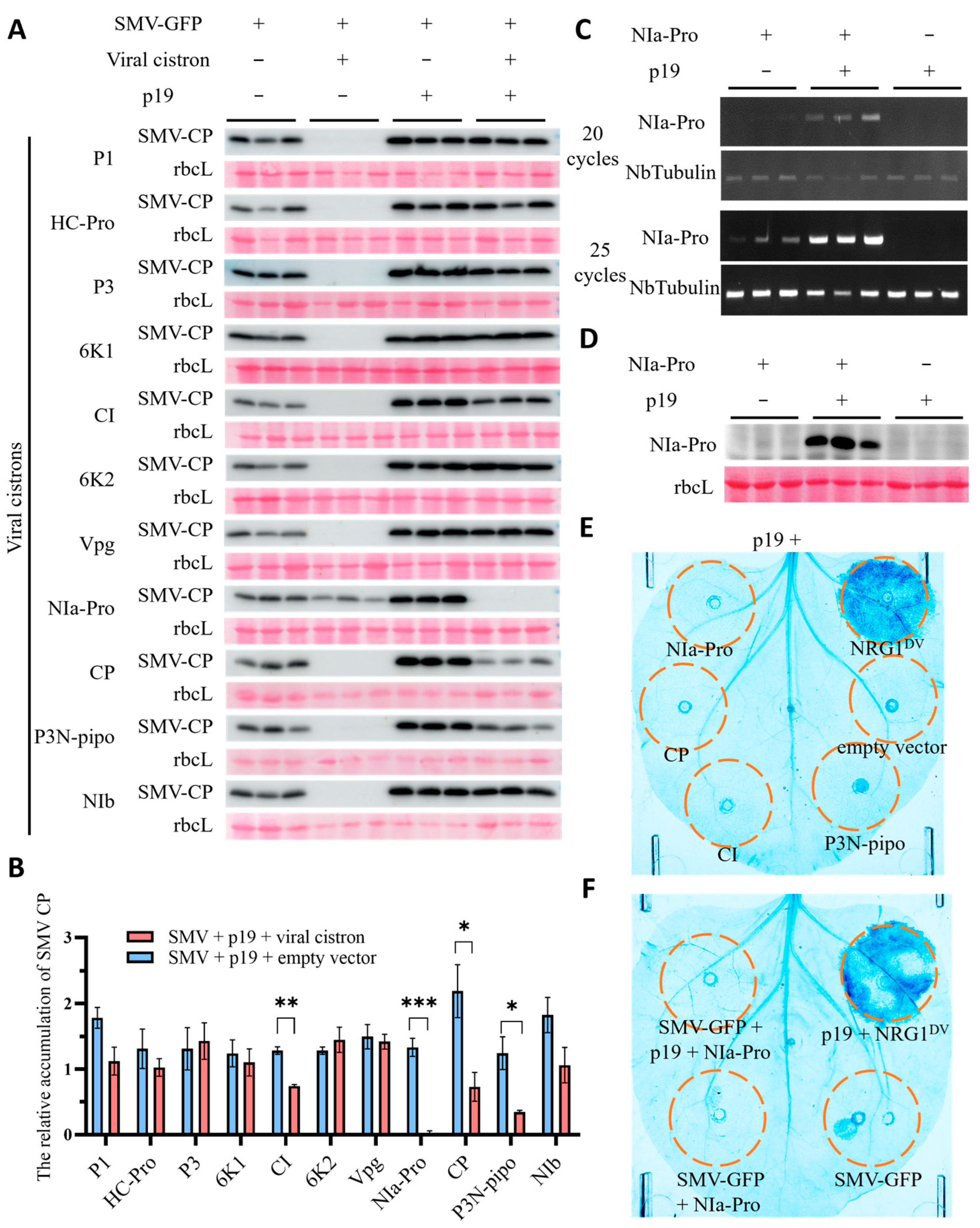
To determine if these viral cistrons function similarly to UTR by inducing RNA silencing to suppress SMV, we coexpressed the VSR p19 with each of the viral cistrons and SMV-GFP to assess whether p19 could alleviate the repression conferred by these viral cistrons. To better observe the effect of p19 in regulating PDR, we coinfiltrated the Agrobacterium harboring p19 at an OD600 of 0.8 and reduced the OD600 of Agrobacterium carrying viral cistrons to 0.1. Subsequent observations revealed that p19 significantly weakened the virus repression conferred by most cistrons (P1, HC-Pro, P3, 6K1, 6K2, VPg, and NIb) at 7 dpi (Figure 5A,B, Supplementary Figure S3). NIa-pro was less effective in SMV repression than in the previous experiment (Figure 4) in the absence of p19, likely due to the reduced OD600 of Agrobacterium carrying the NIa-Pro expression vector in this experiment. However, interestingly, NIa-Pro exhibited more pronounced repression of SMV when coexpressed with p19 (Figure 5A,B). Furthermore, CP, P3N-PIPO, and CI-mediated repression were not entirely abolished by p19. RT-PCR and Western blot analysis revealed an elevated mRNA and protein abundance of NIa-Pro induced by p19 at 4 dpi (Figure 5C,D). In order to exclude the possibility that the repression of virus accumulation was triggered by viral proteins-triggered robust immune responses, such as programmed cell death, trypan blue staining was carried out to show the level of cell death. The leaves agroinfiltrated with NRG1DV [44], an auto-activating plant immune receptor, were used as a positive control. The result showed that overexpression of NIa-Pro, CP, P3N-PIPO, and CI in the presence of p19 did not trigger robust immune responses such as programmed cell death (Figure 5E). Moreover, no cell death was observed when p19, NIa-Pro, and SMV-GFP were co-agroinfiltrated (Figure 5F).
These results suggest that the virus repression conferred by most cistrons may be mediated by RNA silencing. Meanwhile, NIa-Pro, CP, P3N-PIPO, and CI also repress SMV in their protein forms, as p19 did not fully abolish the virus repression.
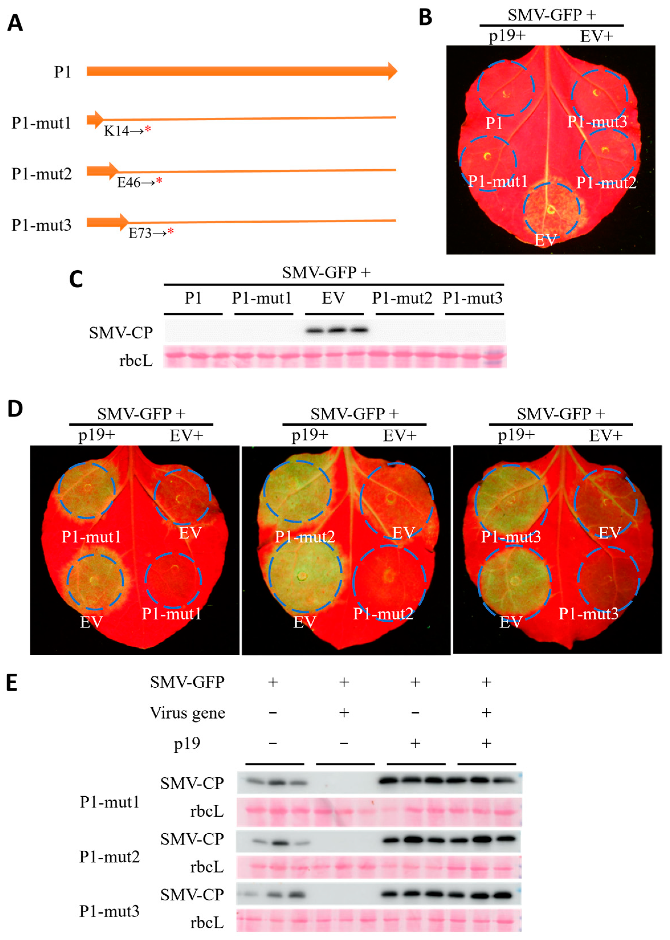
To further discern the involvement of expressed viral RNA in SMV repression, we selected the P1 cistron for further investigation. Stop codons were introduced at various positions in the P1 coding sequence, resulting in mutants translated into non-functional short peptides comprising 13, 45, or 72 amino acid residues (Figure 6A). Surprisingly, the expression of these P1 constructs retained high SMV repression activity, similar to the wild-type P1 (Figure 6B,C). Additionally, co-agroinfiltration of p19 with these mutants demonstrated a significant reduction in SMV repression activity (Figure 6D,E), similar to p19 alleviating dsRNA-mediated virus repression (Figure 3).
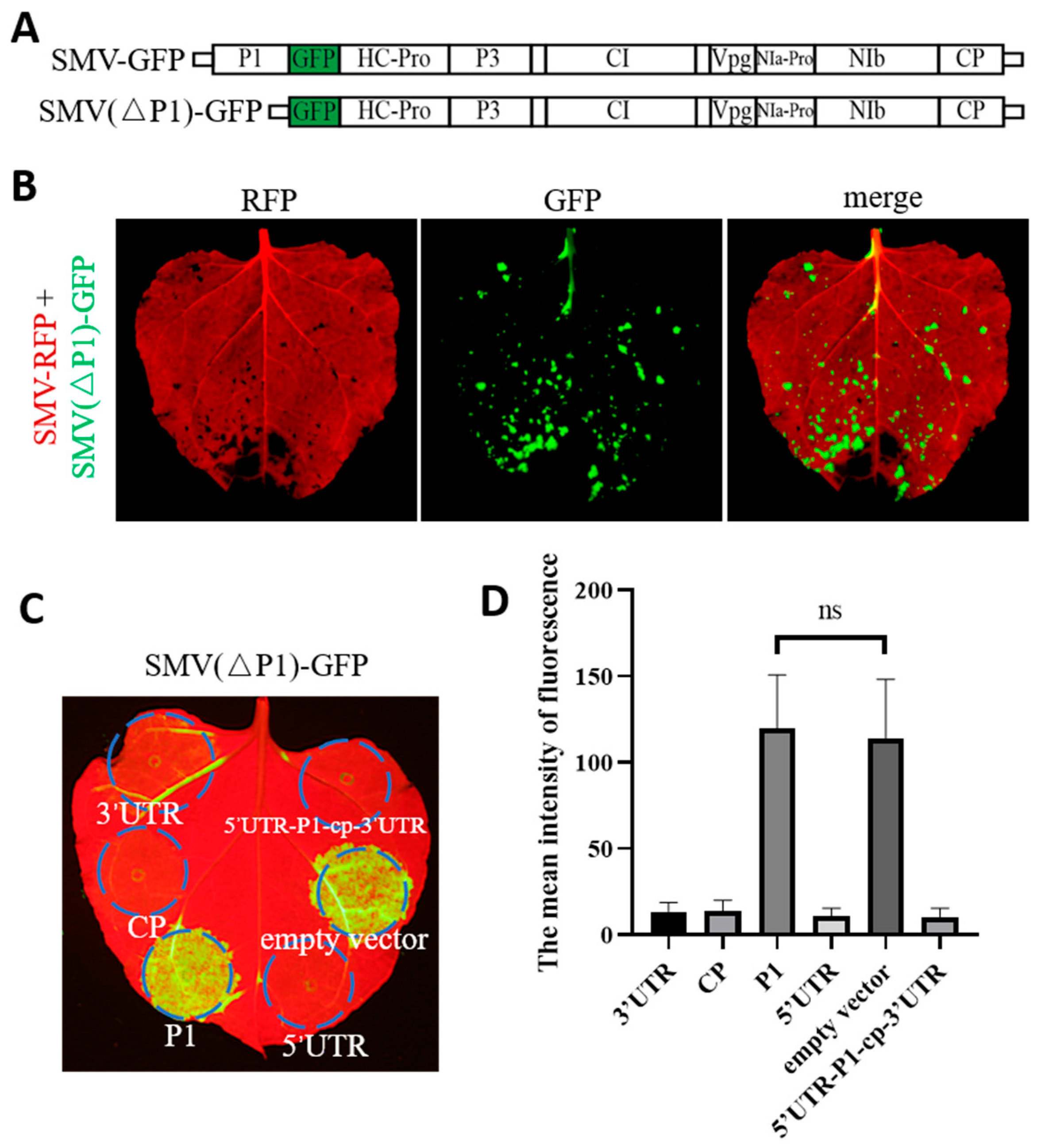
Previous reports on potyviruses have indicated that the P1 cistron can be deleted from the viral genome without completely compromising infection and movement [45,46]. To further assess the role of P1 in SMV repression, we deleted the P1 cistron from SMV-GFP, creating the infectious clone SMV(ΔP1)-GFP (Figure 7A). Co-agroinfiltration of SMV-RFP and SMV(ΔP1)-GFP into N. benthamiana plants resulted in successful systemic infection of both variants (Figure 7B). Further co-agroinfiltration experiments using SMV(ΔP1)-GFP with CP or UTRs showed significant repression of SMV(ΔP1)-GFP infection (Figure 7C), similar to the results observed on SMV-GFP when CP or UTR was expressed (Figure 1 and Figure 4). However, the fluorescence intensity of SMV(ΔP1)-GFP in leaves agroinfiltrated with P1 was consistent with that of the empty vector (Figure 7C,D). These results indicate that P1 could not repress the SMV(ΔP1)-GFP infection due to the lack of P1-coding region within the viral genome. Consequently, our findings strongly suggest that, unlike NIa-Pro, P1-mediated SMV repression occurs through RNA silencing induced by the RNA form of the expressed P1 gene rather than its protein.

4. Discussion

The limitations of innate immune conferred by resistant genes make PDR an appealing alternative. Firstly, the resistance used in breeding is usually qualitative and controlled by one or a few genes. However, this kind of resistance is rare, as most of the resistance is qualitative and controlled by multiple genes [47,48]. When the resistance is controlled by recessive genes, it is also difficult to use in breeding [49]. The selection of recessive genes in the heterozygous state requires the help of molecular markers, which are generally based on accurate gene mapping. Secondly, some resistant genes may be tightly linked to unfavorable traits, which are detrimental to breeding varieties with excellent comprehensive traits. Thirdly, as monogenetic resistance follows a “gene for gene” recognition pattern [50,51], the virus is can easily evade the detection of resistant genes via mutation on the avirulence gene [52,53]. Therefore, this resistance tends to deteriorate after a few years of usage. Furthermore, for some viruses, there are no resistant genes effective enough for resistant breeding [54]. Almost all of the shortcomings of the naturally occurring resistant gene can be overcome by PDR. More than that, PDR targeting multiple viruses can be pyramided by one genetic transformation in 1–2 years. It is also easy to alter the viral gene sequences used for transformation to counteract viral mutations if necessary.
The practice of PDR in molecular breeding has been carried out for decades [25]. Genetically engineered squash simultaneously expressing multiple viral CPs achieved resistance to ZYMV, WMV, and CMV [20]. This genetically engineered squash variety performed well after planting for over 20 years [25]. By introducing the PRSV CP gene into papaya, resistance to PRSV was achieved, and most papaya varieties planted are currently transgenic [25]. The application of these varieties has saved farmers significant losses and reduced the use of insecticides. However, due to public concerns about the safety of genetically modified crops, the application of more cultivars using PDR for virus resistance has not been successful. However, with the increasing understanding of transgenic technology by the public, PDR will undoubtedly continue to contribute greater value to agricultural production.
PDR is a robust concept that can be applied to all plant viruses. While the concept was proposed nearly 40 years ago, previous research has been limited to a small number of viral proteins, such as CP, movement protein, and replicase. The discovery of RNA silencing made it easier to achieve satisfactory resistance with only a part of the viral sequences. Thus, many viral genes are still understudied in the PDR research. We used SMV as the object virus to include all the cistrons in the study. The results showed that the expression of any cistron of SMV can inhibit SMV infection, confirming the effectiveness of the PDR concept.
When PDR was proposed, the existence of post-transcriptional gene silencing (PTGS) had not yet been acknowledged. Researchers observed PTGS based on an experiment regarding PDR on TEV [27]. Meanwhile, with regard to the intact CP gene was involved in the experiment, there was still considerable controversy about whether the protein or RNA mediated PDR [25]. It is also unknown whether the presence of VSR in the viral genome of some viral genus, such as Potyvirus, affects PDR conferred by the RNA silencing mechanism. In this study, the agroinfiltration-mediated transient expression method helped assess the effects of VSR on PDR. The results showed that although SMV has a VSR gene, HC-Pro, which may attenuate PTGS-induced PDR, RNA silencing-mediated PDR is still effective in resistance to SMV for most viral cistrons and UTRs. However, PDR mediated by NIa-Pro appears to not be associated with RNA silencing (Figure 5). This type of PDR may be useful for resistance breeding against viruses with strong VSR. However, it should be noted that PDR mediated by NIa-Pro in this study heavily depended on gene expression levels. Other genes or UTRs can effectively inhibit SMV at an Agrobacterium OD600 of 0.1, whereas the PDR conferred by NIa-Pro is very weak at this concentration.
The overexpression of viral proteins disrupts the processes of the parasite, and the RNA silencing caused by PTGS may not be the entire mechanism of PDR. We found that PDR does seem related to SIE in the experiment on potyviruses [14]. Research on wheat streak mosaic virus (WSMV) and Triticum mosaic virus (TriMV) has identified NIa-Pro and CP proteins as SIE determinants [55]. Another study on TuMV revealed the necessity of P3 and NIa-Pro proteins in conferring the SIE phenotype [56]. In this study, NIa-Pro and CP of SMV also conferred PDR in their protein forms. Unlike the former study, we found that the protein P3N-PIPO might confer PDR instead of P3 (Figure 4), which is regarded as the SIE determinant in TuMV [56]. The intrinsic connection of these results needs to be further explored.

5. Conclusions

This study aimed to find which cistron of the potyvirus genome could be used for PDR, and whether there are different mechanisms of PDR conferred by different cistrons. The results revealed that each cistron of SMV can mediate PDR. Mechanistically, most protein-coding cistrons and UTRs conferred PDR through RNA silencing, but several cistrons (NIa-Pro, CP, P3N-PIPO, and CI) also mediated PDR at the protein level. These results reflect the diversity of the mechanisms of PDR conferred by different viral cistrons and provide valuable insights into the potential applications of PDR in crop protection against SMV. Future research should explore how viral cistrons, such as NIa-Pro, mediate PDR by their protein forms.

Supplementary Materials

The following supporting information can be downloaded at: https://www.mdpi.com/article/10.3390/agronomy14071457/s1, Figure S1. Evaluation of UTR repression ability on cognate viruses of non-potyvirus: title; Figure S2. Relative accumulation analysis of SMV CP; Figure S3. Influence of p19 on viral cistron-mediated repression of SMV; Table S1. Primers used for vector construction. Table S2. Primers used for RT-PCR.

Author Contributions

N.Y.: Investigation, Formal analysis, Writing—original draft. Y.Q.: Investigation, Formal analysis, Writing—original draft. Y.S.: Investigation, Formal analysis, Writing—original draft. K.X.: Conceptualization, Funding acquisition, Methodology, Resources, Writing—review and editing. J.Y.: Conceptualization, Investigation, Formal analysis, Methodology, Writing—original draft, Writing—review and editing. All authors have read and agreed to the published version of the manuscript.

Funding

This research was funded by the National Natural Science Foundation of China (32370158), Jiangsu Province’s Innovation Program (JSSCTD202142).

Data Availability Statement

The original contributions presented in the study are included in the article and Supplementary Material. Further inquiries can be directed to the corresponding author.

Acknowledgments

The authors acknowledge Dawei Li from China Agricultural University for providing the T7 promoter-based TNV-AC infectious clone pMTC27, Xiaoming Zhang from the Institute of Zoology, Chinese Academy of Science, for providing TuMV-GFP infectious clone, and Zhenghe Li from Zhejiang University for providing the p19-HC-Pro-γb plasmid.

Conflicts of Interest

The authors declare no conflicts of interest. The funders had no role in the design of the study; in the collection, analyses, or interpretation of data; in the writing of the manuscript; or in the decision to publish the results.

References

  1. Vincent, S.J.; Coutts, B.A.; Jones, R.A. Effects of introduced and indigenous viruses on native plants: Exploring their disease causing potential at the agro-ecological interface. PLoS ONE 2014, 9, e91224. [Google Scholar] [CrossRef] [PubMed]
  2. Jones, R.A.C. Global Plant Virus Disease Pandemics and Epidemics. Plants 2021, 10, 233. [Google Scholar] [CrossRef] [PubMed]
  3. Lecoq, H. Discovery of the first virus, the tobacco mosaic virus: 1892 or 1898? Comptes Rendus L’academie Sci. Ser. III Sci. 2001, 324, 929–933. [Google Scholar] [CrossRef]
  4. Walker, P.J.; Siddell, S.G.; Lefkowitz, E.J.; Mushegian, A.R.; Adriaenssens, E.M.; Alfenas-Zerbini, P.; Davison, A.J.; Dempsey, D.M.; Dutilh, B.E.; García, M.L. Changes to virus taxonomy and to the International Code of Virus Classification and Nomenclature ratified by the International Committee on Taxonomy of Viruses (2021). Arch. Virol 2021, 166, 2633–2648. [Google Scholar] [CrossRef] [PubMed]
  5. Takahashi, H.; Fukuhara, T.; Kitazawa, H.; Kormelink, R. Virus Latency and the Impact on Plants. Front. Microbiol. 2019, 10, 2764. [Google Scholar] [CrossRef] [PubMed]
  6. Mumford, R.A.; Macarthur, R.; Boonham, N. The role and challenges of new diagnostic technology in plant biosecurity. Food Secur. 2016, 8, 103–109. [Google Scholar] [CrossRef]
  7. Tatineni, S.; Hein, G.L. Plant viruses of agricultural importance: Current and future perspectives of virus disease management strategies. Phytopathology 2023, 113, 117–141. [Google Scholar] [CrossRef] [PubMed]
  8. Folimonova, S.Y. Developing an understanding of cross-protection by Citrus tristeza virus. Front. Microbiol. 2013, 4, 76. [Google Scholar] [CrossRef]
  9. Yeh, S.-D.; Gonsalves, D.; Wang, H.L.; Namba, R.; Chiu, R.J. Control of papaya ringspot virus by cross protection. Plant Dis. 1988, 72, 375–380. [Google Scholar] [CrossRef]
  10. Bohra, A.; Kilian, B.; Sivasankar, S.; Caccamo, M.; Mba, C.; McCouch, S.R.; Varshney, R.K. Reap the crop wild relatives for breeding future crops. Trends Biotechnol. 2022, 40, 412–431. [Google Scholar] [CrossRef]
  11. Hajjar, R.; Hodgkin, T. The use of wild relatives in crop improvement: A survey of developments over the last 20 years. Euphytica 2007, 156, 1–13. [Google Scholar] [CrossRef]
  12. Dwivedi, S.L.; Upadhyaya, H.D.; Stalker, H.T.; Blair, M.W.; Bertioli, D.J.; Nielen, S.; Ortiz, R. Enhancing crop gene pools with beneficial traits using wild relatives. Plant Breed. Rev. 2008, 30, 179–230. [Google Scholar] [CrossRef]
  13. Sanford, J.C.; Johnston, S.A. The concept of parasite-derived resistance—Deriving resistance genes from the parasite’s own genome. J. Theor. Biol. 1985, 113, 395–405. [Google Scholar] [CrossRef]
  14. Abel, P.P.; Nelson, R.S.; De, B.; Hoffmann, N.; Rogers, S.G.; Fraley, R.T.; Beachy, R.N. Delay of disease development in transgenic plants that express the tobacco mosaic virus coat protein gene. Science 1986, 232, 738–743. [Google Scholar] [CrossRef] [PubMed]
  15. Nelson, R.S.; Abel, P.P.; Beachy, R.N. Lesions and virus accumulation in inoculated transgenic tobacco plants expressing the coat protein gene of tobacco mosaic virus. Virology 1987, 158, 126–132. [Google Scholar] [CrossRef] [PubMed]
  16. Register Iii, J.C.; Beachy, R.N. Resistance to TMV in transgenic plants results from interference with an early event in infection. Virology 1988, 166, 524–532. [Google Scholar] [CrossRef] [PubMed]
  17. Osbourn, J.K.; Watts, J.W.; Beachy, R.N.; Wilson, T.M.A. Evidence that nucleocapsid disassembly and a later step in virus replication are inhibited in transgenic tobacco protoplasts expressing TMV coat protein. Virology 1989, 172, 370–373. [Google Scholar] [CrossRef] [PubMed]
  18. Gonsalves, D. Transgenic papaya: Development, release, impact and challenges. Adv. Virus Res. 2006, 67, 317–354. [Google Scholar] [CrossRef] [PubMed]
  19. Scorza, R.; Callahan, A.; Dardick, C.; Ravelonandro, M.; Polak, J.; Malinowski, T.; Zagrai, I.; Cambra, M.; Kamenova, I. Genetic engineering of Plum pox virus resistance: ‘HoneySweet’ plum—From concept to product. Plant Cell Tissue Organ Cult. (PCTOC) 2013, 115, 1–12. [Google Scholar] [CrossRef]
  20. Fuchs, M.; Klas, F.E.; McFerson, J.R.; Gonsalves, D. Transgenic Melon and Squash Expressing Coat Protein Genes of Aphid-borne Viruses do not Assist the Spread of an Aphid Non- transmissible Strain of Cucumber Mosaic Virus in the Field. Transgenic Res. 1998, 7, 449–462. [Google Scholar] [CrossRef]
  21. Arce-Ochoa, J.P.; Dainello, F.; Pike, L.M.; Drews, D. Field performance comparison of two transgenic summer squash hybrids to their parental hybrid line. HortScience 1995, 30, 492–493. [Google Scholar] [CrossRef]
  22. Clough, G.H.; Hamm, P.B. Coat protein transgenic resistance to watermelon mosaic and zucchini yellows mosaic virus in squash and cantaloupe. Plant Dis. 1995, 79, 1107–1109. [Google Scholar] [CrossRef]
  23. Fuchs, M.; Gonsalves, D. Resistance of transgenic hybrid squash ZW-20 expressing the coat protein genes of zucchini yellow mosaic virus and watermelon mosaic virus 2 to mixed infections by both potyviruses. Nat. Biotechnol. 1995, 13, 1466–1473. [Google Scholar] [CrossRef]
  24. Kaniewski, W.; Ilardi, V.; Tomassoli, L.; Mitsky, T.; Layton, J.; Barba, M. Extreme resistance to cucumber mosaic virus (CMV) in transgenic tomato expressing one or two viral coat proteins. Mol. Breed. 1999, 5, 111–119. [Google Scholar] [CrossRef]
  25. Lindbo, J.A.; Falk, B.W. The impact of “coat protein-mediated virus resistance” in applied plant pathology and basic research. Phytopathology 2017, 107, 624–634. [Google Scholar] [CrossRef] [PubMed]
  26. Lindbo, J.A.; Dougherty, W.G. Untranslatable transcripts of the tobacco etch virus coat protein gene sequence can interfere with tobacco etch virus replication in transgenic plants and protoplasts. Virology 1992, 189, 725–733. [Google Scholar] [CrossRef]
  27. Lindbo, J.A.; Silva-Rosales, L.; Proebsting, W.M.; Dougherty, W.G. Induction of a Highly Specific Antiviral State in Transgenic Plants: Implications for Regulation of Gene Expression and Virus Resistance. Plant Cell 1993, 5, 1749–1759. [Google Scholar] [CrossRef] [PubMed]
  28. Galvez, L.C.; Banerjee, J.; Pinar, H.; Mitra, A. Engineered plant virus resistance. Plant Sci. 2014, 228, 11–25. [Google Scholar] [CrossRef] [PubMed]
  29. Latif, M.F.; Tan, J.; Zhang, W.; Yang, W.; Zhuang, T.; Lu, W.; Qiu, Y.; Du, X.; Zhuang, X.; Zhou, T.; et al. Transgenic expression of artificial microRNA targeting soybean mosaic virus P1 gene confers virus resistance in plant. Transgenic Res. 2024, 33, 149–157. [Google Scholar] [CrossRef]
  30. Revers, F.; Garcia, J.A. Molecular biology of potyviruses. Adv. Virus Res. 2015, 92, 101–199. [Google Scholar] [CrossRef]
  31. Dougherty, W.G.; Lindbo, J.A.; Smith, H.A.; Parks, T.D.; Swaney, S.; Proebsting, W.M. RNA-mediated virus resistance in transgenic plants: Exploitation of a cellular pathway possibly involved in RNA degradation. Mol. Plant-Microbe Interact. MPMI 1994, 7, 544–552. [Google Scholar] [CrossRef] [PubMed]
  32. Yin, J.; Hong, X.; Luo, S.; Tan, J.; Zhang, Y.; Qiu, Y.; Latif, M.F.; Gao, T.; Yu, H.; Bai, J.; et al. The Characterization of the Tobacco-Derived Wild Tomato Mosaic Virus by Employing Its Infectious DNA Clone. Biology 2022, 11, 1467. [Google Scholar] [CrossRef] [PubMed]
  33. Xi, D.; Li, J.; Han, C.; Li, D.; Yu, J.; Zhou, X. Complete nucleotide sequence of a new strain of Tobacco necrosis virus A infecting soybean in China and infectivity of its full-length cDNA clone. Virus Genes 2008, 36, 259–266. [Google Scholar] [CrossRef] [PubMed]
  34. Sun, K.; Zhao, D.; Liu, Y.; Huang, C.; Zhang, W.; Li, Z. Rapid Construction of Complex Plant RNA Virus Infectious cDNA Clones for Agroinfection Using a Yeast-E. coli-Agrobacterium Shuttle Vector. Viruses 2017, 9, 332. [Google Scholar] [CrossRef] [PubMed]
  35. Yin, J.; Wang, L.; Jin, T.; Nie, Y.; Liu, H.; Qiu, Y.; Yang, Y.; Li, B.; Zhang, J.; Wang, D.; et al. A cell wall-localized NLR confers resistance to Soybean mosaic virus by recognizing viral-encoded cylindrical inclusion protein. Mol. Plant 2021, 14, 1881–1900. [Google Scholar] [CrossRef] [PubMed]
  36. Fang, X.-D.; Yan, T.; Gao, Q.; Cao, Q.; Gao, D.-M.; Xu, W.-Y.; Zhang, Z.-J.; Ding, Z.-H.; Wang, X.-B. A cytorhabdovirus phosphoprotein forms mobile inclusions trafficked on the actin/ER network for viral RNA synthesis. J. Exp. Bot. 2019, 70, 4049–4062. [Google Scholar] [CrossRef]
  37. Nawaz-ul-Rehman, M.S.; Prasanth, K.R.; Xu, K.; Sasvari, Z.; Kovalev, N.; de Castro Martin, I.F.; Barajas, D.; Risco, C.; Nagy, P.D. Viral Replication Protein Inhibits Cellular Cofilin Actin Depolymerization Factor to Regulate the Actin Network and Promote Viral Replicase Assembly. PLoS Pathog. 2016, 12, e1005440. [Google Scholar] [CrossRef] [PubMed]
  38. Zhang, W.; Qiu, Y.; Zhou, L.; Yin, J.; Wang, L.; Zhi, H.; Xu, K. Development of a Viral RdRp-Assisted Gene Silencing System and Its Application in the Identification of Host Factors of Plant (+)RNA Virus. Front. Microbiol. 2021, 12, 682921. [Google Scholar] [CrossRef] [PubMed]
  39. Cheng, X.; Wang, A. The potyvirus silencing suppressor protein VPg mediates degradation of SGS3 via ubiquitination and autophagy pathways. J. Virol. 2017, 91, 10–1128. [Google Scholar] [CrossRef]
  40. Tatineni, S.; Qu, F.; Li, R.H.; Morris, T.J.; French, R. Triticum mosaic poacevirus enlists P1 rather than HC-Pro to suppress RNA silencing-mediated host defense. Virology 2012, 433, 104–115. [Google Scholar] [CrossRef]
  41. Gupta, A.K.; Tatineni, S. Wheat streak mosaic virus P1 binds to dsRNAs without size and sequence specificity and a GW motif is crucial for suppression of RNA silencing. Viruses 2019, 11, 472. [Google Scholar] [CrossRef] [PubMed]
  42. Valli, A.; Martín-Hernández, A.M.; López-Moya, J.J.; García, J.A. RNA silencing suppression by a second copy of the P1 serine protease of Cucumber vein yellowing ipomovirus, a member of the family Potyviridae that lacks the cysteine protease HCPro. J. Virol. 2006, 80, 10055–10063. [Google Scholar] [CrossRef] [PubMed]
  43. Gao, L.; Ding, X.N.; Li, K.; Liao, W.L.; Zhong, Y.K.; Ren, R.; Liu, Z.T.; Adhimoolam, K.; Zhi, H.J. Characterization of Soybean mosaic virus resistance derived from inverted repeat-SMV-HC-Pro genes in multiple soybean cultivars. Theor. Appl. Genet. 2015, 128, 1489–1505. [Google Scholar] [CrossRef] [PubMed]
  44. Jacob, P.; Kim, N.H.; Wu, F.H.; El Kasmr, F.; Chi, Y.; Walton, W.G.; Furzer, O.J.; Lietzan, A.D.; Sunil, S.; Kempthorn, K.; et al. Plant “helper” immune receptors are Ca2+-permeable nonselective cation channels. Science 2021, 373, 420–425. [Google Scholar] [CrossRef] [PubMed]
  45. Verchot, J.; Carrington, J.C. Evidence that the potyvirus P1 proteinase functions in trans as an accessory factor for genome amplification. J. Virol. 1995, 69, 3668–3674. [Google Scholar] [CrossRef]
  46. Pasin, F.; Simon-Mateo, C.; Garcia, J.A. The hypervariable amino-terminus of P1 protease modulates potyviral replication and host defense responses. PLoS Pathog. 2014, 10, e1003985. [Google Scholar] [CrossRef] [PubMed]
  47. Palloix, A.; Ayme, V.; Moury, B. Durability of plant major resistance genes to pathogens depends on the genetic background, experimental evidence and consequences for breeding strategies. New Phytol. 2009, 183, 190–199. [Google Scholar] [CrossRef] [PubMed]
  48. Vale, F.X.R.; Parlevliet, J.E.; Zambolim, L. Concepts in plant disease resistance. Fitopatol. Bras. 2001, 26, 577–589. [Google Scholar] [CrossRef]
  49. Jiang, G. Molecular markers and marker-assisted breeding in plants. Plant Breed. Lab. Fields 2013, 3, 45–83. [Google Scholar] [CrossRef]
  50. Thompson, J.N.; Burdon, J.J. Gene-for-gene coevolution between plants and parasites. Nature 1992, 360, 121–125. [Google Scholar] [CrossRef]
  51. Van Der Biezen, E.A.; Jones, J.D.G. Plant disease-resistance proteins and the gene-for-gene concept. Trends Biochem. Sci. 1998, 23, 454–456. [Google Scholar] [CrossRef] [PubMed]
  52. García-Arenal, F.; Fraile, A.; Malpica, J.M. Variation and evolution of plant virus populations. Int. Microbiol. 2003, 6, 225–232. [Google Scholar] [CrossRef] [PubMed]
  53. Gallois, J.-L.; Moury, B.; German-Retana, S. Role of the genetic background in resistance to plant viruses. Int. J. Mol. Sci. 2018, 19, 2856. [Google Scholar] [CrossRef] [PubMed]
  54. Gonsalves, D.; Tripathi, S.; Carr, J.B.; Suzuki, J.Y. Papaya ringspot virus. Plant Health Instr. 2010, 10, 1094. [Google Scholar] [CrossRef]
  55. Tatineni, S.; French, R. The Coat Protein and NIa Protease of Two Potyviridae Family Members Independently Confer Superinfection Exclusion. J. Virol. 2016, 90, 10886–10905. [Google Scholar] [CrossRef]
  56. Nunna, H.; Qu, F.; Tatineni, S. P3 and NIa-Pro of Turnip Mosaic Virus Are Independent Elicitors of Superinfection Exclusion. Viruses 2023, 15, 1459. [Google Scholar] [CrossRef]
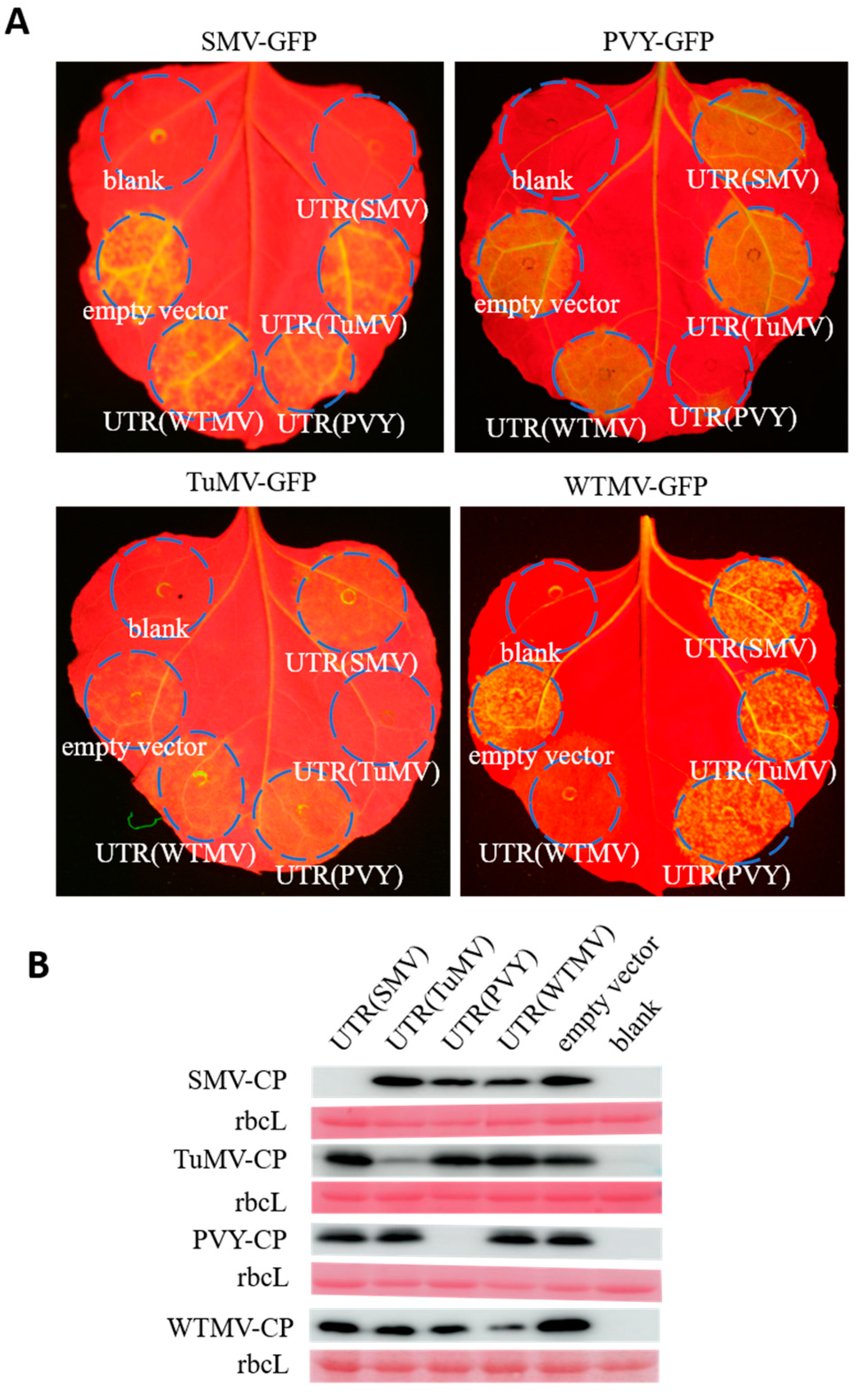
Figure 1. Specific repression of cognate potyviruses infection by UTRs. (A) Co-agroinfiltration of different potyvirus-derived UTRs or an empty vector (OD600 = 0.9) with GFP-tagged virus clones (OD600 = 0.1) into N. benthamiana leaves. Blank represents no expression vector or viral clone that was agroinfiltrated. Photographs were captured seven days post-infiltration under a hand-held LED lamp. (B) Western blot analysis of the samples from panel A. Ponceau S stained rubisco large subunit (rbcL) was used to show protein loading. The experiments were repeated three times with similar results.
Agronomy 14 01457 g001
Figure 2. UTR-mediated repression of SMV specifically attenuated by HC-Pro. (A) Assessment of the influence of HC-Pro on UTR-mediated PDR. Agrobacterium tumefaciens concentrations used were OD600 = 0.1 for all constructs. Photograph was captured under a hand-held LED lamp seven days post-agroinfiltration. (B) Western blot analysis of SMV accumulation in the samples co-agroinfiltrated with viral cistrons and UTR (1), UTR (2), and viral cistrons (3). Ponceau S stained rubisco large subunit (rbcL) was used to show total protein loading. Samples of each treatment on one repeat were taken from the same leaf. In the Western blot of the sample transient expressing CP, two distinct signals are observed: the upper signal corresponds to transiently expressed FLAG-tagged CP, while the lower signal represents CP expressed from the viral genome. (C) Relative accumulation analysis of SMV CP based on the Western blot results in panel B. Relative CP accumulation was determined by computing the ratio of the grey value of CP to the grey value of rubisco large subunit (rbcL). Statistical significance was assessed using one-way ANOVA (* p < 0.05, ** p < 0.01, n = 5). More results can be found in Supplementary Figure S2.
Agronomy 14 01457 g002
Figure 3. Effects of VSRs on UTR-mediated repression of SMV. (A) Assessment of UTR (OD600 = 0.1) repression of SMV (OD600 = 0.1) upon co-agroinfiltration with viral suppressor of RNA silencing (VSR; OD600 = 0.8). The left halves of the leaves were co-agroinfiltrated with an empty vector (pGD-C-FLAG, EV), while p19-HCpro-γb or p19 was co-agroinfiltrated into the other leaf halves. Photographs were captured under a hand-held LED lamp 7 days post-agroinfiltration (dpi). (B) Western blot analysis of the samples from panel (A) to detect CP accumulation of SMV. Ponceau S stained rubisco large subunit (rbcL) was used to show total protein loading. Individual lanes under each treatment represent biological repeats.
Agronomy 14 01457 g003
Figure 4. Effects of individual viral cistrons on SMV repression. (A) Co-agroinfiltration of each SMV cistron (OD600 = 0.9) and SMV-GFP (OD600 = 0.1) into N. benthamiana leaves. Photographs were captured seven days post-infiltration under a hand-held LED lamp. (B) Quantification of GFP fluorescence of leave samples from panel A. Data points indicate distinct biological repeats, with error bars representing standard deviations. Statistical significance was determined via one-way ANOVA analysis (**** p < 0.0001).
Agronomy 14 01457 g004
Figure 5. Western blot analysis of CP accumulation to evaluate the impact of p19 on viral cistron-mediated repression. (A) Ponceau S stained rubisco large subunit (rbcL) was used to show total protein loading. Individual lanes under each treatment represent biological repeats. The corresponding photographs of N. benthamiana leaves can be found in Figure S3. (B) The comparison of SMV CP accumulation based on the Western blot analysis from panel A. Relative CP accumulation was determined by computing the ratio of the grey value of CP to the grey value of rubisco large subunit (rbcL). Statistical significance was assessed using multiple t-test (* p < 0.05, ** p < 0.01, *** p < 0.001, n = 3). The RT-PCR (C) and Western blot (D) analysis to determine the expression of NIa-Pro. The Tubulin of N. benthamiana was used as an internal control in RT-PCR. (E,F) Detection of cell death by trypan blue staining method. NRG1DV was used as the positive control.
Agronomy 14 01457 g005
Figure 6. Effects of P1 cistron mutants on SMV infection. (A) Schematic representation of P1 cistron mutants featuring internal stop codons causing premature translation termination. The codon positions mutated to stop codons (*) are depicted in the diagram. (B) Co-agroinfiltration of P1 cistron mutants (OD600 = 0.1) harboring internal stop codons with SMV-GFP (OD600 = 0.1) into N. benthamiana leaves. EV: empty vector. Photograph captured under a hand-held LED lamp 7 days post-agroinfiltration. (C) Western blot analysis of samples from panel (B) to detect CP accumulation of SMV. Ponceau S stained rubisco large subunit (rbcL) was used to show total protein loading. Individual lanes under each treatment represent biological repeats. (D) Evaluation of the repression exerted by P1 mutants (OD600 = 0.1) on SMV (OD600 = 0.1) when co-agroinfiltrated with the VSR, p19 (OD600 = 0.8). The right half of leaves co-agroinfiltrated with an empty vector (pGD-C-FLAG, EV) serve as controls. (E) Western blot analysis of samples from panel (D). See more details in panel (C).
Agronomy 14 01457 g006
Figure 7. Evaluation of P1 cistron influence on SMV(△P1)-GFP infection. (A) Schematic comparison between SMV-GFP (upper) and SMV(△P1)-GFP (lower) depicting the P1 deletion in the latter. (B) Co-agroinfiltration of SMV(△P1)-GFP and SMV-RFP into N. benthamiana leaves. The photograph was captured under a hand-held LED lamp 14 days post-agroinfiltration. (C) Assessment of the repression exerted by viral cistrons and UTRs (OD600 = 0.9) on SMV(△P1)-GFP (OD600 = 0.1). Photographs were taken under a hand-held LED lamp 7 days post-agroinfiltration. (D) Mean fluorescence intensity analysis of samples from panel C. Statistical significance was assessed by one-way ANOVA (n = 6, ns = no significant).
Agronomy 14 01457 g007
Disclaimer/Publisher’s Note: The statements, opinions and data contained in all publications are solely those of the individual author(s) and contributor(s) and not of MDPI and/or the editor(s). MDPI and/or the editor(s) disclaim responsibility for any injury to people or property resulting from any ideas, methods, instructions or products referred to in the content.