Review

Research Progress on Diseases Caused by the Soil-Borne Fungal Pathogen Rhizoctonia solani in Alfalfa

by
Muhammad Abdullah Akber
and
Xiangling Fang
*
Center for Grassland Microbiome, State Key Laboratory of Herbage Improvement and Grassland Agro-Ecosystems, College of Pastoral Agriculture Science and Technology, Lanzhou University, Lanzhou 730020, China
*
Author to whom correspondence should be addressed.
Agronomy 2024, 14(7), 1483; https://doi.org/10.3390/agronomy14071483 (registering DOI)
Submission received: 1 May 2024 / Revised: 6 June 2024 / Accepted: 29 June 2024 / Published: 9 July 2024
(This article belongs to the Section Grassland and Pasture Science)

Abstract

:
Rhizoctonia solani is a soil-borne fungal species with worldwide distribution and poses serious threats to a wide range of economically important crops such as grain and forage crops. This pathogen has survival capabilities within plants and soil, giving rise to sclerotia and persisting for several months or years. Alfalfa (Medicago sativa) is the most widely grown and important forage crop in China and worldwide. The unique characteristics of alfalfa, such as excellent forage quality, ruminant desirability, and substantial biomass output, distinguish it from other fodder crops, and it is also known as the “Queen of Forages”. However, the production of alfalfa is seriously affected by R. solani, with yield losses of 20% to 60% globally. This review firstly summarizes diseases such as seedling damping-off, root rot, crown rot, root cankers, stem cankers, blight, and stem rot caused by R. solani in alfalfa and the survival mechanism of this pathogen. The techniques for R. solani detection and quantification from plants and soils, as well as management through host resistance, cultural practices, fungicides, and biological control, were then overviewed. This review provides scientific knowledge to enable researchers to efficiently manage R. solani in alfalfa production.

1. Introduction

As we approach 2050, meeting the increasing demand for animal feed is expected to become challenging [1]. However, incorporating forage legume crops into feed production can significantly enhance the quality and availability of fodder [2]. Alfalfa, a perennial forage legume, is one of the most ancient crops, cultivated for over 2000 years and grown in more than 80 countries [3,4]. Currently, it is extensively cultivated and considered the primary source of commercial forage and feed in many countries [1]. Alfalfa is also known as the “Queen of Forage” because of numerous characteristics such as excellent forage quality, palatability for ruminant feed, and high biomass yield [5,6]. The deep root system of alfalfa enables it to absorb water from deep soil layers, and it can be grown in drought areas with only 200 mm of precipitation annually [7,8]. In addition, this crop provides a range of ecosystem services, including enhanced biodiversity and improved soil structure and fertility, as well as benefits such as increased infiltration and flood protection [3,9,10]. Due to all these benefits, alfalfa is being cultivated throughout the dry tropical and temperate regions of the world on approximately 32 million hectares. Its production area in China is 4 million hectares, which ranks first among cultivated grasslands in China [3,9,11]. China is currently the second-largest country for alfalfa cultivation [12]. Moreover, alfalfa plays a crucial role in improving China’s agricultural economic structure by contributing to the expansion of grassland animal husbandry and the sustainability of farming ecosystems [9].
The genus Rhizoctonia was first introduced by de Candole (1815) while identifying an unknown fungus that attacked crops, namely alfalfa (Medicago sativa) and saffron (Crocus sativus). However, Rhizoctonia solani was first described by Kühn in 1858. It is a necrotrophic/saprophytic fungal pathogen that may persist longer in soil and plant parts without a host [5,13,14]. R. solani is pathogenic against approximately 250 host plant species belonging to Moraceae, Poaceae, Solanaceae, Linaceae, Brassicaceae, Fabaceae, Malvaceae, Amaranhtaceae, Rubiaceae, Asteraceae, and Araceae [15]. It is a destructive fungal pathogen and has been reported to cause severe damage and significant yield losses in many economically important crops such as soybean, rice, potato, sugar beet, wheat, and chickpea [14,16,17]. R. solani is a species complex with several related but genetically different isolated groups and has been divided into 14 anastomosis groups (AGs) [18]. AG-2 has been reported as diverse and includes subgroups such as AG-2-1, 2-2 IIIB, 2-2 IV, 2-2 LP, 2-2 WB, 2-3, 2-4, and 2-B1 [19]. However, AG-1, AG-2, AG-3, AG-4, and AG-5 are frequently reported from legume crops [16]. The important diseases caused by R. solani and its subgroups include pre-emergence and post-emergence damping off, root rot, seed rot, hypocotyl rot, cankers, blights, pod rot, limb rot, black scurf, and stem rot [17].
R. solani is associated with serious diseases in alfalfa, including seed rot, seedling damping-off, stem-rot, root rot, root canker, stem canker, crown rots, and blight [20]. In alfalfa, diseases caused by R. solani tend to be more prevalent under elevated temperature and high moisture conditions [21,22]. Indeed, temperature is pivotal in developing R. solani diseases in alfalfa. The literature highlights that the optimum temperature range for these diseases is 21–30 °C [23,24,25,26]. It exhibits a destructive lifestyle that impacts various plant parts, including above-ground (pods, stems, and fruits) and below-ground (roots, hypocotyls, seeds) parts of the alfalfa plant [5,26,27,28,29]. Many studies have been conducted on different aspects of R. solani in other host crops under greenhouse and field conditions. However, limited studies have focused on R. solani in alfalfa (Figure 1), highlighting a major gap in our understanding of this pathogen’s impact on alfalfa yields. Addressing this knowledge gap is essential for developing effective strategies to mitigate yield losses and safeguard alfalfa production. Therefore, this study discusses different alfalfa diseases caused by R. solani and their management approaches, including host resistance, cultural control, biological control, and chemical control. Further, it summarizes the survival structure of R. solani (sclerotia) and its detection and quantification from the rhizosphere and plant.

2. Diseases Caused by R. solani and Worldwide Distribution

2.1. Damping-off

Damping-off disease caused by soil-borne fungal pathogens such as R. solani causes severe yield loss in alfalfa worldwide [29,30,31,32,33]. R. solani is associated with both pre- and post-emergence damping-off diseases of alfalfa; however, post-disease is common [21,34,35]. Addoh [36] described the symptoms associated with the sudden or severe damping-off of alfalfa, such as rapid wilting of plants, tissues becoming water-soaked, and plant death, although the leaves remain attached. However, symptoms in the gradual progression of the disease include brownish stems, gradual stunting of affected parts, and oval-shaped dark brown lesions appearing on the stem. These lesions merge rapidly around the stem and kill the plants. A total of 10 alfalfa genotypes were used against three types of fungus species, including R. solani, Fusarium oxysporum, and Macrophomina. The results showed that R. solani was the most pathogenic, followed by Macrophomina phaseolina and F. oxysporum. In addition, R. solani significantly reduced fresh and dry shoot yield compared to the other two fungus species [34]. In a study conducted in Ismailia Governorate, observations were made regarding fungal diseases affecting alfalfa crops. The collected data revealed that damping-off caused by R. solani was a predominant disease. The percentages of different fungus species for causing pre- and post-emergence damping-off were as follows: R. solani (70.0%, highest), Sclerotium rolfsii (31.25%), F. solani (28.75), and M. phaseolina (17.5%, lowest) [28].

2.2. Root Rot

Root rot disease accounts for 20–40% of the global annual yield loss of alfalfa [37]. Root rot caused by soil-borne fungal pathogens has been frequently reported in major alfalfa-growing regions of China such as northeast China (Jilin, Heilongjiang, and Liaoning), northwest China (Shaanxi, Ningxia Huizu, Gansu, Xinjiang, and Qinghai), and north China (Hebei, Inner Mongolia, and Shanxi). Moreover, in northwest China, alfalfa root rot is severe and has a disease incidence of 30–80% [5]. R. solani has been reported as one of the primary causative agents for root rot disease in alfalfa [5,6,38,39]. McKenzie and Davidson [40] surveyed 2-year-old alfalfa plants from 40 fields for root diseases. R. solani was identified as a causal organism of root rot. They also described the symptoms of alfalfa root rot caused by R. solani as brown to black lesions on root surfaces near the place of lateral root emergence from a primary root. In 1972, Fatemi first reported alfalfa root rot diseases caused by R. solani in the Fars province of Iran and reported the stunted growth of alfalfa plants during the early stages of root disease [41]. Different R. solani AGs are associated with severe root rot diseases of alfalfa in countries, such as AG-1, AG-2, AG-3, AG-4, AG-5, and AG-10 reported in Turkey [42], AG-1 and AG-4 in America [43], AG-1–AG-10 in Saudi Arabia, AG-11, AG-8, and AG-6 in Australia [44], and AG-4 in Iran [45].

2.3. Root Canker

Root canker in alfalfa caused severe losses in southwestern Queensland, Australia, and the Imperial and Palo Verdi Valleys of California [46,47]. Moreover, R. solani AG-6 was reported to cause root canker in all Australian commercial cultivars of alfalfa [48]. In another study, fourteen isolates of R. solani showing black canker symptoms were collected from alfalfa roots. It was concluded from the results that disease-causing strains were related to R. solani AG-4 [49]. Smith [23] identified the root canker disease of alfalfa caused by R. solani in the USA for the first time. The root canker of alfalfa is characterized by elliptical, sunken, and necrotic canker development at the junction of taproots and lateral roots. Hence, plant roots are girdled by these cankers, and plants can die from this disease [47]. Samac et al. [26] indicated that plant death occurs upon the complete invasion of roots by lesions; otherwise, roots can emerge again during fall and support the plant for the following year. Moreover, lesions usually heal in the winter season and turn blackish. The occurrence of root canker in Rhizoctonia is seasonal and strongly influenced by elevated temperatures. The disease’s typical lesions mostly appeared in June, July, August, and September, when temperatures in the soil varied between 21 to 35 °C. In winter, when soil temperatures were between 5 to 10 °C, diseases did not appear [23].

2.4. Crown Rot

The causal organisms of alfalfa crown rot are complex soil-borne fungi and can vary according to geographic location. R. solani is the frequently reported causal organism of crown rot disease in alfalfa [50]. R. solani enters the crown through cut stems and mechanical damage to the crown and root during multiple foliage cuttings [51]. Graham et al. [52] reported that R. solani crown rot is more severe in alfalfa after the second year of growth. In northern Mexico, plants showing crown rot symptoms were collected from 30 fields of alfalfa crops. The results identified nematodes, fungi, and oomycetes as causal organisms, and R. solani was the primary fungus causing crown rot in alfalfa [53]. Another study by Samac et al. [50] revealed that alfalfa plants affected by crown rot become stunted, wilt, and finally die. R. solani damages new shoots, and plants become weaker and more susceptible to attack by saprophytic fungi or worthless soil parasites [54].

2.5. Crown Bud Rot

R. solani has been associated with crown bud rot in forage legumes, including alfalfa [27]. Hawn and Cormack [55] found that R. solani, Fusarium acuminatum, and Fusarium avenaceum are involved in the alfalfa crown bud rot disease complex; however, R. solani was highly pathogenic. In addition, Hwang et al. [56] reported that plant age is a critical factor for this disease. Alfalfa crown bud rot resulted in dark brown to blackish lesions in tissues [52]. R. solani damages crown buds, causing bud killing and moving to the crown, stopping the new buds’ vegetative production. It was also reported that R. solani ramified in alfalfa crown buds inter- and intracellularly [57]. McDonald [58] revealed that R. solani is a vital pathogen that produces crown bud rot problems in alfalfa during summer. Moreover, crown bud rot caused by R. solani is severe during the first month of growth, and plants that are 2 or 3 years old are more susceptible.

2.6. Stem Canker

Stem canker in alfalfa, caused by specific strains of R. solani, is a notable plant disease. Houston [59] showed that stem canker predominantly affects 1–2-year-old alfalfa fields, with higher incidence rates observed during spring, early summer, and fall. Stem canker symptoms include brown-edged, tan cankers that occasionally produce circular shapes resembling growth rings. Brown mycelial strands are usually visible on the canker’s outer layer upon enlargement. Cankers girdle stems, which eventually die and resemble anthracnose-killed stems. The lack of setae in the acervuli of Rhizoctonia stem canker growths separates them from anthracnose stem cankers [26]. R. solani affects alfalfa stem tissues below the soil surface, with cankers extending up to 2.54 cm above the soil line post-infection [59].

2.7. Blights

R. solani-induced blight diseases in alfalfa, extensively documented in the literature [17,27,43,52,60], are particularly prevalent in the southeastern United States [56]. This pathogen persistently targets alfalfa foliage and stems, resulting in rapid tissue collapse and the formation of water-soaked masses, often mistaken for scalding water damage. Diseased plants can rapidly infect adjacent healthy plants. Large and irregularly shaped lesions are observed on less damaged stems and leaves [52,56]. Table 1 summarizes the disease severity induced by R. solani and associated yield reduction (%) in alfalfa, as documented in previous studies.

2.8. Global Distribution

R. solani has affected alfalfa crops across the globe. Alfalfa diseases caused by R. solani have been reported in many countries, including China, Saudi Arabia, Japan, Egypt, Turkey, and Italy. Moreover, in some countries, significant yield losses have been reported by R. solani diseases. For instance, alfalfa root rot and damping-off are the most prevalent in Egypt, root rot is the most common in China, and damping-off is the most prevalent in KSA (Saudi Arabia). Yield losses because of these diseases are reported to be up to 60%, 20–40%, and 26.45% in Egypt [28], China [68], and KSA [69], respectively (Figure 2).

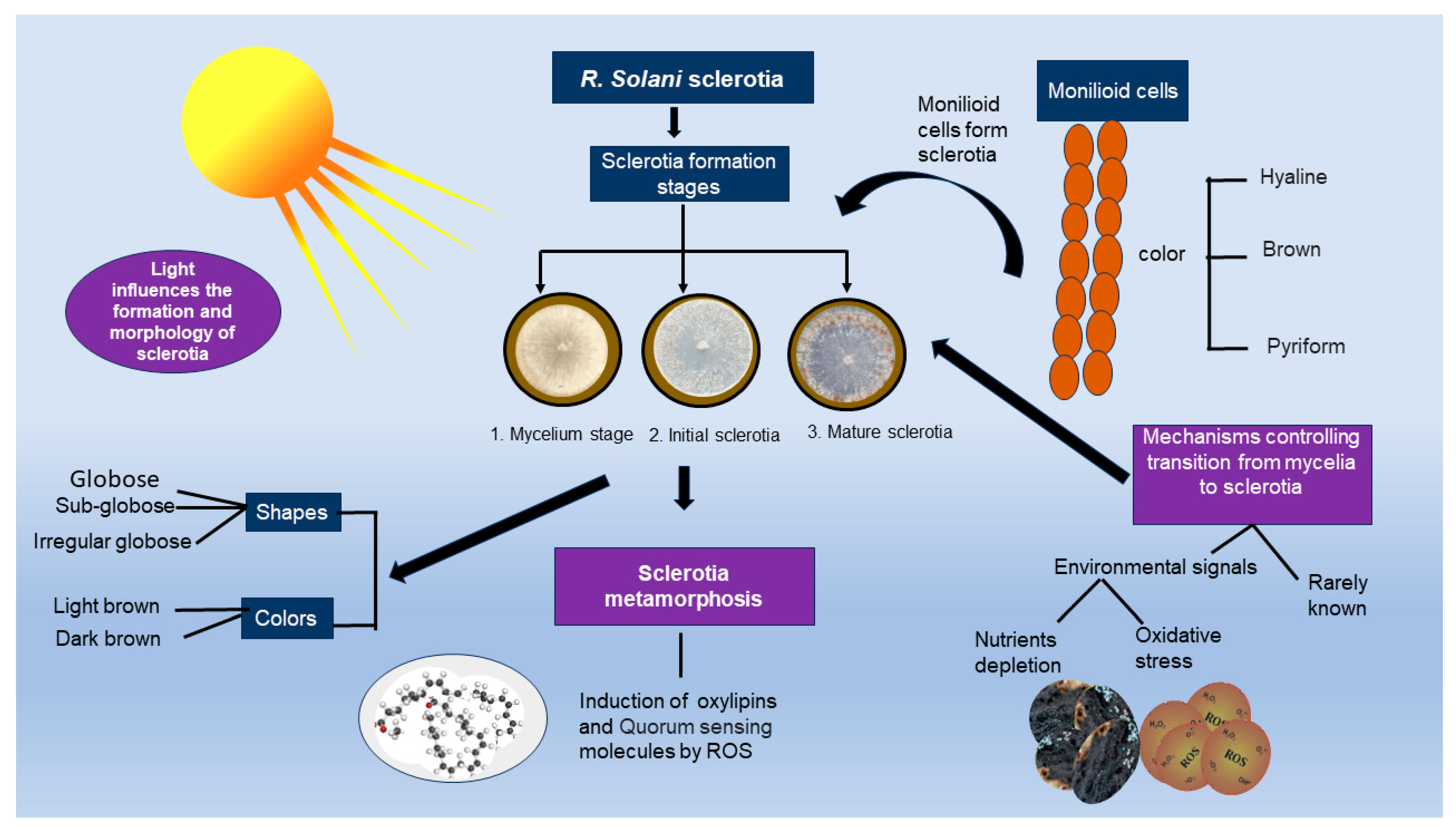
3. Survival Structure of R. solani

R. solani develops sclerotia to survive in soil and plant debris. Sclerotia, known as “nutrient-independent propagules”, persist in soil and crop residues [70]. Sclerotia formation is initiated by undifferentiated hyphae or monilioid cells. These small and ovular cells make chains or clusters. Monilioid cells are hyaline or brown with 30–35 μm and 20–22 μm average length and width, respectively. The chain formation of monilioid cells has been observed on the substrate surfaces and host tissues [71]. Upon germination, sclerotia form mycelia and increase the pathogen’s inoculum density, resulting in disease spread. In most cases, R. solani infection occurs with sclerotia germination to form mycelia that grow toward the host plant [17]. Sclerotia have excellent resistance against unfavorable conditions such as extreme cold or heat and specific chemicals [70]. Sclerotia have three developmental stages: initiation, development, and maturation. Moreover, sclerotia change color (typically light and deep brown) during each developmental stage. Distinct color formation in sclerotia is due to the chemical composition of sclerotia exudates [72]. Aeration and humidity levels are essential factors for sclerotia formation. Light significantly influences sclerotia morphology and formation, and a few isolates of R. solani could not produce sclerotia in dark conditions [73]. Sclerotia can be distinguished into four types: (i) loose, (ii) terminal, (iii) lateral-chained, and (iv) lateral-simple. However, the “loose” type is the most abundant form of R. solani sclerotia [70].
Different studies have reported different periods of R. solani survival, as described below. Pots containing infested soil with R. solani isolates were buried in a field for 40 weeks. Results showed that isolates of AG-1 were recoverable after 86, 211, and 283 days. Isolates of AG-2-1 and AG-2-2 were recovered after 283 days [74]. Similarly, a recent study has reported that R. solani can survive in soil or plant debris as sclerotia for many years [75]. In another study, R. solani sclerotia were buried at a 5 cm depth in field soil and found to be viable after two years of burial with 25–50% germination [76]. The sclerotia of other Rhizoctonia species have different survival capabilities than R. solani. For instance, sclerotia produced by Rhizoctonia cerealis can survive for six months [77]. Sclerotia produced by Rhizoctonia tuliparum survived for ten years; however, the germination percentage after retrieval was <10% [78]. Environmental factors such as water potential, pH, temperature, and fungus nutrition significantly impact the growth and disease development of R. solani [79].
Proteins related to cell defense, amino acid metabolism, genetic information processing, and carbohydrate metabolism may play a role in sclerotia formation and maturation [80]. Regardless of earlier genomic and metabolomics research, the mechanisms regulating R. solani’s transformation from mycelia to sclerotia remain unexplained. The shift from mycelia to monilioid cells and the subsequent development of sclerotia may be associated with several signals, including oxidative stress, nutritional shortage, and other factors that have not yet been studied, such as QS mechanisms [81]. Previous research investigating the survival rates of R. solani sclerotia in alfalfa fields is lacking. As discussed earlier, sclerotia have a crucial role in R. solani disease dissemination; future studies should explore the formation process of sclerotia in alfalfa fields affected by different R. solani diseases. These investigations should focus on unraveling the longevity of sclerotia and understanding how the age of sclerotia influences the severity of alfalfa diseases. Additionally, it is essential to assess the impact of environmental factors on sclerotia survival rates (Figure 3).

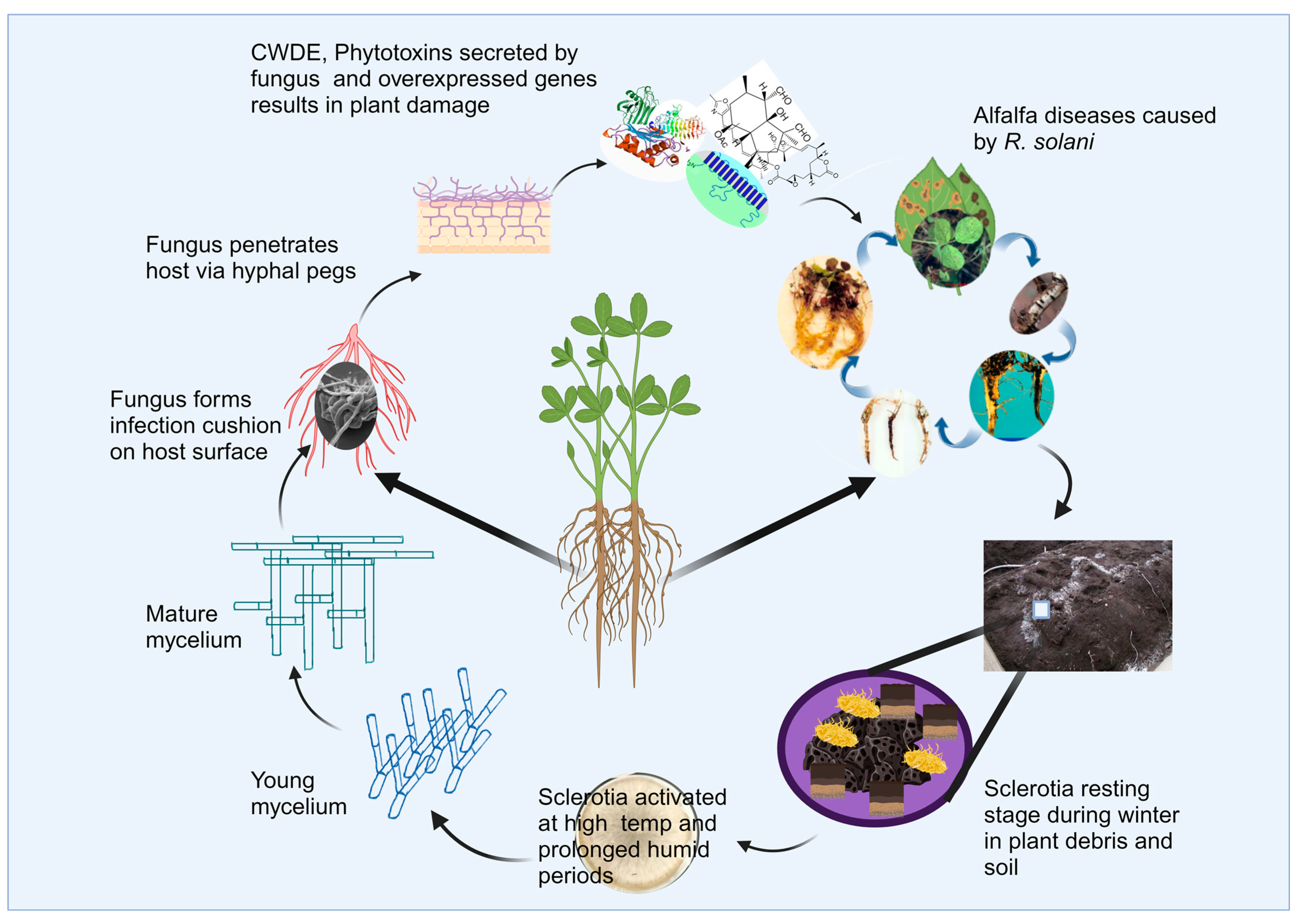
Disease Cycle of R. solani in Alfalfa

Under optimal conditions, such as warm temperatures of around 30 °C and high humidity, dormant sclerotia of R. solani become activated, initiating mycelium growth. This mycelium forms infection cushions, facilitating the penetration of alfalfa taproots through wounds created by young lateral roots. Mycelium then progresses into the cortex region, which induces decay, leading to the sloughing off of cortex tissues [82]. As the infection advances, visible symptoms begin to appear in the alfalfa plants, both above and below ground. Above-ground symptoms may include wilting, stunting, and yellowing of foliage, while below-ground symptoms involve root decay and discoloration. In severe cases, the extent of fungal attack can lead to the death of the alfalfa plants. Following plant death, R. solani can produce new sclerotia on infected alfalfa plant debris, completing the disease cycle. The sclerotia can persist in the soil, serving as a source of inoculum for future infections in subsequent growing seasons (Figure 4).

4. Genetic Diversity and Pathogenic Mechanism of R. solani

4.1. Genetic Diversity

The genetic variability among populations of R. solani is an essential aspect that enhances its potential to cause disease and adapt to various environmental stress conditions and hosts. Researchers have examined the genetic makeup of R. solani populations using multiple molecular markers, including randomly amplified polymorphic DNA (RAPD) markers, restriction fragment length polymorphisms, analysis of sequence variations in ribosomal DNA (rDNA), β-tubulin genes, inter-simple sequence repeats, amplified fragment length polymorphisms, single-nucleotide polymorphisms, and simple sequence repeats [83,84,85,86,87,88,89,90,91]. Significant pathogenicity and variations in genetic composition have been seen in R. solani isolates collected worldwide [92]. Taheri et al. [87] used amplified fragment length polymorphism markers to categorize 150 Indian isolates of R. solani into 33 groups, with a similarity of 80%. Ali et al. [93] classified 29 isolates from Bangladesh into two separate clusters, while Moni et al. [94] grouped 18 isolates into four clusters. However, there was no substantial correlation between the difference in pathogenicity and the genetic groupings found using random amplified polymorphic DNA (RAPD) markers [95]. The internal transcribed spacer (ITS) regions of rDNA have also been demonstrated to be effective for identifying the genetic diversity of AG-1-IA isolates from soybean, AG-1-IB from lettuce, AG-2-1 from cauliflower, and AG-3 from potato [96].
The genome size of R. solani was estimated to range from 36.9 to 42.5 Mb, with 11 chromosomes varying in size from 0.6 to 6 Mb [97]. Later research included the release of a draft genome sequence for the R. solani AG-1-IA strain, which was determined to be 36.94 Mb using advanced next-generation sequencing technologies [98]. Further studies led to the generation of another draft genome sequence for the R. solani AG-1-IA strain, 1802/KB, which was isolated from a widely cultivated rice variety in Malaysia and had a genome size of 28.92 Mb [99]. A detailed study conducted on 38 R. solani isolates from sugar beet and dry bean fields in western Nebraska showed greater morpho-genetic variation. The present study discovered no connection between the isolates of R. solani and their geographical location. It indicates that variables other than geographic distribution have a role in the genetic variation of R. solani in host crops [100]. The isolates from red cabbage were distinguished based on their anastomosis groups by utilizing the retrotransposon-based iPBS (inter priming binding site) amplification DNA profiling method. Pathogenicity tests demonstrated significant variation in disease severity index among the isolates [101]. Another study conducted on 175 isolates of AG-1-IA, which causes rice sheath blight in China, reported a higher degree of haplotype diversity and nucleotide diversity within AG-1-IA. The majority (97.8%) of genetic diversity was found within isolates of populations, while only a tiny portion (2.2%) of the genetic diversity was due to differences between populations. Pathogenicity experiments showed that all isolates caused disease; however, considerable differences were observed in the aggressiveness [96]. Research conducted on different crops worldwide has shown the complex genetics of R. solani. Therefore, understanding the genetic diversity of R. solani populations associated with alfalfa is crucial to facilitate breeding programs for successful disease management.

4.2. Pathogenic Mechanism of R. solani

Bioactive compounds such as toxins, enzymes, and secreted proteins play a major part in the infection process of this pathogen [102]. Notably, toxic substances such as succinic acid, PAA (phenylacetic acid), and furancarboxylic acid have been isolated from R. solani [103]. Substances such as PAA and 3-Methylthiopropionic Acid (MTPA) produced by R. solani AG-3 PT induce cellular destruction, including the breakdown of membranes, the alteration of chloroplast shape, and the swelling of the endoplasmic reticulum [102,104]. In addition, secondary metabolite formation enzymes, such as non-ribosomal peptide synthases (NRPSs), polyketide synthases (PKSs), hybrid NRPS-PKS enzymes, prenyltransferases (DMATSs), and terpene cyclases (TCs), enhance the fungus’ ability to cause disease [105]. R. solani synthesizes a diverse range of carbohydrate-active enzymes (CAZymes) that degrade the cell walls of plants and exhibit considerable activity during the progression of diseases [106]. R. solani AG-1-IA produces 223 carbohydrate-active enzymes (CAZymes), along with an array of cell wall degrading enzymes (CWDEs) such as pectinase, xylanase, and laccase. These enzymes are linked to the pathogenic and saprophytic characteristics of the fungus [98]. In addition, numerous disease-causing fungi secrete proteins that have various functions in triggering disease [107]. These proteins, commonly referred to as effectors, have the dual function of promoting infection and inhibiting the defense mechanisms of the host [108]. The ratio of these effectors differs among fungi, and R. solani AG-1-IA was reported to produce 965 secretory proteins, many of which are still poorly characterized regarding their roles [98]. The effector AGLIP1, present in R. solani AG-1-IA, has been observed to disrupt plants’ defense mechanisms and facilitate disease progression [109]. The primary processes by which R. solani causes disease in alfalfa plants are not well known, particularly in terms of whether specific toxins or enzymes cause the damage. Future studies on the pathogenic mechanism at the molecular level would address a knowledge gap and contribute to the development of appropriate disease management strategies.

5. Detection and Quantification of R. solani

Several methods have been used to quantify R. solani populations. These methods include baiting, screening, plating soil, and direct observation [110,111]. In the baiting method, seeds, plants, and toothpicks are used for fungal colonization. The toothpick-baiting method is widely acknowledged because it is cost-effective and rapid [111]. Moreover, other methods, including dry sieving, elutriation, and wet sieving, have been used to isolate R. solani from concentrated organic fractions of the soil [112]. R. solani produces several diseases in different alfalfa-growing countries. However, we found a significant research gap in the detection and quantification of R. solani from alfalfa. AGs of R. solani have distinct host ranges and different virulence capabilities. The ITS (internal transcribed spacer region) is a crucial barcode for identifying each R. solani AG. We encourage researchers to isolate R. solani from alfalfa-infected plants and soil, confirm pathogenicity using Koch’s Postulates, and identify specific AGs responsible for disease using PCR techniques. We have discussed PCR, qPCR, and selective mediums for detecting R. solani from other hosts to provide valuable insights for future studies on alfalfa.

5.1. Selective Mediums

Due to its low population levels, R. solani is often hard to isolate from soil with the dilution plating method [111]. Recovery of R. solani is also limited to symptomatic plant parts such as roots [112]. A selective medium for isolating R. solani is crucial for research and diagnostic purposes [113]. Some studies have developed effective selective media for this purpose. For example, a study found that a selective medium consisting of Malt Extract Agar (MA) supplemented with gallic acid (400 μg mL−1) and fosetyl-Al (250 μg mL−1) successfully isolated R. solani from soil samples, even when competing fungi such as Macrophomina phaseolina and other soil-borne fungal species were present [114]. Another agar medium containing Dexon, chloramphenicol, streptomycin, gallic acid, and sodium nitrate recovered 90 to 100% of R. solani from the soil and suppressed the growth of other microbes [115]. Recently, six different growth media, including cornmeal agar (CMA), 10% potato dextrose agar (PDA), 50% PDA, water agar (WA), amended clarified V8 (ACV8), and methylene-benomyl-vancomycin (MBV), were evaluated for promoting R. solani growth. However, V8 (ACV8) was the most effective medium for culturing R. solani [116]. In conclusion, it is crucial to prioritize research aimed at refining selective media formulations for improved detection and isolation of R. solani from host plant alfalfa.

5.2. Conventional PCR

R. solani is a destructive soil-borne pathogen with a vast host range and AG groups. There are a lot of similarities among different AG groups of R. solani, making its detection by morphology and isolation challenging [117]. However, molecular techniques such as PCR have been successfully used for R. solani detection and quantification [118]. Protocols are available for detecting specific AGs, including AG-1, AG-2, and AG-3, providing insights into the losses caused by each AG and management options for respective diseases [119]. This method involves the amplification of 5.8S ribosomal DNA and part of the ITS regions using the designed primers in combination with the general fungal primers ITS1F and ITS4B [120]. This method has been used to detect R. solani in various crops, including legumes such as common beans, cowpeas, and chickpeas. A study used and validated a conventional PCR-based assay to detect R. solani AG-1-IA from rice plants. Four sets of reverse and forward primers were used, and the specificity of the primer sets was checked using the primer BLAST tool. Conventional PCR was compared with a LAMP-based assay to develop a simple strategy for on-field pathogen detection. Results showed that traditional PCR was highly sensitive. However, instrumentation and lack of visual confirmation make conventional PCR unsuitable for on-field pathogen detection [121]. To find a reliable detection method for R. solani AG-3 from the soil, PCR assays were combined with the baiting method given by Thornton. The conventional primer set (Rs1F2 and Rs2R1) used in the study was designed from the ITS1 and ITS2 regions of R. solani. The conventional PCR sensitivity was proved suitable for detecting R. solani AG-3 from tuber tissues and plants. The study’s findings also reported that conventional PCR sensitivity could be improved using a second-round nested PCR approach [122]. In another recent work, conventional PCR assays were developed to detect and quantify R. solani responsible for wet rot in pulse crops [123].

5.3. Real-Time Quantitative PCR (qPCR)

qPCR is a sensitive and rapid method with several advantages over traditional diagnostic methods, including being more time-efficient. Unlike conventional PCR, this advanced method can monitor the amplification of DNA molecules during PCR in real-time [124]. Moreover, qPCR has been reported to detect and quantify pathogens isolates from soil and plant parts in small quantities [125]. Fluorescent dyes such as SYBR Green I or sequence-specific DNA probes such as the TaqMan probe monitor reactions throughout the amplification steps [126]. SYBR Green dye is a cheaper monitoring agent; however, its drawback is that it is non-specific. Therefore, during the PCR cycles, it can bind with any DNA, leading to false positive results in pathogen quantification [127]. TaqMan is the most widely used probe out of all probe-based detection methods. Unlike SYBR Green dyes, probes have high specificity, making them more popular for qPCR methods [128].
qPCR has been implemented to detect and quantify many fungal species, such as R. solani [129,130]. For example, conventional and real-time PCR assays were designed to quantify and diagnose R. solani in pulses. Five R. solani-specific primers (ARSF1&R1, ARSF2&R2, ARSF3&R3, ARSF4&R4, and ARSF5&R5) were designed based on the ITS sequences of R. solani isolates from pulse crops. Results revealed that Rt-PCR was specific and sensitive for rapidly detecting and quantifying pathogens in mung beans. Moreover, Rt-PCR detected low levels of genomic DNA from infected plants and did not amplify other soil-borne pathogens [131]. Rt-qPCR assays were developed to isolate the R. solani AGs in soil collected from the Brassica oleracea field. PCR assays were designed by targeting the β-tubulin and ITS regions for different AGs, including AG-1-IA, AG-1-IC, AG-2-1, AG-2-2, AG-4HGI+II, AG-4HGIII, AG-8, AG-3, AG-4HGII, AG-5, and AG-9. Results reported all assays as target group-specific except AG-2-2 [132]. Rt-PCR has also been reported to increase the possibility of a false negative result when detecting pathogens (especially when examining emerging or extremely variable pathogens) [133]. Despite the successful application of qPCR in diagnosing R. solani across various economically important crops (Table 2), a notable gap exists regarding its implementation for detecting and quantifying R. solani in alfalfa. We propose the utilization of qPCR assays to diagnose R. solani in alfalfa plant tissues and field soil to enhance the management of this pathogen.
Although DNA-based methods enable quick pathogen detection, they have some limitations. It has been reported that PCR methods, including conventional and real-time qPCR methods, cannot differentiate between living and dead material. These assays work based on the detection of nucleic acids rather than living cells [134]. Hence, pathogen detection based on DNA can give false results because DNA can exist for a long time, even after the death of the fungus propagules [135]. Therefore, PCR amplification can overestimate the actual number of viable cells. However, consideration of some other practices can help to overcome these issues. For instance, for the detection of fungal species, the use of non-membrane-permeating dyes such as Propidium monoazide (PMA) with PCR-based tools has been reported to reduce the overestimation of cell counts due to the DNA of dead cells [136]. A study reported that including the baiting step before PCR proved helpful for the sensitive and specific detection of living propagules of R. solani AG-3 [137]. Other approaches to minimize the detection of dead cells in pathogen detection include the combination of qPCR with cultures (nutritive media) and using mRNA to indicate viable cells [134].
Table 2. qPCR assays used for Rhizoctonia solani detection and quantification in different crops.
CropAGPrimer and SequenceTargeted RegionAnnealing
T (°C)
PurposeRef.
Wheat and barleyAG-2-1 and AG-8Rs2.1/8F(GTTGTAGCTGGCCCATTCATTTG)ITS1 and ITS263Detection and quantification of Rhizoctonia species from plant and soil[138]
Rs2.1/8R (AGCAGGTGTGAAGCTGCAAAAG)
AG-8Rs8F(GGGGGAATTTATTCATTTATTGGAC)58
Rs8R (GGTGTGAAGCTGCAAAAG)
AG-10Rs10F (GTAGCTGGCCTCTTAATTTG)60
Rs10R (CAAGTGTGAACCTGCAAGAC)
RiceAG-1-IARs1F(GCCTTTTCTACCTTAATTTGGCAG)
Rs2R(GTGTGTAAATTAAGTAGACAGCAAATG)
ITS60Detection and quantification of R. solani AG-1 IA from plant[139]
Mung bean


Soy
bean
AG-1, AG-2-2, AG-2-2LP,
AG-2-3, AG-3, AG-4, AG-5


unknown
ARS (F1: GAGTTGTTGCTGGCCTTTTC)
ARS (R1: TTTTTACGGGTGTCCTCAGC)
ARS (F4: CAACGGATCTCTTGGCTCTC)
ARS (R4: GGTGTCCTCGGCGATAGATA
ARS (F5: ACTAAGTTTCAACAACGGAT)
ARS (R5: TTACTTTGAAGATTTCATGA)
Rso1: RsolF-(GTGAACCAAATCAGACAGA)
Rso1R-(CTACTCTACTGCTTACAG)
ITS


IGS
71

67

52

45.2
Detection and quantification of R. solani from plant


Detection and quantification of R. solani from soil and plant
[131]


[140]
TomatounknownST-RS1-F: (AGTGTTATGCTTGGTTCCACT)
ST-RS1-R: (TCCTCCGCTTATTGATATGC)
ITS259.5Detection and quantification of R. solani from soil[141]
PotatoAG-1-IAAG-1-1A_F(TTGTTGCTGGCCTTTTCTACCT)
AG-1-A_R (ATGGAATTAAATCCACCAACTATTGC)
ITS150Detection of pathogenic AGs and spatial distribution of R. solani in fields[132]
AG-3AG-3_F (TCTACAGGGATTCCAGATTACGC)
AG-3_R (TCACGGATCTTGGAAATCAACA)
β-tubulin
Lettuce


Sugar beet
AG-1-IB


AG-2-2 IIIB
AG1-IB-F3(TGGCCTTTTAACATTGGCATGT)
AG1-IB-R(CCAACCCCAAAGGACCTTGA)

AG22sp2-F(TAGCTGGATCCATTAGTTTG)
5.8SKhot-R(GTTCAAAGATCGATGATTCAC)
ITS


ITS
62


55
Detection and quantification of R. solani and R. solani AG1-IB from soil and plant
Detection and quantification of R. solani AG2-2IIIB from soil and plant
[142]


[143]
TobaccoAG-3RsTqF1(AGAGTTTGGTTGTAGCTGGTCTATTT)
RsTqR4(AGACAGAAGGGTTCAATGACTTATTATA)
ITS60Detection and quantification of R. solani AG-3 from plant[144]

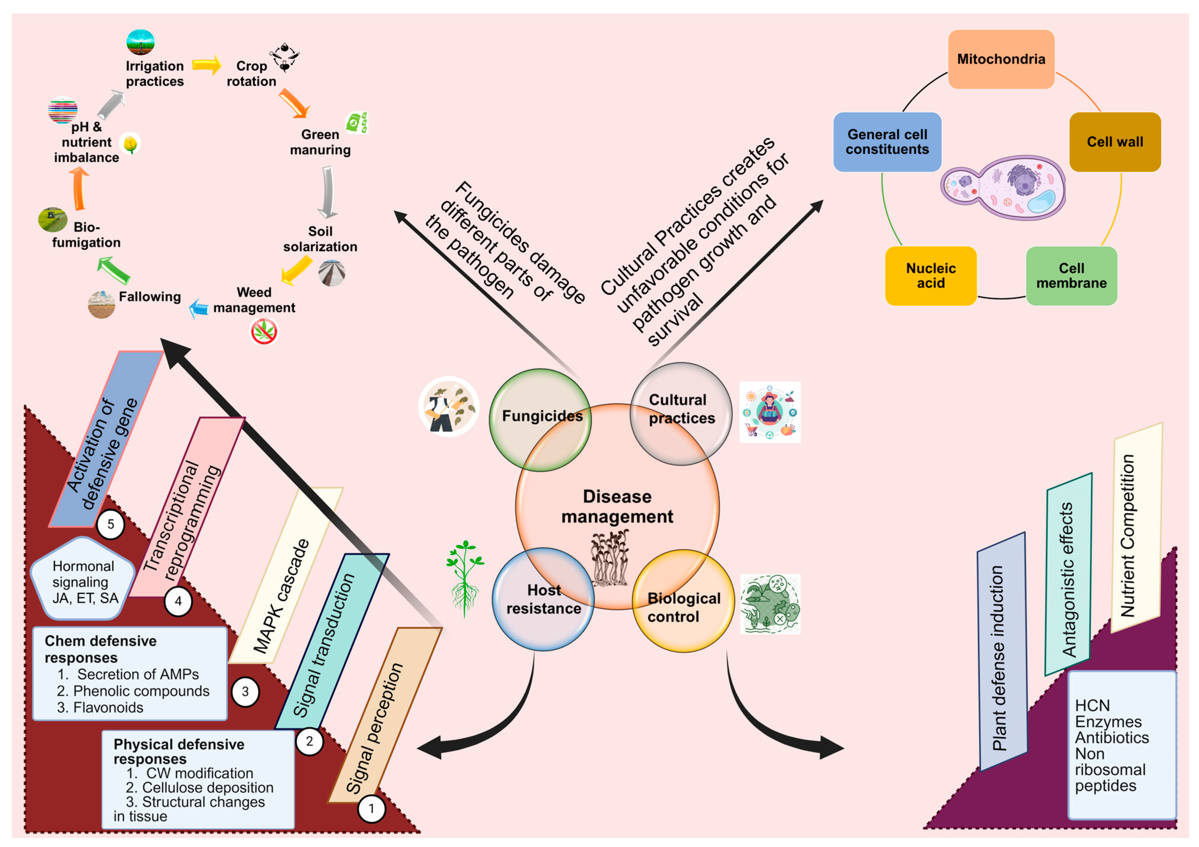
6. Management Approaches

6.1. Host Resistance

Cultivating varieties with disease resistance is the most sustainable and effective way to control alfalfa diseases [145]. Zhang et al. [6] investigated the resistance potential of 68 alfalfa varieties with different worldwide geographic origins against R. solani infection. Only three varieties, including Gannong 9, Trifecta, and Common, displayed a high level of resistance, with the disease indices of shoots and roots, as well as reductions in the dry weight of shoots and roots, all being less than 40%. Some resistant varieties cannot perform well in soils with more than one soil-borne pathogen [14,146]. For example, a recent study revealed that the co-infection of R. solani and F. oxysporum is more destructive to plant growth than a single infection. Moreover, the varieties that are resistant to a single pathogen become susceptible to co-infection, emphasizing the importance of developing alfalfa varieties with resistance against multiple soil-borne pathogens [5]. Three varieties, namely Siwa, Salt America, and New Salt, have been reported as highly resistant to damping-off disease caused by R. solani [34]. Alfalfa genotypes have been checked for resistance against damping-off caused by R. solani. Saponin treatment significantly reduced the mycelial growth of the pathogen. Roots of all genotypes had different saponin and lignin contents. Moreover, it was concluded that breeding for saponin concentration in the alfalfa crop could increase resistance against R. solani diseases such as root rot and damping-off [29]. Several molecular techniques, including CRISPR technology, pyramiding major R genes/QTLs, and host-induced gene silencing, have been used to increase plant resistance to many fungal diseases [147,148,149,150]. Bulked Segregation Analysis (BSA) and Random Amplified Polymorphism (RAPD) were used to examine molecular markers associated with genes that confer resistance toward alfalfa brown spots [151]. As reported from other host–pathogen interactions, implementing the above modern-day technologies might reduce R. solani diseases in alfalfa.
Although R. solani caused significant damage to crop plants, little is known about the host defense responses upon fungus attack. Due to increasing economic losses of legume crops such as alfalfa, chickpeas, and soybeans caused by soil-borne diseases, Medicago truncatula, a model legume plant, has been widely studied for its interactions with legume-infecting strains of R. solani [152]. M. truncatula provides a wide range of genetic and genomic resources, such as a sequenced genome, transformation methods, recombinant inbred lines, and reverse genetics populations, making it an ideal host for studying plant–microbe interactions [153]. It has been reported that different regions of the M. truncatula genome are responsible for its resistance or susceptibility against R. solani strains [152]. Multiple germplasm lines of M. truncatula were screened against R. solani isolates collected from legume fields. The results declared greater differences in susceptibility and resistance, with a majority of genotypes exhibiting susceptibility to R. solani. A17, a reference genotype of M. truncatula, exhibited moderate resistance against the root canker-causing isolate AG6; however, recombinant inbred line population analysis found a single locus producing resistance to R. solani [44]. A study reported that overexpression of IX ERF genes in the roots of a model legume increased resistance against R. solani [154]. Ethylene signaling was a key factor contributing to M. truncatula resistance to R. solani isolates, including AG-8 and legume-specific AG-11 [155]. RNA sequencing of moderately resistant (A17) and highly susceptible (skl) genotypes of a model legume was carried out to unravel the ethylene mechanisms for mediating resistance against R. solani AG-8. Several transcriptional changes were observed in A17, such as ethylene signaling, ROS metabolism, and isoflavonoid biosynthesis. Mass spectrometry detected increased levels of isoflavonoid-related compounds such as liquiritigenin, formononetin, medicarpin, and biochanin A in A17. Overexpressing an isoflavone synthase in M. truncatula roots led to higher isoflavonoid accumulation and resistance against R. solani. Additionally, adding exogenous medicarpin indicated that this phytoalexin might be essential among various isoflavonoids for resistance to R. solani [152]. Given the genomic similarities between alfalfa and M. truncatula, insights gained from studying R. solani interactions in this model legume should be extended to alfalfa to explore resistance mechanisms.

6.2. Cultural Control

Cultural control can reduce soil-borne diseases by creating unfavorable conditions for pathogen growth and survival [73]. Many cultural methods such as crop rotation, mixed cropping, cover cropping, green manuring, bio-fumigation, and pH and nutrient imbalance have been used to manage R. solani diseases in forage legumes [56,156,157,158]. Crop rotations contribute to disease prevention by increasing overall microbial populations that compete with pathogens, reducing inoculum levels when the host is absent, producing toxic compounds that directly inhibit pathogens, and enhancing specific antagonists that combat pathogens [159,160]. Implementing rotation practices has been reported to reduce the severity of R. solani diseases in many important crops [161,162,163,164,165]. The efficacy of crop rotation and green manure treatments in reducing R. solani diseases in alfalfa has been demonstrated. These cultural practices were employed to examine their effect on streptomycete activity and suppress the alfalfa damping-off caused by pathogen complexes including R. solani. Green manuring treatments consisted of canola, sorghum-Sudan grass, buckwheat, and a fallow control. Crop sequences were potato-alfalfa, alfalfa-alfalfa, and corn-alfalfa. Results indicated that crop rotations increased alfalfa yield, and incorporation of green manures resulted in enhanced streptomycete activities and suppressed damping-off. Finally, the data suggested that green manures and crop rotation together can improve crop yield and suppress multiple pathogen complexes by increasing the antagonistic activity within streptomycete [166]. Perennial crops such as alfalfa are reported to increase soil-borne pathogenic populations such as R. solani and allow pathogen multiplication. Therefore, it is recommended that the alfalfa field be rotated with other crops that are not susceptible to R. solani.
Soil pH is an essential factor associated with plant pathogens, and altering the pH levels can reduce pathogenic diseases [156]. It has been proposed that soil pH directly influences the infection and development of plant diseases by affecting soil-borne pathogens [167,168]. Increasing the pH level is recommended for managing soil-borne diseases because a direct correlation has been observed between higher pH levels and lower disease severity caused by fungal pathogens [168,169]. The literature demonstrates that soil pH significantly influences crop diseases caused by R. solani. For instance, a study on sugar beet seedlings affected by R. solani damping-off showed that maximum biomass was produced at a pH near 6.0. In contrast, the minimum biomass was observed at a pH below 5.0 or above 7.0 [170]. Barbetti [171] reported that manipulating pH through the addition of lime influenced the alfalfa root rot complex caused by Fusarium sp., R. solani, and Pythium sp. However, lime application in alfalfa should be further investigated for its influence on various R. solani diseases. A combination of sanitation and bio-fumigation can reduce the survival capabilities of fungal pathogens’ survival structures. However, it may be challenging for farmers to bear production losses from bio-fumigation [172]. Placing seeds deeper in the soil should be avoided because it causes late emergence, which increases the chances of pathogen infection. In addition, avoiding dense planting helps reduce disease spread [173]. Inorganic fertilizer application timings and quantity are critical factors for disease establishment. Suitable nutrition and excellent plant vigor have been reported to reduce crown bud rot caused by R. solani in alfalfa [174]. Over-seeding has been recommended to compensate for the seedling stand losses of alfalfa caused by R. solani seedling diseases [175]. Early cutting is suggested to manage alfalfa foliar blight caused by R. solani [56].

6.3. Biological Control

Beneficial microbes, including bacteria and fungi, have been widely used to improve growth, manage diseases, and enhance plant yield [176]. These microbes release antifungal compounds to suppress pathogens, which disrupt the pathogen’s DNA, cellulose, hemicelluloses, and proteins [177]. Previous studies have investigated the biological control of R. solani in alfalfa. For example, streptomyces reduced disease severity through seed inoculation and/or soil infestation [178]. Similarly, microorganisms belonging to Trichoderma sp., Penicillium sp., and Aspergillus sp. have been suggested for managing Rhizoctonia diseases in alfalfa [179]. In a study conducted by Alsohim [180], it was determined that Pseudomonas fluorescens and its mutants 52-M12 and 45-M19 are effective against R. solani and improved various plant growth parameters, including root weight, root length, and shoot weight, as well as plant fresh and dry biomass. The observed disease suppression effect of these bioagents was attributed to their ability to produce antimicrobial substances. Furthermore, the study highlighted the substantial reduction in R. solani-induced damages in alfalfa fields upon application of these bioagents. Seed treatment with Bacillus amiloliquefacaciens and Paenibacillus polymyxa improved seed germination and increased the fresh and dry weights to 21% [63].
In another study, PGPRs, including Bacillus subtilis, Pseudomonas putida, Pseudomonas fluorescens, Paenibacillus polymyxa, and Sinorhizobium meliloti, were checked against pre- and post-damping-off disease complexes. All four PGPR species produced HCN, siderophore, IAA, solubilized insoluble phosphate, and β-1,3-glucanase activities; however, S. meliloti only produced solubilized insoluble phosphate and IAA. Seed treatment with PGPR significantly reduced damping-off disease in field and greenhouse experiments. Seed treatment with a mixture of PGPR strains and S. meliloti resulted in increased nodules, plant height, dry weight, protein content, and tillers/m2 under field conditions [181]. Additionally, the present study proved that biocontrol agents not only protect alfalfa from R. solani attack but can also help alfalfa plants produce resistance against disease complexes caused by R. solani and other soil-borne fungal pathogens. Moreover, unlike identifying bacterial species based on previous time-consuming and tedious phenotyping approaches, 16S rRNA is an accurate and reliable method for identifying several bacterial strains nowadays [182]. 16S rRNA can identify different types of bacteria, including slow- and fast-growth bacteria and rare and novel species [183]. Therefore, researchers are encouraged to isolate and identify the maximum number of bacterial species to investigate their potential in controlling R. solani alfalfa disease. Antimicrobial peptides (AMPs) are small, soluble molecules involved in plant defense mechanisms. They can inhibit the growth of pathogenic microorganisms by permeabilizing their cell membranes and inactivating intracellular targets within the cytoplasm [184]. The potential of AMPs has been explored against R. solani diseases in economically important crops. For instance, an antimicrobial peptide, AsR416, effectively inhibited the growth of AG1-IA sclerotia, successfully delayed the infection process, and reduced the disease severity of rice sheath blight in vitro and in vivo [185]. Some other studies [186,187] on soybeans and rice have also added similar findings for the effectiveness of different AMPs, including Purothionin, cecropin B, D4E1, Potato snakin-1, and phor21, against R. solani. Considering the need for sustainable farming, AMPs are better alternatives for managing R. solani diseases and improving alfalfa production.

6.4. Fungicides

Chemical control is the predominant method for managing pathogenic diseases in alfalfa and other crops [188]. Alternative options, such as resistant cultivars, often exhibit only partial resistance against pathogens, making them less practical within farming systems [189]. Seed treatment with fungicides containing metalaxyl and fludioxonil has been evaluated against seedling blight and root rot diseases caused by R. solani. Fludioxonil, alone or in combination with metalaxyl, improves seedling survival and reduces root rot under controlled conditions. However, the impact of seed treatment on forage yield under field conditions is limited. Additionally, metalaxyl alone proves ineffective under field and controlled conditions [62]. Berg et al. [190] documented the effectiveness of Stamina, containing the active ingredient pyraclostrobin, in combatting R. solani seedling diseases in alfalfa. Pyraclostrobin stops mitochondrial respiration in fungal cells, preventing energy synthesis for fungal growth and development [191]. Earlier research has also explored fungicide applications in alfalfa cultivation; for example, Hancock [192] observed increased alfalfa yield following the application of benomyl. Ahmad [193] found that seed dressing with Benlate T (a combination of benomyl and Thiram) improved germination rates and plant survival in alfalfa grown in R. solani-infested soil. Furthermore, Rhizolex (Tolclofos-methyl) is effective for controlling alfalfa disease complexes caused by pathogens such as R. solani, Colletotrichum trifolii, Fusarium incarnatum, and Fusarium equiseti [63,65].
Recently, fungicides with different modes of action have been used collectively to control alfalfa soil-borne diseases. For instance, seed treatment with fludioxonil (phenylpyrrole group) and difenoconazole (trizol group) in a 1:4 ratio prevented seedling and rot root diseases caused by Bipolaris sorokiniana. Difenoconazole reduces the participation of the enzyme C14-demethylase in ergosterol production by soil-borne pathogens, whereas fludioxonil stops glucose phosphorylation [194]. We suggest that more trials should be conducted to evaluate the fungicides mentioned above, and others have reported sound results against R. solani diseases. However, high dependency on fungicides has some limitations. Seed treatment with fungicides can suppress endophytes, which protect plants from several pathogens and play an essential role in plant growth. Overdoses of fungicides have also been reported to affect plant metabolism [195]. Moreover, some fungicides are very effective against a single pathogen; however, sometimes, many pathogens are involved in a particular disease. Using a mixture of fungicides could produce better effects on pathogen complexes, but the cost may be difficult for some farmers [196]. Generally, due to its lack of spore production, R. solani is supposed to develop less resistance against different fungicides. However, repeated use of chemicals and remarkable genetic diversity among R. solani AGs resulted in the development of resistance [197]. In a study, RNA-sequencing qPCR analysis revealed that increased expression of ATP-binding cassette and major facilitator superfamily transporter genes produced resistance in R. solani against the fungicide thifluzamide (a succinate dehydrogenase inhibitor) [197]. Furthermore, a cross-resistance assay showed that fungicides such as SYP-14288 induced multidrug resistance (MDR) in R. solani against several fungicides with the same or different MOAs [198]. One potential approach for tackling multidrug resistance (MDR) in R. solani is to utilize metabolic enzyme inhibitors. The inhibitors can target the enzymes responsible for detoxifying and metabolizing fungicides [199]. In conclusion, adopting integrated disease management practices, such as using fungicides with biocontrol agents and cultural practices, could be a good option for managing R. solani in alfalfa (Figure 5).

7. Future Directions in Alfalfa Resistance Research

The management of R. solani diseases in alfalfa is critical and future investigation areas could significantly advance our knowledge towards controlling this pathogen. Several recent developments and trends can potentially enhance alfalfa resistance and crop health [200]. Integrated use of genome-wide association studies (GWAS) with gene editing and synthetic biology could accelerate the molecular breeding of alfalfa to produce resistance against pathogens. For example, GWAS efficiently found the ZmFBL41 gene, which confers resistance to maize-banded leaf and sheath blight [201]. The genes facilitating pathogens to cause infection in host plants are known as susceptible (S) genes. Disruption of these S genes may hinder the compatibility of pathogens and hosts, leading to the development of broad-spectrum disease resistance [202]. Based on similar research studies [203,204,205], it is suggested that modifying S genes could increase alfalfa resistance against R. solani diseases. Plant pathogens are observed to secrete various effector proteins to weaken host immunity during the infection process. For instance, Phytophthora sojae secretes PsXEG1 in soybeans, which advances pathogenicity through its glycoside hydrolase activity [206]. Identifying R. solani-secreted effector proteins and studying their molecular mechanism in triggering the alfalfa immunity system will enhance theoretical understanding and allow the application of resistant genes. Studies on various plant–microbiome interactions have proven that the plant immune system has a strong relationship with its rhizosphere microbiome [207]. In China, artificial inoculation of the endophytic fungus Epichloë bromicola, belonging to wild barley, has increased the seed yield and biomass ratio in cultivated barley [208]. Therefore, researching the alfalfa microbiome would significantly contribute to future research on resistance against soil-borne diseases caused by R. solani in alfalfa. New pest and disease control technologies could improve alfalfa resistance, such as developing immune inducers, small RNAs, and host-induced gene silencing (HIGS) technology [209]. For example, silencing of the virulence gene VdH1 of Verticillium dahliae through HIGS technology improved cotton resistance against V. dahliae diseases [210].

8. Conclusions and Future Perspectives

The review highlights significant research gaps in understanding host–pathogen interactions between alfalfa and R. solani. Future studies should prioritize detecting and diagnosing R. solani in alfalfa fields, exploring management strategies, and conducting genomic comparisons between resistant and susceptible varieties. The rhizosphere is a reservoir of microorganisms, such as bacteria and fungi, that could harm or benefit the host plant. Root exudates alter the rhizosphere microbiome; however, several factors such as crop genotypes, growth stage, and biotic and abiotic factors such as soil types and pathogens influence the quality and quantity of root exudates. It has also been reported that host plants recruit disease-suppressive microbes during pathogen infection. Therefore, it would be interesting and crucial to investigate how the alfalfa rhizosphere microbiome behaves during R. solani infection. Alfalfa-resistant cultivars that can survive against R. solani and potentially persist against multiple pathogens should be introduced, as R. solani has also been reported to cause alfalfa diseases in the form of pathogen complexes. However, we suggest isolating PGPRs from alfalfa fields, testing their efficacy against R. solani in labs, and then applying them in greenhouse and field trials for disease control. These integrated management strategies can reduce R. solani impact, safeguard alfalfa yields, and ensure high-quality fodder for livestock.

Author Contributions

M.A.A. wrote the original manuscript, performed editing, and created the schematics. X.F. provided the main idea, and supervised and finalized the manuscript. All authors have read and agreed to the published version of the manuscript.

Funding

This work was supported by the National Key Research and Development Program of China (2022YFD1300802), and Gansu Provincial Science and Technology Major Projects (23ZDNA009).

Institutional Review Board Statement

All applicable international, national, and/or institutional guidelines were followed.

Informed Consent Statement

All authors of this paper consent to publish manuscripts and figures in this Journal.

Data Availability Statement

Data are contained within the article.

Acknowledgments

We are thankful to Chu Shiyu for his contribution in revising our manuscript.

Conflicts of Interest

All authors declare no conflicts of interest.

References

  1. Kulkarni, K.P.; Tayade, R.; Asekova, S.; Song, J.T.; Shannon, J.G.; Lee, J.-D. Harnessing the potential of forage legumes, alfalfa, soybean, and cowpea for sustainable agriculture and global food security. Front. Plant Sci. 2018, 9, 1314. [Google Scholar] [CrossRef]
  2. Ćupina, B.; Mikić, A.; Stoddard, F.L.; Krstić, Đ.; Justes, E.; Bedoussac, L.; Fustec, J.; Pejić, B. Mutual legume intercropping for forage production in temperate regions. Genet. Biofuels Local Farming Syst. 2011, 7, 347–365. [Google Scholar]
  3. Chen, H.; Zeng, Y.; Yang, Y.; Huang, L.; Tang, B.; Zhang, H.; Hao, F.; Liu, W.; Li, Y.; Liu, Y. Allele-aware chromosome-level genome assembly and efficient transgene-free genome editing for the autotetraploid cultivated alfalfa. Nat. Commun. 2020, 11, 2494. [Google Scholar] [CrossRef] [PubMed]
  4. Putnam, D.; Meccage, E. Profitable alfalfa production sustains the environment. In Proceedings of the 2022 World Alfalfa Congress, San Diego, CA, USA, 14–17 November 2022; pp. 14–17. [Google Scholar]
  5. Fang, X.; Zhang, C.; Wang, Z.; Duan, T.; Yu, B.; Jia, X.; Pang, J.; Ma, L.; Wang, Y.; Nan, Z. Co-infection by soil-borne fungal pathogens alters disease responses among diverse alfalfa varieties. Front. Microbiol. 2021, 12, 664385. [Google Scholar] [CrossRef] [PubMed]
  6. Zhang, C.; Yu, S.; Hui, T.; Wang, Z.; Yu, B.; Ma, L.; Nan, Z.; Fang, X. Varieties with a high level of resistance provide an opportunity to manage root rot caused by Rhizoctonia solani in alfalfa. Eur. J. Plant Pathol. 2021, 160, 1–7. [Google Scholar] [CrossRef]
  7. Radović, J.; Sokolović, D.; Marković, J. Alfalfa-most important perennial forage legume in animal husbandry. Biotechnol. Anim. Husb. 2009, 25, 465–475. [Google Scholar] [CrossRef]
  8. Ali, G.; Wang, Z.; Li, X.; Jin, N.; Chu, H.; Jing, L. Deep soil water deficit and recovery in alfalfa fields of the Loess Plateau of China. Field Crops Res. 2021, 260, 107990. [Google Scholar] [CrossRef]
  9. Fang, X.; Zhang, C.; Nan, Z. Research advances in Fusarium root rot of alfalfa (Medicago sativa). Acta Prataculturae Sin. 2019, 28, 169–183. [Google Scholar]
  10. Blume, L.; Hoischen-Taubner, S.; Sundrum, A. Alfalfa-a regional protein source for all farm animals. Landbauforschung 2021, 71, 1–13. [Google Scholar]
  11. Yang, Q.; Kang, J.; Zhang, T.; Liu, F.; Long, R.; Sun, Y. Distribution, breeding and utilization of alfalfa germplasm resources. Chin. Sci. Bull 2016, 61, 261–270. [Google Scholar]
  12. Wang, T.; Zhang, W.-H. Priorities for the development of alfalfa pasture in northern China. Fundam. Res. 2023, 3, 225–228. [Google Scholar] [CrossRef] [PubMed]
  13. Drizou, F.; Graham, N.S.; Bruce, T.J.; Ray, R.V. Development of high-throughput methods to screen disease caused by Rhizoctonia solani AG 2-1 in oilseed rape. Plant Methods 2017, 13, 1–14. [Google Scholar] [CrossRef] [PubMed]
  14. Wille, L.; Messmer, M.M.; Studer, B.; Hohmann, P. Insights to plant–microbe interactions provide opportunities to improve resistance breeding against root diseases in grain legumes. Plant Cell Environ. 2019, 42, 20–40. [Google Scholar] [CrossRef] [PubMed]
  15. Senapati, M.; Tiwari, A.; Sharma, N.; Chandra, P.; Bashyal, B.; Ellur, R.; Bhowmick, P.; Bollineni, H.; Kurungara, V.; Singh, A.; et al. Rhizoctonia solani Kühn Pathophysiology: Status and Prospects of Sheath Blight Disease Management in Rice. Front. Plant Sci. 2022, 13, 881116. [Google Scholar] [CrossRef] [PubMed]
  16. Hane, J.K.; Anderson, J.P.; Williams, A.H.; Sperschneider, J.; Singh, K.B. Genome sequencing and comparative genomics of the broad host-range pathogen Rhizoctonia solani AG8. PLoS Genet. 2014, 10, e1004281. [Google Scholar] [CrossRef]
  17. Ajayi-Oyetunde, O.O.; Bradley, C.A. Rhizoctonia solani: Taxonomy, population biology and management of rhizoctonia seedling disease of soybean. Plant Pathol. 2018, 67, 3–17. [Google Scholar] [CrossRef]
  18. Gónzalez, D.; Rodriguez-Carres, M.; Boekhout, T.; Stalpers, J.; Kuramae, E.E.; Nakatani, A.K.; Vilgalys, R.; Cubeta, M.A. Phylogenetic relationships of Rhizoctonia fungi within the Cantharellales. Fungal Biol. 2016, 120, 603–619. [Google Scholar] [CrossRef]
  19. Broders, K.; Parker, M.; Melzer, M.; Boland, G. Phylogenetic diversity of Rhizoctonia solani associated with canola and wheat in Alberta, Manitoba, and Saskatchewan. Plant Dis. 2014, 98, 1695–1701. [Google Scholar] [CrossRef]
  20. Basbagci, G.; Dolar, F.S. First report of binucleate Rhizoctonia AG-K causing root rot on chickpea. Arch. Phytopathol. Plant Protect. 2020, 53, 640–652. [Google Scholar] [CrossRef]
  21. Fowler, M.C.; Miller-Garvin, J.; Regulinski, D.; Viands, D. Association of Alfalfa Radicle Lenght with Rhizoctonia Damping-Off. Crop Sci. 1999, 39, 659–661. [Google Scholar] [CrossRef]
  22. Gondal, A.S.; Rauf, A.; Naz, F. Anastomosis Groups of Rhizoctonia solani associated with tomato foot rot in Pothohar Region of Pakistan. Sci. Rep. 2019, 9, 3910. [Google Scholar] [CrossRef]
  23. Smith, O.F. Rhizoctonia root canker of Alfalfa (Medicago sativa). Phytopathology 1943, 33, 1081–1085. [Google Scholar]
  24. Smith, O.F. Effect of soil temperature on the development of Rhizoctonia root canker of alfalfa. Phytopathology 1946, 36, 638–642. [Google Scholar]
  25. Irvine, W.A. Interaction of Meloidogyne hapla and Rhizoctonia solani in Alfalfa. Ph.D. Thesis, Iowa State University of Science and Techonlogy, Ames, IA, USA, 1964. [Google Scholar]
  26. Samac, D.A.; Rhodes, L.H.; Lamp, W.O. Compendium of Alfalfa Diseases and Pests; The American Phytopathological Society: St. Paul, MN, USA, 2015. [Google Scholar]
  27. Eken, C.; Demirci, E. Identification and pathogenicity of Rhizoctonia solani and binucleate Rhizoctonia anastomosis groups isolated from forage legumes in Erzurum, Turkey. Phytoparasitica 2003, 31, 76–80. [Google Scholar] [CrossRef]
  28. EL-Garhy, A.M.; El-Wakil, D.A. Fungal diseases of alfalfa in Ismailia governorate. Egypt. J. Agric. Res. 2014, 92, 1219–1231. [Google Scholar] [CrossRef]
  29. Azzam, C.R.; Abd El-Naby, Z.M.; Abd El-Rahman, S.S.; Omar, S.A.; Ali, E.F.; Majrashi, A.; Rady, M.M. Association of saponin concentration, molecular markers, and biochemical factors with enhancing resistance to alfalfa seedling damping-off. Saudi J. Biol. Sci. 2022, 29, 2148–2162. [Google Scholar] [CrossRef] [PubMed]
  30. Mohamed, I. Pathological and Physiological Studies on Some Diseases of Alfalfa, Medicago sativa L. in Egypt and Their Control. Ph.D. Thesis, Azhar University, Cairo, Egypt, 1995. [Google Scholar]
  31. Omar, S.; Rammah, A. Evaluation of some clover and alfalfa lines to Verticillium wilt disease. Egyp. J. Agr. Res 1992, 70, 1055–1063. [Google Scholar]
  32. El-Morsy, G.; Belal, G. Newly recorded diseases of lucern (alfalfa) in Egypt. Foliar Egypt. J. Agric. Res 1997, 75, 543–550. [Google Scholar]
  33. Morsy, K.M. Biological Control of Damping-off, Root Rot and Wilt Diseases of Faba Bean by Cyanobacteria (Blue-Green Algal) Culture Filtrate. Egypt. J. Phytopathol. 2011, 39, 159–171. [Google Scholar] [CrossRef]
  34. Abd El-Naby Zeinab, M.; Azzam, C.R.; Abd El-Rahman, S.S. Evaluation of ten alfalfa populations for forage yield, protein content, susceptibility to seedling damping-off disease and associated biochemical markers with levels of resistance. J. Am. Sci. 2014, 10, 73–85. [Google Scholar]
  35. Melzer, M.S.; Yu, H.; Labun, T.; Dickson, A.; Boland, G.J. Characterization and pathogenicity of Rhizoctonia spp. from field crops in Canada. Can. J. Plant Pathol. 2016, 38, 367–374. [Google Scholar] [CrossRef]
  36. Addoh, P.G. Studies on Damping-Off of Alfalfa Cuttings in the Greenhouse. Master’s Thesis, Kansas State University, Manhattan, KS, USA, 1961. [Google Scholar]
  37. Wang, X.; Ding, T.; Li, Y.; Guo, Y.; Li, Y.; Duan, T. Dual inoculation of alfalfa (Medicago sativa L.) with Funnelliformis mosseae and Sinorhizobium medicae can reduce Fusarium wilt. J. Appl. Microbiol. 2020, 129, 665–679. [Google Scholar] [CrossRef] [PubMed]
  38. Guo, Y.-X.; Zhang, M.; Guan, Y.-Z.; Liu, F.; Guo, Z.-P.; Zhang, H.-R.; Yang, X.-B.; Wang, C.-Z. Pathogenicity of Rhizoctonia solani to alfalfa seedlings and disease resistance of alfalfa varieties. Acta Prataculturae Sin. 2015, 24, 72. [Google Scholar]
  39. Luo, D.; Tian, H.; Zhang, C.; Fang, X. Advances in the research on plant root rot caused by Rhizoctonia solani. China Plant Prot. 2020, 40, 23–31. [Google Scholar]
  40. McKenzie, J.; Davidson, J. Prevalence of alfalfa crown and root diseases in the Peace River Region of Alberta and British Columbia. Can. Plant Dis. Surv. 1975, 55, 121–125. [Google Scholar]
  41. Fatemi, J. Fungi associated with alfalfa root rot in Iran. Phytopathol. Mediterr. 1972, 11, 163–165. [Google Scholar]
  42. Demir, D.; Eken, C.; Çelik, E.; Alkan, N. Effect of Rhizoctonia spp. on Polyphenol Oxidase Enzyme Activity in Alfalfa Seedling. Res. J. Biotechnol. 2021, 16, 47–50. [Google Scholar] [CrossRef]
  43. Vincelli, P.; Herr, L. 2 Diseases of Alfalfa Caused By Rhizoctonia-Solani AG-1 and AG-4. Plant Dis. 1992, 76, 1283. [Google Scholar] [CrossRef]
  44. Anderson, J.; Lichtenzveig, J.; Oliver, R.; Singh, K. Medicago truncatula as a model host for studying legume infecting Rhizoctonia solani and identification of a locus affecting resistance to root canker. Plant Pathol. 2013, 62, 908–921. [Google Scholar] [CrossRef]
  45. Naseri, B. The potential of agroecological properties in fulfilling the promise of organic farming: A case study of bean root rots and yields in Iran. Adv. Resting-State Funct. MRI, 2023; 203–236. [Google Scholar]
  46. Erwin, D. Important diseases of Alfalfa in southern California. Plant Dis. Rep. 1956, 40, 380–383. [Google Scholar]
  47. Irwin, J. Factors contributing to poor lucerne persistence in southern Queensland. Aust. J. Exp. Agric. 1977, 17, 998–1003. [Google Scholar] [CrossRef]
  48. Anderson, J.R.; Bentley, S.; Irwin, J.A.G.; Mackie, J.M.; Neate, S.; Pattemore, J.A. Characterisation of Rhizoctonia solani isolates causing root canker of lucerne in Australia. Australas. Plant Pathol. 2004, 33, 241–247. [Google Scholar] [CrossRef]
  49. Kronland, W.C.; Stanghellini, M.E. Clean slide technique for the observation of anastomosis and nuclear condition of rhizoctonia solani. Phytopathology 1988, 78, 820–822. [Google Scholar] [CrossRef]
  50. Samac, D.A.; Lamb, J.F.; Kinkel, L.L.; Hanson, L. Effect of wheel traffic and green manure treatments on forage yield and crown rot in alfalfa (Medicago sativa). Plant Soil 2013, 372, 349–359. [Google Scholar] [CrossRef]
  51. Sathoff, A.E.; Velivelli, S.; Shah, D.M.; Samac, D.A. Plant Defensin Peptides have Antifungal and Antibacterial Activity Against Human and Plant Pathogens. Phytopathology 2019, 109, 402–408. [Google Scholar] [CrossRef] [PubMed]
  52. Graham, J.; Kreitlow, K.; Faulkner, L. Diseases. Alfalfa Sci. Technol. 1972, 15, 497–526. [Google Scholar]
  53. Velásquez-Valle, R.; Reveles-Torres, L.R.; Talavera-Correa, H. Microorganismos asociados con la pudrición de corona de alfalfa en el norte centro de México. Rev. Mex. Fitopatol. Mex. J. Phytopathol. 2018, 36, 414–422. [Google Scholar] [CrossRef]
  54. Lehmann, L.C. Studies of Root and Crown Rots on Alfalfa (Medicago sativa L.); The University of Arizona: Tucson, AZ, USA, 1967. [Google Scholar]
  55. Hawn, E.; Cormack, M. Crown bud rot of Alfalfa. Phytopathology 1952, 42, 510–515. [Google Scholar]
  56. Hwang, S.-F.; Howard, R.J.; Chang, K.-F. Forage and Oilseed Legume Diseases Incited by Rhizoctonia Species. In Rhizoctonia Species: Taxonomy, Molecular Biology, Ecology, Pathology and Disease Control; Sneh, B., Jabaji-Hare, S., Neate, S., Dijst, G., Eds.; Springer: Dordrecht, The Netherlands, 1996; pp. 289–301. [Google Scholar]
  57. Hawn, E.J. Histological Study on Crown Bud Rot of Alfalfa. Can. J. Bot. 1959, 37, 1247–1249. [Google Scholar] [CrossRef]
  58. McDonald, W.C. The distribution and pathogenicity of the fungi associated with crown and root rotting of alfalfa in Manitoba. Can. J. Agric. Sci. 1955, 35, 309–321. [Google Scholar]
  59. Houston, B.R.; Erwin, D.; Stanford, E.; Allen, M.; Hall, D.; Paulus, A. Diseases of Alfalfa in California. Gire. Calif. Agric. Ext. Serv. 1960, 485, 20. [Google Scholar]
  60. Eken, C.; Demirci, E. The distribution and pathogenicity of the fungal pathogens determined from alfalfa plants in Erzurum province. Ziraat Fakültesi Derg. Atatürk Üniversitesi 2001, 32, 143–150. [Google Scholar]
  61. Grandfield, C.; Hansing, E.; Hackerott, H. Losses Incurred in Asexual Propagation of Alfalfa Clones 1. Agron. J. 1948, 40, 804–808. [Google Scholar] [CrossRef]
  62. Hwang, S.-F.; Wang, H.; Gossen, B.D.; Turnbull, G.D.; Howard, R.J.; Strelkov, S.E. Effect of seed treatments and root pathogens on seedling establishment and yield of alfalfa, birdsfoot trefoil and sweetclover. Plant Pathol. J. 2006, 5, 322–328. [Google Scholar] [CrossRef]
  63. El-Meleigi, M.; Omar, A.; Rogaibah, A.; Ibrahim, G. Efficacy of Bacilli Strains in Growth Promotion and Biological Control of Soilborne Rhizoctonia and Fusarium on Alfalfa (Medicago sativa L.) and Potato (Solanum tuberosum L). Egypt. J. Biol. Pest Control 2017, 27, 85. [Google Scholar]
  64. Barnes, D.; Sarojak, D.; Frosheiser, F.; Anderson, N. Registration of alfalfa germplasm with seedling resistance to Rhizoctonia solani (Reg. Nos. GP 111 to GP 113). CABI Databases 1981, 20, 675. [Google Scholar]
  65. Al-Askar, A.; Ghoneem, K.; Rashad, Y. Management of some seed-borne pathogens attacking alfalfa plants in Saudi Arabia. Afr. J. Microbiol. Res. 2013, 7, 1197–1206. [Google Scholar] [CrossRef]
  66. ChewMadinaveitia, Y.I.; SantamariaCesar, J. Estimate of losses caused by crown rot of alfalfa (Medicago sativa L.) in the Comarca Lagunera (North of Mexico). ITEA Prod. Veg. 2000, 96, 165–172. [Google Scholar]
  67. Hawn, E.J. Development of Alfalfa Crown Bud Rot (Abstract); Canadian Phytopathology Society: Ottawa, ON, Canada, 1953; pp. 13–21. [Google Scholar]
  68. Cong, L.; Li, M.; Sun, Y.; Cong, L.; Yang, Q.; Long, R.; Kang, J.; Zhang, T. First report of root rot disease caused by Fusarium tricinctum on alfalfa in North China. Plant Dis. 2016, 100, 1503. [Google Scholar] [CrossRef]
  69. Al-Askar, A.A.; Rashad, Y.M. Efficacy of some plant extracts against Rhizoctonia solani on pea. J. Plant Prot. Res. 2010, 50, 239–243. [Google Scholar] [CrossRef]
  70. Datta, S.; Sarkar, M.; Chowdhury, A.; Rakwal, R.; Agrawal, G.K.; Sarkar, A. A comprehensive insight into the biology of Rhizoctonia solani AG1-IA Kühn, the causal organism of the sheath blight disease of rice. J. Plant Pathol. 2021, 104, 79–98. [Google Scholar] [CrossRef]
  71. Butler, E.; Bracker, C. Morphology and cytology of Rhizoctonia solani. Rhizoctonia Solani Biol. Pathol. 1970. [Google Scholar] [CrossRef]
  72. Shu, C.; Chen, J.; Sun, S.; Zhang, M.; Wang, C.; Zhou, E. Two distinct classes of protein related to GTB and RRM are critical in the sclerotial metamorphosis process of Rhizoctonia solani AG-1 IA. Funct. Integr. Genom. 2015, 15, 449–459. [Google Scholar] [CrossRef] [PubMed]
  73. Sumner, D.R. Sclerotia formation by Rhizoctonia species and their survival. In Rhizoctonia Species: Taxonomy, Molecular Biology, Ecology, Pathology and Disease Control; Springer: Berlin/Heidelberg, Germany, 1996; pp. 207–215. [Google Scholar]
  74. Bell, D.; Sumner, D. Survival of Rhizoctonia solani and other soil-borne basidiomycetes in fallow soil. Plant Dis. 1987, 71, 911–915. [Google Scholar] [CrossRef]
  75. Wigg, K.S.; Brainard, S.H.; Metz, N.; Dorn, K.M.; Goldman, I.L. Novel QTL associated with Rhizoctonia solani Kühn resistance identified in two table beet× sugar beet F2: 3 populations using a new table beet reference genome. Crop Sci. 2023, 63, 535–555. [Google Scholar]
  76. Ritchie, F.; Bain, R.; Mcquilken, M. Survival of Sclerotia of Rhizoctonia solani AG 3 PT and Effect of Soil-Borne Inoculum Density on Disease Development on Potato. J. Phytopathol. 2013, 161, 180–189. [Google Scholar] [CrossRef]
  77. Pitt, D. Studies on sharp eyespot disease of cereals II. Viability of sclerotia: Persistence of the causal fungus, Rhizoctonia solani Kühn. Ann. Appl. Biol. 1964, 54, 231–240. [Google Scholar] [CrossRef]
  78. Coley-Smith, J.; Humphreys-Jones, D.; Gladders, P. Long-term survival of sclerotia of Rhizoctonia tuliparum. Plant Pathol. 1979, 28, 128–130. [Google Scholar] [CrossRef]
  79. Mustafa, H.K.; Anwer, S.S.; Zrary, T.J. Influence of pH, agitation speed, and temperature on growth of fungi isolated from Koya, Iraq. Kuwait J. Sci. 2023, 50, 657–664. [Google Scholar]
  80. Kwon, Y.S.; Kim, S.G.; Chung, W.S.; Bae, H.; Jeong, S.W.; Shin, S.C.; Jeong, M.-J.; Park, S.-C.; Kwak, Y.-S.; Bae, D.-W. Proteomic analysis of Rhizoctonia solani AG-1 sclerotia maturation. Fungal Biol. 2014, 118, 433–443. [Google Scholar] [CrossRef]
  81. Nasimi, Z.; Barriuso, J.; Keshavarz, T.; Zheng, A. Molecular, physiological, and biochemical properties of sclerotia metamorphosis in Rhizoctonia solani. Fungal Biol. Rev. 2024, 48, 100351. [Google Scholar] [CrossRef]
  82. Willis, W.G. Fungus Root Diseases of Alfalfa. Master’s Thesis, Kansas State University, Manhattan, KS, USA, 1964. [Google Scholar]
  83. Rosewich, U.L.; Pettway, R.E.; McDonald, B.A.; Kistler, H. High levels of gene flow and heterozygote excess characterize Rhizoctonia solani AG-1 IA (Thanatephorus cucumeris) from Texas. Fungal Genet. Biol. 1999, 28, 148–159. [Google Scholar] [CrossRef]
  84. Singh, A.; Rohilla, R.; Singh, U.; Savary, S.; Willocquet, L.; Duveiller, E. An improved inoculation technique for sheath blight of rice caused by Rhizoctonia solani. Can. J. Plant Pathol. 2002, 24, 65–68. [Google Scholar] [CrossRef]
  85. Fenille, R.C.; Ciampi, M.B.; Kuramae, E.E.; Souza, N.L. Identificação de Rhizoctonia solani associada à soja no Brasil através de seqüências da região rDNA-ITS. Fitopatol. Bras. 2003, 28, 413–419. [Google Scholar] [CrossRef]
  86. Guleria, S.; Aggarwal, R.; Thind, T.; Sharma, T. Morphological and pathological variability in rice isolates of Rhizoctonia solani and molecular analysis of their genetic variability. J. Phytopathol. 2007, 155, 654–661. [Google Scholar] [CrossRef]
  87. Taheri, P.; Gnanamanickam, S.; Höfte, M. Characterization, genetic structure, and pathogenicity of Rhizoctonia spp. associated with rice sheath diseases in India. Phytopathology 2007, 97, 373–383. [Google Scholar] [CrossRef] [PubMed]
  88. Ceresini, P.C.; Shew, H.D.; James, T.Y.; Vilgalys, R.J.; Cubeta, M.A. Phylogeography of the Solanaceae-infecting Basidiomycota fungus Rhizoctonia solani AG-3 based on sequence analysis of two nuclear DNA loci. BMC Evol. Biol. 2007, 7, 1–21. [Google Scholar] [CrossRef] [PubMed]
  89. Ciampi, M.B.; Gale, L.R.; Lemos, E.G.; Ceresini, P.C. Distinctively variable sequence-based nuclear DNA markers for multilocus phylogeography of the soybean-and rice-infecting fungal pathogen Rhizoctonia solani AG-1 IA. Genet. Mol. Biol. 2009, 32, 840–846. [Google Scholar] [CrossRef] [PubMed]
  90. Padasht-Dehkaei, F.; Ceresini, P.C.; Zala, M.; Okhovvat, S.; Nikkhah, M.; McDonald, B.A. Population genetic evidence that basidiospores play an important role in the disease cycle of rice-infecting populations of Rhizoctonia solani AG-1 IA in Iran. Plant Pathol. 2013, 62, 49–58. [Google Scholar] [CrossRef]
  91. Wang, L.; Liu, L.M.; Wang, Z.G.; Huang, S.W. Genetic Structure and Aggressiveness of Rhizoctonia solani AG 1-IA, the Cause of Sheath Blight of Rice in Southern C hina. J. Phytopathol. 2013, 161, 753–762. [Google Scholar] [CrossRef]
  92. Shu, C.-W.; Zou, C.-J.; Chen, J.-L.; Tang, F.; Yi, R.-H.; Zhou, E.-X. Genetic diversity and population structure of Rhizoctonia solani AG-1 IA, the causal agent of rice sheath blight, in South China. Can. J. Plant Pathol. 2014, 36, 179–186. [Google Scholar] [CrossRef]
  93. Ali, M.; Kamal, M.; Archer, S.; Buddie, A.; Rutherford, M. Anastomosis and DNA fingerprinting the rice isolates of Rhizoctonia solani Kuhn using AFLP markers. Bangladesh J. Plant Pathol. 2004, 20, 1–8. [Google Scholar]
  94. Moni, Z.R.; Ali, M.A.; Alam, M.S.; Rahman, M.A.; Bhuiyan, M.R.; Mian, M.S.; Iftekharuddaula, K.M.; Latif, M.A.; Khan, M.A.I. Morphological and genetical variability among Rhizoctonia solani isolates causing sheath blight disease of rice. Rice Sci. 2016, 23, 42–50. [Google Scholar] [CrossRef]
  95. Yi RunHua, Y.R.; Liang ChengYe, L.C.; Zhu XiRu, Z.X.; Zhou ErXun, Z.E. Genetic diversity and virulence variation of rice sheath blight strains (Rhizoctonia solani AG-1 IA) from Guangdong province. J. Trop. Subtrop. Bot. 2002, 10, 161–170. [Google Scholar]
  96. Wang, L.-l.; Liu, L.; Hou, Y.; Li, L.; Huang, S. Pathotypic and genetic diversity in the population of Rhizoctonia solani AG 1-IA causing rice sheath blight in China. Plant Pathol. 2015, 64, 718–728. [Google Scholar] [CrossRef]
  97. Keijer, J. The Initial Steps of the Infection Process in Rhizoctonia Solani. In Rhizoctonia Species: Taxonomy, Molecular Biology, Ecology, Pathology and Disease Control; Sneh, B., Jabaji-Hare, S., Neate, S., Dijst, G., Eds.; Springer: Dordrecht, The Netherlands, 1996; pp. 149–162. [Google Scholar]
  98. Zheng, A.; Lin, R.; Zhang, D.; Qin, P.; Xu, L.; Ai, P.; Ding, L.; Wang, Y.; Chen, Y.; Liu, Y. The evolution and pathogenic mechanisms of the rice sheath blight pathogen. Nat. Commun. 2013, 4, 1424. [Google Scholar] [CrossRef]
  99. Nadarajah, K.; Mat Razali, N.; Cheah Boon, H.; Sahruna Nur, S.; Ismail, I.; Tathode, M.; Bankar, K. Draft Genome Sequence of Rhizoctonia solani Anastomosis Group 1 Subgroup 1A Strain 1802/KB Isolated from Rice. Genome Announc. 2017, 5, 10–1128. [Google Scholar] [CrossRef]
  100. Das, M.M.; Haridas, M.; Sabu, A. Biological control of black pepper and ginger pathogens, Fusarium oxysporum, Rhizoctonia solani and Phytophthora capsici, using Trichoderma spp. Biocatal. Agric. Biotechnol. 2019, 17, 177–183. [Google Scholar]
  101. Erper, I.; Ozer, G.; Kalendar, R.; Avci, S.; Yildirim, E.; Alkan, M.; Turkkan, M. Genetic Diversity and Pathogenicity of Rhizoctonia spp. Isolates Associated with Red Cabbage in Samsun (Turkey). J. Fungi 2021, 7, 234. [Google Scholar] [CrossRef]
  102. Yamamoto, N.; Wang, Y.; Lin, R.; Liang, Y.; Liu, Y.; Zhu, J.; Wang, L.; Wang, S.; Liu, H.; Deng, Q. Integrative transcriptome analysis discloses the molecular basis of a heterogeneous fungal phytopathogen complex, Rhizoctonia solani AG-1 subgroups. Sci. Rep. 2019, 9, 19626. [Google Scholar] [CrossRef]
  103. Li, X.; An, M.; Xu, C.; Jiang, L.; Yan, F.; Yang, Y.; Zhang, C.; Wu, Y. Integrative transcriptome analysis revealed the pathogenic molecular basis of Rhizoctonia solani AG-3 TB at three progressive stages of infection. Front. Microbiol. 2022, 13, 1001327. [Google Scholar] [CrossRef]
  104. Kankam, F.; Long, H.-T.; He, J.; Zhang, C.-h.; Zhang, H.-X.; Pu, L.; Qiu, H. 3-Methylthiopropionic Acid of Rhizoctonia solani AG-3 and Its Role in the Pathogenicity of the Fungus. Plant Pathol. J. 2016, 32, 85. [Google Scholar] [CrossRef]
  105. Slot, J.C.; Rokas, A. Multiple GAL pathway gene clusters evolved independently and by different mechanisms in fungi. Proc. Natl. Acad. Sci. USA 2010, 107, 10136–10141. [Google Scholar] [CrossRef]
  106. Lakshman, D.K.; Alkharouf, N.; Roberts, D.P.; Natarajan, S.S.; Mitra, A. Gene expression profiling of the plant pathogenic basidiomycetous fungus Rhizoctonia solani AG 4 reveals putative virulence factors. Mycologia 2012, 104, 1020–1035. [Google Scholar] [CrossRef]
  107. Dutheil, J.Y.; Mannhaupt, G.; Schweizer, G.; MK Sieber, C.; Münsterkötter, M.; Güldener, U.; Schirawski, J.; Kahmann, R. A tale of genome compartmentalization: The evolution of virulence clusters in smut fungi. Genome Biol. Evol. 2016, 8, 681–704. [Google Scholar] [CrossRef]
  108. Dickman, M.B.; de Figueiredo, P. Death be not proud—Cell death control in plant fungal interactions. PLoS Path. 2013, 9, e1003542. [Google Scholar] [CrossRef]
  109. Li, S.; Peng, X.; Wang, Y.; Hua, K.; Xing, F.; Zheng, Y.; Liu, W.; Sun, W.; Wei, S. The effector AGLIP1 in Rhizoctonia solani AG1 IA triggers cell death in plants and promotes disease development through inhibiting PAMP-triggered immunity in Arab. Thaliana. Front. Microbiol. 2019, 10, 2228. [Google Scholar] [CrossRef]
  110. Harris, K.; Young, I.M.; Gilligan, C.A.; Otten, W.; Ritz, K. Effect of bulk density on the spatial organisation of the fungus Rhizoctonia solani in soil. FEMS Microbiol. Ecol. 2003, 44, 45–56. [Google Scholar] [CrossRef]
  111. Spurlock, T.; Rothrock, C.; Monfort, W. Evaluation of methods to quantify populations of Rhizoctonia in Soil. Plant Dis. 2015, 99, 836–841. [Google Scholar] [CrossRef]
  112. Paulitz, T.; Schroeder, K. A new method for the quantification of Rhizoctonia solani and R. oryzae from soil. Plant Dis. 2005, 89, 767–772. [Google Scholar] [CrossRef]
  113. Salamone, A.L.; Okubara, P.A. Real-time PCR quantification of Rhizoctonia solani AG-3 from soil samples. J. Microbiol. Methods 2020, 172, 105914. [Google Scholar] [CrossRef]
  114. Gangopadhyay, S.; Grover, R. A selective medium for isolating Rhizoctonia solani from soil. Ann. Appl. Biol. 1985, 106, 405–412. [Google Scholar] [CrossRef]
  115. Ko, W.-H.; Hora, F.K. A selective medium for the quantitative determination of Rhizoctonia solani in soil. Phytopathology 1971, 61, 707–710. [Google Scholar] [CrossRef]
  116. Haque, M.E.; Parvin, M.S. Evaluation of three forms of Rhizoctonia solani mediated pathogenicity to sugar beet cultivars in greenhouse studies. bioRxiv 2022. [Google Scholar]
  117. Jambhulkar, P.; Lakshman, D.; Roberts, D.; Sharma, P. The biology, identification and management of Rhizoctonia pathogens. Indian Phytopathol. 2016, 69, 93–106. [Google Scholar]
  118. Harries, E.; Berruezo, L.A.; Galván, M.Z.; Rajal, V.B.; Mercado Cárdenas, G.E. Soil properties related to suppression of Rhizoctonia solani on tobacco fields from northwest Argentina. Plant Pathol. 2020, 69, 77–86. [Google Scholar] [CrossRef]
  119. Vojvodic, M.; Lazic, D.; Mitrović, P.; Pešić, B.; Vico, I.; Bulajić, A. Conventional and real-time PCR assays for detection and identification of Rhizoctonia solani AG-2-2, the causal agent of root rot of sugar beet. Pestic. I Fitomedicina 2019, 34, 19–29. [Google Scholar] [CrossRef]
  120. Salazar, O.; Julian, M.; Rubio, V. Primers based on specific rDNA-ITS sequences for PCR detection of Rhizoctonia solani, R. solani AG 2 subgroups and ecological types, and binucleate Rhizoctonia. Mycol. Res. 2000, 104, 281–285. [Google Scholar] [CrossRef]
  121. Choudhary, P.; Rai, P.; Yadav, J.; Verma, S.; Chakdar, H.; Goswami, S.K.; Srivastava, A.K.; Kashyap, P.L.; Saxena, A.K. A rapid colorimetric LAMP assay for detection of Rhizoctonia solani AG-1 IA causing sheath blight of rice. Sci. Rep. 2020, 10, 22022. [Google Scholar] [CrossRef]
  122. Lees, A.; Cullen, D.; Sullivan, L.; Nicolson, M. Development of conventional and quantitative real-time PCR assays for the detection and identification of Rhizoctonia solani AG-3 in potato and soil. Plant Pathol. 2002, 51, 293–302. [Google Scholar] [CrossRef]
  123. Tripathi, A.; Dubey, S.C.; Akhtar, J.; Kumar, P. Development of PCR-based assays to diagnose the major fungal pathogens infecting pulse crops, potential for germplasm health certification and quarantine processing. World J. Microbiol. Biotechnol. 2023, 39, 74. [Google Scholar] [CrossRef]
  124. Harshitha, R.; Arunraj, D.R. Real-time quantitative PCR: A tool for absolute and relative quantification. Biochem. Mol. Biol. Educ. 2021, 49, 800–812. [Google Scholar] [CrossRef]
  125. Zhang, K.; Wei, J.; Huff Hartz, K.E.; Lydy, M.J.; Moon, T.S.; Sander, M.; Parker, K.M. Analysis of RNA interference (RNAi) biopesticides: Double-stranded RNA (dsRNA) extraction from agricultural soils and quantification by RT-qPCR. Environ. Sci. Technol. 2020, 54, 4893–4902. [Google Scholar] [CrossRef]
  126. Wang, D.; Wang, S.; Du, X.; He, Q.; Liu, Y.; Wang, Z.; Feng, K.; Li, Y.; Deng, Y. ddPCR surpasses classical qPCR technology in quantitating bacteria and fungi in the environment. Mol. Ecol. Resour. 2022, 22, 2587–2598. [Google Scholar] [CrossRef]
  127. Bhat, A.I.; Rao, G.P.; Bhat, A.I.; Rao, G.P. Real-Time Polymerase Chain Reaction. Protocol 2020, 347–356. [Google Scholar] [CrossRef]
  128. Rodríguez, A.; Rodríguez, M.; Córdoba, J.J.; Andrade, M.J. Design of primers and probes for quantitative real-time PCR methods. PCR Primer Des. 2015, 1275, 31–56. [Google Scholar]
  129. Soheili-Moghaddam, B.; Nasr-Esfahani, M.; Mousanejad, S.; Hassanzadeh-Khankahdani, H.; Karbalaie-Khiyavie, H. Biochemical defense mechanism associated with host-specific disease resistance pathways against Rhizoctonia solani AG3-PT potatoes canker disease. Planta 2023, 257, 13. [Google Scholar] [CrossRef] [PubMed]
  130. Priyanka, K.; Dubey, S.; Singh, A. Intergenic spacer region based marker for identification and quantification of Fusarium oxysporum f. sp. ciceris in chickpea plant using real time PCR assay. Res. J. Biotechnol. 2014, 9, 36–40. [Google Scholar]
  131. Dubey, S.C.; Tripathi, A.; Upadhyay, B.K.; Kumar, A. Development of conventional and real time PCR assay for detection and quantification of Rhizoctonia solani infecting pulse crops. Biologia 2016, 71, 133–138. [Google Scholar] [CrossRef]
  132. Budge, G.; Shaw, M.; Colyer, A.; Pietravalle, S.; Boonham, N. Molecular tools to investigate Rhizoctonia solani distribution in soil. Plant Pathol. 2009, 58, 1071–1080. [Google Scholar] [CrossRef]
  133. Agarwal, M.; Tomar, R.S.; Jyoti, A. Detection of water-borne pathogenic bacteria: Where molecular methods rule. Int. J. Multidiscip. Curr. Res. 2014, 2, 351–358. [Google Scholar]
  134. Sanzani, S.M.; Li Destri Nicosia, M.G.; Faedda, R.; Cacciola, S.O.; Schena, L. Use of quantitative PCR detection methods to study biocontrol agents and phytopathogenic fungi and oomycetes in environmental samples. J. Phytopathol. 2014, 162, 1–13. [Google Scholar] [CrossRef]
  135. Elizaquível, P.; Aznar, R.; Sánchez, G. Recent developments in the use of viability dyes and quantitative PCR in the food microbiology field. J. Appl. Microbiol. 2014, 116, 1–13. [Google Scholar] [CrossRef]
  136. Laidlaw, A.M.; Gänzle, M.G.; Yang, X. Comparative assessment of qPCR enumeration methods that discriminate between live and dead Escherichia coli O157: H7 on beef. Food Microbiol. 2019, 79, 41–47. [Google Scholar] [CrossRef] [PubMed]
  137. Thornton, C.R.; Groenhof, A.C.; Forrest, R.; Lamotte, R. A One-Step, Immunochromatographic Lateral Flow Device Specific to Rhizoctonia solani and Certain Related Species, and Its Use to Detect and Quantify R. solani in Soil. Phytopathology 2004, 94, 280–288. [Google Scholar] [CrossRef] [PubMed]
  138. Okubara, P.; Schroeder, K.; Paulitz, T. Identification and quantification of Rhizoctonia solani and R. oryzae using real-time polymerase chain reaction. Phytopathology 2008, 98, 837–847. [Google Scholar] [CrossRef]
  139. Sayler, R.J.; Yang, Y. Detection and quantification of Rhizoctonia solani AG-1 IA, the rice sheath blight pathogen, in rice using real-time PCR. Plant Dis. 2007, 91, 1663–1668. [Google Scholar] [CrossRef]
  140. Rocha, L.F.; Srour, A.Y.; Pimentel, M.; Subedi, A.; Bond, J.P.; Fakhoury, A.; Ammar, H.A. A panel of qPCR assays to detect and quantify soybean soil-borne pathogens. Lett. Appl. Microbiol. 2023, 76, ovac023. [Google Scholar] [CrossRef] [PubMed]
  141. Lievens, B.; Brouwer, M.; Vanachter, A.C.; Lévesque, C.A.; Cammue, B.P.; Thomma, B.P. Quantitative assessment of phytopathogenic fungi in various substrates using a DNA macroarray. Environ. Microbiol. 2005, 7, 1698–1710. [Google Scholar] [CrossRef]
  142. Wallon, T.; Sauvageau, A.; Van der Heyden, H. Detection and quantification of Rhizoctonia solani and Rhizoctonia solani AG1-IB causing the bottom rot of lettuce in tissues and soils by multiplex qPCR. Plants 2020, 10, 57. [Google Scholar] [CrossRef] [PubMed]
  143. Abbas, S.; Bashir, A.; Karlovsky, P. Real-time PCR (qPCR) assay for Rhizoctonia solani anastomoses group AG2-2IIIb. Pak. J. Bot. 2014, 46, 353–356. [Google Scholar]
  144. Zhao, Y.Q.; Wu, Y.H.; Zhao, X.X.; An, M.N.; Chen, J.G. Study on the TaqMan Real-time PCR to the Detection and Quantification of Rhizoctonia solani AG-3 of Tobacco Target Spot. Adv. Mater. Res. 2014, 1010, 80–83. [Google Scholar] [CrossRef]
  145. Rubiales, D.; Fondevilla, S.; Chen, W.; Gentzbittel, L.; Higgins, T.J.V.; Castillejo, M.A.; Singh, K.B.; Rispail, N. Achievements and Challenges in Legume Breeding for Pest and Disease Resistance. Crit. Rev. Plant Sci. 2015, 34, 195–236. [Google Scholar] [CrossRef]
  146. Tollenaere, C.; Susi, H.; Laine, A.-L. Evolutionary and Epidemiological Implications of Multiple Infection in Plants. Trends Plant Sci. 2016, 21, 80–90. [Google Scholar] [CrossRef] [PubMed]
  147. Singh, N.; Joshi, A.; Sahoo, S.; Prasad, B. Resistance breeding and exploitation of wild relatives for new resistance sources. Emerg. Trends Plant Pathol. 2021, 211–247. [Google Scholar]
  148. Makeshkumar, T.; Divya, K.; Asha, S. Transgenic Technology for Disease Resistance in Crop Plants. In Emerging Trends in Plant Pathology; Singh, K.P., Jahagirdar, S., Sarma, B.K., Eds.; Springer: Singapore, 2021; pp. 499–560. [Google Scholar]
  149. Gupta, S.; Kumar, A.; Patel, R.; Kumar, V. Genetically modified crop regulations: Scope and opportunity using the CRISPR-Cas9 genome editing approach. Mol. Biol. Rep. 2021, 48, 4851–4863. [Google Scholar] [CrossRef] [PubMed]
  150. Gao, Y.; Zhang, C.; Han, X.; Wang, Z.Y.; Ma, L.; Yuan, P.; Wu, J.N.; Zhu, X.F.; Liu, J.M.; Li, D.P.; et al. Inhibition of OsSWEET11 function in mesophyll cells improves resistance of rice to sheath blight disease. Mol. Plant Pathol. 2018, 19, 2149–2161. [Google Scholar] [CrossRef]
  151. Gui, Z.; Yuan, Q.-h. Studies on RAPD polymorphic in alfalfa resistant to brown blot. Acta Agrestia Sin. 2002, 10, 274. [Google Scholar]
  152. Liu, Y.; Hassan, S.; Kidd, B.N.; Garg, G.; Mathesius, U.; Singh, K.B.; Anderson, J.P. Ethylene Signaling Is Important for Isoflavonoid-Mediated Resistance to Rhizoctonia solani in Roots of Medicago truncatula. Mol. Plant-Microbe Interact. 2017, 30, 691–700. [Google Scholar] [CrossRef]
  153. Cheng, X.; Wang, M.; Lee, H.K.; Tadege, M.; Ratet, P.; Udvardi, M.; Mysore, K.S.; Wen, J. An efficient reverse genetics platform in the model legume Medicago truncatula. New Phytol. 2014, 201, 1065–1076. [Google Scholar] [CrossRef] [PubMed]
  154. Anderson, J.P.; Lichtenzveig, J.; Gleason, C.; Oliver, R.P.; Singh, K.B. The B-3 ethylene response factor MtERF1-1 mediates resistance to a subset of root pathogens in Medicago truncatula without adversely affecting symbiosis with rhizobia. Plant Physiol. 2010, 154, 861–873. [Google Scholar] [CrossRef] [PubMed]
  155. Varma Penmetsa, R.; Uribe, P.; Anderson, J.; Lichtenzveig, J.; Gish, J.-C.; Nam, Y.W.; Engstrom, E.; Xu, K.; Sckisel, G.; Pereira, M.; et al. The Medicago truncatula ortholog of Arabidopsis EIN2, sickle, is a negative regulator of symbiotic and pathogenic microbial associations. Plant J. 2008, 55, 580–595. [Google Scholar] [CrossRef] [PubMed]
  156. Panth, M.; Hassler, S.C.; Baysal-Gurel, F. Methods for Management of Soil-borne Diseases in Crop Production. Agriculture 2020, 10, 16. [Google Scholar] [CrossRef]
  157. You, M.P.; Barbetti, M.J. Environmental factors determine severity of Rhizoctonia damping-off and root rot in subterranean clover. Australas. Plant Pathol. 2017, 46, 357–368. [Google Scholar] [CrossRef]
  158. Singh, P.; Singh, A.; Singh, V.K.; Singh, O. Green Manuring for Sustainable Agriculture. In Encyclopedia of Green Materials; Baskar, C., Ramakrishna, S., Daniela La Rosa, A., Eds.; Springer: Singapore, 2022; pp. 1–6. [Google Scholar]
  159. Larkin, R.P.; Honeycutt, C.W. Effects of Different 3-Year Cropping Systems on Soil Microbial Communities and Rhizoctonia Diseases of Potato. Phytopathology 2006, 96, 68–79. [Google Scholar] [CrossRef] [PubMed]
  160. Larkin, R.P. Soil Health Paradigms and Implications for Disease Management. Annu. Rev. Phytopathol. 2015, 53, 199–221. [Google Scholar] [CrossRef] [PubMed]
  161. Windels, C.E.; Brantner, J.R. Rhizoctonia inoculum and rotation crop effects on a following sugarbeet crop. Sugarbeet Res. Ext. Rep. 2007, 37, 182–194. [Google Scholar]
  162. Kluth, C.; Varrelmann, M. Maize genotype susceptibility to Rhizoctonia solani and its effect on sugar beet crop rotations. Crop Protect. 2010, 29, 230–238. [Google Scholar] [CrossRef]
  163. Anees, M.; Edel-Hermann, V.; Steinberg, C. Build up of patches caused by Rhizoctonia solani. Soil Biol. Biochem. 2010, 42, 1661–1672. [Google Scholar] [CrossRef]
  164. Bartholomäus, A.; Mittler, S.; Märländer, B.; Varrelmann, M. Control of Rhizoctonia solani in sugar beet and effect of fungicide application and plant cultivar on inoculum potential in the soil. Plant Dis. 2017, 101, 941–947. [Google Scholar] [CrossRef] [PubMed]
  165. Larkin, R.P.; Brewer, M.T. Effects of crop rotation and biocontrol amendments on rhizoctonia disease of potato and soil microbial communities. Agriculture 2020, 10, 128. [Google Scholar] [CrossRef]
  166. Wiggins, B.E.; Kinkel, L.L. Green manures and crop sequences influence alfalfa root rot and pathogen inhibitory activity among soil-borne streptomycetes. Plant Soil 2005, 268, 271–283. [Google Scholar] [CrossRef]
  167. Mazzola, M. Mechanisms of natural soil suppressiveness to soil-borne diseases. Antonie van leeuwenhoek 2002, 81, 557–564. [Google Scholar] [CrossRef]
  168. Ghorbani, R.; Wilcockson, S.; Koocheki, A.; Leifert, C. Soil management for sustainable crop disease control: A review. Environ. Chem. Lett. 2008, 6, 149–162. [Google Scholar] [CrossRef]
  169. Fang, X.; You, M.P.; Barbetti, M.J. Reduced severity and impact of Fusarium wilt on strawberry by manipulation of soil pH, soil organic amendments and crop rotation. Eur. J. Plant Pathol. 2012, 134, 619–629. [Google Scholar] [CrossRef]
  170. Watanabe, K.; Matsui, M.; Honjo, H.; Becker, J.O.; Fukui, R. Effects of soil pH on rhizoctonia damping-off of sugar beet and disease suppression induced by soil amendment with crop residues. Plant Soil 2011, 347, 255–268. [Google Scholar] [CrossRef]
  171. Barbetti, M.J.; Macnish, G.C. Effects of cultivation and cultural practice on root rot of subterranean clover. Aust. J. Exp. Agric. 1984, 24, 550–554. [Google Scholar] [CrossRef]
  172. Wang, Q.; Ma, Y.; Yang, H.; Chang, Z. Effect of biofumigation and chemical fumigation on soil microbial community structure and control of pepper Phytophthora blight. World J. Microbiol. Biotechnol. 2014, 30, 507–518. [Google Scholar] [CrossRef] [PubMed]
  173. Hwang, S.; Gossen, B.; Turnbull, G.D.; Chang, K.-F.; Howard, R. Seedbed preparation, timing of seeding, fertility and root pathogens affect establishment and yield of alfalfa. Can. J. Plant Sci. 2002, 82, 371–381. [Google Scholar] [CrossRef]
  174. Hawn, E.J. Studies on The Epidemiology of Crown Bud Rot of Alfalfa in Southern Alberta. Botany 1958, 36, 239–250. [Google Scholar] [CrossRef]
  175. Tesar, M.; Jackobs, J. Establishing the stand. Alfalfa Sci. Technol. 1972, 15, 415–435. [Google Scholar]
  176. El-Saadony, M.T.; Saad, A.M.; Soliman, S.M.; Salem, H.M.; Ahmed, A.I.; Mahmood, M.; El-Tahan, A.M.; Ebrahim, A.A.M.; Abd El-Mageed, T.A.; Negm, S.H.; et al. Plant growth-promoting microorganisms as biocontrol agents of plant diseases: Mechanisms, challenges and future perspectives. Front. Plant Sci. 2022, 13, 923880. [Google Scholar] [CrossRef]
  177. Akhtar, M.; Nosheen, A.; Keyani, R.; Yasmin, H.; Naz, R.; Mumtaz, S.; Hassan, M.N. Biocontrol of Rhizoctonia solani in Basmati Rice by the Application of Lactobacillus rhamnosus and Weissella confusa. Sci. Rep. 2022, 13, 13855. [Google Scholar] [CrossRef] [PubMed]
  178. El-Khash, M.N. Some Rhizoctonia solani (Kuhn)-Alfalfa (Medicago sativa L.) Relationships. Ph.D. Thesis, The University of Arizona, Tucson, AZ, USA, 1969. [Google Scholar]
  179. Asiry, K.; Ali, I. Effect of Some Biocontrol Agents Against Rhizoctonia solani in vitro. J. King Abdulaziz Univ. 2021, 30, 31–38. [Google Scholar]
  180. Alsohim, A.S. Influence of Pseudomonas fluorescens mutants produced by transposon mutagenesis on in vitro and in vivo biocontrol and plant growth promotion. Egypt. J. Biol. Pest Control 2020, 30, 19. [Google Scholar] [CrossRef]
  181. Sarhan, E.; Shehata, H. Potential Plant Growth-promoting Activity of Pseudomonas spp. and Bacillus spp. as Biocontrol Agents Against Damping-off in Alfalfa. Plant Pathol. J. 2014, 13, 8–17. [Google Scholar] [CrossRef]
  182. Johnson, J.S.; Spakowicz, D.J.; Hong, B.-Y.; Petersen, L.M.; Demkowicz, P.; Chen, L.; Leopold, S.R.; Hanson, B.M.; Agresta, H.O.; Gerstein, M.; et al. Evaluation of 16S rRNA gene sequencing for species and strain-level microbiome analysis. Nat. Commun. 2019, 10, 5029. [Google Scholar] [CrossRef] [PubMed]
  183. Bickel, S.; Or, D. The chosen few—Variations in common and rare soil bacteria across biomes. ISME J. 2021, 15, 3315–3325. [Google Scholar] [CrossRef]
  184. Swidergall, M.; Ernst, J.F. Interplay between Candida albicans and the antimicrobial peptide armory. Eukaryot. Cell 2014, 13, 950–957. [Google Scholar] [CrossRef]
  185. Nassimi, Z.; Taheri, P.; Kong, X.; Dong, W.; Tarighi, S. The antimicrobial peptide AsR416 can inhibit the growth, sclerotium formation and virulence of Rhizoctonia solani AG1-IA. Eur. J. Plant Pathol. 2021, 160, 469–485. [Google Scholar] [CrossRef]
  186. Oard, S.; Rush, M.; Oard, J. Characterization of antimicrobial peptides against a US strain of the rice pathogen Rhizoctonia solani. J. Appl. Microbiol. 2004, 97, 169–180. [Google Scholar] [CrossRef] [PubMed]
  187. Das, K.; Datta, K.; Sarkar, S.N.; Datta, S.K. Expression of antimicrobial peptide snakin-1 confers effective protection in rice against sheath blight pathogen, Rhizoctonia solani. Plant Biotechnol. Rep. 2021, 15, 39–54. [Google Scholar] [CrossRef]
  188. Jiang, D.; Xu, C.; Han, W.; Harris-Shultz, K.; Ji, P.; Li, Y.; Zhao, T. Identification of fungal pathogens and analysis of genetic diversity of Fusarium tricinctum causing root rots of alfalfa in north-east China. Plant Pathol. 2021, 70, 804–814. [Google Scholar] [CrossRef]
  189. Li, W.; Deng, Y.; Ning, Y.; He, Z.; Wang, G.-L. Exploiting broad-spectrum disease resistance in crops: From molecular dissection to breeding. Annu. Rev. Plant Biol. 2020, 71, 575–603. [Google Scholar] [CrossRef] [PubMed]
  190. Berg, L.E.; Miller, S.S.; Dornbusch, M.R.; Samac, D.A. Seed rot and damping-off of alfalfa in Minnesota caused by Pythium and Fusarium species. Plant Dis. 2017, 101, 1860–1867. [Google Scholar] [CrossRef] [PubMed]
  191. Huang, X.; Yang, S.; Li, B.; Wang, A.; Li, H.; Li, X.; Luo, J.; Liu, F.; Mu, W. Comparative toxicity of multiple exposure routes of pyraclostrobin in adult zebrafish (Danio rerio). Sci. Total Environ. 2021, 777, 145957. [Google Scholar] [CrossRef]
  192. Hancock, J.G. Seedling diseases of alfalfa in California. Plant Dis. 1983, 67, 1203–1208. [Google Scholar] [CrossRef]
  193. Ahmad, S.M.F. Studies on damping-off disease of alfalfa in Saudi Arabia. In Proceedings of the First Conference on the Biological Aspects of Saudi Arabia, Riyadh, Saudi Arabia, 15–17 January 1977. [Google Scholar]
  194. Wei, X.; Xu, Z.; Zhang, N.; Yang, W.; Liu, D.; Ma, L. Synergistic Action of Commercially Available Fungicides for Protecting Wheat from Common Root Rot Caused by Bipolaris sorokiniana in China. Plant Dis. 2021, 105, 667–674. [Google Scholar] [CrossRef]
  195. Ayesha, M.S.; Suryanarayanan, T.S.; Nataraja, K.N.; Prasad, S.R.; Shaanker, R.U. Seed Treatment with Systemic Fungicides: Time for Review. Front. Plant Sci. 2021, 12, 654512. [Google Scholar] [CrossRef]
  196. Lamichhane, J.R.; You, M.P.; Laudinot, V.; Barbetti, M.J.; Aubertot, J.N. Revisiting Sustainability of Fungicide Seed Treatments for Field Crops. Plant Dis. 2020, 104, 610–623. [Google Scholar] [CrossRef] [PubMed]
  197. Zhao, C.; Zhang, X.; Hua, H.; Han, C.; Wu, X. Sensitivity of Rhizoctonia spp. to flutolanil and characterization of the point mutation in succinate dehydrogenase conferring fungicide resistance. Eur. J. Plant Pathol. 2019, 155, 13–23. [Google Scholar] [CrossRef]
  198. Cheng, X.; Man, X.; Wang, Z.; Liang, L.; Zhang, F.; Wang, Z.; Liu, P.; Lei, B.; Hao, J.; Liu, X. Fungicide SYP-14288 Inducing Multidrug Resistance in Rhizoctonia solani. Plant Dis. 2020, 104, 2563–2570. [Google Scholar] [CrossRef] [PubMed]
  199. Cheng, X.; Dai, T.; Hu, Z.; Cui, T.; Wang, W.; Han, P.; Hu, M.; Hao, J.; Liu, P.; Liu, X. Cytochrome P450 and Glutathione S-Transferase Confer Metabolic Resistance to SYP-14288 and Multi-Drug Resistance in Rhizoctonia solani. Front. Microbiol. 2022, 13, 806339. [Google Scholar] [CrossRef] [PubMed]
  200. Yang, B.; Zhao, Y.; Guo, Z. Research progress and prospect of alfalfa resistance to pathogens and pests. Plants 2022, 11, 2008. [Google Scholar] [CrossRef] [PubMed]
  201. Li, N.; Lin, B.; Wang, H.; Li, X.; Yang, F.; Ding, X.; Yan, J.; Chu, Z. Natural variation in ZmFBL41 confers banded leaf and sheath blight resistance in maize. Nat. Genet. 2019, 51, 1540–1548. [Google Scholar] [CrossRef] [PubMed]
  202. Schie, C.V.; Takken, F. Susceptibility Genes 101: How to Be a Good Host. Annu. Rev. Phytopathol. 2014, 52, 551–581. [Google Scholar] [CrossRef] [PubMed]
  203. Büschges, R.; Hollricher, K.; Panstruga, R.; Simons, G.; Wolter, M.; Frijters, A.; Van, D.R.; Van, D.; Diergaarde, P.; Groenendijk, J. The barley Mlo gene: A novel control element of plant pathogen resistance. Cell 1997, 88, 695–705. [Google Scholar] [CrossRef] [PubMed]
  204. Wang, Y.; Cheng, X.; Shan, Q.; Zhang, Y.; Liu, J.; Gao, C.; Qiu, J.L. Simultaneous editing of three homoeoalleles in hexaploid bread wheat confers heritable resistance to powdery mildew. Nat. Biotechnol. 2014, 32, 947–951. [Google Scholar] [CrossRef]
  205. Li, S.; Lin, D.; Zhang, Y.; Deng, M.; Chen, Y.; Lv, B.; Li, B.; Lei, Y.; Wang, Y.; Zhao, L.; et al. Genome-edited powdery mildew resistance in wheat without growth penalties. Nature 2022, 602, 455–460. [Google Scholar] [CrossRef]
  206. Ma, Z.; Song, T.; Zhu, L.; Ye, W.; Wang, Y.; Shao, Y.; Dong, S.; Zhang, Z.; Dou, D.; Zheng, X.; et al. A Phytophthora sojae glycoside hydrolase 12 protein is a major virulence factor during soybean infection and is recognized as a PAMP. Plant Cell 2015, 27, 2057–2072. [Google Scholar] [CrossRef] [PubMed]
  207. Ma, K.W.; Niu, Y.; Jia, Y.; Ordon, J.; Copeland, C.; Emonet, A.; Geldner, N.; Guan, R.; Stolze, S.C.; Nakagami, H. Coordination of microbe-host homeostasis by crosstalk with plant innate immunity. Nat. Plants 2021, 7, 814–825. [Google Scholar] [CrossRef] [PubMed]
  208. Li, C.; Wang, Z.; Chen, T.; Nan, Z. Creation of novel barley germplasm using an Epichloë endophyte. Chin. Sci. Bull. 2021, 66, 2608–2617. [Google Scholar] [CrossRef]
  209. Yang, B.; Yang, S.; Zheng, W.; Wang, Y. Plant immunity inducers: From discovery to agricultural application. Stress Biol. 2022, 2, 5. [Google Scholar] [CrossRef] [PubMed]
  210. Zhang, T.; Zhao, Y.L.; Zhao, J.H.; Wang, S.; Jin, Y.; Chen, Z.Q.; Fang, Y.Y.; Hua, C.L.; Ding, S.W.; Guo, H.S. Cotton plants export microRNAs to inhibit virulence gene expression in a fungal pathogen. Nat. Plants 2016, 2, 16153. [Google Scholar] [CrossRef]
Figure 1. Keyword co-occurrence bibliometric analysis (Alfalfa and Rhizoctonia solani). The data were extracted from the Web of Science database and indexed until 31 December 2023. A total of 34 items were clustered into 6 clusters (shown in various colors).
Agronomy 14 01483 g001
Figure 2. Countries where the alfalfa crop has been affected by Rhizoctonia solani and major yield losses.
Agronomy 14 01483 g002
Figure 3. The structure and formation of Rhizoctonia solani sclerotia.
Agronomy 14 01483 g003
Figure 4. Disease cycle of Rhizoctonia solani in alfalfa.
Agronomy 14 01483 g004
Figure 5. Disease control mechanisms through host resistance, fungicides, cultural practices, and biological control.
Agronomy 14 01483 g005
Table 1. Diseases caused by Rhizoctonia solani in alfalfa reported worldwide and yield losses.
Disease NameCountry/RegionDisease Everity (%)Yield Losses (%)References
Damping-offUSA
Turkey
Canada
Riyadh
Egypt
95.090.0[61]
62.2__[27]
87.2__[62]
26.1__[28]
__21.1[34]
70.0__[28]
85.0__[29]
Root rot Canada68.0__[40]
97.085.1[62]
Ryadh19.7__[28]
Al-Qasim17.051.5[63]
China≥60.0≥60.0[6]
68.356.7[5]
Seed rotUSA____[64]
Riyadh31.6__[65]
Egypt27.0__[28]
Crown rot Mexico82.524.3[66]
Crown bud rotCanada80.6__[67]
Stem canker USA____[43]
BlightsChina≥60.0≥60.0[6]
80.055.5[5]
Disclaimer/Publisher’s Note: The statements, opinions and data contained in all publications are solely those of the individual author(s) and contributor(s) and not of MDPI and/or the editor(s). MDPI and/or the editor(s) disclaim responsibility for any injury to people or property resulting from any ideas, methods, instructions or products referred to in the content.

