Article

Transcriptome Insights into Resistance Mechanisms Against Soybean Mosaic Virus Strain SC4 in Soybean

Soybean Research Institute & MARA National Center for Soybean Improvement & MARA Key Laboratory of Biology and Genetic Improvement of Soybean & State Innovation Platform for Integrated Production and Education in Soybean Bio-Breeding & State Key Laboratory for Crop Genetics and Germplasm Enhancement and Utilization & Jiangsu Collaborative Innovation Center for Modern Crop Production, Nanjing Agricultural University, Nanjing 210095, China
*
Authors to whom correspondence should be addressed.
These authors contributed equally to this work.
Agronomy 2025, 15(4), 906; https://doi.org/10.3390/agronomy15040906 (registering DOI)
Submission received: 1 March 2025 / Revised: 3 April 2025 / Accepted: 3 April 2025 / Published: 5 April 2025
(This article belongs to the Special Issue Recent Advances in Legume Crop Protection)

Abstract

:
Soybean, an economically valuable oil and protein crop, is vulnerable to numerous biotic stresses throughout its growth period. Soybean mosaic virus (SMV), a destructive plant pathogen, induces substantial yield reduction and seed quality deterioration globally. In China, a total of 22 distinct SMV strains have been documented, with SMV-SC4 being a widely spread strain. The Chinese cultivar Kefeng-1 (KF) is resistant to this strain. To investigate the resistance mechanism, transcriptional analysis was performed at 0, 6, 24, and 48 h post-inoculation of SC4 in KF (Resistant) and NN1138-2 (NN) (Susceptible). A total of 1201 core differentially expressed genes (DEGs) were identified as active ones against SC4 infection, with most originating from the resistant cultivar at the early infection stages. Gene ontology enrichment analysis indicated that the DEGs directly involved in signal transduction and those related to plant stress response contributed to KF resistance indirectly, including protein phosphorylation, protein kinase activity, oxidation–reduction, oxidoreductase activity, catalytic activity, metal ion transport, and response to auxin. A total of 27 genes in “Signal transduction” with most of them were disease resistance conserved domains, 52 genes active in oxidoreductase activity involving in removing ROS from SMV attack, and 8 genes in “Response to auxin”, a phytohormone that plays a role in biotic stress response in addition to growth and development. These genes expressed more differentially in the resistant versus susceptible cultivar. Our findings provide insights into the molecular networks related to soybean response to SMV, which may be relevant in understanding soybean resistance against the viral infections.

1. Introduction

Plant viruses are significant threats for crop yield and quality with their impact becoming a global issue. Legumes, particularly soybeans [Glycine max (L.) Merr.], have been severely affected by viruses, especially the soybean mosaic virus (SMV) of the genus Potyvirus [1]. Fortunately, researchers have isolated, classified, and categorized SMV strains based on their differential responses in different resistant soybean lines. In China, 22 SMV strains (SC1-SC22) have been identified with SC4 being a widely spread and moderately virulent pathogenic strain [2,3], while seven strains (G1–G7) have been reported in the United States [4,5] and five strains (A–E) in Japan [6].
Naturally, plants possess defense systems to combat pathogenic attacks, and so is for soybeans. Recognition receptors present on cell surface detect strain specific associated molecules or internal signals produced during infection which initiate the first line of defense known as pattern-triggered immunity (PTI), which cause a basic level of resistance. Inside the host cells, pathogen effectors are recognized by the receptors, activating the second line of defense called effector-triggered immunity (ETI) which offers a stronger defensive response [7]. The interplay between PTI and ETI, constituting a complex regulatory network, activates coordinated signaling cascades. These biochemical events encompass reactive oxygen species (ROS) burst and oscillatory calcium ion fluxes, which function as pivotal second messengers in plant immune signaling pathways [8,9]. Transmembrane transport and metal ion transport are involved in the movement of secondary metabolites. Similarly, protein kinase activity and signal transduction play roles in natural resistance against biotic and abiotic stresses in plants [10,11,12,13]. The RNA interference (RNAi) pathway serves as a fundamental component of plant antiviral defense mechanisms, executing sequence-specific degradation of viral RNAs through RNA silencing. This mechanism likely contributes to the observed differences in gene expression between the resistant and susceptible soybean varieties. Critical molecular determinants, including genes encoding pattern recognition receptors, signal transduction components, and resistance (R) proteins, have been identified as key mediators orchestrating SMV resistance mechanisms.
The plant defense system is also mediated by several hormones, including auxin, salicylic acid (SA), cytokinin, ethylene, jasmonic acid (JA), and abscisic acid (ABA). Auxin not only directly participates in signaling pathways for resistance but also regulates these hormones through signal transduction [14]. Although auxin primarily governs plant growth and development under various environmental conditions, its low concentration can control gene expression via precise transcription factors and proteins modified for biotic responses in signaling networks [15].
In China, resistance to SMV is present in many cultivars, such as RN-9, Qihuang-1, and Dabaima, especially Kefeng-1 [16,17,18]. However, the specific pathways and mechanisms leading to defense against SMV in Kefeng-1 remain unclear. The present study aims to investigate the molecular mechanism of resistance against SC4 in Kefeng-1. Transcriptomic analysis was conducted on two extreme cultivars—Kefeng-1 (resistant to SC4) and Nannong1138-2 (susceptible to SC4)—to identify transcriptomic patterns after SC4 inoculation. Through this analysis, we expect to identify gene networks and pathways that may control the resistance mechanism against SMV infection.

2. Materials and Methods

2.1. Plant Materials, SMV Strain, and SMV Inoculation

Kefeng-1 (KF) is a soybean cultivar that has been verified to be resistant to most of SMV strains, while Nannong1138-2 (NN) is a susceptible cultivar that is susceptible to all known SMV strains in China. The SMV strain SC4, sourced by the National Center for Soybean Improvement (Nanjing, China), was collected, purified, and thoroughly identified. The inoculum was prepared by grinding leaf tissue containing the virus with 0.01 M phosphate-buffered saline (pH 7.2) using a mortar and pestle, as described in a previous study [2]. When the first true leaf unfolded, the inoculum was gently and quickly brushed onto the leaves, and the plants were cultivated in an incubator with alternating day and night cycles. Three biological replicates of leaves were collected at 0, 6, 24, and 48 h post-inoculation (hpi), frozen in liquid nitrogen, and stored at −80 °C.

2.2. Serological Determination of Sequenced Materials

A double antibody sandwich enzyme-linked immunosorbent assay (DAS-ELISA) was used to detect the SMV content in Kefeng-1 and NN1138-2 after inoculation with SMV SC4 strain. We followed an antibody diagnostic kit (V094-R2, Nanodiaincs, Fayetteville, AR, USA) manufacturer’s instructions and read the absorption value at 405 nm with the Infinite 200PRO (TECAN, Männedorf, Switzerland). The positive criterion was that the OD405 value was significantly higher than twice the negative control value.

2.3. Total RNA Extraction and cDNA Library Construction for Transcriptome Analysis

RNA was isolated from 24 leave samples using the Trizol reagent kit (Invitrogen, Carlsbad, CA, USA) as per manufacturer’s instructions. The RNA quality was measured with an Agilent 2100 Bioanalyzer (Agilent Technologies, Palo Alto, CA, USA) and confirmed by RNase-free agarose gel electrophoresis. Subsequently eukaryotic mRNA was enriched with Oligo (dT) beads, fragmented, and converted into cDNA using the NEBNext Ultra RNA Library Prep Kit for Illumina (NEB #7530, New England Biolabs, Ipswich, MA, USA). The purified double-stranded cDNA fragments were end-repaired, A -tailed, and ligated to Illumina sequencing adapters. The ligation reaction was purified using AMPure XP Beads (1.0×) and PCR-amplified. The generated cDNA library was subsequently sequenced on an Illumina Novaseq6000 platform by Gene Denovo Biotechnology Co., Ltd. (Guangzhou, China).

2.4. Principal Component Analysis

A principal component analysis (PCA) was carried out with the R package ( version 2.19.1) g models (http://www.r-project.org/, accessed on 28 February 2025). PCA is a statistical technique that transforms hundreds of thousands of correlated variables, such as gene expression data, into a set of linearly uncorrelated variables known as principal components. This method is extensively used to uncover the underlying structure and relationships within a dataset.

2.5. Differentially Expressed Genes (DEGs)

Differential expression analysis of RNA was conducted with DESeq2 [19] for comparison between two different groups and edgeR [20] for pairwise sample comparisons. Genes or transcripts with the false discovery rate (FDR) below 0.05 and an absolute fold change of ≥2 was classified as differentially expressed.

2.6. Gene Ontology Analysis of DEGs

Differentially expressed genes (DEGs) were assigned to the corresponding terms in the gene ontology (GO) database (http://www.geneontology.org/, accessed on 28 February 2025), the count of DEGs for each term was determined to generate a list of genes with specific GO functions. A hypergeometric test was then used to find GO terms that were significantly enriched (p ≤ 0.05) enriched in DEGs compared to background.

2.7. Pathway Enrichment Analysis

Pathway-based analysis provides deeper insights into biological functions of genes. KEGG [21], a chief public database pathway for information, was used for this purpose. In our study, pathway enrichment analysis identified metabolic and signal transduction pathways that were significantly enriched among the differentially expressed genes to whole genome background. The calculated p-values were adjusted with an FDR of threshold of ≤0.05. Pathways meeting this criterion were deemed significantly enriched in the DEGs.

3. Results

3.1. Transcriptome or RNA-Seq Analysis

DAS-ELISA results showed that KF with robust resistance and NN with susceptibility were suitable materials for exploring the mechanism of resistance by transcriptomics (Table S1). A total of 1.13 × 10⁹ base pairs of Raw Data (deposited in the NCBI Sequence Read Archive (SRA) under accession number PRJNA1062726) were obtained for 24 samples, with an average clean data coverage of 99.73% (Table S1). The GC content after filtration ranged between 41.95% and 44.50%, and the Q30 of each sample was above 93.24% (Table S2). These results indicate high randomness and reliability of the sequencing fragments. Mapping with the reference genome G. max Wm82.a2. v1 showed an average total coverage of 95.62% (Table S3), indicating high assembly integrity. For quality control, PCA was performed on expression files from all samples to ensure that samples from the same group were similar (Figure S1). The figure shows that samples from the same treatment time and material clustered together, while samples from different materials at the same treatment time were relatively close, indicating similar expression patterns. However, one sample from NN at 24 hpi deviated from normal clustering and was excluded from further analysis.

3.2. Identification of DEGs at Four-Time Stages After SC4-Inoculation

RNA-seq analysis revealed that at each time point (0, 6, 24, and 48 hpi) after virus infection, the number of DEGs between the two cultivars ranged from 1148 to 2425, with a balance between up-regulated and down-regulated genes (Figure 1A). Significantly more DEGs were observed between soybean cultivars NN and KF at the KF-0h-vs.-KF-6h and NN-0h-vs.-NN-6h stages compared to other time points, particularly for up-regulated genes. As time progressed, the number of DEGs gradually returned to a lower level compared to untreated plants (Figure 1B), suggesting that genes active in the early stages of infection may play pivotal roles in responding to SMV infection.
To understand how plants respond to invading pathogens, we analyzed all DEGs in both cultivars by comparing four groups: KF0h vs. KF6h, NN0h vs. NN6h, KF0h vs. NN0h and KF6h vs. NN6h. The DEGs in groups KF0h vs. KF6h and NN0h vs. NN6h were 5731 (464 + 4466 + 190 + 611) and 2504 (308 + 157 + 1787 + 252), respectively, with an additional 10,994 shared DEGs (270 + 629 + 576 + 9519). The DEGs between the other two groups (KF0h vs. NN0h and KF6h vs. NN6h) were 1791 (499 + 464 + 576 + 252) and 2154 (606 + 308 + 611 + 629), with 1250 shared DEGs (633 + 157 + 190 + 270). After accounting for shared genes, the total DEGs associated with resistance and susceptibility were 9340 [(5731 + 2504) + (499 + 606)] or [(5731 + 2504) + (1791 + 2154)] − [(464 + 576 + 629 + 611 + 308 + 252)] (Figure S2).
Similarly, at 24 hpi, the DEGs in groups KF0h vs. KF24h and NN0h vs. NN24h) were 6133 (594 + 5017 + 328 + 194) and 1185 (132 + 80 + 180 + 793), respectively, with an additional 5019 shared DEGs (173 + 195 + 436 + 4215). The DEGs between the other two groups (KF0h vs. NN0h and KF24h vs. NN24h) were 1934 (724 + 594 + 180 + 436) and 1039 (384 + 132 + 328 + 195), with 1107 shared DEGs (660 + 194 + 173 + 80). After excluding shared DEGs, the total DEGs linked to resistance and susceptibility were 8426 [(6133 + 1185) + (724 + 384)] or [(6133 + 1185) + (1934 + 1039)] − [(594 + 436 + 195 + 328 + 132 + 180)] (Figure S3).
At 48 hpi, the DEGs in groups KF0h vs. KF48h and NN0h vs. NN48h were 1331 (287 + 773 + 173 + 98) and 4174 (1364 + 194 + 324 + 2292), respectively, with 2277 shared DEGs (226 + 560 + 1357 + 134). The DEGs between the other two groups (KF0h vs. NN0h and KF48h vs. NN48h) were 1535 (790 + 287 + 324 + 134) and 3002 (905 + 1364 + 173 + 560), with 1506 shared DEGs (988 + 98 + 226 + 194). After excluding shared genes, the total DEGs associated with resistance and susceptibility were 7200 [(1331 + 4174) + (790 + 905)] or [(1331 + 4174) + (1535 + 3002)] − [(287 + 134 + 506 + 173 + 324 + 1364)] (Figure S4).
The Venn diagram and group-wise calculation of DEGs indicate that nearly double the number of DEGs (5731) were present in KF cultivar compared to NN cultivar at 6 hpi, increasing to 6133 at 24 hpi. This suggests that most genes in active pathways originate from the resistant cultivar at the early stages of infection, peaking at 24 hpi. However, at 48 hpi, the total number of DEGs decreased in KF cultivar while increasing in NN cultivar. The reduced activity of genes in the resistant cultivar at 48 hpi compared to 6 and 24 hpi suggests that most genes related to the defense mechanism are active during the initial to 24 hpi of infection. Once the pathogen is controlled, these genes return to normal levels, whereas in the susceptible cultivar, the infection continues, involving more genes.
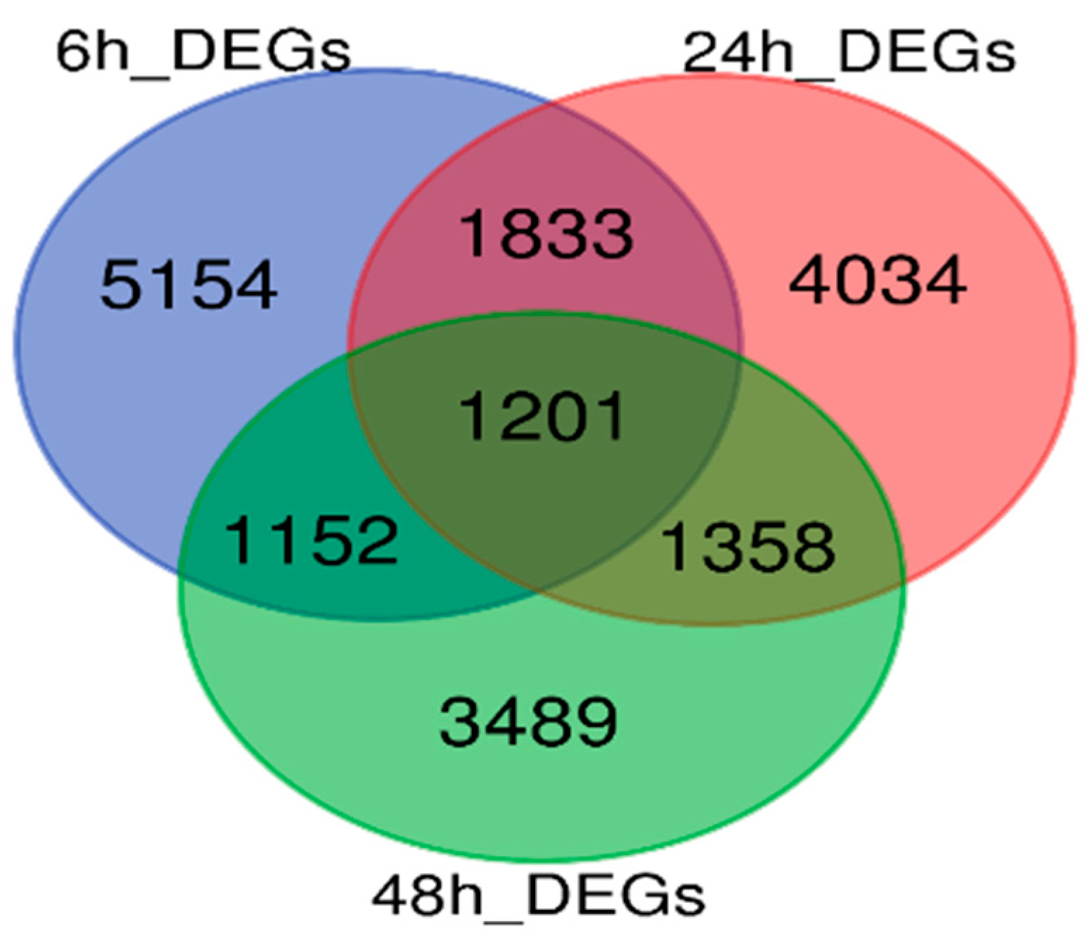
To further narrow down the most relevant DEGs involved in the resistance process, total DEGs at all three time points were assessed to identify shared DEGs. A total of 18,221 DEGs were identified, with 5154, 4034, and 3489 DEGs uniquely expressed at 6 hpi, 24 hpi, and 48 hpi, respectively. Additionally, 1833 DEGs were shared between 6 hpi and 24 hpi, 1152 between 6 hpi and 48 hpi, 1358 between 24 hpi and 48 hpi, and 1201 DEGs were shared across all three time points (Figure 2). These 1201 genes, expressed at all time points after SMV inoculation, may be involved in the resistance in both cultivars. To explore and compare the functions of these 1201 shared genes and the total 18,221 DEGs, GO enrichment analyses were performed separately.

3.3. GO Enrichment Analysis of the Total 18,221 DEGs and 1201 Joint DEGs Among Stages

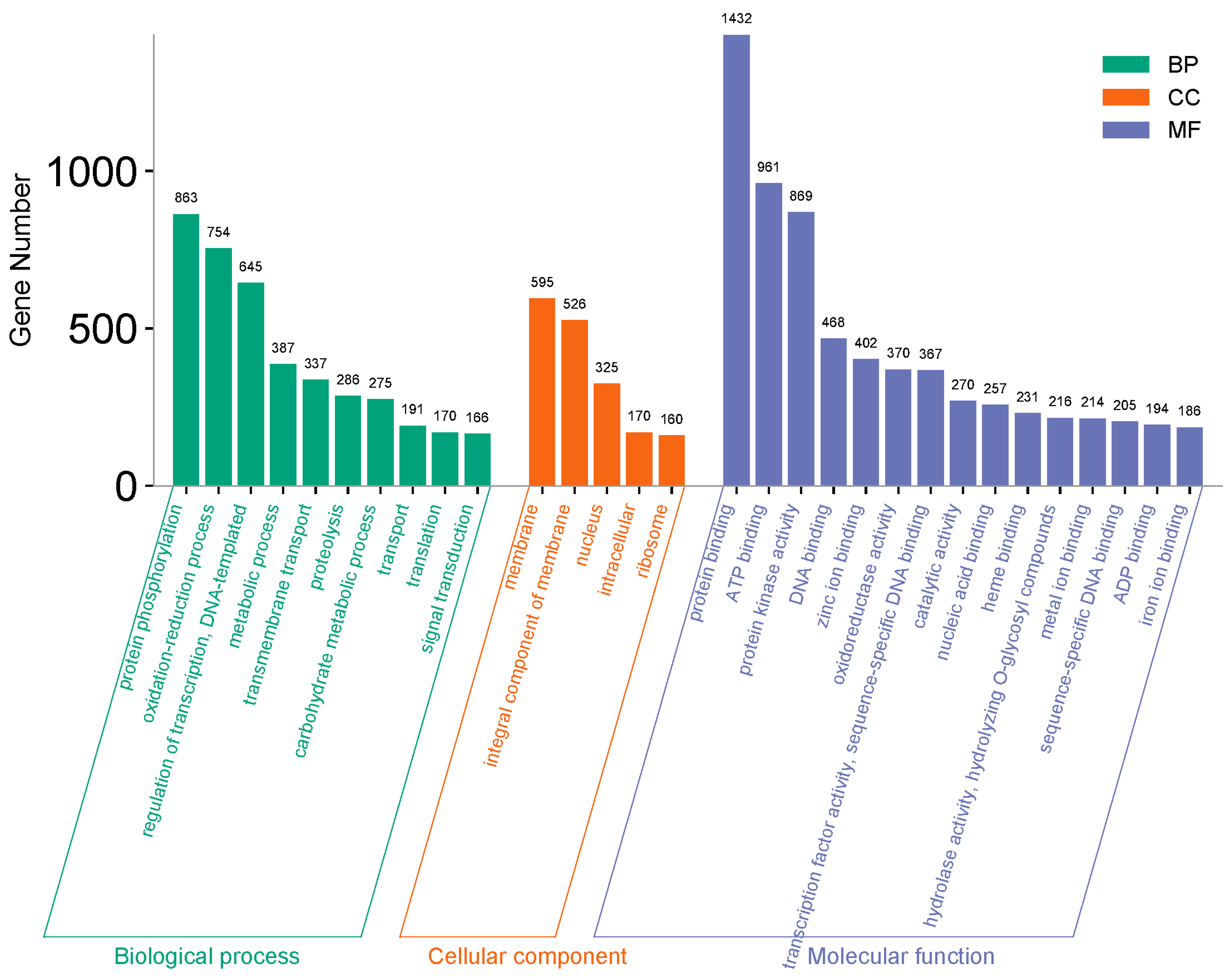
GO enrichment analysis was divided into three major categories: “Biological process”, “Cellular component”, and “Molecular function”, with 30 subcategories (Figure 3). Nearly half of the total DEGs were found in the “molecular function” category, primarily distributed among protein, DNA, ion, and ADP binding, which are mainly associated with regular growth and development activities. Additionally, 869 genes were active in protein kinase activity, 370 in oxidoreductase activity, and 270 in catalytic activity, which are related to diseases resistance signaling in plants. The other half of the DEGs were primarily found in the “biological process” category, distributed among protein phosphorylation, oxidation-reduction Process, transport, transmembrane transport, and signal transduction. Very few DEGs were found in the “Cellular Component” category. Protein phosphorylation and oxidation-reduction processes were the major categories, with 1637 DEGs associated with disease resistance, supported by transmembrane transport, transport and signal transduction, which contained 337, 191, and 166 DEGs, respectively.
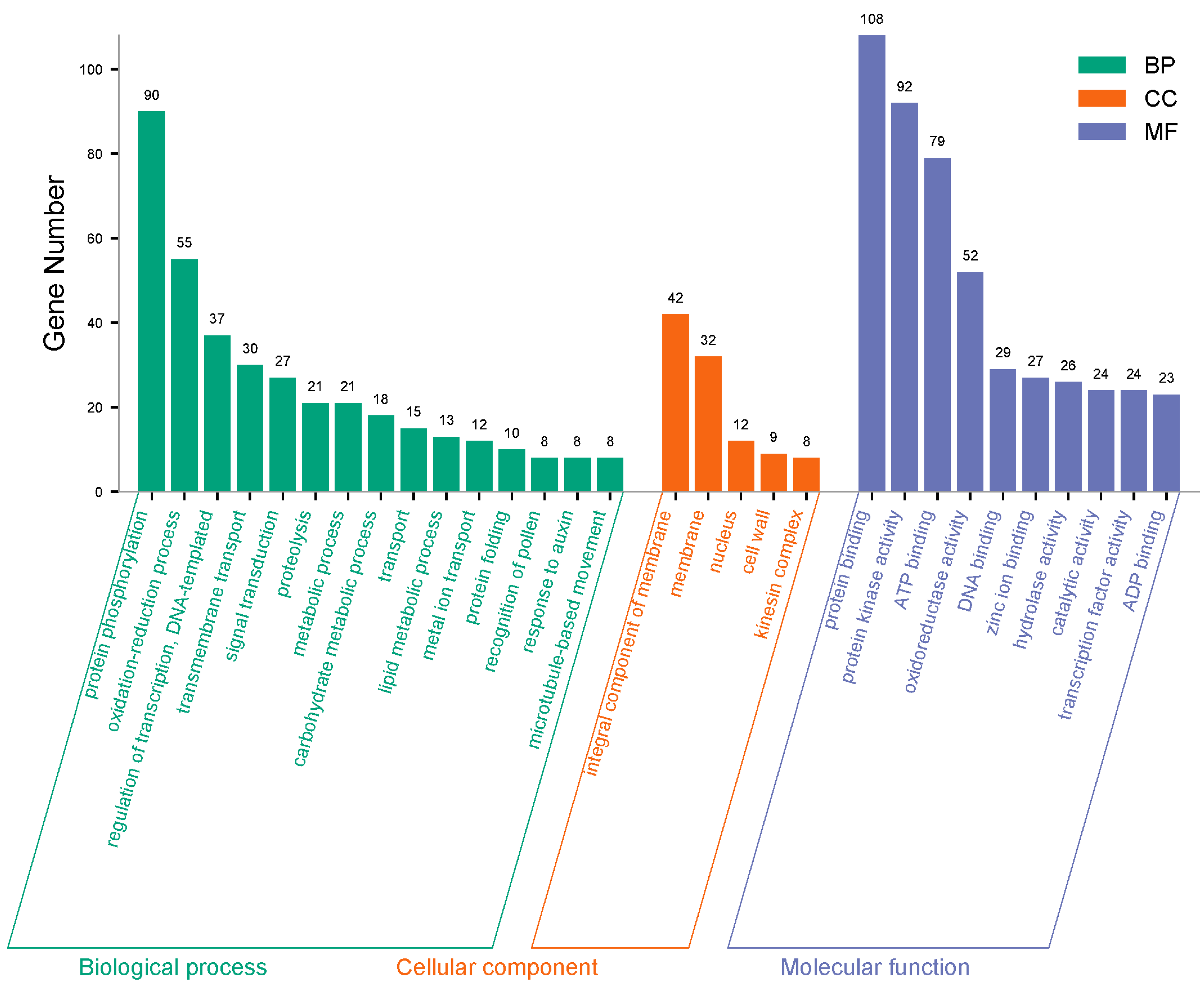
To further narrow down the DEGs and identify the most likely mechanisms against SMV-SC4, a GO function analysis of 1201 joint DEGs was performed (Figure 4). The results revealed that a larger portion of the DEGs was associated with GO term “Biological process”, with the majority falling into the subclass of protein phosphorylation, which included 90 DEGs. This was followed by oxidation-reduction processes (55 DEGs), regulation of transcription and DNA-templated processes (37 DEGs), transmembrane transport (30 DEGs), and signal transduction (27 DEGs). All these processes are related to disease resistance mechanisms. Additionally, transport, metal ion transport, and response to auxin contained 15, 12, and 8 DEGs, respectively, which are supportive activities for signal transduction. Other processes such as proteolysis, metabolic process, carbohydrate metabolic process, lipid metabolic process, protein folding, recognition of pollen, and microtubule-based movement also had DEGs, but these are typically involved in regular cellular functions. In the “Cellular component” category, a total of 103 genes were distributed across subclasses: integral components of the membrane (42 DEGs), Membrane (32 DEGs), Nucleus (12 DEGs), cell wall (9 DEGs), and kinesin complex (8 DEGs). Similarly, the “Molecular function” category had DEGs distributed across subclasses: protein binding (108 DEGs), protein kinase activity (92 DEGs), ATP binding (79 DEGs), oxidoreductase activity (52 DEGs), DNA binding (29 DEGs), zinc ion binding (27 DEGs), hydrolase activity (26 DEGs), catalytic activity (24 DEGs), transcription factor activity (24 DEGs), and ADP binding (23 DEGs) (Figure 4). The joint DEGs active during the resistance process in protein phosphorylation, Transmembrane transport, oxidation-reduction processes, and signal transduction in the biological process, supported by protein kinase activity and oxidoreductase activity in molecular function, contribute to resistance against SMV.

3.4. Protein Phosphorylation, Signal Transduction and Protein Kinase Activity in Early Defense Response to SMV

Plants protect themselves from pathogen attacks by initiating a complex natural defense response. Plant-pathogen interactions trigger signal transduction cascades that mobilize defense mechanisms, ultimately leading to disease resistance responses [10]. Signal transduction, linked with protein phosphorylation and protein kinase activity, collectively activates the plant’s defense system against pathogen attacks. Metal ions play a vital role in plant signal transmission as secondary messengers, and the accumulation and movement of secondary metabolites, facilitated by transmembrane transport, are also crucial for plant growth and defense functions [10,11,12,13].
In the Venn diagrams, as DEGs in KF cultivar increased at 6 and 24 hpi, genes active in signal transduction also increased during the early hours of pathogen attack. By 48 hpi, their number decreased in KF cultivar as the infection was controlled, but increased in NN cultivar as pathogen damage continued. Similarly, in the GO functional analysis of total DEGs and joint DEGs, signal transduction and protein kinase activity play a vital role in disease signaling response. Signal transduction serves as a channel through which other processes like transmembrane transport, metal ion transport, and response to auxin participate in the defense mechanism. Genes involved in metal ion transport and response to auxin were only observed when narrowing down to key genes. These genes are indirectly associated with the signal transduction process, as auxin is a key hormone that not only regulates growth and development but also modulates other hormones in signaling pathways. A few DEGs were also active in the cell wall under “Molecular function”, which may be involved in the defense mechanism, as the cell wall is related to transport and communication functions (Figure 5).

3.5. Oxidation-Reduction and Oxidoreductase Activity in Resistance

When a pathogen invades plant tissue, the production of reactive oxygen species (ROS) is observed. Although ROS is produced during regular growth and development, its concentration increases during stress, causing redox imbalance. Therefore, it is essential to reduce excessive ROS levels to mitigate their toxic effects. However, moderate ROS levels act as signaling molecules that interact with other signaling pathways to activate the plant’s defense system [22]. In the GO functional analysis histograms of 1201 joint DEGs and total 18,221 DEGs, genes were observed in these two terms under “Biological process” and “Molecular function”. The second-highest number of active genes (754) in the total DEGs histogram and 55 genes in the joint DEGs histogram were associated with the oxidation-reduction process, indicating its significant contribution to the defense mechanism of KF cultivar. Similarly, 370 and 52 genes in the total and joint DEGs histograms, respectively, were active in oxidoreductase activity (Figure 3 and Figure 4). Genes active in these redox activities are involved in removing high concentrations of ROS generated in response to SMV attack. Another important term under molecular function is catalytic activity.
Many catalase enzymes in plants convert H2O2 into water and oxygen when plants face environmental stresses [23]. These enzymes are present at major sites where H2O2 is produced, such as cytosol, mitochondria, and chloroplasts. Various catalases play versatile role in the crop plant system. The conversion of H2O2 by catalases within specific cells or organelles at specific times during growth and development directly or indirectly affects signal transduction in plants. In summary, signal transduction is the key process through which all other defense mechanisms are linked to produce the final output.

3.6. Genes Related to Signal Transduction and Response to Auxin in Soybean

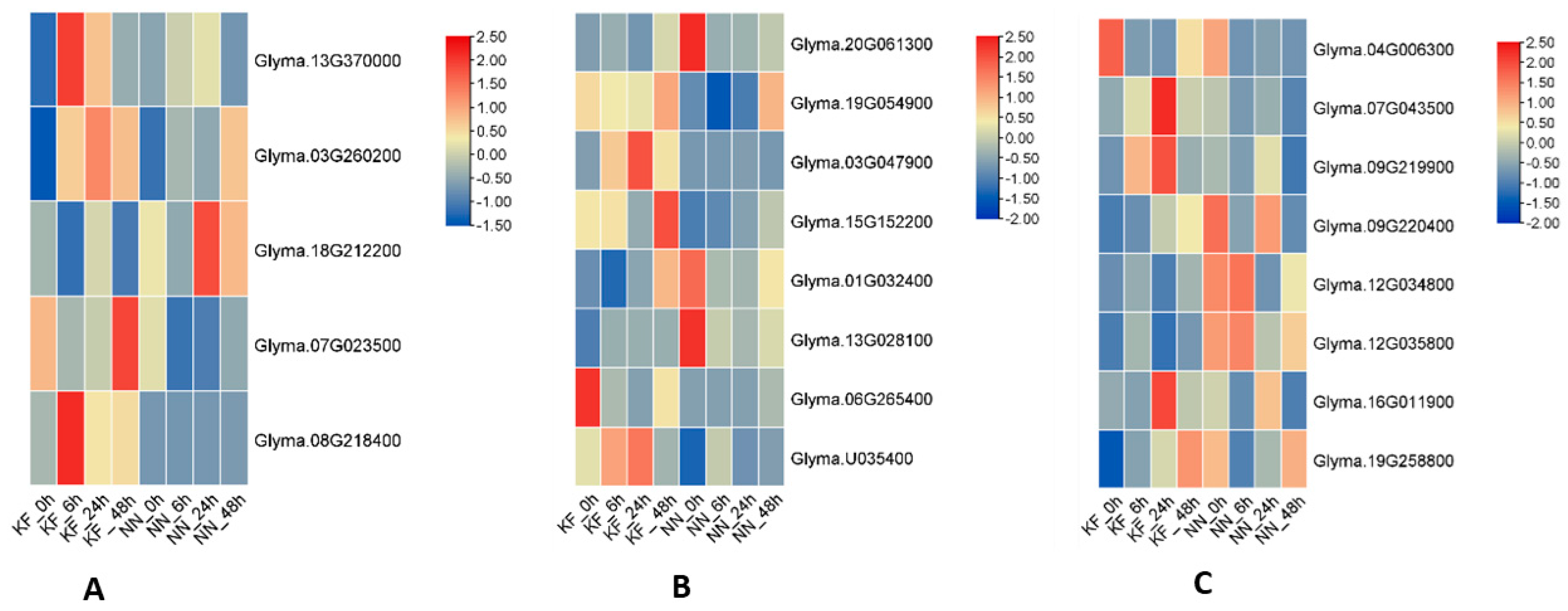
A considerable number of resistance genes in plants have been identified and classified into eight fundamental classes, which can be further divided into various super families based on their functional protein domains. Most of the genes cloned to date belong to the NBS-LRR kinase super families, which provide resistance against a wide range of pathogens, including viruses, bacteria, and pests [24]. In the GO enrichment analysis of the current study, 27 genes related to signal transduction (Table S4) were identified, most of which belong to specific disease-resistant groups and super families, such as Casitas B-lineage lymphoma (CBL)- interacting serine/threonine-protein kinase. Five genes from this family showed deferential expression in both cultivars at different time points. Glyma13G370000, Glyma07G023500 and Glyma08G218400 were upregulated in KF cultivar, while Glyma03G260200 and Glyma18G212200 were upregulated in NN (Figure 6A). Eight genes with predicted disease resistance functions showed expression in the resistant cultivar at almost all three points, except for Glyma19G054900, which was expressed in the susceptible cultivar (Figure 6B). A few genes were also found with predicted functions related to tobacco mosaic virus (TMV) resistance. Another group of eight genes was found in the “Response to auxin” subclass (Table S5). Their differential expression, calculated using FPKM values from RNA-seq data, showed that Glyma04G006300, Glyma07G043500, Glyma09G219900, and Glyma16G011900 were more highly expressed in KF cultivar, while Glyma09G220400, Glyma12G034800, Glyma12G035800, and Glyma19G258800 were more highly expressed in NN cultivar (Figure 6C).
In conclusion, the transcriptomic study revealed that genes involved in signal transduction directly, and genes active in other categories related to plant stress response, such as protein phosphorylation, protein kinase activity, oxidation-reduction, oxidoreductase activity, catalytic activity, metal ion transport, and response to auxin, indirectly contribute to the resistance mechanism in KF cultivar through signal transduction.

4. Discussion

4.1. Transcriptome Study on the Defense Mechanism Against Soybean Mosaic Virus

In the plant defense system against viruses, there are often different defense mechanisms. One common defense mechanism, part of the innate immune system, involves pattern-triggered immunity (PTI), where recognition factors are usually localized on the cell surface [8,9,25]. Another, more powerful defense mechanism is effector-triggered immunity (ETI), which is often specific to certain strains and can mediate rapid necrosis of virus-infected areas, preventing further spread. The genes inducing this response are often referred to as resistance (R) genes.
The activation of PTI and ETI involves different types of receptors—Pattern Recognition Receptors (PRRs) for PTI and NOD-like Receptors (NLRs) for ETI—and different early signal transduction processes. However, there is significant overlap in downstream outputs, such as Calcium Flux, Reactive Oxygen Species (ROS) Burst, Transcriptional Reprograming, and Phytohormone Signaling [26]. In the present study, we identified genes for example Glyma13G370000 (CBL-interacting serine/threonine-protein kinase 5), Glyma08G218400 (CBL-interacting serine/threonine-protein kinase 5-like), Glyma03G047900 (Disease resistance protein RML1A-like), GlymaU035400 (R1protein) (Table S4) in sub-categories signal transduction with their higher expression in the resistant cultivar compare to susceptible cultivar (Figure 6A,B). It can be speculated that these genes may be involved in ETI mechanism removal ROS by translating into specific amino acids to encounter the attack of specific SMV strain, in our case SC4on soybean cultivar Kefeng-1 (KF) which also give signals to other genes to cooperate with them to get rid of the infection at early stage. In short, ETI is the main defense system which makes KF cultivar resistant against SC4.

4.2. Resistance to SMV Is Mediated by a Network of Processes and Signaling Pathways

Soybean plants defend against SMV invasion by initiating a multicomponent defense response. Proteins coded by the disease resistance genes recognize specific SMV strain attacks and bind to specific pathogen (virus)-derived virulence (Avr) proteins. This initiates an internal signaling cascade, activating the host plant’s defense arsenal, leading to localized cell death at the infection site and systemic acquired resistance (SAR) across the plant, preventing the further infection spread and causing a hypersensitive response (HR) [24]. Interactions between pathogens and plants, signal initiation, and signal transduction exhibit resistance in many plants against various pathogens [27]. In the current study, 27 genes active in signal transduction under the biological process were identified as involved in resistance against virus attacks. Most of these genes have predicted disease resistance functions and belong to groups specialized for disease resistance, such as CBL-interacting serine/threonine-protein kinase, and were more highly expressed in the resistant cultivar compared to the susceptible cultivar in the RNA seq dataset. Previously, genes with specific conserved domains belonging to the NBS–LRR or LRR kinase super families have been cloned for resistance against bacteria, nematodes, viruses, and fungi [27,28,29,30].
In addition to signal transduction, DEGs were active in Oxidation-reduction, Protein kinase activity, oxidoreductase activity, metal ion transport, and transmembrane transport, all of which are part of the resistance mechanism. Protein kinase activity involves the removal of a phosphate group from an ATP molecule and its attachment to an acceptor molecule with a free hydroxyl group. Kinase enzymes in this activity regulate many cellular functions, including signal transduction, cell division, differentiation, growth, and development. Metal ions play a vital role in plant signal transmission as secondary messengers, and the accumulation and movement of secondary metabolites, facilitated by transmembrane transport, are also crucial for plant growth and defense functions [12,13].
Moreover, 55 genes in oxidation-reduction and 52 in oxidoreductase activity were expressed in the GO enrichment analysis histogram. Oxidation-reduction induced by pathogens is usually accompanied by a large amount of (ROS), which signals successful infection identification and activates plant defenses [31]. Production of ROS during ETI is essential for eliciting a more robust defense response. A defining characteristic of ETI is an intense and sustained burst of ROS. When high levels of ROS reached, they are associated with the hypersensitive response. In the initial stage, an oxidative burst usually occurs at the virus invasion site, sending signals to activate the entire defense system. Subsequently, ROS accumulate to higher levels, playing an important signaling role in activating plant defenses and triggering hypersensitive cell death (HR) [32]. These findings demonstrate the role of oxidation-reduction, signal transduction, and ROS production in the initial resistance of KF cultivar.

4.3. Role of Hormones in the Defense Mechanism

Plant hormones significantly influence the soybean plant defense system. Jasmonic acid, ethylene, Salicylic acid and auxins are the essential phytohormones that regulate various aspects of plant immunity, including pathogen (virus) recognition, signal transduction, and initiation of genes related to defense. Additionally, plant growth regulating hormones such as cytokinin and gibberellic acid modulate immune responses through intricate regulatory networks [33]. Auxin, naturally existing as IAA, is synthesized from pyruvic acid through various pathways. It rapidly modifies the expression of many genes by eliminating the inhibitory function of AUX/IAA proteins. Its role as a fundamental part of the hormone signaling system in plants under biotic stress has been described by many researchers [34,35,36,37,38]. For example, auxin-sensitive factors in tomatoes play an intermediate role between auxin action and biotic response. In two viral diseases of tomato—Tomato spotted wilt virus (TSWV) and Tomato brown rugose fruit virus (ToBRFV)—auxin plays a signaling role in the defense system [39,40].
In the current transcriptomic study on SMV resistance, eight genes expressed in the “Response to auxin” subclass under the “Biological Process” category may be involved in the resistance mechanism of KF cultivar, as their differential expression was higher in KF cultivar. These genes were active during the middle stage of infection in the GO term enrichment analysis, indicating auxin’s involvement in the defense system against the virus. The movement of auxin into, out of, or within cells, facilitated by transporter or pores, and the auxin-mediated signaling pathway, are crucial for plant growth and development. However, auxin’s involvement in defense responses has also been suggested. Ghanashyam and Jain (2009) studied the function of auxin-inducible genes under biotic stress conditions and observed upregulated differential expression in rice [38]. Auxin is involved in the TOR (target of rapamycin) signaling pathway, which responds to environmental stress and acts as a signal inducer in translation in plants [41,42].
Recent transcriptomic studies on soybean mosaic virus (SMV) resistance across different resistant cultivars and strains indicate that three major pathways are involved in the resistance process: plant-pathogen interaction pathways, hormone signal transduction pathways, and mitogen-activated protein kinase (MAPK) signaling pathways. These pathways are regulated by clusters of genes [43,44]. A study by Zhu et al. [44] investigated SMV strain N1 resistance in the soybean cultivar Dongnong 93-046 using transcriptomic analysis. They identified 41,189 differentially expressed genes (DEGs) associated with the resistance mechanism, with 9196 DEGs showing FPKM values above 10. Their study highlighted 894 core genes involved in plant-pathogen interaction, linoleic acid metabolism, and plant hormone signaling transduction.
In our current study, we analyzed transcriptomic responses to SMV strain SC4 using two soybean cultivars with contrasting resistance profiles: Kefeng-1 (resistant) and NN1138-2 (susceptible). We identified 18,221 DEGs across both varieties, with 1201 core DEGs expressed at 0, 6, 24, and 48 h after SMV inoculation. These core DEGs were consistently active throughout the resistance process, from Virus recognition to defense activation. Some genes were preferentially expressed in the resistant cultivar (KF) during early infection stages but remained inactive in the susceptible cultivar (NN). Conversely, certain genes were highly upregulated in the susceptible cultivar at later infection stages but not in the resistant cultivar, providing valuable insights into the genetic basis of resistance and susceptibility. Additionally, we identified 27 genes involved in signal transduction, most of which contained specific domains associated with disease resistance, including tobacco mosaic virus (TMV) resistance domains. These findings suggest that our study provides a more comprehensive view of SMV resistance mechanisms compared to previous studies.

5. Conclusions

Signal transduction, linked with protein phosphorylation and protein kinase activity, collectively activates the plant’s defense system against pathogen attacks. Metal ions play a vital role in plant signal transmission as secondary messengers, and the accumulation and movement of secondary metabolites, facilitated by transmembrane transport, are also crucial for plant growth and defense functions. Many catalase enzymes in plants convert H2O2 into water and oxygen when plants face environmental stresses. These enzymes are present at major sites where H2O2 is produced, such as cytosol, mitochondria, and chloroplasts. Various catalases play versatile role in the crop plant system. The conversion of H2O2 by catalases within specific cells or organelles at specific times during growth and development directly or indirectly affects signal transduction in plants. In summary, signal transduction is the key process through which all other defense mechanisms are linked to produce the final output. Our transcriptomic study reveals the similar results; genes involved in signal transduction directly, and genes active in other categories related to plant stress response such as protein phosphorylation, protein kinase activity, oxidation–reduction, oxidoreductase activity, catalytic activity, metal ion transport, and response to auxin indirectly contribute to the resistance mechanism in KF cultivar through signal transduction.

Supplementary Materials

The following supporting information can be downloaded at: https://www.mdpi.com/article/10.3390/agronomy15040906/s1, Figure S1. Principal component analysis (PCA) plot; Figure S2. Venn diagram of Differentially expressed genes (DEGs) in resistant and susceptible cultivars at 6 h post-inoculation (hpi), across four comparison groups, including shared DEGs; Figure S3. Venn diagram of Differentially expressed genes (DEGs) in resistant and susceptible cultivars at 24 h post-inoculation (hpi), across four comparison groups, including shared DEGs; Figure S4. Venn diagram of Differentially expressed genes (DEGs) in resistant and susceptible cultivars at 48 h post-inoculation (hpi), across four comparison groups, including shared DEGs; Table S1. The average coverage of clean data across 24 samples reached; Table S2. The GC content after filtration ranged from 41.95% to 44.50%, and the Q30 score for each sample exceeded 93.24%; Table S3. The average coverage across all samples; Table S4. Twenty-seven genes associated with signal transduction; Table S5. Eight genes related to “Response to auxin” from GO.

Author Contributions

J.G. and K.L. planned the experiment, M.M.R. and H.J. executed the experiment and drafted the whole manuscript, while S.G. assisted in execution and write up. All authors have read and agreed to the published version of the manuscript.

Funding

This work was supported by grants from the National Key Research and Development Program of China (2024YFD1201404), China Agriculture Research System of MOF and MARA (No. CARS-04), Jiangsu Collaborative Innovation Center for Modern Crop Production (JCIC-MCP), and Collaborative Innovation Center for Modern Crop Production co-sponsored by Province and Ministry (CIC-MCP).

Data Availability Statement

The original contributions presented in this study are included in the article/Supplementary Materials. Further inquiries can be directed to the corresponding author(s).

Conflicts of Interest

All authors declared that they have no conflicts of interest.

References

  1. Adams, M.J.; Antoniw, J.F.; Beaudoin, F. Overview and Analysis of the Polyprotein Cleavage Sites in the Family Potyviridae. Mol. Plant Pathol. 2005, 6, 471–487. [Google Scholar] [CrossRef] [PubMed]
  2. Li, K.; Yang, Q.H.; Zhi, H.J.; Gai, J.Y. Identification and Distribution of Soybean Mosaic Virus Strains in Southern China. Plant Dis. 2010, 94, 351–357. [Google Scholar] [CrossRef] [PubMed]
  3. Wang, D.; Tian, Z.; Li, K.; Li, H.; Huang, Z.; Hu, G.; Zhi, H. Identification and Variation Analysis of Soybean Mosaic Virus Strains in Shandong, Henan and Anhui Provinces of China. Soybean Sci. 2013, 32, 806–809. [Google Scholar]
  4. Cho, E.K.; Goodman, R.M. Strains of Soybean Mosaic Virus: Classification Based on Virulence in Resistant Soybean Cultivars. Phytopathology 1979, 69, 467–470. [Google Scholar]
  5. Cho, E.K.; Goodman, R.M. Evaluation of Resistance in Soybeans to Soybean Mosaic Virus Strains 1. Crop Sci. 1982, 22, 1133–1136. [Google Scholar] [CrossRef]
  6. Takahashi, K.; Tanaka, T.; Iida, W.; Tsuda, Y. Studies on Virus Diseases and Causal Viruses of Soybean in Japan. Bull. Tohoku Natl. Agric. Exp. Stn. 1980, 62, 1–130. [Google Scholar]
  7. Yu, X.-Q.; Niu, H.-Q.; Liu, C.; Wang, H.-L.; Yin, W.; Xia, X. PTI-ETI Synergistic Signal Mechanisms in Plant Immunity. Plant Biotechnol. J. 2024, 22, 2113–2128. [Google Scholar] [CrossRef]
  8. Lin, H.; Wang, M.; Chen, Y.; Nomura, K.; Hui, S.; Gui, J.; Zhang, X.; Wu, Y.; Liu, J.; Li, Q.; et al. An MKP-MAPK Protein Phosphorylation Cascade Controls Vascular Immunity in Plants. Sci. Adv. 2022, 8, eabg8723. [Google Scholar] [CrossRef]
  9. Li, P.; Zhao, L.; Qi, F.; Htwe, N.M.P.S.; Li, Q.; Zhang, D.; Lin, F.; Shang-Guan, K.; Liang, Y. The Receptor-like Cytoplasmic Kinase RIPK Regulates Broad-Spectrum ROS Signaling in Multiple Layers of Plant Immune System. Mol. Plant 2021, 14, 1652–1667. [Google Scholar] [CrossRef]
  10. Ding, L.N.; Li, Y.T.; Wu, Y.Z.; Li, T.; Geng, R.; Cao, J.; Zhang, W.; Tan, X.L. Plant Disease Resistance-Related Signaling Pathways: Recent Progress and Future Prospects. Int. J. Mol. Sci. 2022, 23, 16200. [Google Scholar] [CrossRef]
  11. Liu, J.Z.; Lam, H.M. Signal Transduction Pathways in Plants for Resistance against Pathogens. Int. J. Mol. Sci. 2019, 20, 2335. [Google Scholar] [CrossRef] [PubMed]
  12. Nogia, P.; Pati, P.K. Plant Secondary Metabolite Transporters: Diversity, Functionality, and Their Modulation. Front. Plant Sci. 2021, 12, 758202. [Google Scholar] [CrossRef] [PubMed]
  13. Shi, X.; Bao, J.; Lu, X.; Ma, L.; Zhao, Y.; Lan, S.; Cao, J.; Ma, S.; Li, S. The Mechanism of Ca2+ Signal Transduction in Plants Responding to Abiotic Stresses. Environ. Exp. Bot. 2023, 216, 105514. [Google Scholar] [CrossRef]
  14. Mazzoni-Putman, S.M.; Brumos, J.; Zhao, C.; Alonso, J.M.; Stepanova, A.N. Auxin Interactions with Other Hormones in Plant Development. Cold Spring Harb. Perspect. Biol. 2021, 13, a039990. [Google Scholar] [CrossRef]
  15. Gomes, G.L.B.; Scortecci, K.C. Auxin and Its Role in Plant Development: Structure, Signalling, Regulation and Response Mechanisms. Plant Biol. 2021, 23, 894–904. [Google Scholar] [CrossRef]
  16. Wang, D.; Ma, Y.; Liu, N.; Yang, Z.; Zheng, G.; Zhi, H. Fine Mapping and Identification of the Soybean R SC4 Resistance Candidate Gene to Soybean Mosaic Virus. Plant Breed. 2011, 130, 653–659. [Google Scholar] [CrossRef]
  17. Rui, R.; Liu, S.; Karthikeyan, A.; Wang, T.; Niu, H.; Yin, J.; Yang, Y.; Wang, L.; Yang, Q.; Zhi, H.; et al. Fine-Mapping and Identification of a Novel Locus Rsc15 Underlying Soybean Resistance to Soybean Mosaic Virus. Theor. Appl. Genet. 2017, 130, 2395–2410. [Google Scholar] [CrossRef]
  18. Zheng, G.; Yang, Y.; MA, Y.; Yang, X.; Chen, S.; Ren, R.; Wang, D.; Yang, Z.; Zhi, H. Fine Mapping and Candidate Gene Analysis of Resistance Gene R to Soybean Mosaic Virus in Qihuang 1. J. Integr. Agric. 2014, 13, 2608–2615. [Google Scholar] [CrossRef]
  19. Love, M.I.; Huber, W.; Anders, S. Moderated Estimation of Fold Change and Dispersion for RNA-Seq Data with DESeq2. Genome Biol. 2014, 15, 550. [Google Scholar] [CrossRef]
  20. Robinson, M.D.; McCarthy, D.J.; Smyth, G.K. EdgeR: A Bioconductor Package for Differential Expression Analysis of Digital Gene Expression Data. Bioinformatics 2010, 26, 139–140. [Google Scholar] [CrossRef]
  21. Kanehisa, M. KEGG: Kyoto Encyclopedia of Genes and Genomes. Nucleic Acids Res. 2000, 28, 27–30. [Google Scholar] [CrossRef] [PubMed]
  22. Suman, S.; Bagal, D.; Jain, D.; Singh, R.; Singh, I.K.; Singh, A. Biotic Stresses on Plants: Reactive Oxygen Species Generation and Antioxidant Mechanism. In Frontiers in Plant-Soil Interaction; Aftab, T., Hakeem, R.K., Eds.; Academic Press: London, UK, 2021; pp. 381–411. [Google Scholar]
  23. Sharma, I.; Ahmad, P. Catalase. In Oxidative Damage to Plants; Academic Press: London, UK, 2014; pp. 131–148. [Google Scholar]
  24. Padder, B.A. Plant Disease Resistance Genes: From Perception to Signal Transduction. In Plant Signaling: Understanding the Molecular Crosstalk; Hakeem, K., Rehman, R., Tahir, I., Eds.; Springer: New Delhi, India, 2014; pp. 345–354. [Google Scholar]
  25. Bigeard, J.; Colcombet, J.; Hirt, H. Signaling Mechanisms in Pattern-Triggered Immunity (PTI). Mol. Plant 2015, 8, 521–539. [Google Scholar] [CrossRef] [PubMed]
  26. Yuan, M.; Ngou, B.P.M.; Ding, P.; Xin, X.F. PTI-ETI Crosstalk: An Integrative View of Plant Immunity. Curr. Opin. Plant Biol. 2021, 62, 102030. [Google Scholar] [CrossRef]
  27. Ijaz, S.; Haq, I.U.; Babar, M.; Nasir, B. Disease Resistance Genes’ Identification, Cloning, and Characterization in Plants. In Cereal Diseases: Nanobiotechnological Approaches for Diagnosis and Management; Springer Nature: Singapore, 2022; pp. 249–269. [Google Scholar]
  28. Ameline Torregrosa, C.; Wang, B.B.; O’Bleness, M.S.; Deshpande, S.; Zhu, H.; Roe, B.; Young, N.D.; Cannon, S.B. Identification and Characterization of Nucleotide-Binding Site-Leucine-Rich Repeat Genes in the Model Plant Medicago Truncatula. Plant Physiol. 2008, 146, 5–21. [Google Scholar] [CrossRef]
  29. Bai, S.; Liu, J.; Chang, C.; Zhang, L.; Maekawa, T.; Wang, Q.; Xiao, W.; Liu, Y.; Chai, J.; Takken, F.L.W.; et al. Structure-Function Analysis of Barley NLR Immune Receptor MLA10 Reveals Its Cell Compartment Specific Activity in Cell Death and Disease Resistance. PLoS Pathog. 2012, 8, e1002752. [Google Scholar] [CrossRef]
  30. Cao, Y.; Mo, W.; Li, Y.; Xiong, Y.; Wang, H.; Zhang, Y.; Lin, M.; Zhang, L.; Li, X. Functional Characterization of NBS-LRR Genes Reveals an NBS-LRR Gene That Mediates Resistance against Fusarium Wilt. BMC Biol. 2024, 22, 45. [Google Scholar] [CrossRef]
  31. Liu, C.; Liu, Q.; Mou, Z. Redox Signaling and Oxidative Stress in Systemic Acquired Resistance. J. Exp. Bot. 2024, 75, 4535–4548. [Google Scholar] [CrossRef]
  32. Weralupitiya, C.; Eccersall, S.; Meisrimler, C.-N. Shared Signals, Different Fates: Calcium and ROS in Plant PRR and NLR Immunity. Cell Rep. 2024, 43, 114910. [Google Scholar] [CrossRef]
  33. Shafqat, A.; Abbas, S.; Ambreen, M.; Siddiqa Bhatti, A.; Kausar, H.; Gull, T. Exploring the Vital Role of Phytohormones and Plant Growth Regulators in Orchestrating Plant Immunity. Physiol. Mol. Plant Pathol. 2024, 133, 102359. [Google Scholar] [CrossRef]
  34. Kalsi, H.S.; Karkhanis, A.A.; Natarajan, B.; Bhide, A.J.; Banerjee, A.K. AUXIN RESPONSE FACTOR 16 (StARF16) Regulates Defense Gene StNPR1 upon Infection with Necrotrophic Pathogen in Potato. Plant Mol. Biol. 2022, 109, 13–28. [Google Scholar] [CrossRef]
  35. Liu, L.; Yahaya, B.S.; Li, J.; Wu, F. Enigmatic Role of Auxin Response Factors in Plant Growth and Stress Tolerance. Front. Plant Sci. 2024, 15, 1398818. [Google Scholar] [CrossRef]
  36. Bouzroud, S.; Gouiaa, S.; Hu, N.; Bernadac, A.; Mila, I.; Bendaou, N.; Smouni, A.; Bouzayen, M.; Zouine, M. Auxin Response Factors (ARFs) Are Potential Mediators of Auxin Action in Tomato Response to Biotic and Abiotic Stress (Solanum lycopersicum). PLoS ONE 2018, 13, e0193517. [Google Scholar] [CrossRef]
  37. Ghanashyam, C.; Jain, M. Role of Auxin-Responsive Genes in Biotic Stress Responses. Plant Signal. Behav. 2009, 4, 846–848. [Google Scholar] [CrossRef] [PubMed]
  38. Guilfoyle, T.J.; Hagen, G. Auxin Response Factors. Curr. Opin. Plant Biol. 2007, 10, 453–460. [Google Scholar] [CrossRef]
  39. Werghi, S.; Herrero, F.A.; Fakhfakh, H.; Gorsane, F. Auxin Drives Tomato Spotted Wilt Virus (TSWV) Resistance through Epigenetic Regulation of Auxin Response Factor ARF8 Expression in Tomato. Gene 2021, 804, 145905. [Google Scholar]
  40. Vaisman, M.; Hak, H.; Arazi, T.; Spiegelman, Z. The Impact of Tobamovirus Infection on Root Development Involves Induction of Auxin Response Factor 10a in Tomato. Plant Cell Physiol. 2023, 63, 1980–1993. [Google Scholar] [CrossRef]
  41. Schepetilnikov, M.; Ryabova, L.A. Auxin Signaling in Regulation of Plant Translation Reinitiation. Front. Plant Sci. 2017, 8, 1014. [Google Scholar] [CrossRef]
  42. Schepetilnikov, M.; Ryabova, L.A. Recent Discoveries on the Role of TOR (Target of Rapamycin) Signaling in Translation in Plants. Plant Physiol. 2018, 176, 1095–1105. [Google Scholar] [CrossRef]
  43. Niu, J.; Zhao, J.; Guo, Q.; Zhang, H.; Yue, A.; Zhao, J.; Yin, C.; Wang, M.; Du, W. WGCNA Reveals Hub Genes and Key Gene Regulatory Pathways of the Response of Soybean to Infection by Soybean Mosaic Virus. Genes 2024, 15, 566. [Google Scholar] [CrossRef]
  44. Zhu, H.; Li, R.; Fang, Y.; Zhao, X.; Teng, W.; Li, H.; Han, Y. Weighted Gene Co-Expression Network Analysis Uncovers Critical Genes and Pathways Involved in Soybean Response to Soybean Mosaic Virus. Agronomy 2024, 14, 2455. [Google Scholar] [CrossRef]
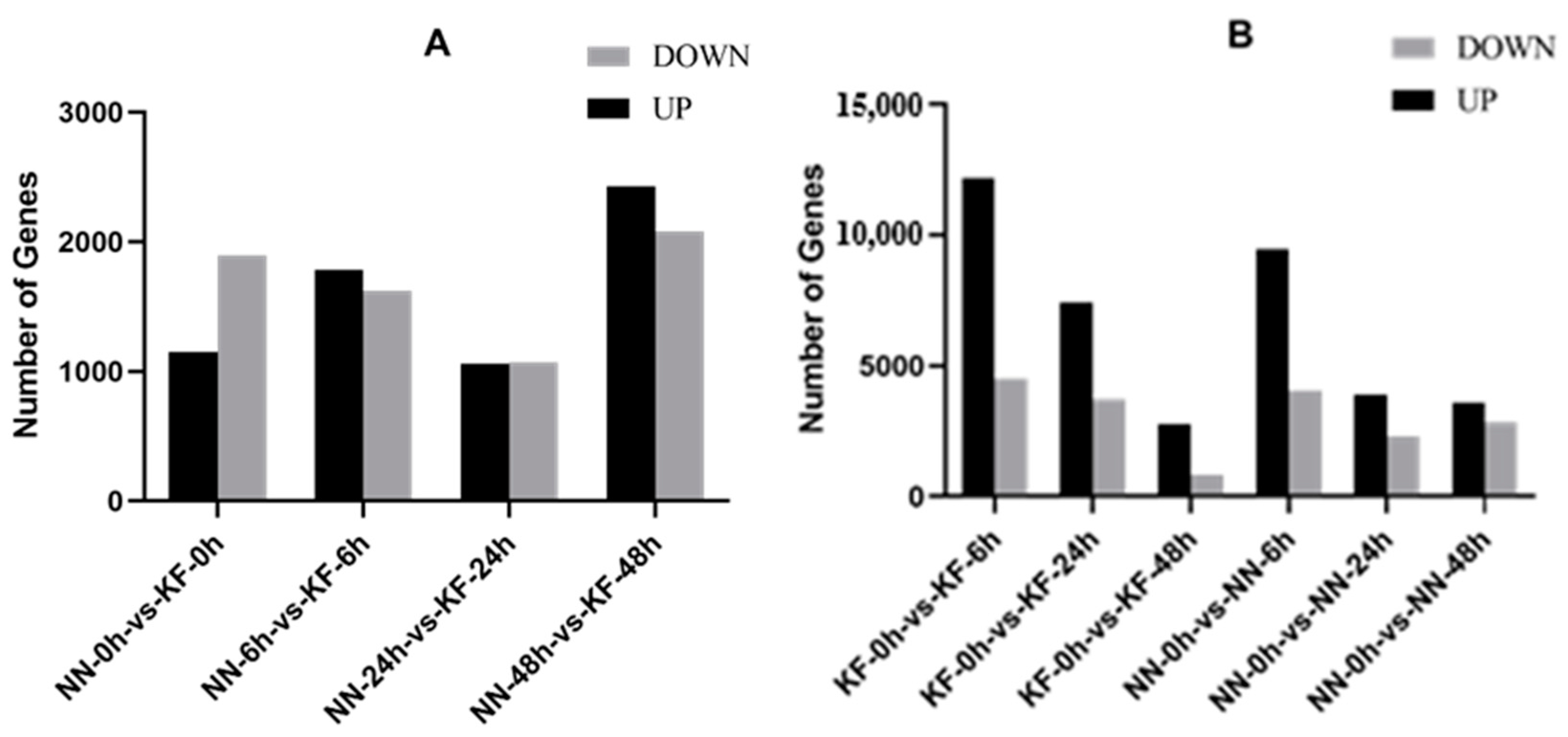
Figure 1. Comparative analysis of differentially expressed genes (DEGs) across four distinct stages following SC4 infection on soybean. (A) Comparative analysis of DEGs between soybean cultivars NN1138-2 (NN) and Kefeng-1 (KF) at each of the four stages. (B) Stage-specific comparative analysis of DEGs between NN1138-2 (NN) and Kefeng-1 (KF) after SC4 inoculation.
Agronomy 15 00906 g001
Figure 2. Venn diagram illustrating the unique and overlapping differentially expressed genes (DEGs) at 6, 24, and 48 h post-inoculation of SMV SC4 on soybean.
Agronomy 15 00906 g002
Figure 3. GO functional analysis histogram for totally identified 18,221 DEGs over four time points of soybean after SMV SC4 inoculation. Biological processes (BP) highlighted in dark cyan, cellular components (CC) in sienna, and molecular functions (MF) in steel blue.
Agronomy 15 00906 g003
Figure 4. GO functional analysis histogram for 1201 jointly identified DEGs over four time points of soybean after SMV SC4 inoculation. Biological processes (BP) represented in dark cyan, cellular components (CC) in sienna, and molecular functions (MF) in steel blue.
Agronomy 15 00906 g004
Figure 5. Model of signal transduction in response to pathogen (SMV SC4) attack on soybean plant. Illustrating the involvement of cell wall, plasma membrane, protein phosphorylation, redox reactions, and secondary signaling pathways in regulating resistance mechanisms.
Agronomy 15 00906 g005
Figure 6. Heatmap generated from FPKM values derived from RNA-seq data of interaction of soybean with SMV SC4 strain. Depicting the expression profiles of genes involved in: (A) (CBL)- interacting serine/threonine-protein kinase, (B) disease resistance proteins, (C) response to auxin.
Agronomy 15 00906 g006
Disclaimer/Publisher’s Note: The statements, opinions and data contained in all publications are solely those of the individual author(s) and contributor(s) and not of MDPI and/or the editor(s). MDPI and/or the editor(s) disclaim responsibility for any injury to people or property resulting from any ideas, methods, instructions or products referred to in the content.