Article

Harnessing the Diversity of Burkholderia spp. Prophages for Therapeutic Potential

by
Hayley R. Nordstrom
1,
Marissa P. Griffith
1,
Vatsala Rangachar Srinivasa
1,
Nathan R. Wallace
1,
Anna Li
2,
Vaughn S. Cooper
2,3,
Ryan K. Shields
1 and
Daria Van Tyne
1,3,*
1
Division of Infectious Diseases, University of Pittsburgh School of Medicine, Pittsburgh, PA 15213, USA
2
Department of Microbiology and Molecular Genetics, University of Pittsburgh School of Medicine, Pittsburgh, PA 15213, USA
3
Center for Evolutionary Biology and Medicine, University of Pittsburgh School of Medicine, Pittsburgh, PA 15213, USA
*
Author to whom correspondence should be addressed.
Cells 2024, 13(5), 428; https://doi.org/10.3390/cells13050428
Submission received: 22 January 2024 / Revised: 24 February 2024 / Accepted: 27 February 2024 / Published: 29 February 2024
(This article belongs to the Special Issue Bacteriophages and Their Enzymes as Antibacterial Agents)

Abstract

:
Burkholderia spp. are often resistant to antibiotics, and infections with these organisms are difficult to treat. A potential alternative treatment for Burkholderia spp. infections is bacteriophage (phage) therapy; however, it can be difficult to locate phages that target these bacteria. Prophages incorporated into the bacterial genome have been identified within Burkholderia spp. and may represent a source of useful phages for therapy. Here, we investigate whether prophages within Burkholderia spp. clinical isolates can kill conspecific and heterospecific isolates. Thirty-two Burkholderia spp. isolates were induced for prophage release, and harvested phages were tested for lytic activity against the same 32 isolates. Temperate phages were passaged and their host ranges were determined, resulting in four unique phages of prophage origin that showed different ranges of lytic activity. We also analyzed the prophage content of 35 Burkholderia spp. clinical isolate genomes and identified several prophages present in the genomes of multiple isolates of the same species. Finally, we observed that Burkholdera cenocepacia isolates were more phage-susceptible than Burkholderia multivorans isolates. Overall, our findings suggest that prophages present within Burkholderia spp. genomes are a potentially useful starting point for the isolation and development of novel phages for use in phage therapy.

1. Introduction

Burkholderia is a genus of gram-negative bacteria that encompasses nearly 50 different species [1]. These organisms are abundant in the environment, are found readily in soil and water, and are associated with the rhizospheres of several species of plants [2,3]. Burkholderia spp. are also opportunistic human pathogens, particularly the members of the Burkholderia cepacia complex (Bcc), a group of at least 20 species which possess high levels of intrinsic resistance to multiple classes of antibiotics [4]. Two Bcc species make up the majority of Burkholderia spp. clinical isolates in the United States: Burkholderia multivorans and Burkholderia cenocepacia [5,6]. While infections caused by Burkholderia spp. are relatively rare in healthy people, these bacteria cause difficult-to-treat infections in patients with compromised immune systems or chronic conditions such as cystic fibrosis (CF) and chronic granulomatous disease [7,8]. Chronic infection with the Bcc, and in particular B. cenocepacia, in CF patients is associated with increased morbidity and mortality, decreased lung function, and shorter life expectancy [4,8,9,10]. Treatment of Burkholderia spp. infections is further complicated by intrinsic resistance to many classes of antibiotics [11,12]. This makes Burkholderia spp. some of the most challenging bacteria to eradicate and increases the risk of transmission to vulnerable patients. Current treatment protocols for managing Burkholderia spp. infections typically include long courses of multiple antibiotics resulting in high rates of treatment failure [13]. Concern over these clinically challenging pathogens has led to a need for alternative treatment strategies.
Bacteriophage (phage) therapy is the use of naturally occurring viruses that infect bacteria to treat infections. Compassionate use of phage therapy has risen dramatically in the last decade, with numerous successful cases reported [14,15,16]. While phage therapy for Burkholderia spp. infections would be advantageous, to date, only three compassionate use cases of Burkholderia-targeting phages have been reported, with mixed results regarding clinical efficacy [17,18,19]. Studies investigating the use of phages to treat Burkholderia spp. infection in vivo have shown efficacy both in Galleria mellonella [20,21] and murine lung infection models [22,23]. Phage therapy for Burkholderia spp. infections nonetheless is currently limited by the large genomic diversity of the Burkholderia genus as well as the relatively small number of Burkholderia-targeting phages that have been isolated to date [5]. One alternative source of Burkholderia-targeting phages are prophages found within the genomes of Burkholderia spp. isolates [24]. This is because prophages that have integrated into bacterial genomes are the result of successful prior phage infection and therefore could be useful for phage therapy [25,26]. Engineering of prophages to render them obligately lytic and deploy them for phage therapy is a strategy that has been used previously, though not in Burkholderia [14]. Prophages found within the genomes of Burkholderia spp. may therefore represent a source of phages for potential therapeutic use.
In this study, we investigated whether temperate phages from Burkholderia spp. clinical isolates could lyse other clinical isolates belonging to the same (conspecific) or different (heterospecific) species. We induced prophage release from Burkholderia spp. clinical isolates using mitomycin C and propagated the phages that lysed alternative Burkholderia spp. isolates. We isolated four different phages with varying activities in this manner. Additionally, we characterized the prophage content of 35 Burkholderia spp. clinical isolates and explored associations between bacterial species, prophage content, and phage susceptibility. Taken together, this study represents a first step toward addressing the limited availability of Burkholderia-targeting phages, presents an alternative strategy for phage discovery, and uncovers valuable insights regarding prophage carriage among Burkholderia spp. clinical isolates.

2. Materials and Methods

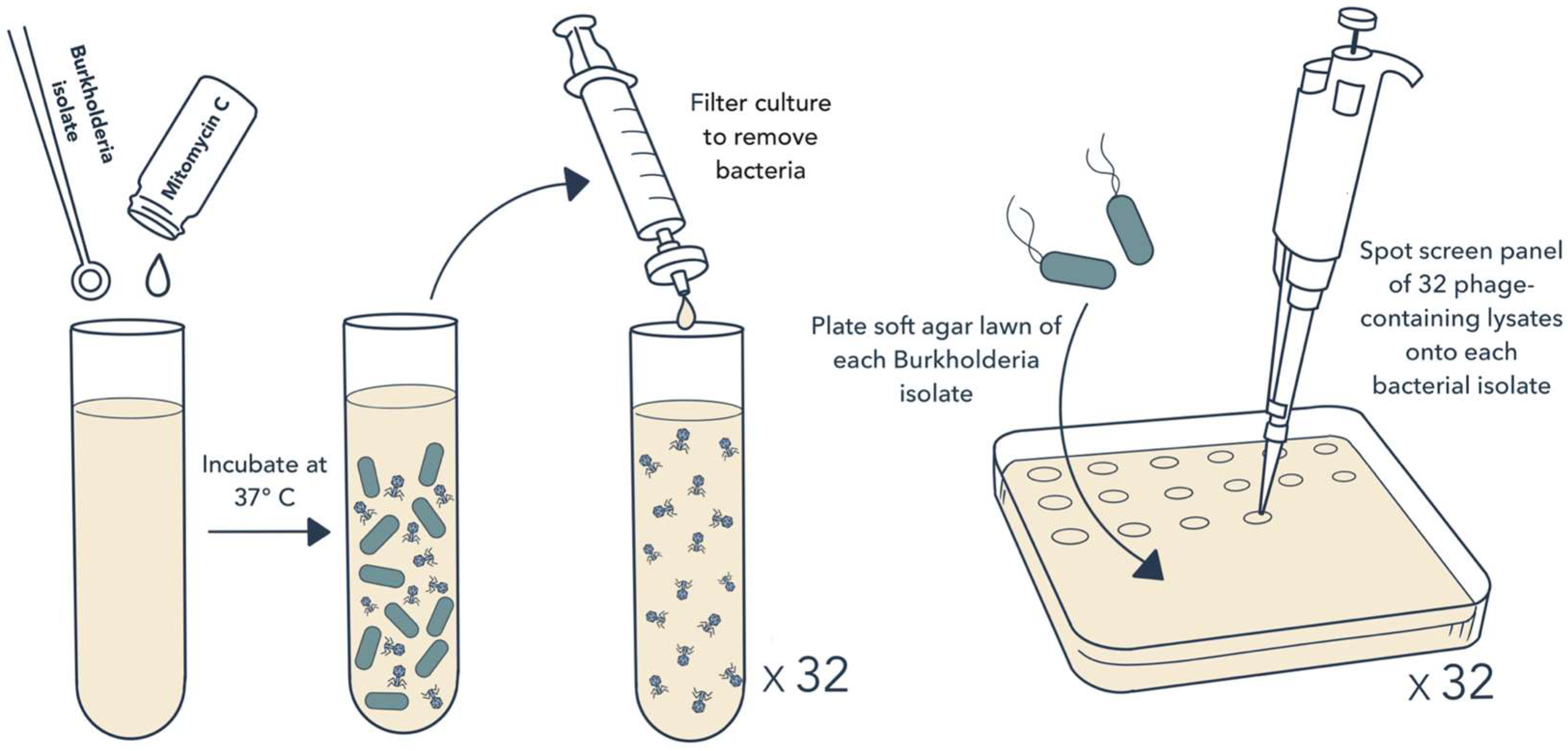
2.1. Bacterial Isolates and Induction of Prophage Release

All Burkholderia spp. isolates used in this study were collected from patients at the University of Pittsburgh Medical Center (UPMC) as part of their routine clinical care. Most isolates were collected as part of the Enhanced Detection System for Healthcare Associated Transmission [27], and others were collected from patients being evaluated for compassionate use phage therapy. Isolate collection was approved by the University of Pittsburgh Institutional Review Board under protocols PRO07060222 and STUDY19110005.
To induce prophage release, 10 µL of stationary phase liquid culture of each bacterial isolate was inoculated into 5 mL of Luria Broth (LB) containing 2.5 µg/mL mitomycin C. Cultures were grown overnight with shaking at 37 °C. The next day, bacterial cells were pelleted, and liquid lysates were filtered through a 0.22 µm syringe filter to remove bacteria. The remaining lysates were presumed to contain phage particles that were released during growth.

2.2. Isolation of Phages and Host Range Testing

Lytic bacteriophage activity was identified with a soft agar overlay assay [28]. Briefly, square petri plates were prepared containing LB bottom agar (LB media with 1.5% agar, 1 mM CaCl2, and 1 mM MgCl2). Bacterial isolates were inoculated into LB media and incubated overnight at 37 °C. The following day, bacterial soft agar lawns were created by mixing 50 µL overnight bacterial culture with 5 mL LB top agar (LB media with 0.5% agarose, 1 mM CaCl2, and 1 mM MgCl2) cooled to 55 °C, plated on top of bottom agar plates, and allowed to solidify. After top agar bacterial lawns had solidified, 5 µL of potential phage-containing lysates were spotted on top of the lawn. Plates were incubated upright at 37 °C overnight. The next day, plates were examined for evidence of lytic phage activity in the form of a spot or clearing of the bacterial lawn. Clearance zones were “picked” with a pipette tip, transferred into 100 µL of suspension media (SM) buffer (50 mM TrisCl pH 7.5, 100 mM NaCl, 8 mM MgSO4), and were incubated overnight at 37 °C. Phage-mediated killing was confirmed by plating 10-fold serial dilutions of phage lysate onto a bacterial lawn in top agar, as above. Each phage was passaged four times before the generation of high-titer stocks.
To prepare high-titer liquid lysates of each phage, individual plaques picked after four rounds of plaque purification were transferred to 100 µL of SM buffer and were then mixed with 100 µL of overnight culture of the propagating bacterial isolate. The mixture was incubated at room temperature for 15 min, then mixed with 10mL of LB top agar, and overlaid onto large (15 cm) bottom agar plates and allowed to set. Plates were incubated overnight at 37 °C. Plates showing semi-confluent lysis were flooded with 10 mL of SM buffer and were incubated at 37 °C for 2 h. The SM lysate was then collected and centrifuged at 4000× g for 10 min to pellet bacteria. Supernatants were filter sterilized through a 0.22 µm membrane syringe filter and were stored for future use.
Host range testing was performed using the soft agar overlay spot screening method [28]. Briefly, 5 µL of 10-fold serial dilutions of each phage lysate was spotted onto top agar lawns of each bacterial isolate and incubated at 37 °C overnight. The following day, each phage-bacteria pairing was assessed for clearance of the bacterial lawn. For pairings where lysis was noted, phage titer in plaque-forming units (PFU)/mL was recorded.

2.3. Whole Genome Sequencing and Analysis

Bacterial genomic DNA was extracted from 1 mL overnight cultures grown in LB media using a Qiagen DNeasy Blood and Tissue Kit (Qiagen, Germantown, MD, USA) following the manufacturer’s protocol. Phage genomic DNA was extracted from aliquots of high-titer liquid lysate using the same kit or via phenol chloroform extraction followed by ethanol precipitation. Briefly, 500 µL phenol/chloroform/isoamyl alcohol (25:24:1) was added to 500 µL of each lysate, and samples were vortexed and then centrifuged at 16,000× g for 1 min. The upper aqueous phase was transferred to a new tube and 500 µL of chloroform was added. Samples were vortexed and centrifuged again at 16,000× g for 1 min, and the upper aqueous phase was again transferred to a new tube. Then, 1 µL glycogen, 0.1 × volume 3 M sodium acetate, and 2.5 × volume 100% ethanol were added, and samples were incubated overnight at −20 °C. The next day, samples were centrifuged at 16,000× g for 30 min at 4 °C, and then the supernatant was removed and the DNA pellet was washed with 150 µL 70% ethanol. DNA pellets were resuspended in 100 µL nuclease-free water. All DNA were quantified with a Qubit fluorimeter (Thermo Fisher Scientific, Waltham, MA, USA). Bacterial and phage genomes were sequenced on the Illumina platform at the Microbial Genome Sequencing (MiGS) Center (Pittsburgh, PA, USA). Illumina library construction and sequencing were conducted using an Illumina Nextera DNA Sample Prep Kit with 150 bp paired-end reads, and libraries were sequenced on the NextSeq 550 sequencing platform (Illumina, San Diego, CA, USA).
Genomes were assembled with SPAdes v3.11 to generate contigs with a 200 bp minimum length cut-off [29]. Phage contigs were extracted from each assembly and were separated from contaminating host bacterial sequences by examining the differential read coverage of each contig, and with BLASTN. Assembled genomes were annotated with RAST [30]. A core genome SNP alignment was generated with snippy v4.6 (https://github.com/tseemann/snippy, accessed on 16 November 2021), then the core alignment was used to construct a phylogenetic tree with RAxML v8.2.12 with the GTRCAT substitution model and 1000 iterations [31]. Bacterial species were assigned by average nucleotide identity comparisons with previously sequenced Burkholderia species using fastANI v1.33 [32]. Prophages were identified in each bacterial genome using PHASTER (https://phaster.ca/) [33]. Prophages of any length that were predicted to be intact, questionable, or incomplete by PHASTER were included. Prophage sequences were compared to one another with BLASTN v2.12 [34], and clusters of similar prophage sequences were identified as those sharing >90% sequence coverage and >90% sequence identity. A cluster analysis of all prophage sequences was performed and visualized using Cytoscape v3.7.2 [35]. Predicted family and genus of each bacteriophage was determined by the closest BLAST match in the NCBI nonredundant (nr) database.

2.4. EM Imaging

Between 1 and 5 µL of bacteriophage BCC02 suspension was added to a copper grid and negatively stained with 1% uranyl acetate. Phage suspension was imaged by transmission electron microscopy on a JEW 1400 Flash Transmission Electron Microscope. Imaging was performed by the University of Pittsburgh Center for Biologic Imaging (Pittsburgh, PA, USA).

2.5. Statistical Analysis

Statistical analyses were performed using GraphPad Prism v8.0.0 (GraphPad Software, San Diego, CA, USA). Linear regression and two-tailed t-tests were performed to assess significance of associations between prophage abundance and phage susceptibility. A Fisher’s Exact Test was used to assess the association between Bmulti_pp1 and phage resistance in B. multivorans isolates.

3. Results

3.1. Prophage Induction and Isolation

To test whether prophages found within the genomes of Burkholderia spp. clinical isolates could target other Burkholderia spp. isolates, we first created a library of potential phage-containing lysates. To do this, we inoculated 32 Burkholderia spp. clinical isolates collected from 28 unique patients into liquid culture in the presence of the mutagen mitomycin C, which prompts prophage excision (Figure 1) [36,37]. We then performed an all-by-all screen of the 32 phage-containing “source” lysates against the same 32 Burkholderia spp. “target” isolates, using a spot-plaque screening method to perform 1024 pairwise tests (Figure 1). For 11 of these tests, a phage-containing source lysate inhibited the growth of the target bacterial lawn (1.1% hit rate). Each positive pairing was retested to confirm that the observed inhibitory activity was due to phage activity by serially diluting the source lysate and looking for individual plaques. The 10 source–target pairings with confirmed phage activity were then subjected to four rounds of picking and passaging of single plaques to isolate individual phages. Three of these pairings did not maintain lytic activity through picking and passaging, meaning that during one of the passages, no plaques were visible. The seven remaining source–target pairs yielded viable phages, which were amplified into high-titer lysates and designated as BCC02 through BCC08 (Table S1).

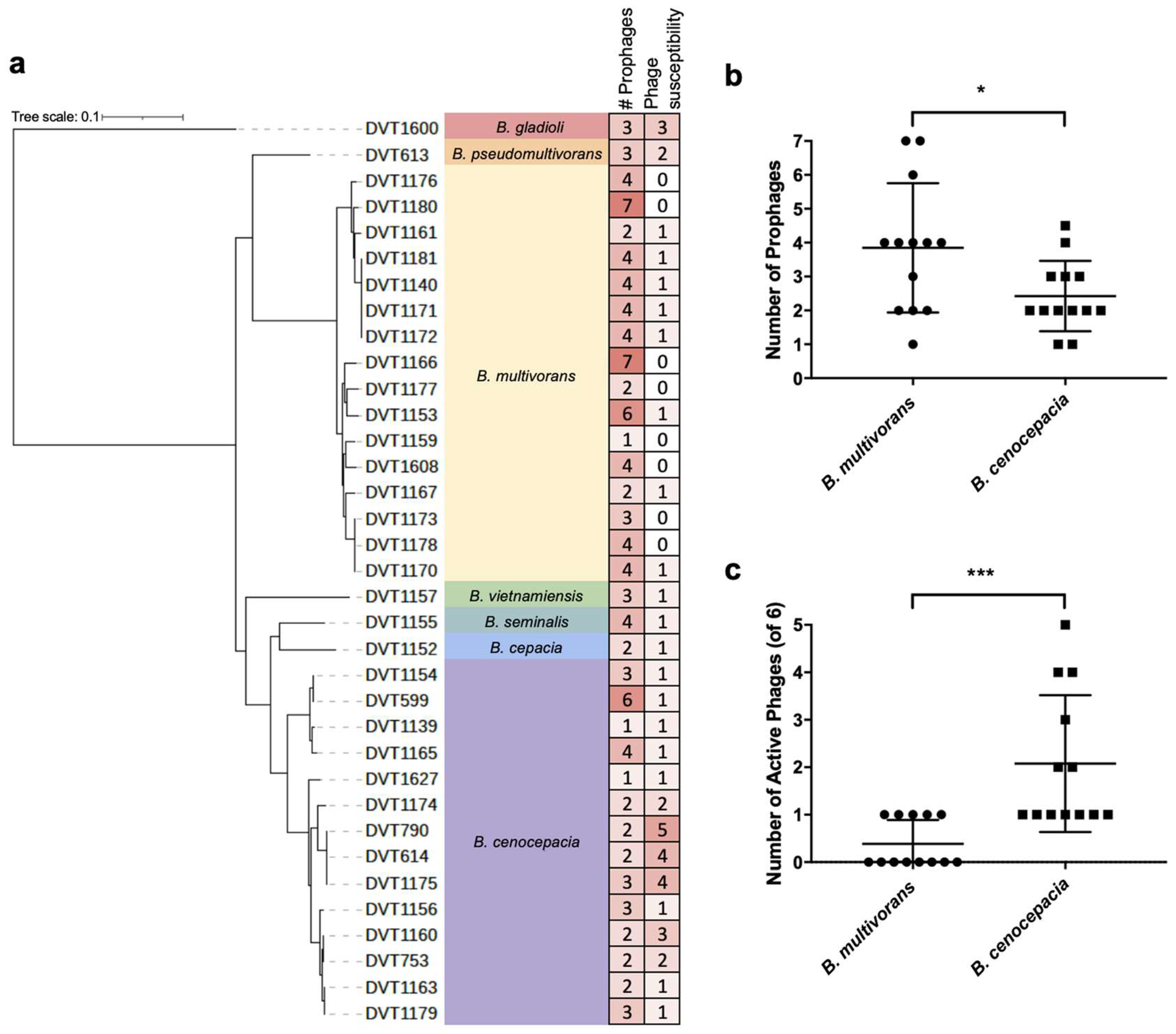
3.2. Whole Genome Sequencing of Burkholderia spp. Clinical Isolates

To explore the genetic diversity of the Burkholderia spp. clinical isolates studied here, we sequenced 35 bacterial genomes, including the 32 isolates used for prophage induction and testing, plus three additional clinical isolates. Isolates were sequenced on the Illumina platform and a core genome phylogeny was constructed (Figure 2). The species of each isolate was determined using fastANI [32] to compare the average nucleotide identity between each genome and available genomes of type strains of Burkholderia spp. The most frequently sampled species were B. multivorans (16 isolates collected from 13 patients) and B. cenocepacia (14 isolates collected from 13 patients). One isolate each belonged to Burkholderia gladioli, Burkholderia pseudomultivorans, Burkholderia vietnamiensis, Burkholderia seminalis, and Burkholderia cepacia (Figure 2). All isolates except DVT1600 (Burkholderia gladioli) fell into the B. cepacia complex (Figure 2).

3.3. Phage Host-Range Screening

To determine the infectivity profile of each isolated temperate phage as well as the phage susceptibility of each Burkholderia spp. clinical isolate, we performed an all-by-all phage activity screen. We tested all seven prophage-derived phages as well as two additional Burkholderia-targeting bacteriophages of environmental origin, DSMZ107315 and Bch7 [19], against all 35 Burkholderia spp. clinical isolates (Figure 2). Eight isolates, all of which were B. multivorans, were resistant to all phages tested. However, the other 27 isolates (77.1% of all isolates tested) were susceptible to at least one phage. We observed variability in the host range of each phage, with phages able to lyse between 2 (BCC06) and 12 (Bch7) of the 35 isolates. All phages were able to infect and lyse multiple isolates, and in most cases, phages were able to lyse bacteria belonging to different species (Figure 2). We observed similar host ranges for phages BCC02, BCC03, and BCC04, as well as for phages BCC05 and BCC06. This finding, along with the fact that these groups contained phages derived from the same donor bacterial isolates (Table S1), suggested that these might represent duplicate isolations of the same phage. Whole genome sequencing confirmed this, as described further below.

3.4. Whole Genome Sequencing of Phages

To explore the genetic diversity of the phages tested in this study, phage lysates were subjected to genomic DNA extraction and sequencing on the Illumina platform. High-coverage phage-derived contigs were analyzed and were compared to each other, to bacterial prophage genomes, and to publicly available genomes (Table 1). Phage genomes ranged in size from 23 to 68 kb and varied in GC content from 54.7 to 67.1% (Table 1). Phages BCC02, BCC03, and BCC04, which were all isolated from the DVT1180 source isolate, were found to be genetically identical to one another. Phage BCC02 therefore was chosen as the representative phage for follow-up work. Likewise, phages BCC05 and BCC06, which were both isolated from the DVT1166 source isolate, were found to be identical except for one single nucleotide polymorphism within a gene predicted to encode a phage tail fiber protein, suggesting divergent evolution during the course of phage isolation. However, as we observed similar host ranges between BCC05 and BCC06, for the purposes of our analysis, we also considered these two phages to be identical and chose BCC05 as the representative phage. The family and genus of each phage were predicted based on sequence comparisons with previously described phages deposited in the NCBI using nucleotide BLAST (Table 1). Transmission electron microscopy (TEM) imaging of phage BCC02 confirmed that it had a contractile tail and icosahedral head (Figure 3). Overall, genomic analyses revealed that our approach yielded four novel temperate bacteriophages targeting Burkholderia spp. (BCC02, BCC05, BCC07, and BCC08) that were distinct from each other and from other known phages.

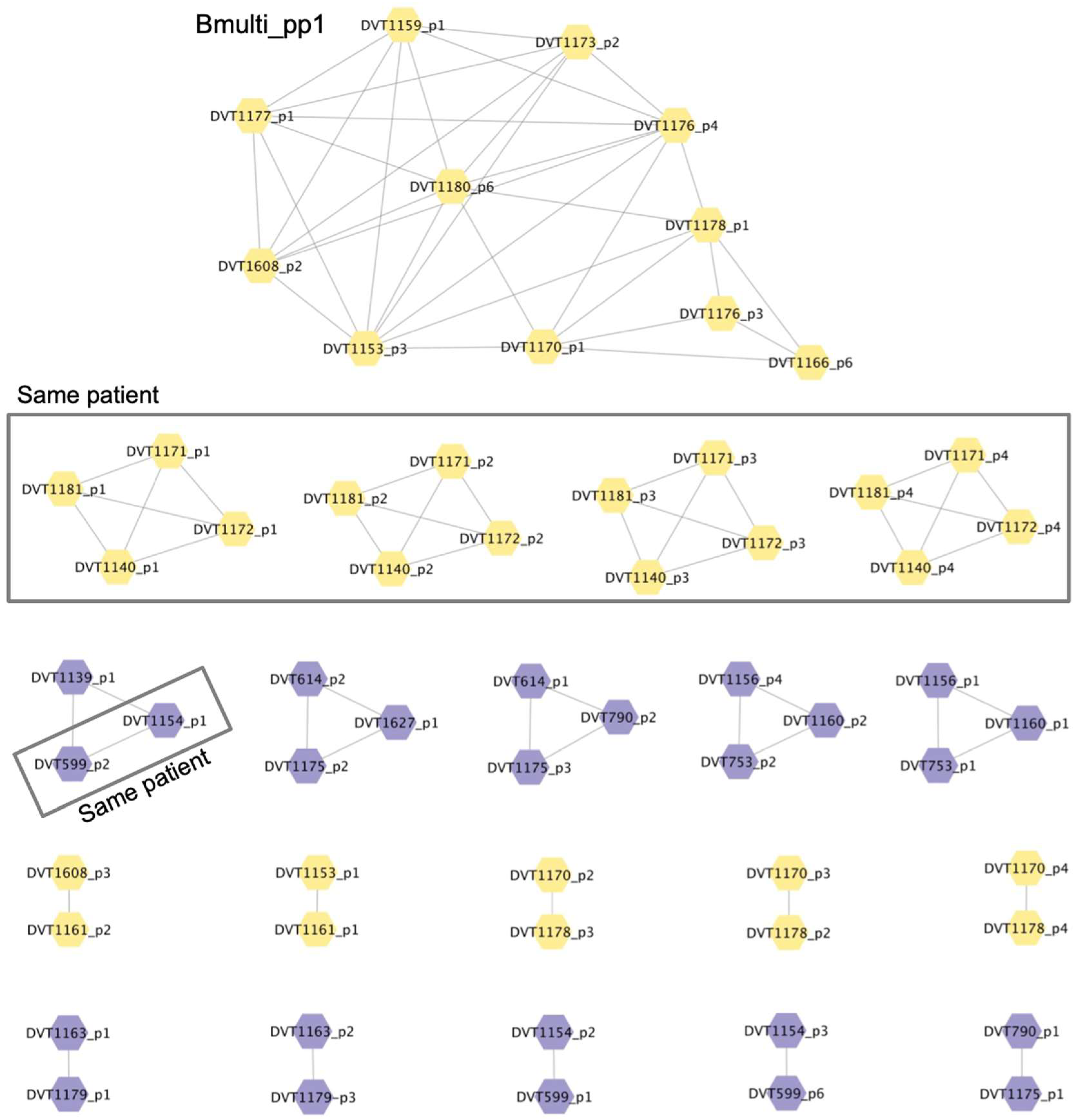
3.5. Analysis of Prophage Carriage in Burkholderia spp. Clinical Isolates

To understand the diversity of prophages present in the Burkholderia spp. clinical isolates we collected, we extracted prophage sequences from the genomes of the 35 isolates used for screening. We searched each isolate genome for sequences of likely prophage origin using PHASTER [33]. A total of 114 prophage sequences were identified (range = 1–7, median = 3 prophages per genome) (Table S2) (Figure 4a). We tested whether prophage abundance was associated with phage susceptibility and found that these two features were only loosely correlated with one another (linear regression p = 0.0675) (Figures S1 and S2). However, we did find that B. multivorans isolates contained more prophages compared with B. cenocepacia isolates (3.8 vs. 2.4 average prophages, p = 0.0266) (Figure 4b). Additionally, B. multivorans isolates were susceptible to fewer phages compared with B. cenocepacia isolates (p = 0.0005) (Figure 4c), in agreement with other studies, demonstrating an inverse correlation between prophage abundance and phage susceptibility in different bacterial species [38,39,40].
To determine the extent of relatedness among prophages encoded in Burkholderia spp. clinical isolate genomes, we compared all extracted prophage sequences to one another using nucleotide BLAST [34]. Prophages that shared >90% sequence coverage and >90% sequence identity were grouped into clusters, and clusters were visualized using Cytoscape [35] (Figure 5) (Table S2). Twenty clusters of similar prophages were identified, which ranged in size from 2 to 4 with the exception of one large cluster containing 11 prophages. Overall, prophage clusters tended to group with related host strains. For example, our screening panel contained two sets of isolates that were gathered at different time points from the same patient; DVT1140, DVT1171, DVT1172, and DVT1181 (B. multivorans) were collected from one patient, while DVT599 and DVT1154 (B. cenocepacia) were collected from another. The prophages present in these isolates clustered together, as expected (Figure 5) (Table S2). Isolated phages BCC02/BCC03/BCC04, BCC05/BCC06, BCC07, and BCC08 did not fall into any of these clusters. Finally, one cluster, which we named Bmulti_pp1, contained 11 prophage sequences from 10 B. multivorans isolates. Presence of this prophage was significantly associated with phage resistance among the B. multivorans isolates we tested (p = 0.035) (Figure S3). This finding suggests that Bmulti_pp1 may provide protection against phage-mediated lysis in B. multivorans.

4. Discussion

The establishment and maintenance of lysogeny in bacterial hosts by temperate phages is a widespread phenomenon that involves a complex interplay of elements both on cellular and ecological levels. These interactions are influenced by many factors, including genetics, cellular development, community dynamics, and environmental conditions [41]. In this study, we show that, in some cases, the relationship between bacteria and their prophages can be exploited to isolate bacteriophages with potential therapeutic use and that prophage induction with mitomycin C is an effective method to accomplish this in Burkholderia spp.
Polylysogeny in Burkholderia appears to be common [24], and prophages present in Burkholderia genomes likely represent a rich hunting ground for clinically useful bacteriophages [42]. Triggering the bacterial DNA damage response using a mutagen like mitomycin C is a simple way to activate the lysogenic-lytic switch for some prophages [43], and the prophages we induced with this method showed lytic activity against both conspecific and heterospecific bacterial isolates. However, further characterization and rational engineering of temperate phages usually is considered to be necessary before they can be used clinically. Engineering of Burkholderia prophages to render them obligately lytic, either through site-directed mutagenesis or experimental evolution, has been previously described [44,45]. Similar approaches could be taken with the prophage-derived phages described here to convert them from temperate to lytic phages. Such exploration of the use of temperate bacteriophages in phage therapy has the potential to expand the pool of useful tools against the escalating threat of multidrug-resistant bacteria when naturally occurring obligately lytic phages are rare.
In contrast to prior studies [46,47], here we performed a systematic screening of lytic phage activity among induced prophages using more than 30 genetically diverse Burkholderia spp. clinical isolates. The four temperate phages we studied showed combined activity against 60% (21/35) of the clinical isolates that were tested. The isolated prophage BCC02, while a temperate phage, was able to lyse 31% (11/35) of the clinical isolates used in our study. This phage is genetically similar to the previously described Burkholderia phage KS5 isolated by Seed et al., which also demonstrated broad lytic activity [48]. The four novel temperate phages isolated and described in this study have potential for further development toward therapeutic application. With the exception of BCC05 and BCC06, the host ranges of phages of prophage origin were comparable to those of environmental origin, in agreement with the previous literature [48,49]. Additionally, identification of phages with varying host ranges and infection dynamics allows for the development of multi-phage cocktails in order to reduce the probability of phage resistance [46].
Our findings demonstrate that clinically significant Burkholderia isolates host a variety of prophage elements, in agreement with previous studies, showing that lysogeny is relatively common in this genus [3,49,50]. While our study was likely underpowered to identify a strong correlation between prophage abundance and phage susceptibility, we found more prophages and greater overall phage resistance in B. multivorans isolates compared to B. cenocepacia isolates. A prior study similarly noted the relative phage resistance of B. multivorans isolates [49]. We also identified one prophage that was associated with resistance to phage lysis, which may indicate some evolutionary or ecological relevance, possibly enabling superinfection immunity. The occurrence of this prophage could indicate ancestral integration maintained through a fitness advantage or direct phage transmission between clinical strains in close contact with one another. Exploring determinants of prophage infectivity and the effects of the Bmulti_pp1 prophage on infection dynamics of other phages require further study.
This study had several limitations. The small volume spot-screening method used to isolate induced phages may have missed phages that were present in lysates at low concentration. We did not investigate the efficiency of temperate phage induction or how well it correlated with the recovery of an active temperate phage. Additionally, slow-growing clinical isolates may have hindered the detection of lytic activity, since some temperate phages are known to induce lysis only when the host density is high [41]. All genomes used for our analysis were draft genomes, and many were not able to be completely assembled within the scope of this project. This could potentially skew our analysis of the number of prophages in each isolate, as some prophages may have spanned multiple contigs. Finally, all work in this study was performed in vitro, thus we are unable to conclude that these newly characterized phages would be useful for clinical therapy without further testing in a relevant in vivo model of infection.

5. Conclusions

Overall, the data generated in this study add to the literature characterizing Burkholderia-targeting bacteriophages, as well as prophage abundance and diversity in clinically relevant Burkholderia species. We isolated four novel phages with lytic activity against a variety of Burkholderia isolates, identified temperate phages with a broad host range, and detected an association between one particular prophage and phage resistance in B. multivorans. The isolated phages of prophage origin may also have clinical utility and are a potentially useful starting point for the development of novel phages for use in phage therapy.

Supplementary Materials

The following supporting information can be downloaded at: https://www.mdpi.com/article/10.3390/cells13050428/s1, Figure S1: Prophage abundance versus phage susceptibility among 35 Burkholderia spp. isolates; Figure S2: Prophage abundance among isolates that are or are not susceptible to each phage used in the study; Figure S3: Prophage Bmulti_pp1 is associated with phage resistance in B. multivorans; Table S1: Burkholderia spp. prophage-derived phages; Table S2: Prophages identified in Burkholderia spp. bacterial isolate genomes.

Author Contributions

Conceptualization, H.R.N. and D.V.T.; methodology, H.R.N., M.P.G. and V.R.S.; investigation, H.R.N., N.R.W. and A.L.; writing—original draft preparation, H.R.N. and D.V.T.; writing—review and editing, H.R.N., M.P.G., V.R.S., N.R.W., A.L., V.S.C., R.K.S. and D.V.T.; visualization, H.R.N. and D.V.T.; funding acquisition, D.V.T. All authors have read and agreed to the published version of the manuscript.

Funding

This research was funded by the Cystic Fibrosis Foundation, grant number BOMBER19R0, and by the Department of Medicine at the University of Pittsburgh School of Medicine.

Institutional Review Board Statement

The study was conducted in accordance with the Declaration of Helsinki, and approved by the Institutional Review Board of the University of Pittsburgh under protocols PRO07060222 and STUDY19110005.

Informed Consent Statement

Patient consent was waived by the Institutional Review Board of the University of Pittsburgh due to the collection and study of bacterial isolates that were collected as part of routine clinical care, contained no human DNA, were stored de-identified from patient information, and which would have otherwise been discarded.

Data Availability Statement

Whole genome sequencing of bacterial isolates and phages have been submitted to the NCBI under BioProject PRJNA1067351. Other data used in the manuscript are available in Supplementary Materials. Bacterial isolates and phages are available from the corresponding author under the terms of a Materials Transfer Agreement.

Acknowledgments

We gratefully acknowledge Donna Stolz for assistance with EM imaging of BCC02. Many clinical Burkholderia spp. isolates were kindly provided by Lee Harrison from the EDS-HAT project.

Conflicts of Interest

The authors declare no conflicts of interest. The funders had no role in the design of the study; in the collection, analyses, or interpretation of data; in the writing of the manuscript; or in the decision to publish the results.

References

  1. Sawana, A.; Adeolu, M.; Gupta, R.S. Molecular Signatures and Phylogenomic Analysis of the Genus Burkholderia: Proposal for Division of This Genus into the Emended Genus Burkholderia Containing Pathogenic Organisms and a New Genus Paraburkholderia Gen. Nov. Harboring Environmental Species. Front. Genet. 2014, 5, 429. [Google Scholar] [CrossRef]
  2. Coenye, T.; Vandamme, P.; Govan, J.R.W.; LiPuma, J.J. Taxonomy and Identification of the Burkholderia cepacia Complex. J. Clin. Microbiol. 2001, 39, 3427–3436. [Google Scholar] [CrossRef]
  3. Mahenthiralingam, E.; Urban, T.A.; Goldberg, J.B. The Multifarious, Multireplicon Burkholderia cepacia Complex. Nat. Rev. Microbiol. 2005, 3, 144–156. [Google Scholar] [CrossRef]
  4. Somayaji, R.; Yau, Y.C.W.; Tullis, E.; LiPuma, J.J.; Ratjen, F.; Waters, V. Clinical Outcomes Associated with Burkholderia cepacia Complex Infection in Patients with Cystic Fibrosis. Ann. Am. Thorac. Soc. 2020, 17, 1542–1548. [Google Scholar] [CrossRef] [PubMed]
  5. Lauman, P.; Dennis, J.J. Advances in Phage Therapy: Targeting the Burkholderia cepacia Complex. Viruses 2021, 13, 1331. [Google Scholar] [CrossRef] [PubMed]
  6. LiPuma, J.J.; Spilker, T.; Gill, L.H.; Campbell, P.W., 3rd; Liu, L.; Mahenthiralingam, E. Disproportionate Distribution of Burkholderia cepacia Complex Species and Transmissibility Markers in Cystic Fibrosis. Am. J. Respir. Crit. Care Med. 2001, 164, 92–96. [Google Scholar] [CrossRef] [PubMed]
  7. Govan, J.R.; Deretic, V. Microbial Pathogenesis in Cystic Fibrosis: Mucoid Pseudomonas Aeruginosa and Burkholderia cepacia. Microbiol. Rev. 1996, 60, 539–574. [Google Scholar] [CrossRef] [PubMed]
  8. Jones, A.; Dodd, M.; Webb, A. Burkholderia cepacia: Current Clinical Issues, Environmental Controversies and Ethical Dilemmas. Eur. Respir. J. 2001, 17, 295–301. [Google Scholar] [CrossRef] [PubMed]
  9. Branstetter, J.W.; Yarbrough, A.; Poole, C. Management of Cepacia Syndrome with a Combination of Intravenous and Inhaled Antimicrobials in a Non-Cystic Fibrosis Pediatric Patient. J. Pediatr. Pharmacol. Ther. 2020, 25, 730–734. [Google Scholar] [CrossRef] [PubMed]
  10. Zahariadis, G.; Levy, M.H.; Burns, J.L. Cepacia-Like Syndrome Caused by Burkholderia multivorans. Can. J. Infect. Dis. 2003, 14, 123–125. [Google Scholar] [PubMed]
  11. Burns, J.L.; Wadsworth, C.D.; Barry, J.J.; Goodall, C.P. Nucleotide Sequence Analysis of a Gene from Burkholderia (Pseudomonas) cepacia Encoding an Outer Membrane Lipoprotein Involved in Multiple Antibiotic Resistance. Antimicrob. Agents Chemother. 1996, 40, 307–313. [Google Scholar] [CrossRef] [PubMed]
  12. Hancock, R.E. Peptide Antibiotics. Lancet 1997, 349, 418–422. [Google Scholar] [CrossRef] [PubMed]
  13. Middleton, P.G.; Kidd, T.J.; Williams, B. Combination Aerosol Therapy to Treat Burkholderia cepacia Complex. Eur. Respir. J. 2005, 26, 305–308. [Google Scholar] [CrossRef] [PubMed]
  14. Dedrick, R.M.; Guerrero-Bustamante, C.A.; Garlena, R.A.; Russell, D.A.; Ford, K.; Harris, K.; Gilmour, K.C.; Soothill, J.; Jacobs-Sera, D.; Schooley, R.T.; et al. Engineered Bacteriophages for Treatment of a Patient with a Disseminated Drug-Resistant Mycobacterium Abscessus. Nat. Med. 2019, 25, 730–733. [Google Scholar] [CrossRef] [PubMed]
  15. Suh, G.A.; Lodise, T.P.; Tamma, P.D.; Knisely, J.M.; Alexander, J.; Aslam, S.; Barton, K.D.; Bizzell, E.; Totten, K.M.C.; Campbell, J.L.; et al. Considerations for the Use of Phage Therapy in Clinical Practice. Antimicrob. Agents Chemother. 2022, 66, e0207121. [Google Scholar] [CrossRef] [PubMed]
  16. Schooley, R.T.; Biswas, B.; Gill, J.J.; Hernandez-Morales, A.; Lancaster, J.; Lessor, L.; Barr, J.J.; Reed, S.L.; Rohwer, F.; Benler, S.; et al. Development and Use of Personalized Bacteriophage-Based Therapeutic Cocktails to Treat a Patient with a Disseminated Resistant Acinetobacter Baumannii Infection. Antimicrob. Agents Chemother. 2017, 61. [Google Scholar] [CrossRef]
  17. Aslam, S.; Courtwright, A.M.; Koval, C.; Lehman, S.M.; Morales, S.; Furr, C.L.; Rosas, F.; Brownstein, M.J.; Fackler, J.R.; Sisson, B.M.; et al. Early Clinical Experience of Bacteriophage Therapy in 3 Lung Transplant Recipients. Am. J. Transplant. 2019, 19, 2631–2639. [Google Scholar] [CrossRef]
  18. Smith, M. Salt in My Soul: An Unfinished Life; Random House: New York, NY, USA, 2019. [Google Scholar]
  19. Haidar, G.; Chan, B.K.; Cho, S.; Kramer, K.H.; Nordstrom, H.R.; Wallace, N.R.; Stellfox, M.E.; Holland, M.; Kline, E.G.; Kozar, J.M.; et al. Phage Therapy in a Lung Transplant Recipient with Cystic Fibrosis Infected with Multidrug-Resistant Burkholderia Multivorans. Transpl. Infect. Dis. 2023, 25, e14041. [Google Scholar] [CrossRef]
  20. Roszniowski, B.; Latka, A.; Maciejewska, B.; Vandenheuvel, D.; Olszak, T.; Briers, Y.; Holt, G.S.; Valvano, M.A.; Lavigne, R.; Smith, D.L.; et al. The Temperate Burkholderia Phage Ap3 of the Peduovirinae Shows Efficient Antimicrobial Activity against B. Cenocepacia of the Iiia Lineage. Appl. Microbiol. Biotechnol. 2017, 101, 1203–1216. [Google Scholar] [CrossRef]
  21. Seed, K.D.; Dennis, J.J. Experimental Bacteriophage Therapy Increases Survival of Galleria Mellonella Larvae Infected with Clinically Relevant Strains of the Burkholderia cepacia Complex. Antimicrob. Agents Chemother. 2009, 53, 2205–2208. [Google Scholar] [CrossRef]
  22. Carmody, L.A.; Gill, J.J.; Summer, E.J.; Sajjan, U.S.; Gonzalez, C.F.; Young, R.F.; LiPuma, J.J. Efficacy of Bacteriophage Therapy in a Model of Burkholderia cenocepacia Pulmonary Infection. J. Infect. Dis. 2010, 201, 264–271. [Google Scholar] [CrossRef]
  23. Semler, D.D.; Goudie, A.D.; Finlay, W.H.; Dennis, J.J. Aerosol Phage Therapy Efficacy in Burkholderia cepacia Complex Respiratory Infections. Antimicrob. Agents Chemother. 2014, 58, 4005–4013. [Google Scholar] [CrossRef]
  24. Roszniowski, B.; McClean, S.; Drulis-Kawa, Z. Burkholderia cenocepacia Prophages-Prevalence, Chromosome Location and Major Genes Involved. Viruses 2018, 10, 297. [Google Scholar] [CrossRef]
  25. Dedrick, R.M.; Smith, B.E.; Garlena, R.A.; Russell, D.A.; Aull, H.G.; Mahalingam, V.; Divens, A.M.; Guerrero-Bustamante, C.A.; Zack, K.M.; Abad, L.; et al. Mycobacterium Abscessus Strain Morphotype Determines Phage Susceptibility, the Repertoire of Therapeutically Useful Phages, and Phage Resistance. mBio 2021, 12. [Google Scholar] [CrossRef]
  26. Raya, R.R.; H’Bert, M. Isolation of Phage Via Induction of Lysogens. Methods Mol. Biol. 2009, 501, 23–32. [Google Scholar]
  27. Sundermann, A.J.; Chen, J.; Kumar, P.; Ayres, A.M.; Cho, S.T.; Ezeonwuka, C.; Griffith, M.P.; Miller, J.K.; Mustapha, M.M.; Pasculle, A.W.; et al. Whole-Genome Sequencing Surveillance and Machine Learning of the Electronic Health Record for Enhanced Healthcare Outbreak Detection. Clin. Infect. Dis. 2022, 75, 476–482. [Google Scholar] [CrossRef]
  28. Russell, D.A.; Clokie, M.R.J.; Kropinski, A.M.; Lavigne, R. Bacteriophages: Methods and Protocols; Springer: Berlin/Heidelberg, Germany, 2018. [Google Scholar]
  29. Bankevich, A.; Nurk, S.; Antipov, D.; Gurevich, A.A.; Dvorkin, M.; Kulikov, A.S.; Lesin, V.M.; Nikolenko, S.I.; Pham, S.; Prjibelski, A.D.; et al. Spades: A New Genome Assembly Algorithm and Its Applications to Single-Cell Sequencing. J. Comput. Biol. 2012, 19, 455–477. [Google Scholar] [CrossRef]
  30. Aziz, R.K.; Bartels, D.; Best, A.A.; DeJongh, M.; Disz, T.; Edwards, R.A.; Formsma, K.; Gerdes, S.; Glass, E.M.; Kubal, M.; et al. The Rast Server: Rapid Annotations Using Subsystems Technology. BMC Genom. 2008, 9, 75. [Google Scholar] [CrossRef] [PubMed]
  31. Stamatakis, A. Raxml Version 8: A Tool for Phylogenetic Analysis and Post-Analysis of Large Phylogenies. Bioinformatics 2014, 30, 1312–1313. [Google Scholar] [CrossRef] [PubMed]
  32. Jain, C.; Rodriguez, R.L.; Phillippy, A.M.; Konstantinidis, K.T.; Aluru, S. High Throughput Ani Analysis of 90k Prokaryotic Genomes Reveals Clear Species Boundaries. Nat. Commun. 2018, 9, 5114. [Google Scholar] [CrossRef] [PubMed]
  33. Arndt, D.; Marcu, A.; Liang, Y.; Wishart, D.S. Phast, Phaster and Phastest: Tools for Finding Prophage in Bacterial Genomes. Brief. Bioinform. 2017, 20, 1560–1567. [Google Scholar] [CrossRef]
  34. Altschul, S.F.; Gish, W.; Miller, W.; Myers, E.W.; Lipman, D.J. Basic Local Alignment Search Tool. J. Mol. Biol. 1990, 215, 403–410. [Google Scholar] [CrossRef] [PubMed]
  35. Shannon, P.; Markiel, A.; Ozier, O.; Baliga, N.S.; Wang, J.T.; Ramage, D.; Amin, N.; Schwikowski, B.; Ideker, T. Cytoscape: A Software Environment for Integrated Models of Biomolecular Interaction Networks. Genome Res. 2003, 13, 2498–2504. [Google Scholar] [CrossRef] [PubMed]
  36. Oliveira, J.; Mahony, J.; Hanemaaijer, L.; Kouwen, T.R.H.M.; Neve, H.; MacSharry, J.; van Sinderen, D. Detecting Lactococcus Lactis Prophages by Mitomycin C-Mediated Induction Coupled to Flow Cytometry Analysis. Front. Microbiol. 2017, 8, 1343. [Google Scholar] [CrossRef]
  37. Otsuji, N.; Sekiguchi, M.; Iijima, T.; Takagi, Y. Induction of Phage Formation in the Lysogenic Escherichia Coli K-12 by Mitomycin C. Nature 1959, 184 (Suppl. S14), 1079–1080. [Google Scholar] [CrossRef] [PubMed]
  38. Bondy-Denomy, J.; Qian, J.; Westra, E.R.; Buckling, A.; Guttman, D.S.; Davidson, A.R.; Maxwell, K.L. Prophages Mediate Defense against Phage Infection through Diverse Mechanisms. ISME J. 2016, 10, 2854–2866. [Google Scholar] [CrossRef] [PubMed]
  39. Dedrick, R.M.; Jacobs-Sera, D.; Bustamante, C.A.G.; Garlena, R.A.; Mavrich, T.N.; Pope, W.H.; Reyes, J.C.C.; Russell, D.A.; Adair, T.; Alvey, R.; et al. Prophage-Mediated Defence against Viral Attack and Viral Counter-Defence. Nat. Microbiol. 2017, 2, 16251. [Google Scholar] [CrossRef]
  40. Harrison, E.; Brockhurst, M.A. Ecological and Evolutionary Benefits of Temperate Phage: What Does or Doesn’t Kill You Makes You Stronger. BioEssays 2017, 39, 1700112. [Google Scholar] [CrossRef]
  41. Howard-Varona, C.; Hargreaves, K.R.; Abedon, S.T.; Sullivan, M.B. Lysogeny in Nature: Mechanisms, Impact and Ecology of Temperate Phages. ISME J. 2017, 11, 1511–1520. [Google Scholar] [CrossRef]
  42. Monteiro, R.; Pires, D.P.; Costa, A.R.; Azeredo, J. Phage Therapy: Going Temperate? Trends Microbiol. 2019, 27, 368–378. [Google Scholar] [CrossRef]
  43. Tomasz, M. Mitomycin C: Small, Fast and Deadly (but Very Selective). Chem. Biol. 1995, 2, 575–579. [Google Scholar] [CrossRef]
  44. Lynch, K.H.; Seed, K.D.; Stothard, P.; Dennis, J.J. Inactivation of Burkholderia cepacia Complex Phage Ks9 Gp41 Identifies the Phage Repressor and Generates Lytic Virions. J. Virol. 2010, 84, 1276–1288. [Google Scholar] [CrossRef]
  45. Yao, G.; Le, T.; Korn, A.M.; Peterson, H.N.; Liu, M.; Gonzalez, C.F.; Gill, J.J. Phage Milagro: A Platform for Engineering a Broad Host Range Virulent Phage for Burkholderia. J. Virol. 2023, 97, e0085023. [Google Scholar] [CrossRef]
  46. Lauman, P.; Dennis, J.J. Synergistic Interactions among Burkholderia cepacia Complex-Targeting Phages Reveal a Novel Therapeutic Role for Lysogenization-Capable Phages. Microbiol. Spectr. 2023, 11, e0443022. [Google Scholar] [CrossRef]
  47. Al-Anany, A.M.; Fatima, R.; Hynes, A.P. Temperate Phage-Antibiotic Synergy Eradicates Bacteria through Depletion of Lysogens. Cell Rep. 2021, 35, 109172. [Google Scholar] [CrossRef] [PubMed]
  48. Seed, K.D.; Dennis, J.J. Isolation and Characterization of Bacteriophages of the Burkholderia cepacia Complex. FEMS Microbiol. Lett. 2005, 251, 273–280. [Google Scholar] [CrossRef]
  49. Langley, R.; Kenna, D.T.; Vandamme, P.; Ure, R.; Govan, J.R.W. Lysogeny and Bacteriophage Host Range within the Burkholderia cepacia Complex. J. Med. Microbiol. 2003, 52 Pt 6, 483–490. [Google Scholar] [CrossRef] [PubMed]
  50. Weiser, R.; Yap, Z.L.; Otter, A.; Jones, B.V.; Salvage, J.; Parkhill, J.; Mahenthiralingam, E. A Novel Inducible Prophage from Burkholderia Vietnamiensis G4 Is Widely Distributed across the Species and Has Lytic Activity against Pathogenic Burkholderia. Viruses 2020, 12, 601. [Google Scholar] [CrossRef]
Figure 1. Approach for prophage induction and testing. To induce prophage release, stationary phase liquid cultures of 32 Burkholderia spp. clinical isolates were individually inoculated into LB media containing mitomycin C. Cultures were grown overnight at 37 °C. The next day, bacterial cells were pelleted and liquid lysates were filtered through a 0.22 μm filter. Filtered lysates were then spotted onto soft agar lawns containing each of the same 32 Burkholderia spp. clinical isolates. Plates were examined for growth inhibition, then inhibitory lysates were retested to confirm phage activity.
Cells 13 00428 g001
Figure 2. Phage activity against genetically diverse Burkholderia spp. clinical isolates. Bacterial isolates are ordered according to their core genome phylogeny and are grouped by species. Grey boxes indicate isolates collected from the same patient. Infectivity is shown as the log10 titer (PFU/mL) of each phage against each isolate. Bolded values indicate the Burkholderia spp. target isolate that each phage was isolated and propagated on. Asterisks mark the source Burkholderia spp. isolate for each phage. Green shading corresponds to phage activity titer, with darker shading indicating a higher titer. Empty cells indicate no phage activity.
Cells 13 00428 g002
Figure 3. Electron micrograph of temperate phage BCC02. Transmission electron micrograph showing Burkholderia phage BCC02. Image was taken at 200,000× magnification. Icosahedral head, tail, and contractile tail sheath are visible for 6 virions.
Cells 13 00428 g003
Figure 4. Prophage abundance and phage susceptibility of diverse Burkholderia spp. clinical isolates. (a) Core genome phylogeny of 35 Burkholderia spp. clinical isolates showing the number of prophages and phage susceptibility of each isolate. For phage susceptibility, BCC02/BCC03/BCC04 and BCC05/BCC06 were each combined into a single count. Darker red shading corresponds with higher values. (b) Prophage abundance among B. multivorans and B. cenocepacia genomes. (c) Phage susceptibility among B. multivorans and B. cenocepacia isolates. p-values are from unpaired two-tailed t-tests. * p < 0.05; *** p < 0.001.
Cells 13 00428 g004
Figure 5. Clusters of prophages encoded by Burkholderia spp. clinical isolate genomes. Bacterial isolate names and prophage numbers are listed inside the nodes of each cluster, and lines connect prophages that share >90% sequence coverage and >90% sequence identity. Yellow nodes indicate prophages in B. multivorans isolate genomes and purple nodes indicate prophages in B. cenocepacia isolate genomes. Isolates from the same patient (two separate patients) are boxed.
Cells 13 00428 g005
Table 1. Genome summary of Burkholderia-targeting phages.
Phage IDSource IsolateSource SpeciesLength (bp)GC %Predicted Genus 1NCBI Similar Phage 2
BCC02/03/04DVT1180B. multivorans34,12664.2Peduovirinae;
Kisquinquevirus
Burkholderia Phage KS5 (GU911303.1)
BCC05/06DVT1166B. multivorans30,95762.8Peduoviridae;
Duodecimduovirus
Burkholderia Phage phiE12-2 (NC_009236.1)
BCC07DVT1155B. seminalis38,21666.7Peduoviridae;
Aptresvirus
Burkholderia Phage Mana (NC_055863.1)
BCC08DVT1155B. seminalis28,70963.7Peduoviridae;
Kayeltresvirus
Burkholderia Phage KL3 (GU911304.1)
Bch7Env 3-68,16654.7BcepfunavirusBurkholderia Phage Maja (MT708549.1)
DSMZ 107315Env 3-22,96761.5Peduovirinae;
Kisquattuordecimvirus
Burkholderia Phage FLC5 (NC_055722.1)
1 Predictions were made based on nucleotide BLAST comparison with available phage genomes in NCBI. 2 Most similar phage identified in NCBI through nucleotide BLAST comparison of sequence coverage and identity. 3 Env = environmental.
Disclaimer/Publisher’s Note: The statements, opinions and data contained in all publications are solely those of the individual author(s) and contributor(s) and not of MDPI and/or the editor(s). MDPI and/or the editor(s) disclaim responsibility for any injury to people or property resulting from any ideas, methods, instructions or products referred to in the content.

