Article

In Silico Evaluation of Putative S100B Interacting Proteins in Healthy and IBD Gut Microbiota

by
Massimiliano Orsini
1,
Rosa Di Liddo
2,
Federica Valeriani
3,
Marzia Mancin
1,
Renata D’Incà
4,
Andrea Castagnetti
5,
Antonio Aceti
6,
Pier Paolo Parnigotto
7,
Vincenzo Romano Spica
3,*,† and
Fabrizio Michetti
8,9,†
1
Istituto Zooprofilattico Sperimentale delle Venezie, Viale dell’Università, 10, 35020 Legnaro PD, Italy
2
Department of Pharmaceutical and Pharmacological Sciences, University of Padova, via Marzolo 5, 35131 Padova, Italy
3
Laboratory of Epidemiology and Biotechnologies, Department of Movement, Human and Health Sciences, University of Rome “Foro Italico”, Piazza Lauro De Bosis, 6, 00135 Rome, Italy
4
Department of Surgery, Oncology and Gastroenterology, Gastroenterology Unit, University Hospital of Padua, 35121 Padua, Italy
5
Wellmicro s.r.l, Via Piero Gobetti, 101, 40129 Bologna, Italy
6
Clinical Infectious Diseases, Sant’Andrea Hospital, Sapienza University of Rome, 00189 Rome, Italy
7
Foundation for Biology and Regenerative Medicine, Tissue Engineering and Signaling T.E.S. onlus Padua, Via De Sanctis 10, Caselle di Selvazzano Dentro, 35030 Padua, Italy
8
Department of Neuroscience, Università Cattolica del Sacro Cuore, Largo Francesco Vito 1, 00168 Rome, Italy
9
IRCCS San Raffaele Scientific Institute, Università Vita-Salute San Raffaele, 20132 Milan, Italy
*
Author to whom correspondence should be addressed.
These authors contributed equally to this work.
Cells 2020, 9(7), 1697; https://doi.org/10.3390/cells9071697
Submission received: 30 April 2020 / Revised: 8 July 2020 / Accepted: 13 July 2020 / Published: 15 July 2020
(This article belongs to the Special Issue Gut Microbiota in Immunity and Inflammatory Diseases)

Abstract

:
The crosstalk between human gut microbiota and intestinal wall is essential for the organ’s homeostasis and immune tolerance. The gut microbiota plays a role in healthy and pathological conditions mediated by inflammatory processes or by the gut-brain axes, both involving a possible role for S100B protein as a diffusible cytokine present not only in intestinal mucosa but also in faeces. In order to identify target proteins for a putative interaction between S100B and the microbiota proteome, we developed a bioinformatics workflow by integrating the interaction features of known domains with the proteomics data derived from metataxonomic studies of the gut microbiota from healthy and inflammatory bowel disease (IBD) subjects. On the basis of the microbiota composition, proteins putatively interacting with S100B domains were in fact found, both in healthy subjects and IBD patients, in a reduced number in the latter samples, also exhibiting differences in interacting domains occurrence between the two groups. In addition, differences between ulcerative colitis and Crohn disease samples were observed. These results offer the conceptual framework for where to investigate the role of S100B as a candidate signalling molecule in the microbiota/gut communication machinery, on the basis of interactions differently conditioned by healthy or pathological microbiota.

1. Introduction

S100B is a calcium-binding protein, which in the central nervous system is concentrated in astrocytes, although it is also expressed in other neural and extra-neural cell types [1]. Namely, the protein is also present, in addition to enteric glial cells, in retinal Muller cells, ependymal cells, oligodendrocytes, Schwann cells, some definite neuron subpopulations [2,3,4,5,6,7] and also in melanocytes, Langerhans cells, dendritic cells of lymphoid organs and some lymphocyte cell types, chondrocytes, Leydig cells, adrenal medulla satellite cells, skeletal muscle satellite cells, and adipocytes, which constitute an additional site of concentration for the protein [8,9,10,11,12,13,14]. When secreted, S100B is believed to have paracrine/autocrine trophic effects at physiological concentrations, but exhibits toxic effects at higher concentrations participating in inflammatory processes crucial for neural disorders, behaving as a damage/danger-associated molecular pattern molecule (DAMP) (for review, [1,15,16,17]). For enteric glia-derived S100B, neither acting mechanisms nor potential targets have been conclusively defined, although a role for the interaction of S100B with its receptor for advanced glycation endproducts (RAGE), has been shown in inflammatory processes of the gastroenteric tract [18,19]. Thus, the enteric S100B is considered as a diffusible cytokine that gains access to the extracellular space participating in immune-inflammatory processes in the gut [18,19,20,21,22,23].
Recently, the protein has also been shown to be present in human faeces, both in healthy and IBD subjects [24]. In the light of the consideration that cellular and molecular interconnectivity at this critical interface between the body and the environment is essentially unknown, S100B present in the intestinal wall and in faeces, and known to actively participate in physiopathological processes of gut mucosa, is a good candidate in playing a key role in the microbiota/gut communication machinery. Thus, the possibility of interactions between gut microbes and S100B protein appears of interest.
With the purpose of exploring this possibility, a study enlightening structural aspects of these potential interactions offers a useful prerequisite for further investigations. Indeed, S100B acts as a dimer or multimer of higher order [25], and it consists of two well-defined protein domains whose interactions are well investigated, namely the EF-hand and the S100 domain (for review, [26]). The Pfam database [27] reports known interactions with more than 36 domains for the former and 15 known interactions for the latter. Consequently, the S100B protein is potentially able to interact with any protein containing one or more of the above-mentioned domains, then hampering or simply modulating its oligomerisation and/or interaction with natural partners (e.g., RAGE). The aim of this work is to investigate if these putative S100B partners are detectable in human gut microbiota, and if they are differentially distributed between healthy and IBD subjects.

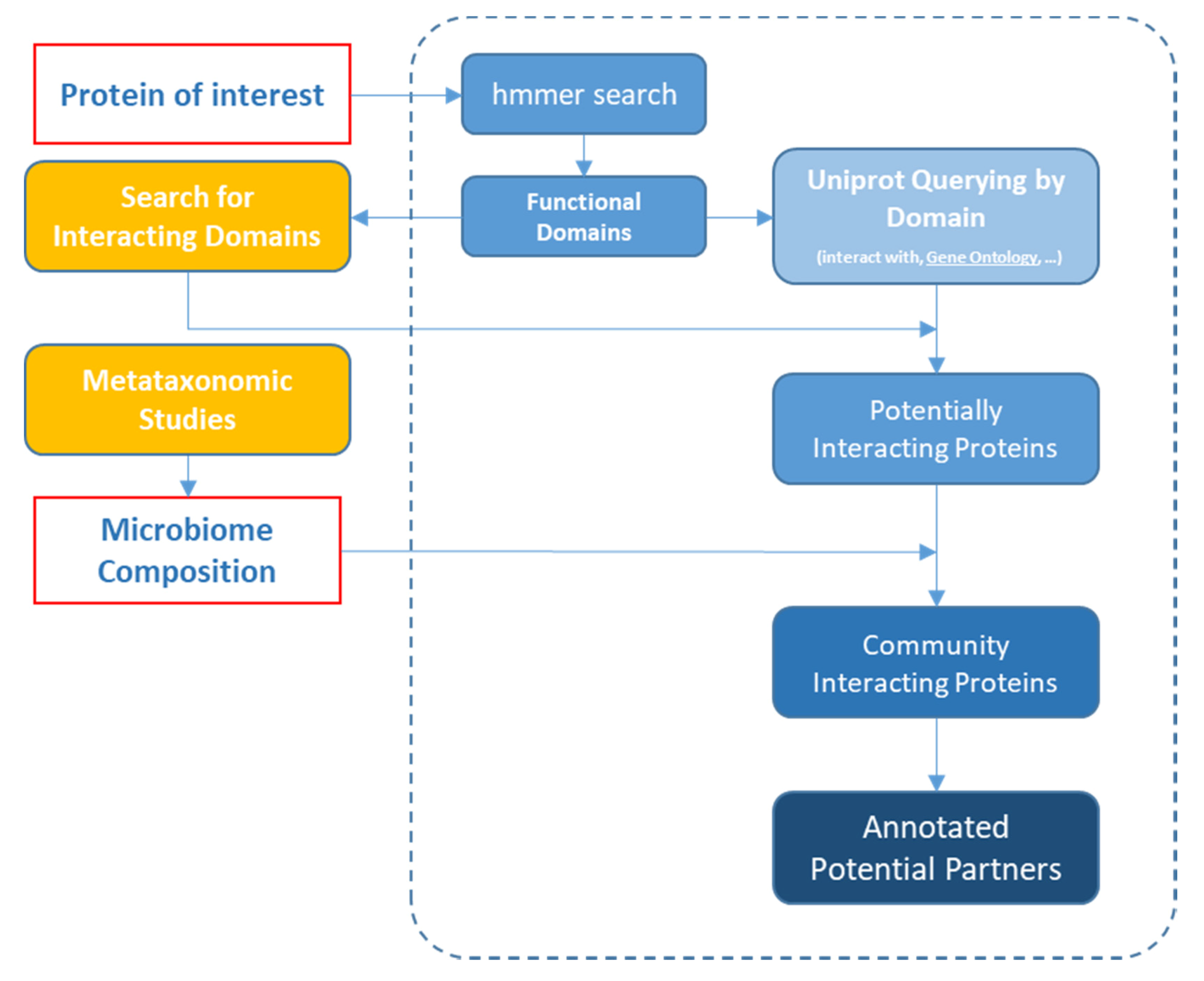
2. Materials and Methods
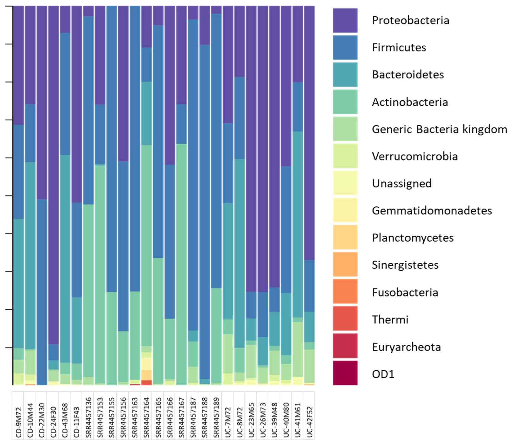
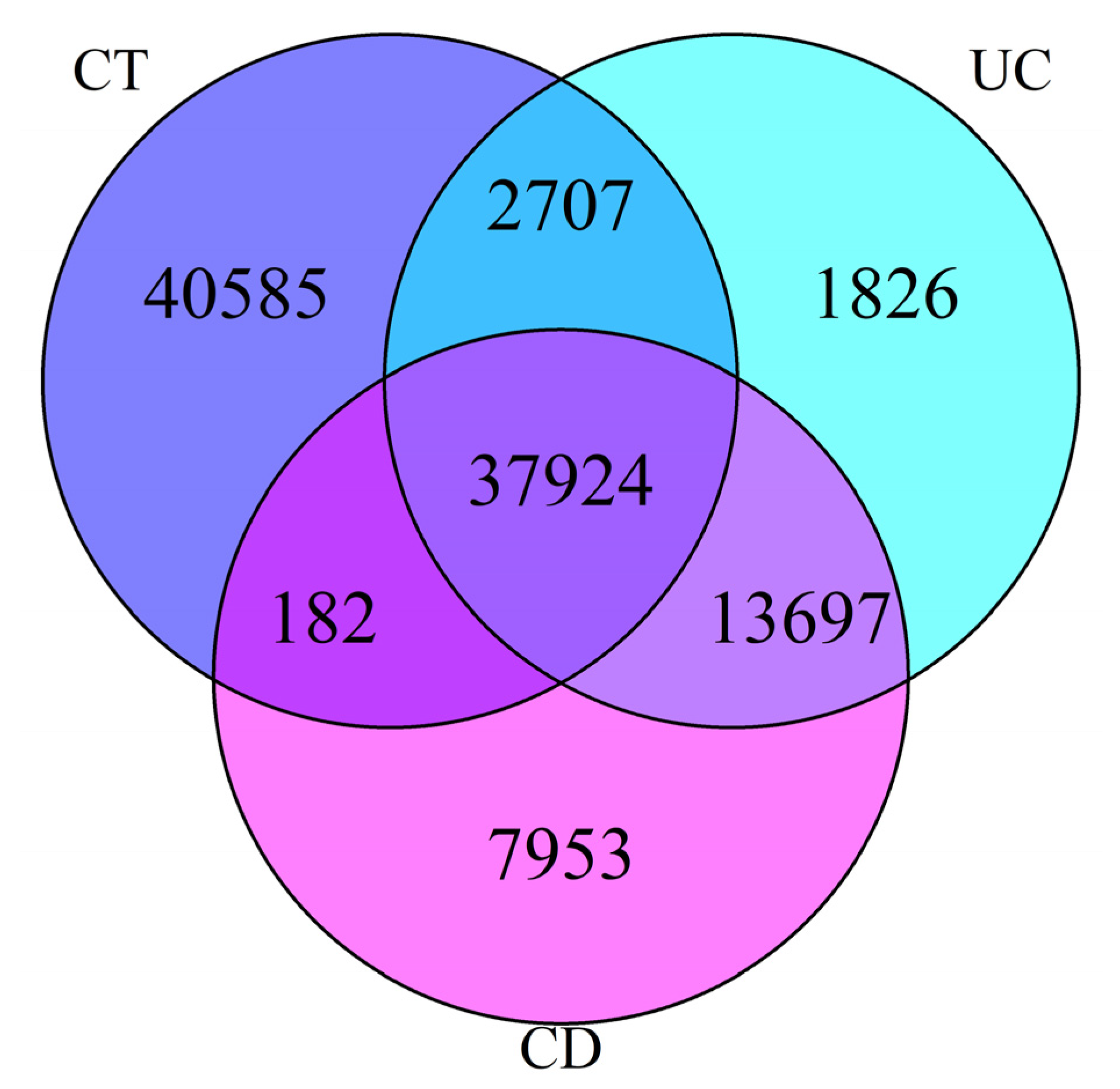
A dedicated pipeline for identifying potential protein interaction partners of human S100B (Uniprot accession ID: P04271) has been implemented. The bioinformatics workflow was designed by integrating the interaction features of S100B known domains with the putative proteomics data derived from metataxonomic studies from gut microbiota (Figure 1).
The general approach was designed to realise a novel framework for evaluating interactions of a candidate protein versus the microbiota proteome S100B protein with the putative domains. Briefly, for a given protein, the functional domains are deduced by profile-sequence search using hmmer [28] and Pfam database [27]. The identified domains were searched through the iPfam database [29] to analyse all the potential interactions. The iPfam database considers the interactions between residues in three-dimensional protein structures (available in PDB [30]) and maps those interactions back to Pfam domains [27]. In parallel, the microbiome composition returned from metataxonomic studies of a population of interest, is used to filter out the Uniprot database retaining only those proteins belonging to the lowest taxonomic level available. A minimum annotation in terms of protein functional domains, cellular localisation and ontology terms are downloaded contextually. The obtained virtual proteomes are then filtered with the aim of retaining only those entries containing at least one target protein-interacting domain, as identified in the previous step. Pearson’s χ2 test was used to compare the frequencies of retrieved domains among groups. Optionally, resulting proteins could be further narrowed by cellular localisation or ontology features. For each protein, the annotation in terms of GO terms [http://geneontology.org] for the three categories was downloaded from the Uniprot database, and successively, the enrichment analysis was carried out using the Fisher’s test, implemented in the topGO R-package [31] and Rgraphviz [32] R-packages. Only those GO terms having an adjusted p-value (q-value) ≤0.01 were considered to be significantly enriched.
We applied it to the microbiota from patients affected by Ulcerative Colitis (UC, n = 8) or Crohn Disease (CD, n = 6) for which faecal S100B was measured using Human S100B (Protein S100-B) ELISA Kit, as previously described [24]. Briefly, faecal samples (25 mg) were dissolved in 2 mL of extraction saline buffer for 15 min at room temperature, and, subsequently, centrifuged at 12,000 g for 10 min. The patients’ samples were collected under the approval of the Ethics Committee of the University Hospital of Padua (protocol no. 46093/AO/2016). Bacteria were recovered by treating the biopsy with 2 cycles of ultrasound bath/vortexing (2 min) to disrupt the biopsy surface. After centrifugation (700 g, 1 min, 4 °C) to pellet debris, bacteria were collected from the supernatant by additional centrifugation (9000 g, 5 min, 4 °C); finally, DNA was isolated from the pellet using Qiagen DNeasy Blood&Tissue and quantified using the NanoDrop ND-100 spectrophotometer (NanoDrop Technologies, Wilmington, DE, USA). The hypervariable region V3-V4 of bacterial 16S rRNA was amplified using eubacterial primers [33], and sequenced with a MiSeq Illumina platform using manufacturer instructions for libraries preparation for 16S metataxonomic studies. The sequencing reaction was carried out on an Illumina MiSeq platform, and 250 bp paired-end reads were generated (Table S2). Control sequences were downloaded from the Short Reads Archive (SRA) [34] from a homogeneous sample of healthy adults of the same ethnicity and from the same sequencing platform. Finally, microbiome composition and diversity indexes were investigated by a full pipeline integrated into the QIIME2 package [35]. A detailed sample description and metagenomics analysis workflow has been provided in the Supplementary Material.

3. Results

The protein interaction pipeline (Figure 1) was realised and applied to investigate in silico the differences in the framework of putative partners of human S100B protein in the virtual gut microbiota of healthy and diseased people.
The hmmer analysis confirmed that S100B protein is composed of two functional domains: the S100 domain (AA 4–46) and the EF-hand domain spanning the residues 53–81. By the iPfam database, the former is potentially responsible of 9 domain-domain interactions, the latter could have interactions with 27 different functional domains, some of them in common with the S100 domain, then totally accounting for 32 unique protein domains (Table 1).
Bacterial communities from the gut of patients with active CD or UC were analysed together with 12 sequence datasets (Tables S1 and S2) from healthy individuals. The microbiome composition of both diseased and control sequences are reported in Figure 2 where they are summarised at the phylum level. Full details about microbiome composition and diversity indexes are reported in the Supplementary Material.
The overall differences between IBD patients and healthy subjects can be clearly appreciated. In particular, the dominant sequences in healthy subjects belong to Firmicutes and Actinobacteria phyla, accounting for over 97% of taxonomy. Among all dominant strains in IBD, Proteobacteria and Bacteroidetes were the most abundant (70–90%), followed by a significant fraction of sequences generically assigned to the Bacteria kingdom (Figure 2). Phylum level analysis also revealed subtle differences between CD and UC samples, where phyla as Fusobacteria and Planctomyces were observed in CD samples only, while OD1 and Synergistetes were detected in UC samples.
Metataxonomic analysis returned a total of 166 clusters of sequences for the three categories; for 74 it was only possible to assign unequivocally the taxonomy at genus level and the relative abundance in the all three groups of samples (Table S4), while other sequence clusters remained unassigned. Of the 74 genera, 15 were in common among the three groups of samples, 8 were found in UC patients only, 9 in CD patients, while 13 genus were specific of control samples (Table S4). The beta diversity analysis, showing the differences between microbial communities from different environments, highlighted that both UC and CD patients tend to cluster together with a high overlap between the two groups, suggesting a common bacterial community structure (Figure S1).
According to the proposed workflow, the Bacterial section of Uniprot database (n = 126,709,838 proteins) was queried using the above mentioned 74 genus (Table S4) describing the microbiome composition of both healthy and diseased people, providing a final pool of 32,344,661 candidate proteins (Table 2). These datasets were further filtered to retain proteins containing at least one domain potentially interacting with the human S100B protein then returning 61,086, 57,344 and 83,008 entries for the CD patients, UC patients and healthy controls (CT), respectively (Table 2, Figure 3). Interestingly, in the samples here used to validate the workflow, a decreased expression of S100B was observed in the faeces of CD patients (43.61–484.00 µg/L) and UC (27.37–937.54 µg/L) patients compared to healthy donors (65.22–2018.00 µg/L) [15].
Altogether these proteins consisted of 107,263 unique entries, differently occurring in the three categories (Table 2). The largest part (38.7%) of them appeared expressed by the healthy microbiome, a consistent fraction was theoretically expressed in all conditions (35.9%), and two smaller subsets appeared peculiar of the disease state, being specific in UC or CD diseased microbiomes, respectively (1.8%) and (7.6%) (Figure 3).
Going more in detail by exploring single functional domain distribution, it was possible to observe that some domains were not present in any of the microbiota proteome that were analysed (PF00340, PF02888, PF03520, PF08763, PF10163, PF12209, PF13833, PF00307, PF00531, PF00613, PF01023, PF05002, PF08457, PF11522, PF12424, PF12515); while other domains were displayed with a frequency close to zero (PF00167, PF00168, PF00992, PF01267, PF01372, PF01387, PF01576) and were not considered further, focusing only on the occurrence of the remaining domains as listed in Table 3.
Most of the selected putative domains showed an occurrence significantly lower than expected (p-value < 0.01), suggesting the presence of specific interactions. The Calcineurin-like phosphoesterase domain (PF00149) is the only domain overrepresented in all samples categories; conversely, the Helix-loop-helix typical of calcium-binding proteins (PF00036), the IQ calmodulin-binding domain (PF00612) and the EF-hand domain (PF12202) were found underrepresented than by chance. Interestingly, three domains were differentially represented in healthy versus diseased sample: namely, the Protein kinase domain (PF00069), the Immunoglobulin domain (PF13895) and the EF-hand_domain_pair (PF13499) were found overrepresented in healthy samples and underrepresented in all IBD sample groups.
In order to capture the function of the identified groups of proteins, the enrichment analysis was conducted for the three gene ontologies (biological process, BP; cellular component, CC; molecular function, MF). None of the three proteins groups showed enrichment for the BP category. Conversely, for molecular functions and cellular component categories, several differences among groups occurred. In the healthy group of proteins, the most enriched MF terms were: hydrolase, di- or tetra-phosphatases and phosphodiesterase, all of them showing a p-value <0.01. The same group in the CC category showed the integral component of the membrane as the most enriched but with a not significant p-value. These data could indicate hypothetical candidate pathways for further investigations, not in silico but experimentally validated.
Even for disease conditions, the bioinformatics approach provided candidate pathways. Proteins from CD diseased samples showed in addition, significant enriched MF terms related to nuclease and in particular exonuclease activities (Table 4).
Moreover, in the CC ontology, in addition to membrane related terms, several other specific terms related to cytoskeleton and movement appeared significantly enriched (p-value <0.05). Finally, the UC diseased samples showed an enrichment profile similar to the healthy samples in terms of MF category (hydrolase, di- or tetra-phosphatases and phosphodiesterase) without exhibiting any of the nucleases related terms observed in the CD samples. Similarly, in the CC category the UC samples showed enriched terms related to cytoskeleton and movement proteins (cytoskeleton, actin cytoskeleton, troponin complex, myofibril, contractile fibre) but differently from CD group did not show any terms related to the membrane protein localisation. On the contrary, CC ontology terms linked to the intracellular environment (organelle part, intracellular organelle part) were observed with a p-value <0.05. The observed data suggest a difference between CD and UC.

4. Discussion

The presence of S100B, both in the gut wall and in faeces suggests the possibility of an interaction between this protein and gut microbiome. This possible interaction is an obvious conceptual prerequisite for a function in this compartment involving together S100B and the gut microbiome. Due to its modular structure, the S100B protein is able to perform a variety of protein–protein interactions. In order to evaluate the hypothesis of possible molecular interactions between S100B and the human microbiome, we developed a bioinformatics approach (Figure 2) and applied it on the microbiome structure of healthy and UC or CD patients, to identify candidate target proteins for a putative interaction between S100B and microbiota proteome. The whole of the results supports the feasibility of a bioinformatics approach for studying in silico possible interactions between candidate proteins secreted in the gut and the microbiota proteome. We found potentially interacting domains both in healthy and IBD microbiome, conceptually supporting a potential interaction between S100B and gut microbes and, in addition, we also found apparent differences between the healthy and IBD groups. S100B is regarded to be associated with several diseases and neurological disorders where the microbiota also seems to play a role (for reviews, [17,23]). Further, in the faeces of patients with UC or CD, S100B levels were also shown to be altered [24] suggesting a dysregulation in these conditions also characterised by a microbiota dysregulation and an inflammatory process of the intestinal mucosa [36,37]. Moreover, the present study shows that gut microbiome protein domains potentially interacting with S100B are quantitatively reduced in IBD. Interestingly, in this respect, S100B in cultured enteroglial cells has been reported to be differently expressed after exposure to probiotic or pathogen bacteria [38]. This observation may support the hypothesis of a possible physiological role of S100B in healthy individuals, as well as a disruption of the putative interactive network in IBD condition.
To our knowledge, this is the first bioinformatics model developed to test a possible interaction between a secreted protein and the lumen microbiota proteome. The bioinformatics workflow was proposed to integrate a classical metataxonomic concept of community composition investigation with a domain-mediated protein-protein interaction approach.
The microflora composition of UC and CD patients was investigated together with a set of microbiomes from healthy people to obtain a taxonomic framework where to investigate proteins potentially interacting with S100B domains. Several differences were observed between the study groups and several candidate proteins were selected. Microbiota results are in agreement with other studies highlighting a strong community shift between healthy and disease state, and, as expected, the healthy microbiome was mainly composed by Firmicutes and Actinobacteria phyla while both UC and CD samples were dominated by Proteobacteria and Bacteroidetes [39,40,41]. These data strongly can impact on the composition of derived proteomes that, however, resulted comparable in size, further supporting the accuracy and suitability of the approach. Nevertheless, as above mentioned, this analysis does not have the purpose of a traditional metagenomics study on microbiota; but rather to obtain a rough microbial signature to narrow and compare the bacterial sets of proteins in a virtual in silico scenario. From the frequency distribution of functional domains emerged that in the virtual-gut proteomes the occurrence of S100B potentially interacting domains is feasible and, more interestingly, in the diseased samples the occurrence of such domains is significantly lower than in the control healthy group. It can be speculated that this may suggest a possible physiological role for S100B in the interaction between microbiota and gut mucosa, thus proposing S100B as a candidate signalling molecule. Thus, in IBD the S100B/microbiota interaction might even participate in pathogenic processes, such as chronic inflammation, which may involve TLR5 and/or TLR9, two recognised regulators of the mucosal immune response to microbiota [42]. Further, going down to a single domain level, it emerged that, differently from the general trend, some domains as PF00069 (Protein-kinase), the PF13499 (EF-hand domain pair) and the PF13895 (Immunoglobulin domain) are considerable over-represented in microbiome samples from healthy subjects respect than expected by chance, but above all, over-represented than in diseased samples (Table 3). This finding is consistent with the unexpected reduced levels of S100B in faeces of IBD patients in comparison with healthy subjects [24]. Although additional data will be needed in order to explain the finding, it seems to hint at a physiological role of S100B in the gut lumen, which is reduced in pathological conditions.
These in silico results open the way to experiments required for their confirmation in the laboratory. In any case, they induce several considerations and further working hypothesis. First of all, it can be speculated that in healthy microbiome more proteins can interact with the S100B, acting as a regulator and contributing in the switching off inflammatory processes or in physiological maintenance of the healthy mucosa. Conversely, in the disease state, the microbiota shift results in a qualitative change and quantitative reduction of exogenous S100B partners carried by the microbiota, then further promoting the inflammatory pathways or potentially influencing the gut-brain axis. The Gene Ontology enrichment analysis further shed light on the differences in the putative S100B interacting proteins in healthy and patients’ subgroups. Excluding the common signature of proteins related to hydrolase activity and membrane localisation, the targets derived from the CD gut proteomes were characterised by an exonucleases activity and a strong evidence of linking to bacterial membrane and cytoskeleton. Conversely, the proteins in the UC virtual proteomes did not show the nuclease activity nor the membrane localisation, but instead displayed an enrichment of cytoskeleton and intracellular organelle related terms. Interestingly, the interaction of S100B with the E. coli troponin, involved in bacterial division, was already reported years ago by Ferguson and Shaw [43].
The hereby proposed approach is based on a qualitative analysis and cannot exclude expression differences or microenvironmental interferences. Moreover, S100B is known to be a calcium-binding protein, although exhibiting calcium-binding affinities relatively weak compared to other members of the same EF-hand superfamily, and its calcium-induced conformational change is believed to facilitate the interaction with target proteins (for review, [44]). A different protein composition driven by selective expression of specific genes or differential abundance should be further considered by a complementary strategy. In this direction, shotgun metagenomics and transcriptomics studies can be helpful in improving the bioinformatics analysis. Indeed, the whole shotgun experiments and metatranscriptomics or metaproteomics can provide further valuable insights in functional features encoded by a microbiome [45,46].

5. Conclusions

A bioinformatics approach is a powerful tool for investigating the interactions between microbiota structure and human proteins. This in silico study addressed large amounts of bacterial proteins from metataxonomic studies from healthy and diseased people, in order to evaluate possible interactions virtually occurring between S100B protein and the respective microbiota proteomes. In particular, the present results offer the conceptual prerequisite to propose S100B as a candidate signalling molecule in the bidirectional microbiota/gut machinery interaction in health and disease. In principle, the same workflow could be applied to functional characterisation of other proteomic data further supporting biological basis to conceive a plausible interaction between a candidate protein and bacterial exogenous domains, then speculating a possible role in modulating physiological or inflammatory processes, as here reported for S100B protein and gut microbiota.

Supplementary Materials

The following are available online at https://www.mdpi.com/2073-4409/9/7/1697/s1, Figure S1: Beta diversity of the dataset microbiomes, Table S1: Dataset, Table S2: Reads count, Table S3: Microbiome composition at the phylum level, Table S4: Genus occurrence in microbiome samples categories, Supplementary Methods.

Author Contributions

Conceptualisation, M.O., V.R.S. and F.M.; software, M.O.; validation, R.D.L. and F.V.; formal analysis, M.M. and A.C.; investigation, R.D., A.A. and P.P.P.; writing—original draft preparation, M.O. and V.R.S.; writing—review and editing, M.O., R.D.L. and F.M.; supervision, V.R.S. and F.M. All authors have read and agreed to the published version of the manuscript.

Funding

This research received no external funding.

Acknowledgments

The authors would like to thank the Contabo Server for providing computing resources, Cineca for providing access to supercomputing calculation thru project ELIXIR-IBB HPC@CINECA "Bioinformatic analysis of S100B interaction in gut" (F.V.). The study was indirectly supported by the University of Padova projects PRID 2016 and DOR 2016-2018 (R.D.L.); Fondazione Ricerca Scientifica Termale (grant assigned to V.R.S.; FoRST CUP H81I18000070008) and Nando-Elsa Peretti Foundation (grant assigned to F.M.; NaEPF 2019-041); Tiziana Zilli for the library assistance; G. Gianfranceschi and E. Scaramucci for their technical support.

Conflicts of Interest

The authors declare no conflict of interest.

References

  1. Michetti, F.; D’Ambrosi, N.; Toesca, A.; Puglisi, M.A.; Serrano, A.; Marchese, E.; Corvino, V.; Geloso, M.C. The S100B story: From biomarker to active factor in neural injury. J. Neurochem. 2019, 148, 168–187. [Google Scholar] [CrossRef] [Green Version]
  2. Ludwin, S.K.; Kosek, J.C.; Eng, L.F. The topographical distribution of S-100 and GFA proteins in the adult rat brain: An immunohistochemical study using horseradish peroxidase-labelled antibodies. J. Comp. Neurol. 1976, 165, 197–207. [Google Scholar] [CrossRef] [PubMed]
  3. Ferri, G.L.; Probert, L.; Cocchia, D.; Michetti, F.; Marangos, P.J.; Polak, J.M. Evidence for the presence of S-100 protein in the glial component of the human enteric nervous system. Nature 1982, 297, 409–410. [Google Scholar] [CrossRef] [PubMed]
  4. Brockes, J.P.; Fields, K.L.; Raff, M.C. Studies on cultured rat Schwann cells. I. Establishment of purified populations from cultures of peripheral nerve. Brain Res. 1979, 165, 105–118. [Google Scholar] [CrossRef]
  5. Didier, M.; Harandi, M.; Aguera, M.; Bancel, B.; Tardy, M.; Fages, C.; Calas, A.; Stagaard, M.; Møllgård, K.; Belin, M.F. Differential immunocytochemical staining for glial fibrillary acidic (GFA) protein, S-100 protein and glutamine synthetase in the rat subcommissural organ, nonspecialized ventricular ependyma and adjacent neuropil. Cell Tissue Res. 1986, 245, 343–351. [Google Scholar] [CrossRef] [PubMed]
  6. Rickmann, M.; Wolff, J.R. S100 protein expression in subpopulations of neurons of rat brain. Neuroscience 1995, 67, 977–991. [Google Scholar] [CrossRef]
  7. Yang, Q.; Hamberger, A.; Hyden, H.; Wang, S.; Stigbrand, T.; Haglid, K.G. S-100 beta has a neuronal localisation in the rat hindbrain revealed by an antigen retrieval method. Brain Res. 1995, 696, 49–61. [Google Scholar] [CrossRef]
  8. Cocchia, D.; Michetti, F.; Donato, R. Immunochemical and immuno-cytochemical localization of S-100 antigen in normal human skin. Nature 1981, 294, 85–87. [Google Scholar] [CrossRef]
  9. Cocchia, D.; Tiberio, G.; Santarelli, R.; Michetti, F. S-100 protein in “follicular dendritic” cells or rat lymphoid organs. An immunochemical and immunocytochemical study. Cell Tissue Res. 1983, 230, 95–103. [Google Scholar] [CrossRef]
  10. Stefansson, K.; Wollmann, R.L.; Moore, B.W.; Arnason, B.G. S-100 protein in human chondrocytes. Nature 1982, 295, 63–64. [Google Scholar] [CrossRef]
  11. Michetti, F.; Dell’Anna, E.; Tiberio, G.; Cocchia, D. Immunochemical and immunocytochemical study of S-100 protein in rat adipocytes. Brain Res. 1983, 262, 352–356. [Google Scholar] [CrossRef]
  12. Lauriola, L.; Maggiano, N.; Sentinelli, S.; Michetti, F.; Cocchia, D. Satellite cells in the normal human adrenal gland and in pheochromocytomas. An immunohistochemical study. Virchows Arch. B Cell Pathol. Incl. Mol. Pathol. 1985, 49, 13–21. [Google Scholar] [CrossRef] [PubMed]
  13. Tubaro, C.; Arcuri, C.; Giambanco, I.; Donato, R. S100B protein in myoblasts modulates myogenic differentiation via NF-kappaB-dependent inhibition of MyoD expression. J. Cell Physiol. 2010, 223, 270–282. [Google Scholar]
  14. Michetti, F.; Rende, M.; Calogero, G.; Dell’Anna, E.; Cocchia, D. Immunochemical detection of S-100 protein in non-nervous structures of the rabbit eye. Brain Res. 1985, 332, 358–360. [Google Scholar] [CrossRef]
  15. Moore, B.W. A soluble protein characteristic of the nervous system. Biochem. Biophys. Res. Commun. 1965, 19, 739–744. [Google Scholar] [CrossRef]
  16. Donato, R.; Cannon, B.R.; Sorci, G.; Riuzzi, F.; Hsu, K.; Weber, D.J.; Geczy, C.L. Functions of S100 proteins. Curr. Mol. Med. 2013, 13, 24–57. [Google Scholar] [CrossRef] [PubMed] [Green Version]
  17. Gong, T.; Liu, L.; Jiang, W.; Zhou, R. DAMP-sensing receptors in sterile inflammation and inflammatory diseases. Nat. Rev. Immunol. 2020, 20, 95–112. [Google Scholar] [CrossRef]
  18. Cirillo, C.; Sarnelli, G.; Esposito, G.; Grosso, M.; Petruzzelli, R.; Izzo, P.; Calì, G.; D’Armiento, F.P.; Rocco, A.; Nardone, G.; et al. Increased mucosal nitric oxide production in ulcerative colitis is mediated in part by the enteroglial-derived S100B protein. Neurogastroenterol. Motil. 2009, 21, 1209–e112. [Google Scholar] [CrossRef]
  19. Cirillo, C.; Sarnelli, G.; Turco, F.; Mango, A.; Grosso, M.; Aprea, G.; Masone, S.; Cuomo, R. Proinflammatory stimuli activates human-derived enteroglial cells and induces autocrine nitric oxide production. Neurogastroenterol. Motil. 2011, 23, e372–e382. [Google Scholar] [CrossRef]
  20. Esposito, G.; Cirillo, C.; Sarnelli, G.; De Filippis, D.; D’Armiento, F.P.; Rocco, A.; Nardone, G.; Petruzzelli, R.; Grosso, M.; Izzo, P.; et al. Enteric glial-derived S100B protein stimulates nitric oxide production in celiac disease. Gastroenterology 2007, 133, 918–925. [Google Scholar] [CrossRef]
  21. Esposito, G.; Capoccia, E.; Sarnelli, G.; Scuderi, C.; Cirillo, C.; Cuomo, R.; Steardo, L. The antiprotozoal drug pentamidine ameliorates experimentally induced acute colitis in mice. J. Neuroinflammation 2012, 9, 277. [Google Scholar] [CrossRef] [PubMed]
  22. Quigley, E.M.M. Microbiota-Brain-Gut Axis and Neurodegenerative Diseases. Curr. Neurol. Neurosci. Rep. 2017, 17, 94. [Google Scholar] [CrossRef] [PubMed]
  23. Zhu, S.; Jiang, Y.; Xu, K.; Cui, M.; Ye, W.; Zhao, G.; Jin, L.; Chen, X. The progress of gut microbiome research related to brain disorders. J. Neuroinflammation 2020, 17, 25. [Google Scholar] [CrossRef] [Green Version]
  24. Di Liddo, R.; Piccione, M.; Schrenk, S.; Dal Magro, C.; Cosma, C.; Padoan, A.; Contran, N.; Scapellato, M.L.; Pagetta, A.; Romano Spica, V.; et al. S100B as a new fecal biomarker of inflammatory bowel diseases. Eur. Rev. Med. Pharmacol. Sci. 2020, 24, 323–332. [Google Scholar] [PubMed]
  25. Ostendorp, T.; Heizmann, C.W.; Kroneck, P.M.H.; Fritz, G. Purification, crystallization and preliminary X-ray diffraction studies on human Ca2+-binding protein S100B. Acta Crystallogr. Sect. F Struct. Biol. Cryst. Commun. 2005, 61, 673–675. [Google Scholar] [CrossRef] [PubMed] [Green Version]
  26. Prez, K.D.; Fan, L. Structural Basis for S100B Interaction with its Target Proteins. J. Mol. Genet. Med. 2018, 12, 366. [Google Scholar] [PubMed]
  27. El-Gebali, S.; Mistry, J.; Bateman, A.; Eddy, S.R.; Luciani, A.; Potter, S.C.; Qureshi, M.; Richardson, L.J.; Salazar, G.A.; Smart, A.; et al. The Pfam protein families database in 2019. Nucleic Acids Res. 2019, 47, D427–D432. [Google Scholar] [CrossRef]
  28. Wheeler, T.J.; Eddy, S.R. nhmmer: DNA homology search with profile HMMs. Bioinformatics 2013, 29, 2487–2489. [Google Scholar] [CrossRef] [Green Version]
  29. Finn, R.D.; Miller, B.L.; Clements, J.; Bateman, A. iPfam: A database of protein family and domain interactions found in the Protein Data Bank. Nucleic Acids Res. 2014, 42, D364–D373. [Google Scholar] [CrossRef]
  30. Berman, H.M.; Battistuz, T.; Bhat, T.N.; Bluhm, W.F.; Bourne, P.E.; Burkhardt, K.; Feng, Z.; Gilliland, G.L.; Iype, L.; Jain, S.; et al. The Protein Data Bank. Acta Crystallogr. D Biol. Crystallogr. 2002, 58, 899–907. [Google Scholar] [CrossRef]
  31. Alexa, A.; Rahnenfuhrer, J. TopGO: Enrichment Analysis for Gene Ontology. R package version 2.40.0. 2020. Available online: https://bioconductor.org/packages/release/bioc/html/topGO.html (accessed on 15 February 2020).
  32. Hansen, K.D.; Gentry, J.; Long, L.; Gentleman, R.; Falcon, S.; Hahne, F.; Sarkar, D. Rgraphviz: Provides Plotting Capabilities for R Graph Objects. Bioconductor version: Release (3.11). 2020. Available online: https://bioconductor.org/packages/release/bioc/html/Rgraphviz.html (accessed on 30 June 2020).
  33. Valeriani, F.; Protano, C.; Gianfranceschi, G.; Leoni, E.; Galasso, V.; Mucci, N.; Vitali, M.; Romano Spica, V. Microflora Thermarum Atlas project: Biodiversity in thermal spring waters and natural SPA pools. Water Sci. Tech-W. Supply 2018, 18, 1472–1483. [Google Scholar] [CrossRef]
  34. Kodama, Y.; Shumway, M.; Leinonen, R. International Nucleotide Sequence Database Collaboration. The Sequence Read Archive: Explosive growth of sequencing data. Nucleic Acids Res. 2012, 40, D54–D56. [Google Scholar] [CrossRef] [Green Version]
  35. Bolyen, E.; Rideout, J.R.; Dillon, M.R.; Bokulich, N.A.; Abnet, C.C.; Al-Ghalith, G.A.; Alexander, H.; Alm, E.J.; Arumugam, M.; Asnicar, F.; et al. Reproducible, interactive, scalable and extensible microbiome data science using QIIME 2. Nat. Biotechnol. 2019, 37, 852–857. [Google Scholar] [CrossRef] [PubMed]
  36. Capoccia, E.; Cirillo, C.; Gigli, S.; Pesce, M.; D’Alessandro, A.; Cuomo, R.; Sarnelli, G.; Steardo, L.; Esposito, G. Enteric glia: A new player in inflammatory bowel diseases. Int. J. Immunopathol. Pharmacol. 2015, 28, 443–451. [Google Scholar] [CrossRef] [PubMed] [Green Version]
  37. de Souza, H.S.P.; Fiocchi, C. Immunopathogenesis of IBD: Current state of the art. Nat. Rev. Gastroenterol. Hepatol. 2016, 13, 13–27. [Google Scholar] [CrossRef] [PubMed]
  38. Turco, F.; Sarnelli, G.; Cirillo, C.; Palumbo, I.; De Giorgi, F.; D’Alessandro, A.; Cammarota, M.; Giuliano, M.; Cuomo, R. Enteroglial-derived S100B protein integrates bacteria-induced Toll-like receptor signalling in human enteric glial cells. Gut 2014, 63, 105–115. [Google Scholar] [CrossRef]
  39. Franzosa, E.A.; Sirota-Madi, A.; Avila-Pacheco, J.; Fornelos, N.; Haiser, H.J.; Reinker, S.; Vatanen, T.; Brantley Hall, A.; Mallick, H.; McIver, L.J.; et al. Gut microbiome structure and metabolic activity in inflammatory bowel disease. Nat. Microbiol. 2019, 4, 293–305. [Google Scholar] [CrossRef]
  40. Halfvarson, J.; Brislawn, C.J.; Lamendella, R.; Vázquez-Baeza, Y.; Walters, W.A.; Bramer, L.M.; D’Amato, M.; Bonfiglio, F.; McDonald, D.; Gonzalez, A.; et al. Dynamics of the human gut microbiome in inflammatory bowel disease. Nat. Microbiol. 2017, 2, 17004. [Google Scholar] [CrossRef] [Green Version]
  41. Marchesi, J.R.; Adams, D.H.; Fava, F.; Hermes, G.D.A.; Hirschfield, G.M.; Hold, G.; Quraishi, M.N.; Kinross, J.; Smidt, H.; Tuohy, K.M.; et al. The gut microbiota and host health: A new clinical frontier. Gut 2016, 65, 330–339. [Google Scholar] [CrossRef] [Green Version]
  42. Lu, Y.; Li, X.; Liu, S.; Zhang, Y.; Zhang, D. Toll-like Receptors and Inflammatory Bowel Disease. Front. Immunol. 2018, 9, 72. [Google Scholar] [CrossRef] [Green Version]
  43. Ferguson, P.L.; Shaw, G.S. Human S100B protein interacts with the Escherichia coli division protein FtsZ in a calcium-sensitive manner. J. Biol. Chem. 2004, 279, 18806–18813. [Google Scholar] [CrossRef] [PubMed] [Green Version]
  44. Baudier, J.; Gentil, B.J. The S100B Protein and Partners in Adipocyte Response to Cold Stress and Adaptive Thermogenesis: Facts, Hypotheses, and Perspectives. Biomolecules 2020, 10, E843. [Google Scholar] [CrossRef] [PubMed]
  45. Schirmer, M.; Franzosa, E.A.; Lloyd-Price, J.; McIver, L.J.; Schwager, R.; Poon, T.W.; Ananthakrishnan, A.N.; Andrews, E.; Barron, G.; Lake, K.; et al. Dynamics of metatranscription in the inflammatory bowel disease gut microbiome. Nat. Microbiol. 2018, 3, 337–346. [Google Scholar] [CrossRef] [PubMed]
  46. Lehmann, T.; Schallert, K.; Vilchez-Vargas, R.; Benndorf, D.; Püttker, S.; Sydor, S.; Schulz, C.; Bechmann, L.; Canbay, A.; Heidrich, B.; et al. Metaproteomics of fecal samples of Crohn’s disease and Ulcerative Colitis. J. Proteomics 2019, 201, 93–103. [Google Scholar] [CrossRef] [PubMed]
Figure 1. Implemented Analytical Workflow. A dedicated bioinformatic approach was developed to test in silico the possible interactions between a candidate protein and the microbiota proteome. The steps inside the dashed box represent the here implemented automatic workflow; data from the side (list of interactions and microbiome composition) have to be manually provided. A module for the automatic download of domain-domain interactions from the Pfam database is currently under development, but any alternative strategy can be applied to feed this automatic workflow.
Cells 09 01697 g001
Figure 2. Microbiome composition. Bacteria community compositions at Phylum level, Relative abundance at phylum and genus level are in Supplementary Tables S3 and S4, respectively.
Cells 09 01697 g002
Figure 3. Distribution of S100B potentially interacting proteins in the three samples category. Abbreviations: CD—Crohn disease; UC—ulcerative colitis; CT—healthy control.
Cells 09 01697 g003
Table 1. S100B potentially interacting domains. The S100B protein is composed of two structural domains: the S100 and the EF-hand. The table reports the list of protein domains potentially interacting with both of them. Some domains (e.g., the FGF domain) are able to interact with both. The S100 domain is also able to interact with itself generating homo-multimers.
DomainPartner Pfam IDDescription
S100BC2PF00168Structural domain involved in targeting proteins to membrane
EF-hand_1PF00036Helix-loop-helix domain or motif found in a large family of Ca-binding proteins.
F-actin_cap_APF01267Domain binding in a Ca independent manner the fast-growing ends of actin filaments
FGFPF00167Fibroblast growth factors, Family of cell signalling proteins
Ig_2PF13895Immunoglobulin domain
IL1PF00340Interleukins
Myosin_tail_1PF01576Myosin domain
S_100PF01023S100 type calcium-binding domain
SGSPF05002Structural domain found in calcyclin binding proteins
EF-handATP_Ca_trans_CPF12424Plasma membrane calcium transporter ATPase C terminal
CA_chan_IQPF08763Voltage-gated calcium channel IQ domain
CaATP_NAIPF12515Ca2+-ATPase N terminal autoinhibitory domain
CaMBDPF02888Calmodulin binding domain
CHPF00307Calponin homology (CH) domain
DeathPF00531Death domain
EF-hand_1PF00036Helix-loop-helix domain or motif found in a large family of Ca-binding proteins.
EF-hand_7PF13499EF-hand domain pair
EF-hand_5PF13202EF hand
EF-hand_6PF13405EF-hand domain
EF-hand_8PF13833EF-hand domain pair
EnY2PF10163Transcription factor e(y)2
F-actin_cap_APF01267F-actin capping protein alpha subunit
FGFPF00167Fibroblast growth factor
IQPF00612IQ calmodulin-binding motif
KCNQ_channelPF03520KCNQ voltage-gated potassium channel
MelittinPF01372Melittin
MetallophosPF00149Calcineurin-like phosphoesterase
Myosin_headPF00063Myosin head
PI3KAPF00613Phosphoinositide 3-kinase family, accessory domain
Pik1PF11522Yeast phosphatidylinositol-4-OH kinase Pik1
PkinasePF00069Protein kinase domain
S_100PF01023S100 type calcium-binding domain
SAC3PF12209Leucine permease transcriptional regulator helical domain
Sfi1PF08457Sfi1 spindle body protein
SynucleinPF01387Synuclein
TroponinPF00992Troponin
Table 2. S100B potentially interacting bacterial proteins in the three categories and their genus of provenance.
CategoryTotal ProteinsFrom GenusS100B Interacting
CD22,066,9815161,086
UC21,402,6845057,344
CT24,306,7403283,008
Abbreviations: CD—Crohn disease; UC—ulcerative colitis; CT—healthy control.
Table 3. The occurrence of S100B interacting functional domains over samples categories. The expected value by chance is reported. In the top those statistically significant (chi-squared Test, p < 0.01). In the bottom part, the domains not significantly supported (p > 0.5). Exp.: Expected Value.
DomainCTExp.CDExp.UCExp.Description
PF0003635110161001797Helix-loop-helix structural domain or motif found in a large family of calcium-binding proteins.
PF00069352553379016544306571344429731Protein kinase domain
PF138951497612691067Immunoglobulin domain
PF00612820918917IQ calmodulin-binding motif
PF00149440224313741838391364109537955Calcineurin-like phosphoesterase
PF13202196234212420310425243010EF hand
PF134992192122669611137241079EF-hand domain pair
PF13405605556505849EF-hand domain
PF00063232323Myosin head (motor domain)
Table 4. Gene ontology enrichment for bacterial candidate proteins, only terms showing a significant (p < 0.01) enrichment have been reported. Category: MF (molecular function), CC (cellular component). Terms: number of proteins annotated with a given GO term. Observed: number of proteins observed with a given GO term. Expected: number of proteins expected with a given GO term. Fisher: p-value according to the exact-Fisher test.
Category GO Term CD UC CT
GOIDDescriptionAnnotatedObservedExpect.FisherObservedObservedFisherObservedExpect.Fisher
MFGO:0016787hydrolase activity241362034516818<1e-301851315394<1e-301595413142<1e-30
GO:0003824catalytic activity282532073119687<1e-301880818020<1e-301619115384<1e-30
GO:0016462pyrophosphatase activity250823631748<1e-3022901600<1e-3017621366<1e-30
GO:0008796bis(5′-nucleosyl)-tetraphosphatase
activity
215520621502<1e-3019901374<1e-3014781173<1e-30
GO:0008803bis(5′-nucleosyl)-tetraphosphatase
(symmetrical) activity
209920141463<1e-3019501339<1e-3014361143<1e-30
GO:0004551nucleotide diphosphatase activity215620621502<1e-3019901375<1e-3014781174<1e-30
GO:0016818hydrolase activity, acting on acid
anhydrides, in phosphorus
containing anhydrides
251723631754<1e-3022901605<1e-3017621371<1e-30
GO:0016817hydrolase activity, acting
on acid anhydrides
251923631755<1e-3022901607<1e-3017621372<1e-30
GO:0004527exonuclease activity911848635<1e-30 663496<1e-30
GO:0004518nuclease activity1004856700<1e-30
GO:0016791phosphatase activity8696456060.0016
GO:0004081bis(5′-nucleosyl)-tetraphosphatase
(asymmetrical) activity
5548380.0020 42300.00065
GO:0004721phosphoprotein phosphatase activity512 3553270.0044452279<1e-30
GO:00041153′,5′-cyclic-AMP phosphodiesterase activity12 1280.0045
CCGO:0016021integral component of membrane51631611550.0065
GO:0031224intrinsic component of membrane51631611550.0065
GO:0044425membrane part51651611550.0069

