Article

Combined Exposure to High-Cholesterol Diet and PM2.5: Brain Injury and Regulatory Mechanism of HIF-1α in ApoE−/− Female Mice

Institute of Environmental Science, Shanxi University, Taiyuan 030006, China
*
Author to whom correspondence should be addressed.
Atmosphere 2024, 15(8), 952; https://doi.org/10.3390/atmos15080952
Submission received: 8 July 2024 / Revised: 30 July 2024 / Accepted: 7 August 2024 / Published: 9 August 2024
(This article belongs to the Special Issue Characterization and Toxicity of Atmospheric Pollutants)

Abstract

:
High-cholesterol diet (HCD) and fine particulate matter (PM2.5) are related to stroke. However, little is known about the combined effects of stroke, especially for females. This study investigated the brain injuries in Apolipoprotein E−/− (ApoE−/−) female mice exposed to HCD plus PM2.5 for 6 months. The protein levels of the genes related to stroke and the blood–brain barrier (BBB) in different groups of mice were measured. The molecular regulation mechanisms were explored. The results showed that HCD and PM2.5 co-exposure altered brain–body weight ratio, behavior, brain pathology, and inflammatory markers in mice relative to exposure to HCD or PM2.5 alone. Co-exposure significantly changed the expressions of HIF-1α and the key genes in its signaling pathway in the brains of mice compared to the single exposure. It suggests that the HIF-1α pathway exerts an important regulatory role in brain injury and behavioral abnormality in female mice after 6-month exposure to HCD plus PM2.5, which are potential mechanisms for HCD and PM2.5-triggering stroke in female individuals.

Graphical Abstract

1. Introduction

Research on the global disease burden showed that air pollution caused 4.9 million deaths in 2017, and ambient fine particulate matter (PM2.5) induced 2.9 million deaths, with an increase of 21.6% compared to 2007 [1]. Numerous epidemiological investigations revealed that PM2.5 increased respiratory, cardiovascular, and cerebrovascular disease incidence and mortality [2,3,4]. PM2.5 correlates positively with central nervous system (CNS) disorders like cognitive decline and stroke [5,6].
There are several ways that PM2.5 may enter the brain. The particles can reach other brain regions from the olfactory bulb via the olfactory nerve and enter the cerebrospinal fluid through extracellular transport around nerves [7,8]. They can also enter the blood circulation by passing through the alveolar–capillary barrier or the gastrointestinal tract after inhalation, finally reaching the brain across the blood–brain barrier (BBB) [9,10]. The population survey data have shown that short-term and long-term exposure to PM2.5 augments the risks of stroke incidence and mortality [11,12]. Experimental data have documented that inflammation and arteriosclerosis are the vital regulatory mechanisms by which PM2.5 exposure triggers stroke [13]. Exposure to PM2.5 positively correlates with neuroinflammation and neurovascular disruption of BBB [14]. In turn, it aggravates brain damage and may trigger stroke [15].
With improved living standards and increased social development, a high-cholesterol diet (HCD) has become a common dietary habit. HCD may cause numerous unhealthy complications (such as obesity and atherosclerosis) with high medical and economic costs [16]. Epidemiological studies have proven that cholesterol is associated with stroke, and insufficient cerebral circulation due to high cholesterol is implicated in the etiology of stroke [17,18]. A study has unequivocally unveiled that unhealthy HCD patterns can evoke basal systemic inflammation [19], a typical pathological feature of stroke [20]. Notably, obese individuals were more susceptible to PM2.5 [21], HCD intake and PM2.5 exposure increased the risk of stroke [12,17]. The exact mechanisms of HCD-caused stroke are unclear. Consequently, studying the toxicological effects and potential regulating mechanisms of co-exposure to HCD and PM2.5 on brain injury has great significance in shedding light on the cause of stroke.
ApoE is a circulating glycoprotein with a molecular weight of 34 kDa, mainly synthesized by the liver and brain, and participates in the synthesis, secretion, processing, and metabolism of lipoproteins. It plays a vital role in lipid metabolism and can bind to triglyceride-rich lipoproteins and regulate the clearance of residues after enzymatic fat decomposition in the circulation [22]. ApoE is a major genetic susceptibility gene for diseases such as Alzheimer’s Disease (AD), stroke, and cerebral hemorrhage and is associated with adverse neurological outcomes after traumatic brain injury [23]. ApoE plays a crucial role in many biological processes of CNS and is closely related to cholesterol metabolism, among which ApoE ε4 is associated with hypercholesterolemia, hyperlipidemia, and coronary heart disease. ApoE regulates CNS by participating in cholesterol/lipid homeostasis, glucose metabolism, tau phosphorylation, mitochondrial function, neuronal atrophy, synaptic function, neuroinflammation, and amyloid β metabolism and aggregation. ApoE structural disorder can also induce BBB dysfunction, which affects cerebrovascular function, leading to the occurrence of CNS diseases.
The deletion and variation in ApoE can lead to metabolic and functional disorders in the body, thus increasing the sensitivity to the pathophysiological results of CNS, such as stroke [23]. ApoE−/− mice were mainly used in models of atherosclerosis, neurodegenerative diseases, and hypercholesterolemia. When the ApoE−/− mice are fed with a normal diet, they will have symptoms such as atherosclerosis and hyperlipidemia, which are typical high-risk factors for stroke [22]. ApoE ε4 mutation was particularly significantly associated with stroke in a dose-dependent manner [24]. As such, utilizing the ApoE−/− model in this study allowed us to more easily discover the effects of HCD and PM2.5 exposure on brain damage.
Hypoxia-inducible factor-1α (HIF-1α) is a sensitive regulatory factor of stroke [25]. It can regulate vascular endothelial growth factor (VEGF) and cyclooxygenase-2 (COX-2) [25,26,27]. COX-2 is related to stroke and has the targeted modulation to metalloprotein-9 (MMP-9) [28]. On the other hand, the protein kinase-B (Akt) may mediate HIF-1α [29,30,31]. Collectively, studying the roles of the HIF-1α/COX-2/VEGF/MMP-9 pathway in the brain injury of mice after HCD plus PM2.5 will help to elucidate the mechanisms by which these two health risk factors incur stroke.
Some studies indicated that there is a higher adverse effect on cognitive function in young people (<65 years old) and women than in older people and men exposed to PM2.5 [32,33]. Others revealed that gender differences exist in PM2.5-induced stroke [34,35]. PM2.5 exposure during pregnancy caused considerable emotional and cognitive impairments in female offspring mice, and the morphological changes in male offspring were not significantly related to the female [36]. Additionally, a study on a group of stroke patients found that obesity had a more significant impact on female brain metabolism than on males [37]. Therefore, exploring the effects of HCD and PM2.5 on brain damage in female individuals will have a positive significance in protecting women’s health.
Taiyuan, China, was selected as the exposure site for severe PM2.5 pollution in this study. The female mice and ApoE−/− model were used to investigate the effects on brain pathology and behavioral damage after chronic (6 months) exposure to HCD and PM2.5. The investigators aimed to uncover the regulatory mechanisms of the HIF-1α pathway responding to the combined effects. It will provide a valuable experimental basis for illustrating the relationship between HCD plus PM2.5 co-exposure and stroke.

2. Materials and Methods
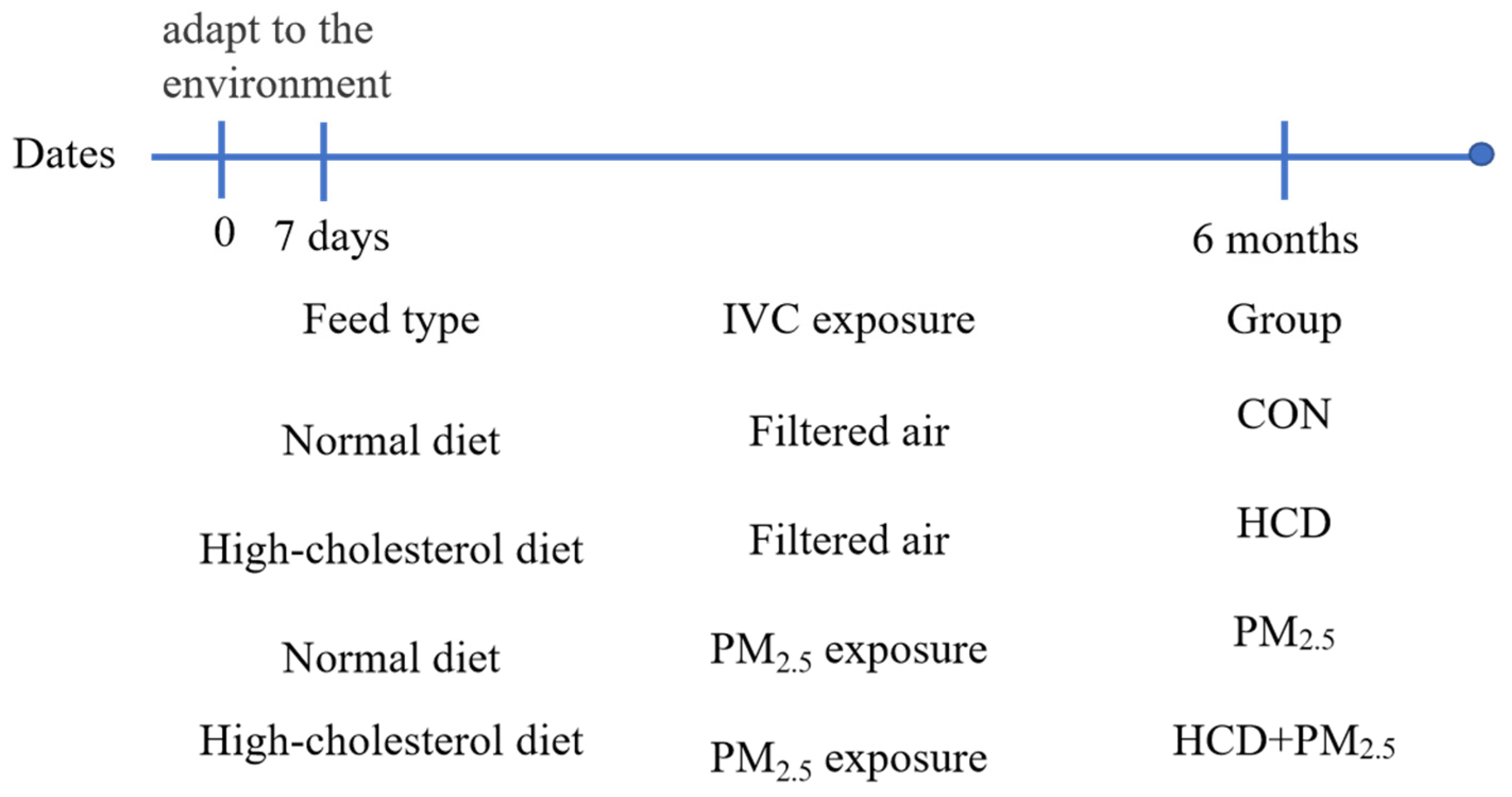
2.1. Animal

Forty C57BL/6 female ApoE−/− mice (4 weeks old) were purchased from Beijing Vital River Laboratory Animal Technology Co., Ltd. (Beijing, China). After acclimating for seven days, they were placed in an individually ventilated cage (IVC) system (Junsheng Laboratory Animal Equipment Co., Ltd., Suzhou, China) (25 °C, 60% humidity) with a 12 h light/12 h dark cycle. The experimental exposure lasted six months, from 1 November 2020 to 30 April 2021, in Taiyuan, China, as shown in Figure 1. The animals were housed in IVC with or without real-ambient PM2.5 inhalation, as described previously [38]. Water was freely available during the experiment. The information on feeding and grouping is illustrated in Figure 1, according to the corresponding exposure and duration. The body weight of mice after finishing was measured. The Animal Ethics Committee of Shanxi University, China, approved all the care, experiments, and treatment protocols.

2.2. Behavioral Test

2.2.1. Buried Pellet Test

Regarding the mice in four groups, two days before the buried pellet test was performed [39], each mouse was fed food pellets (0.5 cm × 0.5 cm × 0.5 cm). On the day of testing, each mouse was individually caged and acclimated to the surroundings for 1 h with free access to food and water. The padding material (about 3 cm) was placed in the experimental test cage, while some food particles (0.5 cm each) were buried below the padding surface, changing the food particle position randomly each time. When the mouse was placed in the test cage’s center, the time it took to find and eat food was recorded. The experiment was over if it could not find food within 300 s. Then, the mouse was set back to the exposure cage, and the latency was recorded as 300 s. After each test, the test cages were cleaned with a 70% ethanol solution. For the subsequent text, the new padding materials in the cage were replaced using new gloves to prevent olfactory cues.

2.2.2. Neurological Score Evaluation

Neurobehavioral functions using the modified Garcia tests previously described were evaluated by an investigator (Chen, S.S.) who knew nothing about group information. The modified Garcia test can assess locomotor activity, limb movement, forelimb extension, climbing, touch of the torso, and vibrissae [40]. The scores for each part of the test ranged from 0 to 3, with a maximum score of 18. Higher scores represent a better neurological function.

2.3. Tissue Collection

Each mouse was weighed the following day following the end of the 3-month or 6-month exposure. Afterward, the mice in various groups were anesthetized with isoflurane and euthanized by exsanguination. The brains were taken and weighed to calculate the brain–body weight ratio (%, brain weight/animal body weight × 100%). A small piece of fresh brain tissue/mouse was cut for hematoxylin and eosin (HE) staining. At the same time, the rest of the tissue was quickly frozen in liquid nitrogen for molecular biology analysis.

2.4. HE Staining Analysis

A piece (3 cm2) of the brain/mouse in each group was cut quickly and fixed in 10% formalin solution. After paraffin embedding and serial sections (5 µm), HE staining analysis was carried out as follows: dewaxing, dyeing, dehydration, transparency, and sealing. The tissue structure of HE staining was observed and photographed with 200× magnification light microscopy. Three non-overlapping photos were randomly selected for each group, and the number of neurons was counted using Image-Pro Plus 6.0. The data were expressed as cells/visual fields [41].

2.5. ELISA

Brain samples in different groups were homogenized and centrifugated (3000 rpm, 4 °C, 15 min). Using special ELISA kits (R&D Company, Minneapolis, MN, USA), the supernatant was taken to determine interleukin-6 (IL-6) and tumor necrosis factor (TNF)-α levels. The experimental operations followed the kit instructions.

2.6. Western Blotting

According to our previously described method [42], brain tissue (50 mg)/mouse in different groups was weighted for protein extraction and Western blotting. The ratio of dilution of p-Akt, HIF-1α, VEGF, COX-2, MMP-9, zonula occludens-1 (ZO-1), and Occludin (Bioss, Beijing, China) was 1:500. The dilution ratio of housekeeping gene β-actin (Sangon, Shanghai, China) was 1:3000. And then, the samples were incubated with a dilution ratio of 1:20,000 anti-rabbit secondary antibody (Yeasen Biotech, Shanghai, China) in the shaker for 2 h at room temperature. The protein bands were scanned and detected with the Odyssey Infrared Imaging System (Li-COR Biosciences, Lincoln, NE, USA). Use β-actin as regarded as an internal parameter for protein expression quantification.

2.7. Statistical Analysis

The experimental data are expressed as means ± standard deviation (SD). Statistical analyses were performed using one-way analysis of variance (ANOVA) using the SPSS 22.0 package of programs for Windows. Post hoc tests were conducted to determine the difference between groups, followed by Fisher’s least significant difference (LSD). The homogeneity of variances was performed before applying ANOVA. The post hoc tests were conducted to explore any significant differences in the toxicology effects of PM2.5 and HCD co-exposure in mouse brain injury compared with the control, PM2.5, or HCD exposure alone. This statistical method defined specific intervention factors to avoid confounding variables and assure accuracy in their analysis. p < 0.05 indicated a significant difference.

3. Results

3.1. Changes in Organ Coefficients of Mice in Different Groups

It can be seen from Table 1 that compared with the CON group, the brain–body weight ratio in the PM2.5 group increased significantly (p < 0.05). There were no significant changes in brain and body weight in mice in the PM2.5 and HCD co-exposure group compared with the control, PM2.5 or HCD exposure alone. There were no significant changes in brain–body weight ratio in mice in the PM2.5 and HCD co-exposure group compared with the PM2.5 or HCD exposure alone.

3.2. Buried Pellet Test

The buried pellet test can assess whether HCD and PM2.5 exposure impair natural behavior in mice. In Figure 2, compared with the 3M-CON group, the 3M-PM2.5 group and the 3M-HCD + PM2.5 group increased the food-seeking latency, respectively. Compared with the 3M-HCD group, the 3M-HCD + PM2.5 group increased the food-seeking latency. The latency of mice to find food was prolonged in the 6M-PM2.5 group and 6M-HCD + PM2.5 group relative to the 6M-CON group and 6M-HCD group. Compared with the 6M-PM2.5 group, the 6M-HCD + PM2.5 group increased the food-seeking latency. These data suggested that HCD or PM2.5 exposure may impair the ability of mice to find food and that this impairment is time-dependent.

3.3. Neurological Score Evaluation

As shown in Figure 3, a further experiment was conducted to test whether HCD and PM2.5 exposure may exacerbate neuronal functional changes. PM2.5 or HCD + PM2.5 exposure exhibited significant behavioral deficits compared to control mice. Compared with the CON group, the behavioral deficits of HCD or PM2.5 exposure for 6 months were more obvious than those for 3 months, indicating that the behavioral deficits caused by HCD and PM2.5 exposure were time-dependent. Combined exposure to HCD and PM2.5 exacerbated this phenomenon more than HCD exposure individually. Such effects were not significant relative to the PM2.5 exposure alone.

3.4. HCD and PM2.5 Caused Pathological Changes

In Figure 4A, some neurons were atrophied, the cytoplasm was deeply stained, and a few nuclei disappeared in the hippocampus of the CON group. Many neurons and neurons were also atrophied, the cytoplasm was hyperchromatic, and some nuclei disappeared in the hippocampus of the exposure groups with ApoE−/− (Figure 4B–D). As quantified in Figure 4E, the number of neurons exposed to PM2.5 and HCD plus PM2.5 was significantly reduced compared with the CON group. Compared with the HCD group, the number of neurons in the HCD + PM2.5 group significantly decreased. The change in neuron number in the PM2.5 plus HCD group was not significant relative to the PM2.5 exposure alone.

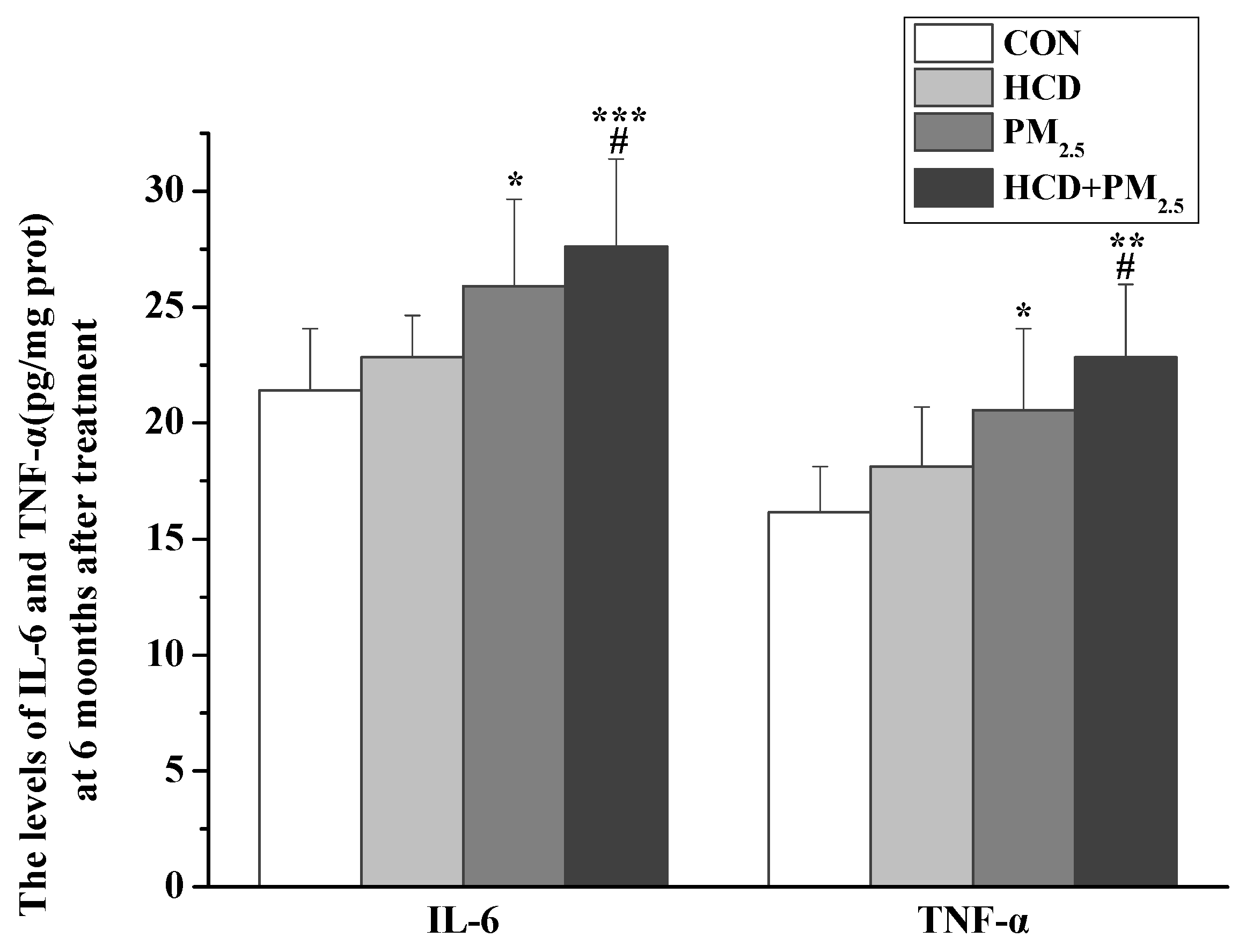
3.5. Effects of HCD and PM2.5 on the Expression of Inflammatory Markers

As shown in Figure 5, PM2.5 and HCD plus PM2.5 exposure increased inflammation significantly compared with the CON group. Compared with the HCD group, the HCD + PM2.5 group increased inflammatory markers markedly. The change in TNF-α and IL-6 levels in the PM2.5 plus HCD group was not significant relative to the PM2.5 exposure alone.

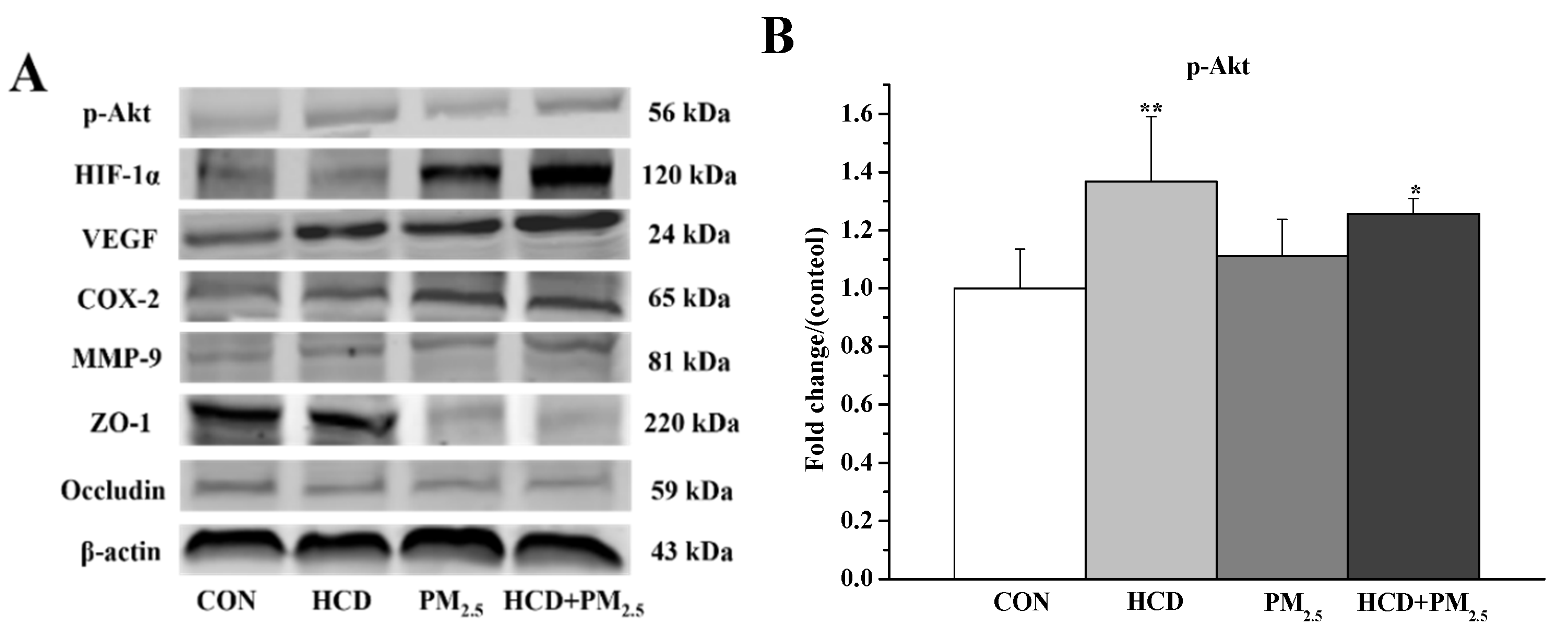
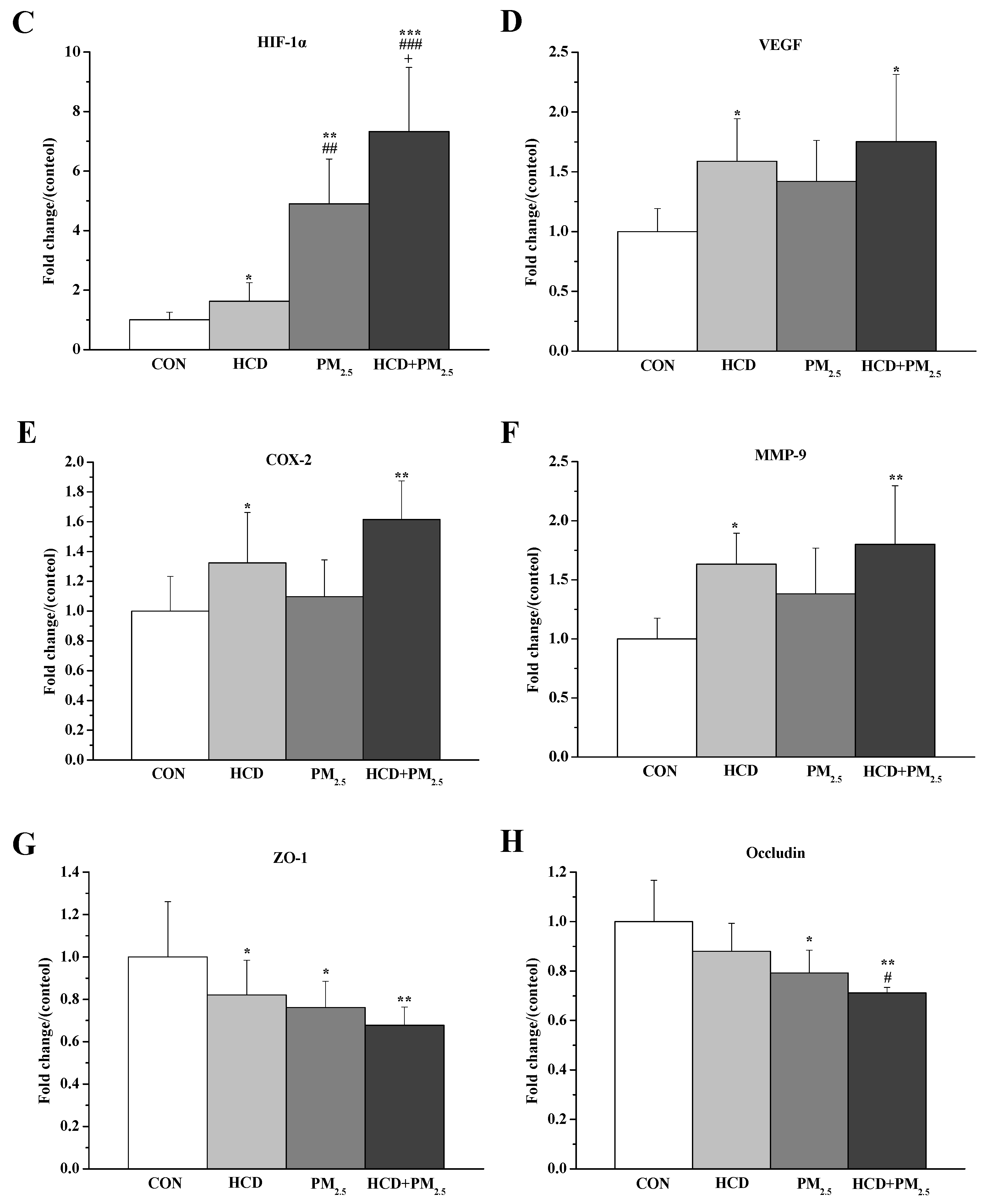
3.6. Effects of HCD and PM2.5 on the Protein Expressions of Stroke and BBB Basement Membrane Damage in the Brains

As shown in Figure 6, in comparison with the CON group, the protein levels of all the genes in the co-exposure group had a significant alteration. The protein levels of all the genes, except Occludin, were markedly changed in the mice after the treatment of HCD relative to the control. The obvious alterations in HIF-1α, ZO-1, and Occludin levels were observed in the mice after PM2.5 exposure. Compared with the HCD group, the HIF-1α and Occludin levels in the co-exposure group were significantly changed. Compared with the PM2.5 group, only the HIF-1α expression increased considerably in the co-exposure group (Figure 6). Especially in Figure 6C, HIF-1α levels in the PM2.5 plus HCD group were significantly increased compared with the PM2.5 or HCD group.

4. Discussion

PM2.5 is a ubiquitous environmental contaminant, notorious for its pervasive presence in the atmosphere and its profound health implications. Similarly, a suboptimal diet, particularly one rich in saturated fats such as an HCD, has been implicated in the exacerbation of various diseases, notably cardiovascular disorders. Epidemiologic studies have illuminated the links between HCD consumption and exposure to PM2.5 with increased incidences of stroke [11,12,17]. Yet, the intricate toxicological mechanisms underlying these associations, especially concerning female physiology, remain inadequately elucidated. The present study embarked on a comprehensive exploration to unravel the molecular regulatory mechanisms governing the synergistic effects of HCD and PM2.5 co-exposure on brain injury in female mice. A multi-faceted experimental design was adopted to achieve this objective, integrating three specific models: the PM2.5 real-exposure model, the HCD model, and the ApoE−/− mice model. The PM2.5 real-exposure model allowed for the simulation of authentic environmental conditions, which can make the subjects exposed to ambient particulate matter levels reflective of real-world pollution scenarios. Simultaneously, the HCD model facilitated the induction of a metabolic state analogous to that observed in humans consuming diets high in cholesterol, thereby creating a milieu conducive to studying diet-related neurological effects. Finally, the employment of ApoE−/− mice, a strain characterized by a genetic deficiency in ApoE, a protein crucial for lipid metabolism and neuronal health, offered a unique perspective on the interplay between genetics, environmental pollutants, and dietary habits.
This study assessed the effects of HCD and PM2.5 co-exposure on brain injury and behavioral changes in female mice. As Figure 2 shows, the buried pellet test, which can evaluate rodents’ olfactory function and odor recognition ability, was processed [43,44,45]. The olfactory dysfunction can be used as an important indicator of learning and memory impairment. In the buried pellet test, HCD intake and PM2.5 exposure increased the mice’s latency in finding food compared to the control. The latency to find food became longer after the co-exposure, and the latency became longer with a more prolonged exposure duration. The modified Garcia test further evaluates the damage of HCD and PM2.5 to mice nerves (Figure 3).
On this basis, the HE staining results of pathological damage to brains are illustrated in Figure 4. The results showed that HCD and PM2.5 could cause pathological damage to the brains of mice. In quantifying the number of neurons, it was found that exposure to PM2.5 or HCD reduced the number of neurons to varying degrees, confirming this damage [46,47].
CNS neuroinflammation, oxidative stress, BBB injury, and neuronal damage can lead to CNS diseases [48]. IL-6 and TNF-α are important neuroinflammatory factors that activate inflammatory reactions and are involved in the pathogenesis of stroke [49]. Multiple reports from rodent studies have confirmed that exposure to traffic PM2.5 leads to an increase in pro-inflammatory cytokines in CNS, including TNF-α and IL-6 [5,50], which is similar to the results of this study. Our results indicated that exposure to PM2.5 and HCD increases IL-6 and TNF-α in the brain (Figure 5). In particular, combined exposure to HCD and PM2.5 had more severe damage than exposure to HCD or PM2.5 alone. It suggests that HCD and PM2.5 may impair certain natural behaviors in mice with time effects through elevated levels of inflammation [44,45].
ApoE deficiency can lead to structural and functional defects in the mammalian brain. It interacts with very low-density lipoprotein receptor and ApoE receptor 2 to convert hydrophobic molecules such as cholesterol and amyloid proteins β to neurons, leading to neuronal damage [51,52]. ApoE−/− mice show significant reductions in dendritic size and synaptic number, as well as impaired learning and memory [53]. The ApoE−/− model enables us to analyze the effects of PM2.5 and HCD on the cerebrovascular system in a baseline model of potential vascular diseases similar to most of the population [54,55]. Animal experiments have shown that exposure to traffic-generated air pollution can cause BBB damage in ApoE−/− male C57BL/6 mice, mediated by changes in MMP-2 and MMP-9 activity and inflammatory signals [56]. However, these studies were only conducted in male mice, so PM2.5 inhalation exposure and HCD feeding studies were performed to determine whether female mice had similar harmful CNS results in their brains. A study has found that obese individuals exposed to PM2.5 have a more significant impact on CNS in women than in men [32,37], which is consistent with our results.
It is known that ApoE deficiency in the brain may cause BBB instability [57]. The ApoE−/− mice in this study observed BBB damage in the brain after exposure to PM2.5 and HCD, consistent with previous research on BBB damage in the mouse brain [56]. Calderón-Garcidueñas et al. found that exposure to air pollution is a risk factor for CNS diseases, which may be mediated by changes in COX-2 and interleukin-1β caused by damage to BBB permeability [58]. This study showed that PM2.5 and HCD caused an increase in p-Akt, HIF-1α, COX-2, VEGF, and MMP-9 in the brain of ApoE−/− mice (Figure 6B–F). According to the above, it can be inferred that HIF-1α regulates COX-2 and VEGF, thereby rapidly generating MMP-9. These results indicated that p-Akt/HIF-1α/COX-2/VEGF/MMP-9 plays an important role in nerve damage caused by PM2.5 and HCD. MMP-9 reduces the expression of ZO-1 and Occludin, thereby increasing the permeability of the BBB basement membrane, damaging BBB, and causing harmful substances to enter the brain, leading to secondary cerebral vascular edema and damage to brain function [59]. VEGF enhanced the expression of MMP-9 and decreased the expression of ZO-1 and Occludin (Figure 6G,H), indicating that the VEGF/MMP-9/ZO-1/Occludin pathway plays a vital role in the permeability of BBB and the intermediate stage of basement membrane rupture caused by PM2.5 and HCD.

5. Conclusions

This study revealed that exposure to HCD and PM2.5 caused brain pathology damage and behavioral abnormality in ApoE−/− female mice through the HIF-1α pathway. (1) Exposure to HCD and PM2.5 triggered abnormal behavioral alterations and significant histopathological changes in the brains of ApoE−/− female mice, accompanied by an inflammatory response. (2) The HIF-1α/COX-2/VEGF/MMP9 regulatory pathway exerted an important role in brain injury in mice caused by HCD and PM2.5, and the enhancement of BBB permeability aggravated brain injury. The study will be beneficial in not only elucidating the molecular mechanisms of the initiation or exacerbation of stroke induced by HCD and PM2.5 but also in providing novel evidence for revealing the adverse effects of HCD and PM2.5 on women’s brain nerve health.

Author Contributions

W.C.: Methodology, Data curation, Formal analysis, Writing—original draft. S.C.: Methodology, Data curation, Formal analysis. L.B.: Methodology, Data curation. R.L.: Project administration, Fund acquiring, Supervision. All authors have read and agreed to the published version of the manuscript.

Funding

This work was supported by the Nature Science Foundation of Shanxi Province in China (201801D121260).

Institutional Review Board Statement

The animal study protocol was approved by the Animal Ethics Committee of Shanxi University, China (Approval No. SXULL-2019036).

Informed Consent Statement

Not applicable.

Data Availability Statement

The data presented in this study are available on request from the corresponding author. The data are not publicly available due to privacy.

Conflicts of Interest

The authors declare no conflicts of interest.

References

  1. Jeffrey, D.S.; Ashkan, A.; Emmanuela, G.; Stephen, S.L.; Degu, A.; Kalkidan, H.A.; Cristiana, A.; Nooshin, A.; Hedayat, A.; Foad, A.; et al. Global, regional, and national comparative risk assessment of 84 behavioural, environmental and occupational, and metabolic risks or clusters of risks for 195 countries and territories, 1990–2017: A systematic analysis for the Global Burden of Disease Study. Lancet 2018, 392, 1923–1994. [Google Scholar]
  2. Martinelli, N.; Olivieri, O.; Girelli, D. Air particulate matter and cardiovascular disease: A narrative review. Eur. J. Intern. Med. 2013, 24, 295–302. [Google Scholar] [CrossRef] [PubMed]
  3. Badyda, A.; Grellier, J.; Dabrowiecki, P. Ambient PM2.5 exposure and mortality due to lung cancer and cardiopulmonary diseases in Polish cities. Adv. Exp. Med. Biol. 2017, 944, 9–17. [Google Scholar] [PubMed]
  4. Fu, P.F.; Guo, X.B.; Cheung, F.M.H.; Yung, K.K.L. The association between PM2.5 exposure and neurological disorders: A systematic review and meta-analysis. Sci. Total Environ. 2019, 655, 1240–1248. [Google Scholar] [CrossRef] [PubMed]
  5. Levesque, S.; Surace, M.J.; McDonald, J.; Block, M.L. Air pollution & the brain: Subchronic diesel exhaust exposure causes neuroinflammation and elevates early markers of neurodegenerative disease. J. Neuroinflamm. 2011, 8, 105. [Google Scholar]
  6. You, R.; Ho, Y.S.; Chang, R.C.C. The pathogenic effects of particulate matter on neurodegeneration: A review. J. Biomed. Sci. 2022, 29, 15. [Google Scholar] [CrossRef]
  7. Oberdorster, G.; Sharp, Z.; Atudorei, V.; Elder, A.; Gelein, R.; Kreyling, W.; Cox, C. Translocation of inhaled ultrafine particles to the brain. Inhal. Toxicol. 2004, 16, 437–445. [Google Scholar] [CrossRef] [PubMed]
  8. Proulx, S.T. Cerebrospinal fluid outflow: A review of the historical and contemporary evidence for arachnoid villi, perineural routes, and dural lymphatics. Cell Mol. Life Sci. 2021, 78, 2429–2457. [Google Scholar] [CrossRef]
  9. Lu, D.; Luo, Q.; Chen, R.; Zhuansun, Y.; Jiang, J.; Wang, W.; Yang, X.; Zhang, L.; Liu, X.; Li, F.; et al. Chemical multi-fingerprinting of exogenous ultrafine particles in human serum and pleural effusion. Nat. Commun. 2020, 11, 2567. [Google Scholar] [CrossRef] [PubMed]
  10. Qi, Y.; Wei, S.; Xin, T.; Huang, C.; Pu, Y.; Ma, J.; Zhang, C.; Liu, Y.; Lynch, I.; Liu, S. Passage of exogeneous fine particles from the lung into the brain in humans and animals. Proc. Natl. Acad. Sci. USA 2022, 119, e2117083119. [Google Scholar] [CrossRef]
  11. Shah, A.S.; Lee, K.K.; McAllister, D.A.; Hunter, A.; Nair, H.; Whiteley, W.; Langrish, J.P.; Newby, D.E.; Mills, N.L. Short term exposure to air pollution and stroke: Systematic review and meta-analysis. BMJ 2015, 350, h1295. [Google Scholar] [CrossRef]
  12. Qiu, H.; Sun, S.; Tsang, H.; Wong, C.M.; Lee, R.S.; Schooling, C.M.; Tian, L. Fine particulate matter exposure and incidence of stroke: A cohort study in Hong Kong. Neurology 2017, 88, 1709–1717. [Google Scholar] [CrossRef] [PubMed]
  13. Gao, L.; Qin, J.X.; Shi, J.Q.; Jiang, T.; Wang, F.; Xie, C.; Gao, Q.; Zhi, N.; Dong, Q.; Guan, Y.T. Fine particulate matter exposure aggravates ischemic injury via NLRP3 inflammasome activation and pyroptosis. CNS Neurosci. Ther. 2022, 28, 1045–1058. [Google Scholar] [CrossRef] [PubMed]
  14. Oppenheim, H.A.; Lucero, J.; Guyot, A.; Herbert, L.M.; McDonald, J.D.; Mabondzo, A.; Lund, A.K. Exposure to vehicle emissions results in altered blood brain barrier permeability and expression of matrix metalloproteinases and tight junction proteins in mice. Part. Fibre Toxicol. 2013, 10, 62–75. [Google Scholar] [CrossRef] [PubMed]
  15. Huang, Y.; Chen, S.; Luo, Y.; Han, Z. Crosstalk between inflammation and the BBB in stroke. Curr. Neuropharmacol. 2020, 18, 1227–1236. [Google Scholar] [CrossRef] [PubMed]
  16. Li, W.G.; Prakash, R.; Chawla, D.; Du, W.T.; Didion, S.P.; Filosa, J.A.; Zhang, Q.G.; Brann, D.W.; Lima, V.V.; Tostes, R.C.; et al. Early effects of high-fat diet on neurovascular function and focal ischemic brain injury. Am. J. Physiol.-Regul. Integr. Comp. Physiol. 2013, 304, R1001–R1008. [Google Scholar] [CrossRef] [PubMed]
  17. Larsson, S.C.; Virtamo, J.; Wolk, A. Dietary fats and dietary cholesterol and risk of stroke in women. Atherosclerosis 2012, 221, 282–286. [Google Scholar] [CrossRef] [PubMed]
  18. Colin-Castelan, D.; Zaina, S. Associations between atherosclerosis and neurological diseases, beyond ischemia-induced cerebral damage. Rev. Endocr. Metab. Dis. 2019, 20, 15–25. [Google Scholar] [CrossRef] [PubMed]
  19. Cai, D.S.; Liu, T.W. Inflammatory cause of metabolic syndrome via brain stress and NF-kappa B. Aging 2012, 4, 98–115. [Google Scholar] [CrossRef]
  20. Diker, S.; Gelener, P.; Eker, A.; Kaymakamzade, B.; Mut, S.; Erem, A.; Balyemez, U. Association between cerebral microbleeds and inflammatory biomarkers in patients with ischemic stroke. Egypt. J. Neurol. Psychiatry Neurosurg. 2022, 58, 43. [Google Scholar] [CrossRef]
  21. Noh, J.; Sohn, J.; Han, M.; Kang, D.R.; Choi, Y.J.; Kim, H.C.; Suh, I.; Kim, C.; Shin, D.C. Long-term effects of cumulative average PM2.5 exposure on the risk of hemorrhagic stroke. Epidemiology 2019, 30 (Suppl. 1), S90–S98. [Google Scholar] [CrossRef] [PubMed]
  22. Barkas, F.; Elisaf, M.; Milionis, H. Statins decrease the risk of stroke in individuals with heterozygous familial hypercholesterolemia: A systematic review and meta-analysis. Atherosclerosis 2015, 243, 60–64. [Google Scholar] [CrossRef] [PubMed]
  23. Verghese, P.B.; Castellano, J.M.; Holtzman, D.M. Apolipoprotein E in Alzheimer’s disease and other neurological disorders. Lancet Neurol. 2011, 10, 241–252. [Google Scholar] [CrossRef]
  24. Qiao, S.Y.; Shang, K.; Chu, Y.H.; Yu, H.H.; Chen, X.; Qin, C.; Pan, D.J.; Tian, D.S. Apolipoprotein E epsilon4 polymorphism as a risk factor for ischemic stroke: A systematic review and meta-analysis. Dis. Markers 2022, 2022, 1407183. [Google Scholar] [CrossRef] [PubMed]
  25. Zhang, L.; Luo, X.L.; Chen, F.; Yuan, W.; Xiao, X.L.; Zhang, X.H.; Dong, Y.R.; Zhang, Y.X.; Liu, Y. LncRNA SNHG1 regulates cerebrovascular pathologies as a competing endogenous RNA through HIF-1/VEGF signaling in ischemic stroke. J. Cell Biochem. 2018, 119, 5460–5472. [Google Scholar] [CrossRef] [PubMed]
  26. Freedman, S.J.; Sun, Z.Y.; Poy, F.; Kung, A.L.; Livingston, D.M.; Wagner, G.; Eck, M.J. Structural basis for recruitment of CBP/p300 by hypoxia-inducible factor-1 alpha. Proc. Natl. Acad. Sci. USA 2002, 99, 5367–5372. [Google Scholar] [CrossRef]
  27. Greer, S.N.; Metcalf, J.L.; Wang, Y.; Ohh, M. The updated biology of hypoxia-inducible factor. EMBO J. 2012, 31, 2448–2460. [Google Scholar] [CrossRef] [PubMed]
  28. Yang, C.; Yang, Y.; DeMars, K.M.; Rosenberg, G.A.; Candelario-Jalil, E. Genetic deletion or pharmacological inhibition of cyclooxygenase-2 reduces blood-brain barrier damage in experimental ischemic stroke. Front. Neurol. 2020, 11, 887. [Google Scholar] [CrossRef] [PubMed]
  29. Laughner, E.; Taghavi, P.; Chiles, K.; Mahon, P.C.; Semenza, G.L. HER2 (neu) signaling increases the rate of hypoxia-inducible factor 1alpha (HIF-1alpha) synthesis: Novel mechanism for HIF-1-mediated vascular endothelial growth factor expression. Mol. Cell Biol. 2001, 21, 3995–4004. [Google Scholar] [CrossRef]
  30. Pez, F.; Dayan, F.; Durivault, J.; Kaniewski, B.; Aimond, G.; Le Provost, G.S.; Deux, B.; Clezardin, P.; Sommer, P.; Pouyssegur, J.; et al. The HIF-1-inducible lysyl oxidase activates HIF-1 via the Akt pathway in a positive regulation loop and synergizes with HIF-1 in promoting tumor cell growth. Cancer Res. 2011, 71, 1647–1657. [Google Scholar] [CrossRef]
  31. Zhang, F.; Ding, T.; Yu, L.; Zhong, Y.; Dai, H.; Yan, M. Dexmedetomidine protects against oxygen-glucose deprivation-induced injury through the I2 imidazoline receptor-PI3K/Akt pathway in rat C6 glioma cells. J. Pharm. Pharmacol. 2012, 64, 120–127. [Google Scholar] [CrossRef] [PubMed]
  32. Zeng, W.; Zhang, Y.C.; Wang, L.; Wei, Y.L.; Lu, R.; Xia, J.J.; Chai, B.; Liang, X. Ambient fine particulate pollution and daily morbidity of stroke in Chengdu, China. PLoS ONE 2018, 13, e0206836. [Google Scholar] [CrossRef]
  33. Kim, H.; Noh, J.; Noh, Y.; Oh, S.S.; Koh, S.B.; Kim, C. Gender difference in the effects of outdoor air pollution on cognitive function among elderly in Korea. Front. Public Health 2019, 7, 375–384. [Google Scholar] [CrossRef]
  34. Petrea, R.E.; Beiser, A.S.; Seshadri, S.; Kelly-Hayes, M.; Kase, C.S.; Wolf, P.A. Gender differences in stroke incidence and poststroke disability in the framingham heart study. Stroke 2009, 40, 1032–1037. [Google Scholar] [CrossRef]
  35. Bushnell, C.; McCullough, L.D.; Awad, I.A.; Chireau, M.V.; Fedder, W.N.; Furie, K.L.; Howard, V.J.; Lichtman, J.H.; Lisabeth, L.D.; Pina, I.L.; et al. Guidelines for the prevention of stroke in women: A statement for healthcare professionals from the American Heart Association/American Stroke Association. Stroke 2014, 45, 1545–1588. [Google Scholar] [CrossRef] [PubMed]
  36. Zhao, C.; Xie, P.S.; Yong, T.; Huang, W.; Liu, J.J.; Wu, D.S.; Ji, F.F.; Li, M.; Zhang, D.D.; Li, R.J.; et al. Airborne fine particulate matter induces cognitive and emotional disorders in offspring mice exposed during pregnancy. Sci. Bull. 2021, 66, 578–591. [Google Scholar] [CrossRef] [PubMed]
  37. Malpetti, M.; Sala, A.; Vanoli, E.G.; Gianolli, L.; Luzi, L.; Perani, D. Unfavourable gender effect of high body mass index on brain metabolism and connectivity. Sci. Rep. 2018, 8, 12584. [Google Scholar] [CrossRef] [PubMed]
  38. Zhao, L.F.; Zhang, M.; Bai, L.R.; Zhao, Y.F.; Cai, Z.W.; Yung, K.K.L.; Dong, C.; Li, R.J. Real-world PM2.5 exposure induces pathological injury and DNA damage associated with miRNAs and DNA methylation alteration in rat lungs. Environ. Sci. Pollut. Res. Int. 2022, 29, 28788–28803. [Google Scholar] [CrossRef]
  39. Hu, Y.; Ding, W.; Zhu, X.; Chen, R.; Wang, X. Olfactory dysfunctions and decreased nitric oxide production in the brain of human P301L tau transgenic mice. Neurochem. Res. 2016, 41, 722–730. [Google Scholar] [CrossRef]
  40. Wu, J.; Zhang, Y.; Yang, P.; Enkhjargal, B.; Manaenko, A.; Tang, J.P.; Pearce, W.J.; Hartman, R.; Obenaus, A.; Chen, G.; et al. Recombinant osteopontin stabilizes smooth muscle cell phenotype via integrin receptor/integrin-linked kinase/rac-1 pathway after subarachnoid hemorrhage in rats. Stroke 2016, 47, 1319–1327. [Google Scholar] [CrossRef]
  41. Zhang, Y.; Chen, Y.; Wu, J.; Manaenko, A.; Yang, P.; Tang, J.; Fu, W.; Zhang, J.H. Activation of dopamine D2 receptor suppresses neuroinflammation through alphaB-crystalline by inhibition of NF-kappaB nuclear translocation in experimental ICH mice model. Stroke 2015, 46, 2637–2646. [Google Scholar] [CrossRef] [PubMed]
  42. Li, R.; Zhao, Y.; Shi, J.; Zhao, C.; Xie, P.; Huang, W.; Yong, T.; Cai, Z.W. Effects of PM2.5 exposure in utero on heart injury, histone acetylation and GATA4 expression in offspring mice. Chemosphere 2020, 256, 127133. [Google Scholar] [CrossRef] [PubMed]
  43. van Zyl, P.J.; Dimatelis, J.J.; Russell, V.A. Behavioural and biochemical changes in maternally separated Sprague-Dawley rats exposed to restraint stress. Metab. Brain Dis. 2016, 31, 121–133. [Google Scholar] [CrossRef] [PubMed]
  44. Lee, B.C.; Kang, I.; Lee, S.E.; Lee, J.Y.; Shin, N.; Kim, J.J.; Choi, S.W.; Kang, K.S. Human umbilical cord blood plasma alleviates age-related olfactory dysfunction by attenuating peripheral TNF-α expression. BMB Rep. 2019, 52, 259–264. [Google Scholar] [CrossRef] [PubMed]
  45. Yeh, C.F.; Chuang, T.Y.; Lan, M.Y.; Chin, Y.C.; Wang, W.H.; Lin, Y.Y. Excessive expression of microglia/macrophage and proinflammatory mediators in olfactory bulb and olfactory dysfunction after stroke. In Vivo 2019, 33, 1893–1899. [Google Scholar] [CrossRef]
  46. Baron, J.C.; Yamauchi, H.; Fujioka, M.; Endres, M. Selective neuronal loss in ischemic stroke and cerebrovascular disease. J. Cereb. Blood Flow. Metab. 2014, 34, 2–18. [Google Scholar] [CrossRef] [PubMed]
  47. Datta, A.; Sarmah, D.; Kalia, K.; Borah, A.; Wang, X.; Dave, K.R.; Yavagal, D.R.; Bhattacharya, P. Advances in studies on stroke-induced secondary neurodegeneration (SND) and its treatment. Curr. Top. Med. Chem. 2020, 20, 1154–1168. [Google Scholar] [CrossRef] [PubMed]
  48. Shou, Y.K.; Huang, Y.L.; Zhu, X.Z.; Liu, C.Q.; Hu, Y.; Wang, H.H. A review of the possible associations between ambient PM2.5 exposures and the development of Alzheimer’s disease. Ecotoxicol. Environ. Safe 2019, 174, 344–352. [Google Scholar] [CrossRef] [PubMed]
  49. Liu, C.; Yang, J.; Guan, L.; Zhu, Y.; Geng, X. Filtered air intervention reduces inflammation and hypothalamus-pituitary-adrenal axis activation in adult male and female rats after PM2.5 exposure. Environ. Sci. Pollut. Res. Int. 2020, 27, 35341–35348. [Google Scholar] [CrossRef]
  50. Gerlofs-Nijland, M.E.; van, B.D.; Cassee, F.R.; Schins, R.P.F.; Wang, K.; Campbell, A. Effect of prolonged exposure to diesel engine exhaust on proinflammatory markers in different regions of the rat brain. Part. Fibre Toxicol. 2010, 7, 12. [Google Scholar] [CrossRef]
  51. Liu, C.C.; Kanekiyo, T.; Xu, H.X.; Bu, G.J. Apolipoprotein E and Alzheimer disease: Risk, mechanisms and therapy. Nat. Rev. Neurol. 2013, 9, 106–118. [Google Scholar] [CrossRef] [PubMed]
  52. Lane-Donovan, C.; Herz, J. The ApoE receptors Vldlr and Apoer2 in central nervous system function and disease. J. Lipid Res. 2017, 58, 1036–1043. [Google Scholar] [CrossRef] [PubMed]
  53. Evola, M.; Hall, A.; Wall, T.; Young, A.; Grammas, P. Oxidative stress impairs learning and memory in ApoE knockout mice. Pharmacol. Biochem. Behav. 2010, 96, 181–186. [Google Scholar] [CrossRef] [PubMed]
  54. Getz, G.S.; Reardon, C.A. Animal models of atherosclerosis. Arterioscler. Thromb. Vasc. Biol. 2012, 32, 1104–1115. [Google Scholar] [CrossRef]
  55. Lo, S.G.; Schlage, W.K.; Boue, S.; Veljkovic, E.; Peitsch, M.C.; Hoeng, J. The Apoe−/− mouse model: A suitable model to study cardiovascular and respiratory diseases in the context of cigarette smoke exposure and harm reduction. J. Transl. Med. 2016, 14, 146. [Google Scholar]
  56. Suwannasual, U.; Lucero, J.; McDonald, J.D.; Lund, A.K. Exposure to traffic-generated air pollutants mediates alterations in brain microvascular integrity in wildtype mice on a high-fat diet. Environ. Res. 2018, 160, 449–461. [Google Scholar] [CrossRef] [PubMed]
  57. Nishitsuji, K.; Hosono, T.; Nakamura, T.; Bu, G.J.; Michikawa, M. Apolipoprotein E regulates the integrity of tight junctions in an isoform-dependent manner in an in vitro blood-brain barrier model. J. Biol. Chem. 2011, 286, 17536–17542. [Google Scholar] [CrossRef] [PubMed]
  58. Calderón-Garcidueñas, L.; Solt, A.C.; Henríquez-Roldán, C.; Torres-Jardon, R.; Nuse, B.; Herritt, L.; Villarreal-Calderon, R.; Osnaya, N.; Stone, I.; Garcia, R.; et al. Long-term air pollution exposure is associated with neuroinflammation, an altered innate immune response, disruption of the blood-brain barrier, ultrafine particulate deposition, and accumulation of amyloid beta-42 and alpha-synuclein in children and young adults. Toxicol. Pathol. 2008, 36, 289–310. [Google Scholar]
  59. Fini, M.E.; Jeong, S.; Wilson, M.R. Therapeutic potential of the molecular chaperone and matrix metalloproteinase inhibitor clusterin for dry eye. Int. J. Mol. Sci. 2020, 22, 116. [Google Scholar] [CrossRef]
Figure 1. Protocols of female animals’ exposure to HCD and PM2.5. Notes: The mice were divided into four groups randomly: CON group, HCD group, PM2.5 group, and HCD + PM2.5 group. Normal diet: carbohydrate, protein, fat, no cholesterol, no bile salt; HCD: carbohydrate, protein, fat, 1.25% cholesterol, 0.5% bile salt.
Atmosphere 15 00952 g001
Figure 2. The effects of HCD and PM2.5 on the score of buried pellet test. The buried pellet test assessed olfactory function and odor recognition ability in mice, revealing the effects of HCD and PM2.5 co-exposure on these sensory and cognitive aspects. Using the one-way ANOVA-LSD method, ** p < 0.01, *** p < 0.001 vs. CON group; ## p < 0.01 vs. HCD group; + p < 0.05 vs. PM2.5 group; 3M and 6M represent female mice exposed for 3 and 6 months; values were expressed as means ± SD (n = 10).
Atmosphere 15 00952 g002
Figure 3. The effects of HCD and PM2.5 on the score of neurological function. Using the one-way ANOVA-LSD method, * p < 0.05, ** p < 0.01, *** p < 0.001 vs. CON group; # p < 0.05, ## p < 0.01 vs. HCD group; 3M and 6M represent female mice exposed for 3 and 6 months, respectively. Values were expressed as means ± SD (n = 10).
Atmosphere 15 00952 g003
Figure 4. Effects of HCD and PM2.5 exposure on hippocampus structure. The image on the bottom left was a larger version of the dark box on the right. Bar = 100 µm (amplification). 6M-ApoE−/− (AD); bar graph showing alteration in the number of neurons in 6M-ApoE−/−, respectively (E). The blue arrows represent neurons. Using the one-way ANOVA-LSD method, ** p < 0.01, *** p < 0.001 vs. CON group; # p < 0.05 vs. HCD group. Values were expressed as means ± SD (n = 3).
Atmosphere 15 00952 g004
Figure 5. The effects of HCD and PM2.5 on the expression of inflammatory markers (using the one-way ANOVA-LSD method, * p < 0.05, ** p < 0.01, *** p < 0.001 vs. CON group; # p < 0.05 vs. HCD group). Values were expressed as means ± SD (n = 6).
Atmosphere 15 00952 g005
Figure 6. Effects of HCD and PM2.5 on the protein expressions of stroke and BBB basement membrane-related genes in ApoE−/− mice. Representative Western blot in brains (A); HCD and PM2.5 further augmented p-Akt, HIF-1α, VEGF, COX-2, and MMP-9 expression (BF); and inhibited the expression of ZO-1 and Occludin (G,H). The relative densities of each protein were normalized against the CON group. Values were expressed as means ± SD (n = 4). Using the one-way ANOVA-LSD method, * p < 0.05, ** p < 0.01, *** p < 0.001 vs. CON group; # p < 0.05, ## p < 0.01 ### p < 0.001 vs. HCD group; + p < 0.05 vs. PM2.5 group.
Atmosphere 15 00952 g006aAtmosphere 15 00952 g006b
Table 1. Changes in brain–body weight ratio (%) in mice of different groups.
GroupsBrain WeightBody WeightBrain–Body Weight Ratio
CON0.40 ± 0.0410.33 ± 1.321.47 ± 0.18
HCD0.39 ± 0.069.45 ± 3.471.52 ± 0.27
PM2.50.44 ± 0.039.59 ± 1.461.68 ± 0.13 *
HCD + PM2.50.42 ± 0.0310.80 ± 1.741.51 ± 0.17
Note: Data are expressed as x ¯ ± SD (n = 6) using the one-way ANOVA-LSD method, compared with the CON group, * p < 0.05.
Disclaimer/Publisher’s Note: The statements, opinions and data contained in all publications are solely those of the individual author(s) and contributor(s) and not of MDPI and/or the editor(s). MDPI and/or the editor(s) disclaim responsibility for any injury to people or property resulting from any ideas, methods, instructions or products referred to in the content.

