Article

Praseodymium(III) Removal from Aqueous Solutions Using Living and Non-Living Arthrospira platensis Biomass

1
Joint Institute for Nuclear Research, Dubna, 141980 Dubna, Russia
2
Doctoral School Biological, Geonomic, Chemical and Technological Science, State University of Moldova, 2009 Chisinau, Moldova
3
Horia Hulubei National Institute for R&D in Physics and Nuclear Engineering, Bucharest Magurele, MG-6, 077125 Bucharest, Romania
4
Institute of Chemistry, 2002 Chisinau, Moldova
5
Institute of Microbiology and Biotechnology, Technical University of Moldova, 2028 Chisinau, Moldova
*
Author to whom correspondence should be addressed.
Water 2023, 15(11), 2064; https://doi.org/10.3390/w15112064
Submission received: 30 March 2023 / Revised: 17 May 2023 / Accepted: 22 May 2023 / Published: 29 May 2023

Abstract

:
Praseodymium, the sixth-most abundant rare-earth element, is widely used in the aircraft industry for the elaboration of refractory substances, coloring materials, lighting equipment, and fiber optical cables. Living and non-living Arthrospira platensis biomass was applied for Pr(III) removal from aqueous solutions. In bioaccumulation experiments, the effect of Pr(III), introduced into the medium in a concentration range of 10–30 mg/L, on biomass productivity, biochemical composition, and antioxidant activity was assessed. The biomass showed high accumulation capacity (more than 99%) toward Pr(III). Supplementation of the cultivation medium with Pr(III) led to a decrease in carbohydrate and lipid content, but it did not significantly influence biomass productivity or the content of proteins and pigments. In experiments with non-living biomass, the effect of pH, Pr(III) concentration, temperature, and contact time on the efficiency of metal removal was investigated. The maximum uptake of Pr(III) was achieved at pH 3.0 after 3.0 min of interaction. The equilibrium data were explained using the Langmuir and Freundlich models, while the kinetics of the process was described by applying pseudo-first-order, pseudo-second-order, and Elovich models. The maximum sorption capacity of Arthrospira platensis biomass calculated from the Langmuir model was 99.3 mg/g. According to the thermodynamic calculations, the process of Pr(III) removal was spontaneous and exothermic in nature. The obtained data can be used for the development of environmentally-friendly technology for Pr(III) recovery from wastewater as well as to understand the effect of Pr(III) on aquatic organisms.

1. Introduction

Rare earth elements (REEs) are a group of 15 lanthanides, as well as Sc and Y, which are categorized into light (La, Ce, Pr, Nd, Pm, and Sm) and heavy (Eu, Gd, Tb, Dy, Ho, Er, Tm, Yb, Lu, and Y) REEs [1,2]. Owing to their chemical, metallurgical, optical, catalytic, magnetic, and electronic properties, REEs are intensively applied in the chemical industry, metallurgy, medicine, electronic device production, petroleum refining [1,2,3,4], nuclear technologies, the production of fertilizers, telecommunications, and security systems [5].
Recently, China has become the leading producer of REEs [2,3] and the strict norms introduced by the state on REEs’ export in 2011 significantly affected the global market through a reduction in REEs’ availability and an increase in their price [1,3].
Under the current conditions, a viable alternative to cover the need for REEs is their recovery from industrial waste, including wastewater, and electrical and electronic compounds [3,5]. In addition, the recovery of REEs is determined by possible contamination of the environment, which might have a negative impact on aquatic organisms and even human health [3,6]. The toxic effects induced by REEs on soil, water organisms, as well as human health, are described in detail in several review papers [6,7,8].
Different technologies, including precipitation, ion exchange, electrochemical processes, flocculation, solvent extraction, and adsorption, have been routinely applied to recover REEs [1,2,3,9]. The mentioned techniques allow for high efficiency of REE recovery to be achieved; however, often, they have severe drawbacks, among which high costs, low selectivity and purity of the extracted elements, high consumption of reagents and energy, the creation of large volumes of sludge [2], and the use of materials derived from non-renewable resources can be mentioned [10]. These limitations do not allow us to consider the traditional techniques as environmentally-friendly approaches which correspond to green engineering principles.
Application of microbial biomass can be regarded as an alternative, cost effective approach for REEs’ recovery from wastewater [11]. Among the biological approaches, biosorption and bioaccumulation have received special attention [10]. When looking at differences between processes, it should be highlighted that biosorption assumes REEs’ removal mainly by using non-living biomass, while bioaccumulation uses only living organisms [12,13]. The biosorption process mainly occurs on the cell surface and includes several mechanisms of interaction: physical adsorption, complexation, ion exchange, and precipitation [14]. The bioaccumulation process is associated with metal ions transport across the cell membrane and accumulation inside the cells. Thus, the process is dependent on microorganisms’ properties as well as the level of their adaptations and metal toxicity [12]. Biosorption in comparison with bioaccumulation does not require nutrients and energy sources and higher removal capacity can be achieved; in addition, the process is rapid and the biomass can be regenerated. Bioaccumulation is a more intricate and pricey process, which can be applied for bioremediation of large polluted territories, as well as to describe the mechanism of metal ions action on living organisms [12,13].
Microorganisms are actively applied for REEs’ biosorption and bioaccumulation from aqueous solutions [11,15,16,17,18]. Among microorganisms, cyanobacteria have shown promising bioremediation properties for heavy metals, including REEs, which enable them to be used for bioremediation and metal recovery on an industrial scale [18,19,20,21,22]. However, according to the published information, there are no data on cyanobacteria use for Pr(III), a REE with numerous industrial applications, recovery from aqueous solution.
The basic aim of the present research was to assess the removal capacity of living and non-living Arthrospira platensis (A. platensis) biomass toward Pr(III). To achieve the goal of the study, the effect of different Pr(III) concentrations on living A. platensis biomass productivity, biochemical composition, and antioxidant activity was investigated. In the case of experiments with non-living biomass, the impact of Pr(III) concentration, pH, contact time, and temperature on biosorption capacity was monitored. In addition, to understand the mechanisms of Pr(III) adsorption, kinetic, equilibrium, and thermodynamic studies were performed.

2. Materials and Methods

2.1. Experiments with Living A. platensis Biomass

In experiments with living biomass (bioaccumulation), cyanobacteria A. platensis CNMN-CB-02 from the collection of non-pathogenic microorganisms (IMB TU, Chisinau, Moldova) was cultivated in SP-1 medium [23]. Cultivation was performed using 100 mL Erlenmeyer flasks at pH 9–10, a temperature of 25–28 °C, and illumination of ~50 µM photons/m2/s. On the first day of biomass cultivation, Pr(III) in concentrations of 10, 20, and 30 mg/L by metal was introduced to the medium, and the biomass was grown for six days.
On the sixth day, biomass separated by filtration was used for biochemical tests. Theconcentration of Pr(III) in the medium (at the beginning and end of the experiment) was determined using a inductively coupled plasma-atomic emission spectrometer (ICP-OES PlasmaQuant PQ 9000 Elite spectrometer, Analytik Jena, Jena, Germany).
The biomass used as the control was cultivated under the same conditions but without adding Pr(III) to the medium.

2.2. Experiments with Non-Living A. platensis Biomass

Experiments with the non-living biomass (biosorption experiments) were performed in order to define the influence of contact time, temperature, initial Pr(III) concentration, and pH on A. platensis biosorption efficiency. To prepare the Pr(III) solutions, Pr(NO3)3·6H2O (Sigma Aldrich, Darmstadt, Germany) of analytical grade, soluble in water, and with strong mineral acids was dissolved in distillated water. For the biosorbent, A. platensis biomass grown in SP-1 medium [23] for six days was used. At the end of the cultivation cycle, the biomass was separated from the medium by filtration, washed several times with distillated water to remove the remnants of salt, dried at 105 °C, and homogenized. For the characterization of A. platensis using scanning electron microscopy and Fourier-transform infrared spectroscopy, the neutron activation analysis is presented in [24]. The zeta potential of the biomass was measured using Zetasizer Nano ZSP (Malvern).
To assess the influence of pH on Pr(III) removal, 0.1 g of dry biomass was mixed with 20 mL of solution containing Pr(III) in a concentration of 10 mg/L for 60 min. The pH of the experimental solutions was adjusted to the required values, 2.0–6.0, using 0.1 M HNO3 or NaOH. To study the kinetics of the process, the samples were withdrawn at predetermined time intervals (3–120 min), maintaining the pH (3.0), the Pr(III) concentration (10 mg/L), the biosorbent dosage (0.1 g), and the temperature (22 °C) constant. To describe the thermodynamics of the process, the temperature of the solution changed from 20 to 50 °C, while the other parameters were kept constant: pH 3.0, Pr(III) concentration 10 mg/L, and time 60 min. In the equilibrium experiments, the concentration of Pr(III) changed from 10 to 100 mg/L, at a fixed pH (3.0), contact time (60 min), and temperature (22 °C). All experiments were performed in 50 mL Erlenmeyer flasks. The experimental solutions were mixed using a rotary shaker at 200 rpm, and at the end of experiment, the solution was filtered using 5–8 µm “White Ribbon Filter” (Sigma-Aldrich, Darmstadt, Germany), and the Pr(III) concentration was determined using the ICP-OES technique.
The adsorption capacity on A. platensis biomass and the efficiency of Pr(III) removal were determined using Equations (1) and (2):
q = V C i C f m
R = C i C f C i × 100
where V is the volume of the Pr(III) solution, L; Ci and Cf are the initial and final Pr(III) concentrations in mg/L, respectively; and m is the weight of the A. platensis, g.

2.3. Biomass Productivity and Biochemical Tests

The biomass productivity, in g/L, was determined by recording the absorbance of the suspension at a wavelength of 750 nm based on the relationship between the cells’ density and absorbance at the aforementioned wavelength. The cell density was transposed into weight based on an experimentally established calibration curve.
For the biochemical tests, the biomass was standardized at a concentration of 10 mg/mL and subjected to freezing–thawing three times. The content of protein was determined using Folin–Cioalteu reagent [23]. The protein content was calculated using a calibration curve for bovine serum albumin and expressed in % of biomass. The content of carbohydrates was determined based on the formation of hydroxymethylfurfural under the interaction of carbohydrates with the Anthon reagent in acid medium [23]. The carbohydrate content was calculated using a calibration curve for glucose and expressed in % of biomass. The phycobiliproteins content was determined by measuring the absorbance of the supernatant at 620 nm for c-phycocyanin and 650 nm for allophycocyanin [23]. The lipid content was determined spectrophotometrically using phosphovanilinic reagent [25]. Calculation of the lipid content was performed according to the calibration curve based on oleic acid. The degree of lipid oxidation, the content of malondialdehyde (MDA), was determined based on reactive products of thiobarbituric acid, while the content of β-carotene and chlorophyll α was measured spectrophotometrically [23].

2.4. Statistical Analysis

The experiments were performed in three replicates. The biochemical tests included three measurements for each repetition. The values in the manuscript are presented as the average ± standard deviation. One-way analysis of variance (ANOVA) and Student’s t-test were applied.

2.5. Data Analysis

The kinetics of the Pr(III) biosorption was described using three kinetics models (Equations (3)–(5)).
The pseudo-first-order model:
q t =   q e   1 e k 1 t
The pseudo-second-order model:
q = q e 2 k 2 t 1 + q e k 2 t
The Elovich model:
q t   = 1 β ln 1 + α β t
where qt is the content of adsorbed Pr(III) (mg/g); t is time (min); k1 (1/min) and k2 (g/mg·min) are the pseudo-first-order and the second-order reaction rate equilibrium constants, respectively; and α (mg/g·min) and β (g/mg) are the constants of the Elovich model.
The equilibrium data are described using the Langmuir and Freundlich isotherm models (Equations (6) and (7)).
q m = q m   bC e 1 + bC e
q m = K F Ce 1 n
where Ce is the Pr(III) concentration at equilibrium (mg/L), qm is the maximum adsorption capacity (mg/g), b(L/mg), and KF and n are the Langmuir and Freundlich equation constants.
The separation factor RL was estimated from Equation (8).
R L = 1 1 + bC i
A RL < 1.0 adsorption is favorable and RL > 1.0 adsorption is unfavorable.
The thermodynamic parameters such as the standard free energy (ΔG°, kJ/mol), enthalpy change (ΔH°, kJ/mol), and entropy change (ΔS°, J/mol·K) were estimated from Equations (9) and (10):
ln K d = Δ S 0 R Δ H 0 R T
Δ G 0 = Δ H 0 T Δ S 0
The distribution coefficient Kd was estimated from Equation (11):
K d = C i C e V m C e
where R is the universal gas constant (8.314 J K−1 mol−1) and T is the temperature (K).

3. Results and Discussion

3.1. Praseodium Uptake by Living A. platensis and Its Effect on Biomass

Traditionally, from a biological point of view, REEs are considered non-essential elements since in most living organisms, they are not involved in the vital processes. The exceptions are some extremophile microorganisms for which some REEs, including Pr(III), are essential, with them being cofactors of homodimeric methanol dehydrogenase (MDH) [26]. At the same time, the biological activity of REEs is significant and is explained by the similarity (ionic radii and coordination numbers) with some essential elements, such as Fe, Zn, Ca, Mg, and Mn. In addition, REEs are characterized by the variability of ionic charges and the ability to form stable complexes with biomolecules, replacing essential metal ions. As a result of the action of REEs, the enzymatic activity and the functioning of membrane ion pumps can be altered [27]. REEs can act either as antioxidants or pro-oxidants, depending on the environment, the nature of the bonds in their compounds, and the concentration of the element [28]. Cyanobacteria and microalgae show a high affinity for REEs by accumulating them in significant amounts mainly due to their similarity with the elements essential for these organisms. Based on the above-mentioned information, it is assumed that A. platensis biomass has a high bioaccumulation capacity toward Pr(III), as well as certain changes in the biochemical composition of the biomass, especially in its antioxidant status. It is expected that these changes will follow a pattern similar to other lanthanides: La, Ce, Nd, and Sm. The results obtained in the present study partially confirmed the hypothesis.
A. platensis showed very high uptake capacity with respect to Pr(III). Regardless of the applied concentration, the efficiency of Pr(III) accumulation by the living biomass was higher than 99% (Figure 1).
Thus, following the introduction of 10 mg/L of Pr(III) into the medium, the efficiency of its uptake by the biomass constituted 99.6% (9.96 mg/g) and at concentrations of 20 and 30 mg/L—99.7% (19.9 and 29.9 mg/g, respectively). Pr(III) was not detected in the control biomass. It is considered that the phosphate groups of phospholipids, lipopolysaccharides, nucleic acids, polyphosphates, etc., play important roles in REEs’ binding [29].
Through studying the influence of Pr(III) on biomass, it was observed that concentrations of 10 and 20 mg/L did not influence the biomass accumulated in the A. platensis culture during a growth cycle (Figure 2). At a metal concentration of 30 mg/L, a slight decrease in this parameter was observed. Although from a statistical point of view the amount of biomass decreased significantly, from a numerical point of view, this is an insignificant decrease, and the amount of A. platensis biomass was within the limits characteristic for this culture. The effect of Pr(III) on biomass was similar to that provoked by other REEs on the cyanobacterium A. platensis. Thus, it was demonstrated that Er, Eu, Sm, Tb, and Nd did not change the amount of A. platensis biomass produced during its growth in the medium supplemented with REEs [23,24,25,30].
Very few studies have examined the effect of REEs on cyanobacteria, and frequently, the obtained results cannot be compared due to different experimental designs. Thus, in the case of Pr(III), there are only several publications that reflect its effects on photosynthetic microorganisms’ growth and physiological processes. Goecke and co-authors [31], studying the modification of the lipid and pigments profiles of two microalgae, Trachydiscus minutus and Parachlorella kessleri, showed that 10 µM of Pr3+ did not cause a change in the cyanobacteria growth rate.
However, REEs are considered as elements with high biological activity toward microalgae and cyanobacteria. Their activity is determined by the REEs’ properties (their ionic radius, coordination numbers, and ability to form stable complexes with biological molecules), which are very similar to those of some essential elements, such as Ca, Mn, Mg, Fe, and Zn [27]. In this regard, it is interesting to investigate Pr(III)’s impact on the biochemical composition of biomass and the content of its main components.
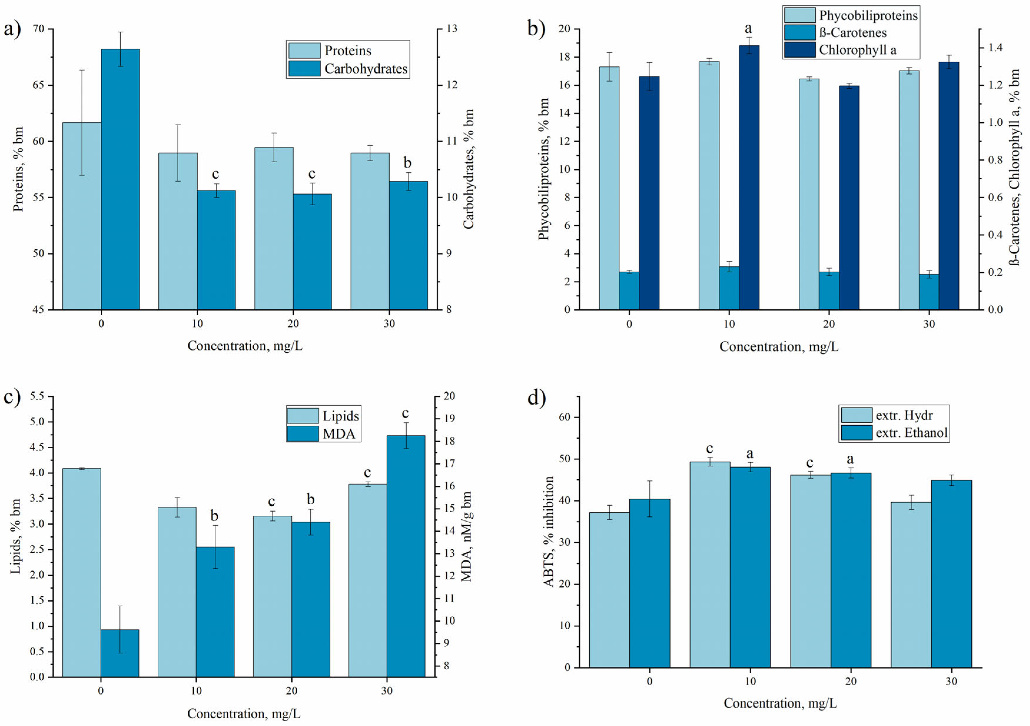
In Figure 3, the changes in the biochemical composition and antioxidant activity of A. platensis under the influence of Pr(III) introduced to the cultivation medium in different concentrations are presented.
Supplementation of nutrient medium with Pr(III) did not influence the content of protein in A. platensis biomass, which varied between 58.95 and 61.65% of the dry biomass in the control and experimental variants, respectively, without statistically relevant differences (Figure 3a). To our knowledge, there are no published data referring to the influence of Pr(III) on the content of protein in cyanobacteria and microalgae; therefore, the obtained data were compared with the values reported for other REEs. It has been shown that depending on the REE and its concentration in the medium, the amount of protein in the spirulina biomass can either increase, decrease, or remain unchanged. For example, at Nd, Y, and Eu concentrations of 30 mg/L, the content of proteins in the A. platensis biomass was lower than in the control; while at the Yb concentration of 20 mg/L, the Nd concentration of 10 mg/L, and the Dy concentration of 30 mg/L, the biomass contained more protein compared to the control. In case of Er, the content of proteins was similar to the control biomass [23,25,30].
In the A. platensis biomass grown on medium supplemented with Pr(III), the content of carbohydrates (Figure 3a) was significantly lower compared to the control. Thus, at the applied Pr(III) concentration, a decrease of 18.7–20.4% compared to the control was observed without significant differences as a function of the element concentration in the culture medium. This phenomenon has also been observed for other REEs. For example, in the case of the medium supplemented with Eu, Er, Nd, or Yb, the A. platensis biomass accumulated 15.8–36.7% less carbohydrates compared to the control [23,24,25,30]. However, the phenomenon cannot be considered as a general one, since Sm, Tb, La, and Dy, on the contrary, significantly increased the level of carbohydrates in A. platensis biomass [30].
The content of total phycobiliproteins (Figure 3b) in the control biomass was 17.3% of the dry biomass, and in the experimental samples, these values oscillated between 16.4 and 17.7% (Figure 3b). Such behavior is less typical for A. platensis culture. As has been shown previously, only Eu and Er did not cause a reduction in the amount of phycobiliproteins in A. platensis [23,25], while other REEs (Nd, Yb, Y, La, Dy, Sm, and Tb) led to a reduction of their content by 11–50% compared to the control. This parameter is considered as a clear indicator of metals toxicity for A. platensis [30]. Phycobiliproteins are characterized by high antioxidant activity and the decrease in their content under the action of different REEs can be associated with their involvement in the inhibition of free radicals. At the same time, on the higher plants, it was demonstrated that the antioxidant effect of Pr(III) can inhibit the oxidative process generated by oxidative stress of a different nature [32,33]. On the other hand, Pr(III) can form with glutathione, a complex with high antioxidant activity [34], which possibly stops the consumption of other cellular antioxidants, including phycobiliproteins. It is very likely that the antioxidant properties of Pr(III) ensure the preservation of a normal level of phycobiliproteins in the spirulina biomass.
The amount of chlorophyll α in the A. platensis biomass rose by 13.2% compared to the control at the Pr(III) concentration of 10 mg/L, while at the other concentrations, the value of this parameter did not change considerably (Figure 3b). The stability of the chlorophyll α content was characteristic for A. platensis biomass grown in the presence of other REEs (Nd, Yb, La, Dy, Tb, and Er), and only some of them influenced it. Thus, depending on the concentration, Y can induce an increase in the content of chlorophyll α (up to 20%), while Sm and Eu can induce a significant decrease in its content [23,25,30]. The content of β-carotene in the native biomass was 0.203% of the dry biomass, and in the experimental samples, it varied between 0.189 and 0.230% of the dry biomass, with the differences being statistically insignificant. This is in line with previously performed studies [23,24,25,30].
In the case of other phycological species, such as Trachydiscus minutus and Parachlorella kessleri, Pr(III) has certain visible effects on the photosynthetic pigments, which differ depending on the species. For example, in Trachydiscus minutus under the influence of Pr(III), a slight increase in the amount of β-carotene and chlorophyll was observed, while the amount of other carotenoids decreased considerably. In contrast, in Parachlorella kessleri, the content of all monitored photosynthetic pigments decreased considerably in the presence of Pr(III) [31]. Thus, as in the case of the other components of the phycological biomass, the influence of REEs is determined by several factors—the species on which the REE is applied, the element, and its concentration in the medium.
The content of lipids in the biomass obtained on the medium supplemented with Pr(III) was significantly lower compared to the control (by approximately 8–23% depending on the concentration of the element) (Figure 3c). This phenomenon has also been observed in previous research. Most of the studied REEs caused a reduction in the amount of lipids in the A. platensis biomass (e.g., Tb, Dy, Yb, and Eu) or did not significantly change this parameter (e.g., Sm and Nd) [25,30]. The modification of the lipid profile in the biomass due to the action of Pr(III) was observed in the case of other phycological species as well. For example, in Trachydiscus minutus and Parachlorella kessleri, although the total lipid content did not change significantly, a quantitative displacement in the direction of the accumulation of saturated fatty acids and a reduction in the amount of polyunsaturated ones was observed [31].
The amount of MDA in A. platensis biomass, an important marker of oxidative stress, increased under the influence of Pr(III) by 38.2–89.8% compared to the control, with the effect having a pronounced dose-dependent character. The increase in the MDA levels in A. platensis biomass grown on medium containing REEs has also been observed in other experiments. Thus, the content of MDA in A. platensis biomass increased under the influence of La, Dy, Sm, Nd, Yb, Eu, Er, and Y; in some cases, even doubling of the value of this parameter was observed [24,25,30].
REEs can affect the stability, permeability, and function of cell membranes by blocking ion channels and binding to specific membrane proteins. The instability of cell membranes leads to intense oxidative degradation of the lipids that make up their composition. It is clear that the significant increase in MDA in the biomass of A. platensis under the influence of Pr(III) is due to the oxidation of lipids, primarily membrane lipids, but the exact mechanism of this phenomenon is not known. A possible explanation for this phenomenon could be the replacement of iron by Pr(III) and the release of ions capable of inducing lipid oxidation through Fenton and Haber–Weiss reactions. As a result, the amount of end products of lipid oxidative degradation, including MDA, increased.
The antioxidant activity of the ethanolic and water extracts obtained from biomass cultivated in the presence of Pr(III) can be seen in Figure 3d. Both types of extracts had very similar antiradical activity against the ABTS cation radical. The extracts of both types obtained from the biomass grown in the presence of 10 and 20 mg/L of Pr(III) were more active than those from the control biomass. The highest increase in the activity of the water extract compared to the control was attested at the Pr(III) concentration of 10 mg/L, with the extract being 32.7% more active against the ABTS cation radical. At the Pr(III) concentration of 20 mg/L, the water extract was 24.2% more active compared to the control, while in the case of the highest Pr(III) concentration, the value was comparable to the control. For the ethanolic extracts, the same change pattern was observed; however, the increase in its values compared to the control was more modest—by 18.8% at the concentration of 10 mg/L and by 15.4% at the concentration of 20 mg/L. The ABTS test values were inversely proportional to the MDA values. Under stress conditions, cells rapidly produce components with antioxidant properties, which determine the activity of biomass extracts. The more pronounced the state of stress, the more intensely the antioxidant compounds are consumed, and under severe stress, cells lose their ability to counteract this process.
In the previously performed studies, changes in the antioxidant activity of A. platensis grown on medium supplemented with REEs were also observed. Thus, addition of the Eu, La, Dy, Sm, and Tb resulted in the growth of antiradical activity, while Nb and Yb led to a considerable decrease in ABTS radical inhibition activity [23,30].

3.2. Praseodium Removal by Non-Living A. platensis Biomass under Different Experimental Conditions

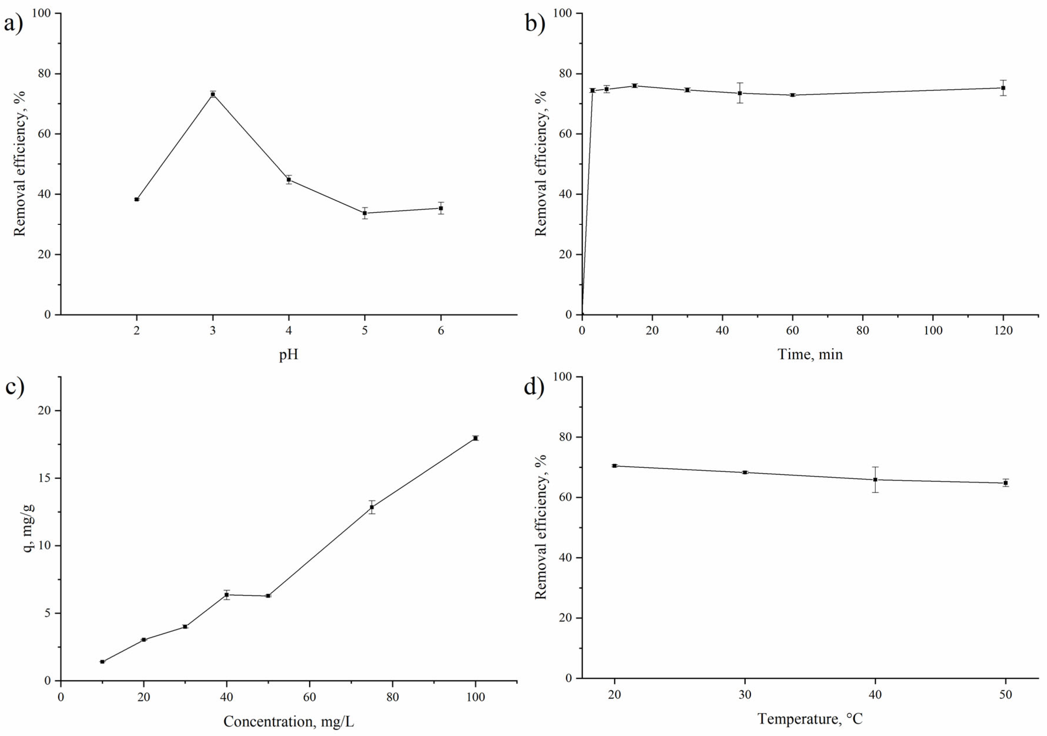
Metal removal by non-living biomass, biosorption, is a complex process affected by several factors. Among them, pH is a critical parameter, since it influences the chemistry of the metals and the surface charge of the functional groups [35]. A. platensis had an isoelectric point at pH 2.85; this suggests that the positive charge of the cell surface was maintained until this pH value; then, the charge of the biosorbent surface became negative [36]. The results presented in Figure 4a confirm this fact. Thus, at pH 2.0, 38% of Pr(III) was removed from the solution. With a strong acidic pH in the solution, a high concentration of H+ ions hampers Pr(III)’s removal due to competition for binding sites [37]. The increase in the pH from 2.0 to 3.0 resulted in the achievement of maximum Pr(III) removal of 73%, while a further increase in the pH value resulted in a continuous decrease in removal efficiency. The increase in the pH value and the decrease in concentration of H+ ions facilitate interaction between Pr(III) and negatively charged functional groups on the A. platensis surface [23,25,37,38]. It is known that at pH values pH ≤ 5, lanthanides exist as Ln3+ [37]. A reduction in Pr(III) removal can be associated with the competition of OH and Pr(III) for binding sites as well as the formation of Pr(OH)3 at pHs higher than 5.0.
The maximum Pr(III) adsorption on the brown seaweeds Turbinaria conoides and Sargassum wightii [38] and green algae Ulva lactuca [39] was attained at pH 5.0, and this was accompanied by the replacement of some of the alkaline earth metals through the ion-exchange mechanism [38]. The maximum adsorption of Pr(III) on D72 resin was achieved at pH 3.0 [40], and on TVEX–PHOR resin, this was achieved at pH 3.5 [41].
Figure 4b shows the effect of the contact time on the efficiency of Pr(III) removal. The process of Pr(III) removal was very quick; 74% of the ions were absorbed from the solution in the first 3 min of the interaction, and then, equilibrium was attained. The biosorption of Pr(III) on Turbinaria conoides and Sargassum wightii with 90% of total biosorption took place within 20 min [38]. The rapid uptake of Pr(III) is explained by the availability of a large number of binding sites on the biomass at the initial stage of biosorption. Lowering of the process is usually associated with occupancy of the binding sites [40,42].
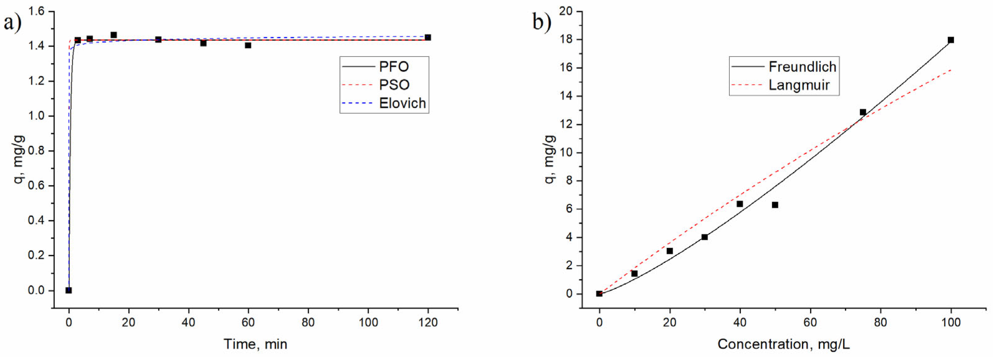
The kinetics of Pr(III) biosorption on A. platensis was analyzed using pseudo-first-order, pseudo-second-order, and Elovich models (Figure 5a, Table 1).
According to the R2 values, all of the applied models can adequately describe the kinetics of sorption. However, the extremally low values of the coefficient α in the Elovich model shows its unsuitability for the description of the experimental data. The close values of the adsorption capacity (calculated and obtained experimentally) as well as the R2 for the pseudo-first-order and pseudo-second-order models were very close indicates their relevance for explanation of the experimental data. However, according to the Akaike information criterion (AIC) test, the pseudo-first-order model was more appropriate to describe the kinetics data. The model suggests that the rate of adsorption is proportional to the number of unoccupied sites on the sorbent surface [43]. The model also assumes that the mass transfer controls the adsorption process due to a concentration difference between the adsorbent surface and the solution [44]. Furthermore, it is considered that in this case, chemisorption can inhibit the sorption kinetics due to the fact that chemical reactions and the diffusion of the liquid in the boundary layer can lead to resistance to mass transfer [43,45]. At the same time, the pseudo-second-order rate constant of 26.5 g/mg min for Pr(III) biosorption on A. platensis was considerably higher than the rate constant of the first-order kinetics (2.13 min−1), suggesting that Pr(III) requires less time to achieve adsorption equilibrium [46].
The influence of Pr(III) concentration on A. platensis biosorption capacity was investigated in the range of 10–100 mg/L (Figure 4c). The adsorption efficiency increased as the concentration of Pr(III) in the solution grew, and the highest adsorption capacity of 17.0 mg/g was achieved at 100 mg/L. The high initial concentration of Pr(III) contributed to the higher number of Pr(III) ions in the aqueous solution, which led to a greater number of interactions between Pr(III) and the available binding sites. Additionally, a higher initial Pr(III) concentration may facilitate metal ions adsorption by enhancement of the mass transfer coefficient, along with providing the driving force to overcome the mass transfer resistances between the solid and liquid phases [47,48]. It should be mentioned that A. platensis maintained high removal efficiency at all studied Pr(III) concentrations.
The Lagmiur and Freundlich models were applied for studying the nonlinear adsorption of Pr(III) on A. platensis (Figure 5b, Table 2). The Freundlich model, according to the higher R2 value, was shown to be more applicable for the description of the experimental data. The n value of 1.2 indicated heterogeneity in the biosorbent surface and the favorable adsorption of Pr(III) [36,40]. Since the Freundlich isotherm model suggests that adsorption takes place on heterogeneous surfaces as a multilayer adsorption [5,41], it can be expected that there is favorable adsorption of Pr(III) onto the heterogeneous surface of A. platensis with a variety of binding sites. In this case, the sorption process is not restricted to one specific class of sites [41]. A large number of the functional groups which can participate in Pr(III) biosorption, including C=O, C-C, C-O-C, P=O, CH2, and NHC(O) have been identified on the surface of A. platensis [25]. Since these functional groups include nitrogen and/or oxygen, which aid in the formation of hydrogen bonds between Pr(III) and cyanobacteria surface, they can facilitate the biosorption process [46]. The sorption of Pr(III) is also possible through ion exchange between Pr(III) and Ca(II) as well as other cations including Na, K, and Mg [42].
The lower values of R2 obtained for the Langmuir model showed that it did not fit well to the adsorption data. The maximum theoretical predicted adsorption of Pr(III) onto A. platensis constituted 99.3 mg/g, and this is higher than the values reported for other sorbents: 57.8 mg/g for crab shell and 49.9 mg/g for orange peel [45] and 30.0 mg/g for Dowex 50WX8 resin [49]. The capacity was comparable with the values obtained for Laminaria digitate (110–120 mg/g) [50] and lower than the values obtained for D72 resin (294 mg/g) [40]. The separation factor values ranged from 0.84 to 0.98, pointing at favorable Pr(III) biosorption. The calculated maximal absorption capacity (Table 2) was greater than the experimentally obtained value (17 mg/g), suggesting that the biosorption of Pr(III) onto A. platensis might occur at higher concentrations of the element [46].
Thus, the mechanisms of Pr(III) biosorption by A. platensis may include electrostatic interaction between Pr(III) and the negatively charged functional groups on the A. platensis surface, ion exchange, or chemical biosorption [36,46].
The effect of temperature on the adsorption of Pr(III) on A. platensis has been examined at temperatures in the range of 20–50 °C. It was observed that the sorption of Pr(III) slightly decreased with the increasing temperature, from 70% at 20 °C to 65% at 50 °C (Figure 4d). Next, from the slope and intercept of the plot (Figure S1), the values of ΔH° and ΔS° were calculated, while ΔG° was evaluated from Equation (11). The obtained parameters are listed in Table 3.
As shown in Table 3, the values of ΔG° ranging from −15.0 to −15.8 kJ/mol suggest that adsorption is a physical process [41]. The negative value of ΔG° indicates the spontaneous and thermodynamical feasibility of the process. The negative value of ΔH° suggests the exothermic type of the sorption, and the value is typical for physisorption [9]. Positive values of ΔS° shows that Pr(III) biosorption is associated with the displacement of more than one water molecule as a result of the increase in entropy [9].
Application of A. platensis in the form of waste from different biotechnological processes will make the process of Pr(III) removal efficient and economically viable. The use of mineral and organic acids, as shown in our previous studies [51,52], will make it possible to regenerate the biosorbent and to apply it for several sorption–desorption cycles.

4. Conclusions

Living and non-living A. platensis biomass showed high affinity toward Pr(III). The high rate of Pr(III) uptake by the living biomass (9.96–29.9 mg/g) was accompanied by a reduction in the amount of lipids and carbohydrates and overaccumulation of malonic dialdehyde. The biomass productivity and the content of proteins and pigments were maintained on the level of the control biomass. The changes observed in the Pr-supplemented biomass indicate a state of oxidative stress for the A. platensis culture, caused by the presence of Pr(III) in the medium. Pr(III) removal by the non-living biomass was a quick and pH-dependent process. The maximum biosorption of 99.3 mg/g was attained at pH 3.0 within 3 min of sorbent interaction with the sorbate. The pseudo-first-order and Freundlich models explained better te kinetics and equilibrium of Pr(III) biosorption. Thermodynamically, Pr(III) biosorption can be described as a spontaneous exothermic process. A. platensis biomass has great potential to be applied as a biosorbent and/or bioaccumulator for Pr(III) recovery from wastewater.

Supplementary Materials

The following supporting information can be downloaded at: https://www.mdpi.com/article/10.3390/w15112064/s1. Figure S1: Plot of lnKd versus 1/T.

Author Contributions

Conceptualization, N.Y., I.Z. and L.C.; methodology, I.Z., L.C. and L.R.; software, D.G.; validation, N.Y., T.C. and L.R.; investigation, N.Y., T.C. and L.R.; resources, I.Z.; writing—original draft preparation, I.Z. and L.C.; writing—review and editing, all authors. All authors have read and agreed to the published version of the manuscript.

Funding

This research received no external funding.

Data Availability Statement

All data are presented in the manuscript.

Conflicts of Interest

The authors declare no conflict of interest.

References

  1. Dev, S.; Sachan, A.; Dehghani, F.; Ghosh, T.; Briggs, B.R.; Aggarwal, S. Mechanisms of biological recovery of rare-earth elements from industrial and electronic wastes: A review. Chem. Eng. J. 2020, 397, 124596. [Google Scholar] [CrossRef]
  2. Bashiri, A.; Nikzad, A.; Maleki, R.; Asadnia, M.; Razmjou, A. Rare Earth Elements Recovery Using Selective Membranes via Extraction and Rejection. Membranes 2022, 12, 80. [Google Scholar] [CrossRef]
  3. Elbashier, E.; Mussa, A.; Hafiz, M.A.; Hawari, A.H. Recovery of rare earth elements from waste streams using membrane processes: An overview. Hydrometallurgy 2021, 204, 105706. [Google Scholar] [CrossRef]
  4. Jyothi, R.K.; Thenepalli, T.; Ahn, J.W.; Parhi, P.K.; Chung, K.W.; Lee, J.Y. Review of rare earth elements recovery from secondary resources for clean energy technologies: Grand opportunities to create wealth from waste. J. Clean. Prod. 2020, 267, 122048. [Google Scholar] [CrossRef]
  5. Negrea, A.; Gabor, A.; Davidescu, C.M.; Ciopec, M.; Negrea, P.; Duteanu, N.; Barbulescu, A. Rare Earth Elements Removal from Water Using Natural Polymers. Sci. Rep. 2018, 8, 316. [Google Scholar] [CrossRef] [PubMed]
  6. Malhotra, N.; Hsu, H.S.; Liang, S.T.; Roldan, M.J.M.; Lee, J.S.; Ger, T.R.; Hsiao, C. Der An updated review of toxicity effect of the rare earth elements (REEs) on aquatic organisms. Animals 2020, 10, 1663. [Google Scholar] [CrossRef]
  7. Laveuf, C.; Cornu, S. A review on the potentiality of Rare Earth Elements to trace pedogenetic processes. Geoderma 2009, 154, 1–12. [Google Scholar] [CrossRef]
  8. Brouziotis, A.A.; Giarra, A.; Libralato, G.; Pagano, G.; Guida, M.; Trifuoggi, M. Toxicity of rare earth elements: An overview on human health impact. Front. Environ. Sci. 2022, 10, 1617. [Google Scholar] [CrossRef]
  9. Anastopoulos, I.; Bhatnagar, A.; Lima, E.C. Adsorption of rare earth metals: A review of recent literature. J. Mol. Liq. 2016, 221, 954–962. [Google Scholar] [CrossRef]
  10. Diep, P.; Mahadevan, R.; Yakunin, A.F. Heavy metal removal by bioaccumulation using genetically engineered microorganisms. Front. Bioeng. Biotechnol. 2018, 6, 157. [Google Scholar] [CrossRef]
  11. Das, N.; Das, D. Recovery of rare earth metals through biosorption: An overview. J. Rare Earths 2013, 31, 933–943. [Google Scholar] [CrossRef]
  12. Timková, I.; Sedláková-Kaduková, J.; Pristaš, P. Biosorption and bioaccumulation abilities of actinomycetes/streptomycetes isolated from metal contaminated sites. Separations 2018, 5, 54. [Google Scholar] [CrossRef]
  13. Filote, C.; Roșca, M.; Hlihor, R.M.; Cozma, P.; Simion, I.M.; Apostol, M.; Gavrilescu, M. Sustainable application of biosorption and bioaccumulation of persistent pollutants in wastewater treatment: Current practice. Processes 2021, 9, 1696. [Google Scholar] [CrossRef]
  14. Javanbakht, V.; Alavi, S.A.; Zilouei, H. Mechanisms of heavy metal removal using microorganisms as biosorbent. Water Sci. Technol. 2014, 69, 1775–1787. [Google Scholar] [CrossRef]
  15. Grosjean, N.; Le Jean, M.; Berthelot, C.; Chalot, M.; Gross, E.M.; Blaudez, D. Accumulation and fractionation of rare earth elements are conserved traits in the Phytolacca genus. Sci. Rep. 2019, 9, 18458. [Google Scholar] [CrossRef]
  16. Čížková, M.; Mezricky, P.; Mezricky, D.; Rucki, M.; Zachleder, V.; Vítová, M. Bioaccumulation of Rare Earth Elements from Waste Luminophores in the Red Algae, Galdieria phlegrea. Waste Biomass Valorization 2021, 12, 3137–3146. [Google Scholar] [CrossRef]
  17. Castillo, J.; Maleke, M.; Unuofin, J.; Cebekhulu, S.; Gómez-Arias, A. Microbial recovery of rare earth elements. In Environmental Technologies to Treat Rare Earth Element Pollution: Principles and Engineering; IWA Publishing: London, UK, 2022; pp. 179–205. ISBN 9781789062236. [Google Scholar]
  18. Andrès, Y.; Le Cloirec, P.; Texier, A.C. Rare earth elements removal by microbial biosorption: A review. Environ. Technol. 2003, 24, 1367–1375. [Google Scholar] [CrossRef]
  19. Al-Amin, A.; Parvin, F.; Chakraborty, J.; Kim, Y.I. Cyanobacteria mediated heavy metal removal: A review on mechanism, biosynthesis, and removal capability. Environ. Technol. Rev. 2021, 10, 44–57. [Google Scholar] [CrossRef]
  20. Sarup, R.; Behl, K.; Joshi, M.; Nigam, S. Heavy metal removal by cyanobacteria. In New Trends in Removal of Heavy Metals from Industrial Wastewater; Elsevier: Amsterdam, The Netherlands, 2021; pp. 441–466. ISBN 9780128229651. [Google Scholar]
  21. Ghorbani, E.; Nowruzi, B.; Nezhadali, M.; Hekmat, A. Metal removal capability of two cyanobacterial species in autotrophic and mixotrophic mode of nutrition. BMC Microbiol. 2022, 22, 58. [Google Scholar] [CrossRef] [PubMed]
  22. Fischer, C.B.; Körsten, S.; Rösken, L.M.; Cappel, F.; Beresko, C.; Ankerhold, G.; Schönleber, A.; Geimer, S.; Ecker, D.; Wehner, S. Cyanobacterial promoted enrichment of rare earth elements europium, samarium and neodymium and intracellular europium particle formation. RSC Adv. 2019, 9, 32581–32593. [Google Scholar] [CrossRef] [PubMed]
  23. Yushin, N.; Zinicovscaia, I.; Cepoi, L.; Chiriac, T.; Rudi, L.; Grozdov, D. Biosorption and Bioaccumulation Capacity of Arthospira platensis toward Europium Ions. Water 2022, 14, 2128. [Google Scholar] [CrossRef]
  24. Yushin, N.; Zinicovscaia, I.; Cepoi, L.; Chiriac, T.; Rudi, L.; Grozdov, D. Application of Cyanobacteria Arthospira platensis for Bioremediation of Erbium-Contaminated Wastewater. Materials 2022, 15, 6101. [Google Scholar] [CrossRef]
  25. Yushin, N.; Zinicovscaia, I.; Cepoi, L.; Chiriac, T.; Rudi, L.; Grozdov, D. Biosorption and Bioaccumulation Capacity of Arthrospira platensis toward Yttrium Ions. Metals 2022, 12, 1465. [Google Scholar] [CrossRef]
  26. Pol, A.; Barends, T.R.M.; Dietl, A.; Khadem, A.F.; Eygensteyn, J.; Jetten, M.S.M.; Op den Camp, H.J.M. Rare earth metals are essential for methanotrophic life in volcanic mudpots. Environ. Microbiol. 2014, 16, 255–264. [Google Scholar] [CrossRef] [PubMed]
  27. Goecke, F.; Zachleder, V.; Vítová, M. Rare earth elements and algae: Physiological effects, biorefi nery and recycling. In Algal Biorefineries: Volume 2: Products and Refinery Design; Springer International Publishing: Cham, Switzerland, 2015; pp. 339–363. ISBN 9783319202006. [Google Scholar]
  28. Valcheva-Traykova, M.; Saso, L.; Kostova, I. Involvement of Lanthanides in the Free Radicals Homeostasis. Curr. Top. Med. Chem. 2014, 14, 2508–2519. [Google Scholar] [CrossRef] [PubMed]
  29. Zinicovscaia, I.; Safonov, A.; Tregubova, V.; Ilin, V.; Cepoi, L.; Chiriac, T.; Rudi, L.; Frontasyeva, M.V. Uptake of metals from single and multi-component systems by spirulina platensis biomass. Ecol. Chem. Eng. S 2016, 23, 401–412. [Google Scholar] [CrossRef]
  30. Zinicovscaia, I.; Cepoi, L.; Rudi, L.; Chiriac, T.; Grozdov, D.; Pavlov, S.; Djur, S. Accumulation of dysprosium, samarium, terbium, lanthanum, neodymium and ytterbium by Arthrospira platensis and their effects on biomass biochemical composition. J. Rare Earths 2021, 39, 1133–1143. [Google Scholar] [CrossRef]
  31. Goecke, F.; Vítová, M.; Lukavský, J.; Nedbalová, L.; Řezanka, T.; Zachleder, V. Effects of rare earth elements on growth rate, lipids, fatty acids and pigments in microalgae. Phycol. Res. 2017, 65, 226–234. [Google Scholar] [CrossRef]
  32. Zhu, Q.; Li, Y.; Gao, S.; Shan, C. Praseodymium enhanced the tolerance of maize seedlings subjected to cadmium stress by up-regulating the enzymes in the regeneration and biosynthetic pathways of ascorbate and glutathione. Plant Soil Environ. 2021, 67, 633–642. [Google Scholar] [CrossRef]
  33. Zhang, C.; Zhang, X.; Shan, C. Effect of praseodymium on the postharvest quality of Lilium longiflorum cut flowers. New Zeal. J. Crop. Hortic. Sci. 2022. [Google Scholar] [CrossRef]
  34. Ziekhrü, M.; Imchen, P.; Phucho, T.; Devi, M.I. Synthesis, characterization, antioxidant and antibacterial studies of praseodymium complex with glutathione. Curr. Sci. 2023, 124, 554–561. [Google Scholar] [CrossRef]
  35. Vijayaraghavan, K.; Sathishkumar, M.; Balasubramanian, R. Interaction of rare earth elements with a brown marine alga in multi-component solutions. Desalination 2011, 265, 54–59. [Google Scholar] [CrossRef]
  36. Fawzy, M.A.; Darwish, H.; Alharthi, S.; Al-Zaban, M.I.; Noureldeen, A.; Hassan, S.H.A. Process optimization and modeling of Cd2+ biosorption onto the free and immobilized Turbinaria ornata using Box–Behnken experimental design. Sci. Rep. 2022, 12, 3256. [Google Scholar] [CrossRef]
  37. Kusaka, E.; Kamata, Y.; Fukunaka, Y.; Nakahiro, Y. Effect of hydrolysed metal cations on the liquid-liquid extraction of silica fines with cetyltrimethylammonium chloride. Colloids Surf. A Physicochem. Eng. Asp. 1998, 139, 155–162. [Google Scholar] [CrossRef]
  38. Vijayaraghavan, K.; Jegan, J. Entrapment of brown seaweeds (Turbinaria conoides and Sargassum wightii) in polysulfone matrices for the removal of praseodymium ions from aqueous solutions. J. Rare Earths 2015, 33, 1196–1203. [Google Scholar] [CrossRef]
  39. Vijayaraghavan, K. Biosorption of lanthanide (praseodymium) using Ulva lactuca: Mechanistic study and application of two, three, four and five parameter isotherm models. J. Environ. Biotechnol. Res. 2015, 1, 10–17. [Google Scholar] [CrossRef]
  40. Xiong, C.; Zhu, J.; Shen, C.; Chen, Q. Adsorption and desorption of praseodymium (III) from aqueous solution using D72 resin. Chinese J. Chem. Eng. 2012, 20, 823–830. [Google Scholar] [CrossRef]
  41. El-Dessouky, S.I.; El-Sofany, E.A.; Daoud, J.A. Studies on the sorption of praseodymium (III), holmium (III) and cobalt (II) from nitrate medium using TVEX-PHOR resin. J. Hazard. Mater. 2007, 143, 17–23. [Google Scholar] [CrossRef]
  42. Boulaiche, W.; Belhamdi, B.; Hamdi, B.; Trari, M. Kinetic and equilibrium studies of biosorption of M(II) (M = Cu, Pb, Ni, Zn and Cd) onto seaweed Posidonia oceanica fibers. Appl. Water Sci. 2019, 9, 173. [Google Scholar] [CrossRef]
  43. Aguiar, A.B.S.; Costa, J.M.; Santos, G.E.; Sancinetti, G.P.; Rodriguez, R.P. Removal of Metals by Biomass Derived Adsorbent in Its Granular and Powdered Forms: Adsorption Capacity and Kinetics Analysis. Sustain. Chem. 2022, 3, 535–550. [Google Scholar] [CrossRef]
  44. Fawzy, M.A.; Gomaa, M. Use of algal biorefinery waste and waste office paper in the development of xerogels: A low cost and eco-friendly biosorbent for the effective removal of congo red and Fe (II) from aqueous solutions. J. Environ. Manag. 2020, 262, 110380. [Google Scholar] [CrossRef] [PubMed]
  45. Varshini, C.J.S.; Das, D.; Das, N. Optimization of parameters for praseodymium(III) biosorption onto biowaste materials using response surface methodology: Equilibrium, kinetic and regeneration studies. Ecol. Eng. 2015, 81, 321–327. [Google Scholar] [CrossRef]
  46. Alharbi, N.K.; Al-Zaban, M.I.; Albarakaty, F.M.; Abdelwahab, S.F.; Hassan, S.H.A.; Fawzy, M.A. Kinetic, Isotherm and Thermodynamic Aspects of Zn2+ Biosorption by Spirulina platensis: Optimization of Process Variables by Response Surface Methodology. Life 2022, 12, 585. [Google Scholar] [CrossRef] [PubMed]
  47. Morales-Barrera, L.; Flores-Ortiz, C.M.; Cristiani-Urbina, E. Single and binary equilibrium studies for Ni2+ and Zn2+ biosorption onto lemna gibba from aqueous solutions. Processes 2020, 8, 1089. [Google Scholar] [CrossRef]
  48. Zinicovscaia, I.; Yushin, N.; Grozdov, D.; Rodlovskaya, E.; Khiem, L.H. Yeast—As Bioremediator of Silver-Containing Synthetic Effluents. Bioengineering 2023, 10, 398. [Google Scholar] [CrossRef]
  49. Masry, B.A.; Abu Elgoud, E.M.; Rizk, S.E. Modeling and equilibrium studies on the recovery of praseodymium (III), dysprosium (III) and yttrium (III) using acidic cation exchange resin. BMC Chem. 2022, 16, 37. [Google Scholar] [CrossRef]
  50. Wang, S.; Hamza, M.F.; Vincent, T.; Faur, C.; Guibal, E. Praseodymium sorption on Laminaria digitata algal beads and foams. J. Colloid Interface Sci. 2017, 504, 780–789. [Google Scholar] [CrossRef]
  51. Zinicovscaia, I.; Yushin, N.; Shvetsova, M.; Frontasyeva, M. Zinc removal from model solution and wastewater by Arthrospira (Spirulina) Platensis biomass. Int. J. Phytoremediation 2018, 20, 901–908. [Google Scholar] [CrossRef]
  52. Zinicovscaia, I.; Yushin, N.; Gundorina, S.; Demčák, Š.; Frontasyeva, M.; Kamanina, I. Biosorption of nickel from model solutions and electroplating industrial effluenusing cyanobacterium arthrospira platensis. Desalin. Water Treat. 2018, 120, 158–165. [Google Scholar] [CrossRef]
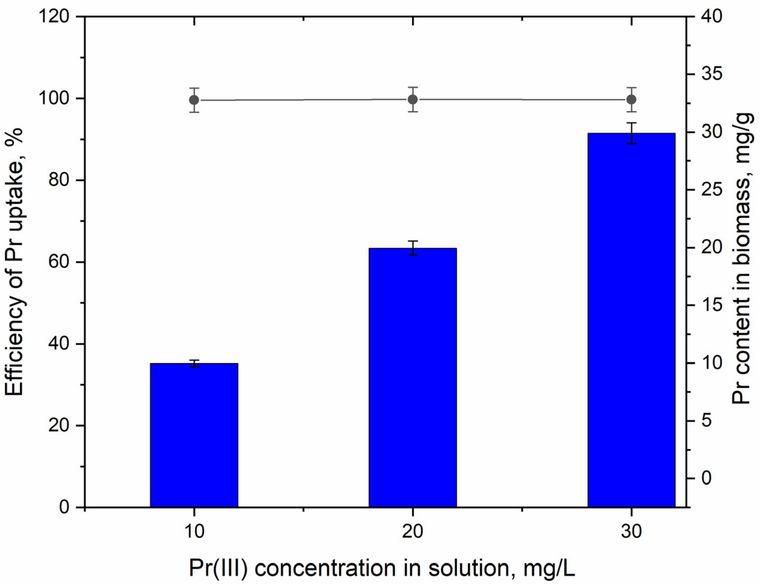
Figure 1. Efficiency of Pr(III) uptake by the living Arthrospira platensis biomass following its addition in the cultivation medium in concentrations of 10–30 mg/L.
Water 15 02064 g001
Figure 2. Effect of Pr(III) introduced to the cultivation medium in concentrations of 10–30 mg/L on the amount of Arthrospira platensis biomass (a—p < 0.05 for the difference between the sample and the control).
Water 15 02064 g002
Figure 3. Effect of Pr(III) introduced to the cultivation medium in concentrations of 10–30 mg/L on the Arthrospira platensis: (a) content of proteins and carbohydrates (b—p < 0.005; c—p < 0.001 for the difference between the experiment and the control for carbohydrates); (b) content of phycobiliproteins, chlorophyll a, and β-carotene (a—p < 0.05 for the difference between the experiment and the control); (c) content of lipids and MDA (b—p < 0.005, c— p< 0.0005 for the difference between the experiment and the control); and (d) antioxidant activity of the extracts obtained from the biomass.
Water 15 02064 g003
Figure 4. Effect of the experimental parameters on Pr(III) removal by A. platensis: (a) pH; (b) contact time; (c) initial Pr(III) concentration; and (d) temperature.
Water 15 02064 g004
Figure 5. (a) Kinetics and (b) equilibrium of Pr(III) biosorption on A. platensis.
Water 15 02064 g005
Table 1. The parameters of the kinetics models applied to describe Pr(III)’s biosorption on A. platensis.
Pseudo-First-OrderPseudo-Second-OrderElovich
qek1R2qek2R2αβR2
1.432.130.9991.4326.50.9983.96 × 10−4375.20.997
Table 2. The parameters obtained by fitting the experimental data with the isotherm models used.
LangmuirFreundlich
qmbR2KfnR2
99.30.00190.9520.0621.20.99
Table 3. Thermodynamic parameters for metal biosorption on A. platensis.
Temperature
K
ΔG°
kJ/mol
ΔH°
kJ/mol
ΔS°
J/mol
293−15.0−7.027.4
303−15.3
313−15.6
323−15.8
Disclaimer/Publisher’s Note: The statements, opinions and data contained in all publications are solely those of the individual author(s) and contributor(s) and not of MDPI and/or the editor(s). MDPI and/or the editor(s) disclaim responsibility for any injury to people or property resulting from any ideas, methods, instructions or products referred to in the content.