Article

Influence of High-Frequency, Low-Voltage Alternating Electric Fields on Biofilm Development Processes of Escherichia coli and Pseudomonas aeruginosa

by
Patthranit Kunlasubpreedee
1,
Tomohiro Tobino
1,2,3,* and
Fumiyuki Nakajima
1,3
1
Department of Urban Engineering, Graduate School of Engineering, The University of Tokyo, Tokyo 113-8656, Japan
2
Collaborative Research Institute for Innovative Microbiology, The University of Tokyo, Tokyo 113-8657, Japan
3
Environmental Science Center, The University of Tokyo, Tokyo 113-0033, Japan
*
Author to whom correspondence should be addressed.
Water 2023, 15(17), 3055; https://doi.org/10.3390/w15173055
Submission received: 20 July 2023 / Revised: 23 August 2023 / Accepted: 24 August 2023 / Published: 26 August 2023
(This article belongs to the Special Issue Sustainable Water Supply, Sanitation and Wastewater Systems)

Abstract

:
A non-chemical solution is needed to control biofilms in water and wastewater treatment systems. High-frequency alternating electric field application offers an alternative approach that does not involve undesired electrode surface reactions. However, the effect of high-frequency alternating electric fields on bacterial cells in the attached-growth mode remains unexplored. This study investigated the impact of such fields on two stages of the biofilm development process: the initial adhesion phase (stage 1) and the early development phase (stage 2). Experiments were conducted using Escherichia coli and Pseudomonas aeruginosa in a three-channel flow cell exposed to alternating electric fields (3.05 V/cm, 20 MHz). The primary outcome of this study demonstrated that alternating electric fields decreased adhered cell numbers at both stages due to their inhibitory effect on growth. The alternating electric fields also triggered cell detachment after the initial attachment stage but not in mature biofilms. Interestingly, despite a reduction in cell counts, the amount of total biofilm biomass remained unaffected, which was likely due to increased cell size via cell elongation compensating for the decrease in numbers. No synergistic effects with respect to hydrodynamic forces were observed. These findings highlight the potential applicability of alternating electric fields to biofilm control and provide implications for water and wastewater engineering applications.

1. Introduction

Attached growth processes, or biofilms, are widely used in water and wastewater treatment systems. Biofilms are clusters of microorganisms immersed in a matrix of extracellular polymeric substances and firmly attached to a surface [1]. The structural characteristics of a biofilm enable nutrient competition, resistance to contaminants, and the creation of a protective microenvironment, ensuring cell survival within the biofilm, thereby contrasting with the nature of free-floating cells [2]. In wastewater treatment systems, biofilms play a crucial role in removing pollutants through diverse mechanisms. They facilitate the breakdown of organic matter, enhance nutrient removal (such as nitrogen and phosphorus), and contribute to contaminant degradation [3]. Moreover, biofilms improve water quality by aiding in the removal of suspended solids and acting as a natural barrier against harmful pathogens [2,4]. Biofilm development also offers advantages for drinking-water treatment processes. Various materials employed in drinking-water treatment, including granular activated carbon, slow and rapid sand filters, and biofilters, foster biofilm formation to enhance water treatment efficiency [5]. The resilience of biofilms to harmful disturbances, together with their ability to retain slow-growing microorganisms, makes their use an advantageous strategy for water and wastewater treatment. Conversely, an overgrowth of biofilm remains a serious issue that deteriorates process performance. In membrane filtration systems, for example, biofouling leads to higher operational costs as more pretreatment processes and energy are required [6,7,8] along with a shorter membrane lifespan [9]. The formation of biofilms in water distribution systems increases the chlorine demand [10] and accelerates pipe corrosion [11]. The successful control of biofilms is key to ensuring the proper functioning of water and wastewater treatment facilities.
To address biofilm-related problems, several strategies for limiting biofilm growth have been developed. Physical methods (such as backwashing, scouring, and ultrasonic treatment) are primarily applied as cleaning processes, regardless of a biofilm’s viability [12]. Such methods are commonly used in biofilm-based wastewater treatment as a means of maintaining healthy biofilm thickness [1]. For instance, Celmer et al. (2008) reported a significant reduction (59%) in biofilm thickness within a membrane biofilm reactor under the application of ultrasound treatment [13]. UV irradiation can successfully eradicate biofilms via inducing DNA damage. Nonetheless, its efficacy depends on the depth of its penetration [14]. According to the research conducted by Ma et al. (2022), the transmittance of UV light (at 282 nm) through the biofilm structure of P. aeruginosa within a bioreactor decreased from 93.6% on day 1 to 33.6% on day 5 [15]. A conventional approach to controlling biofilms in water and wastewater treatment processes involves the application of chemical agents [10]. Sodium hypochlorite (NaOCl) has been utilized to eliminate biofouling on polyvinylidene fluoride (PVDF) membranes [16]. Additionally, free chlorine and monochloramine have been employed as secondary disinfectants in drinking water treatment to target and inhibit biofilm formation [17]. However, large quantities of these chemicals are required to achieve biofilm disinfection, leading to higher costs and more environmental issues. Antibiotics are used less often due to the potential negative impact of residuals entering a water supply [1]. Surface modification is another strategy employed to address biofilm issues [18,19]. Coating surfaces with biocides (quaternary ammonium compounds, single-walled carbon nanotubes, and silver nanoparticles) can suppress biofilm formation [20,21,22]. Nonetheless, biocides’ effectiveness deteriorates over time, leading to only a temporary delay in biofilm prevention [23]. Thus, implementing an alternative technique that enables effective biofilm prevention while minimizing the use of chemicals can bring many benefits.
Electricity-based technologies are an environmentally friendly alternative that circumvents the need for an external chemical dosage [24,25]. According to our literature survey, the utilization of direct current is a more extensively employed method for controlling bacterial cells in water and wastewater engineering in comparison to the application of alternating current. Many previous studies have demonstrated that an electrochemical reaction manipulated using direct current can effectively kill or detach biofilms from substrates [26,27,28]. Nevertheless, many drawbacks remain to be solved. Electrochemical reactions occur when employing conductive electrodes, which produce metal ions and free radicals on an electrode’s surface [29,30,31], leading to a shorter electrode lifespan. Moreover, the byproducts of these electrochemical reactions may initiate secondary chemical reactions that precipitate pollutant production [32,33]. Other limitations of this method are its high energy consumption and safety concerns regarding high voltage and current utilization [34]. The use of a low-frequency alternating current (below 60 Hz) has proven effective in biofilm inactivation [35]. However, this application also generates disinfecting effects through electrochemical reactions, causing undesirable effects similar to those mentioned with respect to direct current application [35,36]. The extremely high voltages employed in alternating current applications (13–35 kV) can successfully eradicate biofilms due to the powerful force exerted by intense electric fields [37], raising safety concerns. Another technique involves microwave radiation, operating within the very high frequency range of 300 MHz to 300 GHz, which can potentially inactivate biofilms by inducing thermal effects [38]. Nevertheless, temperature control remains a critical consideration in this regard. All things considered, the utilization of these electricity-based methods has been constrained.
On the other hand, the application of a high-frequency, low-voltage alternating electric field is gaining attention due to its inherent safety and economic viability. Utilizing such an alternating electric field offers more benefits compared to the utilization of direct current or low-frequency alternating electric fields. This advantage arises from the rapid changes in current direction when applying high frequency levels, thereby minimizing the generation of electrochemical reactions [39,40]. Low-intensity alternating electric fields at high frequencies (100–300 kHz) have found a medical application in tumor-treating fields (TTFields). These fields are utilized to interrupt cancer cell replication through the exertion of forces on charged components within dividing cells. However, the feasibility of this approach for water or wastewater engineering applications requires further investigation [41]. Previous studies have shown that weak alternating electric fields at high frequencies can inhibit bacterial growth. Lee et al. (2013) achieved the effective inactivation of Escherichia coli O157:H7 using a low-intensity alternating electric field (12.5 V/cm) at high frequencies [40]. This method did not cause electrode corrosion or quality deterioration when frequencies above 1 kHz were applied. Mirzaii et al. (2015) found that a low-voltage, high-frequency alternating electric field (10 V/cm, 20 MHz) had antibacterial effects on Staphylococcus aureus and Pseudomonas aeruginosa [42]. Giladi et al. (2008) observed an inhibition of S. aureus and P. aeruginosa growth using low-intensity alternating electric fields at high frequencies (2–4 V/cm, 100 kHz–50 MHz) [43]. The alternating electric fields employed in this study were utilized with completely insulated electrodes, demonstrating the absence of associated electrochemical reactions. However, most existing studies exploring the application of alternating electric fields have predominantly focused on the low-frequency range below 60 Hz, where inactivation effects are primarily attributed to electrochemical reactions. To the best of our knowledge, although high-frequency, low-voltage alternating electric fields have shown potential in controlling bacterial growth, most studies have limited their scopes to exclusively the planktonic growth mode. The impact of high-frequency, low-voltage alternating electric field application on attached growth, i.e., one of the important modes related to water and wastewater treatment processes, has yet to be investigated.
This study aimed to examine the effect of a high-frequency, low-voltage alternating electric field (3.05 V/cm, 20 MHz) on attached growth, for which two stages in biofilm development were considered: the initial adhesion of planktonic cells to surfaces (stage 1) and early development (stage 2). Tests were conducted with E. coli and P. aeruginosa in a three-channel flow cell device. Moreover, the effect of alternating electric fields combined with hydrodynamic forces was also observed. All the tested effects on viability and characteristics were investigated using a colony-forming assay and in terms of protein and polysaccharide components in extracellular polymeric substances (EPSs), biofilm biomass, and the morphology of the adhered cells.

2. Materials and Methods

2.1. Bacterial Strain and Culture Conditions

The experiments were performed using pure cultures of Escherichia coli strain K12 (F+) and Pseudomonas aeruginosa PAO1 as the test biofilms. To prepare the bacterial inoculum, E. coli and P. aeruginosa were individually grown overnight in Luria–Bertani (LB) broth, which consisted of tryptone (10 g/L), yeast extract (5 g/L), and sodium chloride (10 g/L). The growth temperatures were set at 37 °C for E. coli and 30 °C for P. aeruginosa, with agitation at 140 rpm in an orbital shaker for 16 h. The overnight cultures were diluted in fresh LB broth, and their absorbance at 600 nm was measured using a spectrophotometer (UH5300, Hitachi, Japan). The absorbance values were then converted to colony-forming units (CFU/mL) via a calibration curve constructed using cultures with known cell concentrations.

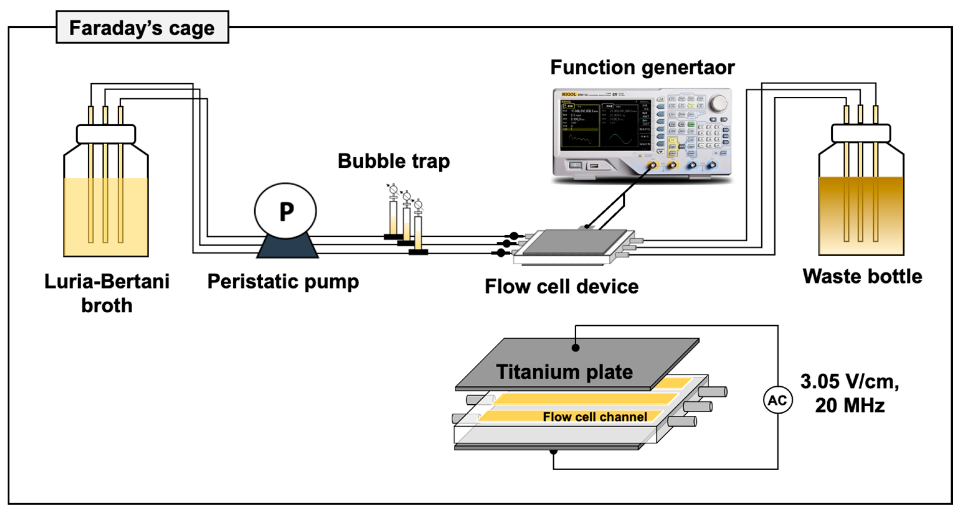
2.2. Flow Cells and Electric Field Exposure Devices

The exposure of the test biofilm (E. coli and P. aeruginosa) to an alternating electric field was conducted using a three-channel flow cell device (ACCFL0001, IBI Scientific, Dubuque, IA, USA). Each channel had an inner surface area of 4.08 cm2 (4 mm width × 40 mm length × 1 mm depth). The flow cell system consisted of a medium bottle for supplying the nutrient source (LB broth), a peristaltic pump (SJ-1211-L, ATTO, Amherst, NY, USA), bubble traps to maintain an air-free environment, injection ports for inoculating the bacterial cultures, a three-channel flow cell for biofilm growth, and a waste container for collecting effluent. All components were assembled and operated at a flow rate of 4.5 ± 0.3 mL/h/channel, as specified by the manufacturer’s recommended flow range of 3–5 mL/h/channel. Referring to a previous study that demonstrated that the influence of an alternating electric field was dependent on the applied intensity and frequency [44], our study exposed biofilms during growth to alternating electric fields at 3.05 V/cm and 20 MHz. Electric field intensity and electric field frequency were restricted owing to limitations in the employed equipment’s capabilities. The exposure time was determined according to the duration of the biofilm development stage being tested in each experiment. This application employed a pair of titanium plates (20 mm wide × 33 mm long) that were positioned on each side of the three-channel flow cell device. Titanium’s outstanding corrosion resistance and biocompatibility favored its use as electrodes [45]. The electrodes were spaced 1.97 mm apart, matching the thickness of the flow cell device, to maximize the intensity of the electric field generated. The alternating electric field, generated by a function generator (DG4102, Rigol, Shanghai, China), was activated in a sine waveform, as it is unaffected by electrical components [46]. The liquid temperature measured at the outlet of the flow cell device revealed no differences between the control group (which was not exposed to an electric field) and the electric-field-exposed group, confirming the absence of thermal effects caused by the applied electric fields. Applied voltage and frequency were monitored using an oscilloscope (DS-1202 Z-E, Rigol, Shanghai, China). To minimize external electromagnetic influences and enhance the integrity of the experimental setup, the flow cell system was placed in a Faraday cage. The experimental setup of the electrical treatment system is illustrated in Figure 1.

2.3. Experimental Conditions

2.3.1. Exposure of Alternating Electric Fields during Biofilm Development Processes

This experiment investigated an alternating electric fields’ effect on two distinct stages during biofilm development: the initial attachment of bacterial cells to the surface (stage 1) and early biofilm development (stage 2). To investigate the effect on stage 1, a cell suspension (106 CFU/mL LB broth) was injected into a flow cell, which was then placed upside-down and exposed to an alternating electric field (3.05 V/cm, 20 MHz) for 2 h without a flowing LB medium. In addition to the cells adhered to the flow-cell’s surface (see Section 2.4 for details), the number of suspended cells remaining in the injected liquid was measured via plate counting. Stage 2 focused on the subsequent growth of the biofilm after stage 1. Cells attached according to the same procedure as that employed in stage 1 but without being treated using an electric field were continuously incubated for the next 16 h under a flow of LB medium and an alternating electric field (3.05 V/cm, 20 MHz). Control tests were conducted in the same manner and in the absence of an alternating electric field. A diagram representing experimental conditions used to test the biofilm development processes in stages 1 and 2 is indicated in Figure S1.

2.3.2. Exposure of Alternating Electric Fields during Cell Detachment

Effects on the detachment of cells after the initial attachment (stage 1) and early development (stage 2) stages were investigated. After the initial attachment of cells in stage 1 (2 h without applying electric fields), the LB medium was replaced with PBS, and PBS was allowed to keep flowing under an alternating electric field (3.05 V/cm, 20 MHz) for 2 h. Similarly, after early biofilm development in stage 2 (16 h without applying an electric field), PBS was fed into the flow cell channel, and the flow cell was incubated for 16 h under an alternating electric field (see the diagram in Figure S2).

2.3.3. Combination of Alternating Electric Fields and Hydrodynamic Shear

To investigate the combined effect of alternating electric fields and hydrodynamic shear forces, experiments were conducted by changing the flow rates of the LB medium in stage 2. Initially, bacterial cells were allowed to complete initial attachment under static conditions for 2 h. Subsequently, the flow of the LB medium was resumed at 0.08 mL/min for 7 min to remove excess suspended cells. The adhered cells were exposed to an alternating electric field (3.05 V/cm, 20 MHz) under a constant flow of LB medium at 0.08, 0.2, 0.4, or 0.6 mL/min (Figure S3). The corresponding Reynolds numbers, shear stresses, and flow velocities for each test flow are provided in Table 1. The formulas used for calculating the flow parameters can be found in the Supplementary Materials (Text S1).

2.4. Quantification of Adhered Cell Numbers and Extracellular Polymeric Substances

After each experiment, adhered cells and biofilm components inside flow cells were detached via sonication (see Text S2 in the Supplementary Materials for details). The resulting suspension was used to analyze the number of cells, protein content, and polysaccharide content in the extracellular polymeric substances (EPSs). Cell concentrations were measured via plate counting. For EPSs, the suspension was centrifuged at 877× g for 5 min, and the supernatant was carefully removed. The pellets were re-suspended to the original volume with MilliQ water and heated in a water bath at 80 °C for 30 min, followed by another centrifugation at 877× g for 15 min. The supernatant was collected and filtered using a 0.45 μm syringe filter prior to quantifying the concentration of proteins and polysaccharides via the phenol-sulfuric acid method [47] and a modified Lowry’s method (Modified Lowry Assay, Thermo Fischer Scientific, Miami, FL, USA) assay using glucose and bovine serum albumin as standards, respectively.

2.5. Quantification of Total Biofilm Biomass via Crystal Violet Assay

The flow cell was gently removed from the system. The liquid in each flow cell channel was replaced with 160 μL of 0.01% crystal violet (CV) solution to stain the attached biomass. Incubation was carried out at room temperature under dark conditions for 15 min. Subsequently, the CV solution was removed, and each channel was washed three times with autoclaved water to remove excess unbound dye. To elute the bound dye, 160 μL of 30% acetic acid was added to each channel and shaken vigorously using a vortex mixer for 3 min. Subsequently, the solution in each channel was carefully collected, and the remaining portion was rinsed with 3 mL of 30% acetic acid. The collected solution was mixed thoroughly using a vortex mixer and then measured for absorbance at 590 nm.

2.6. Scanning Electron Microscopy (SEM)

Bacterial cells were fixed by adding 25% glutaraldehyde (Sigma Aldrich, Burlington, MA, USA) to a final concentration of 2%. The fixed suspension was then placed on ice for 10 min before being filtered through a 0.20 μm white track-etched polycarbonate membrane (Millipore, Darmstadt, Germany). Dehydration was carried out by sequentially incubating the fixed suspension in 50%, 70%, 90%, and 100% ethanol for 5 min at each concentration. Following incubation in 100% ethanol, the cells were soaked three times in 1-butanol (FUJIFILM Wako Pure Chemical Corporation, Osaka, Japan) for 5 min each. After being freeze-dried, each sample was affixed to a specimen stub and coated with a platinum alloy. The morphological characteristics were subsequently examined using a scanning electron microscope (Keyence VE-8800, Shanghai, China) operating at accelerating voltages of 5 kV and 10 kV, with a working distance ranging between 9 and 10 mm. Image J software (version 1.53k) was used to determine cell length.

2.7. Statistical Analysis

The data were calculated and compared between the samples exposed to alternating electric fields and those that were not exposed. Statistical significance was tested using one-way ANOVA in Excel. A p-value < 0.05 was considered statistically significant.

3. Results

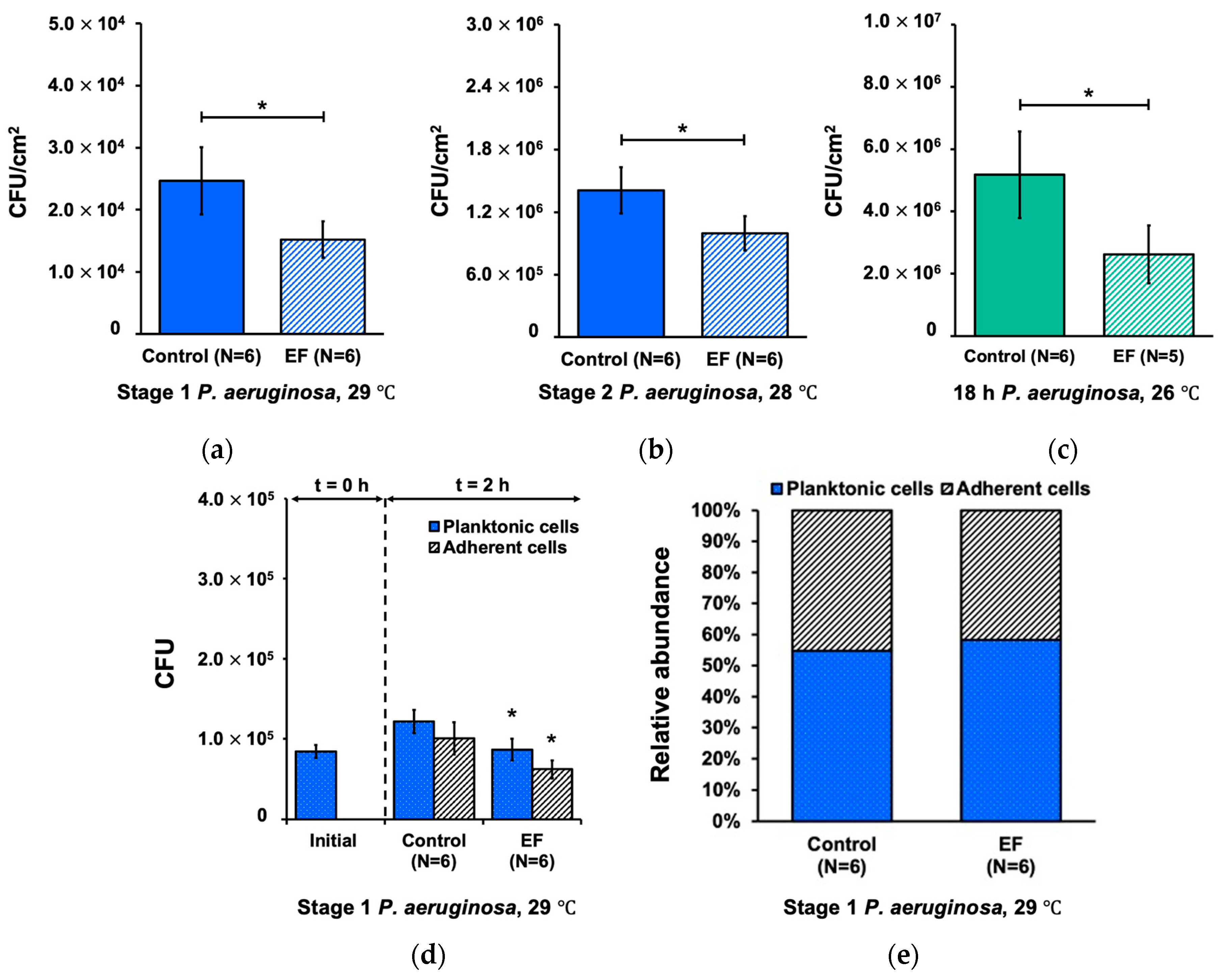
3.1. Effect of Alternating Electric Field on Biofilm Development Processes

Alternating electric fields suppressed the number of P. aeruginosa cells adhered to the inside surface of the flow cells by 38.3% (p < 0.05) in the initial surface adherence stage (stage 1) and by 29.3% (p < 0.05) in the early development stage (stage 2) (Figure 2a,b). When the alternating electric fields were applied continuously throughout the two stages (stages 1 + 2), the overall reduction in adherent cells was 49.4% (Figure 2c), which was relatively consistent with the estimated overall reductions (56.4%) based on the individual reductions obtained for stages 1 and 2.
In addition to the adherent cells, we also sampled and counted the number of suspended cells in the liquid phase inside the flow cells after stage 1. The planktonic cell concentration increased by 44.4% during the 2 h incubation in stage 1, confirming the occurrence of growth. After exposure to an alternating electric field, the number of planktonic cells was 28.9% lower (p < 0.05) than that for the control (Figure 2d), implying that the alternating electric field suppressed the growth of non-adherent cells. The ratio of adhered cells to the total number of cells (adhered + planktonic) was slightly higher in the electric field groups (58.2%) than in the control groups (54.7%) (Figure 2e). This result indicates a small effect of the alternating electric fields on the initial cell attachment process. Taken together, these findings suggest that the decrease in the number of adherent cells under the alternating electric field primarily resulted from the inhibitory effect on the growth of bacterial cells in addition to a slight effect consisting of discouraging cell attachment.
In terms of total biofilm biomass, there was no significant difference observed between the control group and the electric field group in stages 1 and 2 (Figure 3a,b). Similarly, no noticeable effect of the alternating electric field on the total number of EPSs was observed (Figure S6). The EPSs primarily consisted of polysaccharides in the control and electric field groups, with the protein concentration falling below the detection limit (6.9 mg/L). However, when considering the number of EPSs per CFU, opposite trends were observed (Figure 3c,d). The EPS content per CFU under alternating electric fields was significantly higher compared to the control group in stage 1 and stage 2 (p < 0.05).
We repeated the identical experiments using Escherichia coli and obtained similar results (Figures S4 and S5). Therefore, the above effects are not limited to P. aeruginosa and are possibly common to different bacterial species.

3.2. Morphological Characteristics of the Adherent Cells

The SEM images in Figure 4a,b illustrate representative examples of adherent P. aeruginosa cells exposed and non-exposed to an alternating electric field in stage 2. These images indicate that the alternating electric field’s presence impacted cell morphology by increasing cell lengths without causing significant damage to their structural integrity. The treatment group of P. aeruginosa exhibited a broader distribution of cell lengths compared to the control group (Figure 4c). The average lengths of the adherent P. aeruginosa cells were 1.51 µm and 2.22 µm in the control and treatment groups, respectively. A consistent trend was observed when testing using E. coli, where the average cell length was 2.04 µm in the control group and 3.42 µm in the treatment group (Figure S7c).

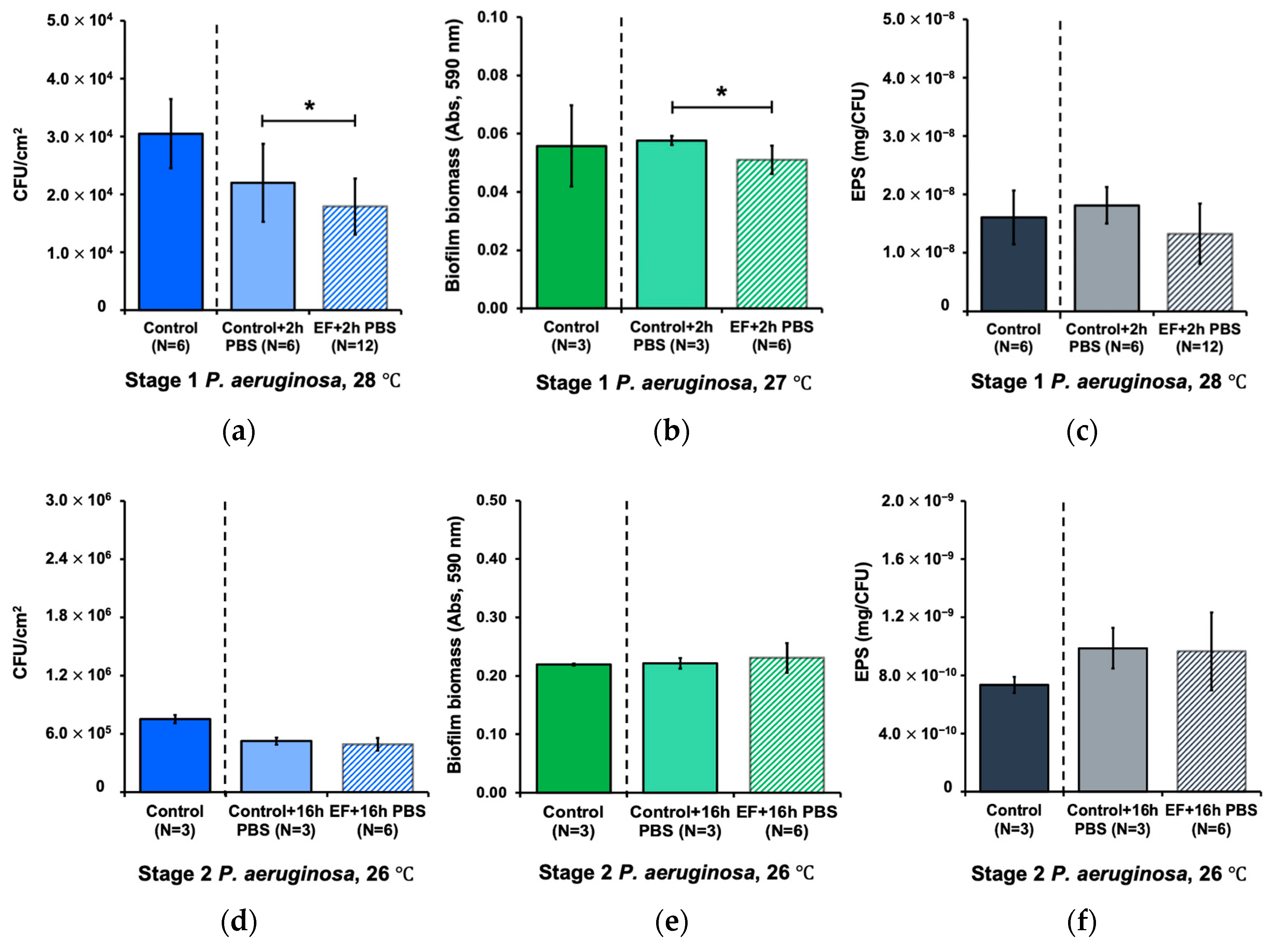
3.3. Effect of Alternating Electric Field on Biofilm Detachment

Figure 5a shows the initial number of adherent P. aeruginosa cells for which attachment was allowed (stage 1) followed by a 2 h exposure to an alternating electric field under a flow of PBS. The number of adhered cells was 18.6% lower with the alternating electric field than that of the control (p < 0.05) (Figure 5a). The total biofilm biomass was reduced (p < 0.05) (Figure 5b), while the EPS content per cell remained constant (p > 0.05) (Figure 5c). These results indicate that the alternating electric field promoted the detachment of cells after their initial adhesion. Consistent results were observed when using E. coli (Figure S8).
However, after the early development phase (stage 2), no effect of the alternating electric field on biofilm detachment was observed. As shown in Figure 5d–f, there was no significant change in the number of adherent cells, total biofilm biomass, and number of EPSs per cell for P. aeruginosa. These insignificant differences were also observed with E. coli (Figure S8). These results imply that the detachment effect of alternating electric fields is dependent on biofilm aging.

3.4. Combined Effect of Alternating Electric Field and Hydrodynamic Shear Forces

We examined the influence of alternating electric fields on biofilm formation under different hydrodynamic shear forces during the early development stage. As seen in Figure 6, increased flow rates (0.2, 0.4, and 0.6 mL/min), without an alternating electric field, suppressed the number of adhered P. aeruginosa cells by 9.0–29.1% compared to the control condition (0.08 mL/min). The more the flow rate increased, the more the suppression of adhered cells was observed. This suppression was further enhanced when an alternating electric field was applied. The highest suppression level of 46.2% was observed when the cells were exposed to an alternating electric field at 0.6 mL/min. There were, however, no or only tiny differences in the total biofilm biomass (Figure 6b) with and without alternating electric field exposure. Although lacking statistical significance, the EPS content per cell tended to increase when the cells were exposed to alternating electric fields, and this result is consistent with the results in Section 3.1. The additional percentage reduction resulting from the combined influence of an alternating electric field and flow velocity, compared to the effect of the flow condition alone, did not demonstrate differences between the various flow conditions (~12.6% relative standard deviation). This suggests the absence of a synergistic effect arising from their cumulative influences. Consistent results were observed when using E. coli (Figure S9).

4. Discussion

Previous studies primarily focused on investigating the impact of direct current application on controlling biofilm overgrowth during water and wastewater treatment processes, which unavoidably resulted in undesirable electrochemical reactions [2,47,48,49]. Unlike the application of a direct current, the application of a high-frequency alternating electric field circumvents any potential electrochemical reactions [39,43]. However, there have been few studies exploring this method’s inhibitory effect on planktonic cells, while the impact on biofilm growth modes remains to be explored. This study investigated biofilm development processes under alternating electric field exposure.
The results of this study demonstrate that high-frequency, low-voltage alternating electric fields (3.05 V/cm, 20 MHz) affected biofilm formation at the initial attachment phase (stage 1) and the early development phase (stage 2) by reducing the number of adherent cells and increasing cell length. The employed frequency and the unchanged liquid temperature at the outlet of the flow cell device in this study ruled out the possibility of microwave irradiation and thermal effects. The significant reduction in the observed planktonic population during the initial attachment phase (stage 1) implies that the inhibition of bacterial growth was responsible for the lower number of adherent cells. This finding aligns with the results of a previous study that observed the inhibitory effect of alternating electric fields (2–4 V/cm, 10 MHz) on planktonic P. aeruginosa and Staphylococcus aureus cells [43]. However, the slight increase in the proportion of planktonic cells suggests that there might be other mechanisms in alternating electric fields that retard the attachment of bacterial cells to solid surfaces.
Using SEM analysis, we revealed that the filamentous formation of adherent cells occurred after their exposure to alternating electric fields. As a mechanism for mammalian cells proposed by Kirson et al. (2007), it is plausible that high-frequency alternating electric fields impact the cell division of bacteria during cytokinesis [50]. This has been attributed to the generation of nonhomogeneous electric fields near the bridge that separates the daughter cells. These nonhomogeneous fields induce unidirectional dielectrophoretic forces on charged and polar particles and molecules, causing the disruption of cellular processes [41,43,50]. This mechanism possibly explains the elongated bacterial cells observed in this study under the high-frequency alternating electric field.
We also observed the effect of alternating electric fields on cell detachment, causing a reduction in both the number of adherent cells and total biofilm biomass. Although further investigation is required to fully elucidate this mechanism, it is plausible that the observed impact was caused by the electrostatic forces present within the biofilm matrix. The application of electric fields induces these electrostatic forces, leading to repulsion between the biofilm and the surface. As a result, the attachment of the biofilm to the substrate is weakened, thereby facilitating its detachment [51,52]. However, the detachment effect was observed only at the initial attachment phase (stage 1), indicating that this effect was biofilm-aging-dependent. Older biofilms tend to be more robust and resistant to detachment compared to younger biofilms. As biofilms mature, they undergo structural and compositional changes that can make them more stable and harder to remove [53,54]. Additionally, Li et al. (2023) found that the volume of the EPS matrices of mature biofilm was higher than that of young biofilm, which indicates that as biofilm becomes mature, more EPSs are produced [55]. Increasing EPS production provides more structural stability to the biofilm and makes it more resistant to detachment [56,57].
The combined effect between alternating electric fields and hydrodynamic forces was also examined in this study. Fluid flow has been shown to be an important factor in biofilm development in terms of structural and physical properties [58,59,60]. In one study, it was observed that the combined effect of hydrodynamic forces and antibiotics promoted biofilm detachment and increased the mass transfer of antibiotics from the bulk solution to the biofilm surface [61]. Meanwhile, several studies have shown that the application of electric fields, including those generated by a low-intensity direct current [62,63,64,65], as well as alternating electric fields (up to 10 MHz) can improve the effectiveness of antibacterial agents against bacterial biofilm [43,66,67]. However, the combined effect of an alternating electric field and hydrodynamic force remains unknown. Especially when the adherent cells form filaments, this cell elongation might promote or suppress the shear stress effect on biofilm development. The results demonstrated that applying an alternating electric field and hydrodynamic forces suppressed the quantity of adherent cells but hardly changed the total biomass of attached cells. This result can be explained by an increase in the EPS content per CFU, which can be attributed to the elongated shape of individual cells when exposed to alternating electric fields. As reported in previous studies, EPS excretion takes place on the surface of bacterial cells [67,68]. The number of EPSs is possibly influenced by the surface area of a bacterial cell. With the same cell radius, a longer cell has a greater number of EPSs compared to a shorter cell. Therefore, the observed increase in the number of EPSs per CFU was likely a result of cell elongation. Additionally, when cells become longer, they contribute to inducing a larger volume per cell (as shown in Table 2). Although fewer cells adhered under alternating electric field exposure compared to the control group, the total cell volumes of the treated and untreated cells were similar (~6.9% difference) because the treated cells had larger individual cell volumes (as presented in Table 2). Moreover, the combined impact of the alternating electric field and flow velocity did not yield a substantial decrease in cell adherence, suggesting that there was no synergistic effect with hydrodynamic shear.
Based on the overall experimental results, it appears that the application of a high-frequency, low-voltage alternating electric field (3.05 V/cm, 20 MHz) may not be effective enough for use in suppressing excessive biofilm growth in water and wastewater treatment processes, including as a measure to mitigate biofouling in membrane filtration or in controlling excess biomass accumulation in attached-growth processes (e.g., trickling filters, rotating biological contactors, and packed bed reactors). The limitation of this method in controlling biofilm growth differentiates it from methods like UV treatment that are more effective in biofilm eradication. Also, the applied frequency was not within the range capable of biofilm inactivation through microwave irradiation.
On the other hand, it is important to avoid overlooking the observed cell elongation resulting from the application of alternating electric fields. Liu & Liu (2006) have highlighted the advantages of filamentous cell shapes in promoting biomass aggregation and enhancing granular sludge stability [69]. Cell elongation might act as a bridge for cell aggregation, thereby enhancing sludge flocculation [70]. Accordingly, utilizing cell elongation as a strategy to enhance or support the attached growth mode in a biofilm may be a viable approach. Further studies are necessary to advance the possible applications of the high-frequency, low-voltage alternating electric field strategy within wastewater engineering. Certain points need to be investigated. Different types of biofilms may exhibit varying responses to electric fields due to variations in their structures and environmental conditions. The optimization of electric field parameters such as strength, frequency, and exposure duration is crucial to ensuring effective biofilm control. Moreover, the consideration of energy consumption, particularly in larger scale implementations, remains an important practical concern.

5. Conclusions

The application of a high-frequency, low-voltage alternating electric field (3.05 V/cm, 20 MHz) demonstrated the ability to reduce the number of adherent cells during the initial adherence process and the early developmental phase for both P. aeruginosa and E. coli. This reduction in cell adherence was accompanied by an increase in cell length, resulting in an insignificant change in the total biofilm biomass. In addition, the alternating electric field promoted cell detachment in the initial attachment phase but was ineffective for the developed biofilm. The field’s combination with hydrodynamic shear was effective in reducing the number of adherent cells during the early biofilm development phase. No synergistic effects were observed between the alternating electric field and hydrodynamic shear.

Supplementary Materials

The following supporting information can be downloaded at: https://www.mdpi.com/article/10.3390/w15173055/s1, Text S1: Calculation of hydrodynamic forces; Text S2: Sterilization of the flow cell system; Text S3: Detachment procedure; Figure S1: Experimental procedure for alternating electric field exposure during biofilm development processes; Figure S2: Operating conditions of alternating electric field exposure during cell detachment; Figure S3: Experimental conditions for investigating the combined effect of alternating electric fields and hydrodynamic shear; Figure S4: Effects of alternating electric fields in terms of adherence of E. coli cells; Figure S5: Effect of alternating electric fields in terms of total biofilm biomass and EPS content; Figure S6: Total EPS amount (mg EPS per cm2) after stage 1 and stage 2 for E. coli and P. aeruginosa under exposure to alternating electric field; Figure S7: Representative SEM images (5000× magnifications) of adherent E. coli cells after alternating electric field exposure; Figure S8: Effects on cell detachment of E. coli after stage 1 and stage 2 under different conditions and parameters; Figure S9: Change in adherent cells and percentage reduction, biofilm biomass, and EPS content in mg per CFU of E. coli after exposure to alternating electric field along with different flow velocities (0.2, 0.4, and 0.6 mL/min).

Author Contributions

Conceptualization, P.K., T.T. and F.N.; methodology, P.K. and T.T.; investigation, P.K.; analysis, P.K. and T.T.; writing—original draft preparation, P.K.; writing—review and editing, T.T. and F.N.; visualization, P.K.; supervision, T.T. and F.N. All authors have read and agreed to the published version of the manuscript.

Funding

This work was supported by JSPS KAKENHI, grant number 22K18824, and by Kurita Water and Environment Foundation, grant number 21E035.

Data Availability Statement

The data presented in this study are available upon request.

Acknowledgments

The authors would like to express their acknowledgement to Tomoko Inoue and Suguru Hakoshima for their technical support and assistance in conducting the experiments for this work.

Conflicts of Interest

The authors declare no conflict of interest.

References

  1. Butler, C.S.; Boltz, J. Biofilm Processes and Control in Water and Wastewater Treatment. In Comprehensive Water Quality and Purification; Elsevier: Amsterdam, The Netherlands, 2014; Volume 3, pp. 90–107. [Google Scholar] [CrossRef]
  2. Saini, S.; Tewari, S.; Dwivedi, J.; Sharma, V. Biofilm Mediated Wastewater Treatment: A Comprehensive Review. Mater. Adv. 2023, 4, 1415–1443. [Google Scholar] [CrossRef]
  3. Chattopadhyay, I.; Usman, T.M.; Varjani, S. Exploring the role of microbial biofilm for industrial effluents treatment. Bioengineered 2023, 13, 6420–6440. [Google Scholar] [CrossRef] [PubMed]
  4. Peng, J.; Kumar, K.; Gross, M.; Kunetz, T.; Wen, Z. Removal of total dissolved solids from wastewater using a revolving algal biofilm reactor. Water Environ. Res. 2020, 92, 766–778. [Google Scholar] [CrossRef] [PubMed]
  5. Bitton, G. Drinking Water Distribution Systems: Biofilm Microbiology. In Microbiology of Drinking Water; John Wiley & Sons, Ltd.: Hoboken, NJ, USA, 2014; pp. 91–115. [Google Scholar] [CrossRef]
  6. Herzberg, M. Osmotic effects of biofouling in reverse osmosis (RO) processes: Physical and physiological measurements and mechanisms. Desalin. Water Treat. 2010, 15, 287–291. [Google Scholar] [CrossRef]
  7. Sweity, A.; Oren, Y.; Ronen, Z.; Herzberg, M. The influence of antiscalants on biofouling of RO membranes in seawater desalination. Water Res. 2013, 47, 3389–3398. [Google Scholar] [CrossRef]
  8. Gutman, J.; Fox, S.; Gilron, J. Interactions between biofilms and NF/RO flux and their implications for control—A review of recent developments. J. Membr. Sci. 2012, 421–422, 1–7. [Google Scholar] [CrossRef]
  9. Hoek, E.M.V.; Weigand, T.M.; Edalat, A. Reverse osmosis membrane biofouling: Causes, consequences and countermeasures. Npj Clean Water 2022, 5, 45. [Google Scholar] [CrossRef]
  10. Lu, W.; Kiéné, L.; Lévi, Y. Chlorine demand of biofilms in water distribution systems. Water Res. 1999, 33, 827–835. [Google Scholar] [CrossRef]
  11. Lee, S.H.; O’Connor, J.T.; Banerji, S.K. Biologically mediated corrosion and its effects on water quality in distribution systems. J. Am. Water Work. Assoc. 1980, 72, 636–645. [Google Scholar] [CrossRef]
  12. Flemming, H.-C.; Griebe, T.; Schaule, G. Antifouling strategies in technical systems—A short review. Water Sci. Technol. 1996, 34, 517–524. [Google Scholar] [CrossRef]
  13. Celmer, D.; Oleszkiewicz, J.; Cicek, N. Impact of ultrasound treatment and performance of a membrane biofilm reactor for tertiary hydrogen driven denitrification of municipal wastewater. In Proceedings of the Twentieth Jubilee-National, the Eighth Internatioanal and Technical Conference: Water Supply and Water Engineering, Warsaw, Poland, 15–18 June 2008. [Google Scholar]
  14. Luo, X.; Zhang, B.; Lu, Y.; Mei, Y.; Shen, L. Advances in application of ultraviolet irradiation for biofilm control in water and wastewater infrastructure. J. Hazard. Mater. 2022, 421, 126682. [Google Scholar] [CrossRef] [PubMed]
  15. Ma, B.; Seyedi, S.; Wells, E.; McCarthy, D.; Crosbie, N.; Linden, K.G. Inactivation of biofilm-bound bacterial cells using irradiation across UVC wavelengths. Water Res. 2022, 217, 118379. [Google Scholar] [CrossRef] [PubMed]
  16. Piasecka, A.; Bernstein, R.; Ollevier, F.; Meersman, F.; Souffreau, C.; Bilad, R.M.; Cottenie, K.; Vanysacker, L.; Denis, C.; Vankelecom, I. Study of biofilms on PVDF membranes after chemical cleaning by sodium hypochlorite. Sep. Purif. Technol. 2015, 141, 314–321. [Google Scholar] [CrossRef]
  17. Lee, W.H.; Wahman, D.G.; Bishop, P.L.; Pressman, J.G. Free Chlorine and Monochloramine Application to Nitrifying Biofilm: Comparison of Biofilm Penetration, Activity, and Viability. Environ. Sci. Technol. 2011, 45, 1412–1419. [Google Scholar] [CrossRef]
  18. Brant, K.M.; Gilg, M.R. Testing the relative effectiveness of traditional and non-traditional antifouling substrates on barnacle and macroalgae settlement. Mar. Biol. Res. 2014, 10, 1027–1032. [Google Scholar] [CrossRef]
  19. Woods, C.M.; Floerl, O.; Jones, L. Biosecurity risks associated with in-water and shore-based marine vessel hull cleaning operations. Mar. Pollut. Bull. 2012, 64, 1392–1401. [Google Scholar] [CrossRef]
  20. Renner, L.D.; Weibel, D.B. Physicochemical regulation of biofilm formation. MRS Bull. 2011, 36, 347–355. [Google Scholar] [CrossRef]
  21. Schiffman, J.D.; Elimelech, M. Antibacterial Activity of Electrospun Polymer Mats with Incorporated Narrow Diameter Single-Walled Carbon Nanotubes. ACS Appl. Mater. Interfaces 2011, 3, 462–468. [Google Scholar] [CrossRef]
  22. Schiffman, J.D.; Wang, Y.; Giannelis, E.P.; Elimelech, M. Biocidal Activity of Plasma Modified Electrospun Polysulfone Mats Functionalized with Polyethyleneimine-Capped Silver Nanoparticles. Langmuir 2011, 27, 13159–13164. [Google Scholar] [CrossRef]
  23. Sultana, S.T.; Babauta, J.T.; Beyenal, H. Electrochemical biofilm control: A review. Biofouling 2015, 31, 745–758. [Google Scholar] [CrossRef]
  24. Wang, H.; Guo, L.; Ren, X.; Gao, M.; Jin, C.; Zhao, Y.; Ji, J.; She, Z. Enhanced aerobic granular sludge by static magnetic field to treat saline wastewater via simultaneous partial nitrification and denitrification (SPND) process. Bioresour. Technol. 2022, 350, 126891. [Google Scholar] [CrossRef]
  25. Anastas, P.T.; Kirchhoff, M.M. Origins, Current Status, and Future Challenges of Green Chemistry. Acc. Chem. Res. 2002, 35, 686–694. [Google Scholar] [CrossRef]
  26. Liu, W.K.; Brown, M.R.; Elliott, T.S. Mechanisms of the bactericidal activity of low amperage electric current (DC). J. Antimicrob. Chemother. 1997, 39, 687–695. [Google Scholar] [CrossRef] [PubMed]
  27. van der Borden, A.; van der Mei, H.; Busscher, H. Electric block current induced detachment from surgical stainless steel and decreased viability of Staphylococcus epidermidis. Biomaterials 2005, 26, 6731–6735. [Google Scholar] [CrossRef] [PubMed]
  28. Schmidt-Malan, S.M.; Karau, M.J.; Cede, J.; Greenwood-Quaintance, K.E.; Brinkman, C.L.; Mandrekar, J.N.; Patel, R. Antibiofilm Activity of Low-Amperage Continuous and Intermittent Direct Electrical Current. Antimicrob. Agents Chemother. 2015, 59, 4610–4615. [Google Scholar] [CrossRef] [PubMed]
  29. Gad, A.; Jayaram, S.H. Effect of Electric Pulse Parameters on Releasing Metallic Particles From Stainless Steel Electrodes during PEF Processing of Milk. IEEE Trans. Ind. Appl. 2014, 50, 1402–1409. [Google Scholar] [CrossRef]
  30. Pataro, G.; Falcone, M.; Donsì, G.; Ferrari, G. Metal release from stainless steel electrodes of a PEF treatment chamber: Effects of electrical parameters and food composition. Innov. Food Sci. Emerg. Technol. 2014, 21, 58–65. [Google Scholar] [CrossRef]
  31. Tzedakis, T.; Basseguy, R.; Comtat, M. Voltammetric and coulometric techniques to estimate the electrochemical reaction rate during ohmic sterilization. J. Appl. Electrochem. 1999, 29, 819–826. [Google Scholar] [CrossRef]
  32. Morren, J.; Roodenburg, B.; de Haan, S.W. Electrochemical reactions and electrode corrosion in pulsed electric field (PEF) treatment chambers. Innov. Food Sci. Emerg. Technol. 2003, 4, 285–295. [Google Scholar] [CrossRef]
  33. Rodaite-Riseviciene, R.; Saule, R.; Snitka, V.; Saulis, G. Release of Iron Ions From the Stainless Steel Anode Occurring during High-Voltage Pulses and Its Consequences for Cell Electroporation Technology. IEEE Trans. Plasma Sci. 2014, 42, 249–254. [Google Scholar] [CrossRef]
  34. Zhou, J.; Wang, T.; Yu, C.; Xie, X. Locally enhanced electric field treatment (LEEFT) for water disinfection. Front. Environ. Sci. Eng. 2020, 14, 78. [Google Scholar] [CrossRef]
  35. Chen, C.W.; Lee, H.-M.; Chang, M.B. Inactivation of Aquatic Microorganisms by Low-Frequency AC Discharges. IEEE Trans. Plasma Sci. 2008, 36, 215–219. [Google Scholar] [CrossRef]
  36. Stoner, G.; Cahen, G.; Sachyani, M.; Gileadi, E. The mechanism of low frequency a.c. electrochemical disinfection. Bioelectrochem. Bioenerg. 1982, 9, 229–243. [Google Scholar] [CrossRef]
  37. Mosqueda-Melgar, J.; Raybaudi-Massilia, R.M.; Martín-Belloso, O. Influence of treatment time and pulse frequency on Salmonella Enteritidis, Escherichia coli and Listeria monocytogenes populations inoculated in melon and watermelon juices treated by pulsed electric fields. Int. J. Food Microbiol. 2007, 117, 192–200. [Google Scholar] [CrossRef] [PubMed]
  38. Mawioo, P.M.; Rweyemamu, A.; Garcia, H.A.; Hooijmans, C.M.; Brdjanovic, D. Evaluation of a microwave based reactor for the treatment of blackwater sludge. Sci. Total Environ. 2016, 548–549, 72–81. [Google Scholar] [CrossRef] [PubMed]
  39. Shawki, M.M.; Gaballah, A. The effect of low AC electric field on bacterial cell death. Rom. J. Biophys. 2015, 25, 163–172. [Google Scholar]
  40. Lee, S.-Y.; Ryu, S.; Kang, D.-H. Effect of Frequency and Waveform on Inactivation of Escherichia coli O157:H7 and Salmonella enterica Serovar Typhimurium in Salsa by Ohmic Heating. Appl. Environ. Microbiol. 2013, 79, 10–17. [Google Scholar] [CrossRef]
  41. Kirson, E.D.; Gurvich, Z.; Schneiderman, R.; Dekel, E.; Itzhaki, A.; Wasserman, Y.; Schatzberger, R.; Palti, Y. Disruption of Cancer Cell Replication by Alternating Electric Fields. Cancer Res. 2004, 64, 3288–3295. [Google Scholar] [CrossRef]
  42. Mirzaii, M.; Alfi, A.; Kasaeian, A.; Norozi, P.; Nasiri, M.; Sarokhalil, D.D.; Khoramrooz, S.S.; Fazli, M.; Davardoost, F. Antibacterial effect of alternating current against Staphylococcus aureus and Pseudomonas aeroginosa. Russ. Open Med. J. 2015, 4, e0203. [Google Scholar] [CrossRef]
  43. Giladi, M.; Porat, Y.; Blatt, A.; Wasserman, Y.; Kirson, E.D.; Dekel, E.; Palti, Y. Microbial Growth Inhibition by Alternating Electric Fields. Antimicrob. Agents Chemother. 2008, 52, 3517–3522. [Google Scholar] [CrossRef]
  44. Kunlasubpreedee, P.; Tobino, T.; Nakajima, F. Effects of high frequency-alternating electric fields on bacterial cell growth in planktonic and biofilm modes. In Proceedings of the Thirteenth International Symposium on Southeast Asian Water Environment, Bangkok, Thailand, 13–15 December 2022. [Google Scholar]
  45. Berbel, L.O.; Banczek, E.D.P.; Karousis, I.K.; Kotsakis, G.A.; Costa, I. Determinants of corrosion resistance of Ti-6Al-4V alloy dental implants in an In Vitro model of peri-implant inflammation. PLoS ONE 2019, 14, e0210530. [Google Scholar] [CrossRef]
  46. Hoseinzadeh, E.; Wei, C.; Farzadkia, M.; Rezaee, A. Effects of Low Frequency-Low Voltage Alternating Electric Current on Apoptosis Progression in Bioelectrical Reactor Biofilm. Front. Bioeng. Biotechnol. 2020, 8, 2. [Google Scholar] [CrossRef] [PubMed]
  47. DuBois, M.; Gilles, K.A.; Hamilton, J.K.; Rebers, P.A.; Smith, F. Colorimetric method for determination of sugars and related substances. Anal. Chem. 1956, 28, 350–356. [Google Scholar] [CrossRef]
  48. Kerdi, S.; Qamar, A.; Vrouwenvelder, J.S.; Ghaffour, N. Biofilm removal efficacy using direct electric current in cross-flow ultrafiltration processes for water treatment. J. Membr. Sci. 2021, 620, 118808. [Google Scholar] [CrossRef]
  49. Long, Y.; Yu, Y.; Yin, X.; Li, J.; Carlos, C.; Du, X.; Jiang, Y.; Wang, X. Effective anti-biofouling enabled by surface electric disturbance from water wave-driven nanogenerator. Nano Energy 2019, 57, 558–565. [Google Scholar] [CrossRef]
  50. Kirson, E.D.; Dbalý, V.; Tovaryš, F.; Vymazal, J.; Soustiel, J.F.; Itzhaki, A.; Mordechovich, D.; Steinberg-Shapira, S.; Gurvich, Z.; Schneiderman, R.; et al. Alternating electric fields arrest cell proliferation in animal tumor models and human brain tumors. Proc. Natl. Acad. Sci. USA 2007, 104, 10152–10157. [Google Scholar] [CrossRef]
  51. Poortinga, A.T.; Bos, R.; Busscher, H.J. Controlled electrophoretic deposition of bacteria to surfaces for the design of biofilms. Biotechnol. Bioeng. 2000, 67, 117–120. [Google Scholar] [CrossRef]
  52. Ueshima, M.; Tanaka, S.; Nakamura, S.; Yamashita, K. Manipulation of bacterial adhesion and proliferation by surface charges of electrically polarized hydroxyapatite. J. Biomed. Mater. Res. 2002, 60, 578–584. [Google Scholar] [CrossRef]
  53. Stewart, P.S. Antimicrobial Tolerance in Biofilms. Microbiol. Spectr. 2015, 12, 1221–1236. [Google Scholar] [CrossRef]
  54. Ngoumelah, D.D.; Harnisch, F.; Kretzschmar, J. Benefits of Age–Improved Resistance of Mature Electroactive Biofilm Anodes in Anaerobic Digestion. Environ. Sci. Technol. 2021, 55, 8258–8266. [Google Scholar] [CrossRef] [PubMed]
  55. Li, H.; Liu, H.; Zhang, L.; Hieawy, A.; Shen, Y. Evaluation of extracellular polymeric substances matrix volume, surface roughness and bacterial adhesion property of oral biofilm. J. Dent. Sci. 2023; in press. [Google Scholar] [CrossRef]
  56. Jiao, Y.; Tay, F.R.; Niu, L.-N.; Chen, J.-H. Advancing antimicrobial strategies for managing oral biofilm infections. Int. J. Oral Sci. 2019, 11, 28. [Google Scholar] [CrossRef] [PubMed]
  57. Fang, H.H.; Chan, K.-Y.; Xu, L.-C. Quantification of bacterial adhesion forces using atomic force microscopy (AFM). J. Microbiol. Methods 2000, 40, 89–97. [Google Scholar] [CrossRef] [PubMed]
  58. Purevdorj, B.; Costerton, J.W.; Stoodley, P. Influence of Hydrodynamics and Cell Signaling on the Structure and Behavior of Pseudomonas aeruginosa Biofilms. Appl. Environ. Microbiol. 2002, 68, 4457–4464. [Google Scholar] [CrossRef]
  59. Battin, T.J.; Kaplan, L.A.; Newbold, J.D.; Cheng, X.; Hansen, C. Effects of Current Velocity on the Nascent Architecture of Stream Microbial Biofilms. Appl. Environ. Microbiol. 2003, 69, 5443–5452. [Google Scholar] [CrossRef]
  60. Paul, E.; Ochoa, J.C.; Pechaud, Y.; Liu, Y.; Liné, A. Effect of shear stress and growth conditions on detachment and physical properties of biofilms. Water Res. 2012, 46, 5499–5508. [Google Scholar] [CrossRef]
  61. Moreira, J.; Simões, M.; Melo, L.; Mergulhão, F. The combined effects of shear stress and mass transfer on the balance between biofilm and suspended cell dynamics. Desalin. Water Treat. 2015, 53, 3348–3354. [Google Scholar] [CrossRef]
  62. Blenkinsopp, S.A.; Khoury, A.E.; Costerton, J.W. Electrical enhancement of biocide efficacy against Pseudomonas aeruginosa biofilms. Appl. Environ. Microbiol. 1992, 58, 3770–3773. [Google Scholar] [CrossRef]
  63. Costerton, J.W.; Ellis, B.; Lam, K.; Johnson, F.; Khoury, A.E. Mechanism of electrical enhancement of efficacy of antibiotics in killing biofilm bacteria. Antimicrob. Agents Chemother. 1994, 38, 2803–2809. [Google Scholar] [CrossRef]
  64. Rabinovitch, C.; Stewart, P.S. Removal and Inactivation of Staphylococcus epidermidis Biofilms by Electrolysis. Appl. Environ. Microbiol. 2006, 72, 6364–6366. [Google Scholar] [CrossRef]
  65. van der Borden, A.J.; van der Werf, H.; van der Mei, H.C.; Busscher, H.J. Electric Current-Induced Detachment of Staphylococcus epidermidis Biofilms from Surgical Stainless Steel. Appl. Environ. Microbiol. 2004, 70, 6871–6874. [Google Scholar] [CrossRef] [PubMed]
  66. Pareilleux, A.; Sicard, N. Lethal Effects of Electric Current on Escherichia coli. Appl. Microbiol. 1970, 19, 421–424. [Google Scholar] [CrossRef] [PubMed]
  67. Angelin, J.; Kavitha, M. Exopolysaccharides from probiotic bacteria and their health potential. Int. J. Biol. Macromol. 2020, 162, 853–865. [Google Scholar] [CrossRef]
  68. Karygianni, L.; Ren, Z.; Koo, H.; Thurnheer, T. Biofilm Matrixome: Extracellular Components in Structured Microbial Communities. Trends Microbiol. 2020, 28, 668–681. [Google Scholar] [CrossRef]
  69. Liu, Y.; Liu, Q.-S. Causes and control of filamentous growth in aerobic granular sludge sequencing batch reactors. Biotechnol. Adv. 2006, 24, 115–127. [Google Scholar] [CrossRef]
  70. Liang, Y.; Pan, Z.; Guo, T.; Feng, H.; Yan, A.; Ni, Y.; Li, J. Filamentous Bacteria and Stalked Ciliates for the Stable Structure of Aerobic Granular Sludge Treating Wastewater. Int. J. Environ. Res. Public Health 2022, 19, 15747. [Google Scholar] [CrossRef] [PubMed]
Figure 1. Flow cell system used in this study.
Water 15 03055 g001
Figure 2. Effects of alternating electric fields on P. aeruginosa biofilm. Number of adherent cells after (a) stage 1, (b) stage 2, and (c) 18 h of continuous exposure (stage 1 + stage 2). (d) Total number of planktonic and adherent cells in CFU after stage 1. (e) Relative abundance of planktonic and adherent cells. EF represents the biofilm group under alternating electric field exposure. * denotes a statistically significant difference (p < 0.05).
Water 15 03055 g002
Figure 3. Effect of alternating electric fields in terms of total biofilm biomass of P. aeruginosa biofilm after (a) stage 1 and (b) stage 2. Content of EPS after (c) stage 1 and (d) stage 2. EF indicates biofilm group under alternating electric field exposure. * denotes a statistically significant difference (p < 0.05).
Water 15 03055 g003
Figure 4. Representative SEM images (5000× magnifications) of adherent P. aeruginosa cells after (b) alternating electric field exposure during stage 2 and (a) under control conditions (the porous structure in the background is the surface of the track-etched polycarbonate membrane). (c) Cell length distribution of adherent P. aeruginosa cells that were and were not exposed to an alternating electric field. EF indicates biofilm under alternating electric field exposure.
Water 15 03055 g004
Figure 5. Effects on cell detachment for P. aeruginosa after stage 1 and stage 2 under different conditions and applied parameters. The conditions for testing the effect on cell detachment after stage 1 consisted of 2 h of LB medium (control) and 2 h of LB medium followed by 2 h of PBS in the absence of an electric field (control + 2 h PBS) and 2 h of LB medium followed by 2 h of PBS with an alternating electric field (EF + 2 h PBS). The conditions for testing after stage 2 were as follows: 16 h application of LB medium (control), 16 h application of LB medium followed by 16 h of PBS in the absence of an electric field (control + 16 h PBS), and 16 h application of LB medium followed by 16 h of PBS with an alternating electric field (EF + 16 h PBS). In each condition, the effects after stages 1 and 2 were investigated in terms of the number of adherent cells (a,d), total biofilm biomass (b,e), and number of EPSs in mg per CFU (c,f), respectively. * represents a significant difference according to our statistical test between the control with PBS and EF with PBS (p < 0.05).
Water 15 03055 g005
Figure 6. Change in (a) adherent cells and percentage reduction, (b) biofilm biomass, and (c) EPS content in mg per CFU for P. aeruginosa after exposure to alternating electric field along with different flow velocities (0.2, 0.4, and 0.6 mL/min). The data are shown in percentages normalized to 0.08 mL/min without an alternating electric field. * represents statistical difference compared to non-exposed adherent cells at each flow rate (p < 0.05).
Water 15 03055 g006
Table 1. Flow parameters under each flow rate condition.
Flow Rate (mL/min)Velocity (mm/s)Reynolds NumberShear Rate (Pa)
0.080.330.600.002
0.20.831.50.004
0.41.73.00.009
0.62.54.50.013
Table 2. Cell volume and total cell volume of untreated and treated adherent cells of P. aeruginosa calculated from the average cell length obtained from the experiment (cell volume was calculated by assuming there was no effect on cell radius (0.5 µm)).
ConditionMean Cell Length (µm) Volume Per Cell (µm3) Total Adherent Cells (Cells/Channel)Total Volume (µm3)
Control cells1.471.15 4.99   × 106 5.74   × 106
Treated cells2.391.88 3.26   × 106 6.14   × 106
Disclaimer/Publisher’s Note: The statements, opinions and data contained in all publications are solely those of the individual author(s) and contributor(s) and not of MDPI and/or the editor(s). MDPI and/or the editor(s) disclaim responsibility for any injury to people or property resulting from any ideas, methods, instructions or products referred to in the content.

