Article

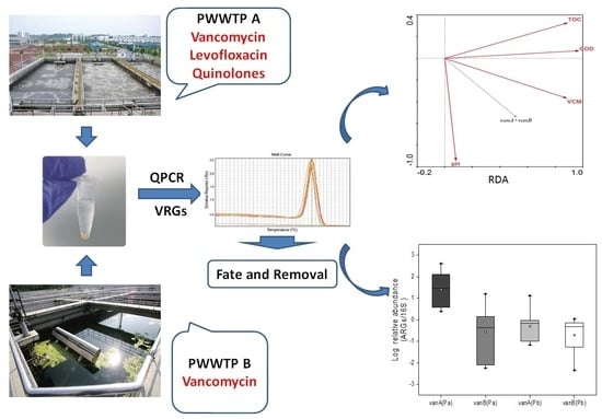
Fate and Proliferation of Vancomycin Resistance Genes in Two Typical Pharmaceutical Wastewater Treatment Plants

1
Nanjing Institute of Environmental Science, Ministry of Ecology and Environment, Nanjing 210042, China
2
Key Laboratory of Pesticide Environmental Assessment and Pollution Control, Ministry of Ecology and Environment, Nanjing 210042, China
*
Authors to whom correspondence should be addressed.
Water 2024, 16(1), 114; https://doi.org/10.3390/w16010114
Submission received: 19 November 2023 / Revised: 14 December 2023 / Accepted: 26 December 2023 / Published: 28 December 2023

Abstract

:
The emergence of vancomycin-resistant Enterococcus (VRE) and vancomycin resistance genes (VRGs) complicates the application of vancomycin, which is a last-line agent for human infectious diseases, and pharmaceutical wastewater treatment plants (PWWTPs) are widely thought to be important sources of corresponding antibiotic resistance genes (ARGs). In this study, two VRGs (vanA and vanB) were evaluated in two PWWTPs using quantitative polymerase chain reaction (qPCR) analysis to characterize the occurrence and fate of VRGs. The VRG concentration tended to decrease throughout all processing stages, while anaerobic treatment promoted the propagation of antibiotic-resistant bacteria and led to an increase in VRG abundance. Finally, the absolute concentrations of vanA and vanB exceeded 104 copies/mL in the effluents, and a significant amount of VRGs was transferred to sludge at 1.68 × 10¹⁷ copies/d. Redundancy analysis demonstrated that the relative abundance of ARGs was significantly correlated with the concentrations of vancomycin and COD. Furthermore, the relative abundance of vanA was increased in wastewater with multiple antibiotics, while the relative abundance of vanB only increased in the presence of vancomycin. This observation implied different intrinsic resistance mechanisms for different VRG subtypes. Overall, in this report, we describe the first comprehensive study on the fate and behavior of VRGs with different physicochemical or biochemical treatments and different antibiotic selection pressures in PWWTPs; this report provides important references for the environmental spread of VRGs.

Graphical Abstract

1. Introduction

Antibiotics are antibacterial drugs that can achieve antibacterial and bactericidal effects by interfering with the physiological processes or cell structures necessary for the growth of bacteria. Since the 1940s, penicillin has been widely utilized in clinical medicine, and antibiotics have played an irreplaceable role in alleviating illness and extending human life. However, with the misuse of antibiotics, the number of drug-resistant bacterial strains has increased significantly. The glycopeptide antibiotic vancomycin is the front-line treatment for infections with multidrug-resistant Gram-positive bacterial strains, such as methicillin-resistant Staphylococcus aureus (MRSA) and methicillin-resistant Staphylococcus epidermidis (MRSE) [1]; furthermore, this antibiotic is known as a last-resort agent against resistant strains of bacteria.
However, with the emergence of vancomycin-resistant Enterococcus (VRE), this last line of defense is facing collapse, and this is a major problem for the treatment of nosocomial infections caused by microorganisms [2,3]. The most common VRE species identified worldwide is Enterococcus faecium [4]. Approximately nine gene clusters confer glycopeptide resistance, including vanA, vanB, vanC, vanD, vanE, vanG, vanL, vanM, and vanN, which have been detected in enterococci [5,6]. Among these gene clusters, vanA and vanB are considered to be the most important as these genes are transferable. In particular, the vanA cluster has the ability to transfer from vancomycin-resistant enterococci to a variety of Gram-positive microorganisms, such as Staphylococcus aureus [7,8], by mobile genetic elements, such as Tn1546, and vanA shows high resistance to teicoplanin [9]. Resistant bacteria have the potential to spread rapidly across the entire planet, and there is evidence that antibiotic resistance genes (ARGs) can transfer to humans through water or food pollution [10] and, thereby, pose enormous challenges in clinical therapy.
Vancomycin is prohibited from being used in livestock and poultry farming in China [11] and is also infrequently used for treating humans owing to the many potential side-effects [12,13,14]. Therefore, vancomycin is rarely detected in the aquatic environment. However, wastewater from pharmaceutical factories often contains extremely high concentrations of antibiotics and relevant ARGs [15,16,17]. In general, antibiotic resistance bacteria (ARB) and ARGs cannot be completely eliminated in urban wastewater treatment plants [18] or in novel treatment units designed for the removal of antibiotics and ARGs [19,20,21]. Pharmaceutical wastewater treatment plants (PWWTPs) of vancomycin wastewaters could still become major sources for the transmission of vancomycin resistance genes (VRGs) and vancomycin-resistant bacteria into the natural environment. PWWTPs contaminated with VRGs and vancomycin-resistant bacteria represent an ideal model for investigating the fate and removal of VRGs. However, the prevalence and proliferation of VRGs in pharmaceutical wastewater have not been fully elucidated.
In this study, two subtypes of VRGs (vanA and vanB) that confer resistance to vancomycin were evaluated in two typical PWWTPs in Eastern China. The occurrence and fate of vanA and vanB resistance genes were investigated in PWWTPs that employed a variety of biotreatment operations. The correlations between VRGs, antibiotics, and other environmental factors were assessed to explore the mechanism underlying the proliferation of certain ARG subtypes under different selective pressures exerted by antibiotics.

2. Materials and Methods

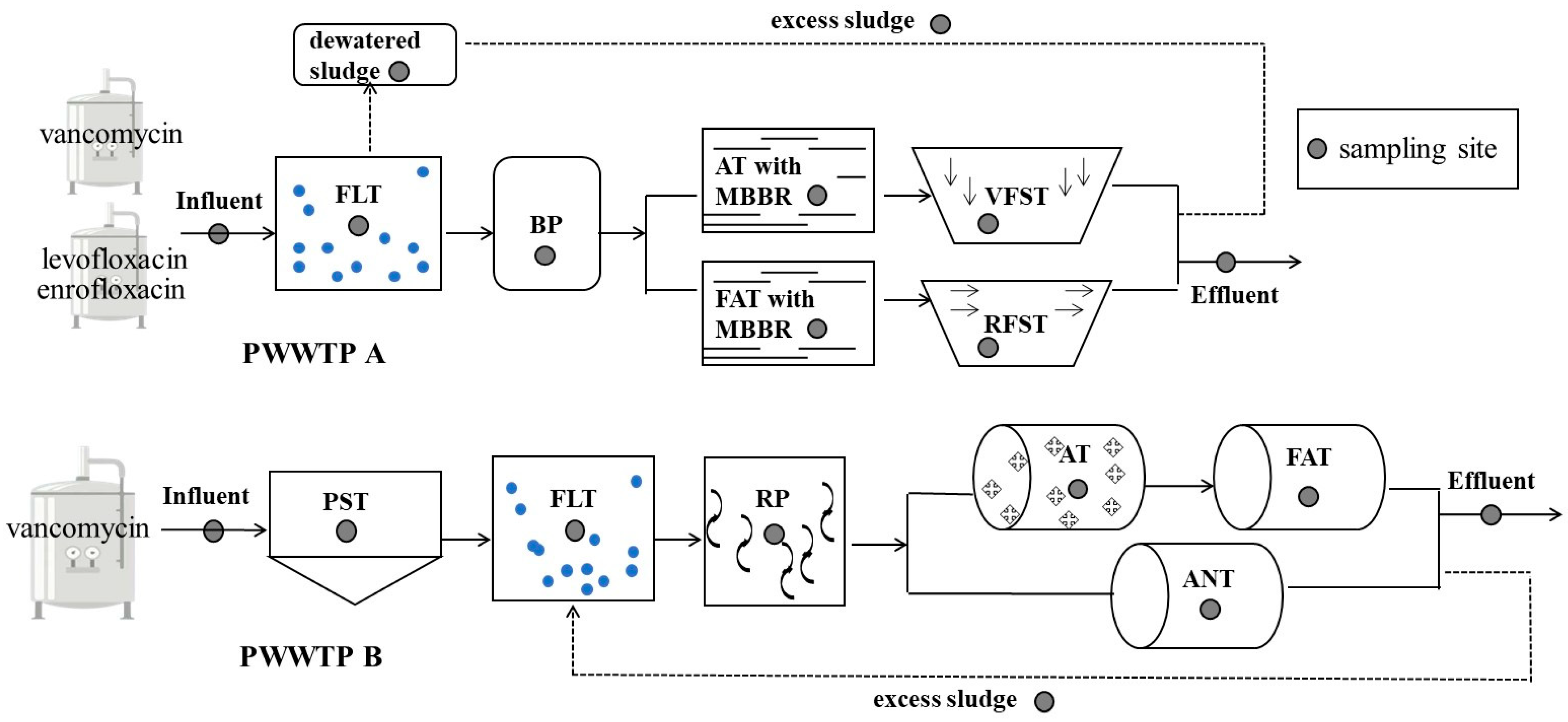
2.1. Characterization of PWWTPs and Sample Collection

Two PWWTPs utilizing various treatment processes in Eastern China were researched in this study. The wastewater from the PWWTPs was primarily contaminated with vancomycin. The main processes and operations employed in PWWTP A consist of flotation, a buffer pool, a facultative tank and vertical flow sedimentation tank, an aerobic tank, and a radial flow sedimentation tank (Figure 1 PWWTP A); in this plant, the capacity for wastewater treatment is up to 1200 m3 per day, the capacity for treating biochemical sludge (defined as having a moisture content of approximately 60%) is approximately 30 ton/d, and the capacity for treating dewatered sludge is 12 ton/d. PWWTP B utilized a modified A2/O process consisting of flotation pre-treatment and facultative, aerobic, and anaerobic tanks (Figure 1 PWWTP B). The daily wastewater treatment capacity was up to 2880 m3, and excess sludge was approximately 80–90 ton/d. The operational parameters of the PWWTPs are shown in Table 1.
Water and sludge samples were collected from different processes and stored in ice bottles at 4 °C, and detailed information on the sampling sites is presented in Figure 1. To avoid confounding effects associated with hydraulic loading fluctuations, raw wastewater and process effluents were taken as 24 h flow-proportional composite samples. All water samples were collected in 10 L amber glass bottles. The sludge samples were generally taken at the outlet of every treatment step. Each sample was placed into a plastic container and immediately chilled in an icebox and transported under cool conditions to the laboratory and then stored in the dark at −20 °C until DNA was extracted (within 3 days).

2.2. Sample Preparation and DNA Extraction

Sludge samples were centrifuged at 4000× g for 10 min at room temperature, and 0.5 g of the pellet was used for DNA extraction. Excess sludge samples were divided into two subsamples: one was subjected to DNA extraction and the other was used to determine the moisture content for further ARG quantification. DNA was obtained using the PowerSoil DNA Isolation Kit (MoBio Laboratories, Carlsbad, CA, USA) following the manufacturer’s protocol. Vacuum filtration apparatus was utilized to concentrate water samples with 0.45 μm filter papers to separate undissolved substances first, and 0.22 μm polyethersulfone ultrafiltration membranes were subsequently employed to intercept the bacteria until the filter clogged. All filter papers and membranes were collected together and stored at −80 °C before DNA extraction was undertaken. The total volumes of each water sample were recorded for the concentration calculations described below. A Water DNA Kit (OMEGA, New York, NY, USA) was utilized to extract total DNA from the filter papers and polyethersulfone ultrafiltration membranes prepared previously according to the manufacturer’s protocol. The concentration and quality of the extracted total DNA were determined through analysis with a spectrophotometer (Nanodrop 1000, Thermo, Waltham, MA, USA) and through 1.5% agarose gel electrophoresis, respectively.

2.3. Construction of qPCR Calibration Curve

Two vancomycin resistance genes (vanA and vanB) were analyzed in this study. The complete sequences acquired from NCBI were employed to design primers by online primer design tools available on the website http://www.ncbi.nlm.nih.gov/tools/primer-blast/index.cgi (accessed on 13 May 2021). Detailed information regarding the DNA sequence accession number and primers is listed in Table S1. To detect the presence of targeted ARGs, the polymerase chain reaction (PCR) method was utilized. All PCR analyses were performed in 25 μL reaction mixtures, including 2× Taq Master Mix (CW Biotech Co., Ltd., Beijing, China), 1 μL individual primers (0.4 μM), 1 μL template DNA (100 ng), and 9.5 μL RNase-Free water. The amplification of the ARGs was undertaken using a thermal cycle (Bio-Rad, Hercules, CA, USA), beginning with a denaturation step for 5 min at 94 °C followed by 35 amplification cycles for 30 s at 94 °C, annealing for 30 s at various temperatures (Table S1), and extension at 72 °C for 1 min followed by a final extension of 6 min at 72 °C.
The PCR products were analyzed via 1.5% agarose gel electrophoresis. Fresh PCR products from samples confirmed to contain target genes were purified using an Easy Pure Quick Gel Extraction Kit (TransGen Biotech, Beijing, China). After purification, PCR products were ligated into the pEasy-T3 vector (TransGen Biotech, China) and subsequently cloned into Trans1-T1 phage-resistant chemically competent cells (TransGen Biotech, China). Positive clones were screened by PCR to verify the cloning of target genes, and they were subsequently sequenced. The sequencing results were analyzed by the BLAST alignment tool (http://www.ncbi.nlm.nih.gov/blast/ accessed on 8 July 2021). Positive clones containing target genes were chosen as the calibration for real-time PCR. An AxyPrep Plasmid Miniprep kit (Axygen, Corning, NY, USA) was utilized to extract plasmids carrying target genes according to the manufacturer’s protocol.
The concentration and quality of plasmids were determined by analysis with a spectrophotometer (NanoDrop ND-1000c, Thermo) and agarose gel electrophoresis, respectively. The gene copies per microliter of plasmid were converted using the relationship described by Pei [22]. Tenfold serial plasmids with target genes ranging in concentration from 109 to 101 were tested in triplicate to generate calibration curves. The linear coefficients (R2) were higher than 0.990, and their efficiencies were determined to be between 90% and 105% for all calibration curves (Figure S1). According to the standard curves, the copy numbers of ARGs were calculated using the CT value of unknown samples.

2.4. Real-Time PCR

Real-time PCR technology was utilized to quantify the abundance of select genes extracted from all samples. To normalize the variance in the abundance of ARGs caused by differences in background bacterial abundance and DNA extraction efficiency, the 16S rRNA gene was also quantified. The 10 μL qPCR mixtures consisted of 5 μL of 2XU IstraSYBR Mixture (CW Biotech Co., Ltd., China), 0.2 μL of each primer at a concentration of 10 μM, 1 μL of DNA template, and 3.6 μL of sterile water. qPCR assays to amplify and quantify genes were performed in triplicate using a CFX96 TouchTM Real-time PCR Detection System (Bio Rad, Hercules, CA, USA). The qPCR protocol was as follows: 10 min at 95 °C for initial denaturation followed by 40 cycles of 10 s at 95 °C and 30 s at various annealing temperatures (Table S1) followed by a final extension step for 30 s at 72 °C. A melt curve stage was incorporated in every reaction to verify specificity, with the temperature ramping from 65 °C to 95 °C by 0.5 °C every 5 s.

2.5. Statistical Analysis

Data were analyzed using Origin Pro 9.1 (Origin Lab Corporation, Northampton, MA, USA). Correlations between the concentrations of ARGs, 16S rRNA, and wastewater-receiving capacity were analyzed. UNIANOVA (performed by specifying the removal value as the dependent variable and wastewater treatment system as a factor) was employed to analyze the results between each pair of different sampling sites with SPSS Version 17.5. p-values < 0.05 were considered to be significant.
The concentrations of ARGs and 16S rRNA in treatment units were expressed as the mean ± S.D. The concentrations of ARGs in activated sludge were expressed as the mean ± S.D. Redundancy analysis (RDA, software package CANOCO version 4.5) was employed to assess the correlations between ARG reductions and wastewater quality parameters (i.e., COD, TOC, pH, and vancomycin concentration; see Table S2). In the analysis of the difference in the relative abundances of vanA and vanB between the two PWWTPs, the significance boxplot with a t-test was plotted by using the OmicShare tools platform (https://www.omicshare.com/tools/) accessed on 7 December 2023.

3. Results and Discussion

3.1. Prevalence of ARGs in PWWTPs

The abundance of ARGs (vanA and vanB) in the water samples varied considerably with different treatment procedures, as shown in Figure 2. Target genes were all detected in the influent and each unit of the two PWWTPs, except for vanB in the RFST unit of PWWTP A. Furthermore, the 16S rRNA gene (a surrogate for total bacteria) was quantified in different treatment units to illustrate the propagation and fate of resistant bacteria in PWWTPs [23].
For PWWTP A, the absolute concentrations of vanA and vanB ranged from 1.11 × 10⁵ to 1.05 × 10¹⁰ and 0 to 3.65 × 10⁶ copies/mL, while the 16S rRNA abundance ranged from 4.47 × 10⁴ to 2.60 × 10⁷ copies/mL. For PWWTP B, the absolute concentrations of vanA and vanB ranged from 2.70 × 10⁴ to 1.27 × 10¹⁰ and 9.38 × 10³ to 4.60 × 10⁷ copies/mL, while the 16S rRNA abundance ranged from 3.15 × 10⁴ to 9.57 × 10⁸ copies/mL. The prevalence of VRGs significantly rose in these two PWWTPs when compared to the normal sewage treatment plant [17,19,24].
The concentrations of vanA were higher by almost 1–3 orders of magnitude than those of vanB in most samples, and the results demonstrated that vanA represented the dominant gene subtype conferring resistance to vancomycin. This result is consistent with the findings of previous studies, and vanA has been determined to be highly transferable to other important pathogens [25].

3.2. Fate of ARGs in Different Wastewater Treatment Processes

3.2.1. Effects of Different Treatments

In the whole treatment process flow, the variation in ARG and 16S rRNA gene concentrations among various treatment processes was determined to characterize the general trends through different treatment units (Figure 2). The abundance of target ARGs was largely decreased in the biological treatment, except in the ANT unit of PWWTP B. The ANT unit of PWWTP B was a distributary treatment stage for the excessive wastewater. The concentrations of ARGs and 16S rRNA genes in the latter process were frequently higher than those in the former process. This result is inconsistent with the findings of previous studies, in which the concentrations of ARGs were significantly increased in biological treatments [26,27]. Figure 2 shows that the concentrations of ARGs and 16S rRNA genes decreased by approximately 1–2 orders of magnitude in FAT and AT units compared to the former unit in PWWTP A, while a notable decrease was not observed in FAT of PWWTP B. The FAT and AT units were treated with membrane bioreactors (MBBRs) in PWWTP A. Concentrations of VRGs and 16S rRNA genes in the MBBR effluent were determined to be reduced by 1–3 orders of magnitude compared to conventional process utilities; notably, previous research [28] determined that MBBRs were the most effective process for antibiotic-resistant bacteria reduction, indicating that the intracellular ARGs are retained by the membrane. These results indicate that absorption and interception on biofilms could have a positive impact on the removal efficiency of ARGs [29].
A study by Tao et al. [30] observed that in an aerobic tank, oxygen may reduce the overall amount of bacteria. Increasing the processing time under aerobic treatment may help decrease the discharge of antibiotic-insensitive bacteria into aquatic environments [31]. Conversely, anaerobic treatment might promote the propagation of antibiotic-resistant bacteria and increase the probability of releasing antibiotic-resistant bacteria [32]. The same trend was also observed in our study. In PWWTPs, the abundance of 16S rRNA in the aerobic treatment tank was lower, and this abundance increased in the anaerobic units of PWWTP B.

3.2.2. Mass Balance Analysis

To characterize the proliferation of ARGs flowing into and out of each treatment unit of PWWTPs, both PWWTPs were subjected to mass balance analysis (measured in copies/d) by multiplying the volumetric flow rates by the corresponding gene concentration. The results demonstrated that the release of entire VRGs was 6.78 × 10¹⁵ copies/d in final effluents and was not detected in excess sludge for PWWTP A. Compared with raw influents at 1.13 × 10¹⁶ copies/d, a significant decrease was detected. This phenomenon indicates that a reduction may mostly occur in MBBR treatment systems, as described in Section 3.2.1. Guo et al. [27] also presumed the attenuation to be caused by the removal of total antibiotics. In PWWTP B, the emission of total ARGs was 1.05 × 10¹⁴ copies/d in final effluents and 1.68 × 10¹⁷ copies/d in excess sludge compared with 8.79 × 10¹⁴ copies/d for raw influents. The abundance of all ARGs in sludge was almost 3 orders of magnitude higher than that in effluents, as well as 16S rRNA. One explanation for this finding may be that the ARGs were transported to the sludge in the primary sedimentation tank and flotation tank, as in the study of Wang et al. [25], who reported that the abundance of ARGs decreased with physicochemical treatment. Notably, an effective sedimentation process might be important for reductions in extracellular DNA [33].

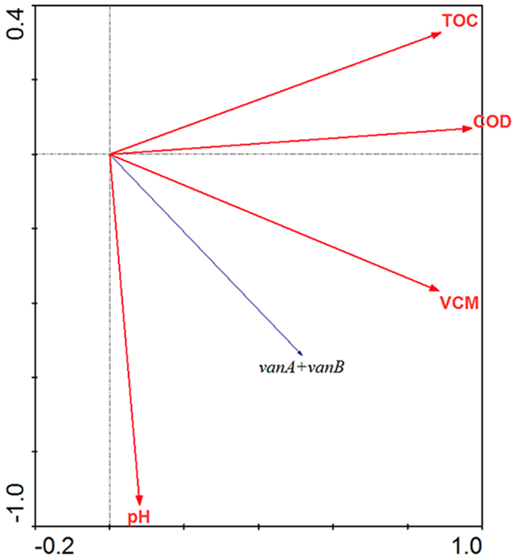
3.2.3. Redundancy Analysis

Because of the diversity of PWWTPs, many physical/chemical/biological water parameters may affect the proliferation and fate of ARGs in PWWTPs. Redundancy analysis (RDA) was performed to confirm the relationship between the relative abundance of ARG subtypes and various environmental factors (Figure 3). PH, COD, TOC, and vancomycin (VCM) were considered. The redundancy analysis indicated that the vancomycin concentration was positively related to the abundance of ARGs (vanA and vanB). In PWWTPs, high concentrations of antibiotic residues loaded in influents appeared to be unavoidable, and they might promote the propagation and persistence of relevant typical ARGs. The concentrations of antibiotics in each process in the two PWWTPs are shown in Table S2. Antibiotics were not able to inhibit the microbiological activities in a sewage treatment plant, since the minimum inhibitory concentrations (MICs) of some specific bacteria in wastewater were two- to tenfold higher than the antibiotic residues detected in the present study [25,34]. However, wastewater contained extremely high levels of antibiotics in PWWTPs, which were higher than MICs in many cases. For example, Qiu et al. [15] obtained the MIC of VCM for some typical sensitive bacteria from the European Committee on Antimicrobial Susceptibility Testing (EUCAST), when the highest, moderate, and lowest MICs were 23.76, 0.50, and 0.13 μg/mL. Takashi et al. [17] also found that two enterococcal strains were resistant to a high concentration of vancomycin (>128 μg/mL) via the MIC test. Furthermore, Zhang et al. [35] found the level of corresponding ARGs more often relies on the existence and concentration of antibiotics when chromosomal mutations occur in bacteria. Our previous study also observed that ARGs, which tend to be located in chromosomes, might be much more likely to show positive correlations with antibiotic concentrations [27]. A recent study observed that vancomycin resistance genes tended to be chromosomally encoded by metagenomic assembly and binning approaches [36], which might be attributed to the regulation of a two-component system consisting of a histidine kinase and a response regulator localized on a chromosomal operon [37,38]. Consistent with our findings, it was observed that the proliferation of vanA generally occurred in the early steps of the treatment processes. Similarly, the proliferation of vanB generally occurred in the biological units of the treatment processes. This observation indicated that ARGs normally increased during the initial stages of the treatment processes, which may be explained by the selective pressure of high vancomycin concentrations.
COD and TOC correlated with ARGs through redundancy analysis, which is in keeping with the findings of a report by [39], who observed that most ARB and ARG amplification phenomena were positively correlated with the COD of raw sewage. This finding may be attributed to COD, which changes the microbial community structure [18]. Most bacteria were expected to adhere to suspended solids or colloids in wastewater, causing reductions in ARB/ARGs to be positively related to COD to a certain extent.

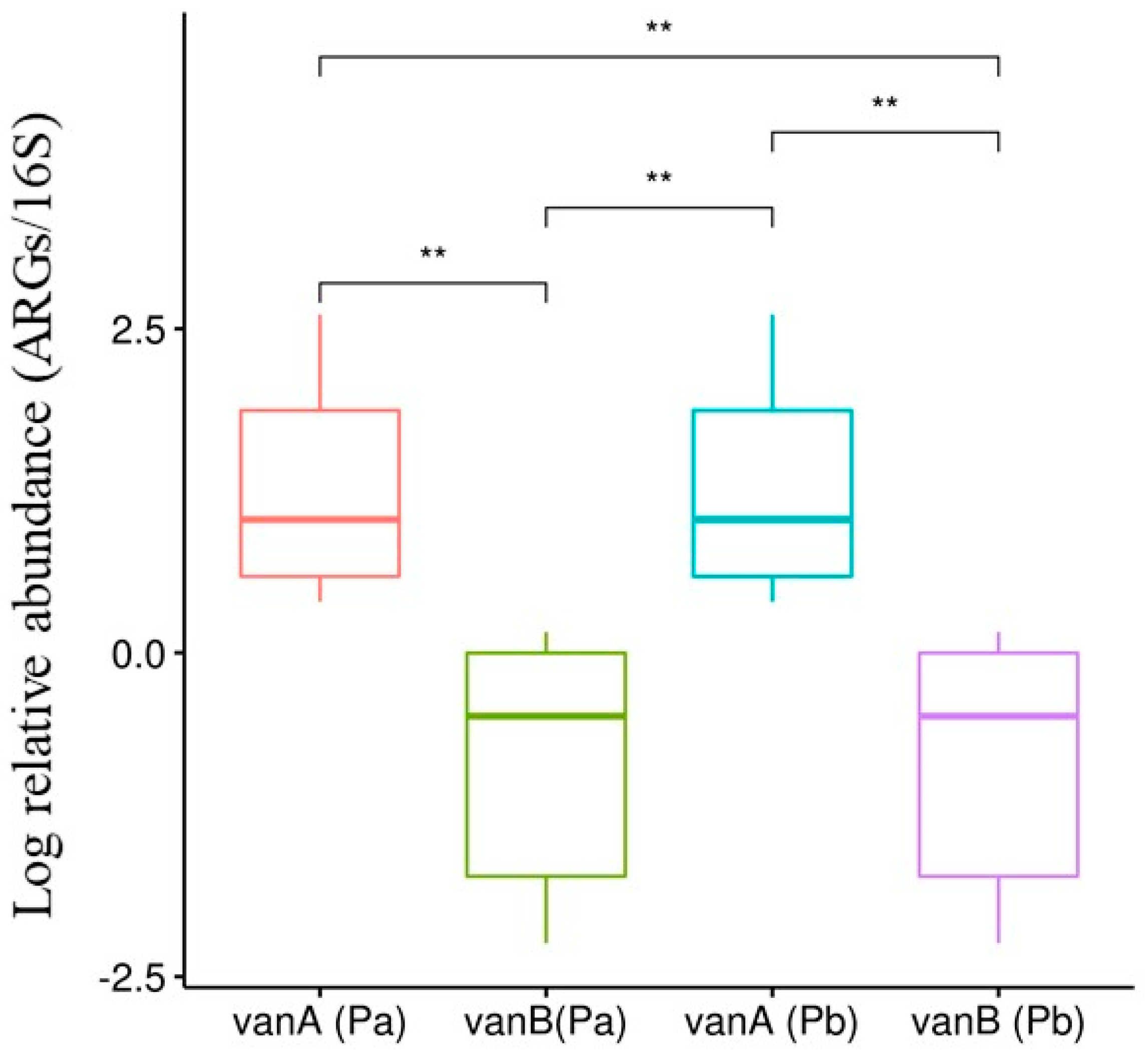
3.3. Selection and Proliferation of Different ARG Subtypes in PWWTPs

The subtypes (vanA and vanB) both acquired resistance to glycopeptide antibiotics. The relative abundances of vanA and vanB in the two PWWTPs are shown in Figure 4. The vancomycin concentrations in the process flow of the two PWWTPs did not differ significantly (Table S2). It was clearly demonstrated that the relative abundance of vanA in PWWTP A was significantly higher than that of vanA in PWWTP B, while the relative abundance of vanB was at the same level in both PWWTPs. Previous studies [25] indicated that the relative abundance of representative subtypes of ARGs in the effluents was significantly higher than that observed in plants free from antibiotics. In PWWTP A, wastewater containing three types of antibiotics were treated: vancomycin, levofloxacin, and enrofloxacin. In PWWTP B, wastewater only contained vancomycin. This observation indicated that the relative abundance of vanA significantly increased in wastewater with multiple antibiotics (p < 0.05), while there was no significant variation in vanB. Previous studies reported that vanA-carrying enterococci can be selected by vancomycin and teicoplanin, while vanB-carrying enterococci can only be selected by vancomycin [40], which may imply differences between the proliferation mechanisms underlying vanA and vanB. According to a study performed by Aydin et al. [32], a consequence of antibiotic combinations is greater than the sum of their independent activities. In summary, vanA could be induced to increase by multiple antibiotic combinations, while vanB was only selected by vancomycin. It was not surprising to observe that higher proliferation would occur in vanA under the selective pressure of multi-antibiotic combinations in our study. Furthermore, the risk and threat of vancomycin resistance genes may be expected to be more severe in vancomycin pharmaceutical factories that produce a variety of antibiotics.

4. Conclusions

In this study, using real-time PCR, the prevalence and fate of two ARGs (vanA and vanB) were evaluated in different wastewater samples undergoing the different treatment processes utilized by two PWWTPs. The two VRGs were widely detected in the PWWTPs, and their concentration tended to decrease throughout the processing stages, except for anaerobic treatment, which promoted the propagation of antibiotic-resistant bacteria. By performing a mass balance analysis, it was found that a significant number of ARGs were transferred to sludge. Thus, the dispersion of effluent and sludge represents an important means by which vancomycin resistance genes may be released into the environment. Through redundancy analysis, the relative abundance of VRGs was significantly correlated with the concentration of vancomycin and COD. ARGs, which tend to be located in chromosomes, are much more likely to show positive correlations with antibiotic concentrations. In addition, the relative abundance of vanA increased significantly in wastewater with multiple antibiotics, while the relative abundance of vanB increased due to the intrinsic resistance mechanisms related to the corresponding antibiotic.

Supplementary Materials

The following supporting information can be downloaded at: https://www.mdpi.com/article/10.3390/w16010114/s1, Table S1. Primers used in the PCR/qPCR assays. Table S2. The concentrations of vancomycin, pH, COD and TOC in different units. Table S3. Equations of standard curves. Table S4 Distribution of ARGs, 16S rRNA in the treatment process flow (copy/mL). Figure S1. The linear coefficients (R2) for calibration curve in real-time PCR.

Author Contributions

Investigation, X.Z.; Writing—original draft, X.G.; Writing—review & editing, M.S.; Visualization, N.N.; Project administration, N.W. All authors have read and agreed to the published version of the manuscript.

Funding

This research was funded by the Fundamental Research Funds for NIES [GYZX230105/GYZX230102] and the National Key Research and Development Program of China [2023YFF0611000].

Data Availability Statement

Data are contained within the article.

Conflicts of Interest

The authors declare no conflict of interest.

References

  1. Chavers, L.S.; Moser, S.A.; Funkhouser, E.; Benjamin, W.; Chavers, P.; Stamm, A.; Waites, K.; Nys, S.; Bruinsma, N.; Filius, P.; et al. Association between antecedent intravenous antimicrobial exposure and isolation of vancomycin-resistant enterococci. Microb. Drug Resist. 2003, 9 (Suppl. S1), 69–77. [Google Scholar] [CrossRef] [PubMed]
  2. Shankar, N.; Baghdayan, A.S.; Gilmore, M.S. Modulation of virulence within a pathogenicity island in vancomycin-resistant Enterococcus faecalis. Nature 2002, 417, 746–750. [Google Scholar] [CrossRef] [PubMed]
  3. Sahlström, L.; Rehbinder, V.; Albihn, A.; Aspan, A.; Bengtsson, B. Vancomycin resistant enterococci (VRE) in Swedish sewage sludge. Acta Vet. Scand. 2009, 51, 1–9. [Google Scholar] [CrossRef] [PubMed]
  4. Werner, G.; Coque, T.M.; Hammerum, A.M.; Hope, R.; Hryniewicz, W.; Johnson, A.; Klare, I.; Kristinsson, K.G.; Leclercq, R.; Lester, C.H.; et al. Emergence and spread of vancomycin resistance among enterococci in Europe. Eurosurveillance 2008, 13, 19046. [Google Scholar] [CrossRef] [PubMed]
  5. Werner, G. Current trends of emergence and spread of vancomycin-resistant enterococci. In Antibiotic Resistant Bacteria-A Continuous Challenge in the New Millennium; IntechOpen: Rijeka, Croatia, 2012; pp. 303–354. [Google Scholar]
  6. Oravcova, V.; Hadelova, D.; Literak, I. Vancomycin-resistant Enterococcus faecium with vanA gene isolated for the first time from wildlife in Slovakia. Vet. Microbiol. 2016, 194, 43–47. [Google Scholar] [CrossRef] [PubMed]
  7. Hiramatsu, K.; Kayayama, Y.; Matsuo, M.; Aiba, Y.; Saito, M.; Hishinuma, T.; Iwamoto, A. Vancomycin-intermediate resistance in Staphylococcus aureus. J. Glob. Antimicrob. Resist. 2014, 2, 213–224. [Google Scholar] [CrossRef] [PubMed]
  8. Hasan, M.A.; Khan, M.A.; Sharmin, T.; Mazumder, M.H.H.; Chowdhury, A.S. Identification of putative drug targets in Vancomycin-resistant Staphylococcus aureus (VRSA) using computer aided protein data analysis. Gene 2016, 575, 132–143. [Google Scholar] [CrossRef] [PubMed]
  9. Arthur, M.; Courvalin, P. Genetics and mechanisms of glycopeptide resistance in enterococci. Antimicrob. Agents Chemother. 1993, 37, 1563–1571. [Google Scholar] [CrossRef]
  10. Wilcks, A.; van Hoek, A.H.A.M.; Joosten, R.G.; Jacobsen, B.B.; Aarts, H.J. Persistence of DNA studied in different ex vivo and in vivo rat models simulating the human gut situation. Food Chem. Toxicol. 2004, 42, 493–502. [Google Scholar] [CrossRef]
  11. Ministry of Agriculture and Rural Affairs of the People’s Republic of China, 2020. Announcement No. 560. Available online: http://www.moa.gov.cn/gk/tzgg_1/gg/202001/t20200106_6334375.htm (accessed on 15 January 2023). (In Chinese)
  12. Kemeç, Z.; Kahya, Y.; Atay, A.; Demir, M.; Gürel, A. Vancomycin dependent pancytopenia-a rare side effect: A case report. Int. J. Med. Rev. Case Rep. 2019, 3, 339. [Google Scholar] [CrossRef]
  13. Selvaraj, P.K.; Khasawneh, F.A. Linear IgA bullous dermatosis: A rare side effect of vancomycin. Ann. Saudi Med. 2013, 33, 397–399. [Google Scholar] [CrossRef] [PubMed]
  14. Aydogan, E.; Egilmez, A.I. Eosinophilia; side effect of vancomycin which is not often detected: Case report. Medicine 2018, 7, 453–455. [Google Scholar] [CrossRef]
  15. Qiu, P.; Guo, X.; Zhang, Y.; Chen, X.; Wang, N. Occurrence, fate, and risk assessment of vancomycin in two typical pharmaceutical wastewater treatment plants in Eastern China. Environ. Sci. Pollut. Res. 2016, 23, 16513–16523. [Google Scholar] [CrossRef] [PubMed]
  16. Pal, C.; Bengtsson-Palme, J.; Kristiansson, E.; Larsson, D.G.J. Co-occurrence of resistance genes to antibiotics, biocides and metals reveals novel insights into their co-selection potential. BMC Genom. 2015, 16, 964. [Google Scholar] [CrossRef]
  17. Takashi, F.; Reina, H.; Tohru, M. Quantification of vancomycin-resistant enterococci and corresponding resistance genes in a sewage treatment plant. J. Environ. Sci. Health. Part A Toxic/Hazard. Subst. Environ. Eng. 2015, 50, 989–995. [Google Scholar]
  18. Novo, A.; André, S.; Viana, P.; Nunes, O.C.; Manaia, C.M. Antibiotic resistance, antimicrobial residues and bacterial community composition in urban wastewater. Water Res. 2013, 47, 1875–1887. [Google Scholar] [CrossRef] [PubMed]
  19. Shi, Z.J.; Zhao, R.X.; Wan, J.J.; Li, B.; Shen, Y.; Zhang, S.; Luo, G. Metagenomic analysis reveals the fate of antibiotic resistance genes in two-stage and one-stage anaerobic digestion of waste activated sludge. J. Hazard. Mater. 2021, 406, 124595. [Google Scholar] [CrossRef]
  20. Hou, J.; Chen, Z.Y.; Gao, J.; Xie, Y.; Li, L.; Qin, S.; Wang, Q.; Mao, D.; Luo, Y. Simultaneous removal of antibiotics and antibiotic resistance genes from pharmaceutical wastewater using the combinations of up-flow anaerobic sludge bed, anoxic-oxic tank, and advanced oxidation technologies. Water Res. 2019, 159, 511–520. [Google Scholar] [CrossRef]
  21. Li, Z.Y.; Dai, R.B.; Yang, B.C.; Chen, M.; Wang, X.; Wang, Z. An electrochemical membrane biofilm reactor for removing sulfonamides from wastewater and suppressing antibiotic resistance development: Performance and mechanisms. J. Hazard. Mater. 2021, 404, 124198. [Google Scholar] [CrossRef]
  22. Pei, R.; Kim, S.C.; Carlson, K.H.; Pruden, A. Effect of river landscape on the sediment concentrations of antibiotics and corresponding antibiotic resistance genes (ARG). Water Res. 2006, 40, 2427–2435. [Google Scholar] [CrossRef]
  23. Mao, D.; Yu, S.; Rysz, M.; Luo, Y.; Yang, F.; Li, F.; Hou, J.; Mu, Q.; Alvarez, P.J.J. Prevalence and proliferation of antibiotic resistance genes in two municipal wastewater treatment plants. Water Res. 2015, 85, 458–466. [Google Scholar] [CrossRef] [PubMed]
  24. Yang, Y.; Li, B.; Zou, S.; Fang, H.H.; Zhang, T. Fate of antibiotic resistance genes in sewage treatment plant revealed by metagenomic approach. Water Res. 2014, 62, 97–106. [Google Scholar] [CrossRef] [PubMed]
  25. Freitas, A.R.; Novais, C.; Tedim, A.P.; Francia, M.V.; Baquero, F.; Peixe, L.; Coque, T.M. Microevolutionary events involving narrow host plasmids influences local fixation of vancomycin-resistance in Enterococcus populations. PLoS ONE 2013, 8, e60589. [Google Scholar] [CrossRef] [PubMed]
  26. Wang, J.; Mao, D.; Mu, Q.; Luo, Y. Fate and proliferation of typical antibiotic resistance genes in five full-scale pharmaceutical wastewater treatment plants. Sci. Total Environ. 2015, 526, 366–373. [Google Scholar] [CrossRef] [PubMed]
  27. Guo, X.Y.; Yan, Z.; Zhang, Y.; Xu, W.; Kong, D.; Shan, Z.; Wang, N. Behavior of antibiotic resistance genes under extremely high-level antibiotic selection pressures in pharmaceutical wastewater treatment plants. Sci. Total Environ. 2018, 612, 119–128. [Google Scholar] [CrossRef] [PubMed]
  28. Munir, M.; Wong, K.; Xagoraraki, I. Release of antibiotic resistant bacteria and genes in the effluent and biosolids of five wastewater utilities in Michigan. Water Res. 2011, 45, 681–693. [Google Scholar] [CrossRef]
  29. Breazeal, M.V.R.; Novak, J.T.; Vikesland, P.J.; Pruden, A. Effect of wastewater colloids on membrane removal of antibiotic resistance genes. Water Res. 2013, 47, 130–140. [Google Scholar] [CrossRef]
  30. Tao, C.W.; Hsu, B.M.; Ji, W.T.; Hsu, T.-K.; Kao, P.-M.; Hsu, C.-P.; Shen, S.-M.; Shen, T.-Y.; Wan, T.-J.; Huang, Y.-L. Evaluation of five antibiotic resistance genes in wastewater treatment systems of swine farms by real-time PCR. Sci. Total Environ. 2014, 496, 116–121. [Google Scholar] [CrossRef]
  31. Lin, H.; Li, H.; Chen, L.; Li, L.; Yin, L.; Lee, H.; Yang, Z. Mass loading and emission of thirty-seven pharmaceuticals in a typical municipal wastewater treatment plant in Hunan Province, Southern China. Ecotoxicol. Environ. Saf. 2018, 147, 530–536. [Google Scholar] [CrossRef]
  32. Aydin, S.; Ince, B.; Ince, O. Development of antibiotic resistance genes in microbial communities during long-term operation of anaerobic reactors in the treatment of pharmaceutical wastewater. Water Res. 2015, 83, 337–344. [Google Scholar] [CrossRef]
  33. Börjesson, S.; Mattsson, A.; Lindgren, P.E. Genes encoding tetracycline resistance in a full-scale municipal wastewater treatment plant investigated during one year. J. Water Health 2010, 8, 247–256. [Google Scholar] [CrossRef] [PubMed]
  34. Chisholm, S.A.; Mouton, J.W.; Lewis, D.A.; Nichols, T.; Ison, C.A.; Livermore, D.M. Cephalosporin MIC creep among gonococci: Time for a pharmacodynamic rethink? J. Antimicrob. Chemother. 2010, 65, 2141–2148. [Google Scholar] [CrossRef] [PubMed]
  35. Zhang, Q.C.; Guillaume, L.; David, L.; Kim, H.; Robin, K.; Tung, C.K.; Pourmand, N.; Austin, R.H. Acceleration of emergence of bacterial antibiotic resistance in connected microenvironment. Science 2011, 333, 1764–1767. [Google Scholar] [CrossRef] [PubMed]
  36. Zhao, R.X.; Yu, K.; Zhang, J.Y.; Zhang, G.; Huang, J.; Ma, L.; Deng, C.; Li, X.; Li, B. Deciphering the mobility and bacterial hosts of antibiotic resistance genes under antibiotic selection pressure by metagenomic assembly and binning approaches. Water Res. 2020, 186, 116318. [Google Scholar] [CrossRef] [PubMed]
  37. Koteva, K.; Hong, H.J.; Wang, X.D.; Nazi, I.; Hughes, D.; Naldrett, M.J.; Buttner, M.J.; Wright, G.D. A vancomycin photoprobe identifies the histidine kinase VanSsc as a vancomycin receptor. Nat. Chem. Biol. 2010, 6, 327–329. [Google Scholar] [CrossRef]
  38. Charlebois, A.; Jalbert, L.A.; Harel, J.; Masson, L.; Archambault, M. Characterization of genes encoding for acquired bacitracin resistance in Clostridium perfringens. PLoS ONE 2012, 7, e44449. [Google Scholar] [CrossRef]
  39. Yuan, Q.B.; Guo, M.T.; Yang, J. Monitoring and assessing the impact of wastewater treatment on release of both antibiotic-resistant bacteria and their typical genes in a Chinese municipal wastewater treatment plant. Environ. Sci. Process. Impacts 2014, 16, 1930–1937. [Google Scholar] [CrossRef]
  40. Klare, I.; Konstabel, C.; Mueller-Bertling, S.; Werner, G.; Strommenger, B.; Kettlitz, C.; Borgmann, S.; Schulte, B.; Jonas, D.; Serr, A.; et al. Spread of ampicillin/vancomycin-resistant Enterococcus faecium of the epidemic-virulent clonal complex-17 carrying the genes esp and hyl in German hospitals. Eur. J. Clin. Microbiol. Infect. Dis. 2005, 24, 815–825. [Google Scholar] [CrossRef]
Figure 1. The flow chart layouts of the treatment processes in the two pharmaceutical wastewater treatment plants and the sampling site location. The black triangle represents the specific sampling location and the dotted line represents the recycled sludge. PWWTP A: Influent; FLT: flotation tank; BP: buffer pool; FAT: facultative tank; VFST: vertical flow sedimentation tank; AT: aerobic tank; RFST: radial flow sedimentation tank; Effluent. PWWTP B: Influent; PST: primary sedimentation tank; FLT: Flotation tank; RP: regulation pool; FAT: facultative tank; AT: aerobic tank; ANT: anaerobic tank; Effluent.
Water 16 00114 g001
Figure 2. Distribution of ARGs and 16S rRNA in the treatment process flow. The correspondence between sampling sites and sample name are as shown in Figure 1.
Water 16 00114 g002
Figure 3. Redundancy analysis (RDA) of the ARGs, antibiotics and environmental factors. The angles between the arrows indicate the sign of the correlation between the ARGs and the antibiotics and environmental factors.
Water 16 00114 g003
Figure 4. Log relative abundance of ARGs subtypes in different PWWTPs. Pa: PWWTP A; Pb: PWWTP B; **: p < 0.01.
Water 16 00114 g004
Table 1. Characteristics of pharmaceutical wastewater treatment plant.
PWWTPsLatitude and
Longitude
Influent (m3/d)Effluent (m3/d)Sludge (ton/d)Influent/Effluent COD
(mg/L)
Influent/Effluent NH3-N (mg/L)Influent/Effluent pHAntibiotics Contained
A29°30′12″ N,
120°54′42″ E
120012004211,000/289300/39.49/7.50vancomycin,
levofloxacin,
enrofloxacin
B28°40′33″ N,
121°27′56″ E
288028809015,000/500200/106.35/8.16vancomycin
Disclaimer/Publisher’s Note: The statements, opinions and data contained in all publications are solely those of the individual author(s) and contributor(s) and not of MDPI and/or the editor(s). MDPI and/or the editor(s) disclaim responsibility for any injury to people or property resulting from any ideas, methods, instructions or products referred to in the content.

