Review

Algae in Recreational Waters: An Overview within a One Health Perspective

by
Federica Valeriani
1,
Federica Carraturo
2,
Giusy Lofrano
1,
Veronica Volpini
1,
Michela Giovanna Izzo
3,
Agnese Bruno
1,
Marco Guida
2 and
Vincenzo Romano Spica
1,*
1
Department of Movement, Human and Health Sciences, University of Rome Foro Italico, 00135 Rome, Italy
2
Department of Biology, University of Naples Federico II, 80126 Naples, Italy
3
GeneS, Research Start Up, 00187 Rome, Italy
*
Author to whom correspondence should be addressed.
Water 2024, 16(7), 946; https://doi.org/10.3390/w16070946
Submission received: 23 December 2023 / Revised: 21 March 2024 / Accepted: 22 March 2024 / Published: 25 March 2024
(This article belongs to the Section Biodiversity and Functionality of Aquatic Ecosystems)

Abstract

:
Recreational water activities are widely recognized to have a positive impact on our physical and mental well-being. However, recreational water sources and their management are also a risk factor for human health due to different agents, including the overgrowth of cyanobacteria and algae. The presence of cyanobacteria and algae in recreational waters represents a One Health threat because of their potential release and the overuse of biocides. These organisms have the potential to metabolize organic matter and produce thermophilic and thermotolerant toxins. Moreover, different species of algae are involved in biofilm formation processes, thus impacting water quality and safety and also posing risks to the environment and animal and human health. Different species of algae participate in biofilm formation and have an impact on managing water and equipment maintenance. By searching literature databases, e.g., PubMed, we reviewed the state of the art, providing basic definitions, taxonomy, and epidemiological or medical issues related to the recreational uses of water. Methods of treatments and monitoring were summarized, considering both traditional and innovative strategies. Public health and surveillance approaches focus on the detection of toxins, the formation of biofilms, and the understanding of the benthonic and planktonic components as part of the larger microbial biodiversity. The review process allowed us to acknowledge that this is the first comprehensive overview of algae in recreational waters carried out within a wider One Health outlook.

1. Introduction

Water is a fundamental element for human health and well-being [1]. Recreational water activities are widely known to positively affect our physical and mental health and fitness. These activities, such as swimming in oceans, lakes, rivers, pools, or spas, are popular leisure activities for several communities and major attractions for tourists and sporting events. However, freshwaters and coastal water bodies are experiencing rising pollution because of the escalating pressure of human activities, as well as the impact of climate change. Contributing factors include untreated sewage overflows, animal excretion run-off from nearby farms, and algal blooms from high nutrient loads [2]. Water source safety represents one of the main factors influencing human health, as well as impacting ecosystems. Ensuring the proper surveillance and management of waters intended for recreational uses implies a substantial economic impact in the sectors of tourism, wellness, and sports events, and a positive cascade across the entire water protection chain, by diffusing awareness of the resource and respect of policies. A crucial and yet neglected aspect in recreational waters is linked to algae and cyanobacteria (also known as Cyanobacteriota or Cyanophyta), whose pathogenic species are potentially capable of metabolizing organic matter and producing thermophilic and thermotolerant toxins, often biologically active neurotoxins, posing severe health risks for humans, animals, and environments [3]. Among the several types of aquatic habitats, recreational waters and thermal mineral basins represent a favorable reservoir for phytoplankton growth. The uncontrolled proliferation of algal species on the surface layers of spa pools leads to the generation of unpleasant coloring, further affecting the appearance and smell of the water in the pool and also impacting pipelines and treatment plants [4]. Besides being ubiquitous in water environments, low algae concentrations do not generally represent health risks for human health. The main concerns in pools are related to the visual impact, unpleasant odor dissuading bathers from accessing thermal or rehabilitative treatments, and activities in swimming pools or water parks [5,6]. In addition, the macroscopic presence of algae is often only the visible part of a more complex microflora component including different bacteria and protozoa species, which may represent a risk to health by contact, inhalation, or drinking [1]. Therefore, this field is part of a huge world of studies on microalgae, represented by over 36,000 publications mainly published in the last decade, of which almost 160 are related to recreational waters and are available in PubMed databases.
The aim of this paper is to perform a comprehensive overview of algae management in recreational water, using a One Health approach. The text initially provides basic information on algae, including their definition, classification, and epidemiological data. This is to ensure accessibility to readers who may not be experts in the field. Thus, this paper aims to provide technical guidance related to the treatment and surveillance of algae in recreational waters.

2. Definition and Classification of Algae and Cyanobacteria

Algae are aquatic organisms devoid of true roots, stems, and leaves, which do not have distinct multicellular structures. Algae are ancient photosynthetic organisms that shaped the biosphere. Historically, algae were divided into two groups based on their size: macroalgae and microalgae. Brown algal seaweeds are macroscopic algae, while green, red, and golden-brown are considered microalgae [7].
Cyanobacteria, known as blue-green algae, have a prokaryotic organization, while all the other algal groups are eukaryotic [7]. The taxonomic diversity is also represented by metabolic factors, where the non-plant algal groups employ heterotrophy and phagotrophy, while the plant-related groups utilize photosynthesis.

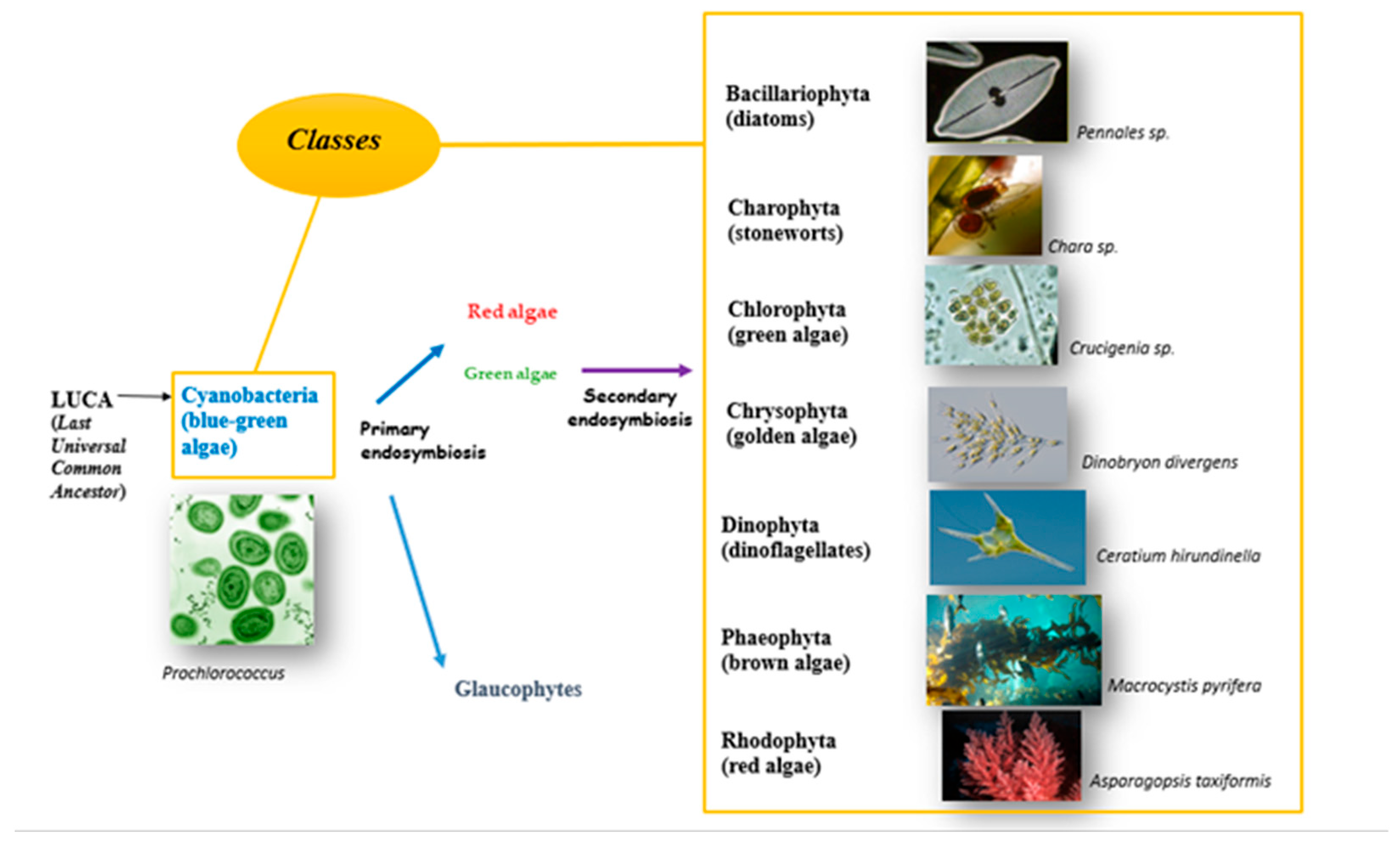
2.1. Evolution of Eukaryotic Photosynthetic Algae

An archaeal cell engulfed an alpha-proteobacterium, resulting in the first eukaryotic cell about 4 billion years ago (Figure 1), which is called the Last Universal Common Ancestor (LUCA) [8]. The First Eukaryotic Common Ancestor (FECA) appeared approximately 2.5 billion years ago, and all eukaryotic cells are believed to have originated from it [9]. The Last Eukaryotic Common Ancestor (LECA), the ancestor of the crown group of eukaryotes, emerged about 1.2 billion years ago [9]. For the crown group eukaryotes, a red algae fossil (Bioangiomorpha pubescens, currently indicated as Heteromorpha pubescens) is the first attribution dating back to 1 billion years ago [10]. Green algae first appeared in the fossil record with Perittonannus antiquus (formerly, Proterocladus antiquus) around 950 million years ago, indicating a divergence from red algae. This led to a significant increase in algal diversity, as supported by molecular data [8,11]. Oxygen photosynthesis originated in the prokaryotic cyanobacteria, and then it took place in eukaryotes at least 1 billion years ago by the uptake of the beta-cyanobacterium Gloeomargarita lithophora by a phagotrophic eukaryotic cell. Subsequently, the cyanobacterium evolved into the primary plastid as a photosynthetic organelle [12]. This endosymbiotic event marked the beginning of Archaeplastida, a monophyletic supergroup that includes the following three lineages: green algae plus land plants, red algae, and glaucophytes [13,14]. One other independent primary photosynthetic symbiosis is known, where the cyanobacterium belonging to the Synechococcus/Prochlorococcus group of alpha-cyanobacteria was engulfed by the cercozoan amoebae of the genus Paulinella and evolved into the chromatophore organelle [15]. Then, 120 million years ago, secondary and tertiary endosymbiosis took place, giving rise to complex plastid organization [7]. Green algal plastid endosymbiosis led to the establishment of euglenids and chlorarachniophytes. The event of the red algal plastid formed cryptophytes, haptophye, dinoflagellates, and stramenopiles (diatoms, brown algae).

2.2. Algal Taxonomy and Phylogeny

The phylogeny and taxonomy of algae are constantly under review due to the increase in groupings, particularly within the golden-brown group, where secondary endosymbiosis often occurs [7]. Algae classification follows two main approaches. The use of taxonomic groupings not sticking to monophyly but providing an overarching system was recently proposed [11]. In contrast, Adl et al. [14] use a hierarchical system based on monophyly. Chromista are included in the five-eukaryote kingdom (Protozoa, Chromista, Fungi, Plantae or Archaeplastida, Animalia). The kingdom comprises the golden-brown groups of algae (chlorophyll c-containing plastids of red algae origin) and most marine algae with heterotrophic protists, i.e., dinoflagellates [15]. Adl et al. [14] do not use the traditional taxonomical higher categories but recognize two overarching domains in eukaryotes including Amorpha and Diaphretickes. According to Adl et al. [14], Archaeplastida is a monophyletic clade under the Diaphoretickes, and they include Stramenopiles, Alveolata, and Rhizaria. Dinoflagellates are in the Alveolata group, the seaweeds are most closely associated with the Stramenopiles, while seaweeds, diatoms, and dinoflagellates are not related to the other traditional algal groups of Viridiplantae (formerly, Chloroplastida, red and green algae) [14]. Harvey is considered the algologist who proposed the first descriptive algal classification. Several other classifications have been proposed based on a variety of characteristics including morphological, physiological, and biochemical traits, and, more recently, molecular characteristics have also been considered [16,17,18,19,20]. Fritsch’s algal classification is widely accepted [20]. It is based on pigmentation, flagellar arrangement, reserve food material, the presence or absence of an organized nucleus in the cell, and the playback mode. He classified algae into eight phyla and 11 classes (Figure 1) [16,17,18,19,20]. A recent study demonstrated that based on genetic differences and not just morphological ones, it is possible to classify algae into 12 phyla [19].
Algae can range in size from a single cell to millions of cells, spanning seven orders of magnitude. The organisms comprise unicellular organisms (microalgae), such as Chlorella, diatoms, and Prototheca, and multicellular organisms (macroalgae), for example, green and brown seaweeds [21]. The macroalgae species Macrocystis pyrifera, also known as giant kelp, reaches 60 m in length, while microalgae are a smaller heterogeneous group, with organisms of sizes that range from 1 µm to 1 mm, such as Chlorella, which lives in freshwater or the soil and has spherical cells with a diameter ranging from 2 µm to 10 µm [17]. Microalgae (microphytes) are single-celled organisms that can convert solar energy into chemical energy through photosynthesis [16]. Macroalgae (seaweed) have a multicellular organization whit and developed anatomical arrangements that resemble stems, roots, and leaves of higher plants for functions such as anchorage, transport, photosynthesis, and reproduction. This specialization indicates a level of complexity and evolutionary advancement [16]. Algae depend on carbon, nitrogen, phosphorus, and micronutrients for metabolic processes [21]. Growth rate and cellular composition depend on several environmental factors such as light, temperature, pH, and salinity [22]. Sustainable high productivity requires synergistic interactions between multiple environmental variables and nutritional factors. Photosynthetic algae have a broad distribution on Earth, and they grow naturally in their environments, resulting in biomass production. Algae are ubiquitous organisms, and they can be categorized ecologically based on their habitats as follows: (i) planktonic algae grow suspended in water; (ii) neustonic algae grow on the water surface; (iii) cryophilic algae are found in snow and ice; (iv) thermophilic algae thrive in hot springs; (v) edaphic algae grow on or in soil; (vi) epizoic algae grow on animals such as turtles and sloths; (vii) epiphytic algae grow on fungi; (viii) corticolous algae grow on the bark of trees; (ix) epilithic algae grow on rocks; (x) endolithic algae grow in porous rocks or coral; and (xi) chasmolithic algae grow in rock fissures. Moreover, some algae live inside other organisms, and these are called endosymbionts. Endozoic endosymbionts live in protozoa and animals such as shelled gastropods, whereas endophytic endosymbionts live in fungi, plants, and other algae.

3. Cyanobacterial and Other Harmful Algae Blooms in Recreational Waters

In recreational waters, algal blooms can be harmful because some algal species and cyanobacteria contain secondary metabolites that are toxic to humans and animals [23,24]. Harmful algal blooms (HABs) result from the proliferation of diverse algal species, mainly including cyanobacteria (also known as blue-green algae), diatoms, dinoflagellates, and green algae. These harmful blooms can be caused by many types of phytoplankton. However, the three main types of phytoplankton that produce most of the blooms that harm people and animals include (i) cyanobacteria (sometimes called blue-green algae), (ii) dinoflagellates (sometimes called microalgae or red tide), and (iii) diatoms (sometimes called microalgae or red tide).
The two main toxin-producing algal groups are cyanobacteria (i.e., blue-green algae) and dinoflagellates; from an epidemiological point of view, algal blooms produced by cyanobacteria are usually isolated from freshwater, while dinoflagellate blooms are mostly detected in seawater [24,25,26,27,28]. Furthermore, many diatoms and dinoflagellates produce potent neurotoxins such as Beta-N-Methylamino-L-Alanine (BMAA), saxitoxins, and their various isomers [29]. About 30 of the estimated 2.000 dinoflagellate species on earth produce toxins that cause human illness, mostly contracted from consuming shellfish or fish that bioaccumulated neurotoxic amino acids [29,30]. The World Health Organization published the guidebook “Toxic cyanobacteria in water”, in 2021, which provides detailed information on the cyanotoxin-related phenomenon. Recreational exposure to cyanotoxins is possible through ingestion, aspiration, and inhalation. Human fatalities are known only from exposure to cyanotoxins via hemodialysis. Although a small number of severe health effects have been plausibly attributed to recreational water exposure, many of the health effects that have been associated with recreational exposure to cyanobacteria are mild and self-limiting, such as irritation of the skin, mucous membranes, and gastrointestinal tract; hay fever-like symptoms; nausea; and fever [23]. The main human health concern is ingestion or nasal uptake of the toxins that cyanobacterial scums may contain. Although no human deaths have been unequivocally attributed to recreational exposure, numerous deaths of livestock, pets, and wild animals have been caused by the consumption of water containing toxic cyanobacteria. This gives rise to concern regarding accidental ingestion of water containing cyanotoxins during recreational activities [23].

3.1. Algae and Cyanobacteria in Spa and Thermal Spring Water Sources

Thermal spring basins are globally exploited for recreational and medical purposes. These waters, as well as the muds, are rich in minerals and bio-active compounds resulting from the metabolic pathways of the autochthonous microbiota and microflora [4], the last of which play an essential role in the beneficial effects of these sources. Most health authorities worldwide and those in European Union countries have established that thermal water springs (i.e., spa water) should be left untreated; nonetheless, the decision to not employ disinfection treatments may pose health risks related to bathing in thermal mineral water pools [3] that do not undergo continuous water renewal. Most national and international regulations assume that the use of chemical disinfectants (e.g., sodium hypochlorite or hydrogen peroxide) on thermal basins is in contrast to their healing effects, considering the high sensitivity of microbiota and microflora composition to common environmental biocides [31]. Despite the substantial lack of studies focusing on the mode of action of different spa waters, the World Health Organization (WHO) recognizes therapeutical water, such as hydrothermal sources, among the oldest global healing means, as a traditional medicine strategy intended for therapeutic and prophylactic purposes towards several pathologies [32] in addition to being considered part of the cultural and medical traditions in most Central European countries [33]. Worldwide, health authorities [34,35,36] and, in particular, the Italian Ministry of Health, do not allow common drinking water disinfection treatments on thermal mineral waters. For example, Health Ministry Decree 25/2012, regulating treatment strategies for water intended for human consumption, specifies that thermal mineral water sources are excluded from any disinfection treatment [37]. Also at the regional level, the chemical disinfection of thermal water is forbidden, while physical technologies are encouraged [37,38,39].
Although water disinfection would sensibly reduce health risks, further ensuring the adequate health status of spa basins, the sanitation treatment may strongly affect non-pathogenic and autochthonous microbiota, leading to a strong reduction in the healing effect of the sources [3]. Moreover, while microbial diversity of spa water can control the survival and proliferation of pathogenic microbiota [40,41,42] by means of the production of bacteriocin-like active molecules, demonstrated to be able to reduce both Coliforms and Staphylococci [43,44], the potentially beneficial microorganisms may not be capable of contrasting biofilm formation in the pools’ walls and water distribution systems. Spa water microbiota may host virulent species of Legionella spp. and Mycobacterium spp., which are capable of surviving in amoebic organisms, thus exploiting the production of biofilms and literally protecting the pathogens from extremophilic conditions and hiding them from endogenous microbiota [45]. The most common family of algae occurring worldwide is Bacillariophyceae, which is dominant in both western and eastern Europe, according to the algological studies [4,46].
Kiliç et al. [4] conducted research on the Delicermik-Koprukoy hot spring water (Erzurum, northeast Turkey), a sodium–calcium–bicarbonate–carbon dioxide chemically classified source, and reported the abundant growth of algae belonging to Bacillariophyta (diatoms), Chlorophyta (green algae), Cyanobacteriota (blue-green algae), and Euglenida (euglenids) [4,47]. Similarly, Giorgio et al. [46] studied the microflora composition of spring water and mud samples from a thermal mineral basin in Naples, Italy, and reported the isolation of different green algae, cyanobacteria, and diatom taxa. The isolation of Cyanophyceae, Chlorophyceae, and Bacillariophyceae, overall non-pathogenic microorganisms, was confirmed by similar studies reporting the ability of the algal species to produce macromolecules with therapeutic properties [48,49,50,51]. The cyanobacteria Anabaena sp., Leptolyngbya sp., and Nostoc sp. produce compounds showing cytotoxic effects towards human cancer lines [51,52]. Strains of Nostoc sp. are capable of producing lipophilic extracts showing antibacterial activities and antiviral proteins [53], while Chlorella sp. algal extracts have antitumoral and anti-immunostimulatory activities [54]. A polysaccharidic compound isolated from strains of Coccomyxa sp. was found to be effective in contrasting influenza A virus infection [55]. Species of Nostoc sp. have also been identified as producers of microcystins, which are harmful toxins used as indicators in water intended for human consumption (according to the Italian regulation D. Lgs. 18/2023), while Anabaena sp. strains produce anatoxin-a and anatoxin-b [56,57].
Stoyneva [58] described Bulgarian algal flora in thermal springs, underlining the detection of over 200 species, consisting of cyanoprokaryotes, green, yellow-green, and red algae, diatoms, and Glaucophytes. In general, Chlorophyta, whose 75 taxa were isolated, were the most abundant [58]. The isolation of diatoms was furthermore highlighted by Lai et al. [59], who analyzed 65 diatom genera (including 196 species) and their assemblages in thermal springs connected to rivers in Regione Sardegna (Italy).
Ulcay et al. [60] realized a report aiming to identify algal species in Germencik (Alangüllü) thermal water springs (Turkey). Overall, 21 cyanobacteria (more tolerant to the majority of ecological extreme conditions, such as high-water temperature), five Bacillariophyceae, and one Zygnemophyceae taxa were isolated. The characterized species belonged to Aphanothece, Chroococcus, Pseudanabaena, Spirulina, Leptolyngbya, Heteroleibleinia, Phormidium, Oscillatoria, Navicula, Achnanthidium, Kamptonema, Rhopalodia, Amphora, Surirella, and Spirogyra genera. The most interesting species were Leptolyngbya subtilis, Leptolyngbya Thermobia, and Heteroleibleinia kossinskajae. Leptolyngbya species were also isolated from salty water springs in Hawaii, Sri Lanka, Australia, Japan, the USA, the Czech Republic [61], water sources in the Netherlands [62], and thermal water springs in Greece [63]. Monitoring the algal flora in thermal water springs located in Egypt allowed for the isolation of 209 species from eight algal phyla including Cyanobacteriota (91 species), Chlorophyta (59), Bacillariophyta (52), Chrysophyceae, Xanthophyceae, Euglenida, Dinophyceae, and Charophyta [64]. Gupta (2017) showed that most of the algal flora isolated from thermal water springs located in Jharkhand, India, belonged to the Cyanophyceae (25 strains), Bacillariophyceae (four), Chlorophyceae (two), and Euglenophyceae (two) families [65].
Several studies have reported the isolation of toxin-producing algae, such as Synechocystis spp. and Raphidiopsis raciborskii (previously known as Cylindrospermopsis raciborskii), from hot spring water in Europe. The possible chemical treatment of toxin-producing algae and cyanobacteria may lead to cell disruption and consequent release of harmful toxins into the water environment, thus increasing exposure to bathers [3,66,67].

3.2. Environmental Epidemiology and Ecotoxicity

The attention of worldwide Health Authorities is particularly focused on HAB-forming algae and cyanobacterial organisms that are not directly pathogenic to humans. HABs are indeed not able to multiply in the human body as common pathogenic microorganisms; rather, they produce secondary metabolites as toxins, showing elevated toxicity in both animals and humans [23]. The formation of algal blooms usually depends on the excess of molecules containing phosphorous and nitrogen in water environments. However, although this represents the main determinant for algal bloom formation, in some cases, it is restrictive for a proper evaluation of toxin risks. Indeed, certain cyanobacteria, such as Planktothrix rubescens, decrease in response to eutrophic conditions instead [68]. HABs in recreational water might usually derive from the excess of fertilizers applied in soils for agricultural purposes [69]. It is essential to consider that toxins constitute a portion of algal cells’ lifecycle: for example, in cyanobacteria, following cellular death, the lysis of the outer wall occurs, and cyanotoxins are generally released in water environments. Some species are also able to release toxins before cell lysis [70]. Cyanobacterial algal blooms are easily visible, producing a green-blue layer on water surfaces (i.e., planktonic cyanobacteria, typical of Microcystis sp., Dolichospermum sp., and Aphanizomenon sp. strains) and growing on the surfaces of rocks and sediments (i.e., benthic cyanobacteria), or they may be invisible, whereas algal species sink underneath the surface [69,71].
A percentage ranging between 25% and 75% of naturally occurring CyanoHABs are potentially toxic [24]. HABs in recreational water sources (pools, spas, rivers, lakes) are capable of directly impacting public health by swallowing water (algal toxicity is higher when ingestion route is involved), inhaling contaminated aerosols, and skin exposure (i.e., contact) during recreational activities (e.g., swimming) [23]. Beyond the capability of impacting aquatic ecosystems, the spectrum of clinical manifestation results is highly variable, ranging from mild skin irritation (i.e., dermatoxins, causing itching and rashes able to affect epidermis and dermis layers) to compromising neurological (i.e., neurotoxins) or gastrointestinal issues (since algal toxins are capable of migrating through the bloodstream to the liver (i.e., hepatoxins), brain, or nervous system), and reaching severe neurodegenerative diseases such as Alzheimer’s disease, long-term liver damage, or cancer [69].
Global warming and related climate change consistently contribute to the growth of cyanobacterial HABs across the world, affecting public health and aquatic biodiversity. In fact, a recent study showed that increased pathogenic Vibrio sp. occurs when algal toxins increase [72].

3.3. Ecotoxicity of Algal Blooms and Related Toxins

HAB-associated toxins are dangerous not only to humans but also to livestock and pet animals. Nevertheless, the few studies focused on the characterization of potential impacts in terms of ecotoxicity, cyanotoxins on terrestrial plants (or even, on other non-harmful algae), aquatic organisms, and wildlife, are not well characterized; consequently, the effects of sustained exposure to cyanotoxins on aquatic life are poorly understood [73]. Studies, reporting data on algal and cyanobacterial toxin ecotoxicity are mainly focused on the effects of five cyanotoxin classes (i.e., predominantly microcystins, followed by cylindrospermopsin, anatoxin-a, saxitoxins, and nodularin) on aquatic invertebrates, fishes, amphibians, and birds exposed to the toxins in freshwater habitats. The majority of the research has been conducted employing a fish or aquatic invertebrate model, assessing mortality, bioaccumulation, and biochemical responses as measurable endpoints. The evaluation of the available data indicates that the 8 μg/L microcystins threshold value for recreational water regulated by the U.S. Environmental Protection Agency (U.S. EPA) is protective for acute toxicity in aquatic organisms but not presumably against chronic toxicity [74]. Considering that in water ecosystems, small invertebrates such as water fleas (Ceriodaphnia spp. and Daphnia spp.) feed on the blue-green algae and that such invertebrates are themselves prey for other invertebrates and fish species, it is presumable that where small invertebrates are negatively affected by toxins produced by blue-green algae, the entire food chain has the potential to be affected.
Experiments conducted by Shamohamadloo et al. [75] on water fleas and mayfly nymphs (Hexagenia spp.) exposed to different levels of cyanobacterial toxins showed that exposure to toxic species did not affect mayfly nymphs, which may potentially have reduced the negative effects (possibly their larger size compared with water fleas provides better tolerability to toxins). On the contrary, some water flea species reached 100% mortality at low toxins doses [75]. Similarly, Smutná et al. [76] exposed Daphnia magna to varying cyanobacterial bloom samples (containing both cyanobacteria and cyanotoxins) in a series of acute (48 h) and chronic (21-day) toxicity assays. Overall, 75% of their samples showed high acute toxicity on a crustacean bioindicator, and most samples evidenced significant lethal effects (with 35.6 mg/L biomass LC50 values) in chronic toxicity tests. Smutná et al. [76] underlined how toxicity levels were independent of the microcystin contents in the samples, which allowed them to hypothesize that not only toxins but also other cyanobacterial components (e.g., lipopolysaccharides, peptides, and other unidentified metabolites) are responsible for the ecotoxicity of complex cyanobacterial blooms [76]. Indeed, previous studies evidenced how an increase in mesozooplankton was inversely correlated with the abundance of potentially toxic cyanobacteria (e.g., Microcystis sp., Anabaena sp., and Cylindrospermopsis sp.). In contrast, a microzooplankton community was not affected; rather, it was consistently present [77].
Palíková et al. [78] assessed the effects of different cyanobacterial biomasses (containing different concentrations of microcystins) on the embryolarval development of carp (Cyprinus carpio), demonstrating that regardless of microcystin content or type in samples, the analyzed biomasses were highly toxic. Samples dominated by Aphanizomenon sp. and Planktothrix sp. strains resulted in a consistent mortality rate, while specimens in which Microcystis spp. were isolated induced lower effects, opening the possibility that microcystins may not be the main factor responsible for HAB toxicity. This hypothesis is supported by a recent review of research focusing attention on the HAB threat to aquatic biota related to the potential bioaccumulation and toxicity in fish [79]; nonetheless, the bioaccumulation potential is lower compared with zooplankton and invertebrates [80]. Cyanotoxins were demonstrated to negatively impact the antioxidant system of fish species, beyond affecting the mitochondrial and endoplasmic reticulum (by means of an increase in intracellular reactive oxygen species) [81,82]. In addition, immunomodulatory, inflammatory, antimicrobial, and endocrine responses were detected in animals exposed to both microcystins and cylindrospermopsins [79,83,84,85,86]. Most interestingly, less common algal groups, such as the haptophyte Prymnesium parvum and the euglenoid Euglena sanguinea, are also capable of producing harmful algal blooms (HABs), whose toxins dangerously affect aquatic biota but do not show effects on human health. The species Prymnesium parvum was described as being responsible for some of the worst HAB-related ecological disasters occurring in inland waters [87]. Also, avian wildlife in both freshwater and marine ecosystems is heavily affected by HABs and related toxins. For example, monitoring conducted in Chesapeake Bay (USA) from 2000 to 2020 resulted in the identification of several mortality events presumably associated with toxic algae and HAB events [88].

3.4. Environmental Epidemiology of HAB, Algal, and Cyanobacterial Toxins

Risk assessment strategies for the protection of public health connected to HABs mainly consist of avoiding environmental concentrations exceeding hazardous levels for humans at points of exposure [71]. The European Union drinking-water legislation framework consists of the Drinking Water Directive, revised with the EU Directive 2184/2020 [36,89], which establishes the World Health Organization (WHO) threshold guideline value of 1 μg/L for the parameter “total microcystin-LR (free plus cell-bound)” to be mandatorily evaluated if HAB presence in treated drinking water is considered hazardous [90]. Microcystin-LR (produced by cyanobacterial strains of Microcystis sp., Anabaena sp., Anabaenopsis sp., Aphanizomenon sp., Planktothrix sp., Oscillatoria sp., Phormidium sp.) was selected as a cyanobacterial and algal toxin indicator in water intended for human consumption since it represents the most widely distributed and dangerous toxin (classified by the WHO as a 2B carcinogen) [91,92,93]. However, considering that microcystin-LR may not be a reliable indicator for any other toxin, it is necessary to improve tailored strategies to mitigate toxin release at various levels (by avoiding favorable algal proliferation conditions, selecting the proper water treatment, etc.) [94]. It is indeed essential to augment HAB surveillance systems worldwide, especially integrating national and international reporting activities with information regarding the algal and cyanobacterial genera responsible for registered harmful algal bloom events.
For example, since 2016, the U.S. Center for Disease Control and Prevention (U.S. CDC) is supported by the One Health Harmful Algal Bloom System (OHHABS, until 2011, covered by the Harmful Algal Bloom-related Illness Surveillance System, i.e., HABISS) in the collection of information to assess environmental risk and prevent illnesses caused by blooms of harmful algae and cyanobacteria [95]. In the 2016–2018 three-year period, 18 U.S. states reported 421 harmful algal bloom events, of which 81% were classified as confirmed. The summer season gathered the highest percentage (98%) of reported events, with peaks registered in July at a 27% rate. Almost the totality of events was reported from freshwater reservoirs, whereas about 40% described the evidence of visible scum. The specific research on algal and cyanobacterial toxins was performed on 83% of algal bloom events, and overall, 94% of the samples highlighted the presence of microcystins, while the latter samples were contaminated with other types of toxins.
HAB monitoring continued after 2018, which was improved with U.S. Environmental Protection Agency risk-based guidance for the quantification of algal and cyanobacterial toxins, aimed at increasing the completeness and accuracy of public health surveillance. Also in 2019, among the 242 reported algal blooms, the majority of the events were predominantly evidenced between July and October. In 2019, environmental testing for algal toxins or species was performed for 88 reported HABs reported, and 53% were confirmed for the presence of toxins, of which 93% were identified as microcystins, followed by anatoxin-a, cylindrospermopsin, saxitoxin, nodularin, and hemolytic toxins. The analysis of the blooms’ composition at the molecular level allowed for the identification of Microcystis sp., Pseudo-nitzschia sp., Anabaena sp., Aphanizomenon sp., Lyngbya sp., Oscillatoria sp., Anabaenopsis sp., Gymnodiniales sp. (belonging to the toxins producer Dinoflagellate Family), Phormidium sp., and Planktothrix sp. In 2020, 227 HAB events were reported by 13 states, where the summer months, particularly July (with a 26% reporting rate), were the periods with a higher number of events. Strain identification analysis at the genus level allowed for the confirmation of the same genera isolated in 2019, beyond which further dinoflagellates (Gonyuaulacales sp. and Dinophysis sp.) and cyanobacteria, such as Nodularia sp., were connected to the detection of nodularin toxin in one sample. The last reported OHHABS data refer to 2021, where 368 HAB events were reported with a 50% higher retrieval rate compared with the previous three years. Also in 2021, most events were registered during the summer season, with a 25% peak in August, culminating in an overall 85% event confirmation. Environmental testing allowed for determining that the majority of HAB events reported in 2021 were caused by cyanobacteria (mostly Microcystis spp., Cylindrospermopsin spp., and Planktothrix spp.), dinoflagellates, and diatoms (mainly strains of Pseudo-nizschia sp.), predominantly producing microcystins, anatoxins-a, saxitoxins, and cylindrospermopsins [96].
The effect of toxin-producing HABs on freshwater and marine ecosystems, and the impact on public health, are extremely harmful to some business sectors like the tourism and fishing industries. Indeed, in the EU, the impact on such industries in terms of the annual cost of HABs is estimated to exceed EUR 918 million [54]. A substantial amount of data has been gathered in the Harmful Algal Event Database (HAEDAT), which is a meta-database that collects records of harmful algal events. HAEDAT contains records provided by the North Atlantic area since 1985 and by the North Pacific area since 2000. The last HAEDAT report shared useful data on the total number of harmful algae events globally reported by single countries during the 1980–2015 period. The highest number of notifications belonged to France (894 events), the United States (620), Canada (521), Portugal (475), Japan (450), and Norway (305) [91,97].
The microcystin-LR monitoring of twenty-four recreational water reservoirs in eastern Cuba identified concentrations exceeding the WHO limits for drinking water in about 30% of the sources [98]. The evaluation of HABs in a drinking water treatment plant in Macapa (Brazil) reported values of 2.1 µg/L in 2015, coinciding with a sensible increase in Limnothrix planktonica (cyanobacterium) density [99]. Monitoring performed in Qatar on drinking water sources resulted in the detection of microcystin-LR concentrations that exceeded the WHO threshold value (up to 1.33 µg/L) [100]. Douma et al. [101] conducted a study in Morocco that exposed mice to cyanobacterial HAB algal biomass extracted from freshwater and aimed to evaluate toxicity by calculating the lethal dose 50 (LD50). Their results demonstrated how toxicity in mice was positively associated with the quantity of microcystin-LR present in the biomass [101]. Research conducted in southern Spain drinking water treatment plants from 2016 to 2019 showed the prevalence of strains of Merismopedia sp. (generally toxic, but no toxins were detected in the analyzed samples), Planktothrix agardhii (more typical of northern Spain, than southern), Woronichinia naegeliana, and Microcystis aeruginosa (usually both rarely isolated) [102].
In Italy, the first observations of algal blooms and related toxins date back to 1977. Noteworthily, in 1985, an impressive blooming of toxic cyanobacteria affected two large artificial reservoirs in the Sardinia Region (the Medio Flumendosa and the Mulargia, built to supply drinking water). In both sites, the dominant presence of Planktothrix rubescens, Microcystis aeruginosa, and Dolichospermum planctonicum (formerly classified as Anabaena planctonica) was found [102,103]. Nowadays, this phenomenon is periodically documented by Italian Health Authorities in the peninsula in several regions [104,105]. It is, however, difficult to establish a list of the most widespread species, considering the lack of quantitative data on abundance; moreover, the data mostly relate to sporadic episodes and not to annual series. Also, by analyzing the literature, it is found that the most common toxic species belong to the genera Microcystis, Planktothrix, Aphanizomenon, Dolichospermum, Anabaena, and Cylindrospermopsis [105,106].
In a One Health approach, environmental epidemiologists, animal health experts, professionals involved in the protection of public health, academics, and stakeholders should act in synergy to empower the awareness and understanding of risks linked to harmful algal bloom events, thus generating networks and strategies to improve the capability to monitor, detect, and quickly report HAB events and connected illnesses (Table 1) [107,108,109,110,111,112,113,114,115,116,117,118,119,120,121].

4. Clinical Epidemiology of HAB, Algal, and Cyanobacterial Toxins

Out of the over 5.000 known extant species of phytoplankton, about 200 can harm human health and food security by producing toxins [122]. These toxins may interfere with the recreational use of coastal or inland waters and cause economic losses. The IOC-UNESCO Taxonomic Reference List currently lists 105 dinoflagellates, 37 marine cyanobacteria, 31 diatoms, eight haptophytes, six raphidophytes, three dictyochophytes, and two pelagophytes that produce toxins [123]. As previously anticipated, in addition to toxic species, HABs can be related to non-toxic microalgae producing high biomass and causing seawater discolorations, anoxia, and mucilage that negatively affect the environment and human activities. The occurrences of harmful microalgal species are currently routinely recorded in the Ocean Biodiversity Information System, OBIS (https://www.obis.org, accessed on 21 March 2024), which is a global database reporting the diversity, distribution, and abundance of all marine organisms. Since 1985, the OBIS has reported harmful algal events impacting human society (Figure 2).
Harmful algal blooms (HABs) can cause several syndromes in humans that mostly depend on the main route of exposure. In fact, the illnesses vary from respiratory symptoms like throat irritation, nasal congestion, cough, and wheezing to gastrointestinal signs like nausea, vomiting, diarrhea, abdominal cramps, and dermatological symptoms like dermatitis, rash, and eye and ear irritation. In addition, algal poisoning often seems to target depurative organs like the liver and kidneys, leading to hepatoenteritis, acute kidney disease, and organ failure with elevated serum enzyme levels [123]. In rare unlucky cases, HABs can cause fatal events.
Estimating the cost of illnesses caused by harmful algal blooms (HABs) on a global scale is challenging, as is calculating the long-term effects of these events [124,125]. While monitoring has been successful in regions such as Europe and North America, where commercial activities like aquaculture are prevalent, there is limited information available for Africa and South America [125]. Despite the well-known toxicity of harmful algae, some people voluntarily consume certain types of algae, such as cyanobacteria, due to their high protein content and potential health benefits, including detoxification, elevated mood and energy, increased alertness, and vivacity [126,127,128]. However, Roy-Lachapelle et al. [129] discovered that certain algae dietary supplements contained cyanotoxins at levels that exceeded tolerable daily intake values. The literature contains numerous articles discussing the health effects of exposure to harmful algae. Indeed, research related to “algae” and “human health” has exponentially increased over the last decade (Table 2).
Table 2 summarizes the main characteristics of 46 studies. There is a clear trend indicating an increasing number of studies conducted over time. A small percentage (28.57%) of all studies reported cases of cyanobacteria, followed by Paralytic Shellfish Poisoning (6.12%). The most recurrent outcome reported is damaged respiratory function described in 63.3% of the studies, followed by 42.8% describing gastrointestinal illness and 36.7% describing dermatologic symptoms. Not all the diseases reported due to contact with water potentially infected with algae were directly caused by algae, but there were suspected cases [164]. However, some cases were confirmed to be directly caused by toxic algae, including cyanobacteria. The studies were mainly composed of case reports (53.9%), surveillance data and ecological studies (23.2%), and anecdotal reports of illness (9.8%). The clinical outcome was most commonly reported in North America (44.2%), followed by Australasia/Oceania (21.8%) and Asia (17.1%). Ingestion through a food vector was the most commonly reported route of exposure (93.7% of studies). However, inhalation and direct contact with seawater were also reported as routes of transmission. A few studies, all describing Ciguatera Poisoning, reported possible transmission through breastfeeding, sexual contact, and placental transfer [177,178]. Todd et al. [177] proposed that reported cases of disease caused by toxic algae from food contamination are underestimated. He suggests a multiplier of 10 to account for the total number of cases [177].

5. Anti-Algae Treatments

The maintenance of modern public swimming pools involves several chemical substances used to ensure water quality and user safety, including sodium and calcium hypochlorites, chloramine, and trichloroisocyanuric acid as disinfectant agents and algaecides [178,179,180,181]. Algae growth in pool waters can be promoted by warm temperatures (T), sunlight exposure times, pH values, carbon dioxide (CO2) levels, phosphate levels, amount of other organic contaminants, and lastly, by disinfection capacity [182]. Chlorine itself can be used to control algae levels. It has been estimated that a chlorine concentration of 10 mg/L for several hours is enough to kill off algae [183].
Algaecides are chemical substances designed to inhibit algae growth [183]. Their use is regulated by EU Biocidal Products Directive 98/8/EC, which was implemented in the member states by May 2000. The control of algae in swimming pools is of great significance since their presence makes the water green or cloudy. Because of the increase in private and public swimming pool facilities, especially in urban areas, and the rising awareness about the importance of pool hygiene and regular maintenance, the swimming pool algaecides trade is growing. According to the Data Bridge Market Research Report (2022), the global algaecides market including application to surface water, home use, hotels and resorts, commercial pools and spas, aquaculture, and agriculture, is expected to account for USD 7127.56 million by 2029 [184]. Recent advancements allowed for the development of multi-functional algaecides able to prevent the growth of algae and contemporarily contribute to water clarification. The formulation of long-lasting algaecides with slow-release properties together with those specifically designed for saltwater pools represent the most emerging trends in this field.
The swimming pool algaecides market includes compounds such as Quaternary Ammonium Compounds (QACs), metallic algaecides, and others. Factors affecting the effectiveness include the species of the target algae concentration, specific characteristics of the product, the duration of exposure, and different environmental parameters, such as alkalinity, pH, dissolved organic carbon (DOC), and conductivity [183].
Depending on specific characteristics, the use of these compounds can produce environmental and health implications. For instance, when QACs are used in combination with chlorine, they can become a precursor of disinfection byproducts (DBPs) [185], and copper-based algaecides can cause metal stains or turn swimming pool water into a clear green solution when copper is oxidized by chlorine [182,186].

5.1. Quaternary Ammonium Compounds (QACs)

Substances referred to as quaternary ammonium compounds (QACs) are characterized by a cationic headgroup and at least one hydrophobic hydrocarbon side chain [187]. QACs are typically classified into three main groups including alkyl-trimethyl-ammonium compounds (ATMACs), benzyl-alkyl-dimethyl-ammonium compounds (BACs), and dialkyl-dimethyl-ammonium compounds (DDACs), whose main characteristics are listed in Table 3 [188].
The two homologs with 12 and 14 carbon atoms in the aliphatic chain (BAC-12 and BAC-14) represent the most used formulations in the wide list of commercially available BAC mixtures. The effectiveness of QACs depends on their structure, and they generally destabilize the cell membrane, causing leakage of intracellular low-molecular-weight material, proteins, and nucleic acids, resulting in rapid cell lysis [191].
Recently, QACs have become candidates recognized as persistent and mobile chemicals (PMs) due to their high mobility (LogKow < 4.5), water solubility, and half-lives [188], as shown in Table 3. The concentrations detected worldwide vary from less than 1 μg/L in surface water to 100 μg/L in wastewater treatment plant effluents and can be as high as 1200 μg/L in raw wastewater. When released in marine ecosystems, QACs cause reduced cellular viability and an alteration in the antioxidant mechanisms of the marine invertebrate Mytilus galloprovincialis at sub-lethal concentrations (~100 μg/L) [192].
Zheng et al. [193] recently published the first detection of quaternary ammonium compounds in breast milk, registering concentrations ranging from 0.33 to 7.4 ng/mL, thus making breastfeeding a significant QAC exposure for infants.

5.2. Metallic Pool Algaecides

Metallic pool algaecides contain copper or silver ions that effectively kill algae. Copper-based algaecides, such as CuSO4, were first introduced in 1904 and are the most widely used algaecides [194]. Copper compounds are highly effective against protista algae cells. Algaecides with copper compounds typically contain organocopper or chelated copper.
Bishop et al. [195] evaluated the responses of eight target algal species (Cymbella tumida, Ankistrodesmus falcatus, Haematococcus lacustris, Pandorina charkowiensis, Eudorina elegans, Nostoc punctiforme, Microcystis aeruginosa and Desmidium sp.) (Figure 3a) and five nontarget animal species (Daphnia magna, Ceriodaphnia dubia, Pimephales promelas, Hyallela azteca, Lepomis macrochirus) (Figure 3b) to exposure of a copper-based algaecide (i.e., copper 5%) in 96-h laboratory toxicity tests. The copper concentrations required to achieve control (i.e., 96-h EC90) of the targeted algae were significantly higher than those corresponding to 96-h LC50 for the selected animal species, indicating significant risks to non-target species. Among those tested, D. magna was the most sensitive invertebrate.
More recently, novel copper-based algaecides have been developed to reduce toxicity towards non-target organisms and to improve efficiency against algae. These formulations include chelator agents that can pass through algal cell membranes and cause cell lysis rapidly [196]. However, the authors who tested these compounds reported the effectiveness and toxicity strictly dependent on water chemistry.

5.3. Algae Removal

The eutrophication caused by the release of urban and industrial discharges containing high concentrations of nutrients in receiving water bodies such as rivers, lakes, and ponds threats drinking water safety since algal organic matter is a precursor of disinfection byproducts (DBPs) during the chlorination process [197,198]. Moreover, the rupture of algal cells in water represents a further threat to human health because of the release of toxic substances, such as microcystins. To manage the eutrophication of lakes, several technologies have been attempted in the last 30 years, including physical, biological, and chemical methods [199]. Flushing and dilution, deep aeration, and mechanical algae removal are the most common physical methods used to remove algae from small lakes but are limited only to these environments due to their high cost and complex operation. Biological methods are based on plant allelopathic, stocking filter-feeding fish, biological activated carbon, biofilm pretreatment, feeding zooplankton, and simulated artificial wetlands. However, their use needs to be carefully evaluated to avoid causing damage to the biological structure and biodiversity of lakes. Chemical methods present low cost and high efficiency: the addiction of chemical reagents into the algae-laden water allows for the inactivation and algal cell death through chemical reactions. Small size and low density make the removal of harmful algae by water treatment facilities difficult. However, more than 95% of algal cells can be removed by drinking water treatment plants (DWTPs) which are constituted by the following units: coagulation, sedimentation, filtration, and disinfection. The coagulation–flocculation (C/F) process is recognized as one of the most efficient treatment methods to remove cyanobacterial blooms, CyanoHABs, in DWTPs. Conventional coagulants and flocculants used for water purification allow for achieving high-performance removal; however, their residuals (e.g., aluminum) in treated water are suspected to be related to neurodegenerative diseases such as Alzheimer’s, as well as neurotoxic disorders and carcinogenic effects [200]. For algae-laden water, a high dosage of coagulants is required. As a consequence, a large amount of sludge is produced. The treatment and management of this sludge result in an increase in the overall treatment costs. Thus, in the last decades, research studies have focused on the development of new eco-friendly substances that are safe, efficient, and cost-effective. Natural coagulants represent a promising and environmentally friendly alternative to conventional ones [201,202]. El Bouaidi et al. [201] reported that 0.5 g/L of Vicia faba seeds and 1 g/L of Opuntia ficus indica cladodes removed up to 85% of M. aeruginosa from treated water. Moreover, the application of natural coagulants allows for reducing the sludge production from DWTPs and the use of the produced sludge in agronomic applications since it is biodegradable and devoid of harmful substances. In order to upgrade DWTPs, the algae removal efficiency can be improved by adding pre-oxidation (pre-chlorination, potassium permanganate, and ozone pre-oxidation), air flotation, and advanced treatment processes (ultrafiltration membrane, ozone, activated carbon filtration, and advanced oxidation process) [202,203,204,205].

6. Algae Detection Methods

Several methods for detecting algae have been developed, but they lack a unified classification standard. The technologies include detection methods based on morphological structure, cytochrome, and nucleic acid, and immunoassays are also included.
Traditionally, algae have been monitored using morphological structure-based detection methods, such as morphological observation under light microscopy [206]. Such methods include the traditional Microscopic Examination Technique (MET), considered the gold standard technique, which is capable of simultaneously providing data on morphology, composition, and abundance within real samples [207]. An evolution of MET is the Image Identification Technique (ITT), developed to overcome the limitation of MET in the study of harmful algae characterized by a size that is too small to be analyzed without an optical microscope [208]. An application of IIT is Imaging Flow Cytometry (IFCM), characterized by combining FCM and computer image processing technology to realize the automation of cell image capture and recognition [209].
Focusing on the advantage of traditional methods for the quantification and identification of algae in recreational waters, the utilization of microscopy allows for both cell counting (commonly, using the relative abundance) and taxonomic analysis of algal cyanobacterial blooms. Usually, epifluorescence and inverted light microscopy with sedimentation chambers are employed, following sample staining with selective dyes (to distinguish autotrophic and heterotrophic cells). Microscopy analysis, however, does not allow for the distinction between toxic and non-toxic species (if not presumptive), resulting in a lack of information in the risk assessment of algal blooms [210]. Structure-based techniques have several limitations such as the need for highly trained personnel, and it is not efficient for analysis in a short and timely manner, especially when dealing with samples.
Therefore, multiple techniques have been developed over the years to achieve better performance from the point of view of speed and accuracy. It is possible to organize all these new strategies into three different macro-groups as follows: (i) cytochrome-based detection techniques (CBDT), (ii) immunoassays, and (iii) nucleic acid-based detection.
The first group of methods, named CBDT (cytochrome-based detection techniques) includes techniques that are mainly based on the investigation of characteristic pigments such as High-Performance Liquid Chromatography (HPLC), Absorption Spectral Analysis (ASA), and Fluorescence Spectral Analysis (FSA) [208]. HPLC is known for being able to perform both qualitative and quantitative type analysis with high reliability, as well as being able to perform parallel analysis. On the other hand, chromatographic techniques need sample pretreatment steps and often require potentially toxic organic solvents that limit their use [211]. ASA techniques measure the absorption of magnetic radiation as a function of a characteristic frequency and wavelength of different spectral ranges: IR and UV-VIS. The absorption of electromagnetic radiation by pigments will produce characteristic spectral bands with different shapes and intensities depending on the type and concentration of the specific pigment. It is therefore used as a diagnostic element, although it is limited in quantification because of the nonlinearity of the response due to mutual interference of multiple pigments. The study of pigment transmission spectra and quantification of photosynthetic components also provide data on algal life. In fact, the amount of photosynthetic pigmentation is a growth index [212]. Many pigments exhibit the phenomenon of fluorescence and thus, the emission of light following excitation. This technique has multiple advantages: first, it allows for quantitative analysis due to the linearity of the response, it is also easy to use, it has high sensitivity, and it does not require any pretreatments, allowing for even on-site and instantaneous analysis due to the portable instrumentation. Moreover, algae have different cell wall compositions, and for this reason, IR micro-spectroscopy allows us to distinguish different species by producing different spectra based on their major components [213].
Aimed at isolating, identifying, and quantifying cyanobacteria, algae, cyanotoxins, and algal toxins, worldwide health authorities maintain that the most useful analytical means are molecular methods, including immunoassays and nucleic acid-based detection [23]. Like many methodologies, molecular evaluations for HAB detection in recreational water hold both advantages and disadvantages. Undoubtedly, limitations in “old generation” methods (e.g., through morphological and cultural classification) might be overcome by integrating the analysis with recently developed innovative methodologies, such as AI/machine learning, biosensor use, and satellite imaging utilization. The present paragraph aims to describe both conventional and recently designed techniques for algae, cyanobacteria, and toxin characterization for use in recreational water ecosystems, such as lysis methodologies for cyanobacterial cells, enzyme-linked immunosorbent assays (ELISA), polymerase chain reaction (PCR), DNA sequencing techniques, etc. [214].
To perform an adequate analysis of cyanotoxins, the total lysis of cyanobacterial cells is needed, especially to measure the concentration of intracellular cyanotoxins. Indeed, the quantification of cyanobacterial density is usually assayed through direct (assaying cell mass) or indirect (analyzing intracellular biomolecules) methodologies [215]. For example, the U.S. EPA requires measuring the total concentration of cyanotoxins, which is the sum of the intracellular and extracellular amounts [216,217]. Cell lysis can be performed using chemicals, ultrasonication, and physical methods [215].
Chemical lysis aimed at disrupting cell membranes exploits chemical compounds such as enzymes (such as proteinase K and lysozyme), surfactants (e.g., Triton-X 100), reducing agents (e.g., β-mercaptoethanol), and organic solvents (e.g., phenol) to disrupt the cell membrane [218]. Ultrasonic cell lysis consists of varying sonic pressure causing cavitation, which is able to break down both cell walls and membranes [219,220]. Physical methodologies to lyse algal and cyanobacterial cells consist of mechanical (e.g., bead beating, commonly using ceramic glass beads) and cryogenic strategies (mainly lyophilization and freeze–thawing) [218,221]. The advantage of using physical methods or ultrasonication is to avoid chemical contamination issues. In general, especially for molecular characterization and -omics methods, bead beating is considered the most efficient cell lysis method [221], although several studies reported the highest lysis yield by utilizing multiple lysis methods in succession, especially to disrupt membranes and walls of the most resilient algal and cyanobacterial species [222,223,224].
For toxin detection and quantification, in addition to microscopy methods, the U.S. EPA usually recommends high-performance liquid chromatography coupled with either mass spectrometry (i.e., LC-MS) or ultraviolet/photodiode array detectors, enzyme-linked immunosorbent assays (i.e., ELISA, exploiting competitive binding between antibodies and the targeted toxins antigens), and protein phosphatase inhibition assays (PPIAs) [217,225]. The above-mentioned methods are highly sensitive to microcystin detection: ELISA-based techniques, for example, are rapid (2 h) and user-friendly as well (not needing sample pre-concentration, and employing small sample volumes), and are able to detect a minimum of 0.02–0.07 ng/mL microcystins. Most commercially available ELISA kits for microcystin detection are based on an assay that is able to bind ADDA β-amino acid (4E, 6E 3-amino-9-methoxy-2,6,8-trimethyl-10-phenyldeca-4, 6-dienoic acid) present in most of the microcystin and nodularin penta- and heptapeptide congeners [226]. Nevertheless, assays based on ELISA are not capable of differentiating microcystin congeners [227,228] and a lack of specificity may be experienced due to cross-reactions with microcystin degradation byproducts and metabolites, leading to overestimation and false-positive results in the assessment of cyanotoxicity [229,230]. In contrast, chromatography coupled with tandem mass spectrometry is highly sensitive, allowing it to discriminate between different cyanotoxin congeners [231,232] using the proper standards, but only a few of them are commercially available [233]. LC-MS-based methods use chromatography (coupled with tandem mass spectrometry) to purify cyanotoxin by affinity-based, hydrophobic/hydrophilic, ionic mobility passing through a separation medium [232].
In the last decade, attention has been focused on the use of the most sensitive molecular biology techniques, consisting of the “old generation” polymerase chain reaction (PCR), the more advanced quantitative real-time PCR (qPCR), and DNA microarrays [217]. Conventional PCR works by using the amplification of organism-specific genomic DNA sequences, providing qualitative outputs (i.e., presence/absence), and qPCR and real-time PCR exploit a dye or a probe that incorporates DNA binding to quantify the DNA target (cyanobacterial or cyanotoxin gene copy number) in purified DNA (e.g., DNA extracted from an environmental sample, including algal blooms) [234]. In addition, the use of reverse transcription qPCR (RT-qPCR) allows for the analysis and characterization of the expression of transcribed cyanotoxin genes, diversifying active cells from quiescent [235].
Molecular methods for identifying and quantifying cyanotoxins use mcyE (microcystin gene cluster) as a target gene to detect all potential microcystin producer species beyond the nodularin synthetase gene clusters [236]. Considering the most commonly used target genes, assays targeting the mcyA (Microcystin Synthetase A) gene allow for the detection of the majority of microcystin-producing strains, whereas assays targeting the mcyE and mcyG genes are specific to Microcystis sp. and Planktothrix sp. strains [237,238]. Gupta and Matthews (2010) targeted conserved signature proteins (CSPs) and conserved signature indels (CSIs), which were found to be highly specific to a particular clade of organisms. The protocols used led to an increase in the detection specificity [239,240,241]. HAB monitoring conducted in the Ohio inland lakes (Ohio, USA) between 2020 and 2022 demonstrated that qPCR and RT-qPCR can be considered extremely useful early warning tools to characterize cyanobacterial blooms and to identify cyanotoxin production [241,242,243]. Furthermore, compared with traditional microscopy and cell counting methods, qPCR is undoubtedly more rapid, providing results in 3 h [242]. Recent advances, associating PCR techniques with high-throughput sequencing of the 16s rRNA gene (e.g., Next-Generation Sequencing), are being employed as risk assessment ameliorating strategies [243]. However, the issue in the exploitation of innovative molecular methodologies is mainly linked to the lack of standardization of such techniques for the detection of harmful algal blooms and related toxins.
Moreover, alternative high-throughput methods for monitoring recreational waters include atomic force microscopy (i.e., AFM), which, in the presence of metal ions, visualizes the formation of microcystins strands, allowing for an investigation of the adhesion properties of algal cells additionally [244,245,246].
Future directions point to the development of devices based on microfluidic (i.e., optical analysis) and DNA capture technologies. Such tools allow for incorporating target-specific probes or fluorophores capable of binding to target molecules in cyanobacterial bloom extracts, returning a specific signal related to the quantity of the investigated molecule [247,248]. The advantage of microfluidic devices is related mainly to their portability, especially to the high sensitivity and the possibility of using small sample volumes [249].
Certainly, the increasing quantity of multiple datasets on recreational water ecosystems suggests the use of artificial intelligence (i.e., AI), machine learning, and -omics technology algorithms as absolutely valuable tools in predicting the proliferation of harmful algal blooms and the consequent production of dangerous toxins (e.g., CyanoHABs tool) [250,251,252]. These tools, whether properly standardized through precise guidelines and policies, may reduce the necessity of onsite expertise in performing cyanobacterial monitoring [253,254].

7. Risk Assessment, Laws, and Water Safety Plans

To support the risk assessment from recreational water exposure, the WHO has established guideline values for cyanotoxin surveillance [1,34]. The potential for cyanobacterial bloom formation or the growth of algal species in a water body is determined by its environmental conditions, such as nutrients and light. Additionally, hydro-physical conditions, such as the rate of water exchange and vertical mixing of the water, can also play a role [1,34].
Due to the complexity of phytoplankton ecology in water bodies, it is highly recommended to involve limnological expertise when assessing the risk of occurrence of cyanobacterial blooms [34]. Two chapters in Toxic Cyanobacteria in Water provide the following guidance: Ibelings et al. [68] provide guidance on phytoplankton ecology and Burch et al. [254] provide guidance on assessing water body conditions and management.
In this sense, the most important pieces of EU legislation that apply to algae are the Habitats Directive [255], the Marine Strategy Framework Directive [256], and the Water Framework Directive [257].
The World Health Organization (WHO) guidebook Toxic Cyanobacteria in Water gives a comprehensive overview of the information and expertise needed to assess the risk of cyanotoxin occurrence, including recreational water use, and to develop effective risk management strategies [34]. The information below is largely summarized from specific chapters of this book, unless cited otherwise. The WHO background documents on four groups of cyanotoxins (microcystins, cylindrospermopsins, anatoxin-a, and saxitoxins) give detailed information on the derivation of WHO guideline values, including those for recreational exposure [1,23,92].
According to the WHO [23], secondary metabolites contained in some species of HABs are toxic to humans and animals. Depending on the water body, HABs grow on sediment or on the surface of submerged macrophytes, as in the case of freshwater, or they can be observed on clumps on sediment down to depths of 30 m, as in marine subtropical and tropical coastal areas. Skin blistering, edema, and deep skin lesions have been observed as a result of exposure to specific toxins from these filaments. Due to differences in growth conditions and health effects, both risk assessment and effective management interventions need to be properly calibrated in freshwater and marine recreational sites. The first legislation on HABs appeared in 1995 when the local Galician Government legally established the monitoring network for marine biotoxins in bivalve mollusks grown in rafts in the Galician Rías [258]. In 1998, the U.S. Congress recognized the threats associated with HABs and hypoxia and established the “Harmful Algal Bloom and Hypoxia Research and Control Act”. In Europe, several regulations mandate the control of HABs and directives have considered the need to seriously evaluate HABs in the evaluation of water quality [255,256,257,258,259]. Considering HABs are a serious threat to health, these regulations prescribe the monitoring of marine biotoxins and toxic phytoplankton. Specific thresholds to trigger control measures including beach closures or seafood trade bans have been established by the European Food Safety Authority [260]. Recently, the algae growth inhibition assay for Skeletonema costatum and Phaeodactylum tricornutum marine was established by standardized technical law [261]. A report dealing with good practices for cyanobacterial and algal bloom detection across Europe was recently published by the European Commission [77]. According to this report, fourteen member states have implemented national monitoring of cyanobacterial/algal blooms in bathing waters in the context of recreational exposure.
Regarding algaecides, the REACH Regulation [262], entered into force in 2007, ensures a high level of protection of human health and the environment from the risks that can be posed by chemicals by mandating that the manufacturers and importers of chemicals substances must identify and manage risks linked to the compounds they produce and sale. When one or more tons per year are manufactured or imported, a registration dossier has to be submitted to the European Chemicals Agency (ECHA), which is responsible for the assessment of the effectiveness and risks of each chemical substance. On the ECHA website [263], a list of the authorized substances is published. The approval of algaecide, its availability, and its use throughout Europe is established by Regulation 528/2012 [264] concerning the availability on the market and the use of biocidal products.

8. Conclusions

Phytoplankton or suspended algae play a key role in the food chains in freshwater ecosystems as these organisms are the main source of organic matter, but they may also represent a risk to human and environmental health. The early detection and widespread monitoring of algal blooms are crucial for effective freshwater resource management and to mitigate negative environmental impacts. Harmful algal blooms, caused by microorganisms/phytoplankton, produce toxins that pose a serious threat to freshwater biodiversity. Scientific research is focused on developing several strategies for predicting, detecting in real time, and monitoring the spatial and temporal occurrence of algal blooms in freshwater lakes or in different waters for recreational uses. The complex and diverse nature of microalgae necessitates a large amount of data and knowledge from multiple disciplines, including species selection, cultivation parameters, reactor design, and conversion technologies for integrating water treatment and plant engineering. Algae are undesirable and sometimes harmful in swimming pools or basins for several rehabilitative, sportive, or ludic activities. However, they are a ubiquitous and fundamental part of the earth’s ecosystems, supporting life through water and light since the beginning of life. The impact of algae on recreational waters has been overhyped or overlooked. It deserves new attention to unravel the underlying biology and redesign its real role in public health, within a broader One Health perspective.

Author Contributions

Conceptualization, V.R.S.; methodology, F.V., F.C. and G.L.; formal analysis, F.V., F.C., V.V., A.B., M.G.I. and G.L.; data curation, F.V., F.C., V.V., A.B., M.G.I. and G.L.; writing—original draft preparation, F.V., F.C., V.V., A.B., M.G.I., M.G. and G.L.; writing—review and editing, F.V., F.C., V.V., A.B., M.G.I., M.G., G.L. and V.R.S.; supervision M.G. and V.R.S. All authors have read and agreed to the published version of the manuscript.

Funding

This study was partially funded by the MIUR-Fund-PON R&I 2014-2020 React-EU and IUSM Projects [CUP H83C23000160001; Prot. 1007-2023].

Data Availability Statement

The data presented in this study are available on request from the corresponding author.

Acknowledgments

The authors are grateful to Elena Scaramucci and Fabrizio Michetti for editing the manuscript and Manuela Camerino and Tiziana Zilli for library assistance.

Conflicts of Interest

The authors declare no conflict of interest.

References

  1. World Health Organization. Guidelines on Recreational Water Quality: Volume 1 Coastal and Fresh Waters; WHO: Geneva, Switzerland, 2003; Available online: https://www.who.int/publications/i/item/9241545801 (accessed on 20 December 2023).
  2. Summers, E.J.; Ryder, J.L. A critical review of operational strategies for the management of harmful algal blooms (HABs) in inland reservoirs. J. Environ. Manag. 2023, 330, 117141. [Google Scholar] [CrossRef]
  3. Varga, C. To treat or not to treat? Misbeliefs in spa water disinfection. Int. J. Biometeorol. 2019, 63, 1135–1138. [Google Scholar] [CrossRef]
  4. Kiliç, S.; Kalkan, E.; Nadaroglu, H. Removal of algae from thermal mud pool: A case study in Koprukoy (Erzurum, Northeast (NE) Turkey) thermal spring area. Bull. Chem. Soc. Ethiop. 2022, 36, 545–553. [Google Scholar] [CrossRef]
  5. Xiao, F.; Xiao, P.; Wang, D. Influence of allochthonous organic matters on algae removal: Organic removal and floc characteristics. Colloids Surf. A Physicochem. Eng. Asp. 2019, 583, 123995. [Google Scholar] [CrossRef]
  6. Qi, J.; Lan, H.C.; Miao, S.Y.; Xu, Q.; Liu, R.; Liu, H.; Qu, J. KMnO4-Fe(II) pretreatment to enhance Microcystis aeruginosa removal by aluminum coagulation: Does it work after long distance transportation. Water Res. 2010, 44, 3617–3624. [Google Scholar]
  7. Gilmour, D.J. Diversity of algae and their biotechnological potential. Adv. Microb. Physiol. 2023, 82, 301–321. [Google Scholar]
  8. Cohen, P.A.; Kodner, R.B. The earliest history of eukaryotic life: Uncovering an evolutionary story through the integration of biological and geological data. Trends Ecol. Evol. 2022, 37, 246–256. [Google Scholar] [CrossRef]
  9. Butterfield, N.J. Bangiomorpha pubescens n. gen., n. sp.: Implication for the evolution of sex, multicellularity and the mesoproterozoic/neoproterozoic radiation of eukaryotes. J. Paleontol. 2011, 26, 386–404. [Google Scholar]
  10. Brocks, J.J. The transition from a cyanobacterial to alga world and the emergence of animals. Emerg. Top. Life Sci. 2018, 2, 181–190. [Google Scholar] [PubMed]
  11. Adl, S.M.; Simpson, A.G.; Lane, C.E.; Lukeš, J.; Bass, D.; Bowser, S.S.; Brown, M.W.; Burki, F.; Dunthorn, M.; Hampl, V.; et al. The revised classification of eukaryotes. J. Eukaryot. Microbiol. 2012, 59, 429–493. [Google Scholar] [CrossRef] [PubMed]
  12. Bolton, J.J. What is aquatic botany?—And why algae are plants: The importance of non-taxonomic terms for groups of organisms. Aquat. Bot. 2016, 132, 1–4. [Google Scholar] [CrossRef]
  13. Ponce-Toledo, R.I.; Lopez-Garcia, P.; Moreira, D. Horizontal and endosymbiotic gene transfer in early plastid evolution. New Phytol. 2019, 224, 618–624. [Google Scholar] [CrossRef]
  14. Adl, S.M.; Bass, D.; Lane, C.E.; Lukeš, J.; Schoch, C.L.; Smirnov, A.; Agatha, S.; Berney, C.; Brown, M.W.; Burki, F.; et al. Revisions to the classification, nomenclature and diversity of eukaryotes. J. Eukaryot. Microbiol. 2019, 66, 4–119. [Google Scholar] [CrossRef]
  15. Cavalier-Smith, T. The kingdom chromista: Origin and systematics. Prog. Phycol. Res. 1986, 4, 309–347. [Google Scholar]
  16. Javed, M.R.; Bilal, M.J.; Mehmood, M.A.; Nashat, N. Microalgae as a Feedstock for Biofuel Production: Current Status and Future Prospects. Energy Res. Dev. 2019, 3, 1–39. [Google Scholar]
  17. Enamala, M.K.; Enamala, S.; Chavali, M.; Donepudi, J.; Yadavalli, R.; Kolapalli, B.; Aradhyula, T.V.; Velpuri, J.; Kuppam, C. Production of biofuels from microalgae—A review on cultivation, harvesting, lipid extraction, and numerous applications of microalgae. Renew. Sustain. Energy Rev. 2018, 94, 49–68. [Google Scholar] [CrossRef]
  18. Nation Museum of Natural Hystory. The Classification. Available online: https://naturalhistory.si.edu/research/botany/research/algae/algae-classification (accessed on 20 December 2023).
  19. Barsani, L.; Gualtieri, P. Algae: Anatomy, Biochemistry, and Biotechnology, 3rd ed.; CRC Press: Boca Raton, FL, USA, 2023. [Google Scholar]
  20. Fritsch, F.E. The Structure and Reproduction of Algae; Cambridge University Press: London, UK, 1935; Volume I. [Google Scholar]
  21. Ghosh, A.; Kiran, B. Carbon Concentration in Algae: Reducing CO2 From Exhaust Gas. Trends Biotechnol. 2017, 35, 806–808. [Google Scholar] [CrossRef]
  22. Ambika, H.D. Positive and negative environmental impacts on algae. In Algae Materials; Elsevier: Amsterdam, The Netherlands, 2023; pp. 343–353. [Google Scholar]
  23. World Health Organization. Guidelines on Recreational Water Quality: Volume 1 Coastal and Fresh Waters; WHO: Geneva, Switzerland, 2021; Available online: https://www.who.int/publications/i/item/9789240031302 (accessed on 21 March 2024).
  24. Centers for Disease Control and Prevention. Avoid Harmful Algae and Cyanobacteria. 2023. Available online: https://www.cdc.gov/habs/be-aware-habs.html (accessed on 7 November 2023).
  25. Dallas, H. Water temperature and riverine ecosystems: An overview of knowledge and approaches for assessing biotic responses, with special reference to South Africa. Water SA 2008, 34, 393–404. [Google Scholar] [CrossRef]
  26. Sompong, U.; Hawkins, P.R.; Besley, C.; Peerapornpisal, Y. The distribution of cyanobacteria across physical and chemical gradients in hot springs in northern Thailand. FEMS Microbiol. Ecol. 2005, 52, 365–376. [Google Scholar] [CrossRef] [PubMed]
  27. Lee, J.; Rai, P.K.; Jeon, Y.J.; Kim, K.H.; Kwon, E.E. The role of algae and cyanobacteria in the production and release of odorants in water. Environ. Pollut. 2017, 227, 252–262. [Google Scholar] [CrossRef] [PubMed]
  28. Winterbourn, M. The distribution of algae and insects in hot spring thermal gradients at Waimangu, New Zealand. N. Z. J. Mar. Freshw. Res. 1969, 3, 459–465. [Google Scholar] [CrossRef]
  29. Zhang, Y.; Whalen, J.K.; Cai, C.; Shan, K.; Zhou, H. Cyanobacteria-diatom/dinoflagellate blooms and their cyanotoxins in freshwaters: A nonnegligible chronic health and ecological hazard. Water Res. 2023, 233, 119807. [Google Scholar] [CrossRef]
  30. Quiblier, C.; Wood, S.; Echenique-Subiabre, I.; Heath, M.; Villeneuve, A.; Humbert, J.-F. A review of current knowledge on toxic benthic freshwater cyanobacteria ecology, toxin production and risk management. Water Res. 2013, 47, 5464–5479. [Google Scholar]
  31. Gere, D.; Róka, E.; Záray, G.; Vargha, M. Disinfection of Therapeutic Spa Waters: Applicability of Sodium Hypochlorite and Hydrogen Peroxide-Based Disinfectants. Water 2022, 14, 690. [Google Scholar] [CrossRef]
  32. World Health Organization. WHO Traditional Medicine Strategy: 2014–2023; World Health Organization: Geneva, Switzerland, 2013; ISBN 9789241506090. [Google Scholar]
  33. Balegová, J. The mineral spa of Rudnok as described by staff colonel Henrik Mayer. Comm. Hist. Art. Med. 2008, 54, 87–92. [Google Scholar]
  34. World Health Organization. Guidelines for Safe Recreational Water Environments; WHO Press: Geneva, Switzerland, 2006; Volume 2, Available online: http://apps.who.int/iris/bitstream/10665/43336/1/9241546808_eng.pdf (accessed on 6 November 2023).
  35. Health and Safety Executive. The Control of Legionella and Other Infectious Agents in Spa-Pool Systems. 2014. Available online: www.hse.gov.uk/pubns/books/hsg282.htm (accessed on 6 November 2023).
  36. EU Council. Directive (EU) 2020/2184 of the European Parliament and of the Council of 16 December 2020 on the Quality of Water Intended for Human Consumption. Off. J. Eur. Union. 2020. Available online: https://eur-lex.europa.eu/legal-content/EN/TXT/PDF/?uri=CELEX:32020L2184&from=ES (accessed on 7 November 2023).
  37. Ministero della Salute. Decreto 7 Febbraio 2012, n. 25 Disposizioni Tecniche Concernenti Apparecchiature Finalizzate al Trattamento dell’Acqua Destinata al Consumo Umano. (12G0044) (GU Serie Generale n. 69 del 22 March 2012). 2012. Available online: https://shorturl.at/gDM35 (accessed on 5 November 2023).
  38. Regione Toscana. Legge Regionale 27 Luglio 2004, n. 38 Norme per la Disciplina della Ricerca, della Coltivazione e dell’Utilizzazione delle Acque Minerali, di Sorgente e Termali. (GU 3a Serie Speciale Regioni n.45 del 13 November 2004). 2004. Available online: https://www.edizionieuropee.it/law/html/208/to4_04_012.html (accessed on 5 November 2023).
  39. Regione Campania. Legge Regionale n. 8 del 29 Luglio 2008. “Disciplina della Ricerca ed Utilizzazione delle Acque Minerali e Termali, delle Risorse Geotermiche e delle Acque di Sorgente”. 2008. Available online: https://www.regione.campania.it/normativa/userFile/documents/attachments/1953_8-2008Storico.pdf (accessed on 5 November 2023).
  40. Sevillano, D.; Romero-Lasta, C.I.; Alou, L.; Gonzáles, N.; González, N.; Collado, L.; Domínguez, A.A.; Arias, C.M.; Corvillo, I.; Armijo, F.; et al. Impact of the biotic and abiotic components of low mineralized natural mineral waters on the growth of pathogenic bacteria of human origin: A key to self-control of spa water quality. J. Hydrol. 2018, 566, 227–234. [Google Scholar] [CrossRef]
  41. Margarucci, L.M.; Romano Spica, V.; Gianfranceschi, G.; Valeriani, F. Untouchability of natural spa waters: Perspectives for treatments within a personalized water safety plan. Environ. Int. 2019, 133, 105095. [Google Scholar] [CrossRef]
  42. Valeriani, F.; Gianfranceschi, G.; Romano Spica, V. The microbiota as a candidate biomarker for SPA pools and SPA thermal spring stability after seismic events. Environ. Int. 2020, 137, 105595. [Google Scholar] [CrossRef]
  43. Giampaoli, S.; Valeriani, F.; Gianfranceschi, G.; Vitali, M.; Delfini, M.; Festa, M.; Bottari, E.; Romano Spica, V. Hydrogen sulfide in thermal spring waters and its action on bacteria of human origin. Microchem. J. 2013, 108, 210–214. [Google Scholar] [CrossRef]
  44. Vachee, A.; Vincent, P.; Struijk, C.B.; Mossel, D.A.A.; Leclerc, H. Study of the fate of the autochtonous bacterial flora of still mineral waters by analysis of restriction fragment length polymorphism of genes coding for rRNA. Syst. Appl. Microbiol. 1997, 20, 492–503. [Google Scholar] [CrossRef]
  45. Lu, J.; Struewing, I.; Vereen, E.; Kirby, A.; Levy, K.; Moe, C.; Ashbolt, N. Molecular detection of Legionella spp. and their associations with Mycobacterium spp., Pseudomonas aeruginosa and amoeba hosts in a drinking water distribution system. J. Appl. Microbiol. 2016, 120, 509–521. [Google Scholar] [CrossRef]
  46. Giorgio, A.; Carraturo, F.; Aliberti, F.; De Bonis, S.; Libralato, G.; Morra, M.; Guida, M. Characterization of microflora composition and antimicrobial activity of algal extracts from Italian thermal muds. J. Nat. Sci. Biol. Med. 2018, 9, 150–158. [Google Scholar]
  47. Kalkan, E. Algae potential of the Delicermik thermal spring area (Köprüköy-Erzurum, NE Turkey). Int. J. Lat. Technol. Eng. Manag. Appl. Sci. 2019, 8, 98–101. [Google Scholar]
  48. Pabuccu, K. A Research on Köprüköy-Deliçermik Algal Flora. Master’s Thesis, Graduate School of Natural and Applied Sciences, Atatürk University, Erzurum, Turkey, 1993; p. 87. [Google Scholar]
  49. Gerwick, L.; Gerwick, W.H.; Coates, R.C.; Engene, N.; Grindberg, R.V.; Jones, A.C.; Sorrels, C.M. Giant marine cyanobacteria produce exciting potential pharmaceuticals. Microbe 2008, 3, 277–284. [Google Scholar] [CrossRef]
  50. Sivonen, K.; Börner, T. Bioactive compounds produced by cyanobacteria. In The Cyanobacteria: Molecular Biology, Genomics and Evolution; Caister Academic Press: Norfolk, UK, 2008; p. 15997. [Google Scholar]
  51. Costa, M.; Garcia, M.; Costa Rodrigues, J.; Costa, M.S.; Ribeiro, M.J.; Fernandes, M.H.; Barros, P.; Barreiro, A.; Vasconcelos, V.; Martins, R. Exploring bioactive properties of marine cyanobacteria isolated from the Portuguese coast: High potential as a source of anticancer compounds. Mar. Drugs 2013, 12, 98–114. [Google Scholar] [CrossRef]
  52. Surakka, A.; Sihvonen, L.M.; Lehtimäki, J.M.; Wahlsten, M.; Vuorela, P.; Sivonen, K. Benthic cyanobacteria from the Baltic sea contain cytotoxic Anabaena, Nodularia, and Nostoc strains and a apoptosis inducing phormidium strain. Environ. Toxicol. 2005, 20, 285–292. [Google Scholar] [CrossRef]
  53. Jaki, B.; Orjala, J.; Heilmann, J.; Linden, A.; Vogler, B.; Sticher, O. Novel extracellular diterpenoids with biological activity from the Cyanobacterium nostoc commune. J. Nat. Prod. 2000, 63, 339–343. [Google Scholar] [CrossRef]
  54. Kralovec, J.A.; Metera, K.L.; Kumar, J.R.; Watson, L.V.; Girouard, G.S.; Guan, Y.; Carr, R.I.; Barrow, C.J.; Ewart, H.S. Immunostimulatory principles from Chlorella pyrenoidosa–Part 1: Isolation and biological assessment in vitro. Phytomedicine 2007, 14, 57–64. [Google Scholar] [CrossRef]
  55. Komatsu, T.; Kido, N.; Sugiyama, T.; Yokochi, T. Antiviral activity of acidic polysaccharides from Coccomyxa gloeobotrydiformi, a green alga, against an in vitro human influenza A virus infection. Immunopharmacol. Immunotoxicol. 2013, 35, 1–7. [Google Scholar] [CrossRef]
  56. Kust, A.; Urajová, P.; Hrouzek, P.; Čapková, K.; Štenclová, L.; Řeháková, K.; Kozlíková-Zapomělová, E.; Lepšová-Skácelová, O.; Lukešová, A.; Mareš, J. A new microcystin producing Nostoc strain discovered in broad toxicological screening of non-planktic Nostocaceae (cyanobacteria). Toxicon 2018, 150, 66–73. [Google Scholar] [CrossRef]
  57. Sanchez, J.A.; Otero, P.; Alfonso, A.; Ramos, V.; Vasconcelos, V.; Aráoz, R.; Molgó, J.; Vieytes, M.R.; Botana, L.M. Detection of anatoxin-a and three analogs in Anabaena spp. cultures: New fluorescence polarization assay and toxin profile by LC-MS/MS. Toxins 2014, 6, 402–415. [Google Scholar] [CrossRef]
  58. Stoyneva, M. Survey on green algae of Bulgarian thermal springs. Biol. Bratisl. Sect. Bot. 2003, 58, 563–574. [Google Scholar]
  59. Lai, G.G.; Wetzel, C.E.; Ector, L.; Lugliè, A.; Padedda, B.M. Composition, structure, and distribution of diatom assemblages in Mediterranean thermal spring ecotones affected by natural and human disturbances. Aquat. Sci. 2023, 85, 55. [Google Scholar] [CrossRef]
  60. Ulcay, S.; Öztürk, M.; Kurt, O. Algae Flora of Germencik-Alangüllü (Aydın, Turkey) Thermal Water. Celal Bayar Univ. J. Sci. 2017, 13, 601–608. [Google Scholar]
  61. Komárek, J.; Anagnostidis, K. Süßwasserflora von Mitteleuropa. In Cyanoprokaryota, 2nd Part: Oscillatoriales; Springer: Berlin/Heidelberg, Germany, 2005; Volume 19, p. 759. [Google Scholar]
  62. Veen, A.; Hof, C.H.J.; Kouwets, F.A.C.; Berkhout, T. Rijkswaterstaat Waterdienst, Informatiehuis Water [Taxa Watermanagement the Netherlands (TWN)]. Available online: http://ipt.nlbif.nl/ipt/resource?r=checklist-twn (accessed on 7 November 2023).
  63. Anagnostidis, K. Untersuchungen über Die Cyanophyceen Einiger Thermen in Griechenland; Institute Systematic Botany & Pflanzengeorge University: Thessaloniki, Greece, 1961; Volume 7, pp. 1–322. [Google Scholar]
  64. Shanab, M.M.A. Algal flora of Ain Helwan, I. Algae of the Worm Spring. Egypt. J. Phycol. 2006, 7, 209–231. [Google Scholar] [CrossRef]
  65. Gupta, R.K. Mini review: Thermal algae of India with special reference to Jharkhand. Biospectra 2017, 12, 15–20. [Google Scholar]
  66. Varga, C. Water hygiene and toxicology: Actual problems and new research trends in Hungary. Acta Biol. Debr. Oecol. Hung. 2012, 29, 9–120. [Google Scholar]
  67. Antunes, J.T.; Leao, P.N.; Vasconcelos, V.M. Cylindrospermopsis raciborskii: Review of the distribution, phylogeography, and ecophysiology of a global invasive species. Front. Microbiol. 2015, 6, 473. [Google Scholar] [CrossRef]
  68. Ibelings, B.W.; Kurmayer, R.; Azevedo, S.M.F.O.; Wood, S.A.; Chorus, I.; Welker, M. Understanding the occurrence of cyanobacteria and cyanotoxins. In Toxic Cyanobacteria in Water; CRC Press: Boca Raton, FL, USA, 2021; pp. 213–294. [Google Scholar]
  69. Centers for Disease Control and Prevention. Harmful Algal Bloom (HAB) Associated Illness. 27 May 2016. Available online: http://www.cdc.gov/habs/general.html (accessed on 7 November 2023).
  70. Environmental Protection Agency. Learn about Cyanobacteria and Cyanotoxins. 1 August 2022. Available online: https://www.epa.gov/cyanohabs/learn-aboutcyanobacteria-and-cyanotoxins (accessed on 13 November 2023).
  71. Chorus, I.; Welker, M. Toxic Cyanobacteria in Water—A Guide io Their Public Health Consequences, Monitoring and Management, 2nd ed.; CRC Press: Boca Raton, FL, USA, 2021. [Google Scholar]
  72. Chatterjee, S.; More, M. Cyanobacterial Harmful Algal Bloom Toxin Microcystin and Increased Vibrio Occurrence as Climate-Change-Induced Biological Co-Stressors: Exposure and Disease Outcomes via Their Interaction with Gut-Liver-Brain Axis. Toxins 2023, 15, 2. [Google Scholar] [CrossRef]
  73. Olker, J.; Banerji, A.; Benesh, K.; Kinziger, B.; Scott, T.; Karschnik, T.; Frisch, J.; Feist, T.; Pilli, A.; Hoff, D. Evidence Map for Ecological Toxicity of Cyanotoxins Using ECOTOXicology Knowledgebase Systematic Protocols; Science Inventory: Nashville, TN, USA, 2023. [Google Scholar]
  74. Mehinto, A.C.; Smith, J.; Wenger, E.; Stanton, B.; Linville, R.; Brooks, B.W.; Sutula, M.A.; Howard, M.D.A. Synthesis of ecotoxicological studies on cyanotoxins in freshwater habitats–Evaluating the basis for developing thresholds protective of aquatic life in the United States. Sci. Total Environ. 2021, 795, 148864. [Google Scholar] [CrossRef]
  75. Shahmohamadloo, R.S.; Poirier, D.G.; Almirall, X.O.; Bhavsar, S.P.; Sibley, P.K. Assessing the toxicity of cell-bound microcystins on freshwater pelagic and benthic invertebrates. Ecotoxicol. Environ. Saf. 2020, 188, 109945. [Google Scholar] [CrossRef]
  76. Smutná, M.; Babica, P.; Jarque, S.; Hilscherová, K.; Maršálek, B.; Haeba, M.; Bláha, L. Acute, chronic and reproductive toxicity of complex cyanobacterial blooms in Daphnia magna and the role of microcystins. Toxicon 2014, 79, 11–18. [Google Scholar] [CrossRef]
  77. Davis, T.W.; Koch, F.; Marcoval, M.A.; Wilhelm, S.W.; Gobler, C.J. Mesozooplankton and microzooplankton grazing during cyanobacterial blooms in the western basin of Lake Erie. Harmful Algae 2020, 15, 26–35. [Google Scholar] [CrossRef]
  78. Palíková, M.; Krejcí, R.; Hilscherová, K.; Babica, P.; Navrátil, S.; Kopp, R.; Bláha, L. Effect of different cyanobacterial biomasses and their fractions with variable microcystin content on embryonal development of carp (Cyprinus carpio L.). Aquat. Toxicol. 2007, 81, 312–318. [Google Scholar] [CrossRef] [PubMed]
  79. Falfushynska, H.; Kasianchuk, N.; Siemens, E.; Henao, E.; Rzymski, P. A Review of Common Cyanotoxins and Their Effects on Fish. Toxics 2023, 11, 118. [Google Scholar] [CrossRef] [PubMed]
  80. Ferrão-Filho, A.D.S.; Kozlowsky-Suzuki, B. Cyanotoxins: Bioaccumulation and effects on aquatic animals. Mar. Drugs 2011, 9, 2729–2772. [Google Scholar] [CrossRef] [PubMed]
  81. Wu, J.L.; Liu, W.X.; Wen, C.G.; Qian, G.M.; Hu, B.Q.; Jian, S.Q.; Yang, G.; Dong, J. Effect of microcystin on the expression of Nrf2 and its downstream antioxidant genes from Cristaria plicata. Aquat. Toxicol. 2020, 225, 105526. [Google Scholar] [CrossRef]
  82. Lundqvist, J.; Pekar, H.; Oskarsson, A. Microcystins activate nuclear factor erythroid 2-related factor 2 (Nrf2) in human liver cells in vitro—Implications for an oxidative stress induction by microcystins. Toxicon 2017, 126, 47–50. [Google Scholar] [CrossRef] [PubMed]
  83. Shaw, G.R.; Seawright, A.A.; Moore, M.R.; Lam, P.K.S. Cylindrospermopsin, a cyanobacterial alkaloid: Evaluation of its toxicological activity. Ther. Drug Monit. 2000, 22, 89–92. [Google Scholar] [CrossRef] [PubMed]
  84. Shu, Y.; Jiang, H.; Yuen, C.N.T.; Wang, W.; He, J.; Zhang, H.; Liu, G.; Wei, L.; Chen, L.; Wu, H. Microcystin-leucine arginine induces skin barrier damage and reduces resistance to pathogenic bacteria in Lithobates catesbeianus tadpoles. Ecotoxicol. Environ. Saf. 2022, 238, 113584. [Google Scholar] [CrossRef]
  85. Xia, H.; Song, T.; Wang, L.; Jiang, L.; Zhou, Q.; Wang, W.; Liu, L.; Yang, P.; Zhang, X. Effects of dietary toxic cyanobacteria and ammonia exposure on immune function of blunt snout bream (Megalabrama amblycephala). Fish Shellfish Immun. 2018, 78, 383–391. [Google Scholar] [CrossRef] [PubMed]
  86. Qiao, Q.; Liang, H.; Zhang, X. Effect of cyanobacteria on immune function of crucian carp (Carassius auratus) via chronic exposure in diet. Chemosphere 2013, 90, 1167–1176. [Google Scholar] [CrossRef] [PubMed]
  87. Patiño, R.; Christensen, V.G.; Graham, J.L.; Rogosch, J.S.; Rosen, B.H. Toxic Algae in Inland Waters of the Conterminous United States—A Review and Synthesis. Water 2023, 15, 2808. [Google Scholar] [CrossRef]
  88. Rattner, B.A.; Wazniak, C.E.; Lankton, J.S.; McGowan, P.C.; Drovetski, S.V.; Egerton, T.A. Review of harmful algal bloom effects on birds with implications for avian wildlife in the Chesapeake Bay region. Harmful Algae 2022, 120, 102319. [Google Scholar] [CrossRef]
  89. European Parliament. Water Protection and Management. Available online: https://www.europarl.europa.eu/factsheets/en/sheet/74/water-protection-and-management (accessed on 13 November 2023).
  90. Li, J.; Parkefelt, L.; Persson, K.M.; Pekar, H. Improving cyanobacteria and cyanotoxin monitoring in surface waters for drinking water supply. J. Water Secur. 2017, 3, 1–8. [Google Scholar] [CrossRef]
  91. Sanseverino, I.; Conduto, D.; Pozzoli, L.; Dobricic, S.; Lettieri, T. Algal Bloom and Its Economic Impact; JRC Technical Reports; European Commission: Brussels, Belgium, 2016. [Google Scholar]
  92. World Health Organization. Guidelines for Drinking-Water Quality: Fourth Edition Incorporating the First Addendum; World Health Organization: Geneva, Switzerland, 2017. [Google Scholar]
  93. IARC. Ingested Nitrate and Nitrite, and Cyanobacterial Peptide Toxins; International Agency for Research on Cancer: Lyon, France, 2010. [Google Scholar]
  94. Hurtado, I.; Pouget, L.; Fernández, S.; Cascales, P. Monitoring and forecasting cyanobacteria risk for a drinking water plant in Spain. Water Supply 2022, 22, 6296–6307. [Google Scholar] [CrossRef]
  95. Lim, C.C.; Yoon, J.; Reynolds, K.; Gerald, L.B.; Ault, A.P.; Heo, S.; Bell, M.L. Harmful algal bloom aerosols and human health. EBioMedicine 2023, 93, 104604. [Google Scholar] [CrossRef]
  96. Centers for Disease Control and Prevention (CDC). Summary Report—One Health Harmful Algal Bloom System (OHHABS), United States, 2021; U.S. Department of Health and Human Services, CDC: Atlanta, GA, USA, 2023. Available online: https://www.cdc.gov/habs/data/2021-ohhabs-data-summary.html (accessed on 11 November 2023).
  97. Harmful Algae Event Database. HAEDAT. IOC-ICES-PICES. 2023. Available online: http://haedat.iode.org/ (accessed on 13 November 2023).
  98. Tito, J.C.R.; Luna, L.M.G.; Noppe, W.N.; Hubert, I.A. First Report on Microcystin-LR Occurrence in Water Reservoirs of Eastern Cuba, and Environmental Trigger Factors. Toxins 2022, 14, 209. [Google Scholar] [CrossRef]
  99. Oliveira, E.D.C.; Castelo-Branco, R.; Silva, L.; Silva, N.; Azevedo, J.; Vasconcelos, V.; Faustino, S.; Cunha, A. First Detection of Microcystin-LR in the Amazon River at the Drinking Water Treatment Plant of the Municipality of Macapa, Brazil. Toxins 2019, 11, 669. [Google Scholar] [CrossRef]
  100. Chatziefthimiou, A.D.; Metcalf, J.S.; Glover, W.B.; Banack, S.A.; Dargham, S.R.; Richer, R.A. Cyanobacteria and cyanotoxins are present in drinking water impoundments and groundwater wells in desert environments. Toxicon 2016, 114, 75–84. [Google Scholar] [CrossRef] [PubMed]
  101. Douma, M.; Ouahid, Y.; del Campo, F.F.; Loudiki, M.; Mouhri, K.; Oudra, B. Identification and quantification of cyanobacterial toxins (microcystins) in two Moroccan drinking-water reservoirs (Mansour Eddahbi, Almassira). Environ. Monit. Assess. 2010, 160, 439–450. [Google Scholar] [CrossRef]
  102. Bruno, M.; Gucci, P.M.B.; Pierdominici, E.; Sestili, P.; Joppolo, A.; Volterra, L. Anatoxin-a and previously unknown toxin in Anabaena planctonica from blooms found in Lake Mulargia (Italy). Toxicon 1994, 32, 369–373. [Google Scholar] [CrossRef]
  103. Bruno, M.; Gucci, P.M.B.; Pierdominici, E.; Sestili, P.; Joppolo, A.; Volterra, L. Production of microcystinlike toxins in different freshwater species of Oscillatoria. Toxicon 1992, 32, 1307–1311. [Google Scholar] [CrossRef]
  104. Messineo, V.; Bogialli, S.; Melchiorre, S.; Sechi, N.; Luglié, A.; Casiddu, P.; Mariani, M.A.; Padedda, B.M.; Di Corcia, A.; Mazza, R.; et al. Cyanobacterial toxins in Italian freshwaters. Limnologica 2009, 39, 95–106. [Google Scholar] [CrossRef]
  105. Istituto Superiore di Sanità. Cyanobacteria in water for human consumption. In State of Knowledge for Risk Assessment; Lucentini, L., Ottaviani, M., Eds.; Rapporti ISTISAN 11/35 Pt. 1; Istituto Superiore di Sanità: Rome, Italy, 2011; Volume 1, xxii; 165p. [Google Scholar]
  106. European Union. S-3 EUROHAB Project. Sentinel-3 Satellite Products for Detecting Eutrophication and Harmful Algal Bloom Events in the French-English Channel. Official Project Website. 2023. Available online: https://www.s3eurohab.eu/ (accessed on 14 November 2023).
  107. Boopathi, T.; Ki, J.S. Impact of environmental factors on the regulation of cyanotoxin production. Toxins 2014, 6, 1951–1978. [Google Scholar] [CrossRef] [PubMed]
  108. Pearson, L.; Mihali, T.; Moffitt, M.; Kellmann, R.; Neilan, B. On the chemistry, toxicology and genetics of the cyanobacterial toxins, microcystin, nodularin, saxitoxin and cylindrospermopsin. Mar. Drugs 2010, 8, 1650–1680. [Google Scholar] [CrossRef]
  109. Weirich, C.A.; Miller, T.R. Freshwater harmful algal blooms: Toxins and children’s health. Curr. Probl. Pediatr. Adolesc. Health Care 2014, 44, 2–24. [Google Scholar] [CrossRef]
  110. Humpage, A.R.; Falconer, I.R. Oral toxicity of the cyanobacterial toxin cylindrospermopsin in male Swiss albino mice: Determination of no observed adverse effect level for deriving a drinking water guideline value. Environ. Toxicol. 2003, 18, 94–103. [Google Scholar] [CrossRef] [PubMed]
  111. Astrachan, N.B.; Archer, B.G.; Hilbelink, D.R. Evaluation of the subacute toxicity and teratogenicity of anatoxin-a. Toxicon 1980, 18, 684–688. [Google Scholar] [CrossRef]
  112. Bumke-Vogt, C.; Mailahn, W.; Chorus, I. Anatoxin-a and neurotoxic cyanobacteria in German lakes and reservoirs. Environ. Toxicol. 1999, 14, 117–125. [Google Scholar] [CrossRef]
  113. van Apeldoorn, M.E.; van Egmond, H.P.; Speijers, G.J.; Bakker, G.J. Toxins of cyanobacteria. Mol. Nutr. Food Res. 2007, 51, 7–60. [Google Scholar] [CrossRef] [PubMed]
  114. Strichartz, G. Structural determinants of the affinity of saxitoxin for neuronal sodium channels. Electrophysiological studies on frog peripheral nerve. J. Gen. Physiol. 1984, 84, 281–305. [Google Scholar] [CrossRef] [PubMed]
  115. Holtcamp, W. The emerging science of BMAA: Do cyanobacteria contribute to neurodegenerative disease? Environ. Health Perspect. 2012, 120, A110–A116. [Google Scholar] [CrossRef]
  116. Jiang, L.; Kiselova, N.; Rosén, J.; Ilag, L.L. Quantification of neurotoxin BMAA (beta-N-methylamino-Lalanine) in seafood from Swedish markets. Sci. Rep. 2014, 4, 6931. [Google Scholar] [CrossRef]
  117. Torokne, A.; Palovics, A.; Bankine, M. Allergenic (sensitization, skin and eye irritation) effects of freshwater cyanobacteria—Experimental evidence. Environ. Toxicol. 2001, 16, 512–516. [Google Scholar] [CrossRef]
  118. Bláhová, L.; Adamovský, O.; Kubala, L.; Švihálková Šindlerová, L.; Zounková, R.; Bláha, L. The isolation and characterization of lipopolysaccharides from Microcystis aeruginosa, a prominent toxic water bloom forming cyanobacteria. Toxicon 2013, 76, 187–196. [Google Scholar] [CrossRef] [PubMed]
  119. Osborne, N.J.T.; Webb, P.M.; Shaw, G.R. The toxins of Lyngbya majuscule and their human and ecological health effects. Environ. Int. 2001, 27, 381–392. [Google Scholar] [CrossRef]
  120. Arthur, K.; Limpus, C.; Balazs, G.; Capper, A.; Udy, J.; Shaw, G.; Keuper-Bennett, U.; Bennett, P. The exposure of green turtles (Chelonia mydas) to tumour promoting compounds produced by the cyanobacterium Lyngbya majuscula and their potential role in the aetiology of fibropapillomatosis. Harmful Algae 2008, 7, 114–125. [Google Scholar] [CrossRef]
  121. Churro, C.; Dias, E.; Valrio, E. Risk Assessment of Cyanobacteria and Cyanotoxins, the Particularities and Challenges of Planktothrix spp. Monitoring. In Novel Approaches and Their Applications in Risk Assessment; IntechOpen: London, UK, 2012. [Google Scholar]
  122. Simon, N.; Cras, A.L.; Foulon, E.; Lemée, R. Diversity and evolution of marine phytoplankton. Comptes Rendus Biol. 2009, 332, 159–170. [Google Scholar] [CrossRef]
  123. Poniedziałek, B.; Rzymski, P.; Wiktorowicz, K. Experimental immunologyFirst report of cylindrospermopsin effect on human peripheral blood lymphocytes proliferation in vitro. Cent. Eur. J. Immunol. 2012, 37, 314–317. [Google Scholar] [CrossRef]
  124. Kouakou, C.R.C.; Poder, T.G. Economic impact of harmful algal blooms on human health: A systematic review. J. Water Health 2019, 17, 499–516. [Google Scholar] [CrossRef] [PubMed]
  125. Moore, S.K.; Trainer, V.L.; Mantua, N.J.; Parker, M.S.; Laws, E.A.; Backer, L.C.; Fleming, L.E. Impacts of climate variability and future climate change on harmful algal blooms and human health. Environ. Health Glob. Access Sci. Source 2008, 7, S4. [Google Scholar] [CrossRef] [PubMed]
  126. Dietrich, D.; Hoeger, S. Guidance values for microcystins in water and cyanobacterial supplement products (blue-green algal supplements): A reasonable or misguided approach? Toxicol. Appl. Pharmacol. 2005, 203, 273–289. [Google Scholar] [CrossRef] [PubMed]
  127. Drobac, D.; Tokodi, N.; Simeunović, J.; Baltić, V.; Stanić, D.; Svirčev, Z. Human exposure to cyanotoxins and their effects on health. Arh. Hig. Rada Toksikol. 2013, 64, 119–130. [Google Scholar] [CrossRef] [PubMed]
  128. Ananya, K.A.; Iffat, A. Cyanobacteria “the blue green algae” and its novel applications: A brief review. Int. J. Innov. Appl. Stud. 2014, 7, 251–261. [Google Scholar]
  129. Roy-Lachapelle, A.; Solliec, M.; Bouchard, M.F.; Sauvé, S. Detection of Cyanotoxins in Algae Dietary Supplements. Toxins 2017, 9, 76. [Google Scholar] [CrossRef]
  130. Codd, G.A.; Steffensen, D.A. Toxic blooms of cyanobacteria in Lake Alexandrina, South Australia–Learning from history. Mar. Freshw. Res. 1994, 45, 731–736. [Google Scholar] [CrossRef]
  131. Dillenberg, H.O.; Dehnel, M.K. Toxic waterbloom in Saskatchewan, 1959. Can. Med. Assoc. J. 1960, 83, 1151–1154. [Google Scholar]
  132. Griffiths, D.J.; Saker, M.L. The Palm Island mystery disease 20 years on: A review of research on the cyanotoxin cylindrospermopsin. Environ. Toxicol. 2003, 18, 78–93. [Google Scholar] [CrossRef]
  133. Hawkins, P.R.; Runnegar, M.T. Severe hepatotoxicity caused by the tropical cyanobacterium (blue-green alga) Cylindrospermopsis raciborskii (Woloszynska) Seenaya and Subba Raju isolated from a domestic water supply reservoir. Appl. Environ. Microbiol. 1985, 50, 1292–1295. [Google Scholar] [CrossRef] [PubMed]
  134. Jeffery, B.; Barlow, T. Amnesic shellfish poison. Food Chem. Toxicol. 2004, 42, 545–557. [Google Scholar] [CrossRef]
  135. Teixeira, M.D.G.; Costa, M.D.C. Gastroenteritis epidemic in the area of the Itaparica Dam, Bahia, Brazil. Bull. Pan Am. Health Organ. 1993, 27, 244–253. [Google Scholar]
  136. Azevedo, S.M.; Carmichael, W.W. Human intoxication by microcystins during renal dialysis treatment in Caruaru-Brazil. Toxicology 2002, 441–446, 181–182. [Google Scholar] [CrossRef]
  137. Backer, L.C.; Fleming, L.E. Recreational exposure to aerosolized brevetoxins during Florida red tide events. Harmful Algae 2003, 2, 19–28. [Google Scholar] [CrossRef]
  138. Stewart, I.; Webb, P.M. Epidemiology of recreational exposure to freshwater cyanobacteria—An international prospective cohort study. BMC Public Health 2006, 6, 93. [Google Scholar] [CrossRef] [PubMed]
  139. Bean, J.A.; Fleming, L.E. Florida Red Tide Toxins (Brevetoxins) and Longitudinal Respiratory Effects in Asthmatics. Harmful Algae 2011, 10, 744–748. [Google Scholar] [CrossRef] [PubMed]
  140. Kirkpatrick, B.; Fleming, L.E. Aerosolized red tide toxins (brevetoxins) and asthma: Continued health effects after 1 h beach exposure. Harmful Algae 2011, 10, 138–143. [Google Scholar] [CrossRef] [PubMed]
  141. Li, A.; Ma, J.; Cao, J.; McCarron, P. Toxins in mussels (Mytilus galloprovincialis) associated with diarrhetic shellfish poisoning episodes in China. Toxicon 2012, 60, 420–425. [Google Scholar] [CrossRef] [PubMed]
  142. Giannuzzi, L.; Sedan, D. An acute case of intoxication with cyanobacteria and cyanotoxins in recreational water in Salto Grande Dam, Argentina. Mar. Drugs 2011, 9, 2164–2175. [Google Scholar] [CrossRef] [PubMed]
  143. Lévesque, B.; Gervais, M.C. Prospective study of acute health effects in relation to exposure to cyanobacteria. Sci. Total Environ. 2014, 1, 397–403. [Google Scholar] [CrossRef] [PubMed]
  144. Trevino-Garrison, I.; DeMent, J. Human illnesses and animal deaths associated with freshwater harmful algal blooms-Kansas. Toxins 2015, 7, 353–366. [Google Scholar] [CrossRef] [PubMed]
  145. Berdalet, E.; Pavaux, A. Environmental, human health and socioeconomic impacts of Ostreopsis spp. Blooms in the NW Mediterranean. Harmful Algae 2022, 119, 102320. [Google Scholar] [CrossRef] [PubMed]
  146. Veldee, M.V. An Epidemiological Study of Suspected Water-borne Gastroenteritis. Am. J. Public Health Nations Health 1931, 21, 1227–1235. [Google Scholar] [CrossRef]
  147. Backer, L.C.; Kirkpatrick, B. Occupational exposure to aerosolized brevetoxins during Florida red tide events: Effects on a healthy worker population. Environ. Health Perspect. 2005, 113, 644–649. [Google Scholar] [CrossRef]
  148. Lee, J.; Woo, S.Y.; Kim, Y.W.; Kim, S.J.; Pyo, J.; Cho, K.H. Dynamic calibration of phytoplankton blooms using the modified SWAT model. J. Clean. Prod. 2022, 343, 131005. [Google Scholar] [CrossRef]
  149. Figgatt, M.; Muscatiello, N. Harmful Algal Bloom-Associated Illness Surveillance: Lessons From Reported Hospital Visits in New York, 2008–2014. Am. J. Public Health 2016, 106, 440–442. [Google Scholar] [CrossRef]
  150. Wu, J.; Hilborn, E.D. Acute health effects associated with satellite-determined cyanobacterial blooms in a drinking water source in Massachusetts. Environ. Health 2021, 20, 83. [Google Scholar] [CrossRef]
  151. Hilborn, E.D.; Roberts, V.A. Centers for Disease Control and Prevention (CDC). Algal bloom-associated disease outbreaks among users of freshwater lakes–United States, 2009–2010. MMWR Morb. Mortal. Wkly. Rep. 2014, 63, 11–15. [Google Scholar]
  152. Roberts, V.A.; Vigar, M.; Backer, L.; Veytsel, G.E.; Hilborn, E.D.; Hamelin, E.I.; Vanden Esschert, K.L.; Lively, J.Y.; Cope, J.R.; Hlavsa, M.C.; et al. Surveillance for Harmful Algal Bloom Events and Associated Human and Animal Illnesses—One Health Harmful Algal Bloom System, United States, 2016–2018. MMWR Morb. Mortal. Wkly. Rep. 2020, 69, 1889–1894. [Google Scholar] [CrossRef]
  153. Backer, L.C.; Carmichael, W. Recreational exposure to low concentrations of microcystins during an algal bloom in a small lake. Mar. Drugs 2008, 6, 389–406. [Google Scholar] [CrossRef]
  154. Backer, L.C.; McNeel, S.V. Recreational exposure to microcystins during algal blooms in two California lakes. Toxicon 2010, 55, 909–921. [Google Scholar] [CrossRef] [PubMed]
  155. Stumpf, R.P.; Li, Y. Quantifying Karenia brevis bloom severity and respiratory irritation impact along the shoreline of Southwest Florida. PLoS ONE 2022, 17, e0260755. [Google Scholar] [CrossRef] [PubMed]
  156. Diaz, R.E.; Friedman, M.A. Neurological illnesses associated with Florida red tide (Karenia brevis) blooms. Harmful Algae 2019, 82, 73–81. [Google Scholar] [CrossRef] [PubMed]
  157. Tichadou, L.; Glaizal, M.; Armengaud, A.; Grossel, H.; Lemée, R.; Kantin, R.; Lasalle, J.L.; Drouet, G.; Rambaud, L.; Malfait, P.; et al. Health impact of unicellular algae of the Ostreopsis genus blooms in the Mediterranean Sea: Experience of the French Mediterranean coast surveillance network from 2006 to 2009. Clin. Toxicol. 2010, 48, 839–844. [Google Scholar] [CrossRef] [PubMed]
  158. Lavery, A.; Backer, L.; Daniel, J. Evaluation of Electronic Health Records to Monitor Illness from Harmful Algal Bloom Exposure in the United States. J. Environ. Health 2021, 839, 8–14. [Google Scholar] [PubMed]
  159. Abdullah, L.; Ferguson, S.; Niedospial, D.; Patterson, D.; Oberlin, S.; Nkiliza, A.; Bartenfelder, G.; Hahn-Townsend, C.; Parks, M.; Crawford, F.; et al. Exposure-response relationship between K. brevis blooms and reporting of upper respiratory and neurotoxin-associated symptoms. Harmful Algae 2022, 117, 102286. [Google Scholar] [CrossRef] [PubMed]
  160. Mchau, G.J.; Makule, E.; Machunda, R.; Gong, Y.Y.; Kimanya, M. Harmful algal bloom and associated health risks among users of Lake Victoria freshwater: Ukerewe Island, Tanzania. J. Water Health 2019, 17, 826–836. [Google Scholar] [CrossRef]
  161. Vila, M.; Abós-Herràndiz, R.; Isern-Fontanet, J.; Alvarez, J.; Berdalet, E. Establishing the link between Ostreopsis cf. Ovata blooms and human health impacts using ecology and epidemiology. Sci. Mar. 2016, 80, 107–115. [Google Scholar] [CrossRef]
  162. Lavery, A.M.; Backer, L.C.; Roberts, V.A.; DeVies, J.; Daniel, J. Evaluation of Syndromic Surveillance Data for Studying Harmful Algal Bloom-Associated Illnesses–United States, 2017–2019. MMWR. Morb. Mortal. Wkly. Rep. 2021, 70, 1191–1194. [Google Scholar] [CrossRef]
  163. Illoul, H.; Hernández, F.R.; Vila, M.; Adjas, N.; Younes, A.A.; Bournissa, M.; Koroghli, A.; Marouf, N.; Rabia, S.; Ameur, F.L.K. The genus Ostreopsis along the Algerian coastal waters (SW Mediterranean Sea) associated with a human respiratory intoxication episode. Cryptogam. Algol. 2012, 2, 209–216. [Google Scholar] [CrossRef]
  164. Figgatt, M.; Hyde, J.; Dziewulski, D.; Wiegert, E.; Kishbaugh, S.; Zelin, G.; Wilson, L. Harmful Algal Bloom-Associated Illnesses in Humans and Dogs Identified Through a Pilot Surveillance System–New York, 2015. MMWR. Morb. Mortal. Wkly. Rep. 2017, 66, 1182–1184. [Google Scholar] [CrossRef] [PubMed]
  165. Fleming, L.E.; Kirkpatrick, B.; Backer, L.C.; Bean, J.A.; Wanner, A.; Dalpra, D.; Tamer, R.; Zaias, J.; Cheng, Y.S.; Pierce, R.; et al. Initial evaluation of the effects of aerosolized Florida red tide toxins (brevetoxins) in persons with asthma. Environ. Health Perspect. 2005, 113, 650–657. [Google Scholar] [CrossRef] [PubMed]
  166. Fleming, L.E.; Bean, J.A.; Kirkpatrick, B.; Cheng, Y.S.; Pierce, R.; Naar, J.; Nierenberg, K.; Backer, L.C.; Wanner, A.; Reich, A.; et al. Exposure and effect assessment of aerosolized red tide toxins (brevetoxins) and asthma. Environ. Health Perspect. 2009, 117, 1095–1100. [Google Scholar] [CrossRef] [PubMed]
  167. Hoagland, P.; Jin, D.; Beet, A.; Kirkpatrick, B.; Reich, A.; Ullmann, S.; Fleming, L.E.; Kirkpatrick, G. The human health effects of Florida red tide (FRT) blooms: An expanded analysis. Environ. Int. 2014, 68, 144–153. [Google Scholar] [CrossRef] [PubMed]
  168. Hoagland, P.; Jin, D.; Polansky, L.Y.; Kirkpatrick, B.; Kirkpatrick, G.; Fleming, L.E.; Reich, A.; Watkins, S.M.; Ullmann, S.G.; Backer, L.C. The costs of respiratory illnesses arising from Florida gulf coast Karenia brevis blooms. Environ. Health Perspect. 2009, 117, 1239–1243. [Google Scholar] [CrossRef]
  169. Kirkpatrick, B.; Fleming, L.E.; Backer, L.C.; Bean, J.A.; Tamer, R.; Kirkpatrick, G.; Kane, T.; Wanner, A.; Dalpra, D.; Reich, A.; et al. Environmental exposures to Florida red tides: Effects on emergency room respiratory diagnoses admissions. Harmful Algae 2006, 5, 526–533. [Google Scholar] [CrossRef] [PubMed]
  170. Chomérat, N.; Antajan, E.; Auby, I.; Bilien, G.; Carpentier, L.; Casamajor, M.-N.d.; Ganthy, F.; Hervé, F.; Labadie, M.; Méteigner, C.; et al. First Characterization of Ostreopsis cf. ovata (Dinophyceae) and Detection of Ovatoxins during a Multispecific and Toxic Ostreopsis Bloom on French Atlantic Coast. Mar. Drugs 2022, 20, 461. [Google Scholar] [CrossRef] [PubMed]
  171. Kirkpatrick, B.; Bean, J.A.; Fleming, L.E.; Kirkpatrick, G.; Grief, L.; Nierenberg, K.; Reich, A.; Watkins, S.; Naar, J. Gastrointestinal Emergency Room Admissions and Florida Red Tide Blooms. Harmful Algae 2010, 9, 82–86. [Google Scholar] [CrossRef]
  172. Vila, M.; Masó, M.; Sampedro, N.; Illoul, H.; Arin, L.; Garcés, E.; Giacobbe, M.G.; Àlvarez, J.; Camp, J. The genus Ostreopsis in recreational waters of the Catalan Coast and Balearic Islands (NW Mediterranean Sea): Is this the origin of human respiratory difficulties? In Proceedings of the 12th International Conference on Harmful Algae, Copenhagen, Denmark, 4–8 September 2006; UNESCO: Copenhagen, Denmark, 2008; pp. 334–336. [Google Scholar]
  173. Gallitelli, M.; Ungaro, N.; Addante, L.M.; Procacci, V.; Silveri, N.G.; Sabbà, C. Respiratory illness as a reaction to tropical algal blooms occurring in a temperate climate. JAMA 2005, 293, 2599–2600. [Google Scholar]
  174. Durando, P.; Ansaldi, F.; Oreste, P.; Moscatelli, P.; Marensi, L.; Grillo, C.; Gasparini, R.; Icardi, G.; Collaborative Group for the Ligurian Syndromic Algal Surveillance. Ostreopsis ovata and human health: Epidemiological and clinical features of respiratory syndrome outbreaks from a two-year syndromic surveillance, 2005–2006, in north-west Italy. Eurosurveillance 2007, 12, E070607. [Google Scholar]
  175. Milian, A.; Nierenberg, K.; Fleming, L.E.; Bean, J.A.; Wanner, A.; Reich, A.; Backer, L.C.; Jayroe, D.; Kirkpatrick, B. Reported respiratory symptom intensity in asthmatics during exposure to aerosolized Florida red tide toxins. J. Asthma Off. J. Assoc. Care Asthma 2007, 44, 583–587. [Google Scholar] [CrossRef]
  176. Fleming, L.E.; Kirkpatrick, B.; Backer, L.C.; Bean, J.A.; Wanner, A.; Reich, A.; Zaias, J.; Cheng, Y.S.; Pierce, R.; Naar, J.; et al. Aerosolized red-tide toxins (brevetoxins) and asthma. Chest 2007, 131, 187–194. [Google Scholar] [CrossRef]
  177. Todd, E.C.D. Preliminary estimates of costs of foodborne disease in the United States. J. Food Prot. 1989, 52, 595–601. [Google Scholar] [CrossRef]
  178. Chowdhury, R.; Warnakula, S.; Kunutsor, S.; Crowe, F.; Ward, H.A.; Johnson, L.; Franco, O.H.; Butterworth, A.S.; Forouhi, N.G.; Thompson, S.G.; et al. Association of dietary, circulating, and supplement fatty acids with coronary risk: A systematic review and meta-analysis. Ann. Intern. Med. 2014, 160, 398–406. [Google Scholar] [CrossRef]
  179. Carter, R.A.A.; Joll, C.A. Occurrence and formation of disinfection by-products in the swimming pool environment: A critical review. J. Environ. Sci. 2017, 58, 19–50. [Google Scholar] [CrossRef]
  180. Heilgeist, S.; Sahin, O.; Sekine, R.; Stewart, R.A. Mapping the Complex Journey of Swimming Pool Contaminants: A Multi-Method Systems Approach. Water 2022, 14, 2062. [Google Scholar] [CrossRef]
  181. PWTAG–Pool Water Treatment Advisory Group. Swimming Pool Water: Treatment and Quality Standards for Pools and Spas; BC Publications: Victoria, BC, Canada, 2009; Available online: https://www.pwtag.org/standards/ (accessed on 13 November 2023).
  182. Bélanger, A.S.; Brouard, J.S.; Charlebois, P.; Otis, C.; Lemieux, C.; Turmel, M. Distinctive architecture of the chloroplast genome in the chlorophycean green alga Stigeoclonium helveticum. Mol. Genet. Genom. MGG 2006, 276, 464–477. [Google Scholar] [CrossRef] [PubMed]
  183. Martić, A.; Čižmek, L.; Ul’yanovskii, N.V.; Paradžik, T.; Perković, L.; Matijević, G.; Vujović, T.; Baković, M.; Babić, S.; Kosyakov, D.S.; et al. Intra-Species Variations of Bioactive Compounds of Two Dictyota Species from the Adriatic Sea: Antioxidant, Antimicrobial, Dermatological, Dietary, and Neuroprotective Potential. Antioxidants 2023, 12, 857. [Google Scholar] [CrossRef]
  184. Data Bridge Market Research. Global Algae Products Market–Industry Trends and Forecast to 2029. Available online: https://www.databridgemarketresearch.com/reports/global-algae-products-market (accessed on 27 February 2024).
  185. Bures, F. Quaternary ammonium compounds: Simple in structure, complex in application. Top. Curr. Chem. 2019, 377, 14. [Google Scholar] [CrossRef] [PubMed]
  186. Liu, H.; Chen, S.; Zhang, H.; Wang, N.; Ma, B.; Liu, X.; Niu, L.; Yang, F.; Xu, Y.; Zhang, X. Effects of copper sulfate algaecide on the cell growth, physiological characteristics, the metabolic activity of Microcystis aeruginosa and raw water application. J. Hazard. Mater. 2023, 445, 130604. [Google Scholar] [CrossRef] [PubMed]
  187. Juergensen, L.; Busnarda, J.; Caux, P.Y.; Kent, R.A. Fate, behavior, and aquatic toxicity of the fungicide DDAC in the Canadian environment. Environ. Toxicol. 2000, 15, 174–200. [Google Scholar] [CrossRef]
  188. Frank, T. Reregistration Eligibility Decision for Alkyl Dimethyl Benzyl Ammonium Chloride (ADBAC); Report; US Environmental Protection Agency Office of Prevention, Pesticides, and Toxic Substances: Washington, DC, USA, 2006; pp. 10–44. [Google Scholar]
  189. Chapman, J.S. Biocide resistance mechanisms. Int. Biodeterior. Biodegrad. 2003, 51, 133–138. [Google Scholar] [CrossRef]
  190. Faggio, C.; Pagano, M.; Alampi, R.; Vazzana, I.; Felice, M.R. Cytotoxicity, haemolymphatic parameters, and oxidative stress following exposure to sub-lethal concentrations of quaternium-15 in Mytilus galloprovincialis. Aquat. Toxicol. 2006, 180, 258–265. [Google Scholar] [CrossRef]
  191. Michalek, K.; Ventura, A.; Sanders, T. Mytilus hybridisation and impact on aquaculture: A minireview. Mar. Genom. 2016, 27, 3–7. [Google Scholar] [CrossRef]
  192. Moore, G.T.; Kellerman, K.F. U.S. Department of Agriculture, Bureau of Plant Industry—Bulletin No. 76; Leopold Classic Library: Amsterdam, The Netherlands, 1994; Volume 64, p. 904. [Google Scholar]
  193. Bishop, W.M.; Johnson, B.M.; Rodgers, J.H., Jr. Comparative responses of target and nontarget species to exposures of a copper-based algaecide. J. Aquat. Plant Manag. 2014, 52, 65–70. [Google Scholar]
  194. Kang, L.; Mucci, M.; Fang, J.; Lürling, M. New is not always better: Toxicity of novel copper based algaecides to Daphnia magna. Ecotoxicol. Environ. Saf. 2022, 241, 113817. [Google Scholar] [CrossRef] [PubMed]
  195. Peng, Y.; Yang, X.; Ren, B.; Zhang, Z.; Deng, X.; Yin, W.; Yang, S. Algae removal characteristics of the ultrasonic radiation enhanced drinking water treatment process. J. Water Process Eng. 2023, 55, 104154. [Google Scholar] [CrossRef]
  196. Sheng, D.; Bu, L.; Zhu, S.; Wu, R.; Shi, Z.; Zhou, S. Pre-oxidation coupled with charged covalent organic framework membranes for highly efficient removal of organic chloramines precursors in algae-containing water treatment. Chemosphere 2023, 333, 138982. [Google Scholar] [CrossRef]
  197. Zhang, Y.; Luo, P.; Zhao, S.; Kang, S.; Wang, P.; Zhou, M.; Lyu, J. Control and remediation methods for eutrophic lakes in the past 30 years. Water Sci. Technol. 2020, 81, 1099–1113. [Google Scholar] [CrossRef] [PubMed]
  198. Rondeau, V.; Commenges, D.; Jacqmin-Gadda, H.; Dartigues, J.F. Relation between aluminum concentrations in drinking water and Alzheimer’s disease: An 8-year follow-up study. Am. J. Epidemiol. 2000, 152, 59–66. [Google Scholar] [CrossRef]
  199. El Bouaidi, W.; Libralato, G.; Tazart, Z.; Enaime, G.; Douma, M.; Ounas, A.; Loudiki, M. Nature-based coagulants for drinking water treatment: An ecotoxicological overview. Water Environ. Res. 2022, 94, e10782. [Google Scholar] [CrossRef]
  200. El Bouaidi, W.; Libralato, G.; Douma, M.; Ounas, A.; Yaacoubi, A.; Lofrano, G.; Loudiki, M. A review of plant-based coagulants for turbidity and cyanobacteria blooms removal. Environ. Sci. Pollut. Res. 2022, 29, 42601–42615. [Google Scholar] [CrossRef]
  201. El Bouaidi, W.; Essalhi, S.; Douma, M.; Tazart, Z.; Ounas, A.; Enaime, G.; Yaacoubi, A.; Loudiki, M. Evaluation of the potentiality of Vicia faba and Opuntia ficus indica as eco-friendly coagulants to mitigate Microcystis aeruginosa blooms. Desalin. Water Treat. 2020, 196, 198–213. [Google Scholar] [CrossRef]
  202. Ma, J.; Jia, B.; Li, S.; Kong, Y.; Nie, Y.; Zhang, H.; Gao, T. Enhanced coagulation of covalent composite coagulant with potassium permanganate oxidation for algae laden water treatment: Algae and extracellular organic matter removal. Chem. Eng. J. Adv. 2023, 13, 100427. [Google Scholar] [CrossRef]
  203. Zeng, S.; Liu, Y.; Wang, Y.; Wang, Y.; Zhou, Y.; Li, L.; Ren, L. Photo-Fenton self-cleaning carbon fibers membrane supported with Zr-MOF@ Fe2O3 for effective phosphate removal from algae-rich water. Chemosphere 2023, 323, 138175. [Google Scholar] [CrossRef] [PubMed]
  204. Fang, X.; Zhang, D.; Chang, Z.; Li, R.; Meng, S. Phosphorus removal from water by the metal-organic frameworks (MOFs)-based adsorbents: A review for structure, mechanism, and current progress. Environ. Res. 2024, 243, 117816. [Google Scholar] [CrossRef]
  205. Barkallah, M.; Elleuch, J.; Smith, K.F.; Chaari, S.; Ben Neila, I.; Fendri, I.; Michaud, P.; Abdelkafi, S. Development and application of a real-time PCR assay for the sensitive detection of diarrheic toxin producer Prorocentrum lima. J. Microbiol. Meth. 2020, 178, 106081. [Google Scholar] [CrossRef] [PubMed]
  206. Chin Chwan Chuong, J.J.; Rahman, M.; Ibrahim, N.; Heng, L.Y.; Tan, L.L.; Ahmad, A. Harmful Microalgae Detection: Biosensors versus Some Conventional Methods. Sensors 2022, 22, 3144. [Google Scholar] [CrossRef] [PubMed]
  207. Liu, F.; Zhang, C.; Wang, Y.; Chen, G. A review of the current and emerging detection methods of marine harmful microalgae. Sci. Total Environ. 2022, 815, 152913. [Google Scholar] [CrossRef] [PubMed]
  208. Park, J.; Kim, Y.; Kim, M.; Lee, W.H. A novel method for cell counting of microcystis colonies in water resources using a digital imaging flow cytometer and microscope. Environ. Eng. Res. 2019, 24, 397–403. [Google Scholar] [CrossRef]
  209. Sanseverino, I.; Conduto António, D.; Loo, R.; Lettieri, T. Cyanotoxins: Methods and Approaches for Their Analysis and Detection; Joint Research Centre (JRC) Technical Reports; European Commission: Brussels, Belgium, 2017. [Google Scholar]
  210. Ghosh, P.; Mukherji, S. Fate, detection technologies and toxicity of heterocyclic PAHs in the aquatic and soil environments. Sci. Total Environ. 2023, 892, 164499. [Google Scholar] [CrossRef]
  211. Liu, J.Y.; Zeng, L.H.; Ren, Z.H. Recent application of spectroscopy for the detection of microalgae life information: A review. Appl. Spectrosc. Rev. 2019, 55, 26–59. [Google Scholar] [CrossRef]
  212. Murdock, J.N.; Wetzel, D.L. FT-IR Microspectroscopy Enhances Biological and Ecological Analysis of Algae. Appl. Spectrosc. Rev. 2009, 44, 335–361. [Google Scholar] [CrossRef]
  213. Saleem, F.; Jiang, J.L.; Atrache, R.; Paschos, A.; Edge, T.A.; Schellhorn, H.E. Cyanobacterial Algal Bloom Monitoring: Molecular Methods and Technologies for Freshwater Ecosystems. Microorganisms 2023, 11, 851. [Google Scholar] [CrossRef]
  214. Marrone, B.L.; Banerjee, S.; Talapatra, A.; Gonzalez-Esquer, C.R.; Pilania, G. Toward a Predictive Understanding of Cyanobacterial Harmful Algal Blooms through AI Integration of Physical, Chemical, and Biological Data. ACS ES&T Water 2023, 4, 844–858. [Google Scholar]
  215. Jin, C.; Mesquita, M.M.F.; Deglint, J.L.; Emelko, M.B.; Wong, A. Quantification of Cyanobacterial Cells via a Novel Imaging-Driven Technique with an Integrated Fluorescence Signature. Sci. Rep. 2018, 8, 9055. [Google Scholar] [CrossRef] [PubMed]
  216. United States Environmental Protection Agency. Determination of Cyanotoxins in Drinking and Ambient Freshwaters. Available online: https://www.epa.gov/cyanohabs/determination-cyanotoxins-drinking-and-ambient-freshwaters (accessed on 15 December 2023).
  217. Islam, M.S.; Aryasomayajula, A. A Review on Macroscale and Microscale Cell Lysis Methods. Micromachines 2017, 8, 83. [Google Scholar] [CrossRef]
  218. Pandur, Ž.; Dular, M.; Kostanjšek, R.; Stopar, D. Bacterial Cell Wall Material Properties Determine, E. coli Resistance to Sonolysis. Ultrason. Sonochem 2022, 83, 105919. [Google Scholar] [CrossRef] [PubMed]
  219. Brown, R.B.; Audet, J. Current Techniques for Single-Cell Lysis. J. R. Soc. Interface 2008, 5, S131–S138. [Google Scholar] [CrossRef] [PubMed]
  220. Danaeifar, M. New Horizons in Developing Cell Lysis Methods: A Review. Biotechnol. Bioeng. 2022, 119, 3007–3021. [Google Scholar] [CrossRef]
  221. Dahlgren, K.K.; Gates, C.; Lee, T.; Cameron, J.C. Proximity-Based Proteomics Reveals the Thylakoid Lumen Proteome in the Cyanobacterium synechococcus sp. PCC 7002. Photosynth. Res. 2021, 147, 177–195. [Google Scholar] [CrossRef] [PubMed]
  222. Mehta, K.K.; Evitt, N.H.; Swartz, J.R. Chemical Lysis of Cyanobacteria. J. Biol. Eng. 2015, 9, 10. [Google Scholar] [CrossRef] [PubMed]
  223. Morin, N.; Vallaeys, T.; Hendrickx, L.; Natalie, L.; Wilmotte, A. An Efficient DNA Isolation Protocol for Filamentous Cyanobacteria of the Genus Arthrospira. J. Microbiol. Methods 2010, 80, 148–154. [Google Scholar] [CrossRef] [PubMed]
  224. Qian, S.S.; Chaffin, J.D.; DuFour, M.R.; Sherman, J.J.; Golnick, P.C.; Collier, C.D.; Nummer, S.A.; Margida, M.G. Quantifying and Reducing Uncertainty in Estimated Microcystin Concentrations from the ELISA Method. Environ. Sci. Technol. 2015, 49, 14221–14229. [Google Scholar] [CrossRef] [PubMed]
  225. Fischer, W.J.; Garthwaite, I.; Miles, C.O.; Ross, K.M.; Aggen, J.B.; Chamberlin, A.R.; Towers, N.R.; Dietrich, D.R. Congener-Independent Immunoassay for Microcystins and Nodularins. Environ. Sci. Technol. 2001, 35, 4849–4856. [Google Scholar] [CrossRef]
  226. Shoemaker, J.A.; Tettenhorst, D.R.; de la Cruz, A. Single Laboratory Validated Method for Determination of Microcystins and Nodularin in Ambient Freshwaters by Solid Phase Extraction and Liquid Chromatography/Tandem Mass Spectrometry (LC/MS/MS). Available online: https://www.epa.gov/sites/default/files/2017-11/documents/microcystin_method_for_ambient_water_nov_2017.pdf (accessed on 16 December 2023).
  227. Zaffiro, A.; Rosenblum, L.; Wendelken, S.C. Method 546: Determination of Total Microcystins and Nodularins in Drinking Water and Ambient Water by Adda Enzyme-Linked Immunosorbent Assay. Available online: https://www.epa.gov/sites/default/files/2016-09/documents/method-546-determination-total-microcystins-nodularins-drinking-water-ambient-water-adda-enzyme-linked-immunosorbent-assay.pdf (accessed on 16 December 2023).
  228. He, X.; Stanford, B.D.; Adams, C.; Rosenfeldt, E.J.; Wert, E.C. Varied Influence of Microcystin Structural Difference on ELISA Cross-Reactivity and Chlorination Efficiency of Congener Mixtures. Water Res. 2017, 126, 515–523. [Google Scholar] [CrossRef]
  229. Mountfort, D.O.; Holland, P.; Sprosen, J. Method for Detecting Classes of Microcystins by Combination of Protein Phosphatase Inhibition Assay and ELISA: Comparison with LC-MS. Toxicon 2005, 45, 199–206. [Google Scholar] [CrossRef]
  230. Greer, B.; McNamee, S.E.; Boots, B.; Cimarelli, L.; Guillebault, D.; Helmi, K.; Marcheggiani, S.; Panaiotov, S.; Breitenbach, U.; Akçaalan, R.; et al. A Validated UPLC-MS/MS Method for the Surveillance of Ten Aquatic Biotoxins in European Brackish and Freshwater Systems. Harmful Algae 2016, 55, 31–40. [Google Scholar] [CrossRef]
  231. Coskun, O. Separation Techniques: Chromatography. North. Clin. Istanb. 2016, 3, 156–160. [Google Scholar]
  232. Kumar, P.; Rautela, A.; Kesari, V.; Szlag, D.; Westrick, J.; Kumar, S. Recent Developments in the Methods of Quantitative Analysis of Microcystins. J. Biochem. Mol. Toxicol. 2020, 34, e22582. [Google Scholar] [CrossRef]
  233. Kralik, P.; Ricchi, M. A Basic Guide to Real Time PCR in Microbial Diagnostics: Definitions, Parameters, and Everything. Front. Microbiol. 2017, 8, 108. [Google Scholar] [CrossRef]
  234. Barón-Sola, Á.; Campo, F.F.d.; Sanz-Alférez, S. Dynamics of Cylindrospermopsin Production and Toxin Gene Expression in Aphanizomenon ovalisporum. Adv. Microbiol. 2016, 6, 381–390. [Google Scholar] [CrossRef]
  235. Jungblut, A.-D.; Neilan, B.A. Molecular Identification and Evolution of the Cyclic Peptide Hepatotoxins, Microcystin and Nodularin, Synthetase Genes in Three Orders of Cyanobacteria. Arch. Microbiol. 2006, 185, 107–114. [Google Scholar] [CrossRef]
  236. Ngwa, F.; Madramootoo, C.; Jabaji, S. Monitoring Toxigenic Microcystis Strains in the Missisquoi Bay, Quebec, by PCR Targeting Multiple Toxic Gene Loci. Environ. Toxicol. 2014, 29, 440–451. [Google Scholar] [CrossRef]
  237. Furukawa, K.; Noda, N.; Tsuneda, S.; Saito, T.; Itayama, T.; Inamori, Y. Highly Sensitive Real-Time PCR Assay for Quantification of Toxic Cyanobacteria Based on Microcystin Synthetase A Gene. J. Biosci. Bioeng. 2006, 102, 90–96. [Google Scholar] [CrossRef]
  238. Gupta, R.S.; Mathews, D.W. Signature Proteins for the Major Clades of Cyanobacteria. BMC Evol. Biol. 2010, 10, 24. [Google Scholar] [CrossRef]
  239. Gupta, R.S. Impact of Genomics on Clarifying the Evolutionary Relationships amongst Mycobacteria: Identification of Molecular Signatures Specific for the Tuberculosis-Complex of Bacteria with Potential Applications for Novel Diagnostics and Therapeutics. High-Throughput 2018, 7, 31. [Google Scholar] [CrossRef] [PubMed]
  240. Duan, X.; Zhang, C.; Struewing, I.; Li, X.; Allen, J.; Lu, J. Cyanotoxin-Encoding Genes as Powerful Predictors of Cyanotoxin Production during Harmful Cyanobacterial Blooms in an Inland Freshwater Lake: Evaluating a Novel Early-Warning System. Sci. Total Environ. 2022, 830, 154568. [Google Scholar] [CrossRef] [PubMed]
  241. Lu, J.; Struewing, I.; Wymer, L.; Tettenhorst, D.R.; Shoemaker, J.; Allen, J. Use of qPCR and RT-qPCR for Monitoring Variations of Microcystin Producers and as an Early Warning System to Predict Toxin Production in an Ohio Inland Lake. Water Res. 2020, 170, 115262. [Google Scholar] [CrossRef] [PubMed]
  242. Crevecoeur, S.; Edge, T.A.; Watson, L.C.; Watson, S.B.; Greer, C.W.; Ciborowski, J.J.H.; Diep, N.; Dove, A.; Drouillard, K.G.; Frenken, T.; et al. Spatio-Temporal Connectivity of the Aquatic Microbiome Associated with Cyanobacterial Blooms along a Great Lake Riverine-Lacustrine Continuum. Front. Microbiol. 2023, 14, 1073753. [Google Scholar] [CrossRef] [PubMed]
  243. Marcuello, C. Present and Future Opportunities in the Use of Atomic Force Microscopy to Address the Physico-Chemical Properties of Aquatic Ecosystems at the Nanoscale Level. Int. Aquat. Res. 2022, 14, 231–240. [Google Scholar]
  244. Pillet, F.; Dague, E.; Ilić, J.P.; Ružić, I.; Rols, M.-P.; DeNardis, N.I. Changes in Nanomechanical Properties and Adhesion Dynamics of Algal Cells during Their Growth. Bioelectrochem. Amst. Neth. 2019, 127, 154–162. [Google Scholar] [CrossRef] [PubMed]
  245. Ceballos-Laita, L.; Marcuello, C.; Lostao, A.; Calvo-Begueria, L.; Velazquez-Campoy, A.; Bes, M.T.; Fillat, M.F.; Peleato, M.-L. Microcystin-LR Binds Iron, and Iron Promotes Self-Assembly. Environ. Sci. Technol. 2017, 51, 4841–4850. [Google Scholar] [CrossRef]
  246. Wei, Y.-J.; Zhao, Y.-N.; Zhang, X.; Wei, X.; Chen, M.-L.; Chen, X.-W. Biochemical Analysis Based on Optical Detection Integrated Microfluidic Chip. TrAC Trends Anal. Chem. 2023, 158, 116865. [Google Scholar] [CrossRef]
  247. Knob, R.; Hanson, R.L.; Tateoka, O.B.; Wood, R.L.; Guerrero-Arguero, I.; Robison, R.A.; Pitt, W.G.; Woolley, A.T. Sequence-Specific Sepsis-Related DNA Capture and Fluorescent Labeling in Monoliths Prepared by Single-Step Photopolymerization in Microfluidic Devices. J. Chromatogr. A 2018, 1562, 12–18. [Google Scholar] [CrossRef] [PubMed]
  248. Khan, M.; Mao, S.; Li, W.; Lin, J.-M. Microfluidic Devices in the Fast-Growing Domain of Single-Cell Analysis. Chem. Eur. J. 2018, 24, 15398–15420. [Google Scholar] [CrossRef]
  249. Gaur, A.; Pant, G.; Jalal, A.S. Computer-Aided Cyanobacterial Harmful Algae Blooms (CyanoHABs) Studies Based on Fused Artificial Intelligence (AI) Models. Algal Res. 2022, 67, 102842. [Google Scholar] [CrossRef]
  250. Baek, S.-S.; Pyo, J.; Pachepsky, Y.; Park, Y.; Ligaray, M.; Ahn, C.-Y.; Kim, Y.-H.; Chun, J.A.; Cho, K.H. Identification and Enumeration of Cyanobacteria Species Using a Deep Neural Network. Ecol. Indic. 2020, 115, 106395. [Google Scholar] [CrossRef]
  251. Hennon, G.M.M.; Dyhrman, S.T. Progress and promise of omics for predicting the impacts of climate change on harmful algal blooms. Harmful Algae 2020, 91, 101587. [Google Scholar] [CrossRef]
  252. Zhu, M.; Wang, J.; Yang, X.; Zhang, Y.; Zhang, L.; Ren, H.; Wu, B.; Ye, L. A Review of the Application of Machine Learning in Water Quality Evaluation. Eco-Environ. Health 2022, 1, 107–116. [Google Scholar] [CrossRef]
  253. Almuhtaram, H.; Kibuye, F.A.; Ajjampur, S.; Glover, C.M.; Hofmann, R.; Gaget, V.; Owen, C.; Wert, E.C.; Zamyadi, A. State of Knowledge on Early Warning Tools for Cyanobacteria Detection. Ecol. Indic. 2021, 133, 108442. [Google Scholar] [CrossRef]
  254. Burch, M.; Brookes, J.; Chorus, I. Assessing and controlling the risk of cyanobacterial blooms: Waterbody conditions. In Toxic Cyanobacteria in Water, 2nd ed.; Chorus, I., Welker, M., Eds.; World Health Organization: Geneva, Switzerland, 2021. [Google Scholar]
  255. European Union. Council Directive 92/43/EEC of 21 May 1992 on the Conservation of Natural Habitats and of Wild Fauna and Flora. Council Directive 92/43/EEC. Available online: https://eur-lex.europa.eu/legal-content/EN/TXT/?uri=CELEX%3A31992L0043 (accessed on 21 March 2024).
  256. European Union. Directive 2008/56/EC of the European Parliament and of the Council of 17 June 2008 Establishing a Framework for Community Action in the Field of Marine Environmental Policy (Marine Strategy Framework Directive). Directive 2008/56/EC. Available online: https://eur-lex.europa.eu/legal-content/EN/TXT/?uri=CELEX%3A32008L0056 (accessed on 21 March 2024).
  257. European Union. Directive 2000/60/EC of the European Parliament and of the Council of 23 October 2000 Establishing a Framework for Community Action in the Field of Water Policy. Directive 2000/60/EC. Available online: https://eur-lex.europa.eu/legal-content/EN/TXT/?uri=CELEX:32000L0060 (accessed on 21 March 2024).
  258. Pazos, Y.; Maneiro, J. Algal bloom detection, monitoring and prediction in the Galician Rías (NW Spain). In Algal Bloom Detection, Monitoring and Prediction; Catena, G., Funari, E., Eds.; 3rd Workshop on Public Health; Istituto Superiore di Sanitá: Roma, Italy, 1999; Volume 95. [Google Scholar]
  259. Directive 2006/7/EC of the European Parliament and of the Council of 15 February 2006 Concerning the Management of Bathing Water Quality and Repealing Directive 76/160/EEC. Directive 2006/7/EC. Available online: https://eur-lex.europa.eu/legal-content/EN/TXT/?uri=CELEX:32006L0007 (accessed on 21 March 2024).
  260. European Food Safety Authority. 2009 EU Report on Pesticide Residues. EFSA J. 2011, 9, 2430. [Google Scholar]
  261. UNI EN ISO 10253:2017; Water Quality. Marine Algal Growth Inhibition Test with Skeletonema sp. and Phaeodactylum tricornutum. ISO: Geneva, Switzerland, 2017.
  262. Regulation (EC) No 1907/2006 of the European Parliament and of the Council of 18 December 2006 Concerning the Registration, Evaluation, Authorisation and Restriction of Chemicals (REACH), Establishing a European Chemicals Agency, Amending Directive 1999/45/EC and Repealing Council Regulation (EEC) No 793/93 and Commission Regulation (EC) No 1488/94 as Well as Council Directive 76/769/EEC and Commission Directives 91/155/EEC, 93/67/EEC, 93/105/EC and 2000/21/EC. Available online: https://eur-lex.europa.eu/legal-content/en/ALL/?uri=CELEX%3A32006R1907 (accessed on 21 March 2024).
  263. REACH—Registration, Evaluation, Authorisation and Restriction of Chemicals Regulation. Registered Substances Factsheets. Available online: https://echa.europa.eu/it/information-on-chemicals/registered-substances (accessed on 27 February 2024).
  264. Regulation (EU) No 528/2012 of the European Parliament and of the Council of 22 May 2012 Concerning the Making Available on the Market and Use of Biocidal Products Text with EEA Relevance. Regulation (EU) No 528/2012. Available online: https://eur-lex.europa.eu/legal-content/EN/TXT/?uri=CELEX%3A32012R0528 (accessed on 21 March 2024).
Figure 1. Schematic representation of the evolution of algae according to primary and secondary endosymbiotic events [9,15] and classification according to the National Museum of Botany Smithsonian based on morphological traits [8,11,15]. Sources of the images are public repositories and Wikipedia.
Water 16 00946 g001
Figure 2. Temporal trends in harmful algal bloom (HAB) events showing an increase in the number of reported events per year, with statistically significant increases observed from 1980 onwards (Ocean Biodiversity Information System, https://www.obis.org, accessed on 21 March 2024). The future trend underlines a continuous increase, as confirmed by the R2 value (R2 = 0.74).
Water 16 00946 g002
Figure 3. Responses of eight target algal species (a) and five non-target animal species (b) to exposures to a copper-based algaecide (Data from ref. [194]).
Water 16 00946 g003
Table 1. Main toxin-producing genera, produced toxins and their classification, and the WHO threshold value in recreational water (information collected from the report “Algal Bloom and its economic impact”, JRC Technical Report, 2016) [54] (Reference values collected from “WHO Guidelines on Recreational Water Quality: Volume 1 Coastal and Fresh Waters” [23]).
Toxin-Producing GeneraToxin CategoryToxin Classification (Based on Effects on Humans and
Animals)
WHO Guideline Value in
Recreational
Water
References
Anabaenopsis, Aphanizomenon, Dolichospermum (formerly, Ananbaena), Mycrocystis, Oscillatoria, Phormidium, PlanktothrixMicrocystinsHepatotoxins24 µg/L[107,108]
Nodularia, NostocNodularinsHepatotoxinsNot established[107,108,109]
Aphanizomenon, Cylindrospermopsis, Dolichospermum (formerly, Ananbaena), Lyngbya, Oscillatoria, Raphidiopsis, UmezakiaCylindrospermopsinCytotoxins6 µg/L[107,110]
Aphanizomenon, Cylindrospermopsis, Dolichospermum (formerly, Ananbaena), OscillatoriaAnatoxinsNeurotoxins60 µg/L[111,112,113]
Aphanizomenon, Cylindrospermopsis, Dolichospermum (formerly, Ananbaena), Lyngbya, Planktothrix RaphidiopsisSaxitoxinsNeurotoxins30 µg/L[107,113,114]
Aphanizomenon, Dolichospermum (formerly, Ananbaena), Mycrocistis, Nodularia, Nostocβ-Methylamino L-Alanine (BMAA)NeurotoxinsNot established[115,116]
Anacystis, Dolichospermum (formerly, Ananbaena), Microcystis, Oscillatoria, Schizothrix, SynechococcusLypopolysaccharidesDermatoxinsNot established[117,118]
LyngbyaLyngbyatoxinsDermatoxinsNot established[119,120]
Lyngbya, Oscillatoria, SchizothrixAplysiatoxinsDermatoxinsNot established[121]
Table 2. Main suspected and confirmed cases of human illnesses resulting from exposure to cyanobacterial or other harmful algae blooms.
OrganismExposure RoutePopulation SizeOutcome SyndromeType of Recreational Water References
Nodularia spumigenaIngestion1UndescribedLake water[130]
Anabaena circinalisIngestion>2Gastrointestinal illnessLake water[131]
Raphidiopsis raciborskiiIngestion150Kidney and liver diseaseFreshwater[132]
Raphidiopsis raciborskiiIngestion138HepatoenteritisFreshwater[133]
Pseudo-nitzschia spp. and Nitzschia spp.Ingestion150Gastrointestinal and/or neurological illnessSeawater[134]
Cyanobacteria Ingestion 2000Gastroenteritis and deathsFreshwater[135,136]
Karenia brevisInhalation129HepatoenteritisSeawater[137]
Many OrganismsIngestion1331Respiratory irritationLake water[138]
Karenia brevisInhalation125Respiratory illnessSeawater[139]
Karenia brevisInhalation52Respiratory illnessSeawater[140]
Many organismsIngestion>200Gastrointestinal symptoms Seawater[141]
Cyanobacteria Ingestion1Intoxication Fresh water[142]
Cyanobacteria Ingestion466Gastrointestinal symptoms Lake water[143]
Cyanobacteria Direct contact, ingestion, inhalation 13Gastrointestinal illness and Hepatoenteritis Lake water[144]
Ostreopsis spp.Inhalation16Respiratory diseasesSeawater[145]
Anabaena flos-aquaeIngestion>6Gastrointestinal illnessRiver water[146]
Karenia brevisInhalation 28Respiratory symptoms and headacheSeawater[147]
Cyanobacteria Ingestion and inhalation -Neurodegenerative diseasesFreshwater[148]
Microcystin and
Cylindrospermopsin
-228-Freshwater[149]
Cyanobacteria Ingestion -Gastrointestinal, respiratory,
and dermal illnesses
Freshwater[150]
Cyanobacteria Ingestion, inhalation, dermal contact 41Gastrointestinal, neurological, respiratory, and dermal illnesses Lake water[151]
Many organismsInhalation 14Gastrointestinal, dermatologic, respiratory, neurological, cardiopulmonary, genitourinary diseasesFreshwater[152]
Microcystis aeruginosaIngestion and inhalation 97Respiratory and dermatological diseasesLake water[153]
Microcystis spp.Inhalation 81Respiratory and dermatological diseasesLake water[154]
Karenia brevisInhalation -Respiratory tract irritationSeawater [155]
Karenia brevisInhalation -Headache Seawater [156]
Ostreopsis spp.Inhalation, dermal contact 47Gastrointestinal, dermatologic, respiratory, neurological, cardiopulmonary, genitourinary diseasesSeawater [157]
-Ingestion, inhalation, dermal contact 380 + 178General flu-like symptomsLake water [158]
Karenia brevisInhalation 258Gastrointestinal, respiratory, and dermal illnesses Seawater [159]
Many OrganismsIngestion 432Gastrointestinal, respiratory, and dermal illnesses Lake water and well water [160]
Ostreopsis spp.Inhalation 16General malaise and respiratory illnessSeawater [161]
Cyanobacteria Inhalation, ingestion or
dermal contact
-Respiratory, gastrointestinal, neurologic, and dermatologic illnessLake water [162]
Ostreopsis spp.Inhalation 300Respiratory and dermatological diseasesSeawater [163]
Cyanobacteria Inhalation, ingestion or
dermal contact
32Fatigue, dermatological, and neurologic illnessFreshwater [164]
Karenia brevisInhalation 59Respiratory diseasesSeawater [165]
Karenia brevisInhalation 87Respiratory, gastrointestinal, and dermatologic illnessSeawater [166]
Karenia brevisInhalation, ingestion -Gastrointestinal and respiratory symptoms Seawater [167]
Karenia brevisInhalation -Respiratory diseasesSeawater [168]
Red tideInhalation -Respiratory diseasesSeawater [169]
Ostreopsis spp.Inhalation, direct contact674Fatigue, respiratory, and
dermatological diseases
Seawater [170]
Red tideInhalation -Gastrointestinal illness Seawater [171]
Ostreopsis spp.Inhalation 62Respiratory diseases and migraineSeawater [172]
Ostreopsis spp.Inhalation, ingestion or
dermal contact
28Respiratory diseases Seawater [173]
Ostreopsis ovataInhalation, ingestion or
dermal contact
209Respiratory diseasesSeawater [174]
Red tideInhalation 97Respiratory diseasesSeawater [175]
BrevitoxinInhalation 97Respiratory diseasesSeawater [176]
Table 3. Chemical characteristics of QACs.
QACFormulaCAS-NumberMolecular
Weight
Half-LifeLog KocLog Dow/Kow
ATMACsCH3(CH3)7-17 N(CH3)3ClC8:10108-86-8C8: 207.8-C8: 2.78C8: −1.05
C10: 10108-86-9C10: 235.8C10: 3.30C10: −0.189
C12: 112-00-5C12: 263.9C12: 3.82C12: 0.857
C14: 4574-04-3C14: 291.9C14: 4.34C14: 1.77
C16: 112-02-7C16: 320.0C16: 4.36C16: 2.43
C18: 112-03-8C18: 348.0C18: 5.38C18: 3.25
DDACs[CH3(CH3)7-17]2 N(CH3)2ClC8: 5538-94-3C8: 306.0180 days in river water
(EPA, 2017)
1048 days in soil
[189]
C8: 4.64C8: 1.57
C10: 7173-51-5C10: 362.1C10: 5.68C10: 2.59
C12: 3401-74-9C12: 418.2C12: 6.73C12: 4.31
C14: 10108-91-5C14: 474.3C14: 7.71C14: 6.25
C16: 1812-53-9C16: 530.4C16: 8.81C16: 9.98
C18:107-64-2C18: 586.5C18: 9.86C18: 9.52
BACsCH3(CH3)5-17N(Cl) (CH3)2 CH2 C6H5C6: 22559-57-5C6: 255.8379 days (in water at pH 9)
[190]
C6: 3.87C6: −0.763
C8: 959-55-7C8: 283.9C8: 4.39C8: 0.233
C10: 965-32-2C10: 311.9C10: 4.91C10: 1.31
C12: 139-07-1C12: 340.0C12: 5.43C12: 2.10
C14: 139-08-2C14: 368.0C14: 5.96C14: 2.78
C16: 122-18-9C16: 396.1C16: 6.48C16: 3.54
C18: 122-19-0C18: 424.1C18: 7.00C18: 4.28
Disclaimer/Publisher’s Note: The statements, opinions and data contained in all publications are solely those of the individual author(s) and contributor(s) and not of MDPI and/or the editor(s). MDPI and/or the editor(s) disclaim responsibility for any injury to people or property resulting from any ideas, methods, instructions or products referred to in the content.

