Article

Phytoplankton Composition During the Ice-Free Period of Lakes on Horseshoe Island (Antarctica) by Metagenomic Analysis

1
Department of Basic Science, Faculty of Fisheries, Ataturk University, 25240 Erzurum, Türkiye
2
Department of Biology, Faculty of Science, Ataturk University, 25240 Erzurum, Türkiye
3
Department of Fisheries and Aquaculture Engineering, Faculty of Agriculture, Ankara University, 06110 Ankara, Türkiye
4
Department of Molecular Biology and Genetics, Faculty of Science, Ataturk University, 25240 Erzurum, Türkiye
*
Author to whom correspondence should be addressed.
Water 2025, 17(7), 975; https://doi.org/10.3390/w17070975
Submission received: 18 February 2025 / Revised: 20 March 2025 / Accepted: 21 March 2025 / Published: 27 March 2025
(This article belongs to the Section Biodiversity and Functionality of Aquatic Ecosystems)

Abstract

:
The phytoplankton communities in lakes change seasonally within competitive areas, referred to as seasonal succession, which results in high compositional diversity if conditions remain stable. However, glacial lakes are generally far from human and terrestrial influences due to their location so very few species can be identified and large changes in phytoplankton composition cannot be anticipated. Nonetheless, molecular techniques, as well as classical methods, help us to determine the existence of different species. Additionally, these techniques allow us to evaluate the ecology of glacial lakes from different perspectives with developing technology. Horseshoe Island is located in the area known as Marguerite Bay on the Peninsula region in western Antarctica. This study was carried out to determine phytoplankton genome biodiversity by using the metagenomic analysis method used in 18S rRNA, 16S rRNA, and 23S rRNA gene analyses. 16S rRNA and 23S rRNA gene analyses revealed that bacteria belong to broadly distributed Cyanobacteria taxa, whereas 18S rRNA gene analysis revealed other eukaryotic phytoplankton groups. This method was used for the first time for Horseshoe Island lakes (Col 1, Col 2, Skua, and Zano), and species belonging to Cyanobacteria, Chlorophyta, Ochrophyta, and Bacillariophyta were identified. As a result, the phytoplankton genomic diversity of shallow and oligotrophic glacial lakes was determined, and benthic algal species were also identified in the water samples. These results indicate that benthic algae associated with the sediment can also contribute to aquatic phytoplankton communities in addition to oligotrophic lake phytoplankton biodiversity. Cyanobacterial biodiversity can also be recognized as a sentinel by which to monitor adaptation responses to climate change in this rapidly warming region.

1. Introduction

Lentic systems are bodies of standing water that exhibit unique internal dynamics and are influenced by the geological structure and climate of their surrounding basin [1]. The regional differences in climate and environmental conditions are directly related to species diversity, especially the structure of phytoplankton communities. Phytoplankton communities change every year within the competition area, which is referred to as seasonal succession [2]. This situation results in the separation of lake phytoplankton communities from other aquatic ecosystems or the addition of many generations of each species to aquatic ecosystems. This process depends on the conditions influencing the lentic system; if the conditions remain stable, competitive pressures is the best adapt communities dominated by one or a few species. However, changes in conditions will result in high compositional diversity [3]. Research on saprobic and trophic indicator species on phytoplankton has long been the topic of many studies [4,5,6]. These studies, which were based on classical identification methods, have been transformed into molecular biology-based approaches with the development of technology. The basis of these approaches is that they can be performed using tissue, organelles, and other biological materials released into the environment where the organisms live rather than by collecting and examining living organisms from nature.
Molecular techniques have been employed to identify microorganisms in natural environments since the 1990s [7,8]. Specifically, methods involving the amplification and sequencing of distinct rRNA gene regions have revealed unexpected diversity among microbial prokaryotes and eukaryotes across various environments, leading to the discovery of previously undescribed taxa and new lineages [9,10]. Moreover, new developments in this field have made it possible to classify microbial organisms without culturing them [11]. Metagenomic analysis is used to isolate and sequence DNA from uncultivated organisms directly from their natural environments [12]. Environmental DNA (eDNA) analysis is the most widely used method to examine species distributions in ecosystems. The ability to detect target species from water samples from eDNA has the potential to be used in scientific studies without harming aquatic species or their habitats, thus creating an important solution for the ecology and protection of these species [13].
The inclusion of direct DNA extraction and cloning analyses in environmental genomic studies of ecosystems in Antarctica began approximately 20 years ago [14,15]. Molecular studies of small eukaryotic organisms first began in the deep sea at the Antarctic polar region [9], and diatom biodiversity and bacteria in marine ecosystems were also examined [16,17,18,19,20,21]. The first study in an Antarctic aquatic ecosystem was an investigation into lakes [22]. In metagenome studies in glacial lakes, Verrucomicrobia genomes under ice were detected, and it was reported that the number of species belonging to this bacterium had increased [23]. In addition, in some freshwater ecosystems, gene research studies have been carried out with bacterial substructures to conduct phytoplankton studies [24]. Recently, the use of novel molecular methods employing high-throughput sequencing technologies (e.g., 454 pyrosequencing, Illumina HiSeq, and Illumina MiSeq) has significantly expanded the literature on microbial diversity [25,26]. These new technologies have recently been used for Cyanobacteria and microbial eukaryotes in different Antarctic lakes [27,28,29,30,31].
Horseshoe Island is situated in the Peninsula region of West Antarctica. In the north, it features volcanic mountains. The island’s most significant natural feature is the glaciers that stretch from east to west [32,33]. It also has numerous large and small shallow freshwater lakes, which remain ice-covered for most of the year. The most well-known of these lakes are Col 1, Col 2, Skua, and Zano. In this study, the aim was to investigate the distribution of phytoplankton diversity on Horseshoe Island freshwater lakes using eDNA and phototrophic biodiversity.

2. Materials and Methods

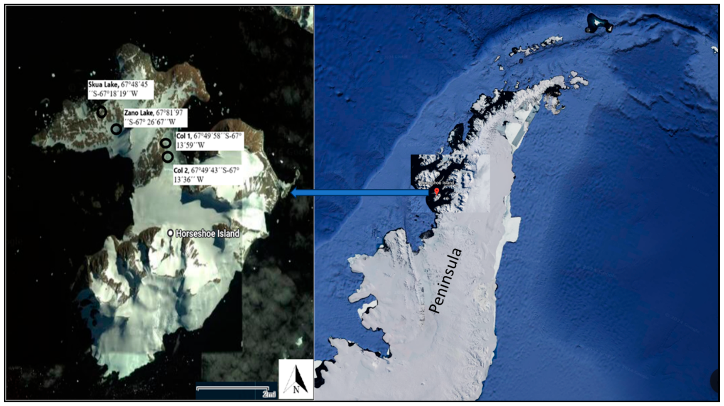
2.1. Site Description

Horseshoe Island, the site of the Turkish Scientific Camp, is situated on the western coast of the GRAHAM territory. Four lakes (Col 1, Col 2, Skua, and Zano) on the island were sampled in February 2023 during the Seventh Turkish Antarctic Expedition (TAE VII). The coordinates of these lakes are shown on maps, and the maximum depths (m) of the Col 1, Col 2, Skua, and Zano Lakes were 1.5, 3, 3, and 1 m, respectively (Figure 1).

2.2. Sampling Methods and Preservation

Water samples were collected using a Ruttner sampler (HydroBios) from one station in each lake for molecular analyses. The samples intended for molecular analyses were stored in Falcon tubes, both as water samples and through OE67 CA Membrane 0.45 µm Whatman filter paper samples, were preserved in polyethylene lid containers by adding an ethanol solution and led with parafilm. These samples were then frozen at –20 °C in containers with polyethylene lids and transported to Türkiye.

2.3. Amplification of Barcoding Regions, Processing, and Bioinformatics Analysis

This study examines the phytoplankton biodiversity in Horseshoe Island’s freshwater lakes using molecular techniques for the first time. Amplification tests employed universal primers from Table 1. The sequence reads were processed using the SILVA project’s amplicon analysis pipeline (SILVAngs 1.4) [34]. Each read was aligned with the SILVA Incremental Aligner (SINA SINA v1.2.10 for ARB SVN (revision 21008)) [35] against the SILVA SSU rRNA SEED and underwent quality control [34]. Reads exceeding 50 aligned nucleotides and those with more than 2% large or homopolymer sequences were excluded. Contaminants, artefacts, and low-quality alignments (50 alignment identities and 40 alignment scores reported by SINA) were also removed from further analysis. After quality control, dereplication identified identical reads. Unique reads were clustered into operational taxonomic units (OTUs) per sample, and each OTU reference read was classified. Dereplication and clustering were performed using VSEARCH (version 2.17.0; https://github.com/torognes/vsearch, accessed on 1 October 2023) [36] with identity criteria of 1.00 and 0.98. Classification was conducted using BLASTn (2.11.0+; http://blast.ncbi.nlm.nih.gov/Blast.cgi, accessed on 1 October 2023) [37] with standard settings, referencing the nonredundant SILVA SSU Ref dataset (release 138.1; http://www.arb-silva.de, accessed on 1 October 2023). OTU reference read classifications were mapped to all reads assigned to the respective OTU, providing quantitative data on individual reads per taxonomic path, accounting for PCR and sequencing biases and multiple rRNA operons. Reads with weak or no classifications (where (% sequence identity + % alignment coverage)/2 was below 93) were left unclassified and assigned to the meta group No Relative in the SILVAngs fingerprint and Krona charts [38]. This method was first applied in the studies by Klindworth et al. [39] and Ionescu et al. [40]. SILVAngs uses the NBI cloud (http://cloud.denbi.de, accessed on 1 October 2023) for project processing. The NBI cloud, an academic resource funded by BMBF1, provides free computational resources and storage for large-scale scientific projects to academic users.
Bioinformatics analyses were carried out in a Linux/Unix terminal. The quality of sequences in the “.fastq” format was evaluated using the FASTQC program. Subsequent analyses were performed using the “ObiTools” package. Sequences with a Phred score above 30 were aligned and merged using “illuminapairedend.” The merged reads were then cleaned, primers were removed, and duplicates and unnecessary data were eliminated from each sample header. Sequences with more than 10 repeats and longer than 100 base pairs were labeled as “C10l100.” Taxonomic association analysis of the “.fasta” files generated from the ObiTools package was conducted using SILVAngs.

3. Results

In this study, the primer sequences of the rRNA barcode regions of the phytoplankton species were successfully amplified in all the lakes; the V5–V7 regions of the 16S rRNA gene were amplified with the primer pair 799F–1191R (799F: 5′-AACMGGATTAGATACCCKG-3′ and 1191R 5′-ACGTCATCCCCACCTTCC-3′); the 18S gene was amplified with the universal eukaryotic primer pair 18S–V9F (TTGTACACACCGCCCGTCGC) and 18S–V9R (CCTTCYGCAGGTTCACCTAC); and the barcode region of the 23S rRNA gene was amplified with the primer pair p23SrV_f1 (5′-GGACAGAAAGACCCTATGAA-3′) and p23SrV_r1 (5′-TCAGCCTGTTATCCCTAGAG-3′).
An examination of water and filter samples from the study lakes revealed that, in particular, species belonging to Amorphous and Bacteria were detected at high rates; species belonging to phytoplankton groups were also identified, although at lower levels (Figure 2). Bacillariophyta, Chlorophyta, Cyanobacteria, and Ochrophyta were found in the phytoplankton taxonomic groups. The distributions of dominating taxonomic groups, depending on the lakes and the OTUs graphs, are shown in Figure 3.
As a result of 16S rRNA and 23S rRNA metagenomic analyses, a total of 111 Cyanobacteria taxa were identified. The highest Cyanobacterial diversity were detected in Lake Col 1 (107 taxa) and second in Lake Skua (76 taxa). Considering the distribution of species within the Cyanobacteria group, Allocoleopsis franciscana and Leptolyngbya boryana were the most abundant species in all lakes. However, the highest number of species belonging to a genus was identified in Synechococcus sp. (15 species), and this species was identified in almost all stations, followed by Nostoc sp., which was identified with 12 different species (Table S1). Species in the orders Leptolyngbyaceae, Oscillatoriales, and Synechococcaceae were determined to be dominant. The most abundant species for Lake Col 1 was Allocoleopsis franciscana; this species was followed by Leptothermofonsia sichuanensis and Leptolyngbya boryana. The most identified Cyanobacteria species in Col 2, Suka, and Zano Lakes were Synechococcus sp. and Thermostichus lividus (Figure 4).
As a result of 18S rRNA metagenomic analysis, 51 genera belonging to the Bacillariophyta, Chlorophyta, Dinophyceae, Ochrophyta, and Rhodophyta were identified. Bacteria had universally higher alpha diversity than eukaryotes, with the majority of microeukaryotic taxa being relatively abundant in Lake Col 1, Col 2, and Skua. Chlorophyta and Chrysophyta were found in Lake Col 1, Lake Col 2, and Lake Skua. However, species belonging to the Bacillariophyta were also determined in Lake Col 1, Lake Col 2, Lake Zano, and Lake Skua. (Figure S1). The OTUs and percentage distributions of Actinochloris, Amphidinium, Bolidomonas (newly recorded as Triparma), Bathycoccus, Chaetoceros, Chlamydopodium, Chloromonas, Chloromulina, Chlamydomonas, Chlamydopodium, Chlorella, Choricystis, Chrysoxys, Coccomyxa, Cryptomonas, Cyclotella, Cylindrotheca, Dictyococcus, Dinobryon, Eucampia, Gloeotila, Gomphonema, Goniomonas, Gyrodinium, Hindakia, Karlodinium, Komma, Leptocylindrus, Luticola, Mallomonas, Monomastix, Mychonastes, Nephroselmis, Ochromonas, Oikomonas, Paraphysomonas, Pedinellales, Peridinium, Pinnularia, Phaeodactylum, Pleurastrum, Polysiphonia boldii, Ptychodiscus, Rhodella violacea, Spumella, Teleaulax, Tetracystis, Tetradesmus, Trebouxia, Ulnaria, and Ulothrix are given in Figues S2–S20.

4. Discussion

In studies examining phytoplankton and benthic species diversity in Antarctica, Chlorophyta, Chrysophyta, Diatoms, Dinoflagellates, and prokaryotic Cyanobacteria were generally found [30,46,47,48,49,50,51,52,53]. Vick-Majors et al. [27] reported that bacterial and eukaryotic communities in Lakes FRX and Bonney were strongly controlled by vertically stratified water columns. However, these communities also responded to seasonal change. Additionally, bacterial, archaeal, and eukaryotic cells, including Cryptomonadales, Chloroplastida, Cyanobacteria, and Dinoflagellates, were found in permanently ice-covered lakes within the OTUs of phototrophic organisms. The researchers identified several microeukaryotes that exhibited specific reactions to salinity in the high-salinity lakes of East Antarctica and noted that ecological drift predominantly influenced these organisms in approximately 72% of the cycles [28].
Phytoplankton compositions have been identified from lakes in different regions of Antarctica using classical methods, and Pyramimonas sp. and Pyramimonas gelicola [54] and filamentous algae belonging to Chlorophyta (Klebsormidium, Mougeotia, Ulothrix moniliformis, and Zygnema) were found on King George Island [55], whereas Chlamydomonas, Geminigera cryophila, and Isochrysis have been identified in lakes of the McMurdo Dry Valleys, where no crustaceans, zooplankton, or fish have been found [56]. Rogers et al. [57] reported that in V5, there were 16 sequences closely related to those from single-celled eukaryotic species in Lake Vostok. These included sequences most similar to species from Excavata, with over 90% shared identity with Rhizaria, Amoebozoa, Chromalveolata. hey also had nearly 100% identity to Stephanodiscus sp., Hantzschia sp., Aphanomyces euteiches, Botrydiopsis constricta, Cryptomonas paramecium, and Halosiphon tomentosus. Additionally, they found a sequence with 100% identity to Perkinsea, an obligate parasite of mollusks. In this study, the highest density Cyanobacteria biodiversity was found in V5–V7 regions of the 16S rRNA gene and 23S rRNA were detected. However, filamentous (Ulothrix) and unicellular (e.g., Gyrodinium, Peridinium) phytoplankton (single-celled eukaryotic species) that can adapt to salinity were identified in a similar manner in Horseshoe Island lakes in V9 regions of the 18S rRNA.
The studies reported that Nostocaceae is dominant, accounting for almost half of the Cyanobacteria, compared with Oscillatoriaceae and Chroococcaceae, and that Ulothrix sp. was found to be dominant just in permanently submerged stream biofilms. In addition, the Leptolyngbyaceae and Phormidiaceae and Nostocacean morphotypes (e.g., Tolypothrix and Nostoc) were reported to be abundant in areas with high dehydration as well as in areas with low nutrient salts [31]. In the research carried out in two lakes in the Peninsula region from 2000–2001, Achnanthes linkei (61.9%), Navicula (10.8%), Navicula molesta (8.5%), and Gomphonema (8%) were identified. Diadesmis perpusilla (5.5%) and Achnanthes muelleri (2.1%) were also identified using classical methods [58]. In this research, for Lake Col 1, Cylindrotheca (23%) and Phaeodactylum (17%) were from the Bacillariophyta group; Polysiphonia boldii and Rhodella violacea, which generally live in marine environments, although some lineages live in brackish and freshwater ecosystems, were the most identified species from Rhodophyta. The presence of these species in freshwater glacial lakes has not been explained and remains a question. In Lake Col 2, Nitzschia alba (13%) and Cylindrotheca (88%) from the Bacillariophyta group were present. In Skua Lake, Bacillariophyta group species were detected, of which 23% were Phaeodactylum tricornutum and 15% were Cylindrotheca. Diatoms such as Phaeodactylum tricornutum and Cylindrotheca can be found in both freshwater and marine environments. In general, the determination of benthic algal species is due to the mixing of these algae or their sequences in the water column of lakes. It is likely that their genetic material could be accessed and/or identified more easily, or ocean water could be mixing into the lakes as groundwater or wave spray, allowing these species to be more easily detected, as they formed dominant groups.
Diatoms form benthic and planktonic algal communities that are widely distributed in different habitats from freshwater to marine water. They were also the first group to be studied in Antarctica. In the first studies conducted using classical identification methods, 63 taxa were reported in 1965, and 750 taxa were reported in 1979 [59]. Moreover, dinoflagellates (Gymnodinium sp. and Gonyaulax sp.) constitute another intensively identified group. In the distribution of phytoplankton, although the species of Cosmarium, Euastrum, and Closterium from Desmidiales have been detected in low numbers and rarely, the species Ochromonas spp., Chromulina spp., and Pseudokephyrion spp. were reported to be present in high amounts in the Chrysophyta (Heterokontophyta/Ochrophyta) group. In addition, Lizotte [49] reported the phytoplankton compositions of Chlorophyta, Chrysophyta, diatoms, and dinoflagellate and prokaryotic Cyanobacteria in Antarctic lakes. In this research, Bacillariophyta and Rhodophyta formed the dominant groups in the 18S rRNA gene amplification, and the Chlorophyta was recorded in 47% of the Skua Lake samples, 41% of the Zana Lake samples, 29% of the Col 1 Lake samples, and 39% of the Col 2 Lake samples. It has been concluded that the diversity of phytoplankton has increased in Antarctic lakes.
Cyanobacteria are the dominant photosynthetic producers in Antarctic lakes, and at the same time, some species (e.g., from Nostocales, Oscillatoriales) can also fix atmospheric nitrogen, which is a scarce resource in oligotrophic lakes [60]; benthic microbial mats constitute the first level of the food web in Antarctic lakes affected by environmental changes caused by climate [61,62,63]. Cyanobacterial biodiversity can also be used as a sentinel by which to monitor climate change adaptation responses in this rapidly warming region. The species diversity of Cyanobacteria was found to be higher than that of other eukaryotes in the 16S rRNA and 23S rRNA gene amplifications, while some bacterial groups were detected using 18S rRNA gene amplification, even though the species diversity of Cyanobacteria was not established. It was reported that Proteobacteria and Cyanobacteria were the dominant groups identified in the 16S rRNA gene amplification [29]. Alongside filamentous taxa like Leptolyngbya and Phormidium, which are common genera in microbial mats in Antarctic lakes, other phenotypes associated with different taxa, including Geitlerinema, Pseudanabaena, Synechococcus, Chamaesiphon, Calothrix, and Coleodesmium, were also identified [64]. This situation can be interpreted as a natural result of the phytoplankton composition of the lakes; however, it is anticipated that with the development of metagenome methods, data belonging to different phytoplankton groups can be revealed.
Global worming causes changes in water quality in natural lakes, leading to alterations in the diversity of phytoplankton species. Due to climate change, the precipitation regime changes, thus altering the flow rates of streams feeding the lakes and the retention times of lake waters. Irregularities in the precipitation regime cause changes in the duration of lake ice cover, which, in turn, changes the phytoplankton composition [65]. Antarctic microbiota mainly depends on cyanobacteria. In Antarctic ecosystems, cyanobacteria play the role of primary producers; their survival and development in extreme environments are primarily due to their ability to produce biofilms (especially microbial mats) [66]. The responsiveness of microorganisms and their dominant influence on ecosystem functioning make them suitable indicators for the early detection of climate-related environmental changes [29]. Recent studies using molecular techniques in the benthic area of Antarctica have reported that the Nostocaceae family dominates, accounting for nearly half of the Cyanobacteria abundance, in comparison to the Oscillatoriaceae and Chroococcaceae families [31,54]. These taxa have the capability to fix atmospheric nitrogen, a scarce resource in predominantly oligotrophic lakes [60,61]. In this study, in addition to the aforementioned species, Anabaena cylindrica, Microcystis aeruginosa, Planktothrix agardhii, and Pseudanabaena sp. were found. This indicates that the lakes are undergoing eutrophication, primarily driven by climate change. However, addressing this issue requires integrating data on their diversity, biogeographic range, and evolutionary history, along with information on their responses to changes and the functional attributes underlying their resilience.
Within the Cyanobacteria, Pseudoanabaenales, Oscillatoriales, Synechococcales, Nostocales, and Chroococcales orders were found in microbial mat samples of Antartic lakes including Lake Col1 [29]. Here, we found the some Chrysophyceae members, such as Chrysoxys, Chromulina, Dinobryon, Ochromonas, Oikomonas, Paraphysomonas, Spumella spp., as well as different taxa such as Hindakia sp., Monomastix sp., Dictyococcus sp., Mychonastes sp., and Bathycoccus sp. from Chlorophyta. Moreover, within Cyanobacteria, species such as Chamaesiphon, Leptolyngbya, Pseudoanabaena, and Synechococcus spp. were detected. These species were recorded for the first time in Col2 and Zano lakes.

5. Conclusions

In our study, conducted using metagenome techniques, different species were detected in addition to previously identified phytoplankton and benthic species. Cryptista were commonly found in the Col 1 and Col 2 lakes, while Cyanobacteria were widely found in Lake Skua. Although sediment sampling and analysis were not performed, the presence of benthic species suggests that benthic and sediment-associated algal sequences also contribute to aquatic phytoplankton communities. However, given the absence of species previously identified using classical methods, we recommend the following: (1) using both molecular and classical methods together when examining phytoplankton communities; and (2) conducting studies in conjunction with functional gene analysis, metagenomics, or transcription analyses to explore the relationships revealed by our molecular ecological network analysis.

Supplementary Materials

The following supporting information can be downloaded at: https://www.mdpi.com/article/10.3390/w17070975/s1, Figure S1: Total eukaryota biodiversity of Lake Col 1 by high-throughput sequencing; Figure S2: Bacillariophyceae biodiversity of Lake Col 1 by high-throughput sequencing; Figure S3: Chlorophyta biodiversity of Lake Col 1 by high-throughput sequencing; Figure S4: Cryptophyceae biodiversity of Lake Col 1 by high-throughput sequencing; Figure S5: Ochrophyta biodiversity of Lake Col 1 by high-throughput sequencing; Figure S6: Total eukaryota biodiversity of Lake Col 2 by high-throughput sequencing; Figure S7: Bacillariophyceae biodiversity of Lake Col 2 by high-throughput sequencing; Figure S8: Chlorophyta biodiversity of Lake Col 2 by high-throughput sequencing; Figure S9: Cryptophyceae biodiversity of Lake Col 2 by high-throughput sequencing; Figure S10: Ochrophyta biodiversity of Lake Col 2 by high-throughput sequencing; Figure S11: Total eukaryota biodiversity of Lake Skua by high-throughput sequencing; Figure S12: Bacillariophycidae biodiversity of Lake Skua by high-throughput sequencing; Figure S13: Chlorophyta biodiversity of Lake Skua by high-throughput sequencing; Figure S14: Cryptophyceae biodiversity of Lake Skua by high-throughput sequencing; Figure S15: Ochrophyta biodiversity of Lake Skua by high-throughput sequencing; Figure S16: Total eukaryota biodiversity of Lake Zano by high-throughput sequencing; Figure S17: Bacillariophyceae biodiversity of Lake Zano by high-throughput sequencing; Figure S18: Chlorophyta biodiversity of Lake Zano by high-throughput sequencing; Figure S19: Cryptophyceae biodiversity of Lake Zano by high-throughput sequencing; Figure S20: Ochrophyta biodiversity of Lake Zano by high-throughput sequencing; Table S1: Cyanobacteria biodiversity in the lakes.

Author Contributions

Ö.F., M.K. and N.D. wrote the main manuscript text; M.F.T. prepared Figure 2 and Figure 3; and all of authors reviewed the manuscript. All authors have read and agreed to the published version of the manuscript.

Funding

This study was funding by TUBITAK (Project Number: 122Y200).

Data Availability Statement

The original contributions presented in this study are included in the article/Supplementary Material. Further inquiries can be directed to the corresponding author.

Acknowledgments

This research was carried out under the auspices of the Presidency of the Republic of Türkiye, supported by the Ministry of Industry and Technology, and coordinated by the TUBITAK MAM Polar Research Institute.

Conflicts of Interest

The authors declare no conflicts of interest.

References

  1. Wetzel, R.G. Limnology; W.B. Sounders Company: London, UK, 2001. [Google Scholar]
  2. Sommer, U. The periodicity of phytoplankton in Lake Constance (Bodensee) in comparison to other deep lakes of central Europe. Hydrobiologia 1986, 138, 1–7. [Google Scholar] [CrossRef]
  3. Scheffer, M.; Rinaldi, S.; Huisman, J.; Weissing, F.J. Why plankton communities have no equilibrium: Solutions to the paradox. Hydrobiologia 2003, 491, 9–18. [Google Scholar] [CrossRef]
  4. Thunmark, S. Zur soziologie des süsswasserplanktons. Eine methodisch ökologische studie. Folia Limnol. Skand 1945, 3, 1–66. [Google Scholar]
  5. Nygaard, G. Hidrobiological studies on some Danish ponds and lakes. Part II: The quotient hypothesis and some little known plankton organisms. K Dan Vidensk Selsk Biol. Skr. 1949, 7, 1–293. [Google Scholar]
  6. Lepistö, L.; Rosenström, U. The most typical phytoplankton taxa in four types of boreal lakes. Hydrobiologia 1998, 369/370, 89–97. [Google Scholar] [CrossRef]
  7. Amann, R.I.; Ludwig, W.; Schleifer, K.H. Phylogenetic identification and in situ detection of individual microbial cells without cultivation. Microbiol. Rev. 1995, 59, 143–169. [Google Scholar] [CrossRef] [PubMed]
  8. Head, I.; Saunders, J.; Pickup, R. Microbial Evolution, Diversity, and Ecology: A Decade of Ribosomal RNA Analysis of Uncultivated Microorganisms. Microb. Ecol. 1998, 35, 1–21. [Google Scholar] [CrossRef]
  9. López-García, P.; Rodríguez-Valera, F.; Pedrós-Alió, C.; Moreira, D. Unexpected diversity of small eukaryotes in deep-sea Antarctic plankton. Nature 2001, 409, 603–607. [Google Scholar] [CrossRef]
  10. Epstein, S.; López-García, P. “Missing” protists: A molecular prospective. Biodivers. Conserv. 2007, 17, 261–276. [Google Scholar] [CrossRef]
  11. Baker, A.C. Flexibility and Specificity in Coral-Algal Symbiosis: Diversity, Ecology, and Biogeography of Symbiodinium. Annu. Rev. Ecol. Evol. Syst. 2003, 34, 661–689. [Google Scholar] [CrossRef]
  12. Schloss, P.D.; Handelsman, J. Metagenomics for studying unculturable microorganisms: Cutting the Gordian knot. Genome Biol. 2005, 6, 229. [Google Scholar] [CrossRef] [PubMed]
  13. Keskin, E.; Ünal, E. Environmental DNA (eDNA); Nobel Akademic Press: Ankara, Türkiye, 2020. [Google Scholar]
  14. Clark, M.S.; Clarke, A.; Cockell, C.S.; Convey, P.; Detrich, H.W.; Fraser, K.P.P.; Johnston, I.A.; Methe, B.A.; Murray, A.E.; Peck, L.S.; et al. Antarctic genomics. Comp. Funct. Genom. 2004, 5, 230–238. [Google Scholar] [CrossRef] [PubMed]
  15. Peck, L.S.; Clark, M.S.; Clarke, A.; Cockell, C.S.; Convey, P.; Detrich, H.W.; Fraser, K.P.P.; Johnston, I.A.; Methe, B.A.; Murray, A.E.; et al. Genomics: Applications to Antarctic ecosystems. Polar Biol. 2004, 28, 351–365. [Google Scholar] [CrossRef]
  16. Francis, T.B.; Krüger, K.; Fuchs, B.M.; Teeling, H.; Amann, R.I. Candidatus Prosiliicoccus vernus, a spring phytoplankton bloom associated member of the Flavobacteriaceae. Syst. Appl. Microbiol. 2018, 42, 41–53. [Google Scholar] [CrossRef]
  17. Grieb, A.; Bowers, R.M.; Oggerin, M.; Goudeau, D.; Lee, J.; Malmstrom, R.R.; Woyke, T.; Fuchs, B.M. A pipeline for targeted metagenomics of environmental bacteria. Microbiome 2020, 8, 21. [Google Scholar] [CrossRef]
  18. Tominaga, K.; Morimoto, D.; Nishimura, Y.; Ogata, H.; Yoshida, T. In silico Prediction of Virus-Host Interactions for Marine Bacteroidetes with the Use of Metagenome-Assembled Genomes. Front. Microbiol. 2020, 11, 738. [Google Scholar] [CrossRef] [PubMed]
  19. Vorobev, A.; Dupouy, M.; Carradec, Q.; Delmont, T.O.; Annamalé, A.; Wincker, P.; Pelletier, E. Transcriptome reconstruction and functional analysis of eukaryotic marine plankton communities via high-throughput metagenomics and metatranscriptomics. Genome Res. 2020, 30, 647–659. [Google Scholar] [CrossRef]
  20. Fu, J.; Chen, L.; Yang, S.; Li, Y.; Jin, L.; He, X.; He, L.; Ao, X.; Liu, S.; Liu, A.; et al. Metagenome and analysis of metabolic potential of the microbial community in pit mud used for Chinese strong-flavor liquor production. Food Res. Int. 2021, 143, 110294. [Google Scholar] [CrossRef]
  21. Duncan, A.; Barry, K.; Daum, C.; Eloe-Fadrosh, E.; Roux, S.; Schmidt, K.; Tringe, S.G.; Valentin, K.U.; Varghese, N.; Salamov, A.; et al. Metagenome-assembled genomes of phytoplankton microbiomes from the Arctic and Atlantic Oceans. Microbiome 2022, 10, 67. [Google Scholar] [CrossRef]
  22. Unrein, F.; Izaguirre, I.; Massana, R.; Balagué, V.; Gasol, J. Nanoplankton assemblages in maritime Antarctic lakes: Characterisation and molecular fingerprinting comparison. Aquat. Microb. Ecol. 2005, 40, 269–282. [Google Scholar] [CrossRef]
  23. Tran, P.; Ramachandran, A.; Khawasik, O.; Beisner, B.E.; Rautio, M.; Huot, Y.; Walsh, D.A. Microbial life under ice: Metagenome diversity and in situ activity of Verrucomicrobia in seasonally ice-covered Lakes. Environ. Microbiol. 2018, 20, 2568–2584. [Google Scholar] [CrossRef] [PubMed]
  24. Smith, D.R.M.; Uria, A.R.; Helfrich, E.J.N.; Milbredt, D.; van Pée, K.-H.; Piel, J.; Goss, R.J.M. An Unusual Flavin-Dependent Halogenase from the Metagenome of the Marine Sponge Theonella swinhoei WA. ACS Chem. Biol. 2017, 12, 1281–1287. [Google Scholar] [CrossRef] [PubMed]
  25. Sogin, M.L.; Morrison, H.G.; Huber, J.A.; Welch, D.M.; Huse, S.M.; Neal, P.R.; Arrieta, J.M.; Herndl, G.J. Microbial diversity in the deep sea and the underexplored “rare biosphere”. Proc. Natl. Acad. Sci. USA 2006, 103, 12115–12120. [Google Scholar] [CrossRef] [PubMed]
  26. Taib, N.; Mangot, J.-F.; Domaizon, I.; Bronner, G.; Debroas, D. Phylogenetic Affiliation of SSU rRNA Genes Generated by Massively Parallel Sequencing: New Insights into the Freshwater Protist Diversity. PLoS ONE 2013, 8, e58950. [Google Scholar] [CrossRef]
  27. Vick-Majors, T.J.; Priscu, J.C.; Amaral-Zettler, L.A. Modular community structure suggests metabolic plasticity during the transition to polar night in ice-covered Antarctic lakes. ISME J. 2013, 8, 778–789. [Google Scholar] [CrossRef]
  28. Logares, R.; Tesson, S.V.; Canbäck, B.; Pontarp, M.; Hedlund, K.; Rengefors, K. Contrasting prevalence of selection and drift in the community structuring of bacteria and microbial eukaryotes. Environ. Microbiol. 2018, 20, 2231–2240. [Google Scholar] [CrossRef]
  29. Durieu, B.; Lara, Y.; Pessi, I.S.; Wilmotte, A.; Tytgat, B.; Sweetlove, M.; Pinseel, E.; Verleyen, E.; Vyverman, W.; Van der Vijver, B.; et al. Climate Change and Antarctic Microbial Diversity’CCAMBIO; Final Report; British Antarctic Survey: Cambridge, UK, 2019. [Google Scholar]
  30. Li, W.; Morgan-Kiss, R.M. Influence of Environmental Drivers and Potential Interactions on the Distribution of Microbial Communities from Three Permanently Stratified Antarctic Lakes. Front. Microbiol. 2019, 10, 1067. [Google Scholar] [CrossRef]
  31. Rochera, C.; Camacho, A. Limnology and Aquatic Microbial Ecology of Byers Peninsula: A Main Freshwater Biodiversity Hotspot in Maritime Antarctica. Diversity 2019, 11, 201. [Google Scholar] [CrossRef]
  32. Yıldırım, C. Geomorphology of Horseshoe Island, Marguerite Bay, Antarctica. J. Maps 2019, 16, 56–67. [Google Scholar] [CrossRef]
  33. Yirmibeşoğlu, S.; Oktar, Ö.; Özsoy, B. Review of Scientific Research Conducted in Horseshoe Island Where Potential Place for Turkish Antarctic Base. Int. J. Environ. Geoinformat. 2022, 9, 11–23. [Google Scholar] [CrossRef]
  34. Quast, C.; Pruesse, E.; Yilmaz, P.; Gerken, J.; Schweer, T.; Yarza, P.; Peplies, J.; Glöckner, F.O. The SILVA Ribosomal RNA Gene Database Project: Improved Data Processing and Web-Based Tools. Nucleic Acids Res. 2013, 41, D590–D596. [Google Scholar] [CrossRef] [PubMed]
  35. Pruesse, E.; Peplies, J.; Glöckner, F.O. SINA: Accurate high-throughput multiple sequence alignment of ribosomal RNA genes. Bioinformatics 2012, 28, 1823–1829. [Google Scholar] [CrossRef] [PubMed]
  36. Rognes, T.; Flouri, T.; Nichols, B.; Quince, C.; Mahé, F. VSEARCH: A versatile open source tool for metagenomics. PeerJ 2016, 2016, e2584. [Google Scholar] [CrossRef]
  37. Camacho, C.; Coulouris, G.; Avagyan, V.; Ma, N.; Papadopoulos, J.; Bealer, K.; Madden, T.L. BLAST+: Architecture and applications. BMC Bioinform. 2009, 10, 421. [Google Scholar] [CrossRef]
  38. Ondov, B.D.; Bergman, N.H.; Phillippy, A.M. Interactive metagenomic visualization in a Web browser. BMC Bioinform. 2011, 12, 385. [Google Scholar] [CrossRef]
  39. Klindworth, A.; Pruesse, E.; Schweer, T.; Peplies, J.; Quast, C.; Horn, M.; Glöckner, F.O. Evaluation of General 16S Ribosomal RNA Gene PCR Primers for Classical and Next-Generation Sequencing-Based Diversity Studies. Nucleic Acids Res. 2013, 41, e1. [Google Scholar] [CrossRef]
  40. Ionescu, D.; Siebert, C.; Polerecky, L.; Munwes, Y.Y.; Lott, C.; Häusler, S.; Bižić-Ionescu, M.; Quast, C.; Peplies, J.; Glöckner, F.O.; et al. Microbial and Chemical Characterization of Underwater Fresh Water Springs in the Dead Sea. PLoS ONE 2012, 7, e38319. [Google Scholar] [CrossRef]
  41. Rego, A.; Raio, F.; Martins, T.P.; Ribeiro, H.; Sousa, A.G.G.; Séneca, J.; Baptista, M.S.; Lee, C.K.; Cary, S.C.; Ramos, V.; et al. Actinobacteria and Cyanobacteria Diversity in Terrestrial Antarctic Microenvironments Evaluated by Culture-Dependent and Independent Methods. Front. Microbiol. 2019, 10, 1018. [Google Scholar] [CrossRef]
  42. Thijs, S.; Op De Beeck, M.; Beckers, B.; Truyens, S.; Stevens, V.; Van Hamme, J.D.; Weyens, N.; Vangronsveld, J. Comparative evaluation of four bacteria-specific primer pairs for 16S rRNA gene surveys. Front. Microbiol. 2017, 8, 494. [Google Scholar] [CrossRef]
  43. Sherwood, A.R.; Presting, G.G. Universal primers amplify a 23S rDNA plastid marker in eukaryotic algae and cyanobacteria. J. Phycol. 2007, 43, 605–608. [Google Scholar] [CrossRef]
  44. Schiaffino, M.R.; Lara, E.; Fernández, L.D.; Balagué, V.; Singer, D.; Seppey, C.C.W.; Massana, R.; Izaguirre, I. Microbial eukaryote communities exhibit robust biogeographical patterns along a gradient of Patagonian and Antarctic lakes. Environ. Microbiol. 2016, 18, 5249–5264. [Google Scholar] [CrossRef] [PubMed]
  45. Kezlya, E.; Tseplik, N.; Kulikovskiy, M. Genetic Markers for Metabarcoding of Freshwater Microalgae: Review. Biology 2023, 12, 1038. [Google Scholar] [CrossRef] [PubMed]
  46. Schmidt, R.; Mäusbacher, R.; Müller, J. Holocene diatom flora and stratigraphy from sediment cores of two Antarctic lakes (King George Island). J. Paleolimnol. 1990, 3, 55–74. [Google Scholar] [CrossRef]
  47. Spaulding, S.A.; McKnight, D.M.; Stoermer, E.F.; Doran, P.T. Diatoms in sediments of perennially ice-covered Lake Hoare, and implications for interpreting lake history in the McMurdo Dry Valleys of Antarctica. J. Paleolimnol. 1996, 17, 403–420. [Google Scholar] [CrossRef]
  48. Sabbe, K.; Verleyen, E.; Hodgson, D.; Vanhoutte, K.; Vyverman, W. Benthic diatom flora of freshwater and saline lakes in the Larsemann Hills and Rauer Islands, East Antarctica. Antarct. Sci. 2003, 15, 227–248. [Google Scholar] [CrossRef]
  49. Lizotte, M.P. Phytoplankton and primary production. In Polar Lakes and Rivers: Limnology of Arctic and Antarctic Aquatic Ecosystems; Laybourn-Parry, J., Vincent, W.F., Eds.; Oxford University Press: Oxford, UK, 2008; pp. 157–178. [Google Scholar]
  50. Bielewicz, S.; Bell, E.; Kong, W.; Friedberg, I.; Priscu, J.C.; Morgan-Kiss, R.M. Protist diversity in a permanently ice-covered Antarctic Lake during the polar night transition. ISME J. 2011, 5, 1559–1564. [Google Scholar] [CrossRef]
  51. Kong, W.; Ream, D.C.; Priscu, J.C.; Morgan-Kiss, R.M. Diversity and Expression of RubisCO Genes in a Perennially Ice-Covered Antarctic Lake during the Polar Night Transition. Appl. Environ. Microbiol. 2012, 78, 4358–4366. [Google Scholar] [CrossRef]
  52. Kong, W.; Li, W.; Romancova, I.; Prášil, O.; Morgan-Kiss, R.M. An integrated study of photochemical function and expression of a key photochemical gene (psbA) in photosynthetic communities of Lake Bonney (McMurdo Dry Valleys, Antarctica). FEMS Microbiol. Ecol. 2014, 89, 293–302. [Google Scholar] [CrossRef]
  53. Dolhi, J.M.; Teufel, A.G.; Kong, W.; Morgan-Kiss, R.M. Diversity and spatial distribution of autotrophic communities within and between ice-covered Antarctic lakes (McMurdo Dry Valleys). Limnol. Oceanogr. 2015, 60, 977–991. [Google Scholar] [CrossRef]
  54. Izaguirre, I.; Allende, L.; Schiaffino, M.R. Phytoplankton in Antarctic lakes: Biodiversity and main ecological features. Hydrobiologia 2020, 848, 177–207. [Google Scholar] [CrossRef]
  55. Vinocur, A.; Unrein, F. Typology of lentic water bodies at Potter Peninsula (King George Island, Antarctica) based on physical-chemical characteristics and phytoplankton communities. Polar Biol. 2000, 23, 858–870. [Google Scholar] [CrossRef]
  56. Sherwell, S.; Kalra, I.; Li, W.; McKnight, D.M.; Priscu, J.C.; Morgan-Kiss, R.M. Antarctic lake phytoplankton and bacteria from near-surface waters exhibit high sensitivity to climate-driven disturbance. Environ. Microbiol. 2022, 24, 6017–6032. [Google Scholar] [CrossRef] [PubMed]
  57. Rogers, S.O.; Shtarkman, Y.M.; Koçer, Z.A.; Edgar, R.; Veerapaneni, R.; D’Elia, T. Ecology of Subglacial Lake Vostok (Antarctica), Based on Metagenomic/Metatranscriptomic Analyses of Accretion Ice. Biology 2013, 2, 629–650. [Google Scholar] [CrossRef] [PubMed]
  58. Smith, J.A.; Hodgson, D.A.; Bentley, M.J.; Verleyen, E.; Leng, M.J.; Roberts, S.J. Limnology of Two Antarctic Epishelf Lakes and their Potential to Record Periods of Ice Shelf Loss. J. Paleolimnol. 2006, 35, 373–394. [Google Scholar] [CrossRef]
  59. Jones, J. The diversity, distribution and ecology of diatoms from Antarctic inland waters. Biodivers. Conserv. 1996, 5, 1433–1449. [Google Scholar] [CrossRef]
  60. Quesada, A.; Vincent, W.F. Cyanobacteria in the cryosphere: Snow, ice and extreme cold. In Ecology of Cyanobacteria II: Their Diversity in Space and Time; Whitton, B.A., Ed.; Springer: Dordrecht, The Netherlands, 2012; pp. 387–399. [Google Scholar]
  61. Vincent, W.F. Evolutionary origins of Antarctic microbiota: Invasion, selection and endemism. Antarct. Sci. 2000, 12, 374–385. [Google Scholar] [CrossRef]
  62. Jungblut, A.D.; Lovejoy, C.; Vincent, W.F. Global distribution of cyanobacterial ecotypes in the cold biosphere. ISME J. 2009, 4, 191–202. [Google Scholar] [CrossRef]
  63. Verleyen, E.; Sabbe, K.; Hodgson, D.A.; Grubisic, S.; Taton, A.; Cousin, S.; Wilmotte, A.; De Wever, A.; Van der Gucht, K.; Vyverman, W. Structuring effects of climate-related environmental factors on Antarctic microbial mat communities. Aquat. Microb. Ecol. 2010, 59, 11–24. [Google Scholar] [CrossRef]
  64. Pessi, I.S.; Maalouf, P.D.C.; Laughinghouse, H.D.; Baurain, D.; Wilmotte, A. On the use of high-throughput sequencing for the study of cyanobacterial diversity in Antarctic aquatic mats. J. Phycol. 2016, 52, 356–368. [Google Scholar] [CrossRef]
  65. Kalff, J. Limnology; Prentice-Hall, Inc.: Hoboken, NJ, USA, 2003. [Google Scholar]
  66. Velichko, N.; Smirnova, S.; Averina, S.; Pinevich, A. A survey of Antarctic cyanobacteria. Hydrobiologia 2021, 848, 2627–2652. [Google Scholar]
Figure 1. Locations and coordinate information of Lake Col 1, Lake Col 2, Lake Skua, and Lake Zano on Horseshoe Island.
Water 17 00975 g001
Figure 2. Taxonomic fingerprints at the phylum level for the lakes.
Water 17 00975 g002
Figure 3. Dominating taxonomic groups of the lakes: (A) Lake Col 1; (B) Lake Col 2; (C) Lake Skua; (D) Lake Zano. SAR includes Stramenopiles, Alveolates, and Rhizarians. Phytoplankton distribution from Bicosoecida and Ochrophyta (under Stramenopiles) and Dinoflagellata (under Alveolata) were shown.
Water 17 00975 g003
Figure 4. Distribution of cyanobacterial composition in the lakes (%).
Water 17 00975 g004
Table 1. The universal primer sets used in the metagenomic analysis.
Primer SetForward Primer SequenceReverse Primer SequenceReference
CYA359F/CYA781R5′-GGGGAATYTTCCGCAATGGG-3′5′-GACTACWGGGGTATCTAATCCCWTT-3′[41]
799F/1191R5′-AACMGGATTAGATACCCKG-3′5′-ACGTCATCCCCACCTTCC-3′[42]
p23SrV_f1/p23SrV_r15′-GGACAGAAAGACCCTATGAA-3′5′-TCAGCCTGTTATCCCTAGAG-3′[43]
Euk1f/Euk516r5′-CTGGTTGATCCTGCCAG-3′5′-ACCAGACTTGCCCTCC-3′[44]
18S_V9_F1/18S_V9_R5′-TTGTACACACCGCCCTCGC-3′5′-CCTTCYGCAGGTTCACCTAC-3′[45]
Disclaimer/Publisher’s Note: The statements, opinions and data contained in all publications are solely those of the individual author(s) and contributor(s) and not of MDPI and/or the editor(s). MDPI and/or the editor(s) disclaim responsibility for any injury to people or property resulting from any ideas, methods, instructions or products referred to in the content.

