Abstract
The aim of this study was to evaluate the relationship of the ankle-brachial index (ABI) level with kinetic and kinematic parameters of the gait pattern and force-velocity parameters generated by lower limb muscles. Methods: The study group consisted of 65 patients with peripheral arterial disease (PAD). The ABI value, kinetic and kinematic parameters of gait and force-velocity parameters of knee and ankle extensors and flexors were determined in all subjects. The values obtained for right and left limbs as well as the limbs with higher and lower ABI were compared. Results: Regardless of the method of analysis, the values of the gait’s kinematic and kinetic parameters of both lower limbs did not differ significantly. However, significant differences were noted in the values of peak torque, work and power of the extensor muscles of the knee and the flexor muscles of the ankle with the higher and lower ABI. Conclusion: This study demonstrated that a higher degree of ischemia worsened the level of strength, endurance, and performance of ankle flexors and extensors of the knee joint. ABI is not related to the gait pattern. The above-mentioned relationship should be taken into account in the rehabilitation process and methodological assessment.
1. Introduction
Hemodynamic studies with ankle-brachial index (ABI) estimation are not completely reliable in assessing functional capacity in patients with peripheral arterial disease (PAD). This is shown especially in the distance covered [1], which is also verified by McDermott et al. [2] in their 5-year follow-up. However, there are a number of studies that have confirmed this relationship [3,4,5,6]. The discrepancies resulting from the number of subjects and the use of different research protocols were the reason for undertaking an extended analysis of gait pattern and muscle function in patients with PAD and verifying them with ABI results.
The authors point to functional limitations in walking ability, balance, lower limb strength, and general functional performance in individuals presenting with lower ABI scores [2,3,4,7,8,9,10]. Reduced blood supply to muscles through narrowed arterial lumen, deterioration of tissue trophism, persistent inflammation, lower extremity pain, and comorbidities are the most commonly cited causes of functional impairment among patients with PAD [2,8,11].
Atherosclerotic lesions in the vessels of the lower extremities usually are asymmetric, as demonstrated by varying ABI values. Differences in muscle strength of the right and left lower limbs and differences in gait pattern are observed in people with PAD compared to healthy subjects [12,13,14,15]. For this reason, many studies analyze the relationship of the ABI with functional parameters taking into account the right and left side, often indicating the dominant side or considering the results from the lower limb presenting a lower ABI. However, it is noted that not the side but the degree of ischemia may provide more information about the relationship of the ankle-brachial index with functional parameters of individuals with PAD [2,4,7,8,9,10]. Therefore, the aim of this study is to evaluate the relationship of the ankle-brachial index level with kinetic and kinematic parameters of gait pattern and force-velocity parameters generated by lower limb muscles based on the analysis of sides (right and left lower limb) and degree of ischemia (high, low ABI).
2. Material and Method
This work is part of the project “WROVASC—Integrated Center for Cardiovascular Medicine”, co-funded by the European Regional Development Fund, under the Innovative Economy Operational Program for 2007–2013, implemented in the Regional Specialist Hospital in Wrocław, Research and Development Center.
The study was approved by the Ethics Committee of the Medical University of Wroclaw, Poland (Ref.KB-130/2008). These studies were registered in the clinical trial database—the Australian New Zealand Clinical Trials Registry (Trial Id: ACTRN12621000780853).
2.1. Study Group
The study group consisted of 65 patients with peripheral arterial disease (PAD). Inclusion criteria were: age over 40 years, demonstrated chronic lower extremity arterial disease (PAD) class IIa and IIb in the Fontaine classification, intermittent claudication (IC), ankle-brachial index (ABI) lower than 0.9 in at least one limb and different for the right and left limbs, good clinical condition of the patient, and written informed consent to participate in the program.
Exclusion criteria were PAD Fontaine I and III/IV, non-controlled internal and cardiovascular diseases, revascularization procedures performed during the last 3 months, overall poor health, psychiatric illness. The general characteristics of the subjects are shown in Table 1. A detailed history and qualifying examination revealed a femoropopliteal type of occlusion in 61 patients of the study group (82%), an aorto-iliac type in 9 (12%), and multilevel (3 subjects) and peripheral (1 subject) in the remaining patients. Significant results of the qualifying study are shown in Table 2.

Table 1.
General characteristics of participants.
Table 2.
Qualifying examination results.
Patients applied for participation in the program in response to widely disseminated announcements in the media and health centers. Recruitment of patients to participate in the program was based on a medical consultation taking into account the inclusion and exclusion criteria and completion of preliminary examinations. During the medical consultation, which took place in the health care facilities in Wrocław, all patients had the ankle-brachial index measured. On the next day, in the research laboratories of the Wroclaw University of Health and Sport Sciences, a gait test was performed, followed by an assessment of the force-velocity parameters of the lower limb muscles. Individual tests were performed by a qualified technician of the device. A schematic of the study is shown in Figure 1.
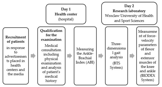
Figure 1.
Schematic diagram of the study.
2.2. Test Methods
2.2.1. Measuring the Ankle-Brachial Index (ABI)
The ankle-brachial index (ABI) was determined by measuring systolic blood pressure at the upper and lower extremities using continuous wave Doppler (Multi Dopplex II Vascular Doppler, Huntleigh, Wales, UK). First, measurements were taken on both brachial arteries, and the higher of the obtained values was considered. Next, arterial pressure was measured on the arteries of the foot (tibialis posterior and dorsal artery of the foot), also taking into account the higher of the obtained values. The ABI index was the quotient of the systolic pressure measured at the foot to the systolic pressure at the arm. The measurements were performed in the supine position of the subject, with the patient staying in the given position for 10 min to normalize hemodynamic parameters before starting the measurements [16].
2.2.2. Gait Biomechanics Testing
To record kinematic and kinetic parameters of gait, an optoelectronic system for three-plane motion analysis BTS Smart-E equipped with six digital cameras operating in the infrared range (1.1 μM) at 200 Hz and two Network Cam AXIS 210A cameras operating in the visible light range at 20 Hz was used. The set-up was complemented by two dynamometer platforms (Kistler 9286) with signal recording at 200 Hz forming the central part of the 10 m path. All devices made measurements synchronously.
Before testing, 22 photoreflective passive markers corresponding to selected anthropometric points were placed on the patient’s body according to the Davis protocol. A schematic of the gait analysis laboratory and the course of the study are shown in Figure 2 and Figure 3.
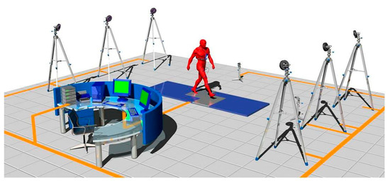
Figure 2.
Schematic diagram of the gait analysis laboratory (source: device manual BTS Smart).
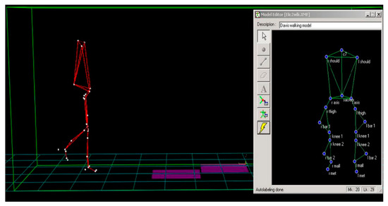
Figure 3.
Schematic diagram of the gait analysis test.
Patients performed 10–15 recorded passes. Only measurements in which each foot made full sole contact with the dynamometer platform during one pass were included for further analysis. Recording was conducted until a minimum of three correct records of the ground reaction forces of both feet were obtained. The data recorded by the cameras was transferred to a computer system, and then, using the Smart software, the signal recorded from the markers was identified and the coordinates of the markers in the three-plane space were determined, which allowed for the calculation of kinematic and dynamic quantities. Digitally processed images from the cameras enabled registration of step length (length STRIDE (m) and double step length, step velocity and transfer phase (velSTRIDE (m/s) and velSWING (m/s)), step duration–transfer phase and support (tSTRIDE (s), tSWING (s) and tSTANCE (s)). The three-plane registration of ground reaction forces enabled the analysis of vertical loading during heel rest (VEmax1), rolling (VEmin) and rebound (VEmax2), anteroposterior load distribution (APmax and min) and lateral load distribution (ML max and min). For further analysis, all ground force response values for each subject were presented as a percentage of body weight (%BW).
The research and analysis of gait biomechanics were performed at the Laboratory of Biomechanical Analysis, AWF in Wrocław.
2.3. Studies on Force-Velocity Parameters of Flexor and Extensor Muscles at the Knee and Ankle-Shin Joint
Strength capacity of muscles—flexors and extensors of the knee joint and flexors and extensors of the ankle and knee joint—was assessed using a Biodex S4 Pro dynamometer (Shirley, NY, USA). An isokinetic mode of operation was used to record the time courses of muscle force moments at an adjustable load in the form of a constant angular velocity of movement. The test protocol was identical for the knee and ankle and knee joint muscles. The loads used were 60, 120, and 180 °/s. At each given load, the subject performed 5 alternating flexion and extension movements separated by a 60 s pause. In each movement there, was an imperative to perform the given action with the maximum possible force in the shortest possible time.
Prior to the measurements, the chair and dynamometer, as well as the appropriate attachment of the device, were set up so that the tip of the dynamometer was an extension of the axis of rotation in the tested joint. The same flexion and extension ranges of motion of 90° for the knee joint and 50° for the ankle and knee joint were established for all subjects. The subject’s thigh and pelvis were stabilized with straps attached to the chair to eliminate movement of adjacent joints. The starting position of the test was the maximum flexion of the lower limb in the tested joint. Before the first measurement and after each load change, each subject performed 3 submaximal flexion and extension movements at the tested joint and 1 maximal movement to familiarize themselves with the set load [17]. The system for the analysis of force-velocity parameters is shown in Figure 4.
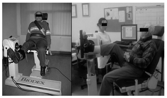
Figure 4.
Measurement of force-velocity parameters of flexor and extensor muscles of the knee and ankle (Biodex System 4; own source).
Muscle function parameters were recorded during the test: peak torque (Pt) (Nm), total work (TW) (J) and average power (aP) (W).
2.4. Statistical Analysis
Descriptive statistics were calculated. The Shapiro–Wilk test was used to check the distribution of all analyzed parameters. When the distribution of a variable was normal, Student’s t-test was used to determine the significance of differences, whereas when the distribution did not have the characteristics of normality, Wilcoxon’s paired rank test was applied. The significance level was p < 0.05. All calculations were performed using Statistica 13.3 software.
3. Results
Analyzing the ABI value showed no statistically significant differences between the average values calculated for the right and left limb for the whole group. However, statistically significant differences were observed when ABI values of the limb with higher and lower index values were compared (Table 3). For both the right and left limb, ABI values that were within normal range or indicative of a mild decrease were observed in more than 50% of the subjects. However, when comparing the limb in which the higher ABI was measured to the limb with the lower ABI, a greater variation in results was observed. In both groups, more than 30% of the subjects had ABI values between 0.89 and 0.70, indicative of a mild reduction. In the limb with the higher ratio, more than 40% of the cases were ABI ≥ 0.90 (normative and borderline values), while in the limb with the lower ratio, more than 70% were individuals whose ABI was in the 0.69–0.40 range indicative of moderate decline (Table 4) [18].
Table 3.
Ankle-brachial index (ABI) values according to the mode of analysis.
Table 4.
Variation in ABI values by mode of analysis.
Given the variation in ABI coefficient values depending on the mode of comparison, the assessment of locomotor symmetry was performed in two steps. In the first step, all analyzed right-to-left limb variables were compared.
In step two, instead of the classic analysis (right to left) in all subjects, the values measured for the limb with the higher ABI (ABI H) were compared to the values measured for the limb with the lower ABI (ABI L).
Analyzing the magnitudes of the ground reaction forces and the timespace parameters of gait, no statistically significant differences were found either between the right and left limbs or between the values obtained by the limb with the higher and lower coefficient (Table 5 and Table 6).
Table 5.
Analysis of three-plane ground reaction force values during gait according to analysis method.
Table 6.
Symmetry of spatiotemporal gait parameters according to the mode of analysis.
Regardless of the load, no statistically significant differences at the level of strength parameters of the analyzed muscles of the right and left limb were found (Table 6, Table 7, Table 8 and Table 9). However, when comparing the strength parameters of the limb with a higher ABI index (ABI H) to the values obtained by the limb with a lower index (ABI L), significant differences were observed. The limb with the higher ABI registered significantly higher peak torque (Pt), total work (TW), and average power (aP) values measured for the ankle flexors and knee extensors at each of the applied loads (Table 7, Table 8, Table 9 and Table 10).
Table 7.
Symmetry of force-velocity parameters of knee extensor muscles according to the mode of analysis.
Table 8.
Symmetry of force-velocity parameters of knee joint flexor muscles according to the mode of analysis.
Table 9.
Symmetry of force-velocity parameters of the extensor muscles of the ankle joint according to the method of analysis.
Table 10.
Symmetry of force-velocity parameters of the flexor muscles of the ankle joint according to the method of analysis.
4. Discussion
Our findings suggest that analysis of higher and lower ankle-brachial index with functional parameters is more appropriate than analysis of limb side. Therefore, our research presents mainly the effect of the degree of lower limb ischemia on gait kinematic, kinetic parameters and force-velocity parameters. Considering the right, left lower limb as well as the extremity with higher or lower ABI index, we could not confirm that the lower limb ischemia had an influence on the gait pattern of patients with PAD. However, our study of gait biomechanics was performed only under pain-free walking conditions and did not examine the gait during ischemic pain. The degree of ischemia may affect the muscle function involved in gait rather than the gait pattern itself. Therefore, gait analysis would require further assessment (e.g., including EMG studies).
An important aspect of the analysis is gait symmetry. From a pathophysiological point of view, severed blood supply to limbs weakens the muscles, which can disturb gait symmetry. However, both of our two-way analyses of temporal-spatial parameters and ground reaction forces did not confirm gait asymmetry in patients with PAD. This is not a normal condition, as previous studies confirm many abnormalities in gait pattern in patients with PAD [14,15,19,20,21]. The symmetrical gait is likely due to the similar ankle-brachial index values presented in the right and left lower limbs in the subjects. The aspect of gait symmetry would need to be verified during the claudication conditions.
As suggested by Sadeghi et al. [22], muscle strength is a good indicator of the ability to propel the body and to control balance during gait. Reduction in muscle strength and endurance along with impaired postural control during gait results from muscle fiber atrophy and altered muscle metabolism among individuals with PAD [12,23,24,25,26]. This contributes to an increased risk of falls and decreased mobility during walking or stair climbing [4,19,27]. The degree of lower extremity arterial ischemia therefore has a significant impact on the level of lower extremity muscle strength, which translates to the walking ability of individuals with PAD [3,5,10,28].
A study by Chen et al. [29], which assessed peak torque at lower limb joints under pain-free and pain-induced walking conditions, found no relationship between lower limb muscle strength and ABI scores. However, we found that force-velocity parameters evaluating peak torque, average power, total work of knee extensors and ankle flexors are dependent on the degree of lower limb ischemia. The limb side plays no role in this relationship. The generation of maximal muscular force, muscular endurance, and the engagement of motor units are closely related to their nutrition, which ensures proper blood flow [12,23,30]. The ability of muscles to undertake efforts of both strength and endurance nature (such as climbing stairs, getting up from a chair, walking a long distance) is of great importance for daily living. Impairment of muscle blood perfusion affects mainly their strength and endurance, and not a gait pattern, which engages muscles in a natural way as a reflex.
Considering the vascular system supplying the muscle groups, the most common hemodynamic abnormalities are observed at the femoropopliteal level, which are diagnosed in at least half of individuals with PAD [13,31,32]. The calf muscles, which are supplied by the posterior tibial and fibular arteries, are responsible for plantar flexion of the foot. In gait, this movement plays an important role in the support phase and propulsion of the foot and, together with the toe flexors, contributes to the acquisition of momentum through the body [20,33,34].
In our study, conducted under isokinetic conditions at angular velocities of 60 and 120 °/s, all force-velocity parameters of the ankle flexors are related to the ankle-brachial index level. This is also confirmed by McDermott et al. [35], who obtained a dependence of ABI values on the peak torque and power of the calf muscles but under isometric conditions. In contrast, at an angular velocity of 180 °/s, which is associated with low resistance and high repetition rates, a difference was obtained only between ABI and average power. Presumably, this is due to the rapid contraction and diastole of muscle fibers over a longer time interval, creating ischemic conditions during muscle work. The pathological reduction in the number and size of slow-twitch muscle fibers and the area of fast-twitch muscle fibers, the reduction in the number of capillaries per number of muscle fibers and motor units [25,26,36,37], are probably contributing to the inability to maintain a high-intensity effort for several seconds (e.g., running up to a tram or bus). It appears that the endurance of the calf muscles in people with PAD is estimated to be 30–40% compared to healthy individuals [26,36]. The study by White et al. [37] further confirms that reduced walking ability may also be influenced by mitochondrial damage, which reduces the oxidative capacity of calf muscles.
The muscles of the knee joint in individuals with PAD are also weakened, less efficient, and less enduring compared to the healthy individuals [2,12,20,23,35]. In our study, the peak torque of the knee joint flexors shows a relationship with the high, low ABI at moderate angular velocity (120 °/s), while at the highest velocity (180 °/s) this relationship was only shown in the average power. Thus, it is interesting that only the knee extensors of subjects with PAD are dependent on the degree of lower limb ischemia at all velocities analyzed. It would seem, firstly: that these muscles do not play an as important role in walking as the knee joint flexors, which was suggested by Camara et al. [23]. Knee extensors during the gait are responsible for postural stability and the risk of falls in people with PAD [17]. Secondly: the arteries supplying these muscles are less frequently constricted compared to those of the distal parts of the lower limbs [31,32,38]. However, the deep femoral artery and its branches are the major collateral blood supply route to the calf muscles in subjects with femoropopliteal arterial occlusion. Therefore, ischemia of the knee extensor muscles can result from a stealing phenomenon. Under conditions of isometric contraction, it has been confirmed that the greater ischemia, the lower power of the knee joint extensors. This relationship was not confirmed by the peak torque parameter [35]. Similarly, McDermott, Criqui et al. [3] demonstrated the relationship of ABI with the power of extensors of the knee joint. It turns out that the power parameter of the extensors of the knee joint is the most important predictor of functional ability in older people [39], which in everyday life is most often expressed in getting up from a chair or climbing stairs.
In physiological gait, the rectus femoris muscle is activated only during the stance to swing transition, and the knee joint extension during the swing phase is performed passively. The three heads of quadriceps femoris (medial, vastus, lateral) are activated during the double support phase and have a stabilizing effect on the knee joint [40]. When analyzing activities of daily living, the function of the quadriceps femoris muscles is therefore more marked in getting up from a chair than in the gait function itself. This confirmed that gait speed of people with PAD is dependent on the speed of rising from a chair [41]. Additionally, in individuals with chronic lower extremity ischemia, knee extensors engage as much as 10% slower during the supporting phase of gait compared to healthy individuals, which is due to impaired neuromuscular conduction function [42,43]. Neuromuscular slowdown also affects the plantar flexors of the foot as a result of chronic ischemia [43].
5. Conclusions
In conclusion, this research suggests that the analysis of the ankle-brachial index based on its higher, lower values gives reliable information about changes in the gait pattern and muscle strength and endurance in PAD patients. The comparison of right to the left lower limbs in PAD patients showed no influence on the analyses of the ABI with the functional parameters. Verification of differences between muscle force-velocity parameters and the severity of the leg ischemia was confirmed in the assessment of the ankle flexors and knee extensors in all analyzed parameters. It means that the higher degree of ABI is connected with worsening of the strength, endurance, and performance of the ankle flexors and knee extensors. Interestingly, ABI is not related to the gait pattern. This should be taken into account during the methodological assessment and planning of the rehabilitation process for people with different degrees of ischemia. Further research is required.
Author Contributions
Conceptualization, A.S. and M.W.; methodology, M.S., K.B. and W.D.; validation M.S., K.B. and W.D.; formal analysis, M.S.; investigation, M.S., K.B. and W.D.; resources, A.S. and M.W.; data curation, M.S. and K.B.; writing—original draft preparation, M.S., K.B. and W.D.; writing—review and editing, M.S., K.B. and W.D.; visualization, M.S.; supervision, A.S.; project administration, A.S. and M.W. All authors have read and agreed to the published version of the manuscript.
Funding
This research was funded by the “WROVASC—Integrated Center for Cardiovascular Medicine” project, co-financed by the European Regional Development Fund under the Innovative Economy Operational Program 2007–2013, and implemented at the Research and Development Center of the Provincial Specialist Hospital in Wroclaw, Poland.
Institutional Review Board Statement
The study was conducted according to the guidelines of the Declaration of Helsinki, and approved by the Ethics Committee of the Medical University of Wroclaw, Poland (Ref.KB-130/2008). These studies were registered in the clinical trial database—the Australian New Zealand Clinical Trials Registry (Trial Id: ACTRN12621000780853).
Informed Consent Statement
Informed consent was obtained from all subjects involved in the study. Written informed consent has been obtained from the patient(s) to publish this paper.
Data Availability Statement
All data supporting reported results can be found in the archives of the Wrovasc Research Centre (https://www.wssobr-wroc.pl/projekty/wrovasc/).
Conflicts of Interest
The authors declare no conflict of interest.
References
- Szuba, A.; Oka, R.K.; Harada, R.; Cooke, J.P. Limb Hemodynamics Are Not Predictive of Functional Capacity in Patients with PAD. Vasc. Med. 2006, 11, 155–163. [Google Scholar] [CrossRef] [PubMed]
- McDermott, M.M.; Guralnik, J.M.; Tian, L.; Liu, K.; Ferrucci, L.; Liao, Y.; Sharma, L.; Criqui, M.H. Associations of borderline and low normal ankle brachial index values with functional decline at five year follow-up. J. Am. Coll. Cardiol. 2009, 53, 1056–1062. [Google Scholar] [CrossRef] [PubMed]
- McDermott, M.M.; Criqui, M.H.; Greenland, P.; Guralnik, J.M.; Liu, K.; Pearce, W.H.; Taylor, L.; Chan, C.; Celic, L.; Woolley, C.; et al. Leg Strength in Peripheral Arterial Disease: Associations with Disease Severity and Lower-Extremity Performance. J. Vasc. Surg. 2004, 39, 523–530. [Google Scholar] [CrossRef] [PubMed]
- McDermott, M.M.; Ferrucci, L.; Guralnik, J.M.; Dyer, A.R.; Liu, K.; Pearce, W.H.; Clark, E.; Liao, Y.; Criqui, M.H. The Ankle–Brachial Index Is Associated with the Magnitude of Impaired Walking Endurance among Men and Women with Peripheral Arterial Disease. Vasc. Med. 2010, 15, 251–257. [Google Scholar] [CrossRef] [PubMed]
- McDermott, M.M.; Greenland, P.; Liu, K.; Guralnik, J.M.; Celic, L.; Criqui, M.H.; Chan, C.; Martin, G.J.; Schneider, J.; Pearce, W.H.; et al. The Ankle Brachial Index Is Associated with Leg Function and Physical Activity: The Walking and Leg Circulation Study. Ann. Intern. Med. 2002, 136, 873–883. [Google Scholar] [CrossRef]
- Miossec, A.; Tollenaere, Q.; Lanéelle, D.; Guilcher, A.; Métairie, A.; Le Pabic, E.; Carel, A.; Le Faucheur, A.; Mahé, G. Arterial Doppler Waveforms Are Independently Associated with Maximal Walking Distance in Suspected Peripheral Artery Disease Patients. Front. Cardiovasc. Med. 2021, 8, 608008. [Google Scholar] [CrossRef] [PubMed]
- Matsushita, K.; Ballew, S.H.; Sang, Y.; Kalbaugh, C.; Loehr, L.R.; Hirsch, A.T.; Tanaka, H.; Heiss, G.; Windham, B.G.; Selvin, E.; et al. Ankle-Brachial Index and Physical Function in Older Individuals: The Atherosclerosis Risk in Communities (ARIC) Study. Atherosclerosis 2017, 257, 208–215. [Google Scholar] [CrossRef] [PubMed]
- McDermott, M.M.; Greenland, P.; Ferrucci, L.; Criqui, M.H.; Liu, K.; Sharma, L.; Chan, C.; Celic, L.; Priyanath, A.; Guralnik, J.M. Lower Extremity Performance Is Associated with Daily Life Physical Activity in Individuals with and without Peripheral Arterial Disease. J. Am. Geriatr. Soc. 2002, 50, 247–255. [Google Scholar] [CrossRef]
- McDermott, M.M.; Liu, K.; Greenland, P.; Guralnik, J.M.; Criqui, M.H.; Chan, C.; Pearce, W.H.; Schneider, J.R.; Ferrucci, L.; Celic, L.; et al. Functional Decline in Peripheral Arterial Disease: Associations with the Ankle Brachial Index and Leg Symptoms. JAMA 2004, 292, 453–461. [Google Scholar] [CrossRef]
- Parmenter, B.J.; Raymond, J.; Dinnen, P.J.; Lusby, R.J.; Fiatarone Singh, M.A. Preliminary Evidence That Low Ankle-Brachial Index Is Associated with Reduced Bilateral Hip Extensor Strength and Functional Mobility in Peripheral Arterial Disease. J. Vasc. Surg. 2013, 57, 963–973. [Google Scholar] [CrossRef]
- Dolan, N.C.; Liu, K.; Criqui, M.H.; Greenland, P.; Guralnik, J.M.; Chan, C.; Schneider, J.R.; Mandapat, A.L.; Martin, G.; McDermott, M.M. Peripheral Artery Disease, Diabetes, and Reduced Lower Extremity Functioning. Diabetes Care 2002, 25, 113–120. [Google Scholar] [CrossRef]
- Dziubek, W.; Bulińska, K.; Stefańska, M.; Woźniewski, M.; Kropielnicka, K.; Jasiński, T.; Jasiński, R.; Pilch, U.; Dąbrowska, G.; Skórkowska-Telichowska, K.; et al. Peripheral Arterial Disease Decreases Muscle Torque and Functional Walking Capacity in Elderly. Maturitas 2015, 81, 480–486. [Google Scholar] [CrossRef] [PubMed]
- Dziubek, W.; Stefańska, M.; Bulińska, K.; Barska, K.; Paszkowski, R.; Kropielnicka, K.; Jasiński, R.; Rachwalik, A.; Woźniewski, M.; Szuba, A. Effects of Physical Rehabilitation on Spatiotemporal Gait Parameters and Ground Reaction Forces of Patients with Intermittent Claudication. J. Clin. Med. 2020, 9, 2826. [Google Scholar] [CrossRef]
- Pietraszewski, B.; Woźniewski, M.; Jasiński, R.; Struzik, A.; Szuba, A. Changes in Gait Variables in Patients with Intermittent Claudication. BioMed Res. Int. 2019, 2019, e7276865. [Google Scholar] [CrossRef]
- Scott-Okafor, H.R.; Silver, K.K.; Parker, J.; Almy-Albert, T.; Gardner, A.W. Lower Extremity Strength Deficits in Peripheral Arterial Occlusive Disease Patients with Intermittent Claudication. Angiology 2001, 52, 7–14. [Google Scholar] [CrossRef]
- Crawford, F.; Welch, K.; Andras, A.; Chappell, F.M. Ankle Brachial Index for the Diagnosis of Lower Limb Peripheral Arterial Disease. Cochrane Database Syst. Rev. 2016, 9, CD010680. [Google Scholar] [CrossRef]
- Davies, G. A Compendium of Isokinetcis Usage and Clinical Rehabilitation Techniques, 4th ed.; S&S Publishers: Winona, MN, USA, 1992. [Google Scholar]
- Firnhaber, J.M.; Powell, C.S. Lower Extremity Peripheral Artery Disease: Diagnosis and Treatment. Am. Fam. Physician 2019, 99, 362–369. [Google Scholar]
- Gardner, A.W.; Forrester, L.; Smith, G.V. Altered Gait Profile in Subjects with Peripheral Arterial Disease. Vasc. Med. 2001, 6, 31–34. [Google Scholar] [CrossRef] [PubMed]
- McCamley, J.D.; Cutler, E.L.; Schmid, K.K.; Wurdeman, S.R.; Johanning, J.M.; Pipinos, I.I.; Myers, S.A. Gait Mechanics Differences between Healthy Controls and Patients with Peripheral Artery Disease after Adjusting for Gait Velocity, Stride Length, and Step Width. J. Appl. Biomech. 2019, 35, 19–24. [Google Scholar] [CrossRef] [PubMed]
- Myers, S.A.; Johanning, J.M.; Stergiou, N.; Celis, R.I.; Robinson, L.; Pipinos, I.I. Gait Variability Is Altered in Patients with Peripheral Arterial Disease. J. Vasc. Surg. 2009, 49, 924–931. [Google Scholar] [CrossRef]
- Sadeghi, H.; Allard, P.; Prince, F.; Labelle, H. Symmetry and Limb Dominance in Able-Bodied Gait: A Review. Gait Posture 2000, 12, 34–45. [Google Scholar] [CrossRef]
- Câmara, L.C.; Ritti-Dias, R.M.; Menêses, A.L.; D’Andréa Greve, J.M.; Filho, W.J.; Santarém, J.M.; Forjaz, C.L.D.M.; Puech-Leão, P.; Wolosker, N. Isokinetic Strength and Endurance in Proximal and Distal Muscles in Patients with Peripheral Artery Disease. Ann. Vasc. Surg. 2012, 26, 1114–1119. [Google Scholar] [CrossRef]
- Lanzarin, M.; Parizoto, P.; Santos, G.M. Analysis of Isokinetic Muscle Function and Postural Control in Individuals with Intermittent Claudication. Braz. J. Phys. Ther. 2016, 20, 48–57. [Google Scholar] [CrossRef][Green Version]
- McDermott, M.M.; Ferrucci, L.; Gonzalez-Freire, M.; Kosmac, K.; Leeuwenburgh, C.; Peterson, C.A.; Saini, S.; Sufit, R. Skeletal Muscle Pathology in Peripheral Artery Disease: A Brief Review. Arterioscler. Thromb. Vasc. Biol. 2020, 40, 2577–2585. [Google Scholar] [CrossRef] [PubMed]
- Regensteiner, J.G.; Wolfel, E.E.; Brass, E.P.; Carry, M.R.; Ringel, S.P.; Hargarten, M.E.; Stamm, E.R.; Hiatt, W.R. Chronic Changes in Skeletal Muscle Histology and Function in Peripheral Arterial Disease. Circulation 1993, 87, 413–421. [Google Scholar] [CrossRef]
- Richardson, J.K.; Eckner, J.T.; Allet, L.; Kim, H.; Ashton-Miller, J.A. Complex and Simple Clinical Reaction Times Are Associated with Gait, Balance, and Major Fall Injury in Older Subjects with Diabetic Peripheral Neuropathy. Am. J. Phys. Med. Rehabil. 2017, 96, 8–16. [Google Scholar] [CrossRef]
- McDermott, M.M.; Ferrucci, L.; Guralnik, J.; Tian, L.; Liu, K.; Hoff, F.; Liao, Y.; Criqui, M.H. Pathophysiological Changes in Calf Muscle Predict Mobility Loss at 2-Year Follow-up in Men and Women with Peripheral Arterial Disease. Circulation 2009, 120, 1048–1055. [Google Scholar] [CrossRef] [PubMed]
- Chen, S.-J.; Pipinos, I.; Johanning, J.; Radovic, M.; Huisinga, J.M.; Myers, S.A.; Stergiou, N. Bilateral Claudication Results in Alterations in the Gait Biomechanics at the Hip and Ankle Joints. J. Biomech. 2008, 41, 2506–2514. [Google Scholar] [CrossRef] [PubMed]
- Kannus, P. Isokinetic Evaluation of Muscular Performance: Implications for Muscle Testing and Rehabilitation. Int. J. Sports Med. 1994, 15 (Suppl. 1), S11–S18. [Google Scholar] [CrossRef]
- Schulte, K.-L.; Hardung, D.; Tiefenbacher, C.; Weiss, T.; Hoffmann, U.; Amendt, K.; Tepe, G.; Heuser, L.; Treszl, A.; Lau, H.-J.; et al. Real-World Outcomes of Endovascular Treatment in a Non-Selected Population with Peripheral Artery Dise—Prospective Study with 2-Year Follow-Up. VASA Z. Gefasskrankh. 2019, 48, 433–441. [Google Scholar] [CrossRef] [PubMed]
- Skórkowska-Telichowska, K.; Kropielnicka, K.; Bulińska, K.; Pilch, U.; Woźniewski, M.; Szuba, A.; Jasiński, R. Insufficient Modification of Atherosclerosis Risk Factors in PAD Patients. Adv. Clin. Exp. Med. 2018, 27, 819–826. [Google Scholar] [CrossRef]
- Chambers, H.G.; Sutherland, D.H. A Practical Guide to Gait Analysis. J. Am. Acad. Orthop. Surg. 2002, 10, 222–231. [Google Scholar] [CrossRef]
- Arias, D.G.; Marappa-Ganeshan, R. Anatomy, Bony Pelvis and Lower Limb, Arteries. In StatPearls; StatPearls Publishing: Treasure Island, FL, USA, 2021. [Google Scholar]
- McDermott, M.M.; Tian, L.; Ferrucci, L.; Liu, K.; Guralnik, J.M.; Liao, Y.; Pearce, W.H.; Criqui, M.H. Associations between Lower Extremity Ischemia, Upper and Lower Extremity Strength, and Functional Impairment with Peripheral Arterial Disease. J. Am. Geriatr. Soc. 2008, 56, 724–729. [Google Scholar] [CrossRef] [PubMed]
- Askew, C.D.; Green, S.; Walker, P.J.; Kerr, G.K.; Green, A.A.; Williams, A.D.; Febbraio, M.A. Skeletal Muscle Phenotype Is Associated with Exercise Tolerance in Patients with Peripheral Arterial Disease. J. Vasc. Surg. 2005, 41, 802–807. [Google Scholar] [CrossRef]
- White, S.H.; McDermott, M.M.; Sufit, R.L.; Kosmac, K.; Bugg, A.W.; Gonzalez-Freire, M.; Ferrucci, L.; Tian, L.; Zhao, L.; Gao, Y.; et al. Walking Performance Is Positively Correlated to Calf Muscle Fiber Size in Peripheral Artery Disease Subjects, but Fibers Show Aberrant Mitophagy: An Observational Study. J. Transl. Med. 2016, 14, 284. [Google Scholar] [CrossRef]
- Aboyans, V.; Desormais, I.; Lacroix, P.; Salazar, J.; Criqui, M.H.; Laskar, M. The General Prognosis of Patients with Peripheral Arterial Disease Differs According to the Disease Localization. J. Am. Coll. Cardiol. 2010, 55, 898–903. [Google Scholar] [CrossRef] [PubMed]
- Byrne, C.; Faure, C.; Keene, D.J.; Lamb, S.E. Ageing, Muscle Power and Physical Function: A Systematic Review and Implications for Pragmatic Training Interventions. Sports Med. 2016, 46, 1311–1332. [Google Scholar] [CrossRef] [PubMed]
- Nene, A.; Byrne, C.; Hermens, H. Is Rectus Femoris Really a Part of Quadriceps? Assessment of Rectus Femoris Function during Gait in Able-Bodied Adults. Gait Posture 2004, 20, 1–13. [Google Scholar] [CrossRef]
- Correia, M.D.A.; Cucato, G.G.; Lanza, F.C.; Peixoto, R.A.O.; Zerati, A.E.; Puech-Leao, P.; Wolosker, N.; Ritti-Dias, R.M. Relationship between Gait Speed and Physical Function in Patients with Symptomatic Peripheral Artery Disease. Clinics 2019, 74, e1254. [Google Scholar] [CrossRef]
- McDermott, M.M.; Guralnik, J.M.; Albay, M.; Bandinelli, S.; Miniati, B.; Ferrucci, L. Impairments of Muscles and Nerves Associated with Peripheral Arterial Disease and Their Relationship with Lower Extremity Functioning: The InCHIANTI Study. J. Am. Geriatr. Soc. 2004, 52, 405–410. [Google Scholar] [CrossRef] [PubMed]
- Savelberg, H.H.C.M.; Ilgin, D.; Angin, S.; Willems, P.J.B.; Schaper, N.C.; Meijer, K. Prolonged Activity of Knee Extensors and Dorsal Flexors Is Associated with Adaptations in Gait in Diabetes and Diabetic Polyneuropathy. Clin. Biomech. 2010, 25, 468–475. [Google Scholar] [CrossRef] [PubMed]


Publisher’s Note: MDPI stays neutral with regard to jurisdictional claims in published maps and institutional affiliations. |
© 2021 by the authors. Licensee MDPI, Basel, Switzerland. This article is an open access article distributed under the terms and conditions of the Creative Commons Attribution (CC BY) license (https://creativecommons.org/licenses/by/4.0/).