Article

Identification of Fusarium oxysporum Causing Leaf Blight on Dendrobium chrysotoxum in Yunnan Province, China

1
College of Resources, Environment and Chemistry, Chuxiong Normal University, Chuxiong 675000, China
2
State Key Laboratory for Conservation and Utilization of Bio-Resources, Yunnan Agricultural University, Kunming 650201, China
3
Dehong Insitute of Tropical Agriculture in Yunnan Province, Ruili 678600, China
*
Authors to whom correspondence should be addressed.
These authors contributed equally to this work and shared first authorship.
Life 2024, 14(3), 285; https://doi.org/10.3390/life14030285
Submission received: 28 December 2023 / Revised: 6 February 2024 / Accepted: 17 February 2024 / Published: 20 February 2024
(This article belongs to the Special Issue Plant Diseases: Interactions, Resistance, Epidemiology, and Control)

Abstract

:
Leaf-blight disease caused by the Fusarium oxysporum is an emerging problem in Dendrobium chrysotoxum production in China. Symptoms of leaf blight were observed on seedlings of D. chrysotoxum cultivated in a nursery in Ruili City, Yunnan Province, China. In this study, we isolated the Fusarium sp. associated with leaf-blight disease of D. chrysotoxum from the diseased seedlings. A pathogenicity test was performed to fulfill Koch’s postulates to confirm the pathogenicity of isolated strains and identified using morphological and molecular techniques. The results revealed that all four isolated Fusarium sp. isolates (DHRL-01~04) produced typical blight symptoms followed by marginal necrosis of leaves on the D. chrysotoxum plants. On the PDA medium, the fungal colony appeared as a white to purplish color with cottony mycelium growth. Microconidia are oval-shaped, whereas macroconidia are sickle-shaped, tapering at both ends with 2–4 septations. The phylogenetic trees were construed based on internal transcribed spacer (ITS), translation elongation factor (EF-1α), and RNA polymerase subunit genes RPB1 and RPB2 genes, respectively, and blasted against the NCBI database for species confirmation. Based on the NCBI database’s blast results, the isolates showed that more than 99% identify with Fusarium oxysporum. To our knowledge, this is the first comprehensive report on the identification of Fusarium oxysporum as the causal agent of Dendrobium chrysotoxum leaf blight in Yunnan Province, China, based on morphological and molecular characteristics.

1. Introduction

Orchidaceae is the chief family of plants, with more than 25,000 plant species worldwide [1]. They are also some of the most vulnerable flowering plants, as many genera are endangered, and almost all are at risk of habitat damage and overclassification [2]. The Dendrobium represents one of the most important genera of the Orchidaceae, first identified by Olaf Swartz in 1799 AD, which comprises approximately 1500 to 2000 species [3]; among them, 74 species and two varieties are native to China [4]. Genus Dendrobium contains species with high medicinal and ornamental values, such as the D. chrysotoxum Lindl. [5], D. fimbriatum Hook [6], D. nobile Lindl. [7], and D. officinale Kimura et Migo [8]. In China, Dendrobium species have traditionally been used as first-rate medicinal herbs to treat various disorders, such as stomach nourishment and diabetes [9]. The stems and leaves of Dendrobium are rich sources of polysaccharides, and one class that contains mannans is used in drugs and has a high market value [10]. Dendrobium plant parts, especially leaves and stems, are used in traditional Chinese medicine (TCM) to control diabetes, rheumatoid arthritis, obesity, and many other diseases and are enlisted in the famous 18 ancient TCM [11]. In China, the history of Dendrobium was written about 2300 years ago as “Shen Nong’s Herbal Classic (the Eastern Han Dynasty)” TCM, and it is cultivated as both a medicinal and ornamental plant [12]. Among the Dendrobium species, D. officinale has the foremost therapeutic properties and is used as “Shihu” in TCM. Many species of Dendrobium are the sources of tonic for astringents, analgesics, antipyretics, antioxidants, antimicrobials, antidiabetics, anticancers, antiinflammatories, antimetastases, and antiangiogenetics because they have alkaloids, aromatic compounds, sesquiterpenoids, and polysaccharides as main components [13].
D. chrysotoxum is known as a “fried egg” orchid and is widely cultivated as an ornamental and medicinal plant [14]. D. chrysotoxum has high medicinal value due to its enrichment of various types of chemical constituents, such as bibenzyls, phenanthrenes, and fluorenones in its plant tissues [15], which have been indicated to possess antiangiogenic, anticataractogenic, antitumor, antidiabetic retinopathy and antiacetylcholine esterase activities by in vitro and in vivo pharmacological experiments [16]. For example, Erianin, as the main active ingredient isolated from Dendrobium chrysotoxum, exhibits potential antioxidant, antiangiogenic, and antitumor activity in various malignancies, including gastric cancer, liver cancer, lung cancer, breast cancer, and osteosarcoma [17]. Additionally, due to the pure bright yellow color, strong fragrance, and long-lasting floral scent in its flowers, D. chrysotoxum has ornamental values as great as potted and cut flowers [18]. It is native to Southeast Asian countries and growing naturally in Assam (India), Burma (Myanmar), Bangladesh, Thailand, Vietnam, and Yunnan (China). It prefers cool to warm environmental conditions and is grown at an elevation of 700 to 1100 m [19]. D. chrysotoxum produced microscopic seeds fenced within an apparent testa cover. Plants are small sized with clustered clavate and pseudobulbs of 12 to 30 cm in height. The pseudobulbs produce up to 20 flowers with high fragrance but are short lived. The flowers are yellow and have orange-colored lips with pleated edges [20]. In China, in nursery and field conditions, D. chrysotoxum plants have been affected by the pathogenic fungi Pythium vexans, the causative agent of stem-rot disease [21].
The genus Fusarium, a large group of filamentous fungi, belongs to the family Nectriaceae and is pathogenic to plants [22]. Based on its scientific and economic importance, Fusarium was recently included in the top-10 most important plant pathogenic fungal genera worldwide [23]. Fusarium oxysporum f. sp. cubense, the causative agent of banana Panama disease, leads to billions of dollars in losses to farmers, and in case of a severe outbreak, the death of the whole crop occurs [24]. The Fusarium graminearum and Fusarium oxysporum f. sp. vasinfectum cause Fusarium head blight (FHB) disease in wheat and Fusarium wilt (FW) disease in cotton, respectively, as devastating diseases in the field [25,26]. Fusarium as pathogens that cause foliar and root diseases in orchids have been reported, including Fusarium oxysporum, Fusarium proliferatum, Fusarium solani, Fusarium subglutinans, and Fusarium fractiflexum [27]. The commonly employed methods are the morphological characterization and ITS sequencing identification of the Fusarium species complex. Fusarium complex is nonspecific and commonly causes root rot, decline in vigor, and wilt symptoms and is responsible for the mortality of taxonomically diverse groups of plants. On the other hand, numerous species of Fusarium oxysporum cause seed rot, damping off, and vascular wilt symptoms in vegetables, fruits, and ornamental plants [28]. Wilt-disease complexes have been reported in many vegetables, field crops, plantation crops, fruit trees, medicinal crops, etc., as the pathogen has a wide host range.
In August 2019, leaf-blight symptoms were first observed in the D. chrysotoxum nursery at the Dehong Institute of Tropical Agriculture, Ruili City, Yunnan Province, China. This disease causes necrosis and dryness of leaf tips after 7 days of seeding, with a disease incidence of 60%. The disease expansion causes plant leaves to fall off, which limits plant growth and continues to spread over 80% of the plants under high temperatures and humidity, becoming a huge obstacle and seriously threatening the production of D. chrysotoxum in Yunnan. This study aimed to isolate and identify the causal agent of leaf blight disease of D. chrysotoxum through morphological and molecular techniques, which will help us develop integrated disease management strategies to minimize production losses.

2. Materials and Methods

2.1. Sample Collection

In August 2019, the samples of Dendrobium chrysotoxum (cv. Jin Gu Zi Chui, accession 20140053) showed leaf blight disease symptoms (yellowing of leaves and marginal necrosis). This disease causes necrosis and dryness at the tips of the leaves. Leaves shedding occurs in the later stages of disease development, which can lead to limited plant growth and continues to spread in approximately 80% of plants under high temperature and humidity conditions in the nursery. The diseased leaf samples (yellowing of leaves and marginal necrosis) were collected from the nursery in Ruili City (24° N, 98° E), Yunnan Province, China. Disease samples were kept in the icebox until delivered to the laboratory and stored at 4 °C for isolation of the pathogenic fungi and future use. Healthy D. chrysotoxum plants were collected from the Dehong Institute of Tropical Agriculture, Ruili City, to confirm the pathogenicity of the isolates.

2.2. Isolation of the Pathogen

The pathogen was isolated from leaf parts, and the diseased leaves were initially washed with sterile water. Then, the margin of diseased leaves was cut into small pieces (0.5 × 2 cm) with a sterilized blade, surface sterilized with a 75% (v/v) ethanol solution for two minutes and washed thrice with the sterilized distilled water. Three to five pieces from each sample were then placed in the Petri plates containing potato–dextrose–agar (PDA) (potato 200 g/L, dextrose 20 g/L, agar 18 g/L, and pH 7.0) and incubated at 28 °C in the dark. After 5 to 7 days of incubation based on colony morphology, fungal hyphae were picked, placed on PDA plates, purified by a single-spore technique, and stored at 4 °C in PDA slants for future use [29].

2.3. Morphological Identification

A single spore was picked with the help of a sterilized needle from the pure culture of each strain, placed in the Petri plates containing PDA as an artificial growth medium, and incubated at 28 °C for 7 to 10 days. Morphological identification was made using different identification keys for Fusarium spp., e.g., vegetative growth on culture characteristics, colony color, and reproductive structure. The spore characteristics (shape and size) were observed under an Olympus BX53 microscope (Tokyo, Japan) [28,30].

2.4. DNA Extraction

Total genomic DNA was extracted from the 7-to-10-day-old cultures of isolated fungi by scraping the surfaces of growing colonies on PDA. For DNA extraction, fresh mycelium from all four isolates was harvested aseptically. Around 1 g of mycelium was used for DNA extraction using the cetyltrimethylammonium bromide (CTAB) method, as described by Brandfass’s report [31]. The concentration and quality of extracted DNA was checked using a NanoDrop spectrophotometer (ND2000, Thermo Scientific, Wilmington, WA, USA) and by gel electrophoresis in 1.0% (w/v) agarose gel prepared in 1 × TAE buffer solutions, and the electrophoresis was run for 25 min at 160 V. The DNA was then stained with ethidium bromide (2 mg/L), and the gel was visualized under ultraviolet (UV) light (Bio-Rad Gel Doc™ XR Imaging system, Wilmington, WA, USA) [32]. The extracted DNA was stored at −20 °C for future use.

2.5. PCR Amplification, Molecular Identification, and Construction of Phylogenetic Tree

The isolated strains were identified by PCR amplification of four different genes, including the internal transcribed spacer (ITS) specific region of rDNA, the largest subunit of RNA polymerase Ⅰ (RPB1), the second-largest subunit of RNA polymerase Ⅱ (RPB2), and translation elongation factor (EF-1α) were amplified using four corresponding primer pairs ITS1/ITS4, EF1/EF2, F7/R8, 5F2/7CR. The primers used in this study to amplify ITS, RPB1, RPB2, and EF-1α genes are listed in Table 1 [33,34,35]. All PCRs were performed in a 25 μL reaction mixture containing 12 μL 2×EasyTaq® PCR SuperMix (Takara Biotechnology Co., Ltd., Dalian, China), 100 ng of genomic DNA as template, 1 μL of each forward and reverse primers (10 uM), and ddH2O adjusted to the final volume. The PCR amplification conditions were as follows: initial denaturation at 94 °C for 5 min, followed by 35 amplification cycles of denaturation at 94 °C for 30 s, annealing at 55 °C for 30 s, extension at 72 °C for 1 min, and final extension at 72 °C for 7 min.
The PCR amplicon products were visualized by the gel electrophoresis in 1.0% (w/v) agarose gel and sent to the company (TSINGKE® Co. Ltd., Beijing, China) for sequencing by the Sanger method. The sequences generated in this study were subjected to a BLAST search in the GenBank nucleotide database (http://www.ncbi.nlm.nih.gov/BLAST, accessed on 14 February 2021), and sequences were deposited in the NCBI GenBank. The phylogenetic trees based on sequences of 4 genes (ITS, RPB1, RPB2, and EF-1α) were constructed separately using the aligned nucleotide sequences with 1000 bootstrap replicates (removing gaps) following the maximum likelihood tree phylogeny of MEGA software version 7.0.21. Further, the DNA sequences based on the MYCOBANK online Fusarium MLTS database (https://fusarium.mycobank.org/, accessed on 22 March 2022) were accessed for species identification [36].

2.6. Pathogenicity Test

According to Koch’s postulates, a pathogenicity test was performed to confirm the pathogenicity of 4 isolated strains. Spore suspension of all four isolated strains was prepared according to Han’s protocols [37]. The 4 isolates suspension was obtained from 7-to-10-day-old cultures grown on a PDA medium, prepared from the actively grown mycelium of the 7-day-old culture in 15 mL sterilized distilled water in each of the Petri dishes, and a sterile inoculation needle was used to scrape the mycelium on the medium surface. Mycelium was harvested aseptically from the medium’s surface, and spore suspension was adjusted to 1 × 10⁶ conidia/mL using a hemocytometer. The collected spores were observed under an optical microscope (Olympus BX53 microscope, Tokyo, Japan). Two-month-old D. chrysotoxum (cv. Jin Gu Zi Chui) plants were sprayed with a 20 mL, 1 × 10⁶ conidia/mL spore suspension, whereas the control plants were treated with sterilized ddH2O (water control). The treated plants were kept in the greenhouse at 28 ± 2 °C and 75% relative humidity [38]. The experiment was repeated thrice for each isolated strain, with five plants in each treatment as replicates.

2.7. Pathogen Reisolation

For pathogen reisolation, the leaf samples from the water control plants and the pathogen-inoculated plants were prepared, incubated, and mycelia subcultured on PDA as to the previous process. DNA was extracted from the reisolated pathogens, and PCR products were sent for Sanger sequencing at TSINGKE® Co. Ltd. (Kunming, China). A BLAST search of the resulting ITS and EF-1α sequences was performed using the NCBI database.

3. Results

3.1. Nursery Observations and Isolations

In August 2019, a disease survey was conducted on D. chrysotoxum (cv. Jin Gu Zi Chui) nurseries (110 seedbeds) in four greenhouses at the Dehong Institute of Tropical Agriculture, Ruili City. After 7 days of seeding, as the disease initiated, necrotic water-soaked corrugated lesions were first observed on the tips of blades (Figure 1A), and later, the necrotic lesion expanded to over half of the leaf blade with a yellow border (Figure 1B). In case of a severe attack, the disease expansion causes plant leaves to fall off (Figure 1C). During the wet summer, the disease incidence rapidly increased, which affected more than 60% of the plants in the nursery. Four isolates, DHRL-01, DHRL-02, DHRL-03, and DHRL-04, were isolated from the infected leaves of D. chrysotoxum. Morphological and molecular characterization were performed for the identification of the isolated pathogen.

3.2. Morphological Identification

The four isolated strains were grown on a PDA medium for seven days for morphological identification. The strain colonies were characterized by an abundant white cottony mycelium and a dark-purple undersurface on the PDA medium (Figure 2A,B). Strains produced three types of spores: microconidia, macroconidia, and chlamydospores. Microconidia were oval to ellipsoid or renal shaped, without septation, and were 2.57–3.38 × 5.25–10.62 µm in size (Figure 2C). Macroconidia were sickle shaped, tapering at both ends, having 2–4 septation, and 19.32–36.55 × 3.44–5.52 µm in size (Figure 2D), while the chlamydospores were produced in the chain (Figure 2E–G). The four isolated strains were identified as Fusarium spp. based on colony morphology.

3.3. Molecular Identification

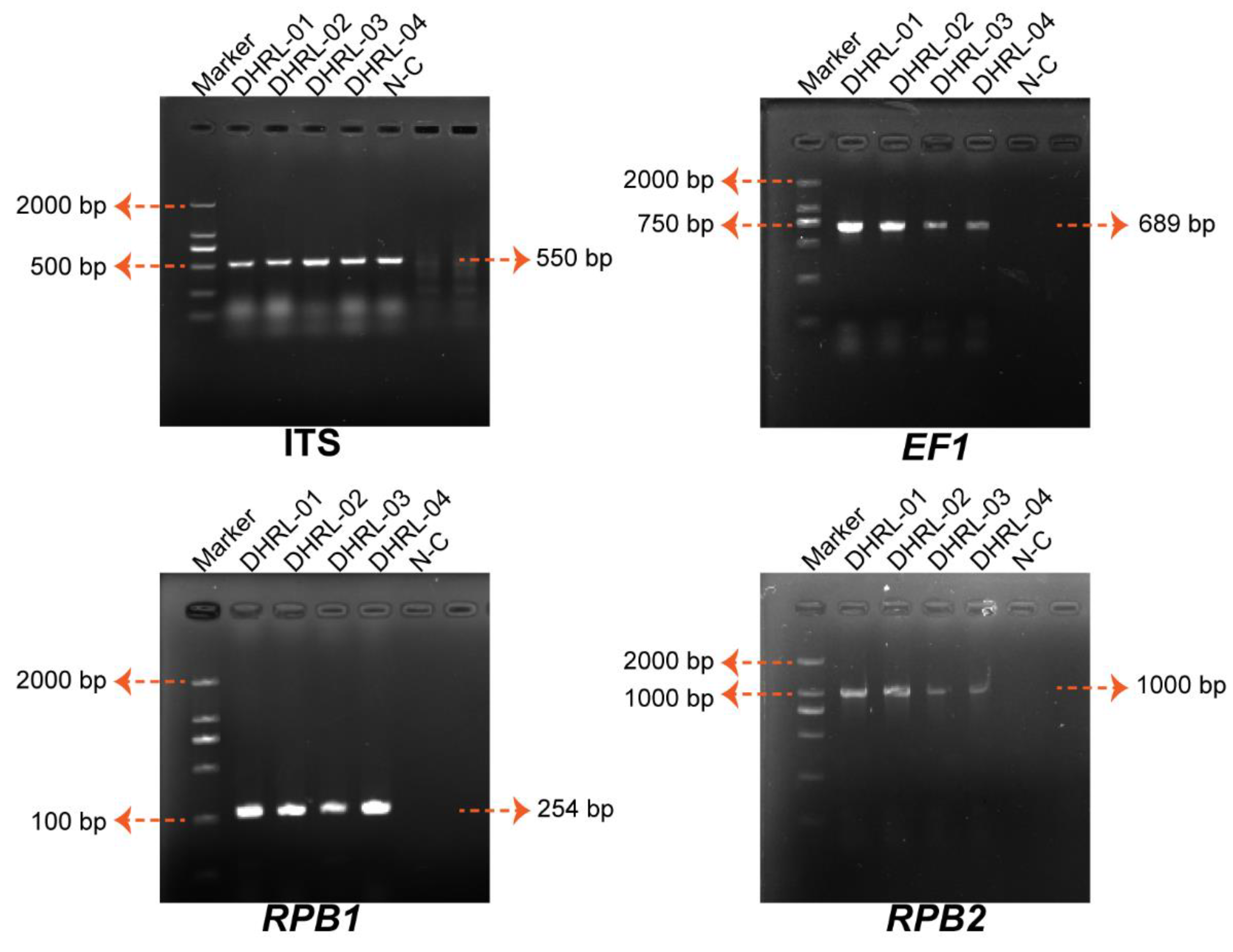
Molecular identification of isolated strains was done by the PCR amplification of ITS, EF-1α, RPB1, and RPB2 genes. Through PCR amplification, the results in a product were obtained at 527–550 bp (ITS), 687–689 bp (EF-1α), 254–256 bp (RPB1), and 995–1018 bp (RPB2) for all isolated strains (Figure 3). The isolated strains were identified as having 99.42–99.62% similarity with the sequences of Fusarium oxysporum in the NCBI database, and all sequences are deposited in the NCBI GenBank (Table 2).
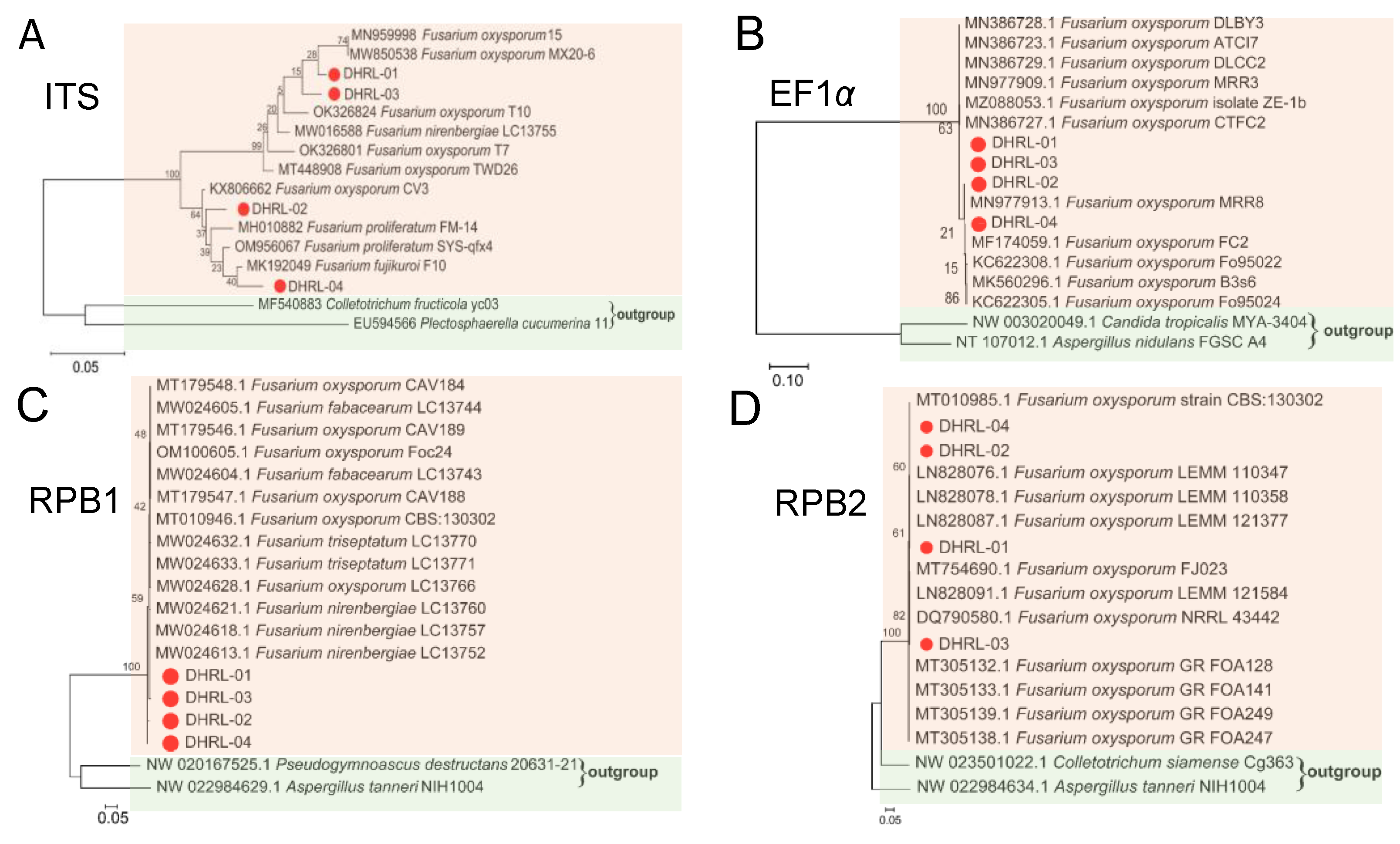
The phylogenetic tree was constructed for each gene, ITS, EF-1α, RPB1, and RPB2, to confirm the NCBI blast results (Figure 4). The phylogenetic tree results based on ITS showed that four isolated strains were grouped in one clade with reference sequences of Fusarium spp. strains, supported by a 100% bootstrap value, but bootstrap values equal to or lower than 64% are shown as isolated strains clustered together as one with Fusarium oxysporum (Figure 4A). The results of phylogenetic trees based on three genes, EF-1α, RPB1, and RPB2 sequences, showed all four isolates (DHRL-01~04) were clustered within the clade corresponding to the species Fusarium oxysporum with 100% bootstrap support (Figure 4B–D). Further research showed the species of the four isolates to be confirmed by the MLST database; the MLST was determined based on polyphasic identification using the Fusarium MLST database, and the identification result shows four isolates have 100% MLST similarity to the Fusarium oxysporum species complex. In total, we confirm the four isolates were Fusarium oxysporum isolated from Dendrobium chrysotoxum plants.

3.4. Pathogenicity Test

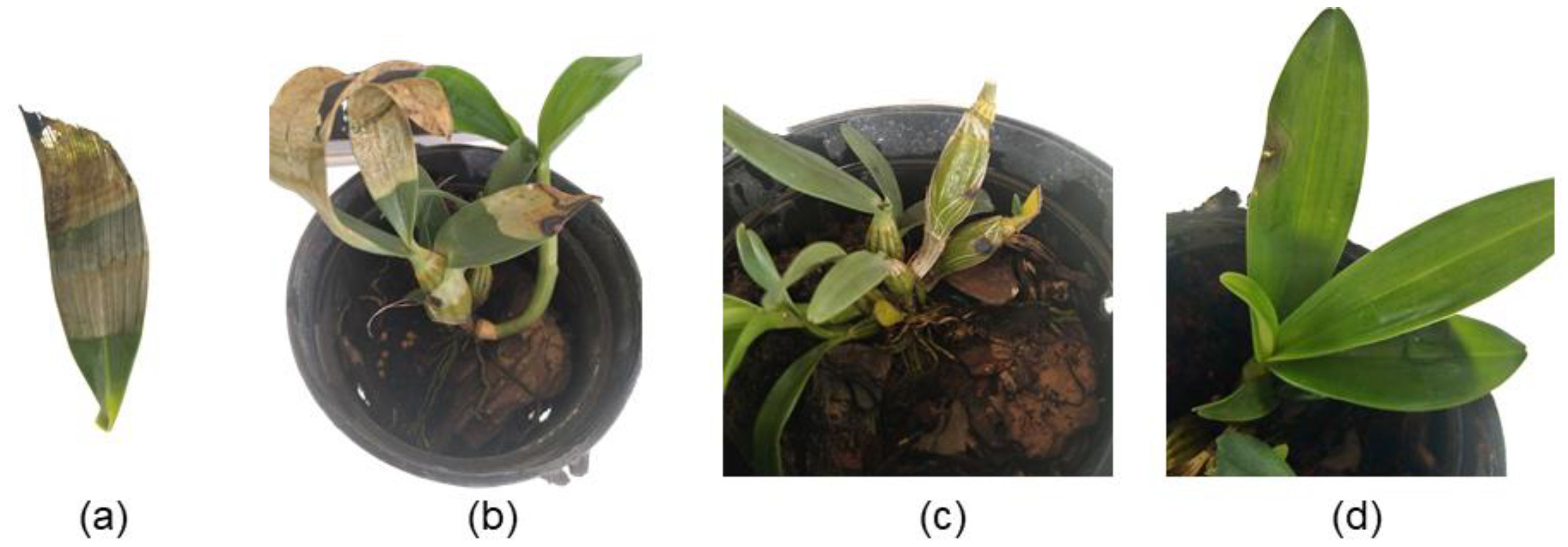
A pathogenicity test was performed to fulfill Koch’s postulates and confirm the virulence of the four isolated strains. Figure 5 shows that the symptoms developed within 30 days after inoculation of the isolate DHRL-01. The disease symptom appeared first on the top leaves of plants as vein clearing, yellowing, and marginal necrosis and later expanded to the bottom until the blight symptoms showed on the whole blade (Figure 5a–c), while the control plants remained healthy (Figure 5d). The other three isolates also caused similar symptoms in D. chrysotoxum leaves that were inoculated with the spore suspension. The pathogenicity test results show that four isolates were observed to be pathogenic by producing symptoms identical to those observed on the naturally infected plants.

3.5. Pathogen Reisolation

After 15 days post-inoculation, we reisolated the four pathogens from leaf samples of pathogen-inoculated plants. The results showed that the same morphological feature in the growth media (PDA: potato–dextrose–agar media) were reisolated from the leaves and were inoculated with pathogens, and a BLAST search of ITS and EF-1α sequencing results performed using the NCBI database showed that reisolated pathogens were the same as the inoculated pathogens with 100% identity, completing Koch’s postulates.

4. Discussion

Some Dendrobium plants are considerably threatened by destruction due to many disease-causing microorganisms [39]. Dendrobium chrysotoxum, an economically important medicinal plant, is mainly planted in Yunnan province. However, the leaf-blight disease reduces its production and may be destructive to the medical and esthetic values of the D. chrysotoxum plant. We applied a polyphasic approach to identify fungal isolates associated with the blight disease of D. chrysotoxum at the seeding stage in a nursery room in Ruili City, China. A combination of morphological characteristics and molecular phylogeny results identified the fungal isolates as Fusarium oxysporium (Figure 2 and Figure 4).
The genus Fusarium comprises the most important and notorious fungi, consisting of diverse species of complex and economically destructive phytopathogens associated with leaf-blight diseases on a wide range of host plants, for example, banana leaf-blight disease, peanut leaf blight, and plum leaf blight [40,41,42]. Some Dendrobium plants have been reported as disease hosts caused by Fusarium spp., for example, dieback disease [43] and soft rot disease of D. officinale in China [44] and leaf blotch of D. antennatum in Malaysia [45]. Still, there are no records of its occurrence in D. chrysotoxum plants. Therefore, this is the first report of Fusarium spp., causing leaf-blight disease in D. chrysotoxum seedlings in China. In this study, four strains of Fusarium oxysporium DHRL-01~04 were isolated from the diseased leaves of D. chrysotoxum.
Additionally, identification and pathogenicity assays were performed for the F. oxysporum associated with the leaf-blight disease of D. chrysotoxum. The pure culture characteristic of Fusarium oxysporum produced white-to-pale cottony mycelium, and a dark-purple undersurface on the PDA medium was accordingly from Fusarium spp. The morphotype of the fungus was examined with microscopy. The sickle shaped, tapering at both ends having septation, was oval to ellipsoid or renal-shaped microconidia and macroconidia, and the shapes and sizes of the microconidia and macroconidia were observed as Fusarium spp. reported in the earlier studies [46,47].
The Fusarium species were isolated based on morphological features alone and confronted several discrepancies, as frequently observed in previous studies [48]. Currently, the species identified by the PCR assay and sequence are required for accurate taxonomical characterization, including the Fusarium species [35]. Sequences of the ITS region have been commonly employed in the phylogenetic analysis of fungus genera at the species level [49]. Based on ITS sequences, the isolated strains were identified as Fusarium oxysporum species (Figure 3A). However, relying only on a single gene for phylogenetic analysis is often uninformative at the species level and aligned across the members of complex or closely related species [50]. The EF1-α gene encodes an essential part of the protein translation machinery, and non-orthologous copies of the gene have not been discovered in the genus to be as helpful and informative for identifying Fusarium until the species level. Many previous studies have reported the EF1-α gene was used to identify Fusarium species [51,52]. The RPB1 and RPB2 genes have highly variable introns that can only be aligned reliably across members of a species complex or several closely related ones; the portions of RPB1 and RPB2 sequenced can easily be aligned across Fusarium [35]. Therefore, in addition to the morphological features and ITS sequencing, the wilt disease associated with Fusarium sp. was characterized based on partial EF-1α, RPB1, and RPB2 sequencing. The sequence-based phylogenetic trees provide essential information on the systematics of the Fusarium species complex. So, we further constructed a phylogenetic tree for EF-1α, RPB1, and RPB2 gene sequences for four strains DHRL-01~04. Further on in the phylogenetic analysis, the sequence of ITS, EF-1α, RPB1, and RPB2 genes used in the Fusarium MLTS database (http://www.westerdijkinstitute.nl/fusarium/, accessed on 22 March 2022) also demonstrated that the sequenced four strains DHRL-01~04 in our study were identified as Fusarium oxysporum species (similar by more than 99%), causing the leaf-blight disease of D. chrysotoxum.
Leaf-blight disease management required multifaceted approaches, and D. chrysotoxum as a medicinal plant needed more attention. It is widely grown in the western part of the country, and very limited fungicides are advised to manage diseases. The excessive use of fungicides harms human health and is environmentally unfriendly [53]. During the crop cycle, leaf-blight disease occurs at any stage of the crop, from seedling to plant maturity. But, younger plants at the nursery, especially in the seeding stage, are more affected by this disease, and seedlings cannot be transplanted. So, considering the above facts, further studies should be needed to determine the appropriate environmental conditions for disease development and how disease spreads.
Chemical soil fumigation and resistant cultivars were employed as the main measures to control the Fusarium wilt disease. However, the broad-spectrum chemical fungicides used to fumigate soil before planting are environmentally damaging [54]. Disease-resistant planting is the most cost-effective, environmentally safe method of controlling wilt disease; breeding for resistance can be difficult when no dominant gene is known [55]. Biological control has been considered an alternative strategy to chemical germicides to control the Fusarium wilts by using antagonistic nonpathogenic microorganisms that have the potency to minimize or recover the harmful effects in numerous crops. Recently, plant-growth-promoting fungi (PGPF), rhizobacterium (PGPR), and arbuscular mycorrhizal fungi (AMF) as biocontrol agent managers of Fusarium wilt disease for crop plants have been heavily reported [56,57]. Additionally, disease-suppressive specific endophytes are used as potential biocontrol agents to manage the disease [58]. In recent years, research on Fusarium taxonomy in China has developed rapidly based on morphology and molecular biology. This experiment targets field prevention and treatment of D. chrysotoxum leaf-blight disease. It provides a reference for further genetic analysis and cultivation of disease-resistant varieties of D. chrysotoxum.

5. Conclusions

In conclusion, based on morphological characterization and molecular phylogenetic analysis by genes ITS, EF-1α, RPB1, and RPB2, the isolated strains were identified as Fusarium oxysporum. We concluded that F. oxysporum causes leaf-blight disease in D. chrysotoxum in China. This is the first study of pathogenic fungi causing leaf-blight disease in Chinese D. chrysotoxum. However, further study should be done to identify the specific disease-suppressive biocontrol agent to control the incidence of F. oxysporum leaf-blight disease.

Author Contributions

G.J. and C.X. conceived and designed the experiments. J.Y., W.A., J.Z. and H.Y. performed the experiments. S.G., Z.W., X.B. and K.L. collected and analyzed the data. J.Y. and W.A. wrote the manuscript. W.A. and J.Y. revised, writing, review, and edit the manuscript. All authors contributed to the final draft of the manuscript. All authors have read and agreed to the published version of the manuscript.

Funding

This study was financially supported by the Project supported by the Open Research Program of State Key Laboratory for Conservation and Utilization of Bio-Resource in Yunnan (gzk2022005), National Natural Science Foundation of China (No. 32260701 and No. 32060601), Yunnan Fundamental Research Projects (NO. 202201AU070194, NO. 202101BA070001-188), Yunnan Ten Thousand Talents Plan Leading Talents of Industrial Technology Project of China (YNWR-CYJS-2019-046), Yunnan Ten Thousand Talents Plan Youth Top Talent Project (YNWR-QNBJ-2020-104), and Yunnan Province Ten Thousand Talents Plan Yunling Famous Teacher Project (GJZ1505).

Institutional Review Board Statement

Not applicable.

Informed Consent Statement

Not applicable.

Data Availability Statement

Data are contained within the article.

Conflicts of Interest

The authors declare no conflicts of interest.

References

  1. Sarsaiya, S.; Jain, A.; Jia, Q.; Fan, X.; Shu, F.; Chen, Z.; Zhou, Q.; Shi, J.; Chen, J. Molecular Identification of Endophytic Fungi and Their Pathogenicity Evaluation against Dendrobium nobile and Dendrobium officinale. Int. J. Mol. Sci. 2020, 21, 316. [Google Scholar] [CrossRef]
  2. Huang, H.; Zi, X.-M.; Lin, H.; Gao, J.-Y. Host-specificity of symbiotic mycorrhizal fungi for enhancing seed germination, protocorm formation and seedling development of over-collected medicinal orchid, Dendrobium devonianum. J. Microbiol. 2018, 56, 42–48. [Google Scholar] [CrossRef]
  3. Hou, B.; Tian, M.; Luo, J.; Ji, Y.; Xue, Q.; Ding, X. Genetic diversity assessment and ex situ conservation strategy of the endangered Dendrobium officinale (Orchidaceae) using new trinucleotide microsatellite markers. Plant Syst. Evol. 2012, 298, 1483–1491. [Google Scholar] [CrossRef]
  4. Hou, B.; Luo, J.; Zhang, Y.; Niu, Z.; Xue, Q.; Ding, X. Iteration expansion and regional evolution: Phylogeography of Dendrobium officinale and four related taxa in southern China. Sci. Rep. 2017, 7, 43525. [Google Scholar] [CrossRef] [PubMed]
  5. Tikendra, L.; Potshangbam, A.M.; Amom, T.; Dey, A.; Nongdam, P. Understanding the genetic diversity and population structure of Dendrobium chrysotoxum Lindl.—An endangered medicinal orchid and implication for its conservation. S. Afr. J. Bot. 2021, 138, 364–376. [Google Scholar] [CrossRef]
  6. Xu, F.-Q.; Fan, W.-W.; Zi, C.-T.; Dong, F.-W.; Yang, D.; Zhou, J.; Hu, J.-M. Four new glycosides from the stems of Dendrobium fimbriatum Hook. Nat. Prod. Res. 2016, 31, 797–801. [Google Scholar] [CrossRef] [PubMed]
  7. Zhang, Y.; Wang, H.; Guo, Q.; Wang, J.; Cui, S.W. Structural characterization and conformational properties of a polysaccharide isolated from Dendrobium nobile Lindl. Food Hydrocoll. 2020, 98, 104904. [Google Scholar] [CrossRef]
  8. Cao, H.; Ji, Y.; Li, S.; Lu, L.; Tian, M.; Yang, W.; Li, H. Extensive Metabolic Profiles of Leaves and Stems from the Medicinal Plant Dendrobium officinale Kimura et Migo. Metabolites 2019, 9, 215. [Google Scholar] [CrossRef] [PubMed]
  9. Hu, J.; Fan, W.; Dong, F.; Miao, Z.; Zhou, J. Chemical Components of Dendrobium chrysotoxum. Chin. J. Chem. 2012, 30, 1327–1330. [Google Scholar] [CrossRef]
  10. Teixeira da Silva, J.A.; Ng, T.B. The medicinal and pharmaceutical importance of Dendrobium species. Appl. Microbiol. Biotechnol. 2017, 101, 2227–2239. [Google Scholar] [CrossRef]
  11. Feng, Y.; Zhang, J.; Wen, C.; Dzah, C.S.; Juliet, I.C.; Duan, Y.; Zhang, H. Recent advances in Agaricus bisporus polysaccharides: Extraction, purification, physicochemical characterization and bioactivities. Process. Biochem. 2020, 94, 39–50. [Google Scholar] [CrossRef]
  12. Kanlayavattanakul, M.; Lourith, N.; Chaikul, P. Biological activity and phytochemical profiles of Dendrobium: A new source for specialty cosmetic materials. Ind. Crop. Prod. 2018, 120, 61–70. [Google Scholar] [CrossRef]
  13. Zha, X.-Q.; Luo, J.-P.; Wei, P. Identification and classification of Dendrobium candidum species by fingerprint technology with capillary electrophoresis. S. Afr. J. Bot. 2009, 75, 276–282. [Google Scholar] [CrossRef]
  14. Gong, C.-Y.; Yu, Z.-Y.; Lu, B.; Yang, L.; Sheng, Y.-C.; Fan, Y.-M.; Ji, L.-L.; Wang, Z.-T. Ethanol extract of Dendrobium chrysotoxum Lindl ameliorates diabetic retinopathy and its mechanism. Vasc. Pharmacol. 2014, 62, 134–142. [Google Scholar] [CrossRef] [PubMed]
  15. Shang, Z.-Z.; Xu, T.-T.; Wang, C.-Q.; Li, Q.-M.; Zha, X.-Q.; Pan, L.-H.; Luo, J.-P. Bioactivity-guided investigation for isolation and immunoregulatory potential of polysaccharides from Dendrobium chrysotoxum stems. Process. Biochem. 2021, 104, 124–131. [Google Scholar] [CrossRef]
  16. Pan, L.-H.; Wang, J.; Ye, X.-Q.; Zha, X.-Q.; Luo, J.-P. Enzyme-assisted extraction of polysaccharides from Dendrobium chrysotoxum and its functional properties and immunomodulatory activity. LWT Food Sci. Technol. 2015, 60, 1149–1154. [Google Scholar] [CrossRef]
  17. Wang, Y.; Chu, F.; Lin, J.; Li, Y.; Johnson, N.; Zhang, J.; Gai, C.; Su, Z.; Cheng, H.; Wang, L.; et al. Erianin, the main active ingredient of Dendrobium chrysotoxum Lindl, inhibits precancerous lesions of gastric cancer (PLGC) through suppression of the HRAS-PI3K-AKT signaling pathway as revealed by network pharmacology and in vitro experimental verification. J. Ethnopharmacol. 2021, 279, 114399. [Google Scholar] [CrossRef] [PubMed]
  18. Du, Z.; Jin, Y.; Wang, W.; Xia, K.; Chen, Z. Molecular and metabolic insights into floral scent biosynthesis during flowering in Dendrobium chrysotoxum. Front. Plant Sci. 2022, 13, 1030492. [Google Scholar] [CrossRef] [PubMed]
  19. Meng, L.-Z.; Lv, G.-P.; Hu, D.-J.; Cheong, K.L.; Xie, J.; Zhao, J.; Li, S.-P. Effects of Polysaccharides from Different Species of Dendrobium (Shihu) on Macrophage Function. Molecules 2013, 18, 5779–5791. [Google Scholar] [CrossRef] [PubMed]
  20. Roy, J.; Naha, S.; Majumdar, M.; Banerjee, N. Direct and callus-mediated protocorm-like body induction from shoot-tips of Dendrobium chrysotoxum Lindl. (Orchidaceae). Plant Cell Tissue Organ Cult. 2007, 90, 31–39. [Google Scholar] [CrossRef]
  21. Tao, Y.; Zeng, F.; Ho, H.; Wei, J.; Wu, Y.; Yang, L.; He, Y. Pythium vexans Causing Stem Rot of Dendrobium in Yunnan Province, China. J. Phytopathol. 2010, 159, 255–259. [Google Scholar] [CrossRef]
  22. Nhung, N.P.; Thu, P.Q.; Dell, B.; Chi, N.M. First report of canker disease in Dalbergia tonkinensis caused by Fusarium lateritium and Fusarium decemcellulare. Australas. Plant Pathol. 2018, 47, 317–323. [Google Scholar] [CrossRef]
  23. Dean, R.; Van Kan, J.A.L.; Pretorius, Z.A.; Hammond-Kosack, K.E.; Di Pietro, A.; Spanu, P.D.; Rudd, J.J.; Dickman, M.; Kahmann, R.; Ellis, J.; et al. The Top 10 fungal pathogens in molecular plant pathology. Mol. Plant Pathol. 2012, 13, 414–430. [Google Scholar] [CrossRef] [PubMed]
  24. Meitha, K.; Hanisia, R.H.; Signorelli, S.; Fauziah, T.; Iriawati; Esyanti, R.R. Extracellular DNA of Fusarium oxysporum f. sp. cubense as a Priming Agent for Inducing the Resistance of Banana Plantlets. Agronomy 2023, 13, 441. [Google Scholar] [CrossRef]
  25. Matengu, T.T.; Bullock, P.R.; Mkhabela, M.S.; Zvomuya, F.; Henriquez, M.A.; Ojo, E.R.; Fernando, W.G.D. Weather-based models for forecasting Fusarium head blight risks in wheat and barley: A review. Plant Pathol. 2023. [Google Scholar] [CrossRef]
  26. Zhu, Y.; Abdelraheem, A.; Cooke, P.; Wheeler, T.; Dever, J.K.; Hake, K.; Bissonnette, K.; Zhang, J. Comparative analysis of infection process in Upland cottons differing in resistance to Fusarium wilt caused by Fusarium oxysporum f. sp. vasinfectum race. Crop Sci. 2023, 63, 1330–1343. [Google Scholar] [CrossRef]
  27. Srivastava, S.; Kadooka, C.; Uchida, J.Y. Fusarium species as pathogen on orchids. Microbiol. Res. 2018, 207, 188–195. [Google Scholar] [CrossRef]
  28. Gordon, T.R. Fusarium oxysporum and the Fusarium Wilt Syndrome. Annu. Rev. Phytopathol. 2017, 55, 23–39. [Google Scholar] [CrossRef] [PubMed]
  29. Lofgren, L.A.; LeBlanc, N.R.; Certano, A.K.; Nachtigall, J.; LaBine, K.M.; Riddle, J.; Broz, K.; Dong, Y.; Bethan, B.; Kafer, C.W.; et al. Fusarium graminearum: Pathogen or endophyte of North American grasses? New Phytol. 2017, 217, 1203–1212. [Google Scholar] [CrossRef]
  30. Trabelsi, R.; Sellami, H.; Gharbi, Y.; Krid, S.; Cheffi, M.; Kammoun, S.; Dammak, M.; Mseddi, A.; Gdoura, R.; Triki, M.A. Morphological and molecular characterization of Fusarium spp. associated with olive trees dieback in Tunisia. 3 Biotech 2017, 7, 28. [Google Scholar] [CrossRef]
  31. Brandfass, C.; Karlovsky, P. Upscaled CTAB-Based DNA Extraction and Real-Time PCR Assays for Fusarium culmorum and F. graminearum DNA in Plant Material with Reduced Sampling Error. Int. J. Mol. Sci. 2008, 9, 2306–2321. [Google Scholar] [CrossRef]
  32. Shanuja, S.; Iswarya, S.; Gnanamani, A. Marine fungal DHICA as a UVB protectant: Assessment under in vitro and in vivo conditions. J. Photochem. Photobiol. B Biol. 2018, 179, 139–148. [Google Scholar] [CrossRef]
  33. Cabral, C.S.; Fonseca, M.E.d.N.; Brunelli, K.R.; Rossato, M.; Costa, H.; Boiteux, L.S.; Reis, A. Relationships among Brazilian and worldwide isolates of Fusarium oxysporum f. sp. lactucae race 1 inferred from ribosomal intergenic spacer (IGS-rDNA) region and EF- gene sequences. Eur. J. Plant Pathol. 2018, 152, 81–94. [Google Scholar] [CrossRef]
  34. O’Donnell, K.; Sutton, D.A.; Rinaldi, M.G.; Sarver, B.A.J.; Balajee, S.A.; Schroers, H.-J.; Summerbell, R.C.; Robert, V.A.R.G.; Crous, P.W.; Zhang, N.; et al. Internet-Accessible DNA Sequence Database for Identifying Fusaria from Human and Animal Infections. J. Clin. Microbiol. 2010, 48, 3708–3718. [Google Scholar] [CrossRef]
  35. O’donnell, K.; Ward, T.J.; Robert, V.A.R.G.; Crous, P.W.; Geiser, D.M.; Kang, S. DNA sequence-based identification of Fusarium: Current status and future directions. Phytoparasitica 2015, 43, 583–595. [Google Scholar] [CrossRef]
  36. Kumar, S.; Stecher, G.; Tamura, K. MEGA7: Molecular Evolutionary Genetics Analysis Version 7.0 for Bigger Datasets. Mol. Biol. Evol. 2016, 33, 1870–1874. [Google Scholar] [CrossRef]
  37. Han, K.-S.; Park, J.-H.; Back, C.-G.; Park, M.-J. First Report of Fusarium subglutinans Causing Leaf Spot Disease on Cymbidium orchids in Korea. Mycobiology 2015, 43, 343–346. [Google Scholar] [CrossRef] [PubMed]
  38. Wang, C.-J.; Chen, Y.-J.; Jain, Y.-C.; Chung, W.-C.; Wang, C.-L.; Chung, W.-H. Identification of Fusarium proliferatum causing leaf spots on Cymbidium orchids in Taiwan. J. Phytopathol. 2018, 166, 675–685. [Google Scholar] [CrossRef]
  39. Baek, J.M.; Kim, J.-Y.; Ahn, S.-J.; Cheon, Y.-H.; Yang, M.; Oh, J.; Choi, M.K. Dendrobium moniliforme Exerts Inhibitory Effects on Both Receptor Activator of Nuclear Factor Kappa-B Ligand-Mediated Osteoclast Differentiation in vitro and Lipopolysaccharide-Induced Bone Erosion in vivo. Molecules 2016, 21, 295. [Google Scholar] [CrossRef] [PubMed]
  40. Cui, Y.; Wu, B.; Peng, A.; Song, X.; Chen, X. The genome of banana leaf blight pathogen Fusarium sacchari str. FS66 harbors widespread gene transfer from Fusarium oxysporum. Front. Plant Sci. 2021, 12, 629859. [Google Scholar] [CrossRef]
  41. Lu, M.; Zhang, Y.; Li, Q.; Huang, S.; Tang, L.; Chen, X.; Guo, T.; Mo, J.; Ma, L. First Report of Leaf Blight Caused by Fusarium pernambucanum and Fusarium sulawesiense on Plum in Sichuan, China. Plant Dis. 2022, 106, 2759. [Google Scholar] [CrossRef]
  42. Thirumalaisamy, P.P.; Dutta, R.; Jadon, K.S.; Nataraja, M.V.; Padvi, R.D.; Rajyaguru, R.; Yusufzai, S. Association and characterization of the Fusarium incarnatumF. equiseti species complex with leaf blight and wilt of peanut in India. J. Gen. Plant Pathol. 2018, 85, 83–89. [Google Scholar] [CrossRef]
  43. Guo, M.; Li, B.; Wang, R.; Liu, P.; Chen, Q. Occurrence of dieback disease caused by Fusarium equiseti on Dendrobium officinale in China. Crop. Prot. 2020, 137, 105209. [Google Scholar] [CrossRef]
  44. Xiao, C.; Li, R. Detection and Control of Fusarium oxysporum from Soft Rot in Dendrobium officinale by Loop-Mediated Isothermal Amplification Assays. Biology 2021, 10, 1136. [Google Scholar] [CrossRef]
  45. Huda-Shakirah, A.R.; Mohd, M.H. First report of Fusarium sacchari causing leaf blotch of orchid (Dendrobium antennatum) in Malaysia. Crop. Prot. 2021, 143, 105559. [Google Scholar] [CrossRef]
  46. Ibrahim, N.F.; Mohd, M.H.; Nor, N.M.I.M.; Zakaria, L. First Report of Fusarium oxysporum and F. solani Associated With Pineapple Rot in Peninsular Malaysia. Plant Dis. 2015, 99, 1650. [Google Scholar] [CrossRef]
  47. Meena, R.P.; Roy, S. Morphological and molecular characterization of Fusarium sp. causing wilt disease of isabgol (Plantago ovata Forsk.) and its management strategies. J. Appl. Res. Med. Aromat. Plants 2020, 16, 100244. [Google Scholar] [CrossRef]
  48. Sandoval-Denis, M.; Guarnaccia, V.; Polizzi, G.; Crous, P.W. Symptomatic Citrus trees reveal a new pathogenic lineage in Fusarium and two new Neocosmospora species. Persoonia 2018, 40, 1–25. [Google Scholar] [CrossRef] [PubMed]
  49. Kauserud, H. ITS alchemy: On the use of ITS as a DNA marker in fungal ecology. Fungal Ecol. 2023, 65, 101274. [Google Scholar] [CrossRef]
  50. Bickerstaff, J.R.M.; Jordal, B.H.; Riegler, M. Two sympatric lineages of Australian Cnestus solidus share Ambrosiella symbionts but not Wolbachia. Heredity 2024, 132, 43–53. [Google Scholar] [CrossRef] [PubMed]
  51. Hiremani, N.S.; Dubey, S.C. Phylogenetic relationship among Indian population of Fusarium oxysporum f. sp. lentis infecting lentil and development of specific SCAR markers for detection. 3 Biotech 2019, 9, 196. [Google Scholar] [CrossRef]
  52. Abd Murad, N.B.; Mohamed Nor, N.M.I.; Shohaimi, S.; Mohd Zainudin, N.A.I. Genetic diversity and pathogenicity of Fusarium species associated with fruit rot disease in banana across Peninsular Malaysia. J. Appl. Microbiol. 2017, 123, 1533–1546. [Google Scholar] [CrossRef]
  53. Patel, R.; Mehta, K.; Prajapati, J.; Shukla, A.; Parmar, P.; Goswami, D.; Saraf, M. An anecdote of mechanics for Fusarium biocontrol by plant growth promoting microbes. Biol. Control. 2022, 174, 105012. [Google Scholar] [CrossRef]
  54. Dangi, S.R.; Tirado-Corbalá, R.; Gerik, J.; Hanson, B.D. Effect of Long-Term Continuous Fumigation on Soil Microbial Communities. Agronomy 2017, 7, 37. [Google Scholar] [CrossRef]
  55. Van Esse, H.P.; Reuber, T.L.; van der Does, D. Genetic modification to improve disease resistance in crops. New Phytol. 2019, 225, 70–86. [Google Scholar] [CrossRef] [PubMed]
  56. Xu, W.; Wang, K.; Wang, H.; Liu, Z.; Shi, Y.; Gao, Z.; Wang, Z. Evaluation of the biocontrol potential of Bacillus sp. WB against Fusarium oxysporum f. sp. niveum. Biol. Control. 2020, 147, 104288. [Google Scholar] [CrossRef]
  57. Nabi, R.B.S.; Shahzad, R.; Tayade, R.; Shahid, M.; Hussain, A.; Ali, M.W.; Yun, B.-W. Evaluation potential of PGPR to protect tomato against Fusarium wilt and promote plant growth. PeerJ 2021, 9, e11194. [Google Scholar] [CrossRef] [PubMed]
  58. Raymaekers, K.; Ponet, L.; Holtappels, D.; Berckmans, B.; Cammue, B.P. Screening for novel biocontrol agents applicable in plant disease management—A review. Biol. Control. 2020, 144, 104240. [Google Scholar] [CrossRef]
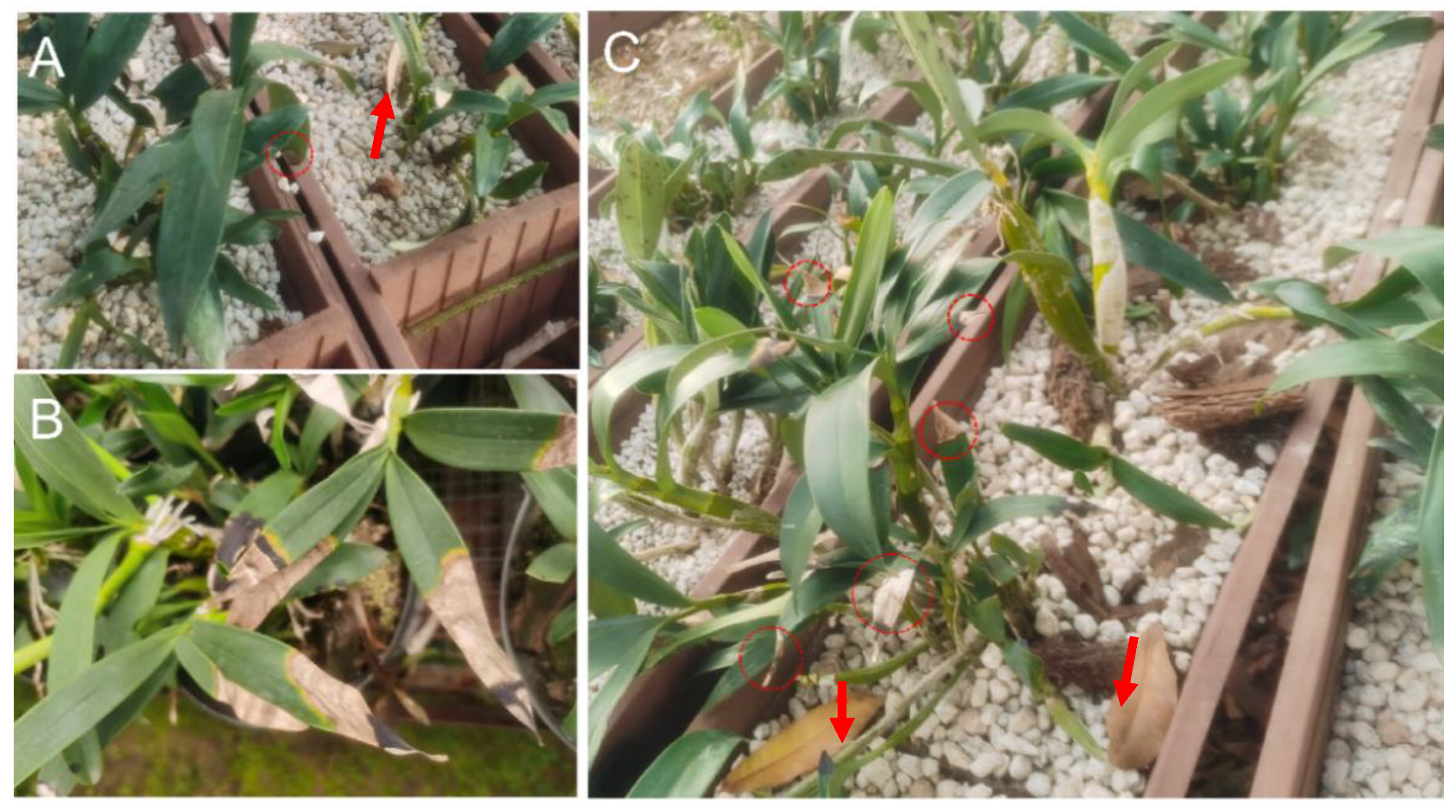
Figure 1. Symptoms of leaf blight on D. chrysotoxum seedlings in the nursery beds. (A) Early water-soaked corrugated lesions on the stem and graft interface; (B) yellowing of leaves and marginal necrosis; (C) plant leaves fall off. Arrows indicated leaves fall off the stem; Circles indicated lesions in (A,C).
Life 14 00285 g001
Figure 2. Morphological characteristics of Fusarium oxysporum isolated from diseased Dendrobium chrysotoxum plants. The colony growth on PDA medium (A, B), microconidia (C), macroconidia (D), and conidia chain bar = 10 μm and macroconidia (EG).
Life 14 00285 g002
Figure 3. One percent agarose gel image of PCR-amplified products of ITS, EF-1α, RPB1, and RPB2 gene of four isolates from Dendrobium chrysotoxum. Marker—2000 bp DNA marker (D2000, Tiangen, Beijing, China), DHRL-01~DHRL-04 were the gene PCR production of 4 isolates, and N-C—negative control.
Life 14 00285 g003
Figure 4. Maximum-likelihood phylogenetic tree generated from ITS (A), EF-1α (B), RPB1 (C), and RPB2 (D) sequences analysis of isolated strains of DHRL-01~04, respectively. Colletotrichum fructicola and Plectosphaerella cucumerina were used as the outgroup of ITS. Colletotrichum fructicola and Aspergilus nidulans were used as the outgroup of EF-1α, Pseudogymnoascus destructans, and Aspergilus tanneri were used as the outgroup of RPB1, Aspergilus tanneri and Aspergilus tanneri were used as the outgroup of RPB2. Scale bars indicate the number of substitutions per site. Bootstrap values are expressed as percentages based on 1000 replicates. Different background colors represent different clusters in the phylogenetic tree.
Life 14 00285 g004
Figure 5. Symptoms produced on Dendrobium chrysotoxum plants inoculated with Fusarium oxysporum. (a,b) yellowing, marginal necrosis, and, later, expansion to the bottom leaves after 15 days post-inoculation with Fusarium oxysporum DHRL-01. (c) Whole leaves became blighted and dropped after 30 days post-inoculation with Fusarium oxysporum DHRL-01, and (d) healthy plants as control (inoculated with sterilized ddH2O) was only physical damage in leaves.
Life 14 00285 g005
Table 1. Primers used in this study.
GenePrimersSequence (5′–3′)Annealing Temperature (°C)
ITSITS1-FTCCGTAGGTGAACCTGCGG55
ITS4-RTCCTCCGCTTATTGATATGC
EF-1αEF1-FATGGGTAAGGARGACAAGAC55
EF2-RGGARGTACCAGTSATCATG
RPB1F7-FCRACACAGAAGAGTTTGAAGG55
R8-RCAATGAGACCTTCTCGACCAGC
RPB25F2-FGGGGWGAYCAGAAGAAGGC55
7CR-RCCCATRGCTTGYTTRCCCAT
Table 2. Nucleotide BLAST results from the Fusarium MLST databases for the FOSC GenBank and accession numbers of the Fusarium oxysporum isolated from Dendrobium chrysotoxum plants.
IsolateFusarium MLST (Number)Fusarium MLST
Similarity %
GenBank Accession Number
ITSEF-1αRPB1RPB2
DHRL-01Fusarium oxysporum species complex (NRRL 22549)100MW599746MW703468MW703472MW703476
DHRL-02Fusarium oxysporum species complex (NRRL 22549)100MW599747MW703469MW703473MW703477
DHRL-03Fusarium oxysporum species complex (NRRL 22549)100MW599748MW703470MW703474MW703478
DHRL-04Fusarium oxysporum species complex (NRRL 22549)100MW599749MW703471MW703475MW703479
Disclaimer/Publisher’s Note: The statements, opinions and data contained in all publications are solely those of the individual author(s) and contributor(s) and not of MDPI and/or the editor(s). MDPI and/or the editor(s) disclaim responsibility for any injury to people or property resulting from any ideas, methods, instructions or products referred to in the content.