In the original publication [1], there was a mistake in Figure 1 as published. The plumb line drawn for the calculation of the cervical sagittal vertical axis should originate from the center of the C2 body. In the original figure, the line incorrectly begins at the anterior body. The corrected Figure 1 appears below. The authors state that the scientific conclusions are unaffected. This correction was approved by the Academic Editor. The original publication has also been updated.
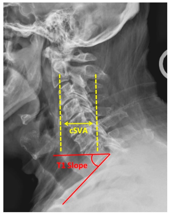
Figure 1.
Lateral cervical spine radiograph demonstrating cervical sagittal vertical axis (cSVA) and T1 slope.
Reference
- Foley, D.; Hardacker, P.; McCarthy, M. Emerging Technologies within Spine Surgery. Life 2023, 13, 2028. [Google Scholar] [CrossRef] [PubMed]

Disclaimer/Publisher’s Note: The statements, opinions and data contained in all publications are solely those of the individual author(s) and contributor(s) and not of MDPI and/or the editor(s). MDPI and/or the editor(s) disclaim responsibility for any injury to people or property resulting from any ideas, methods, instructions or products referred to in the content. |
© 2024 by the authors. Licensee MDPI, Basel, Switzerland. This article is an open access article distributed under the terms and conditions of the Creative Commons Attribution (CC BY) license (https://creativecommons.org/licenses/by/4.0/).