Case Report

Cutaneous Sarcoidosis Induced by Laser Therapy: Case Report and Review of the Literature

by
Hanna Cisoń
1,
Magdalena Simon-Błażewicz
2,
Joanna Suseł
3,
Marianna Suseł
1,
Zdzisław Woźniak
4,
Rafał Białynicki-Birula
1 and
Jacek C. Szepietowski
1,*
1
Department of Dermatology, Venereology and Allergology, Wroclaw Medical University, 50-367 Wroclaw, Poland
2
Medplus Medical Doctor’s Office M. Simon-Błażewicz, 58-100 Świdnica, Poland
3
JM Susel Clinic, 51-504 Wroclaw, Poland
4
Department of General and Experimental Pathology, Wroclaw Medical University, 50-367 Wroclaw, Poland
*
Author to whom correspondence should be addressed.
Life 2024, 14(6), 773; https://doi.org/10.3390/life14060773
Submission received: 13 May 2024 / Revised: 9 June 2024 / Accepted: 15 June 2024 / Published: 17 June 2024

Abstract

:
Background: Sarcoidosis, characterized by non-caseating epithelioid granulomas, presents diagnostic and therapeutic challenges. Method: Here we present a 38-year-old woman who exhibited erythematous and infiltrated skin lesions on her facial region following fractional laser treatment. Results: Histological analysis confirmed cutaneous sarcoidosis. Initial interventions with topical clobetasol and oral chloroquine provided transient relief. Subsequent outpatient management comprised topical tacrolimus and clobetasol, as well as systemic methotrexate, later substituted with prednisone. Gradual tapering resulted in lesion reduction. Conclusions: This case underscores the intricate nature of cutaneous sarcoidosis and the necessity for personalized therapeutic approaches. The association with cosmetic procedures highlights the importance of understanding potential triggers. The presented case highlights and reminds the medical community that lasers are not only used for therapeutic purposes but can also induce specific responses through laser therapy. Notably, while laser therapy is frequently employed in treating cutaneous sarcoidosis, its role in inducing sarcoidosis warrants further investigation.

1. Introduction

Sarcoidosis is a multifaceted systemic condition typified by granuloma formation, characteristically non-caseating epithelioid granulomas, in diverse organ systems. Scientific inquiry has illuminated the interplay of genetic susceptibility and environmental elements as key contributors to the etiology of an augmented immune response, culminating in the development of sarcoidosis [1]. The lungs and thoracic lymph nodes are the predominant sites of involvement, observed in over 90% of cases. Nevertheless, it is important to note that nearly every other organ system can potentially exhibit manifestations of the condition [2]. Dermatological involvement is a prevalent occurrence, with documented incidence rates exhibiting a range between 9% and 37% [3,4,5].
Sarcoidosis is a disease that affects individuals across all age groups, independent of their racial or ethnic background. However, it is most frequently diagnosed in individuals aged between 20 and 39 years [1]. It is noteworthy that this condition exhibits a higher prevalence among female individuals, non-smokers, and those residing in rural environments [6]. In the European context, there is a distinct geographic variation in the incidence of the disease, with northern regions reporting a considerably higher occurrence, estimated at approximately 60 cases per 100,000 individuals, in contrast to southern European countries like Italy, where the incidence is notably lower, at less than 10 cases per 100,000 individuals [7]. Exposure to a range of substances, including wood stove emissions, soil components, tree pollen, inorganic particulates, insecticides, and silica, has been linked to an increased susceptibility to sarcoidosis development [8,9].
Furthermore, it is postulated that certain microorganisms, such as Mycoplasma species, Leptospira species, Herpesviruses, Retroviruses, Chlamydia pneumoniae, Borrelia burgdorferi, Pneumocystis jirovecii, and Propionibacterium species, may be a potential triggers of sarcoidosis [10,11,12]. Furthermore, individuals subjected to interferon α therapy for the treatment of hepatitis C infection have been reported to manifest sarcoidosis as a notable consequence [13]. Moreover, drug-induced sarcoidosis-like reactions have been documented in connection with various pharmaceutical agents, including checkpoint inhibitors, anti-tumor necrosis factor (TNF), and anti-interleukin biological agents [14].
Here, we present the case of a 38-year-old female patient presenting with erythematous and infiltrated skin lesions on her facial region. These cutaneous manifestations became apparent approximately one month after ablative fractional CO2 laser treatment, as confirmed by histological examination revealing cutaneous sarcoidosis. Our patient represents the third reported case of laser-induced cutaneous sarcoidosis in the currently available literature. The literature search was conducted in the PubMed and Google Scholar databases, using combinations of the following relevant keywords: cutaneous sarcoidosis, laser therapy, and laser. Written informed consent for use of the patient’s data for this study, histopathologic preparation of the tissues, and publication of the relevant data was obtained from the family of our deceased patient.

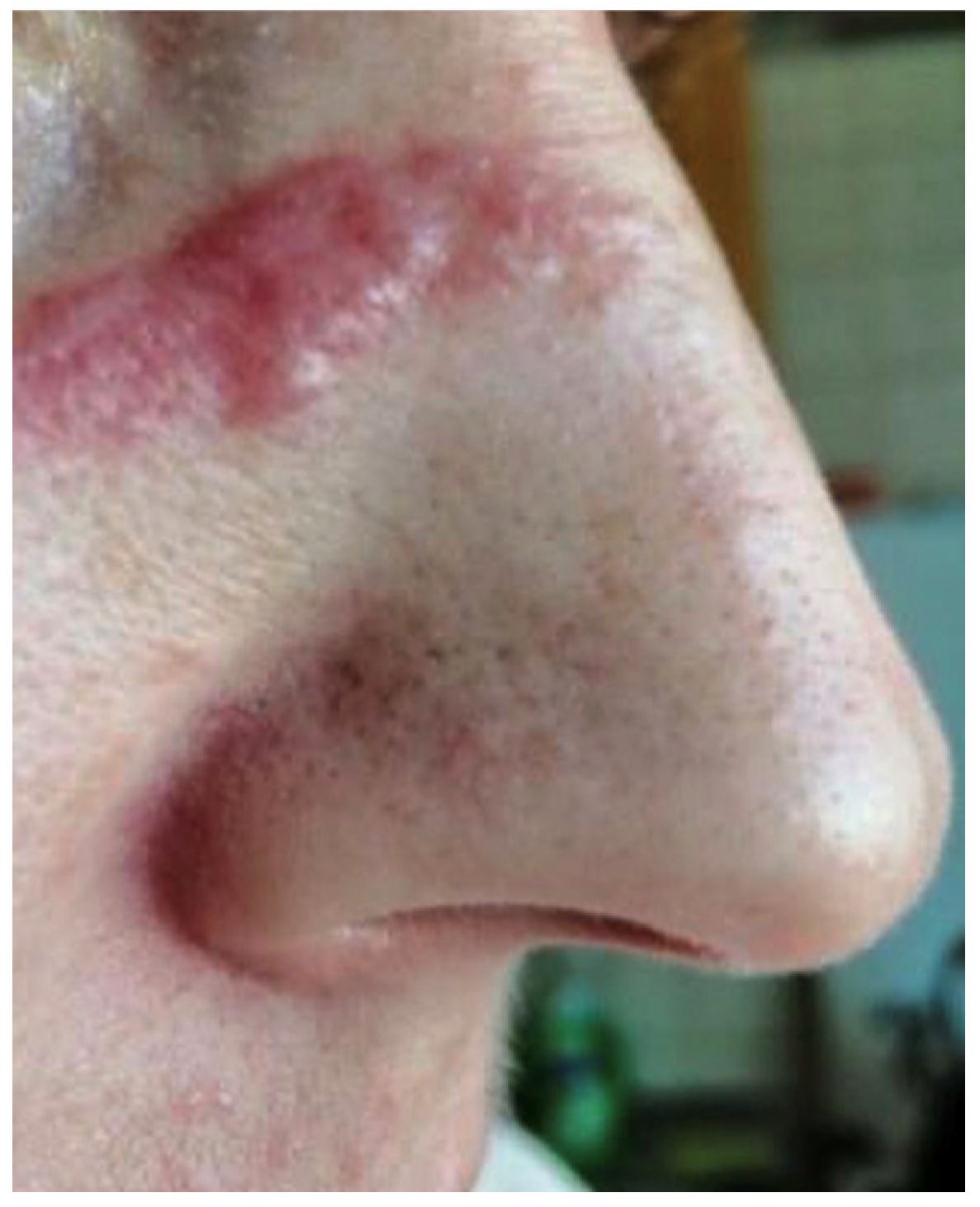
2. Clinical Case Presentation

A 38-year-old female patient was admitted to the Department of Dermatology, Venereology and Allergology in Wrocław, Poland, due to the presence of erythematous and infiltrated skin lesions on her facial region (Figure 1 and Figure 2).
Cutaneous manifestations manifested approximately one month post ablative fractional CO2 laser (wavelength of 10,600 nm and a fluence of 5 J/cm2 with a pulse duration of <1 ms) treatment. The laser therapy, conducted by a non-medical aesthetician, utilized a 125 mm handpiece, an 8 mm spot size, 25% density, and 12 Watts. The procedure consisted only of one session.
It is also noteworthy that in the patient’s medical history there was information about a 7-month course of isotretinoin therapy for acne four weeks prior to the laser procedure.
Isotretinoin was administered orally to the patient at a dosage of 20 mg twice daily, demonstrating favorable tolerability and maintaining results within normal parameters. Notably, complete remission of pathological manifestations was achieved during the final 3 months of isotretinoin therapy.
Initially, in the outpatient department, oral acyclovir was prescribed, suspecting herpes virus etiology for the skin lesions; however, this treatment failed to yield any improvement. Subsequently, a therapeutic regimen involving topical clobetasol propionate 0.5 mg/g and oral chloroquine at a dose of 250 mg b.i.d. for 2 months was initiated, due to suspicion of cutaneous sarcoidosis, leading to a reduction in erythema and infiltration. Regrettably, upon discontinuation of this treatment, the skin lesions recurred (Figure 3). Upon the resumption of topical clobetasol, no significant improvement was observed. In the course of outpatient diagnostics, a skin biopsy was performed. The histological examination revealed granulomatous inflammation mainly in superficial dermis without involvement of the subcutis. The confluent granulomata were composed of epithelioid histiocytes with abundant eosinophilic cytoplasm and oval vesicular nuclei. Few multinucleated giant cells were present. The granulomas were surrounded with few surrounding lymphocytes (‘naked granulomas’) and a rim of dermal fibrosis. Discrete small central foci of fibrinoid necrosis were present. The epidermis displayed mild acanthosis. The histological features confirmed cutaneous sarcoidosis (Figure 4).
Six months after the first hospitalization, the patient was again admitted to our department, where a thorough diagnostic workup was conducted, encompassing chest X-rays focused on lung hilum, abdominal ultrasound, and peripheral lymph node ultrasound. These investigations did not reveal any significant abnormalities. Following her hospital discharge, the patient continued her treatment regimen as an outpatient, consisting of topical tacrolimus (0.1% ointment) and clobetasol (cream 0.05%), resulting in a modest improvement.
Two months later, during her next hospitalization due to cutaneous sarcoidosis, erythematous and infiltrated skin changes were once again observed, primarily on her forehead, cheeks, and nose, albeit with a lesser degree of severity compared to her prior hospitalization. Laboratory tests performed at that time revealed results within the normal range, and the Quantiferon test was negative. Consequently, a decision was made to initiate methotrexate therapy at a dosage of 15 mg per week administered s.c., and this treatment was continued as part of her management plan. The methotrexate was changed to prednisone due to unsatisfactory treatment outcomes.
In conclusion, the gradual tapering of prednisone doses (20-10-5 mg p.d.) demonstrated favorable outcomes, leading to a reduction in cutaneous manifestations (Figure 5).

3. Discussion and Review of the Literature

3.1. Symptoms of Cutaneous Sarcoidosis

Cutaneous manifestations of sarcoidosis can be categorized into two distinct classes: specific and nonspecific lesions [3].
Specific lesions are characterized by histological confirmation of typical sarcoid granulomas and are often chronic and asymptomatic, requiring therapeutic intervention. These lesions include macules, papules, plaques, lupus pernio, and subcutaneous nodules, known as Darier–Roussy nodules [3,5,15]. Less common variations consist of skin changes associated with scars and tattoos, as well as hypopigmented, angiolupoid, psoriasiform, erythrodermic, ulcerative, ichthyosiform, verrucous, and lichenoid forms [3,15,16].
Nonspecific lesions, on the other hand, result from an inflammatory reaction pattern. Erythema nodosum, a frequently observed nonspecific skin manifestation, is often associated with acute sarcoidosis and is typically indicative of a favorable prognosis [17]. Understanding and distinguishing these cutaneous presentations is essential for proper diagnosis and management of sarcoidosis.

3.2. Diagnosis of Cutaneous Sarcoidosis

When there is a clinical suspicion of sarcoidosis, the performance of a skin biopsy is imperative. In our study, histological examination was typical and revealed granulomatous inflammation in superficial dermis. Confluent granulomata with few multinucleated giant cells were surrounded with minimal lymphocytic infiltration and a rim of fibrosis. However, discrete small central foci of fibrinoid necrosis were present.
Subsequent to the confirmation of cutaneous sarcoidosis through biopsy, it is essential to undertake a comprehensive evaluation to assess the potential for systemic involvement [18].

3.3. Treatment of Cutaneous Sarcoidosis

Topical treatments are preferred, if possible, especially for cutaneous or bronchial sarcoidosis. Severe, persisting cases require long-term immunosuppressive drugs, with glucocorticoids as the primary choice [19]. Methotrexate or rarely azathioprine, mycophenolate mofetil, and immunomodulators like hydroxychloroquine are alternative options. Tumor necrosis factor-alpha inhibitors, such as adalimumab and infliximab, are reserved as third-line agents but may be considered earlier for severe cases, organ involvement, or intolerance to standard drugs [18].

3.4. Cosmetic Procedure and Cutaneous Sarcoidosis

In the contemporary scientific literature, there exist documented instances of cutaneous sarcoidosis presenting in locations where previous blepharoplasty, tattooing, skin piercing, injections, or permanent makeup procedures, such as micro-blading, have been performed [5,20,21,22].

3.5. Laser Therapy and Cutaneous Sarcoidosis

While the existing literature discusses the treatment of cutaneous sarcoidosis with laser therapy [23,24,25,26,27,28,29,30,31,32,33,34,35] (Table 1), there are also singular occurrences describing laser therapy causing cutaneous sarcoidosis [36,37] (Table 2).

3.5.1. Pulsed Dye Laser

The Pulsed Dye Laser (PDL) was designed with the objective of implementing selective thermolysis for vascular lesions, with hemoglobin serving as the principal chromophore.
In a study by Roos et al. [24], a 63-year-old Caucasian woman presented with well-defined nodules on her back, emerging over a 2-week period. Histological examination confirmed cutaneous sarcoidosis. Fractionated Pulsed Dye Laser (FPDL) treatment resulted in complete clearance at treated sites after 4 weeks, contrasting with untreated sites.
Dong et al. [25] reported a 46-year-old Asian woman with longstanding left facial lesions, diagnosed with cutaneous sarcoidosis. Treatment included acitretin (10 mg q.d.), hydroxychloroquine (200 mg b.i.d.), and PDL therapy (595 nm, 6 ms, 7 mm, 14 J/cm2). Improvement was observed after 10 PDL sessions over 15 months.
Hollzman et al. [26] described a 10-year-old boy with a solitary, inflammatory, erythematous, ulcerated plaque, diagnosed with cutaneous sarcoidosis. PDL treatment (595 nm, 0.5 ms, 7 mm, 7.6–7.8 J/cm2) in three sessions at 6-week intervals led to favorable outcomes.
Also, Goodman et al. [27] presented a 39-year-old woman with cutaneous sarcoidosis and stable pulmonary health. Flashlamp PDL treatment (585 nm, 460 ms, 5 mm, 8 J/cm2) effectively reduced erythema, with optimal improvement at 8 J/cm2.

3.5.2. Carbon Dioxide Laser

The CO2 laser is an ablative modality that targets water as a chromophore.
In Zaouak et al.’s study [28], a 49-year-old woman with histologically confirmed cutaneous sarcoidosis underwent experimental treatment with ablative fractional CO2 laser. Three sessions were administered with a 2-month interval between each, yielding satisfactory results.
O’Donoghue et al. [29] reported CO2 laser treatment for lupus pernio nodular component recontouring in three patients. One patient required post-treatment intralesional triamcinolone and oral prednisolone due to systemic involvement but remained recurrence-free over 6 years. Another patient maintained a disease-free status for 14 months with laser treatment alone. The third patient, receiving laser treatment with intralesional steroids, experienced partial recurrence after nine months, with improvement lasting from nine months to six years.
In Stack et al.’s study [30], a 31-year-old man with cutaneous sarcoidosis was treated with a CO2 laser, followed by injection of a steroid preparation into the excised regions, resulting in satisfactory outcomes.

3.5.3. Intense Pulse Light

IPL employs a flashlamp to release polychromatic light spanning a wide wavelength spectrum, typically ranging from approximately 400 to 1400 nm.
Piccolo et al. [31] treated a 26-year-old woman with three nodules on her pinna, diagnosed as sarcoidosis after unsuccessful intralesional corticosteroid therapy. Following four IPL sessions (500 nm, 2 pulses of 5–10 ms, 12–16 J/cm2), there was a significant reduction in the vascular component and lesion consistency, along with alleviated pain.

3.5.4. Combined Laser Therapies

Within the existing literature, instances emerge detailing the therapeutic approach to cutaneous sarcoidosis through the amalgamation of diverse laser modalities [32,33,34,35].
In the study by Emer J et al. [32], correction of lupus pernio on the nose was performed using PDL 545 nm and nonablative fractional resurfacing (NAFR, 1550 nm). Following the initial application of PDL (7 mm, 0.45 ms, 8 J/cm2), NAFR was subsequently conducted (70 mJ, treatment level 6, 8 passes). The patient expressed satisfaction with the outcomes and declined any suggested further treatments.
In investigations by Grema H et al. [33] and Ekbäck et al. [34], treatment of cutaneous sarcoidosis with PDL yielded unsatisfactory outcomes. But further treatment with a Q-switched ruby laser (10 J/cm2) [33] and a frequency-doubled YAG laser (532 nm, 50 ms, 12–16 J/cm2) [34] led to success.
Momen et al. [35] reported on several cases of cutaneous sarcoidosis treated with lasers. One patient, unresponsive to systemic treatments, saw a 50–60% improvement in upper lip erythema and plaque depth after CO2 and PDL sessions. Three other patients received laser treatments for lupus pernio on the nose, an erythematous forehead plaque (PDL), and cheek nodules/plaques (CO2 laser).

3.6. Laser as a Factor Inducing Cutaneous Sarcoidosis

In our investigation, the temporal proximity between the laser therapy intervention in the patient and the emergence of cutaneous sarcoidosis suggests a potential association beyond mere coincidence. The existing literature presents only two instances documenting the induction of cutaneous sarcoidosis following laser therapy [36,37].
In the investigation conducted by Kim Hr et al. [36], an observational case is detailed featuring a 71-year-old male subject with a facial scarring history spanning five decades. The individual reported a progressive enlargement of the scar subsequent to three sessions of PDL treatment administered at a dermatology clinic three years prior. Histological examination identified numerous non-caseating granulomas indicative of cutaneous sarcoidosis.
In the study by Kormeilii T. et al. [37], a clinical case was delineated involving a 33-year-old Caucasian male who initially presented with hypertrophic scars on his left glabellar region and left upper lip, resulting from a past automobile accident. The patient underwent CO2 laser resurfacing to ameliorate the visibility of facial scars. However, a progression was noted, with an enlargement observed in the scars on his forehead two years post-treatment. Subsequent histopathological analysis was consistent with a diagnosis of cutaneous sarcoidosis.

3.7. Isotretinoin and Cutaneous Sarcoidosis

In our investigation, the patient was orally administered isotretinoin for common acne prior to the laser therapy procedure. However, no description was found regarding the induction of cutaneous sarcoidosis by isotretinoin therapy. Conversely, reports exist detailing the resolution of cutaneous lesions in a patient diagnosed with cutaneous sarcoidosis following isotretinoin therapy [38,39].

4. Conclusions

In summary, cutaneous sarcoidosis manifests a spectrum of specific and nonspecific lesions, indicative of the intricate nature of its clinical expressions. Specific lesions, characterized by typical sarcoid granulomas, encompass diverse forms such as macules, papules, plaques, lupus pernio, and Darier–Roussy nodules. Nonspecific lesions, arising from an inflammatory reaction pattern, include erythema nodosum. The accurate diagnosis and effective management of cutaneous sarcoidosis necessitate a comprehensive understanding of these diverse cutaneous presentations.
In the literature, cutaneous sarcoidosis has been linked to various cosmetic procedures, and the role of laser therapy in both the treatment and, in rare instances, the induction of cutaneous sarcoidosis has been systematically documented. Our review accentuates distinct cases wherein laser therapy, encompassing Pulsed Dye Laser and CO2 laser modalities, exhibited varied efficacy in managing cutaneous sarcoidosis. Particularly noteworthy is the induction of remission by laser therapy in specific cases, underscoring its potential as a therapeutic avenue for cutaneous sarcoidosis. Our patient represents the third reported case of laser-induced cutaneous sarcoidosis in the existing literature.
Moreover, the presented cases underscore the imperative of a tailored therapeutic approach and emphasize the ongoing necessity for research endeavors aimed at elucidating optimal treatment modalities. In the context of cutaneous sarcoidosis post-laser therapy, meticulous consideration of individual patient characteristics and treatment responses is paramount. These documented cases contribute significantly to the evolving understanding of cutaneous sarcoidosis and its management, providing valuable insights into the intricate interplay between therapeutic interventions and ensuing clinical outcomes.

Author Contributions

H.C., M.S.-B., J.S., M.S., R.B.-B. and J.C.S. contributed to the study design, analyzed and interpreted the patient’s clinical data regarding the disease, and prepared the manuscript. Z.W. performed the histological examination of the biopsy. H.C., M.S. and R.B.-B. contributed to collecting the relevant literature, data analysis, review, and critical interpretation. Conceptualization, H.C. and R.B.-B.; methodology, H.C.; validation, H.C. and R.B.-B.; formal analysis, R.B.-B. and J.C.S.; investigation, H.C., M.S.-B., J.S. and M.S.; resources, H.C., M.S.-B., J.S. and M.S.; data curation, H.C., M.S.-B., J.S. and M.S.; writing—original draft preparation, H.C.; writing—review and editing, R.B.-B. and J.C.S.; visualization, H.C.; supervision, R.B.-B. and J.C.S.; project administration, R.B.-B. and J.C.S. All authors have read and agreed to the published version of the manuscript.

Funding

The APC was funded by Wroclaw Medical University as academic support, with no influence on the research, content of the manuscript, or publication (SUBZ.C260.22.073).

Institutional Review Board Statement

This study was conducted in accordance with the Declaration of Helsinki, and approved by the Ethics Committee at the Medical University in Wroclaw, Poland on 9 March 2023 (No. 234/2023).

Informed Consent Statement

Written informed consent for use of the patient’s data for this study, histopathologic preparation of the tissues, and publication of the relevant data was obtained.

Data Availability Statement

The data presented in this study are available upon reasonable request from the corresponding author.

Conflicts of Interest

The authors declare no conflicts of interest. The funders provided only academic support for article APC and had no role in the design of the study; in the collection, analyses, or interpretation of data; in the writing of the manuscript; or in the decision to publish the results.

References

  1. Valeyre, D.; Prasse, A.; Nunes, H.; Uzunhan, Y.; Brillet, P.-Y.; Müller-Quernheim, J. Sarcoidosis. Lancet 2014, 383, 1155–1167. [Google Scholar] [CrossRef] [PubMed]
  2. Haimovic, A.; Sanchez, M.; Judson, M.A.; Prystowsky, S. Sarcoidosis: A comprehensive review and update for the dermatologist: Part II. Extracutaneous disease. J. Am. Acad. Dermatol. 2012, 66, 719.e1–719.e10. [Google Scholar] [CrossRef] [PubMed]
  3. Hutchinson, J. Statement on Sarcoidosis. Joint Statement of the American Thoracic Society (ATS), the European Respiratory Society (ERS) and the World Association of Sarcoidosis and Other Granulomatous Disorders (WASOG) adopted by the ATS Board of Directors and by the ERS Executive Committee, February 1999. Am. J. Respir. Crit. Care Med. 1999, 160, 736–755. [Google Scholar] [CrossRef]
  4. Mañá, J.; Marcoval, J. Skin manifestations of sarcoidosis. Presse Med. 2012, 41 Pt 2, e355–e374. [Google Scholar] [CrossRef] [PubMed]
  5. Haimovic, A.; Sanchez, M.; Judson, M.A.; Prystowsky, S. Sarcoidosis: A comprehensive review and update for the dermatologist: Part I. Cutaneous disease. J. Am. Acad. Dermatol. 2012, 66, 699.e1–699.e18. [Google Scholar] [CrossRef] [PubMed]
  6. Jain, R.; Yadav, D.; Puranik, N.; Guleria, R.; Jin, J.-O. Sarcoidosis: Causes, Diagnosis, Clinical Features, and Treatments. J. Clin. Med. 2020, 9, 1081. [Google Scholar] [CrossRef] [PubMed]
  7. Arkema, E.V.; Cozier, Y.C. Epidemiology of sarcoidosis: Current findings and future directions. Ther. Adv. Chronic Dis. 2018, 9, 227–240. [Google Scholar] [CrossRef]
  8. Newman, K.L.; Newman, L.S. Occupational causes of sarcoidosis. Curr. Opin. Allergy Clin. Immunol. 2012, 12, 145–150. [Google Scholar] [CrossRef]
  9. Vihlborg, P.; Bryngelsson, I.-L.; Andersson, L.; Graff, P. Risk of sarcoidosis and seropositive rheumatoid arthritis from occupational silica exposure in Swedish iron foundries: A retrospective cohort study. BMJ Open 2017, 7, e016839. [Google Scholar] [CrossRef]
  10. Newman, L. Aetiologies of sarcoidosis. Eur. Respir. Monogr. 2005, 32, 23–48. [Google Scholar]
  11. Vidal, S.; de la Horra, C.; Martín, J.; Montes-Cano, M.A.; Rodríguez, E.; Respaldiza, N.; Rodríguez, F.; Varela, J.M.; Medrano, F.J.; Calderón, E.J. Pneumocystis jirovecii colonisation in patients with interstitial lung disease. Clin. Microbiol. Infect. 2006, 12, 231–235. [Google Scholar] [CrossRef] [PubMed]
  12. Drake, W.P.; Newman, L.S. Mycobacterial antigens may be important in sarcoidosis pathogenesis. Curr. Opin. Pulm. Med. 2006, 12, 359–363. [Google Scholar] [CrossRef] [PubMed]
  13. Salameh, H.; Trien, R.; Cooper, C.J.; Paez, D.; Colon, E.; Ajmal, S. Interferon-alpha-induced sarcoidosis in a patient being treated for hepatitis C. Am. J. Case Rep. 2014, 15, 235–238. [Google Scholar] [CrossRef] [PubMed]
  14. Cohen Aubart, F.; Lhote, R.; Amoura, A.; Valeyre, D.; Haroche, J.; Amoura, Z.; Lebrun-Vignes, B. Drug-induced sarcoidosis: An overview of the WHO pharmacovigilance database. J. Intern. Med. 2020, 288, 356–362. [Google Scholar] [CrossRef] [PubMed]
  15. Marchell, R.M.; Judson, M. Cutaneous Sarcoidosis. Semin. Respir. Crit. Care Med. 2010, 31, 442–451. [Google Scholar] [CrossRef] [PubMed]
  16. Marchell, R.M.; Judson, M.A. Chronic cutaneous lesions of sarcoidosis. Clin. Dermatol. 2007, 25, 295–302. [Google Scholar] [CrossRef] [PubMed]
  17. Pérez-Garza, D.M.; Chavez-Alvarez, S.; Ocampo-Candiani, J.; Gomez-Flores, M. Erythema Nodosum: A Practical Approach and Diagnostic Algorithm. Am. J. Clin. Dermatol. 2021, 22, 367–378. [Google Scholar] [CrossRef] [PubMed]
  18. Franzen, D.P.; Brutsche, M.; Nilsson, J.; Böni, C.; Daccord, C.; Distler, O.; Elsener, D.; Funke-Chambour, M.; Gruner, C.; Hayward-Könnecke, H.; et al. Sarcoidosis—A multisystem disease. Swiss Med. Wkly. 2022, 152, w30049. [Google Scholar] [CrossRef] [PubMed]
  19. Grunewald, J.; Grutters, J.C.; Arkema, E.V.; Saketkoo, L.A.; Moller, D.R. Sarcoidosis. Nat. Rev. Dis. Primers 2019, 5, 45, Correction in Nat. Rev. Dis. Primers 2019, 5, 49. [Google Scholar] [CrossRef]
  20. Kwon, S.H.; Jeong, K.M.; Baek, Y.S.; Jeon, J. Linear scar sarcoidosis on thin blepharoplasty line mimicking a hypertrophic scar: A case report. SAGE Open Med. Case Rep. 2018, 6, 2050313X18803991. [Google Scholar] [CrossRef]
  21. Kanakamedala, A.D.; Maamari, R.N.; Couch, S.M. Tattoo-associated lacrimal gland enlargement and sarcoidosis. Am. J. Ophthalmol. Case Rep. 2023, 32, 101889. [Google Scholar] [CrossRef] [PubMed]
  22. Bae, K.-N.; Shin, K.; Kim, H.-S.; Ko, H.-C.; Kim, B.; Kim, M.-B. Scar Sarcoidosis: A Retrospective Investigation into Its Peculiar Clinicopathologic Presentation. Ann. Dermatol. 2022, 34, 28–33. [Google Scholar] [CrossRef] [PubMed]
  23. Cliff, S.; Felix, R.H.; Singh, L.; Harland, C.C. The successful treatment of lupus pernio with the flashlamp pulsed dye laser. J. Cutan. Laser Ther. 1999, 1, 49–52. [Google Scholar] [CrossRef] [PubMed]
  24. Roos, S.; Raulin, C.; Ockenfels, H.-M.; Karsai, S. Successful treatment of cutaneous sarcoidosis lesions with the flashlamp pumped pulsed dye laser: A case report. Dermatol. Surg. 2009, 35, 1139–1140. [Google Scholar] [CrossRef] [PubMed]
  25. Dong, W.; Li, C.; Shi, Y.; Zhang, W. Combined pulsed-dye laser and medical therapy for treatment of cutaneous sarcoidosis lesions: A case report. J. Int. Med. Res. 2021, 49, 300060521997745. [Google Scholar] [CrossRef]
  26. Holzmann, R.D.; Astner, S.; Forschner, T.; Sterry, G. Scar sarcoidosis in a child: Case report of successful treatment with the pulsed dye laser. Dermatol. Surg. 2008, 34, 393–396. [Google Scholar] [CrossRef]
  27. Goodman, M.M.; Alpern, K. Treatment of lupus pernio with the flashlamp pulsed dye laser. Lasers Surg. Med. 1992, 12, 549–551. [Google Scholar] [CrossRef]
  28. Zaouak, A.; Koubaa, W.; Hammami, H.; Fenniche, S. Unconventional use of fractional ablative CO2 laser in facial cutaneous sarcoïdosis. Dermatol. Ther. 2017, 30, e12569. [Google Scholar] [CrossRef]
  29. O’Donoghue, N.B.; Barlow, R.J. Laser remodelling of nodular nasal lupus pernio. Clin. Exp. Dermatol. 2006, 31, 27–29. [Google Scholar] [CrossRef]
  30. Stack, B.C.; Hall, P.J.; Goodman, A.L.; Perez, I.R. CO2 laser excision of lupus pernio of the face. Am. J. Otolaryngol. 1996, 17, 260–263. [Google Scholar] [CrossRef]
  31. Piccolo, D.; Di Marcantonio, D.; Crisman, G.; Cannarozzo, G.; Sannino, M.; Chiricozzi, A.; Chimenti, S. Unconventional use of intense pulsed light. BioMed Res. Int. 2014, 2014, 618206. [Google Scholar] [CrossRef] [PubMed]
  32. Emer, J.; Uslu, U.; Waldorf, H. Improvement in lupus pernio with the successive use of pulsed dye laser and nonablative fractional resurfacing. Dermatol. Surg. 2014, 40, 201–202. [Google Scholar] [CrossRef]
  33. Grema, H.; Greve, B.; Raulin, C. Scar sarcoidosis—Treatment with the Q-switched ruby laser. Lasers Surg. Med. 2002, 30, 398–400. [Google Scholar] [CrossRef] [PubMed]
  34. Ekbäck, M.; Molin, L. Effective laser treatment in a case of lupus pernio. Acta Derm. Venereol. 2005, 85, 521–522. [Google Scholar] [CrossRef]
  35. Momen, S.; Al-Niaimi, F.; Barlow, R.; Mallipeddi, R. The use of lasers in cutaneous sarcoid: Is there a role? J. Cosmet. Laser Ther. 2016, 18, 335–338. [Google Scholar] [CrossRef]
  36. Kim, H.-R.; Kim, S.-J.; Im, M.; Lee, Y.; Seo, Y.-J.; Lee, J.-H. Scar Sarcoidosis Induced by Pulsed Dye Laser Treatment. Ann. Dermatol. 2016, 28, 509–510. [Google Scholar] [CrossRef]
  37. Kormeili, T.; Neel, V.; Moy, R.L. Cutaneous sarcoidosis at sites of previous laser surgery. Cutis 2004, 73, 53–55. [Google Scholar] [PubMed]
  38. Georgiou, S.; Monastirli, A.; Pasmatzi, E.; Tsambaos, D. Cutaneous sarcoidosis: Complete remission after oral isotretinoin therapy. Acta Derm. Venereol. 1998, 78, 457–459. [Google Scholar] [CrossRef]
  39. Waldinger, T.P.; Ellis, C.N.; Quint, K.; Voorhees, J.J. Treatment of cutaneous sarcoidosis with isotretinoin. Arch. Dermatol. 1983, 119, 1003–1005. [Google Scholar] [CrossRef]
Figure 1. The patient’s forehead with the numerous erythematous, infiltrated skin lesions.
Life 14 00773 g001
Figure 2. The erythematous, infiltrated skin lesions on the nose of the patient.
Life 14 00773 g002
Figure 3. The recurrence of the intensity of infiltration on the left forehead.
Life 14 00773 g003
Figure 4. The histological features confirmed cutaneous sarcoidosis (H&E staining, 100×).
Life 14 00773 g004
Figure 5. The healing due to prednisone therapy—the less infiltrated skin lesions on the right cheek.
Life 14 00773 g005
Table 1. The use of lasers in the treatment of cutaneous sarcoidosis.
Authors
and Type of the Study
Clinical PicturePatient Age/GenderLaser/SettingTreatment NumberOutcomeFollow Up
Roos et al.
case report
[24]
Nodules on back63 FFPDL 585 nm
0.5 ms, 12 mm, 6 J/cm2
1After four weeks, lesions had completely resolvedPrednisolone added at 4 weeks for systemic disease; at 13 months no recurrence of lesions
Dong et al.
case report
[25]
Erythematous papules on the left part of the face46 FPDL 595 nm
6 ms, 7 mm, 14 J/cm2
10 treatments during 15 monthsThe plaque was thinner, normal skin had appeared; telangiectasia was less obviousAcitretin (10 mg per day) and hydroxychloroquine (200 mg twice a day) along with PDL therapy
Holzmann et al.
case report
[26]
Erythematous papule on varicella
scar site on cheeks
10 MPDL 595 mm,
0.5 ms, 7 mm, 7.6–7.8 J/cm2
3 treatments
at 6-week intervals
Clinical remission after
three treatments
No recurrence at 12 months
Goodman et al.
case report
[27]
Lupus pernio granulomatous papules on nasal ala 39 F PDL 585 nm,
460 ms, 5 mm, 8 J/cm2
2 treatments 7 months apart Erythema and papular components showed a 75% improvement from baseline after six months Papules recurred at 2 months, and erythema at 6 months. By 15 months, a significant reappearance of both erythema and papules necessitated a third treatment
Zaouak et al.
case report
[28]
Two infiltrated papulo-nodular plaques on the cheek49 FAblative fractional CO2 laser
125 mm handpiece, 10 mm spot, 25% density, 10 Watts
3 sessions with a 2-month intervalComplete resolution of the two facial plaquesNo signs of relapse after 6 months
O’Donoghue et al.
case series
[29]
Lupus pernio nose; diffuse violaceous involvement and fleshy nodules on ala rim
 
Violaceous nodule on nasal bridge, nasal swelling and erythema
 
Coalescent fleshy violaceous nodule on nasal tip and left nasal ala
55F


57 M


58 F
Sarcoid nodules underwent debulking and recontouring with a CO2 laser in “paint mode” (18 W, 6 mm spot size); for cosmetic refinement, patients 1 and 3 received a resurfacing pass (14 W, 6 mm spot size), accompanied by intralesional triamcinolone acetonide (TAC) injection to prevent recurrence New contour maintainedSix years maintained on 5 mg prednisolone, mild hypopigmentation


At 14 months nasal contour was stable
 
At 9 months, partial recurrence with nasal swelling in conjunction with flare of systemic sarcoid
Stack et al.
case report
[30]
Nodules on nose causing nasal obstruction31 MExcision with CO2 laser and intralesional TAC 24 months no recurrence
Piccolo et al.
case report
[31]
Three nodules located on the anterior and posterior aspects of the pinna26 FIPL 500 nm
2 pulses 5–10 ms, 12–16 J/cm2
4Reduction of the vascular component and in the consistency of the lesionsImprovement persisted throughout the 6 months
Emer et al.
case report
[32]
Lupus pernio on the noseNot shownPDL 595 nm
7 mm, 0.45 ms, 8 J/cm2
 
NAFR 1550 nm
(70 mJ, treatment level 6, 8 passes)
1Reduction of lesionsN.D.
Grema et al.
case report
[33]
Scar sarcoid on right elbow
and both knees (red-brown
discoloration)
50 FQSRL,
four treatments
 
Previously treated with PDL
with no effect 585 nm, 7 mm, 0.5 ms, 5.5–5.6 J/cm2
4Lightened after four treatmentsNo recurrence at three years
Ekbäck et al.
case report
[34]
Lupus pernio of cheek57 FPDL 585 nm,
45 ms, 6.75–7 J/cm2
 
Frequency-doubled YAG laser 532 nm, 50 ms, 12–16 J/cm2
10 treatments over 3 years
 
2 treatments over 7 months
Limited effect, less redness and thinner lesions
 
Complete healing
N/D
Momen et al.
case reports
[35]
Erythematous plaque on upper lip
 
 
Nodules and plaques across cheek
 
Erythematous triangular plaque on forehead
 
Lupus pernio
One female, age not shown
 
Two patients, gender and age not shown
 
Three females, age not shown
 
One female, age not shown
Single pass of CO2 laser and two treatments with PDL
 
Two treatments with CO2 laser
 
 
Singular PDL
 
Seven treatments with PD
Self-reported improvement of 50–60%
PDL—Pulsed Dye Laser, IPL—Intense Pulse Light, NAFR—Nonablative Fractional Resurfacing, QSRL—Q-switched Ruby Laser.
Table 2. The use of lasers as a cause/progression of cutaneous sarcoidosis.
Author and Type of the StudyClinicalPatient Age/GenderLaser/SettingTreatmentOutcome
Kim Hr et al.
case report
[36]
The enlargement of the scar 71 M Enlargement of the scar subsequent to three sessions of PDL Hydroxychloroquine (100 mg b.i.d.) along with topical pimecrolimus and topical steroids Reduction of the scar
Kormeilii T et al.
case report
[37]
Hypertrophic scars on his left glabellar region and left side of upper lip 33 M Progression two years after CO2 laser resurfacing N/DN/D
Disclaimer/Publisher’s Note: The statements, opinions and data contained in all publications are solely those of the individual author(s) and contributor(s) and not of MDPI and/or the editor(s). MDPI and/or the editor(s) disclaim responsibility for any injury to people or property resulting from any ideas, methods, instructions or products referred to in the content.

