
Comprehensive Assessment of Medial Knee Joint Instability by Valgus Stress MRI

 , ,
, ,  ,
, 
Abstract
:1. Introduction
2. Materials and Methods
2.1. Study Design and Sample Size Estimation
2.2. MRI-Compatible Loading Device
2.3. Human Cadaveric Knee Joint Specimens
2.4. Pre-Imaging Preparations and Image Acquisition
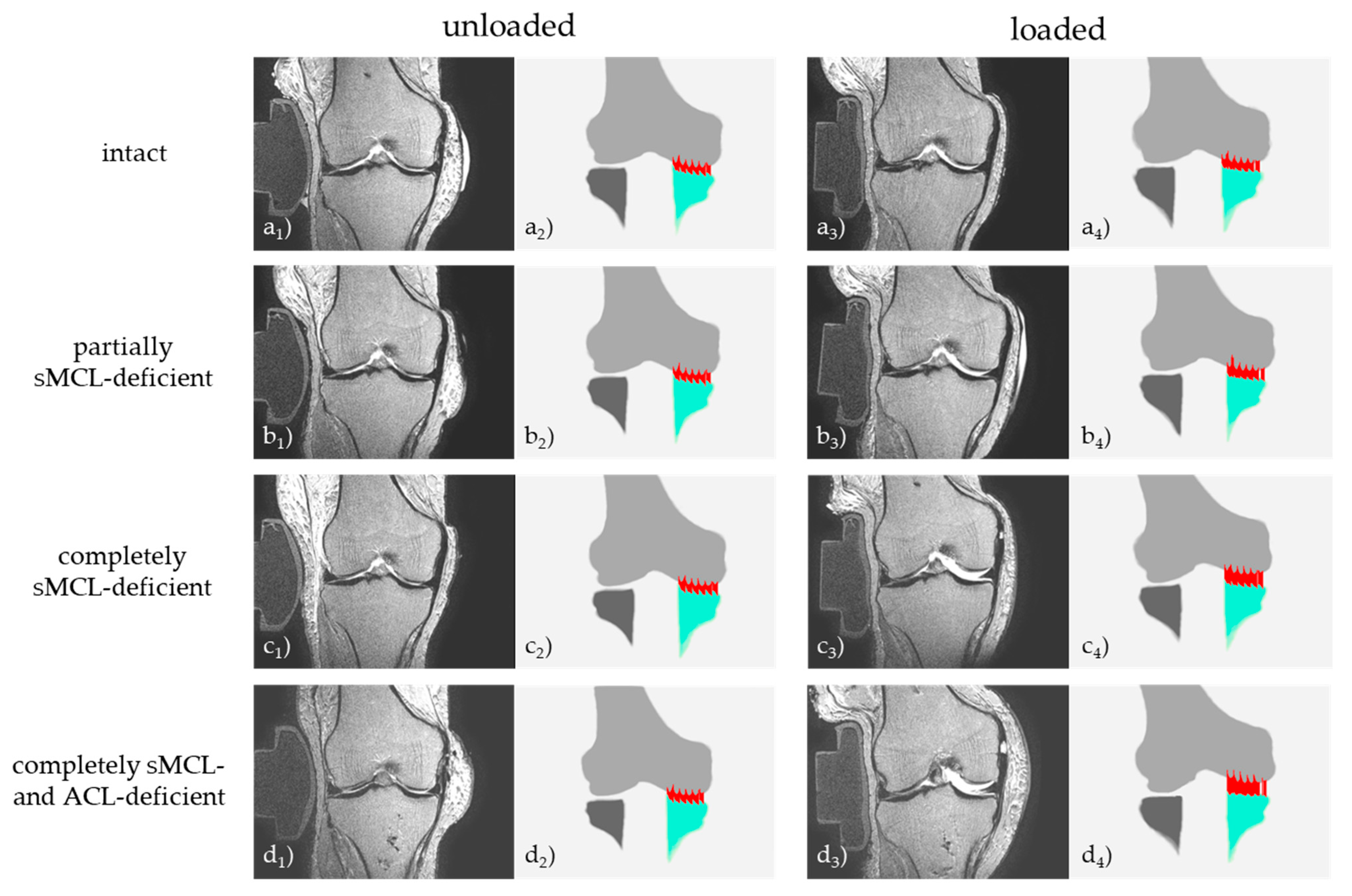
2.5. Graded Knee Joint Ligament Injuries
2.6. Image Post-Processing and Analysis
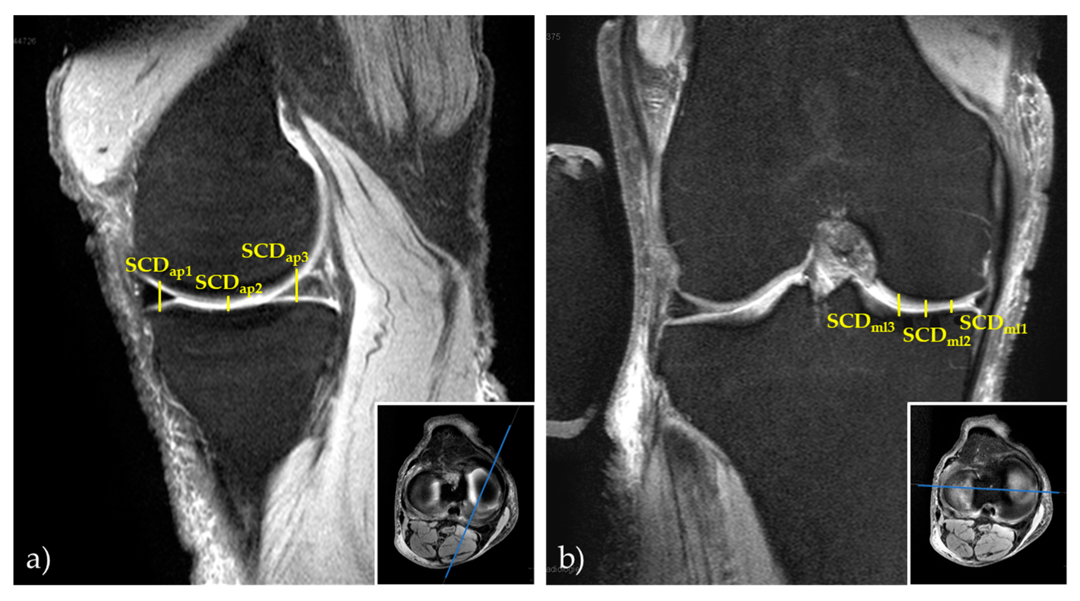
2.6.1. Manual 2D Reference Measurements
2.6.2. Computed 3D Measurements
2.7. Statistical Analysis
3. Results
4. Discussion
5. Conclusions
Supplementary Materials
Author Contributions
Funding
Institutional Review Board Statement
Informed Consent Statement
Data Availability Statement
Acknowledgments
Conflicts of Interest
References
- Bollen, S. Epidemiology of knee injuries: Diagnosis and triage. Br. J. Sports Med. 2000, 34, 227–228. [Google Scholar] [CrossRef] [Green Version]
- Rothenberg, P.; Grau, L.; Kaplan, L.; Baraga, M.G. Knee Injuries in American Football: An Epidemiological Review. Am. J. Orthop. 2016, 45, 368–373. [Google Scholar]
- Majewski, M.; Susanne, H.; Klaus, S. Epidemiology of athletic knee injuries: A 10-year study. Knee 2006, 13, 184–188. [Google Scholar] [CrossRef] [PubMed]
- Roach, C.J.; Haley, C.A.; Cameron, K.L.; Pallis, M.; Svoboda, S.J.; Owens, B.D. The epidemiology of medial collateral ligament sprains in young athletes. Am. J. Sports Med. 2014, 42, 1103–1109. [Google Scholar] [CrossRef]
- Lundblad, M.; Waldén, M.; Magnusson, H.; Karlsson, J.; Ekstrand, J. The UEFA injury study: 11-year data concerning 346 MCL injuries and time to return to play. Br. J. Sports Med. 2013, 47, 759–762. [Google Scholar] [CrossRef] [PubMed]
- Fetto, J.F.; Marshall, J.L. Medial collateral ligament injuries of the knee: A rationale for treatment. Clin. Orthop. Relat. Res. 1978, 132, 206–218. [Google Scholar] [CrossRef]
- Elkin, J.L.; Zamora, E.; Gallo, R.A. Combined Anterior Cruciate Ligament and Medial Collateral Ligament Knee Injuries: Anatomy, Diagnosis, Management Recommendations, and Return to Sport. Curr. Rev. Musculoskelet. Med. 2019, 12, 239–244. [Google Scholar] [CrossRef] [PubMed]
- Phisitkul, P.; James, S.L.; Wolf, B.R.; Amendola, A. MCL injuries of the knee: Current concepts review. Iowa Orthop. J. 2006, 26, 77–90. [Google Scholar] [PubMed]
- Wijdicks, C.A.; Griffith, C.J.; Johansen, S.; Engebretsen, L.; LaPrade, R.F. Injuries to the medial collateral ligament and associated medial structures of the knee. J. Bone Jt. Surg. Am. Vol. 2010, 92, 1266–1280. [Google Scholar] [CrossRef]
- Kastelein, M.; Wagemakers, H.P.; Luijsterburg, P.A.; Verhaar, J.A.; Koes, B.W.; Bierma-Zeinstra, S.M. Assessing medial collateral ligament knee lesions in general practice. Am. J. Med. 2008, 121, 982–988.e2. [Google Scholar] [CrossRef]
- Laprade, R.F.; Bernhardson, A.S.; Griffith, C.J.; Macalena, J.A.; Wijdicks, C.A. Correlation of valgus stress radiographs with medial knee ligament injuries: An in vitro biomechanical study. Am. J. Sports Med. 2010, 38, 330–338. [Google Scholar] [CrossRef]
- Sawant, M.; Narasimha Murty, A.; Ireland, J. Valgus knee injuries: Evaluation and documentation using a simple technique of stress radiography. Knee 2004, 11, 25–28. [Google Scholar] [CrossRef]
- Lee, Y.S.; Han, S.H.; Jo, J.; Kwak, K.S.; Nha, K.W.; Kim, J.H. Comparison of 5 different methods for measuring stress radiographs to improve reproducibility during the evaluation of knee instability. Am. J. Sports Med. 2011, 39, 1275–1281. [Google Scholar] [CrossRef]
- James, E.W.; Williams, B.T.; LaPrade, R.F. Stress radiography for the diagnosis of knee ligament injuries: A systematic review. Clin. Orthop. Relat. Res. 2014, 472, 2644–2657. [Google Scholar] [CrossRef] [PubMed] [Green Version]
- Kleinbaum, Y.; Blankstein, A. Mild to moderate medial collateral ligament (MCL) injuries of the knee: Sonographic findings and sonographic valgus stress test. J. Musculoskelet. Res. 2008, 11, 9–14. [Google Scholar] [CrossRef]
- Lutz, P.M.; Feucht, M.J.; Wechselberger, J.; Rasper, M.; Petersen, W.; Wörtler, K.; Imhoff, A.B.; Achtnich, A. Ultrasound-based examination of the medial ligament complex shows gender- and age-related differences in laxity. Knee Surg. Sports Traumatol. Arthrosc. 2021, 29, 1960–1967. [Google Scholar] [CrossRef] [PubMed]
- Slane, L.C.; Slane, J.A.; Scheys, L. The measurement of medial knee gap width using ultrasound. Arch. Orthop. Trauma Surg. 2017, 137, 1121–1128. [Google Scholar] [CrossRef] [PubMed]
- De Maeseneer, M.; Shahabpour, M.; Pouders, C. MRI spectrum of medial collateral ligament injuries and pitfalls in diagnosis. JBR-BTR 2010, 93, 97–103. [Google Scholar] [CrossRef] [Green Version]
- Farshad-Amacker, N.A.; Potter, H.G. MRI of knee ligament injury and reconstruction. J. Magn. Reson. Imaging 2013, 38, 757–773. [Google Scholar] [CrossRef] [PubMed]
- Halinen, J.; Koivikko, M.; Lindahl, J.; Hirvensalo, E. The efficacy of magnetic resonance imaging in acute multi-ligament injuries. Int. Orthop. 2009, 33, 1733–1738. [Google Scholar] [CrossRef] [PubMed] [Green Version]
- Derby, E.; Imrecke, J.; Henckel, J.; Hirschmann, A.; Amsler, F.; Hirschmann, M.T. How sensitive and specific is 1.5 Tesla MRI for diagnosing injuries in patients with knee dislocation? Knee Surg. Sports Traumatol. Arthrosc. 2017, 25, 517–523. [Google Scholar] [CrossRef]
- Barbier, O.; Galaud, B.; Descamps, S.; Boisrenoult, P.; Leray, E.; Lustig, S.; Bonnevialle, P.; Laffargue, P.; Paillot, J.L.; Rosset, P.; et al. Relevancy and reproducibility of magnetic resonance imaging (MRI) interpretation in multiple-ligament injuries and dislocations of the knee. Orthop. Traumatol. Surg. Res. 2013, 99, 305–311. [Google Scholar] [CrossRef] [PubMed] [Green Version]
- Svantesson, E.; Hamrin Senorski, E.; Alentorn-Geli, E.; Westin, O.; Sundemo, D.; Grassi, A.; Čustović, S.; Samuelsson, K. Increased risk of ACL revision with non-surgical treatment of a concomitant medial collateral ligament injury: A study on 19,457 patients from the Swedish National Knee Ligament Registry. Knee Surg. Sports Traumatol. Arthrosc. 2019, 27, 2450–2459. [Google Scholar] [CrossRef] [Green Version]
- Westermann, R.W.; Spindler, K.P.; Huston, L.J.; Wolf, B.R. Outcomes of Grade III Medial Collateral Ligament Injuries Treated Concurrently With Anterior Cruciate Ligament Reconstruction: A Multicenter Study. Arthrosc. J. Arthrosc. Relat. Surg. 2019, 35, 1466–1472. [Google Scholar] [CrossRef]
- Said, O.; Schock, J.; Krämer, N.; Thüring, J.; Hitpass, L.; Schad, P.; Kuhl, C.; Abrar, D.; Truhn, D.; Nebelung, S. An MRI-compatible varus-valgus loading device for whole-knee joint functionality assessment based on compartmental compression: A proof-of-concept study. Magma 2020, 33, 839–854. [Google Scholar] [CrossRef] [PubMed]
- Winkelmeyer, E.-M.; Schock, J.; Wollschläger, L.M.; Schad, P.; Huppertz, M.S.; Kotowski, N.; Prescher, A.; Kuhl, C.; Truhn, D.; Nebelung, S. Seeing Beyond Morphology-Standardized Stress MRI to Assess Human Knee Joint Instability. Diagnostics 2021, 11, 1035. [Google Scholar] [CrossRef]
- Rossi, R.; Dettoni, F.; Bruzzone, M.; Cottino, U.; D’Elicio, D.G.; Bonasia, D.E. Clinical examination of the knee: Know your tools for diagnosis of knee injuries. Sports Med. Arthrosc. Rehabil. Ther. Technol. 2011, 3, 25. [Google Scholar] [CrossRef] [PubMed] [Green Version]
- Lane, J.G.; Amiel, D. Ligament Histology, Composition, Anatomy, Injury, and Healing Mechanisms. In Bio-Orthopaedics; Springer: Berlin, Germany, 2017; pp. 291–312. [Google Scholar] [CrossRef]
- Yushkevich, P.A.; Piven, J.; Hazlett, H.C.; Smith, R.G.; Ho, S.; Gee, J.C.; Gerig, G. User-guided 3D active contour segmentation of anatomical structures: Significantly improved efficiency and reliability. NeuroImage 2006, 31, 1116–1128. [Google Scholar] [CrossRef] [Green Version]
- Grood, E.S.; Noyes, F.R.; Butler, D.L.; Suntay, W.J. Ligamentous and capsular restraints preventing straight medial and lateral laxity in intact human cadaver knees. J. Bone Jt. Surg. Am. Vol. 1981, 63, 1257–1269. [Google Scholar] [CrossRef]
- LaPrade, M.D.; Kennedy, M.I.; Wijdicks, C.A.; LaPrade, R.F. Anatomy and biomechanics of the medial side of the knee and their surgical implications. Sports Med. Arthrosc. Rev. 2015, 23, 63–70. [Google Scholar] [CrossRef] [PubMed] [Green Version]
- Pedersen, R.R. The Medial and Posteromedial Ligamentous and Capsular Structures of the Knee: Review of Anatomy and Relevant Imaging Findings. Semin. Musculoskelet. Radiol. 2016, 20, 12–25. [Google Scholar] [CrossRef]
- Robinson, J.R.; Bull, A.M.; Thomas, R.R.; Amis, A.A. The role of the medial collateral ligament and posteromedial capsule in controlling knee laxity. Am. J. Sports Med. 2006, 34, 1815–1823. [Google Scholar] [CrossRef]
- Warren, L.A.; Marshall, J.L.; Girgis, F. The prime static stabilizer of the medical side of the knee. J. Bone Jt. Surg. Am. Vol. 1974, 56, 665–674. [Google Scholar] [CrossRef]
- Piedade, S.R.; Servien, E.; Lavoie, F.; Neyret, P. Classification of knee laxities. In The Knee Joint: Surgical Techniques and Strategies; Springer: Paris, France, 2012; pp. 85–93. [Google Scholar] [CrossRef]
- Jacobsen, K. Stress radiographical measurement of the anteroposterior, medial and lateral stability of the knee joint. Acta Orthop. Scand. 1976, 47, 335–344. [Google Scholar] [CrossRef] [PubMed]
- Gruber, G.; Martens, D.; Konermann, W. [Value of ultrasound examination in lesion of the medial collateral ligament of the knee joint]. Zeitschrift Orthopadie Grenzgebiete 1998, 136, 337–342. [Google Scholar] [CrossRef]
- Marchant, M.H., Jr.; Tibor, L.M.; Sekiya, J.K.; Hardaker, W.T., Jr.; Garrett, W.E., Jr.; Taylor, D.C. Management of medial-sided knee injuries, part 1: Medial collateral ligament. Am. J. Sports Med. 2011, 39, 1102–1113. [Google Scholar] [CrossRef]
- Haimes, J.L.; Wroble, R.R.; Grood, E.S.; Noyes, F.R. Role of the medial structures in the intact and anterior cruciate ligament-deficient knee. Limits of motion in the human knee. Am. J. Sports Med. 1994, 22, 402–409. [Google Scholar] [CrossRef]
- Engebretsen, L.; Lind, M. Anteromedial rotatory laxity. Knee Surg. Sports Traumatol. Arthrosc. 2015, 23, 2797–2804. [Google Scholar] [CrossRef] [Green Version]
- Hughston, J.C.; Andrews, J.R.; Cross, M.J.; Moschi, A. Classification of knee ligament instabilities. Part I. The medial compartment and cruciate ligaments. J. Bone Jt. Surg. Am. Vol. 1976, 58, 159–172. [Google Scholar] [CrossRef]
- Wierer, G.; Milinkovic, D.; Robinson, J.R.; Raschke, M.J.; Weiler, A.; Fink, C.; Herbort, M.; Kittl, C. The superficial medial collateral ligament is the major restraint to anteromedial instability of the knee. Knee Surg. Sports Traumatol. Arthrosc. 2021, 29, 405–416. [Google Scholar] [CrossRef] [PubMed]
- Jacobsen, K. Radiologic technique for measuring instability in the knee joint. Acta Radiol. Diagn. 1977, 18, 113–125. [Google Scholar] [CrossRef]
- Noyes, F.; Grood, E. The strength of the anterior cruciate ligament in humans and Rhesus. J. Bone Joint. Surg. Am. 1976, 58, 1074–1082. [Google Scholar] [CrossRef] [PubMed]
- Woo, S.L.-Y.; Hollis, J.M.; Adams, D.J.; Lyon, R.M.; Takai, S. Tensile properties of the human femur-anterior cruciate ligament-tibia complex: The effects of specimen age and orientation. Am. J. Sports Med. 1991, 19, 217–225. [Google Scholar] [CrossRef] [PubMed]
- Fishkin, Z.; Miller, D.; Ritter, C.; Ziv, I. Changes in human knee ligament stiffness secondary to osteoarthritis. J. Orthop. Res. 2002, 20, 204–207. [Google Scholar] [CrossRef]
- Sims, W.F.; Jacobson, K.E. The posteromedial corner of the knee: Medial-sided injury patterns revisited. Am. J. Sports Med. 2004, 32, 337–345. [Google Scholar] [CrossRef]
- Robinson, J.R.; Sanchez-Ballester, J.; Bull, A.M.; Thomas Rde, W.; Amis, A.A. The posteromedial corner revisited. An anatomical description of the passive restraining structures of the medial aspect of the human knee. J. Bone Jt. Surg. Br. Vol. 2004, 86, 674–681. [Google Scholar] [CrossRef] [PubMed] [Green Version]
- Craft, J.A.; Kurzweil, P.R. Physical examination and imaging of medial collateral ligament and posteromedial corner of the knee. Sports Med. Arthrosc. Rev. 2015, 23, e1–e6. [Google Scholar] [CrossRef]
- Shultz, S.J.; Shimokochi, Y.; Nguyen, A.D.; Schmitz, R.J.; Beynnon, B.D.; Perrin, D.H. Measurement of varus-valgus and internal-external rotational knee laxities in vivo--Part II: Relationship with anterior-posterior and general joint laxity in males and females. J. Orthop. Res. Off. Publ. Orthop. Res. Soc. 2007, 25, 989–996. [Google Scholar] [CrossRef] [Green Version]
- Yoo, J.C.; Ahn, J.H.; Sung, K.S.; Wang, J.H.; Lee, S.H.; Bae, S.W.; Ahn, Y.J. Measurement and comparison of the difference in normal medial and lateral knee joint opening. Knee Surg. Sports Traumatol. Arthrosc. 2006, 14, 1238–1244. [Google Scholar] [CrossRef]
- Boguszewski, D.V.; Cheung, E.C.; Joshi, N.B.; Markolf, K.L.; McAllister, D.R. Male-Female Differences in Knee Laxity and Stiffness: A Cadaveric Study. Am. J. Sports Med. 2015, 43, 2982–2987. [Google Scholar] [CrossRef]


| PDw fs | PDw fs | PDw fs | T1w | T2w | T2w | |
|---|---|---|---|---|---|---|
| Sequence Type | 2D TSE | 2D TSE | 2D TSE | 2D TSE | 2D TSE | 2D TSE |
| Orientation | ax | sag | cor | cor | ax | cor |
| Type of fat saturation | SPAIR | SPAIR | SPAIR | n/a | n/a | n/a |
| Repetition Time [ms] | 4776 | 7595 | 4495 | 671 | 3283 | 3000 |
| Echo time [ms] | 30 | 30 | 30 | 9 | 80 | 80 |
| Turbo spin-echo factor | 13 | 15 | 13 | 3 | 14 | 14 |
| Field of view [mm] | 160 × 160 | 164 × 144 | 160 × 160 | 160 × 160 | 160 × 160 | 160 × 160 |
| Acquisition matrix [pixels] | 400 × 312 | 352 × 256 | 400 × 300 | 368 × 317 | 352 × 295 | 352 × 297 |
| Reconstruction matrix [pixels] | 512 × 512 | 512 × 512 | 512 × 512 | 448 × 448 | 512 × 512 | 512 × 512 |
| Scan percentage [%] | 79.4 | 79.3 | 79.4 | 86.4 | 85.0 | 85.0 |
| Flip angle [°] | 90 | 90 | 90 | 90 | 90 | 90 |
| Number of signal averages | 1 | 1 | 1 | 1 | 1 | 1 |
| Slices | 33 | 40 | 31 | 30 | 40 | 30 |
| Slice Thickness/Gap [mm] | 3.0/0.3 | 3.0/0.3 | 3.0/0.3 | 3.0/0.3 | 1.5/0.0 | 1.5/0.3 |
| Duration [min:s] | 03:59 | 05:19 | 04:39 | 06:40 | 03:17 | 03:42 |
| Intact | Partially sMCL-Deficient | Completely sMCL-Deficient | Completely sMCL- and ACL-deficient | p-Value (‡) | ||||||
|---|---|---|---|---|---|---|---|---|---|---|
| UL | LO | UL | LO | UL | LO | UL | LO | |||
| Manual 2D Measurements | SCDml1 | 3.7 ± 0.7 | 4.8 ± 1.0 | 3.9 ± 0.7 | 5.6 ± 1.0 | 4.0 ± 0.7 | 9.2 ± 1.4 | 4.7 ± 1.0 | 10.6 ± 1.4 | <0.001 |
| SCDml2 | 3.7 ± 0.6 | 4.7 ± 0.9 | 3.9 ± 0.7 | 5.3 ± 0.8 | 4.2 ± 0.6 | 8.7 ± 1.5 | 4.5 ± 0.9 | 9.6 ± 1.5 | ||
| SCDml3 | 4.8 ± 0.9 | 5.9 ± 0.9 | 4.9 ± 0.9 | 6.5 ± 1.0 | 5.3 ± 0.9 | 10.0 ± 1.5 | 5.4 ± 1.3 | 10.6 ± 1.6 | ||
| SCDap1 | 6.2 ± 1.4 | 7.6 ± 1.5 | 6.4 ± 1.2 | 8.3 ± 1.5 | 6.4 ± 1.5 | 11.9 ± 2.1 | 7.1 ± 1.4 | 14.4 ± 2.2 | ||
| SCDap2 | 3.9 ± 0.5 | 4.8 ± 0.7 | 4.1 ± 0.4 | 5.4 ± 0.8 | 4.4 ± 0.6 | 8.9 ± 1.6 | 4.8 ± 0.8 | 9.8 ± 1.5 | ||
| SCDap3 | 7.4 ± 1.1 | 8.0 ± 1.2 | 7.8 ± 1.2 | 8.4 ± 1.1 | 8.0 ± 1.3 | 10.8 ± 1.4 | 7.9 ± 1.5 | 10.8 ± 1.9 | ||
| Computed 3D Measurements | SCDmean | 5.8 ± 0.8 | 7.0 ± 0.7 | 6.1 ± 0.7 | 7.7 ± 0.7 | 6.2 ± 0.7 | 11.0 ± 1.4 | 6.6 ± 1.1 | 12.1 ± 1.4 | <0.001 |
| SCDml1 | 3.7 ± 0.8 | 4.9 ± 0.8 | 4.1 ± 0.6 | 5.7 ± 0.8 | 4.1 ± 0.7 | 9.4 ± 1.3 | 4.7 ± 0.9 | 10.7 ± 1.5 | ||
| SCDml2 | 3.9 ± 0.7 | 4.9 ± 0.9 | 4.1 ± 0.7 | 5.5 ± 1.0 | 4.3 ± 0.6 | 8.9 ± 1.4 | 4.6 ± 0.8 | 9.9 ± 1.6 | ||
| SCDml3 | 5.0 ± 0.9 | 5.9 ± 0.9 | 5.3 ± 0.8 | 6.4 ± 0.8 | 5.4 ± 0.9 | 10.0 ± 1.4 | 5.4 ± 1.0 | 10.6 ± 1.3 | ||
| SCDap1 | 5.7 ± 1.0 | 7.6 ± 1.3 | 5.7 ± 1.1 | 8.1 ± 1.4 | 6.0 ± 1.3 | 12.2 ± 1.8 | 6.7 ± 1.9 | 14.4 ± 2.0 | ||
| SCDap2 | 3.8 ± 0.8 | 4.9 ± 0.9 | 4.1 ± 0.7 | 5.5 ± 1.0 | 4.2 ± 0.6 | 8.8 ± 1.4 | 4.7 ± 0.8 | 9.8 ± 1.5 | ||
| SCDap3 | 7.1 ± 1.4 | 7.8 ± 1.0 | 7.9 ± 1.3 | 8.3 ± 1.7 | 7.7 ± 1.6 | 10.8 ± 1.9 | 7.7 ± 1.1 | 10.8 ± 2.3 | ||
| p-value (†) | SCDml1 | 0.871 | 0.798 | 0.156 | 0.748 | 0.785 | 0.314 | 0.976 | 0.397 | |
| SCDml2 | 0.180 | 0.061 | 0.269 | 0.051 | 0.062 | 0.056 | 0.102 | 0.051 | ||
| SCDml3 | 0.082 | 0.770 | 0.103 | 0.333 | 0.277 | 0.924 | 0.872 | 0.989 | ||
| SCDap1 | 0.126 | 0.984 | 0.101 | 0.233 | 0.168 | 0.347 | 0.187 | 0.836 | ||
| SCDap2 | 0.827 | 0.668 | 0.960 | 0.846 | 0.086 | 0.363 | 0.171 | 0.856 | ||
| SCDap3 | 0.356 | 0.494 | 0.635 | 0.677 | 0.372 | 0.832 | 0.554 | 0.839 | ||
Publisher’s Note: MDPI stays neutral with regard to jurisdictional claims in published maps and institutional affiliations. |
© 2021 by the authors. Licensee MDPI, Basel, Switzerland. This article is an open access article distributed under the terms and conditions of the Creative Commons Attribution (CC BY) license (https://creativecommons.org/licenses/by/4.0/).
Share and Cite
Ciba, M.; Winkelmeyer, E.-M.; Schock, J.; Schad, P.; Kotowski, N.; Nolte, T.; Wollschläger, L.M.; Knobe, M.; Prescher, A.; Kuhl, C.; et al. Comprehensive Assessment of Medial Knee Joint Instability by Valgus Stress MRI. Diagnostics 2021, 11, 1433. https://doi.org/10.3390/diagnostics11081433
Ciba M, Winkelmeyer E-M, Schock J, Schad P, Kotowski N, Nolte T, Wollschläger LM, Knobe M, Prescher A, Kuhl C, et al. Comprehensive Assessment of Medial Knee Joint Instability by Valgus Stress MRI. Diagnostics. 2021; 11(8):1433. https://doi.org/10.3390/diagnostics11081433
Chicago/Turabian StyleCiba, Malin, Eva-Maria Winkelmeyer, Justus Schock, Philipp Schad, Niklas Kotowski, Teresa Nolte, Lena Marie Wollschläger, Matthias Knobe, Andreas Prescher, Christiane Kuhl, and et al. 2021. "Comprehensive Assessment of Medial Knee Joint Instability by Valgus Stress MRI" Diagnostics 11, no. 8: 1433. https://doi.org/10.3390/diagnostics11081433
APA StyleCiba, M., Winkelmeyer, E.-M., Schock, J., Schad, P., Kotowski, N., Nolte, T., Wollschläger, L. M., Knobe, M., Prescher, A., Kuhl, C., Truhn, D., & Nebelung, S. (2021). Comprehensive Assessment of Medial Knee Joint Instability by Valgus Stress MRI. Diagnostics, 11(8), 1433. https://doi.org/10.3390/diagnostics11081433