Peripartum Cardiomyopathy: Diagnostic and Prognostic Value of Cardiac Magnetic Resonance in the Acute Stage
Abstract
:1. Introduction
2. Materials and Methods
2.1. Study Population
2.2. Cardiac Magnetic Resonance Imaging
2.3. Cardiac Image Analysis
2.4. Statistical Analysis
3. Results
3.1. General Characteristics
3.2. CMR Imaging Results
3.3. Subgroup Analyses of CMR Parameters in Patients with Follow-Up
3.4. Association between Imaging Parameters and LVEF Recovery
4. Discussion
Supplementary Materials
Author Contributions
Funding
Institutional Review Board Statement
Informed Consent Statement
Data Availability Statement
Conflicts of Interest
References
- Bauersachs, J.; König, T.; van der Meer, P.; Petrie, M.C.; Hilfiker-Kleiner, D.; Mbakwem, A.; Hamdan, R.; Jackson, A.M.; Forsyth, P.; de Boer, R.A.; et al. Pathophysiology, diagnosis and management of peripartum cardiomyopathy: A position statement from the Heart Failure Association of the European Society of Cardiology Study Group on peripartum cardiomyopathy. Eur. J. Heart Fail. 2019, 21, 827–843. [Google Scholar] [CrossRef] [PubMed]
- Arany, Z.; Elkayam, U. Peripartum Cardiomyopathy. Circulation 2016, 133, 1397–1409. [Google Scholar] [CrossRef] [PubMed]
- Isogai, T.; Kamiya, C.A. Worldwide Incidence of Peripartum Cardiomyopathy and Overall Maternal Mortality. Int. Heart J. 2019, 60, 503–511. [Google Scholar] [CrossRef] [PubMed] [Green Version]
- Hilfiker-Kleiner, D.; Sliwa, K. Pathophysiology and epidemiology of peripartum cardiomyopathy. Nat. Rev. Cardiol. 2014, 11, 364–370. [Google Scholar] [CrossRef] [PubMed]
- Davis, M.B.; Arany, Z.; McNamara, D.M.; Goland, S.; Elkayam, U. Peripartum Cardiomyopathy: JACC State-of-the-Art Review. J. Am. Coll. Cardiol. 2020, 75, 207–221. [Google Scholar] [CrossRef]
- Biteker, M.; Ilhan, E.; Biteker, G.; Duman, D.; Bozkurt, B. Delayed recovery in peripartum cardiomyopathy: An indication for long-term follow-up and sustained therapy. Eur. J. Heart Fail. 2012, 14, 895–901. [Google Scholar] [CrossRef] [Green Version]
- Goland, S.; Bitar, F.; Modi, K.; Safirstein, J.; Ro, A.; Mirocha, J.; Khatri, N.; Elkayam, U. Evaluation of the clinical relevance of baseline left ventricular ejection fraction as a predictor of recovery or persistence of severe dysfunction in women in the United States with peripartum cardiomyopathy. J. Card. Fail. 2011, 17, 426–430. [Google Scholar] [CrossRef]
- Luetkens, J.A.; Faron, A.; Isaak, A.; Dabir, D.; Kuetting, D.; Feisst, A.; Schmeel, F.C.; Sprinkart, A.M.; Thomas, D. Comparison of Original and 2018 Lake Louise Criteria for Diagnosis of Acute Myocarditis: Results of a Validation Cohort. Radiol. Cardiothorac. Imaging 2019, 1, e190010. [Google Scholar] [CrossRef]
- Luetkens, J.A.; Homsi, R.; Sprinkart, A.M.; Doerner, J.; Dabir, D.; Kuetting, D.L.; Block, W.; Andrié, R.; Stehning, C.; Fimmers, R.; et al. Incremental value of quantitative CMR including parametric mapping for the diagnosis of acute myocarditis. Eur. Heart J. Cardiovasc. Imaging 2016, 17, 154–161. [Google Scholar] [CrossRef]
- Luetkens, J.A.; Schlesinger-Irsch, U.; Kuetting, D.L.; Dabir, D.; Homsi, R.; Doerner, J.; Schmeel, F.C.; Fimmers, R.; Sprinkart, A.M.; Naehle, C.P.; et al. Feature-tracking myocardial strain analysis in acute myocarditis: Diagnostic value and association with myocardial oedema. Eur. Radiol. 2017, 27, 4661–4671. [Google Scholar] [CrossRef]
- Schelbert, E.B.; Elkayam, U.; Cooper, L.T.; Givertz, M.M.; Alexis, J.D.; Briller, J.; Felker, G.M.; Chaparro, S.; Kealey, A.; Pisarcik, J.; et al. Myocardial Damage Detected by Late Gadolinium Enhancement Cardiac Magnetic Resonance Is Uncommon in Peripartum Cardiomyopathy. J. Am. Heart Assoc. 2017, 6, e005472. [Google Scholar] [CrossRef] [PubMed] [Green Version]
- Ersbøll, A.S.; Bojer, A.S.; Hauge, M.G.; Johansen, M.; Damm, P.; Gustafsson, F.; Vejlstrup, N.G. Long-Term Cardiac Function After Peripartum Cardiomyopathy and Preeclampsia: A Danish Nationwide, Clinical Follow-Up Study Using Maximal Exercise Testing and Cardiac Magnetic Resonance Imaging. J. Am. Heart Assoc. 2018, 7, e008991. [Google Scholar] [CrossRef] [Green Version]
- Briasoulis, A.; Mocanu, M.; Marinescu, K.; Qaqi, O.; Palla, M.; Telila, T.; Afonso, L. Longitudinal systolic strain profiles and outcomes in peripartum cardiomyopathy. Echocardiography 2016, 33, 1354–1360. [Google Scholar] [CrossRef]
- Sugahara, M.; Kagiyama, N.; Hasselberg, N.E.; Blauwet, L.A.; Briller, J.; Cooper, L.; Fett, J.D.; Hsich, E.; Wells, G.; McNamara, D.; et al. Global Left Ventricular Strain at Presentation Is Associated with Subsequent Recovery in Patients with Peripartum Cardiomyopathy. J. Am. Soc. Echocardiogr. 2019, 32, 1565–1573. [Google Scholar] [CrossRef] [PubMed]
- Cannan, C.; Weeks, S.; Friedrich, M. CMR features of peri-partum cardiomyopathy. J. Cardiovasc. Magn. Reson. 2010, 12, P185. [Google Scholar] [CrossRef] [Green Version]
- Arora, N.P.; Mohamad, T.; Mahajan, N.; Danrad, R.; Kottam, A.; Li, T.; Afonso, L.C. Cardiac magnetic resonance imaging in peripartum cardiomyopathy. Am. J. Med. Sci. 2014, 347, 112–117. [Google Scholar] [CrossRef]
- Sprinkart, A.M.; Luetkens, J.A.; Träber, F.; Doerner, J.; Gieseke, J.; Schnackenburg, B.; Schmitz, G.; Thomas, D.; Homsi, R.; Block, W.; et al. Gradient Spin Echo (GraSE) imaging for fast myocardial T2 mapping. J. Cardiovasc. Magn. Reson. 2015, 17, 12. [Google Scholar] [CrossRef] [Green Version]
- Messroghli, D.R.; Radjenovic, A.; Kozerke, S.; Higgins, D.M.; Sivananthan, M.U.; Ridgway, J.P. Modified Look-Locker inversion recovery (MOLLI) for high-resolution T1 mapping of the heart. Magn. Reson. Med. 2004, 52, 141–146. [Google Scholar] [CrossRef]
- Schulz-Menger, J.; Bluemke, D.A.; Bremerich, J.; Flamm, S.D.; Fogel, M.A.; Friedrich, M.G.; Kim, R.J.; von Knobelsdorff-Brenkenhoff, F.; Kramer, C.M.; Pennell, D.J.; et al. Standardized image interpretation and post-processing in cardiovascular magnetic resonance-2020 update: Society for Cardiovascular Magnetic Resonance (SCMR): Board of Trustees Task Force on Standardized Post-Processing. J. Cardiovasc. Magn. Reson. 2020, 22, 19. [Google Scholar] [CrossRef]
- Luetkens, J.A.; Doerner, J.; Thomas, D.K.; Dabir, D.; Gieseke, J.; Sprinkart, A.M.; Fimmers, R.; Stehning, C.; Homsi, R.; Schwab, J.O.; et al. Acute myocarditis: Multiparametric cardiac MR imaging. Radiology 2014, 273, 383–392. [Google Scholar] [CrossRef] [PubMed] [Green Version]
- Rothman, K.J. No adjustments are needed for multiple comparisons. Epidemiology 1990, 1, 43–46. [Google Scholar] [CrossRef] [PubMed] [Green Version]
- McNamara, D.M.; Elkayam, U.; Alharethi, R.; Damp, J.; Hsich, E.; Ewald, G.; Modi, K.; Alexis, J.D.; Ramani, G.V.; Semigran, M.J.; et al. Clinical Outcomes for Peripartum Cardiomyopathy in North America: Results of the IPAC Study (Investigations of Pregnancy-Associated Cardiomyopathy). J. Am. Coll. Cardiol. 2015, 66, 905–914. [Google Scholar] [CrossRef] [PubMed] [Green Version]
- Mahowald, M.K.; Basu, N.; Subramaniam, L.; Scott, R.; Davis, M.B. Long-term Outcomes in Peripartum Cardiomyopathy. Open Cardiovasc. Med. J. 2019, 13, 13–23. [Google Scholar] [CrossRef]
- Haghikia, A.; Röntgen, P.; Vogel-Claussen, J.; Schwab, J.; Westenfeld, R.; Ehlermann, P.; Berliner, D.; Podewski, E.; Hilfiker-Kleiner, D.; Bauersachs, J. Prognostic implication of right ventricular involvement in peripartum cardiomyopathy: A cardiovascular magnetic resonance study. ESC Heart Fail. 2015, 2, 139–149. [Google Scholar] [CrossRef] [PubMed]
- Lindley, K.J.; Conner, S.N.; Cahill, A.G.; Novak, E.; Mann, D.L. Impact of Preeclampsia on Clinical and Functional Outcomes in Women with Peripartum Cardiomyopathy. Circ. Heart Fail. 2017, 10, e003797. [Google Scholar] [CrossRef]
- Ricke-Hoch, M.; Pfeffer, T.J.; Hilfiker-Kleiner, D. Peripartum cardiomyopathy: Basic mechanisms and hope for new therapies. Cardiovasc. Res. 2020, 116, 520–531. [Google Scholar] [CrossRef] [PubMed]
- Renz, D.M.; Röttgen, R.; Habedank, D.; Wagner, M.; Böttcher, J.; Pfeil, A.; Dietz, R.; Hamm, B.; de Kivelitz, E.; Elgeti, T. Kardiale Bildgebung bei peripartaler Kardiomyopathie: Evaluation eines umfassenden MR-Untersuchungsprotokolls. RöFo 2011, 183, VO316_4. [Google Scholar] [CrossRef]
- Liang, Y.-D.; Xu, Y.-W.; Li, W.-H.; Wan, K.; Sun, J.-Y.; Lin, J.-Y.; Zhang, Q.; Zhou, X.-Y.; Chen, Y.-C. Left ventricular function recovery in peripartum cardiomyopathy: A cardiovascular magnetic resonance study by myocardial T1 and T2 mapping. J. Cardiovasc. Magn. Reson. 2020, 22, 2. [Google Scholar] [CrossRef]
- Zagrosek, A.; Wassmuth, R.; Abdel-Aty, H.; Rudolph, A.; Dietz, R.; Schulz-Menger, J. Relation between myocardial edema and myocardial mass during the acute and convalescent phase of myocarditis—A CMR study. J. Cardiovasc. Magn. Reson. 2008, 10, 19. [Google Scholar] [CrossRef] [Green Version]
- Nii, M.; Ishida, M.; Dohi, K.; Tanaka, H.; Kondo, E.; Ito, M.; Sakuma, H.; Ikeda, T. Myocardial tissue characterization and strain analysis in healthy pregnant women using cardiovascular magnetic resonance native T1 mapping and feature tracking technique. J. Cardiovasc. Magn. Reson. 2018, 20, 52. [Google Scholar] [CrossRef]
- Azibani, F.; Pfeffer, T.J.; Ricke-Hoch, M.; Dowling, W.; Pietzsch, S.; Briton, O.; Baard, J.; Abou Moulig, V.; König, T.; Berliner, D.; et al. Outcome in German and South African peripartum cardiomyopathy cohorts associates with medical therapy and fibrosis markers. ESC Heart Fail. 2020, 7, 512–522. [Google Scholar] [CrossRef] [PubMed] [Green Version]
- Vermes, E.; Childs, H.; Faris, P.; Friedrich, M.G. Predictive value of CMR criteria for LV functional improvement in patients with acute myocarditis. Eur. Heart J. Cardiovasc. Imaging 2014, 15, 1140–1144. [Google Scholar] [CrossRef] [PubMed] [Green Version]
- Moon, J.C.; Messroghli, D.R.; Kellman, P.; Piechnik, S.K.; Robson, M.D.; Ugander, M.; Gatehouse, P.D.; Arai, A.E.; Friedrich, M.G.; Neubauer, S.; et al. Myocardial T1 mapping and extracellular volume quantification: A Society for Cardiovascular Magnetic Resonance (SCMR) and CMR Working Group of the European Society of Cardiology consensus statement. J. Cardiovasc. Magn. Reson. 2013, 15, 92. [Google Scholar] [CrossRef] [PubMed] [Green Version]


| Variable | Patients with PPCM (n = 17) | Healthy Female Controls (n =15) | p-Value |
|---|---|---|---|
| Clinical parameters | |||
| Age (years) | 33 ± 5 | 33 ± 8 | 0.892 |
| Weight (kg) | 77 ± 19 | 67 ± 13 | 0.088 |
| Height (cm) | 170 ± 8 | 170 ± 7 | 0.972 |
| Body mass index (kg/m²) | 27 ± 7 | 23 ± 4 | 0.077 |
| Heart rate (bpm) | 78 ± 27 | 75 ± 11 | 0.052 |
| NT-proBNP (pg/mL) | 8792 ± 12,308 | NA | - |
| Troponin I (ng/L) | 0.12 ± 0.25 | NA | - |
| C-reactive protein (mg/L) | 15.0 ± 11.1 | NA | - |
| White blood cells (G/L) | 10.5 ± 3.7 | NA | - |
| CMR parameters | |||
| Left ventricular ejection fraction (%) | 31 ± 10 | 61 ± 6 | <0.001 |
| Left ventricular end-diastolic volume index (mL/m²) | 121 ± 43 | 73 ± 9 | <0.001 |
| Right ventricular ejection fraction (%) | 32 ± 13 | 57 ± 7 | <0.001 |
| Right ventricular end-diastolic volume index (mL/m²) | 82 ± 24 | 75 ± 11 | 0.300 |
| Cardiac index (L/min/m²) | 3.0 ± 0.7 | 3.3 ± 0.7 | 0.228 |
| Left atrium volume index (mL/m²) | 75 ± 24 | 40 ± 10 | <0.001 |
| Left ventricular mass index (g/m²) | 71 ± 19 | 41 ± 7 | <0.001 |
| Interventricular septal thickness (mm) | 10.3 ± 1.9 | 7.9 ± 1.1 | <0.001 |
| T2 signal intensity ratio | 2.10 ± 0.34 | 1.58 ± 0.21 | <0.001 |
| Visual myocardial edema | 10 (59%) | 0 (0%) | <0.001 |
| Visual late gadolinium enhancement | 2 (12%) | 0 (0%) | 0.484 |
| Late gadolinium enhancement (%) | 3.9 ± 4.7 | 0.6 ± 0.7 | 0.013 |
| Global longitudinal strain (%) | −11.8 ± 4.8 | −22.3 ± 4.2 | <0.001 |
| Global circumferential strain (%) | −12.3 ± 6.3 | −24.1 ± 3.6 | <0.001 |
| Global radial strain (%) | 22.8 ± 14.7 | 37.1 ± 10.2 | 0.004 |
| T1 relaxation time, native (ms) | 1070 ± 51 | 980 ± 28 | 0.001 |
| Extracellular volume fraction (%) | 31.7 ± 7.1 | 27.7 ± 3.2 | 0.235 |
| T2 relaxation time (ms) | 63 ± 5 | 53 ± 2 | <0.001 |
| Variable | Baseline (n = 6) | Follow-Up (n = 6) | p-Value |
|---|---|---|---|
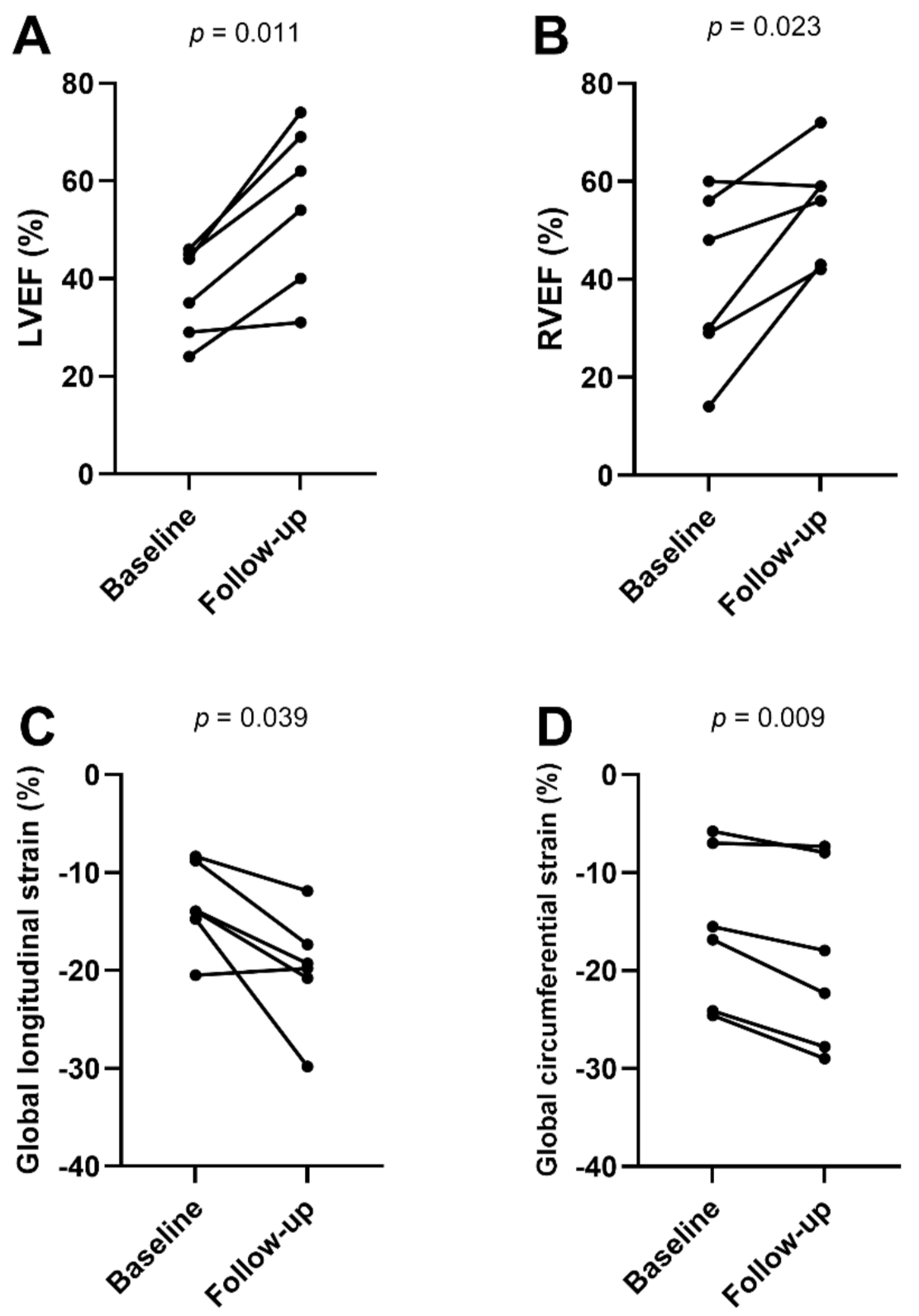
| Left ventricular ejection fraction (%) | 38 ± 9 | 55 ± 17 | 0.011 |
| Left ventricular end-diastolic volume index (mL/m²) | 89 ± 28 | 85 ± 27 | 0.651 |
| Right ventricular ejection fraction (%) | 40 ± 18 | 55 ± 11 | 0.023 |
| Right ventricular end-diastolic volume index (mL/m²) | 66 ± 13 | 71 ± 15 | 0.370 |
| Left atrium volume index (mL/m²) | 56 ± 18 | 42 ± 10 | 0.051 |
| Left ventricular mass index (g/m²) | 61 ± 14 | 52 ± 8 | 0.176 |
| Interventricular septal thickness (mm) | 10 ± 2.8 | 9.1 ± 2.0 | 0.047 |
| T2 signal intensity ratio | 2.1 ± 0.3 | 1.7 ± 0.3 | 0.126 |
| Visual myocardial edema | 3 (50%) | 0 (0%) | 0.25 |
| Visual late gadolinium enhancement | 1 (20%) | 0 (0%) | 0.99 |
| Late gadolinium enhancement (%) | 4.5 ± 3.3 | 5.0 ± 2.6 | 0.363 |
| Global longitudinal strain (%) | −13.5 ± 4.8 | −19.8 ± 5.8 | 0.039 |
| Global circumferential strain (%) | −15.6 ± 8.1 | −18.7 ± 9.5 | 0.009 |
| Global radial strain (%) | 30.1 ± 21.9 | 30.5 ± 17.6 | 0.935 |
| Variable | Univariable Analysis | Multivariable Analysis | ||
|---|---|---|---|---|
| Hazard Ratio | p-Value | Hazard Ratio | p-Value | |
| Age (per year) | 0.89 (0.77–1.03) | 0.116 | ||
| Body mass index (per kg/m²) | 0.99 (0.90–1.09) | 0.841 | ||
| LVEF (per %) | 1.13 (1.02–1.25) | 0.023 | ||
| LVEDVI (per mL/m²) | 0.99 (0.96–1.01) | 0.228 | ||
| LVMI (per g/m²) | 1.01 (0.96–1.05) | 0.790 | ||
| LAI (per mL/m²) | 0.99 (0.96–1.02) | 0.585 | ||
| RVEF (per %) | 1.07 (1.00–1.14) | 0.036 | ||
| RVEDVI (per mL/m²) | 1.01 (0.98–1.04) | 0.422 | ||
| LV GLS (per %) | 0.53 (0.34–0.84) | 0.007 | 0.51 (0.30–0.85) | 0.010 |
| LV GCS (per %) | 0.81 (0.70–0.95) | 0.010 | ||
| LV GRS (per %) | 1.10 (1.02–1.18) | 0.010 | ||
| LGE (per %) | 1.05 (0.92–1.21) | 0.475 | ||
| T2 signal intensity ratio | 1.77 (0.25–12.30) | 0.565 | ||
| Visual myocardial edema (yes/no) | 10.17 (1.17–88.65) | 0.036 | ||
Publisher’s Note: MDPI stays neutral with regard to jurisdictional claims in published maps and institutional affiliations. |
© 2022 by the authors. Licensee MDPI, Basel, Switzerland. This article is an open access article distributed under the terms and conditions of the Creative Commons Attribution (CC BY) license (https://creativecommons.org/licenses/by/4.0/).
Share and Cite
Isaak, A.; Ayub, T.H.; Merz, W.M.; Faron, A.; Endler, C.; Sprinkart, A.M.; Pieper, C.C.; Kuetting, D.; Dabir, D.; Attenberger, U.; et al. Peripartum Cardiomyopathy: Diagnostic and Prognostic Value of Cardiac Magnetic Resonance in the Acute Stage. Diagnostics 2022, 12, 378. https://doi.org/10.3390/diagnostics12020378
Isaak A, Ayub TH, Merz WM, Faron A, Endler C, Sprinkart AM, Pieper CC, Kuetting D, Dabir D, Attenberger U, et al. Peripartum Cardiomyopathy: Diagnostic and Prognostic Value of Cardiac Magnetic Resonance in the Acute Stage. Diagnostics. 2022; 12(2):378. https://doi.org/10.3390/diagnostics12020378
Chicago/Turabian StyleIsaak, Alexander, Tiyasha H. Ayub, Waltraut M. Merz, Anton Faron, Christoph Endler, Alois M. Sprinkart, Claus C. Pieper, Daniel Kuetting, Darius Dabir, Ulrike Attenberger, and et al. 2022. "Peripartum Cardiomyopathy: Diagnostic and Prognostic Value of Cardiac Magnetic Resonance in the Acute Stage" Diagnostics 12, no. 2: 378. https://doi.org/10.3390/diagnostics12020378
APA StyleIsaak, A., Ayub, T. H., Merz, W. M., Faron, A., Endler, C., Sprinkart, A. M., Pieper, C. C., Kuetting, D., Dabir, D., Attenberger, U., Zimmer, S., Becher, U. M., & Luetkens, J. A. (2022). Peripartum Cardiomyopathy: Diagnostic and Prognostic Value of Cardiac Magnetic Resonance in the Acute Stage. Diagnostics, 12(2), 378. https://doi.org/10.3390/diagnostics12020378


