
Root Resorptions on Adjacent Teeth Associated with Impacted Maxillary Canines


 ,
,  , ,
, ,
Abstract
:1. Introduction
2. Materials and Methods
- (1)
- Type of impaction (unilateral, bilateral);
- (2)
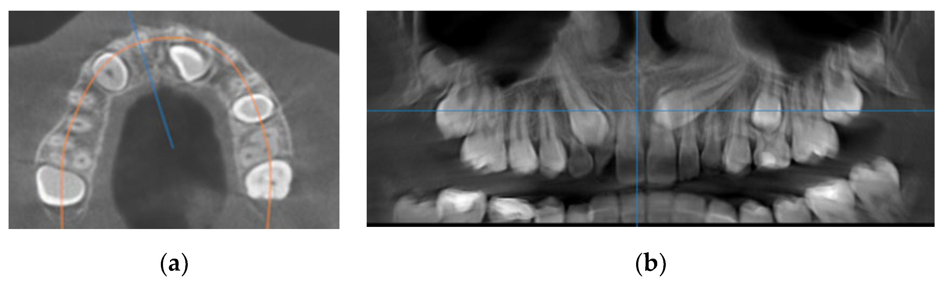
- Sagittal location (labial, palatal or median) using sagittal and coronal CBCT scans (Figure 1a);
- (3)
- Vertical location of the cusp tip in relation to the long axis of the adjacent tooth (on sagittal and axial CBCT scans), which was assigned to one of five categories: subdivided into coronal, cervical third, middle third, apical third of the root, or suprapical;
- (4)
- Horizontal position of the canine cusp tip; the canine was observed to overlap with adjacent teeth using sagittal or coronal CBCT scans. It was assigned according to Ericson and Kurol [17] (sector 1: canine overlapping by up to half the width of the lateral incisor; sector 2: canine overlapping by over half the width of the lateral incisor; sector 3: canine completely overlapping with the lateral incisor; sector 4: canine overlapping by up to half the width of the central incisor; sector 5: canine overlapping over the midline of the maxilla) (Figure 1b);
- (5)
- Distance of the impacted maxillary canine cusp to the midline (measured on axial CBCT scans) (Figure 2a);
- (6)
- Distance of the impacted maxillary canine cusp to the occlusal line (measured on CBCT images in the sagittal plane);Figure 1. (a) Occlusal reference arch–location of impacted maxillary canines in axial plane; (b) horizontal position of canine cusp tip: the canine overlap with adjacent teeth in coronal plane (right maxillary impacted canine in sector 2 ad left maxillary impacted canine in sector 4).Figure 1. (a) Occlusal reference arch–location of impacted maxillary canines in axial plane; (b) horizontal position of canine cusp tip: the canine overlap with adjacent teeth in coronal plane (right maxillary impacted canine in sector 2 ad left maxillary impacted canine in sector 4).
![Diagnostics 12 00380 g001]() Figure 2. (a) The distance of maxillary canine cusp to midline (measured on CBCT images in axial plane); (b) Angle between long axis of impacted maxillary canine and long axis of adjacent lateral incisor measured on CBCT images in sagittal plane.Figure 2. (a) The distance of maxillary canine cusp to midline (measured on CBCT images in axial plane); (b) Angle between long axis of impacted maxillary canine and long axis of adjacent lateral incisor measured on CBCT images in sagittal plane.
Figure 2. (a) The distance of maxillary canine cusp to midline (measured on CBCT images in axial plane); (b) Angle between long axis of impacted maxillary canine and long axis of adjacent lateral incisor measured on CBCT images in sagittal plane.Figure 2. (a) The distance of maxillary canine cusp to midline (measured on CBCT images in axial plane); (b) Angle between long axis of impacted maxillary canine and long axis of adjacent lateral incisor measured on CBCT images in sagittal plane.![Diagnostics 12 00380 g002]()
- (7)
- Angle between the longitudinal axis of the impacted maxillary canine and the long axis of the adjacent central/lateral incisor (measured on CBCT images in the sagittal plan) (Figure 2b);
- (8)
- Angle between the longitudinal axis of the impacted maxillary canine and the maxillary arch midline (measured on CBCT images in the coronal plan) (Figure 3a);Figure 3. (a) Maxillary impacted canine angulation to the midline (measured on CBCT images in the coronal plan); (b) maxillary impacted canine angulation to the occlusal line and the distance canine cusp to occlusal line (measured on CBCT images in the sagittal plane).Figure 3. (a) Maxillary impacted canine angulation to the midline (measured on CBCT images in the coronal plan); (b) maxillary impacted canine angulation to the occlusal line and the distance canine cusp to occlusal line (measured on CBCT images in the sagittal plane).
![Diagnostics 12 00380 g003]() Angle between the longitudinal axis of the impacted maxillary canine and the occlusal line (measured on CBCT images in the sagittal plane) (Figure 3b);
Angle between the longitudinal axis of the impacted maxillary canine and the occlusal line (measured on CBCT images in the sagittal plane) (Figure 3b); - (9)
- RR of the adjacent tooth assessed in the axial plane, using a previously established classification. If RR was suspected, resorption was graded based on the system suggested by Ericson [18] for each tooth into 4 categories: no resorption (intact root surface, the cementum layer may have been lost), slight resorption (resorption up to half of the dentine thickness), moderate resorption (resorption of the dentine midway to the pulp or more, the pulp lining being unbroken), and severe resorption (resorption reaches the pulp). The presence or absence of RR was assessed on 3D MPR views along the long axis of every adjacent root;
- (10)
- Localization of RR (cervical, middle or apical third of root).


3. Results
4. Discussion
5. Conclusions
- -
- The prevalence of impacted maxillary canines and RR on adjacent teeth was higher in female subjects.
- -
- Resorption of adjacent teeth was 60.2%, and slight RR was the most frequent.
- -
- The maxillary lateral incisors were more frequently affected than central incisors.
- -
- The sensitivity of CBCT allows the accurate diagnosis of the location and the degree of RR, alongside the angulation and distance of impacted canines to adjacent teeth.
- -
- The measured parameters on the canines improve the indication of RR (the higher position of the impacted canine, the higher values for the angulation of canine to the midline/lateral incisor and the greater degree of horizontal overlap leading to the impacted canine cusp with adjacent teeth).
- -
- The association between the linear and angular features of the impacted maxillary canine and RR was confirmed.
Author Contributions
Funding
Institutional Review Board Statement
Informed Consent Statement
Conflicts of Interest
References
- Oberoi, S.; Knueppel, S. Three-dimensional assessment of impacted canines and root resorption using cone beam computed tomography. Oral Surg. Oral Med. Oral Pathol. Oral Radiol. 2012, 113, 260–267. [Google Scholar] [CrossRef] [PubMed]
- Yan, B.; Sun, Z.; Fields, H.; Wang, L. Maxillary canine impaction increases root resorption risk of adjacent teeth: A problem of physical proximity. Orthod. Fr. 2015, 86, 169–179. [Google Scholar] [CrossRef] [PubMed]
- Walker, L.; Enciso, R.; Hatcher, D. Three-dimensional craniofacial imaging. Am. J. Orthod. Dentof. Orthop. 2005, 125, 418–423. [Google Scholar] [CrossRef] [PubMed]
- Schroder, A.G.; Guariza-Filho, O.; de Araujo, C.M.; Ruellas, A.C.; Tanaka, O.M.; Porporatti, A.L. To what extent are impacted canines associated with root resorption of the adjacent tooth? A systematic review with meta-analysis. J. Am. Dent. Assoc. 2018, 149, 765.e8–777.e8. [Google Scholar] [CrossRef]
- Ericson, S.; Kurol, J. Incisor root resorptions due to ectopic maxillary canines imaged by computerized tomography: A comparative study in extracted teeth. Angle Orthod. 2000, 70, 276–283. [Google Scholar]
- Ucar, F.I.; Celebi, A.A.; Tan, E.; Topcuoğlu, T.; Sekerci, A.E. Effects of impacted maxillary canines on root resorption of lateral incisors. A cone beam computed tomography study. J. Orofac. Orthop. 2017, 78, 233–240. [Google Scholar] [CrossRef]
- Sosars, P.; Jakobsone, G.; Neimane, L.; Mukans, M. Comparative analysis of panoramic radiography and cone-beam computed to-mography in treatment planning of palatally displaced canines. Am. J. Orthod. Dentofac. Orthop. 2020, 157, 719–727. [Google Scholar] [CrossRef]
- Bjerklin, K.; Guitirokh, C.H. Maxillary incisor root resorption induced by ectopic canines. Angle Orthod. 2011, 81, 800–806. [Google Scholar] [CrossRef] [Green Version]
- Alqerban, A.; Hedesiu, M.; Baciut, M.; Nackaerts, O.; Jacobs, R.; Fieuws, S.; Consortium, S.; Willems, G. Presurgical treatment planning of maxillary canine impactions using panoramic vs cone beam CT imaging. Dentomaxillofac. Radiol. 2013, 42, 20130157. [Google Scholar] [CrossRef] [Green Version]
- Kim, Y.; Hyun, H.K.; Jang, K.T. Morphological relationship analysis of impacted maxillary canines and the adjacent teeth on 3-dimensional reconstructed CT images. Angle Orthod. 2017, 87, 590–597. [Google Scholar] [CrossRef] [Green Version]
- Cernochova, P.; Krupa, P.; Izakovicova-Holla, L. Root resorption associated with ectopically erupting maxillary permanent canines: A computed tomography study. Eur. J. Orthod. 2011, 33, 483–491. [Google Scholar] [CrossRef] [PubMed] [Green Version]
- Bjerklin, K.; Ericson, S. How a computerized tomography examination changed the treatment plans of 80 children with re-tained and ectopically positioned maxillary canines. Angle Orthod. 2006, 76, 43–51. [Google Scholar] [PubMed]
- Dagsuyu, I.M.; Kahraman, F.; Oksayan, R. Three-dimensional evaluation of angular, linear, and resorption features of maxillary impacted canines on cone-beam computed tomography. Oral Radiol. 2018, 34, 66–72. [Google Scholar] [CrossRef] [PubMed]
- Kalavritinos, M.; Benetou, V.; Bitsanis, E.; Sanoudos, M.; Alexiou, K.; Tsiklakis, K.; Tsolakis, A.I. Incidence of incisor root resorption associated with the position of the impacted max-illary canines: A cone beam computed tomographic stud. Am. J. Orthod. Dentof. Orthop. 2020, 157, 73–79. [Google Scholar] [CrossRef]
- Liu, D.G.; Zhang, W.L.; Zhang, Z.Y.; Wu, Y.T.; Ma, X.C. Localization of impacted maxillary canines and observation of adjacent incisor resorption with cone-beam computed tomography. Oral Surg. Oral Med. Oral Pathol. Oral Radiol. Endod. 2008, 105, 91–98. [Google Scholar] [CrossRef]
- Arriola-Guillen, L.E.; Ruiz-Mora, G.A.; Rodrıguez-Cardenas, Y.A.; Aliaga-Del Castillo, A.; Boessio-Vizzotto, M.; Dias-Da Silveira, H.L. Influence of Impacted Maxillary Canine Orthodontic Traction Complexity on Root Resorption of Incisors: A Retrospective Longitudinal Study. Am. J. Orthod. Dentofac. Orthop. 2019, 155, 28–39. [Google Scholar] [CrossRef]
- Ericson, S.; Kurol, J. Early treatment of palatally erupting maxillary canines by extraction of the primary canines. Eur. J. Orthod. 1988, 10, 283–295. [Google Scholar] [CrossRef] [PubMed]
- Ericson, S.; Kurol, P.J. Resorption of incisors after ectopic eruption of maxillary canines: A CT study. Angle Orthod. 2000, 70, 415–423. [Google Scholar]
- Koral, S.; Özçırpıcı, A.A.; Tunçer, N.İ. Association between Impacted Maxillary Canines and Adjacent Lateral Incisors: A Retrospective Study with Cone Beam Computed Tomography. Turk. J. Orthod. 2021, 34, 207–213. [Google Scholar] [CrossRef]
- da Silva Santos, L.M.; Bastos, L.C.; Oliveira-Santos, C.; da Silva, S.J.; Neves, F.S.; Campos, P.S. Cone-beam computed tomogra-phy findings of impacted upper canines. Imaging Sci. Dent. 2014, 44, 287–292. [Google Scholar] [CrossRef] [Green Version]
- Hadler-Olsen, S.; Pirttiniemi, P.; Kerosuo, H.; Limchaichana, N.B.; Pesonen, P.; Kallio-Pulkkinen, S.; Lähdesmäki, R. Root resorptions related to ectopic and normal eruption of maxillary canine teeth—A 3D study. Acta Odontol. Scand. 2015, 73, 609–615. [Google Scholar] [CrossRef] [PubMed]
- Rafflenbeul, F.; Gros, C.I.; Lefebvre, F.; Bahi-Gross, S.; Maizeray, R.; Bolender, Y. Prevalence and risk factors of root resorption of adjacent teeth in maxillary canine impaction, among untreated children and adolescents. Eur. J. Orthod. 2019, 41, 447–453. [Google Scholar] [CrossRef] [PubMed]
- Castro, I.O.; Alencar, A.H.; Valladares-Neto, J.; Estrela, C. Apical root resorption due to orthodontic treatment detected by cone beam computed tomography. Angle Orthod. 2013, 83, 196–203. [Google Scholar] [CrossRef] [PubMed] [Green Version]
- Dekel, E.; Nucci, L.; Weill, T.; Flores-Mir, C.; Becker, A.; Perillo, L.; Chaushu, S. Impaction of maxillary canines and its effect on the position of adjacent teeth and canine development: A cone-beam computed tomography study. Am. J. Orthod. Dentofac. Orthop. 2021, 159, e135–e147. [Google Scholar] [CrossRef] [PubMed]
- Dogramaci, E.; Sheriff, M.; Rossi-Fedele, G.; McDonald, F. Location and severity of root resorption related to impacted maxillary canines: A cone beam computed tomography (CBCT) evaluation. Aust. Orthod. J. 2015, 31, 49. [Google Scholar] [PubMed]
- Alqerban, A.; Jacobs, R.; Fieuws, S.; Willems, G. Predictors of root resorption associated with maxillary canine impaction in panoramic images. Eur. J. Orthod. 2016, 38, 292–299. [Google Scholar] [CrossRef] [Green Version]
- Guarnieri, R.; Cavallini, C.; Vernucci, R.; Vichi, M.; Leonardi, R.; Barbato, E. Impacted maxillary canines and root resorption of adjacent teeth: A retrospective observational study. Med. Oral Patol. Oral Cir. Bucal 2016, 21, e743–e750. [Google Scholar] [CrossRef]
- Alqerban, A.; Jacobs, R.; Fieuws, S.; Willems, G. Radiographic predictors for maxillary canine impaction. Am. J. Orthod. Dentofac. Orthop. 2015, 147, 345–354. [Google Scholar] [CrossRef]
- Ericson, S.; Kurol, J. CT diagnosis of ectopically erupting maxillary canines–A case report. Eur. J. Orthod. 1988, 10, 115–121. [Google Scholar] [CrossRef]
- Yu, J.N.; Gu, Y.G.; Zhao, C.Y.; Liu, K.; Mo, S.C.; Li, H.; Pan, C.Q.; Wang, L. Three-dimensional localization and assessment of maxillary palatal impacted canines with cone-beam computed tomography. Shanghai Kou Qiang Yi Xue 2015, 24, 65–70. [Google Scholar]
- Sajnani, A.K.; King, N.M. The sequential hypothesis of impaction of maxillary canine—A hypothesis based on clinical and radiographic findings. J. Craniomaxillofac. Surg. 2012, 40, e375–e385. [Google Scholar] [CrossRef] [PubMed]
- Cuminetti, F.; Boutin, F.; Frapier, L. Predictive factors for resorption of teeth adjacent to impacted maxillary canines. Int. Orthod. 2017, 15, 54–68. [Google Scholar] [CrossRef] [PubMed]
- Deng, Y.; Sun, Y.; Xu, T. Evaluation of root resorption after comprehensive orthodontic treatment using cone-beam computed tomography (CBCT): Metaanalysis. BMC Oral Health 2018, 18, 116. [Google Scholar] [CrossRef] [PubMed]
- Jiang, F.; Chen, J.; Kula, K.; Gu, H.; Du, Y.; Eckert, G. Root resorptions associated with canine retraction treatment. Am. J. Orthod. Dentofac. Orthop. 2017, 152, 348–354. [Google Scholar] [CrossRef]


| Impacted Maxillary Canines | Root Resorptions | ||
|---|---|---|---|
| Subjects | n = 89 (100%) | n = 47 (52.8%) | |
| Age (mean ± SD) | 18.3 ± 4.1years | 16.7 ± 3.5years | |
| Canines | n = 108 | RR n = 65 (60.2%) | |
| Male Female | 31 (34.8%) 58 (65.2%) | 11 (12.4%) 36 (40.4%) | |
| Canine localization sagittal | Labial Palatal Median | 25 (23.1%) 80 (74.1%) 3 (2.8%) | 12 (13.5%) 35 (39.3%) 0 |
| Unilateral Bilateral | 70 (64.8%) 19 (17.6%) | 39 (43.8%) 8 (8.9%) | |
| Canine localization vertical | Suprapical Apical third Middle third Cervical third Coronal | 7 (6.5%) 26 (24.1%) 34 (31.5%) 25 (23.1%) 16 (14.8%) | 0 32 (29.6%) 21 (19.4%) 12 (11.1%) 0 |
| Canine localization horizontal | Sector 1 Sector 2 Sector 3 Sector 4 Sector 5 | 27 (25%) 18 (16.7%) 39 (36.1%) 21 (19.4%) 3 (2.8%) | 0 (0%) 9 (10.1%) 20 (22.5%) 16 (17.9%) 2 (2.2%) |
| Distance to midline (mean) | 9.25 mm ± 4.4 mm | ||
| Distance to occlusal line (mean) | 11.7 mm, SD ± 3.6 mm | ||
| RR on Adjacent Teeth | Central Incisors No = 89 (82.4%) Yes = 19 (17.6%) | Lateral Incisors No = 71 (65.7%) Yes = 37 (34.3%) | First Premolars No = 99 (91.7%) Yes = 9(8.3%) | Total No = 43 (39.8%) Yes = 65 (60.2%) | |||
| Location of resorption | |||||||
| Apical third | 9 (8.3%) | 17 (15.7%) | 6 (5.5%) | 32 (29.6%) | |||
| Middle third | 7 (6.5%) | 11 (10.2%) | 3 (2.8%) | 21 (19.4%) | |||
| Cervical third | 3 (2.8%) | 9 (8.3%) | 0 | 12 (11.1%) | |||
| Severity of resorption | |||||||
| Slight | 10 (9.2%) | 18 (16.7%) | 5 (4.6%) | 33 (30.5%) | |||
| Moderate | 6 (5.5%) | 11 (10.2%) | 3 (2.8%) | 20 (18.5%) | |||
| Severe | 3 (2.8%) | 8 (7.4%) | 1 (0.9%) | 12 (11.1%) | |||
| Variables | Values | Resorption on Lateral Incisors | p Value | Resorption on Central Incisors | p Value | ||
| No (n = 71) | Yes (n = 37) | No (n = 89) | Yes (n = 19) | ||||
| Anglulation canine to midline | Median (range) | 21.4 (12.4−55.6) | 46.7 (14.3−86.1) | 0.005 * | 25.3 (6.1−75.6) | 43.5 (17.5−64.6) | <0.001 |
| Angulation canine to incisor | Median (range) | 23.6 (11.1−48.5) | 43.1 (18.1−86.4) | 0.004 * | 23.1 (8.2−86.2) | 40.3 (20.4−61.8) | <0.001 |
| Anglulation canine to occlusal line | Median (range) | 55.8 (17.4−75.1) | 43.6 (22.3−78.8) | 0.007 * | 59.4 (3.8−78.6) | 41.4 (17.4−65.5) | 0.004 * |
| Variables | Values | Severity of Root Resorptions on Lateral Incisors | Severity of Root Resorptions on Central Incisors | Severity of Root Resorptions on First Premolars |
|---|---|---|---|---|
| Anglulation canine to midline | rho | 0.293 * | 0.426 * | 0.235 * |
| P | 0.008 | 0.000 | 0.002 | |
| Angulation canine to incisor | rho | 0.300 * | 0.411 * | 0.165 |
| P | 0.007 | 0.000 | 0.000 | |
| Angulation canine to occlusal plane | rho | −0.294 | −0.319 | −0.185 |
| P | 0.008 | 0.004 | 0.004 | |
| Horizontal position of canine | rho | 0.291 * | 0.580 * | 0.320 * |
| P | 0.009 | <0.001 | <0.001 | |
| Vertical position of canine | rho | 0.197 | 0.117 | 0.122 |
| P | 0.079 | 0.301 | 0.255 | |
| Distance of canine to midline | rho | 0.305 * | 0.341 * | −0.174 |
| P | 0.008 | 0.005 | 0.000 | |
| Distance of canine to occlusal line | rho | 0.307 * | 0.320 * | 0.287 * |
| P | 0.006 | 0.004 | 0.006 |
Publisher’s Note: MDPI stays neutral with regard to jurisdictional claims in published maps and institutional affiliations. |
© 2022 by the authors. Licensee MDPI, Basel, Switzerland. This article is an open access article distributed under the terms and conditions of the Creative Commons Attribution (CC BY) license (https://creativecommons.org/licenses/by/4.0/).
Share and Cite
Simić, S.; Nikolić, P.; Stanišić Zindović, J.; Jovanović, R.; Stošović Kalezić, I.; Djordjević, A.; Popov, V. Root Resorptions on Adjacent Teeth Associated with Impacted Maxillary Canines. Diagnostics 2022, 12, 380. https://doi.org/10.3390/diagnostics12020380
Simić S, Nikolić P, Stanišić Zindović J, Jovanović R, Stošović Kalezić I, Djordjević A, Popov V. Root Resorptions on Adjacent Teeth Associated with Impacted Maxillary Canines. Diagnostics. 2022; 12(2):380. https://doi.org/10.3390/diagnostics12020380
Chicago/Turabian StyleSimić, Sanja, Predrag Nikolić, Jelena Stanišić Zindović, Radovan Jovanović, Ivana Stošović Kalezić, Aleksandar Djordjević, and Vesna Popov. 2022. "Root Resorptions on Adjacent Teeth Associated with Impacted Maxillary Canines" Diagnostics 12, no. 2: 380. https://doi.org/10.3390/diagnostics12020380